Identification of a novel cell death-inducing domain ... · des Nanoobjets, CNRS, Université de...
6
Identification of a novel cell death-inducing domain reveals that fungal amyloid-controlled programmed cell death is related to necroptosis Asen Daskalov a,1 , Birgit Habenstein b , Raimon Sabaté c , Mélanie Berbon b , Denis Martinez b , Stéphane Chaignepain a , Bénédicte Coulary-Salin a , Kay Hofmann d , Antoine Loquet b , and Sven J. Saupe a,2 a Institut de Biochimie et de Génétique Cellulaire, CNRS, Université de Bordeaux, 33077 Bordeaux, France; b Institut de Chimie et Biologie des Membranes et des Nanoobjets, CNRS, Université de Bordeaux, 33077 Bordeaux, France; c Institut de Nanociència i Nanotecnologia, Departament Fisicoquímica, Universitat de Barcelona, E-08028 Barcelona, Spain; and d Institute for Genetics, University of Cologne, D-50674 Cologne, Germany Edited by Reed B. Wickner, National Institutes of Health, Bethesda, MD, and approved January 29, 2016 (received for review November 13, 2015) Recent findings have revealed the role of prion-like mechanisms in the control of host defense and programmed cell death cascades. In fungi, HET-S, a cell death-inducing protein containing a HeLo pore-forming domain, is activated through amyloid templating by a Nod-like receptor (NLR). Here we characterize the HELLP protein behaving analogously to HET-S and bearing a new type of N-ter- minal cell death-inducing domain termed HeLo-like (HELL) and a C-terminal regulatory amyloid motif known as PP. The gene encod- ing HELLP is part of a three-gene cluster also encoding a lipase (SBP) and a Nod-like receptor, both of which display the PP motif. The PP motif is similar to the RHIM amyloid motif directing forma- tion of the RIP1/RIP3 necrosome in humans. The C-terminal region of HELLP, HELLP(215-278), encompassing the motif, allows prion propagation and assembles into amyloid fibrils, as demonstrated by X-ray diffraction and FTIR analyses. Solid-state NMR studies reveal a well-ordered local structure of the amyloid core residues and a primary sequence that is almost entirely arranged in a rigid conformation, and confirm a β-sheet structure in an assigned stretch of three amino acids. HELLP is activated by amyloid templating and displays membrane-targeting and cell death-inducing activity. HELLP targets the SBP lipase to the membrane, suggesting a synergy between HELLP and SBP in membrane dismantling. Remarkably, the HeLo-like domain of HELLP is homologous to the pore-forming domain of MLKL, the cell death-execution protein in necroptosis, revealing a transkingdom evolutionary relationship between amy- loid-controlled fungal programmed cell death and mammalian necroptosis. amyloid | prion | programmed cell death | incompatibility | necroptosis P rogrammed cell death (PCD) plays a central role in response to nonself and host defenses in animals, plants, fungi, and bacteria (1–4). Altruistic cell suicide hinders pathogen replication and promotes survival in multicellular organisms, and provides benefits at the population level in unicellular microbes. Questions surrounding the classification, evolutionary origin, and level of transkingdom conservation of PCD modalities remain open and often polemic (5). Classification systems of PCD types are based on cytological or molecular markers that can differ between phyla, making it difficult to merge cell death processes into unifying cat- egories (5). In filamentous fungi, a form of PCD termed heterokaryon in- compatibility occurs in response to conspecific nonself (3). Cell death by incompatibility takes place following fusion of cells from genetically distinct individuals and is controlled by het genes. One of the best-characterized incompatibility systems involves a cell death- inducing protein termed HET-S, which is controlled by amyloid templating. HET-S of Podospora anserina exhibit an N-terminal cell death execution HeLo domain and a C-terminal regulatory prion forming domain (PFD) that adopts a specific β-solenoid amyloid fold, characterized by the stacking of two pseudorepeats of an el- ementary amyloid motif (6–8). When the HET-S PFD is converted to the β-solenoid fold, the HeLo domain undergoes refolding, and an N-terminal transmembrane helix (TMH) is exposed, causing HET-S to behave as a pore-forming toxin (9). On transconformation, HET-S relocates from the cytoplasm to the cell membrane, where it exerts toxicity (10). Conversion of the HET-S PFD region can be achieved by two means. The first described mechanism occurs in the context of HET-S/[Het-s] incompatibility (9). [Het-s] is a variant form of HET-S that has lost the pore-forming activity and can stably propagate as a prion. [Het-s] and HET-S strains are incompatible, because [Het-s] acts as a seed converting the PFD of HET-S, thereby leading to activation of the HeLo cell death-inducing domain. A second, recently described mode of activation involves a Nod-like receptor protein (NLR), termed NWD2, encoded by the gene adjacent to het-S in the genome (11, 12). The NWD2 NLR displays a 21-aa N-terminal region homologous to the HET-s amyloid motif. On binding of a specific ligand to NWD2, these N-terminal extensions adopt an amyloid fold and convert HET-S to the toxic pore-forming state. The NWD2/HET-S system is one of several examples in which activation of cell death and host defense cascades relies on prion- like and/or amyloid polymerization mechanisms (13, 14). Activa- tion of ASC (apoptosis-associated speck-like protein with a caspase recruitment domain) by the NLRP3 human NLR depends on a Significance Programmed cell death plays a central role in host defense in plants, animals, and fungi, but the extent to which cell death modalities are evolutionarily related and mechanistically simi- lar in different kingdoms is unclear. The involvement of prion- like mechanisms in host defense and cell death cascades has been reported in animals and fungi. In fungi, a cell death-inducing pore-forming protein termed HET-S is activated by amyloid templating. Here we characterize a protein termed HELLP, which behaves analogously to HET-S as a membrane-targeting cell death-inducing protein regulated by amyloid templating. We find that HELLP is homologous to MLKL, the protein involved in execution of necroptotic cell death in mammals, thus revealing transkingdom conservation of amyloid-regulated programmed cell death. Author contributions: A.D., B.H., R.S., K.H., A.L., and S.J.S. designed research; A.D., B.H., R.S., M.B., D.M., S.C., and B.C.-S. performed research; A.D., B.H., R.S., D.M., S.C., K.H., A.L., and S.J.S. analyzed data; and B.H., A.L., and S.J.S. wrote the paper. The authors declare no conflict of interest. This article is a PNAS Direct Submission. 1 Present address: Department of Plant and Microbial Biology, University of California, Berkeley, CA 94720. 2 To whom correspondence should be addressed. Email: [email protected]. This article contains supporting information online at www.pnas.org/lookup/suppl/doi:10. 1073/pnas.1522361113/-/DCSupplemental. www.pnas.org/cgi/doi/10.1073/pnas.1522361113 PNAS Early Edition | 1 of 6 MICROBIOLOGY
Transcript of Identification of a novel cell death-inducing domain ... · des Nanoobjets, CNRS, Université de...

Identification of a novel cell death-inducing domainreveals that fungal amyloid-controlled programmedcell death is related to necroptosisAsen Daskalova,1, Birgit Habensteinb, Raimon Sabatéc, Mélanie Berbonb, Denis Martinezb, Stéphane Chaignepaina,Bénédicte Coulary-Salina, Kay Hofmannd, Antoine Loquetb, and Sven J. Saupea,2
aInstitut de Biochimie et de Génétique Cellulaire, CNRS, Université de Bordeaux, 33077 Bordeaux, France; bInstitut de Chimie et Biologie des Membranes etdes Nanoobjets, CNRS, Université de Bordeaux, 33077 Bordeaux, France; cInstitut de Nanociència i Nanotecnologia, Departament Fisicoquímica, Universitatde Barcelona, E-08028 Barcelona, Spain; and dInstitute for Genetics, University of Cologne, D-50674 Cologne, Germany
Edited by Reed B. Wickner, National Institutes of Health, Bethesda, MD, and approved January 29, 2016 (received for review November 13, 2015)
Recent findings have revealed the role of prion-like mechanisms inthe control of host defense and programmed cell death cascades.In fungi, HET-S, a cell death-inducing protein containing a HeLopore-forming domain, is activated through amyloid templating bya Nod-like receptor (NLR). Here we characterize the HELLP proteinbehaving analogously to HET-S and bearing a new type of N-ter-minal cell death-inducing domain termed HeLo-like (HELL) and aC-terminal regulatory amyloid motif known as PP. The gene encod-ing HELLP is part of a three-gene cluster also encoding a lipase(SBP) and a Nod-like receptor, both of which display the PP motif.The PP motif is similar to the RHIM amyloid motif directing forma-tion of the RIP1/RIP3 necrosome in humans. The C-terminal regionof HELLP, HELLP(215-278), encompassing the motif, allows prionpropagation and assembles into amyloid fibrils, as demonstratedby X-ray diffraction and FTIR analyses. Solid-state NMR studiesreveal a well-ordered local structure of the amyloid core residuesand a primary sequence that is almost entirely arranged in a rigidconformation, and confirm a β-sheet structure in an assigned stretchof three amino acids. HELLP is activated by amyloid templatingand displays membrane-targeting and cell death-inducing activity.HELLP targets the SBP lipase to themembrane, suggesting a synergybetween HELLP and SBP in membrane dismantling. Remarkably, theHeLo-like domain of HELLP is homologous to the pore-formingdomain of MLKL, the cell death-execution protein in necroptosis,revealing a transkingdom evolutionary relationship between amy-loid-controlled fungal programmed cell death and mammaliannecroptosis.
amyloid | prion | programmed cell death | incompatibility | necroptosis
Programmed cell death (PCD) plays a central role in responseto nonself and host defenses in animals, plants, fungi, and
bacteria (1–4). Altruistic cell suicide hinders pathogen replicationand promotes survival in multicellular organisms, and providesbenefits at the population level in unicellular microbes. Questionssurrounding the classification, evolutionary origin, and level oftranskingdom conservation of PCD modalities remain open andoften polemic (5). Classification systems of PCD types are basedon cytological or molecular markers that can differ between phyla,making it difficult to merge cell death processes into unifying cat-egories (5).In filamentous fungi, a form of PCD termed heterokaryon in-
compatibility occurs in response to conspecific nonself (3). Celldeath by incompatibility takes place following fusion of cells fromgenetically distinct individuals and is controlled by het genes. One ofthe best-characterized incompatibility systems involves a cell death-inducing protein termed HET-S, which is controlled by amyloidtemplating. HET-S of Podospora anserina exhibit an N-terminal celldeath execution HeLo domain and a C-terminal regulatory prionforming domain (PFD) that adopts a specific β-solenoid amyloidfold, characterized by the stacking of two pseudorepeats of an el-ementary amyloid motif (6–8). When the HET-S PFD is converted
to the β-solenoid fold, the HeLo domain undergoes refolding, andan N-terminal transmembrane helix (TMH) is exposed, causingHET-S to behave as a pore-forming toxin (9). On transconformation,HET-S relocates from the cytoplasm to the cell membrane, whereit exerts toxicity (10).Conversion of the HET-S PFD region can be achieved by two
means. The first described mechanism occurs in the context ofHET-S/[Het-s] incompatibility (9). [Het-s] is a variant form ofHET-S that has lost the pore-forming activity and can stablypropagate as a prion. [Het-s] and HET-S strains are incompatible,because [Het-s] acts as a seed converting the PFD of HET-S,thereby leading to activation of the HeLo cell death-inducingdomain. A second, recently described mode of activation involvesa Nod-like receptor protein (NLR), termed NWD2, encoded bythe gene adjacent to het-S in the genome (11, 12). The NWD2NLR displays a 21-aa N-terminal region homologous to the HET-samyloid motif. On binding of a specific ligand to NWD2, theseN-terminal extensions adopt an amyloid fold and convert HET-Sto the toxic pore-forming state.The NWD2/HET-S system is one of several examples in which
activation of cell death and host defense cascades relies on prion-like and/or amyloid polymerization mechanisms (13, 14). Activa-tion of ASC (apoptosis-associated speck-like protein with a caspaserecruitment domain) by the NLRP3 human NLR depends on a
Significance
Programmed cell death plays a central role in host defense inplants, animals, and fungi, but the extent to which cell deathmodalities are evolutionarily related and mechanistically simi-lar in different kingdoms is unclear. The involvement of prion-like mechanisms in host defense and cell death cascades has beenreported in animals and fungi. In fungi, a cell death-inducingpore-forming protein termed HET-S is activated by amyloidtemplating. Here we characterize a protein termed HELLP, whichbehaves analogously to HET-S as a membrane-targeting celldeath-inducing protein regulated by amyloid templating. Wefind that HELLP is homologous to MLKL, the protein involved inexecution of necroptotic cell death in mammals, thus revealingtranskingdom conservation of amyloid-regulated programmedcell death.
Author contributions: A.D., B.H., R.S., K.H., A.L., and S.J.S. designed research; A.D., B.H.,R.S., M.B., D.M., S.C., and B.C.-S. performed research; A.D., B.H., R.S., D.M., S.C., K.H., A.L.,and S.J.S. analyzed data; and B.H., A.L., and S.J.S. wrote the paper.
The authors declare no conflict of interest.
This article is a PNAS Direct Submission.1Present address: Department of Plant and Microbial Biology, University of California,Berkeley, CA 94720.
2To whom correspondence should be addressed. Email: [email protected].
This article contains supporting information online at www.pnas.org/lookup/suppl/doi:10.1073/pnas.1522361113/-/DCSupplemental.
www.pnas.org/cgi/doi/10.1073/pnas.1522361113 PNAS Early Edition | 1 of 6
MICRO
BIOLO
GY

prion-like polymerization of the PYRIN domain and the HET-s-prion motif is able to substitute for the PYRIN signaling domain inNLRP3-dependent ASC activation (13). Another example is themammalian RIP1/RIP3 kinase complex controlling necroptosis,assembled via short amyloid motifs termed RHIM (15). Onedownstream effector of the RIP1/RIP3 complex in necroptosis isthe mixed lineage kinase domain-like protein (MLKL), which is atarget of RIP3 (16). In fungi, we have identified several other geneclusters analogous to Nwd2/het-S in which an NLR and a putativecell death effector protein are encoded by adjacent genes and inwhich the N-terminal extension of the NLR protein is homologous tothe C-terminal end of the effector (12, 17). One such proposed prionmotif, termed PP, was found to be associated with HeLo domainproteins, lipases, and regulatory NLRs (12). Herein we characterizea protein identified in Chaetomium globosum bearing this motif,termed HELLP.
ResultsIdentification of a PP Motif Gene Cluster. We identified inC. globosum a gene cluster with three adjacent genes, CHGG_01412,CHGG_01413, and CHGG_01414, encoding proteins with PP mo-tifs, corresponding to a HeLo-like domain protein (HELLP), aputative lipase (SBP) and a NLR (PNT1), respectively (Fig. 1A). AnN-terminal transmembrane helix (TMH) is predicted in HELLPthat shows homology to the HET-S TMH (Fig. S1). The SBP lipaseis homologous to SesB, a protein involved in propagation of the σinfectious element of Nectria hematococca (18, 19). HELLP, SBP,and PNT1 share a region of homology of 20–21 residues corre-sponding to the PP motif and located C-terminally in HELLP andSBP and N-terminally in PNT1. The similarity in genome and do-main architecture between the het-S/Nwd2 and Hellp/Sbp/Pnt1 geneclusters suggests that HELLP may represent an HET-S analog ac-tivated by amyloid templating via the PP motif. It has been proposedthat the HET-s prion motif is evolutionarily related to the RHIMamyloid motif controlling necroptotic signaling (20). The PP motif
of HELLP shows similarity to human RIP1 and RIP3 RHIMmotifs(Fig. 1B). The RHIM and PP motif signatures share a centralcommon G-φ-Q-φ-G core centered on a conserved Q residue andflanked by N residues (Fig. 1C).
HELLP Behaves as an HET-S Analog. We hypothesized that HELLPmight behave as HET-S, and thus attempted to recreate an in-compatibility system, analogous to [Het-s]/HET-S, based onHELLP. HET-S cannot form a prion, because amyloid conver-sion of HET-S leads to membrane targeting and toxicity, pre-venting prion propagation. The HeLo domain must be mutatedor truncated to allow for prion formation (7, 8). We reasonedthat if HELLP is a HET-S analog, then truncation of the HELLdomain should allow formation of a PP-based prion, and that strainsbearing this prion should be incompatible with strains expressingfull-length HELLP. A truncated HELLP construct, HELLP(172-278)-RFP, was expressed in P. anserina. HELLP(172-278)-RFPfluorescence was initially diffuse, but dot-like and elongated aggre-gates formed on further subculturing (Fig. 2A). Strains expressingHELLP(172-278)-RFP were then confronted to strains expressingfull-length HELLP-RFP (Fig. 2B). Barrage formation, indicative ofan incompatibility reaction, occurred specifically in confrontationswith strains displaying HELLP(172-278)-RFP aggregates. A shorterconstruct, HELLP(215-278)-GFP, also led to dot formation and toincompatibility with full-length HELLP (Fig. S2).Strains expressing truncated HELLP display two phenotypic
states that we term [π*] and [π]. [π*] strains show diffuse fluores-cence and are compatible with HELLP, whereas [π] strains showaggregates and are incompatible with HELLP (Fig. 2 A and B). [π*]strains were systematically converted to the [π] state when con-fronted with [π] strains (Fig. S2). The rate of spontaneous [π] for-mation was dependent on the growth medium (Table S1). Therewas no cross-infection between [π] and [Het-s] prions. [π] strains didnot convert [Het-s*] to the [Het-s] state, and [Het-s] strains did notconvert [π*] to the [π] state (Fig. S2C).
HELLP Relocates to the Membrane, and Mutations in the TMH AbolishIncompatibility. HET-S relocates from the cytoplasm to the cellmembrane on conversion of the PFD region to the β-solenoidprion fold (9–11). We tested whether HELLP also relocates to thecell membrane on interaction with the prion form of the PP motif.A strain expressing HELLP-RFP was confronted to a strainexpressing HELLP(215-278)-GFP in the [π] prion state. HELLP-RFP had a diffuse cytoplasmic localization in homokaryotic my-celium and relocated to the cell membrane region in fusion cellson interaction with HELLP(215-278)-RFP prion aggregates (Fig.3 and Fig. S3A).
Fig. 1. The PP motif gene cluster. (A) Representation of the CHGG_1412/13/14 locus with three adjacent genes, one encoding a HELL domain proteintermed HELLP, one encoding a predicted lipase related to SesB (SBP), and oneencoding a NB-ARC/TPR STAND protein (PNT1). The PP motif is represented asa pink line. (B) Alignment of the PP motif region of HELLP, SBP, and PNT1together with a portion of the RHIM motif of human RIP1 and RIP3. (C) Se-quence signatures for the central region of PP and the RHIM motif generatedwith Skylign. Letter size corresponds to level of conservation.
Fig. 2. HELLP(172-278)-RFP forms a prion, incompatible with full-lengthHELLP. (A) Micrograph of a P. anserina strain expressing a HELLP(172-278)-RFP fusion protein in either the soluble [π*] state (Upper) or the aggregated[π] state (Lower). (Scale bar: 5 μ.) (B) Confrontation of a strain expressing full-length HELLP and expressing HELLP(172-278)-RFP in the soluble state [π*] oraggregated state [π] as noted. Note the formation of a barrage (marked by awhite arrow) specifically in the confrontation with the strain expressingHELLP(172-278)-RFP in the aggregated state [π].
2 of 6 | www.pnas.org/cgi/doi/10.1073/pnas.1522361113 Daskalov et al.

The N-terminal TMH of HET-S plays a critical role in membranetargeting and toxicity (9). To investigate the role of the predictedHELLP TMH, we mutated two glycines, part of a putative GxxxGglycine-zipper motif, (a motif involved in TMHmultimerization (21)into large hydrophobic residues (G9I and G13I) predicted to dis-rupt the glycine zipper (Fig. S1). Both mutants no longer produceda incompatibility reaction to [π] (Fig. S3B), and instead of beingtargeted to the membrane, HELLP-RFP G9I and G13I mutantsformed cytoplasmic aggregates that partially colocalized with HELLP(215-278)-GFP (Fig. S3C).
The Prion-Forming Domain of HELLP Forms Amyloid Fibrils in Vitro. Invitro, purified HELLP(215-278) spontaneously assembled into∼5-nm-wide fibrils (Fig. S4A). In X-ray diffraction experiments, thefibrils presented a reflection at 4.7 Å, typical of a cross-β structure(Fig. S4B). When analyzed by spectroscopy, fibrils showed a singlestrong amide I band (around 1,630 cm−1), usually associated withparallel β-sheet structures in amyloid aggregates (Fig. S4C). Asdescribed previously for HET-s(218-289) (22), HELLP(215-278)assembled into fibrils in denaturing conditions (8 M urea) as well(Fig. S4D). The HELLP(215-278) fibrils showed a proteinaseK-resistant core (Fig. S5). Two main products corresponding to re-gions 238–278 and 236–278 were identified. The proteinase K-resistantcore encompassed the PP motif per se and extended N-terminallyinto a highly charged region not conserved among HELLP, SBP,and PNT1. We transfected [π*] strains with HELLP(215-278) fi-brils to assay their prion infectivity (Table S2). On transfection of[π*] strains with HELLP(215-278) fibrils, [π] formation rate wasstrongly increased, indicating that HELLP(215-278) fibrils assem-bled in vitro display prion infectivity.
Solid-State NMR Analysis of HELLP Fibrils.We carried out solid-stateNMR (SSNMR) experiments to characterize the structural order inHELLP(215-278) fibrils. A 1D 13C cross-polarization (CP) experi-ment (Fig. 4A) showed strong signals, indicating that a significantpart of the protein was immobilized in the fibrils. MultidimensionalMAS SSNMR was conducted on 15N and 13C isotope- labeledHELLP fibrils. A 2D SSNMR insensitive nuclei enhanced bypolarization transfer (INEPT) experiment was performed to de-tect mobile residues. The 2D 1H-13C INEPT (Fig. 4B) revealedthe buffer carbon signals and a side-chain arginine (Arg) carbon,
indicating the absence of residues that execute nearly isotropicmotion on time scales much shorter than 1 μs.Two-dimensional 13C-13C experiments (Fig. 4C) were recorded
using proton driven spin-diffusion (PDSD) mixing times of 30 and150 ms, leading to isolated cross-peaks encoding intraresidue andinterresidue correlations, respectively. Several residue spin systemswere observable based on their typical chemical shifts, includingthreonine (Thr), isoleucine, alanine, and valine. A detailed analysiswas possible for one apparent Thr residue, because the SSNMRCα-Cβ and Cβ-Cγ resonance peaks of Thr residues lay in an iso-lated part of the 2D 13C-13C correlation spectra. We could assignthe Cα, Cβ, and Cγ resonance frequencies of only one Thr (Fig. 4C,Upper). The sequential contacts connecting the Thr residue to itsamino acid neighbors were detected in a 2D 13C-13C PDSD spec-trum designed to detect interresidual 13C-13C correlations (Fig. 4C,Lower) with a PDSD mixing time of 150 ms (full 2D spectrumshown in Fig. S6). We could tentatively assign the glycine (Gly270)-Thr271 amino acid pair located in the proteinase K core region238–278, because SSNMR Gly Cα resonance peaks are very spe-cific and only one Gly-Thr pair is present in the HELLP(215-278)primary sequence. The neighboring methionine (Met) 272 Cα, Cβ,and Cγ resonance frequencies were assigned owing to their se-quential contacts with the isolated Thr Cβ frequency (Fig. 4B). Thethree-residue stretch, Gly270-Thr271-Met272, of the fibrillar coreregion located in the PP motif appeared to adopt a β-strand con-formation in HELLP(215-278) fibrils, as demonstrated by the sec-ondary chemical shift values (Fig. 4C). The 1D trace of the 150-msPDSD spectrum is shown in Fig. 4E to indicate the spectral reso-lution and sensitivity, representative of the decent structural orderand rigidity of the amyloid core.
The PP Motif Regions of SBP and PNT1 Form Amyloids. The PP motifwas also found at the C terminus of SBP encoded by the geneadjacent to hellp (Fig. 1). Recombinant SBP(219-280) also formedfibrils in vitro with a parallel β-sheet FTIR signature and a pro-teinase K-resistant core encompassing residues 235–277 (Fig. S4 Eand F and Fig. S5). A 26-aa peptide corresponding to the PP motifof PNT1 [PNT1(23-48)] also formed fibrils, as did two other PP-related peptides, corresponding to a PP motif consensus sequenceand to the C-terminal part of the RHIM motif of human RIP3[RIP3(245-270)] (Fig. S4 G and H).
HELLP Targets SBP to the Plasma Membrane. We also analyzedin vivo aggregation of SBP and the SBP/HELLP interaction. Full-length SBP and the region encompassing SBP(219-280) wereexpressed in vivo as GFP fusion proteins. SBP(219-280)-GFP ledto the formation of dot-like aggregates (Fig. S7A). For full-lengthSBP-GFP, diffuse cytoplasmic localization and dot formation wasdetected, suggesting that full-length SBP also can be converted toan aggregated state (Fig. S7A). To explore a possible interactionbetween HELLP and SBP, a strain expressing HELLP-RFP wasconfronted to a strain expressing SBP-GFP and displaying dots. Abarrage formation was observed in the confrontation zone, in-dicative of a cell death reaction (Fig. S7B). In HELLP/SBP fusioncells, relocalization of both SBP and HELLP from the cytoplasmto the membrane was observed (Fig. S7C), suggesting that SBPaggregates are able to activate HELLP, and that HELLP allowstargeting of SBP at the membrane. The combined and synchro-nized activation of HELLP and SBP lipase, together with thepossibility of colocalizing SBP with the membrane-bound HELLP,might synergize membrane disruptive activity.
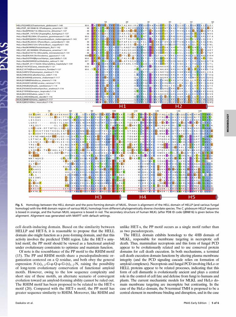
The HELL Domain Is Homologous to the Pore-Forming Domain ofMLKL. The HELL domain occurs in fungi as an N-terminal do-main of NLRs or associated with amyloid-forming motifs (17, 19).To identify distant homologs of HELL outside of the fungal king-dom, we performed profile hidden Markov model searches andfound hits in chordate MLKL, the cell death execution protein in
Fig. 3. HELLP-RFP localizes at the cell periphery on interaction with ag-gregated HELLP(215-278)-GFP. On fusion of cells coexpressing HELLP-RFPand HELLP(215-278)-GFP under the aggregated state [π], HELLP-RFP reloc-alizes to the membrane region. The white arrowhead marks the position ofthe cell fusion point between the HELLP-RFP and HELLP(215-278)-GFP strains.(Scale bar: 5 μ.)
Daskalov et al. PNAS Early Edition | 3 of 6
MICRO
BIOLO
GY

mammalian necroptosis, the target of a phosphorylation by theRIP1/RIP3 complex (16, 23–26). The homology between HELLPand MLKL lies in the N-terminal 4HB domain responsible for celldeath execution by membrane permeation. In HHPred searches,human MLKL was the first hit to HELLP, and HELLP scoredsignificantly better to MLKL than to HET-S (P = 6.8 ×10⁻⁷ toMLKL and 9.2 × 10⁻⁴ to HET-S). Fig. 5 shows the alignment offungal HELL domain proteins and chordate MLKL homologs.Sequence homology began in the N terminus in the region
predicted as TMH in HELLP, and then roughly correlated withthe secondary structure elements of the 4HB region of MLKL.Conservation of a negatively charged residue in position 2 or 3 wascommon to MLKL, HeLo, and HELL domains. Homology be-tween HELL and N-terminal domains of plant NLRs was de-tected as well. These N-terminal domains are of the non-TIR,CCR-type and are related to the RPW8 domain mediating celldeath and membrane targeting (27). HELL, HeLo, and CCR(RPW8) domains occur in similar domain architectures particu-larly as N-terminal domains of NLRs, but also associated with ki-nase domains, with the latter architecture representing the MLKL
domain architecture. As observed in the MLKL and HELL se-quences, a negatively charged residue was frequently found close tothe N termini in RPW8 sequences (Fig. S8). In HELL- and RPW8-related sequences, the region corresponding to the predicted TMHhelix was similar in length, and both types of sequences containedGXXXG-type motifs. This region was shorter in the MLKL se-quences. These analyses revealed homology among the chordateMLKL 4HB domain, the fungal HELL domain, and plant RPW8-related domains, three types of domains that have membrane-tar-geting activity in host-defense related processes.
DiscussionHET-S is a cell death-execution protein regulated by amyloidtemplating (11). We have proposed the existence in fungi of othercell death-effector proteins activated by amyloid signaling (12).Here we have characterized one such protein, HELLP, displayingan N-terminal HELL domain and a C-terminal PP motif. Like het-S,the gene encoding HELLP lies adjacent to a gene encoding anNLR with an N-terminal amyloid-forming motif. We found that theHELL domain is also an amyloid-controlled membrane-targeting
Fig. 4. Conformational analysis of HELLP(215-278) fibrils by solid-state NMR. (A) 1D 13C-detected CP spectrum. (B) 2D 1H-13C INEPT spectrum. (C) Excerpt of 2DPDSD ssNMR 13C-13C spectra centered around the Thr resonance frequency (Upper, 30 ms mixing time; Lower, 150 ms mixing time), optimized to detectintraresidual (30 ms) and sequential (150 ms) 13C-13C correlations. All peak assignments of the Thr271 spin system except for the diagonal Cα-Cα peaks areannotated. The additional peaks visible in the red spectrum reveal interresidual contacts of the Thr spin system. Assignments of Gly270 and Met272 areannotated above the spectral excerpt. The complete aliphatic spectral region of the 2D PDSD (30 ms mixing time) is shown in Fig. S5D. (D) Secondary ssNMRchemical shifts ΔδCα-ΔδCβ of the Gly270-Thr271-Met272 amino acid stretch in HELLP(215-278) (black), revealing their β-strand secondary structure. Thesecondary chemical shifts of a Gly-Thr-Met stretch in typical α-helical and β-strand conformations (blue and orange, respectively) are plotted for eye guidance.(E) 1D trace of the 2D PDSD (150 ms mixing time) at the position of the dotted blue line indicated in C, Lower. The line widths of intense signals are denoted.
4 of 6 | www.pnas.org/cgi/doi/10.1073/pnas.1522361113 Daskalov et al.
cell death-inducing domain. Based on the similarity betweenHELLP and HET-S, it is reasonable to propose that the HELLdomain also might function as a pore-forming domain, and that thisactivity involves the predicted TMH region. Like the HET-s amy-loid motif, the PP motif should be viewed as a functional amyloidunder evolutionary constraints to optimize and maintain function.Of note is the resemblance of the PP motif to the RHIM motif
(15). The PP and RHIM motifs share a pseudopalindromic or-ganization centered on a Q residue, and both obey the generalexpression N-(x)1–2-G-φ-Q-φ-G-(x)1–2-N, raising the possibilityof long-term evolutionary conservation of functional amyloidmotifs. However, owing to the low sequence complexity andshort size of these motifs, an alternate scenario of convergentevolution toward an amyloid-forming ability cannot be ruled out.The RHIM motif has been proposed to be related to the HET-smotif (20). Compared with the HET-s motif, the PP motif hasgreater sequence similarity to RHIM. Moreover, like RHIM and
unlike HET-s, the PP motif occurs as a single motif rather thanas two pseudorepeats.The HELL domain exhibits homology to the 4HB domain of
MLKL, responsible for membrane targeting in necroptotic celldeath. Thus, mammalian necroptosis and this form of fungal PCDappear to be evolutionarily related and to use conserved proteindomains for cell death execution. In both mechanisms, a terminalcell death execution domain functions by altering plasma membraneintegrity (and the PCD signaling cascade relies on formation ofamyloid complexes). Necroptosis and fungal PCD involving HeLo orHELL proteins appear to be related processes, indicating that thisform of cell dismantle is evolutionarily ancient and plays a centralrole in the control of cell fate and defense from fungi to humans andplants. The current mechanistic models for MLKL and HeLo do-main membrane targeting are incomplete but contrasting. In thecase of the HeLo domain, the N-terminal TMH is proposed to be acentral element in membrane binding and disruption, and in the case
Fig. 5. Homology between the HELL domain and the pore-forming domain of MLKL. Shown is alignment of the HELL domain of HELLP (and various fungalhomologs) with the 4HB domain region of various MLKL homologs from different phylogenetically diverse chordate species. The C. globosum HELLP sequenceis boxed in orange, and the human MLKL sequence is boxed in red. The secondary structure of human MLKL (after PDB ID code Q8NB16) is given below thealignment. Alignment was generated with MAFFT with default settings.
Daskalov et al. PNAS Early Edition | 5 of 6
MICRO
BIOLO
GY

of the MLKL domain, it is reported that most of the 4HB domaininserts into the membrane (9, 26). Future studies are needed todetermine the mechanistic similarities and differences in membranealteration by MLKL, HeLo, and HELL domain proteins.
Materials and MethodsPrion Propagation and Incompatibility Assays. Incompatibility phenotypes weredetermined by confronting strains of solid corn meal agar medium. Prionpropagationwas assayed as the ability to transmit the [π] prion from a [π]-donorstrain to a [π*] prion-free tester strain after confrontation on solid medium.Protein transfection experiments with amyloid fibrils of recombinant HELLP(215-278) protein were performed by spotting 100 μL of a 1 mg mL−1 HELLP(215-278) fibril suspension onto a [π*] mycelium. The mycelium was then cutwith a scalpel blade, and the regenerating mycelium was tested for the abilityto produce a barrage reaction to a strain expressing full-length HELLP.
Microscopy. P. anserina hyphae were inoculated on solid medium and cul-tivated for 24–72 h at 26 °C. The medium was then cut out, placed on a glassslide, and examined with a Leica DMRXA microscope equipped with aMicromax CCD (Princeton Instruments) controlled by Metamorph 5.06 soft-ware (Roper Scientific). The microscope was fitted with a Leica PL APO 100×immersion lens.
SSNMR Spectroscopy. SSNMR experiments were performed with 300- and800-MHz 1H Larmor frequency spectrometers (Bruker Biospin) using 3.2- and4-mm MAS probes, respectively, filled with ∼7 mg and ∼14 mg of fibril sam-ples. The MAS frequency was set between 7 and 20 kHz. Sample temperaturewas set to 6 °C with the internal reference DSS (28). A ramped CP with a 1-mscontact time was used for the 1H-13C polarization transfer. The 1D 1H-13C CP
was recorded at 800 MHz 1H Larmor frequency with an MAS frequency of20 kHz. An acquisition time of 20 ms was used for 512 scans, with an interscandelay of 3 s. The 2D 13C-13C PDSD spectra were recorded at 300 MHz 1H Lamorfrequency with an MAS rate of 11 kHz . 13C-13C polarization transfer wasachieved with PDSD with mixing times of 30 ms to correlate intraresiduecarbon atoms and 150 ms to connect sequential atoms. Proton decouplingwith a frequency of 85 kHz was applied during acquisition times, using theSPINAL-64 decoupling sequence (29). Acquisition times of 20 ms (t1) × 24 ms(t2) (for the 30-ms mixing time) and 17 ms (t1) × 24 ms (t2) (for the 150-msmixing time) were chosen for the indirect and direct dimensions, respectively,resulting in a total experiment time of ∼6.5 d. A 1D trace was extracted at thepositions of the Thr resonances, indicated by a dashed blue line in Fig. 4C, fromthe 2D PDSD (for the 150-ms mixing time). Mobile residues were probed usinga 2D 1H-13C INEPT experiment recorded at 300 MHz 1H Lamor frequency for atotal experiment time of ∼2 d. Random coil chemical shifts for the secondarychemical shift calculation were as reported previously (30). Spectra were an-alyzed and figures prepared using the CCPNMR analysis software (31).
ACKNOWLEDGMENTS. We thank Dr. David Gajan for technical assistancewith the NMR measurements. We acknowledge the support provided by theCrystallography Platform of the Institute of Molecular Biology of Barcelona,and particularly Dr. Joan Pous, for help with diffraction assays and spectraanalyses. Financial support for this research from the TGIR-RMN-THC Fr3050CNRS for conducting the research is gratefully acknowledged. This work wassupported, in whole or in part, by L’Agence Nationale de la Recherche(Grants STANDPRION ANR-11-BSV8-0001, to S.J.S; ANR-13-PDOC-0017-01,to B.H.; and ANR-14-CE09-0020-01, to A.L.), IdEx Bordeaux (PEPS 2014,to A.L. and S.J.S.), Fondation pour la Recherche Médicale (Grant FRM-AJE20140630090, to A.L.), the FP7 Program (Grant FP7-PEOPLE-2013-CIG,to A.L.) and the European Research Council under the European Union’sHorizon 2020 Program (Starting Grant Agreement 105945, to A.L.).
1. Dickman MB, Fluhr R (2013) Centrality of host cell death in plant–microbe interac-tions. Annu Rev Phytopathol 51:543–570.
2. Blander JM (2014) A long-awaited merger of the pathways mediating host defenceand programmed cell death. Nat Rev Immunol 14(9):601–618.
3. Pinan-Lucarré B, Paoletti M, Clavé C (2007) Cell death by incompatibility in the fungusPodospora. Semin Cancer Biol 17(2):101–111.
4. Makarova KS, Anantharaman V, Aravind L, Koonin EV (2012) Live virus-free or die:Coupling of antivirus immunity and programmed suicide or dormancy in prokaryotes.Biol Direct 7:40.
5. van Doorn WG (2011) Classes of programmed cell death in plants, compared to thosein animals. J Exp Bot 62(14):4749–4761.
6. Wasmer C, et al. (2008) Amyloid fibrils of the HET-s(218-289) prion form a beta so-lenoid with a triangular hydrophobic core. Science 319(5869):1523–1526.
7. Balguerie A, et al. (2003) Domain organization and structure–function relationship ofthe HET-s prion protein of Podospora anserina. EMBO J 22(9):2071–2081.
8. Greenwald J, et al. (2010) The mechanism of prion inhibition by HET-S. Mol Cell 38(6):889–899.
9. Seuring C, et al. (2012) The mechanism of toxicity in HET-S/HET-s prion in-compatibility. PLoS Biol 10(12):e1001451.
10. Mathur V, Seuring C, Riek R, Saupe SJ, Liebman SW (2012) Localization of HET-S to thecell periphery, not to [Het-s] aggregates, is associated with [Het-s]-HET-S toxicity. MolCell Biol 32(1):139–153.
11. Daskalov A, et al. (2015) Signal transduction by a fungal NOD-like receptor based onpropagation of a prion amyloid fold. PLoS Biol 13(2):e1002059.
12. Daskalov A, Paoletti M, Ness F, Saupe SJ (2012) Genomic clustering and homologybetween HET-S and the NWD2 STAND protein in various fungal genomes. PLoS One7(4):e34854.
13. Cai X, et al. (2014) Prion-like polymerization underlies signal transduction in antiviralimmune defense and inflammasome activation. Cell 156(6):1207–1222.
14. Wu H (2013) Higher-order assemblies in a new paradigm of signal transduction. Cell153(2):287–292.
15. Li J, et al. (2012) The RIP1/RIP3 necrosome forms a functional amyloid signalingcomplex required for programmed necrosis. Cell 150(2):339–350.
16. Sun L, et al. (2012) Mixed lineage kinase domain-like protein mediates necrosis sig-naling downstream of RIP3 kinase. Cell 148(1-2):213–227.
17. Daskalov A, Dyrka W, Saupe SJ (2015) Theme and variations: Evolutionary di-versification of the HET-s functional amyloid motif. Sci Rep 5:12494.
18. Graziani S, Silar P, Daboussi MJ (2004) Bistability and hysteresis of the “Secteur”differentiation are controlled by a two-gene locus in Nectria haematococca. BMC Biol2:18.
19. Dyrka W, et al. (2014) Diversity and variability of NOD-like receptors in fungi. GenomeBiol Evol 6(12):3137–3158.
20. Kajava AV, Klopffleisch K, Chen S, Hofmann K (2014) Evolutionary link betweenmetazoan RHIM motif and prion-forming domain of fungal heterokaryon in-compatibility factor HET-s/HET-s. Sci Rep 4:7436.
21. Mueller BK, Subramaniam S, Senes A (2014) A frequent, GxxxG-mediated, trans-membrane association motif is optimized for the formation of interhelical Cα-H hy-drogen bonds. Proc Natl Acad Sci USA 111(10):E888–E895.
22. Sabaté R, et al. (2007) Prion and non-prion amyloids of the HET-s prion forming do-main. J Mol Biol 370(4):768–783.
23. Murphy JM, et al. (2013) The pseudokinase MLKL mediates necroptosis via a molec-ular switch mechanism. Immunity 39(3):443–453.
24. Cai Z, et al. (2014) Plasma membrane translocation of trimerized MLKL protein isrequired for TNF-induced necroptosis. Nat Cell Biol 16(1):55–65.
25. Chen X, et al. (2014) Translocation of mixed lineage kinase domain-like protein toplasma membrane leads to necrotic cell death. Cell Res 24(1):105–121.
26. Su L, et al. (2014) A plug release mechanism for membrane permeation by MLKL.Structure 22(10):1489–1500.
27. Collier SM, Hamel LP, Moffett P (2011) Cell death mediated by the N-terminal do-mains of a unique and highly conserved class of NB-LRR protein. Mol Plant MicrobeInteract 24(8):918–931.
28. Böckmann A, et al. (2009) Characterization of different water pools in solid-stateNMR protein samples. J Biomol NMR 45(3):319–327.
29. Fung BM, Khitrin AK, Ermolaev K (2000) An improved broadband decoupling se-quence for liquid crystals and solids. J Magn Reson 142(1):97–101.
30. Wang Y, Jardetzky O (2002) Probability-based protein secondary structure identifi-cation using combined NMR chemical-shift data. Protein Sci 11(4):852–861.
31. Vranken WF, et al. (2005) The CCPN data model for NMR spectroscopy: Developmentof a software pipeline. Proteins 59(4):687–696.
6 of 6 | www.pnas.org/cgi/doi/10.1073/pnas.1522361113 Daskalov et al.