Identification of a host collagen inducing factor from the ...
16
RESEARCH ARTICLE Identification of a host collagen inducing factor from the excretory secretory proteins of Trichinella spiralis Mi Kyung Park 1 , Hae-Jin Kim 1 , Min Kyoung Cho 1 , Shin Ae Kang 1 , So Young Park 1,2 , Se Bok Jang 2 , Hak Sun Yu 1 * 1 Department of Parasitology School of Medicine, Pusan National University, Yangsan, Republic of Korea, 2 Department of Molecular Biology, College of Natural Sciences, Pusan National University, Busan, Republic of Korea * [email protected] Abstract Background In a previous study, we found that Trichinella spiralis muscle larva excretory and secretory proteins (ES-P) most likely activate collagen synthesis via TGF-β/Smad signaling, and this event could influence collagen capsule formation. Methodology/Principal findings In order to identify the specific collagen inducing factor, ES-P was fractionated by a Super- dex 200 10/300 GL column. We obtained three large fractions, F1, F2, and F3, but only F3 had collagen gene inducing ability. After immunoscreening, 10 collagen inducing factor can- didates were identified. Among them, TS 15–1 and TS 15–2 were identical to the putative trypsin of T. spiralis. The deduced TS 15–1 (M.W. = 72 kDa) had two conserved catalytic motifs, an N-terminal Tryp_SPc domain (TS 15-1n) and a C-terminal Tryp_SPc domain (TS 15-1c). To determine their collagen inducing ability, recombinant proteins (rTS 15-1n and rTS 15-1c) were produced using the pET-28a expression system. TS 15–1 is highly expressed during the muscle larval stage and has strong antigenicity. We determined that rTS 15-1c could elevate collagen I via activation of the TGF-β1 signaling pathway in vitro and in vivo. Conclusion/Significance In conclusion, we identified a host collagen inducing factor from T. spiralis ES-P using immu- noscreening and demonstrated its molecular characteristics and functions. PLOS Neglected Tropical Diseases | https://doi.org/10.1371/journal.pntd.0006516 November 1, 2018 1 / 16 a1111111111 a1111111111 a1111111111 a1111111111 a1111111111 OPEN ACCESS Citation: Park MK, Kim H-J, Cho MK, Kang SA, Park SY, Jang SB, et al. (2018) Identification of a host collagen inducing factor from the excretory secretory proteins of Trichinella spiralis. PLoS Negl Trop Dis 12(11): e0006516. https://doi.org/ 10.1371/journal.pntd.0006516 Editor: Maria Angeles Go ´mez-Morales, Istituto Superiore di Sanità, ITALY Received: May 8, 2018 Accepted: September 4, 2018 Published: November 1, 2018 Copyright: © 2018 Park et al. This is an open access article distributed under the terms of the Creative Commons Attribution License, which permits unrestricted use, distribution, and reproduction in any medium, provided the original author and source are credited. Data Availability Statement: All relevant data are within the paper and its Supporting Information files. Funding: This research was financially supported by the Ministry of SMEs and Startups [MSS] of the Republic of Korea and Korea Institute for Advancement of Technology[KIAT] through the Regional Specialized Industry Development Program[Grant Number:R0006014]. The funders had no role in study design, data collection and
Transcript of Identification of a host collagen inducing factor from the ...
RESEARCH ARTICLE
Identification of a host collagen inducing
factor from the excretory secretory proteins
of Trichinella spiralis
Mi Kyung Park1, Hae-Jin Kim1, Min Kyoung Cho1, Shin Ae Kang1, So Young Park1,2, Se
Bok Jang2, Hak Sun Yu1*
1 Department of Parasitology School of Medicine, Pusan National University, Yangsan, Republic of Korea,
2 Department of Molecular Biology, College of Natural Sciences, Pusan National University, Busan, Republic
of Korea
Abstract
Background
In a previous study, we found that Trichinella spiralis muscle larva excretory and secretory
proteins (ES-P) most likely activate collagen synthesis via TGF-β/Smad signaling, and this
event could influence collagen capsule formation.
Methodology/Principal findings
In order to identify the specific collagen inducing factor, ES-P was fractionated by a Super-
dex 200 10/300 GL column. We obtained three large fractions, F1, F2, and F3, but only F3
had collagen gene inducing ability. After immunoscreening, 10 collagen inducing factor can-
didates were identified. Among them, TS 15–1 and TS 15–2 were identical to the putative
trypsin of T. spiralis. The deduced TS 15–1 (M.W. = 72 kDa) had two conserved catalytic
motifs, an N-terminal Tryp_SPc domain (TS 15-1n) and a C-terminal Tryp_SPc domain
(TS 15-1c). To determine their collagen inducing ability, recombinant proteins (rTS 15-1n
and rTS 15-1c) were produced using the pET-28a expression system. TS 15–1 is highly
expressed during the muscle larval stage and has strong antigenicity. We determined that
rTS 15-1c could elevate collagen I via activation of the TGF-β1 signaling pathway in vitro
and in vivo.
Conclusion/Significance
In conclusion, we identified a host collagen inducing factor from T. spiralis ES-P using immu-
noscreening and demonstrated its molecular characteristics and functions.
PLOS Neglected Tropical Diseases | https://doi.org/10.1371/journal.pntd.0006516 November 1, 2018 1 / 16
a1111111111
a1111111111
a1111111111
a1111111111
a1111111111
OPEN ACCESS
Citation: Park MK, Kim H-J, Cho MK, Kang SA,
Park SY, Jang SB, et al. (2018) Identification of a
host collagen inducing factor from the excretory
secretory proteins of Trichinella spiralis. PLoS Negl
Trop Dis 12(11): e0006516. https://doi.org/
10.1371/journal.pntd.0006516
Editor: Maria Angeles Gomez-Morales, Istituto
Superiore di Sanità, ITALY
Received: May 8, 2018
Accepted: September 4, 2018
Published: November 1, 2018
Copyright: © 2018 Park et al. This is an open
access article distributed under the terms of the
Creative Commons Attribution License, which
permits unrestricted use, distribution, and
reproduction in any medium, provided the original
author and source are credited.
Data Availability Statement: All relevant data are
within the paper and its Supporting Information
files.
Funding: This research was financially supported
by the Ministry of SMEs and Startups [MSS] of the
Republic of Korea and Korea Institute for
Advancement of Technology[KIAT] through the
Regional Specialized Industry Development
Program[Grant Number:R0006014]. The funders
had no role in study design, data collection and
Author summary
Trichinella spiralis can make collagen capsules in host muscle cells during its life cycle,
which encapsulates muscle stage larvae. Many investigators have tried to reveal the com-
plex mechanism behind this collagen capsule architecture, and it has been suggested that
several serine proteases in excretory-secretory proteins of the parasite are potential colla-
gen capsule inducing factors. In addition, collagen synthesis is activated through the TGF-
β/Smad signaling pathway and these events are closely related with protease activated
receptor 2 which was activated by various serine proteases. In this study, we isolated and
characterized a collagen gene expression inducer from T. spiralis ES-P using immunoscre-
ening and investigated the candidate protein for its usefulness as a wound healing thera-
peutic agent.
Introduction
Trichinella spiralis can make collagen capsules in host muscles during their life cycle that sur-
round muscle stage larvae and might protect the larvae from the host immune system. This
phenomenon can be understood as the parasite creating a simple structure to protect itself, but
when examined closely, numerous different mechanisms are involved in this stage of the para-
site’s life. Division of the host muscle cell nucleus, regulation of host cell cycling, huge eleva-
tion of host collagen gene expression, and generation of new blood vessels around the collagen
capsule are observed during nurse cell formation by T. spiralis [1–4]. The process of nurse cell
formation induces de-differentiation, cell cycle re-entry, arrest of infected muscle cells, and
activation, proliferation, and differentiation of satellite cells. These events are very similar to
those occurring during muscle cell regeneration and repair [2].
In a previous study, we found that T. spiralis excretory and secretory proteins (ES-P) most
likely activate collagen synthesis via TGF-β/Smad signaling, and this event could influence col-
lagen capsule formation [5]. These events were closely related with protease activated receptor
2 (PAR2), which was activated by various serine proteases [5]. However, the question of which
protease in T. spiralis ES-P has a role in collagen gene expression of host muscle cells is still
unanswered. The identification of a specific collagen gene inducer from T. spiralis could be
exploited as a therapeutic and/or cosmetic agent. In this study, we isolated and characterized
the collagen gene expression inducer from T. spiralis ES-P by immunoscreening and investi-
gated the candidate for its usefulness as a wound healing therapeutic agent.
Materials and methods
Isolation of muscle larvae and extraction of whole parasite proteins
The T. spiralis strain (isolate code ISS623) used in this study has been maintained in our labo-
ratory via serial passage in rats. For acquisition of muscle larva, eviscerated mouse carcasses
were cut into pieces, followed by digestion in 1% pepsin 1% hydrochloride digestion fluid (arti-
ficial gastric juice) for 1 hr at 37˚C with stirring. Larvae were collected manually from muscle
digested solution under microscopy and washed 6 times with sterile PBS containing 100 μg/ml
ampicillin, 5 μg/ml kanamycin and 50 μg/ml tetracyclin. After collection, in order to prevent
contamination with the host material, worms were thoroughly and carefully washed several 3
times with PBS. Whole parasite proteins (total extract; TE) was obtained from muscle larva
according to previous study [6]. In brief, muscle larva were rinsed in PBS and homogenized in
50 mM Tris–HCl (pH 7.5) with a glass homogenizer. The homogenates were briefly sonicated
Host collagen inducing factor
PLOS Neglected Tropical Diseases | https://doi.org/10.1371/journal.pntd.0006516 November 1, 2018 2 / 16
analysis, decision to publish, or preparation of the
manuscript.
Competing interests: The authors have declared
that no competing interests exist.
and then centrifuged for 30 min at 12,000 × g and 4˚C. The supernatant (TE) was stored at
-20˚C.
Isolation of adult worm and new born larvae (NBL)
Small intestines were removed on the day 6 after infection from infected rat, opened, sliced by
2 cm, washed with PBS, and incubated for 1 hr at 37˚C in PBS containing antibiotics. Adult
worms were collected on a PBS, washed 3 times with PBS containing antibiotics, and incu-
bated for 24 hrs in serum-free RPMI 1640 medium containing antibiotics. After incubation,
NBL were passed through 40 μl nylon mesh (BC falcon, USA) to be separated from adult
worms.
Extraction of ES-P from muscle larvae and fractionation of ES-P
Muscle larvae were isolated from T. spiralis infected mice (4 weeks after infection) and ES-P
from cultured muscle larvae was obtained according to the previously reported method [5].
The ES-P was fractionated using gel filtration chromatography. ES-P (5 mg) in 10 ml PBS was
applied to a Superdex 200 10/300 GL column (GE Healthcare, Uppsala, Sweden). The flow rate
was 0.25 ml/min. Each 0.5 ml fraction was collected and protein quantity was measured by UV
detection at 260 nm. Three big fractions, F1, F2, and F3, were acquired and used for collagen
gene inducing experiments (Fig 3A).
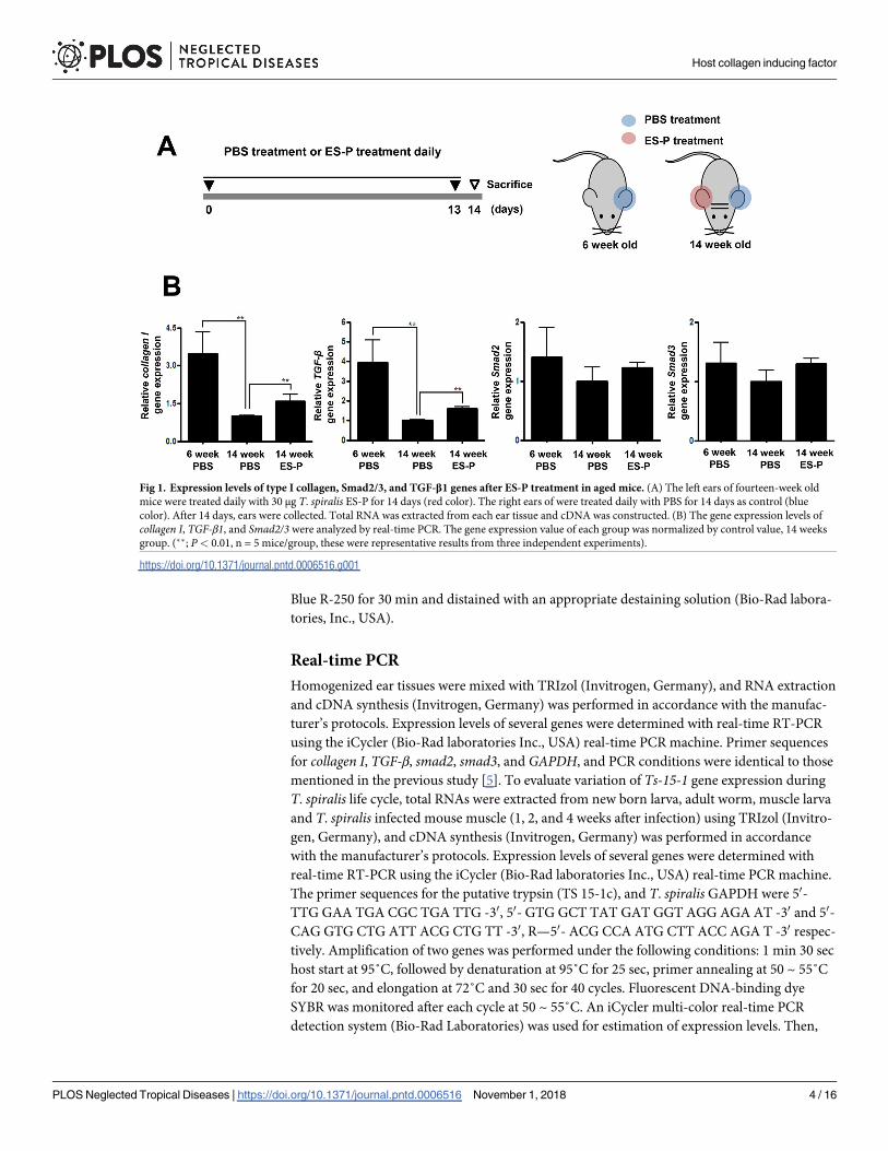
Mouse experiments
Twenty female C57BL/6 mice at the age of 6 weeks and twenty female 14 week-old mice were
purchased from Samtako Co. (Gyeonggi-do, Korea). The skin of the left ear of each mouse was
treated with T. spiralis ES-P (30 μg) or rTS 15-1c (30 μg) in PBS (total volume 50 μl) every day
for 14 days, and that of the right ear was treated with PBS (Figs 1A and 7A). The mice were
housed in a specific pathogen-free facility at the Institute for Laboratory Animals of Pusan
National University.
Cell culture and in vitro stimulation
In order to compare the expression level of type I collagen and their signal pathway related
genes, mouse fibroblast (MEF) cells were used in this study because type I collagen was prefer-
entially synthesized by two cell types, the osteoblast and the fibroblast [7]. MEF cells were iso-
lated from C57BL/6 mouse fetuses 10 days after fertilization [8]. MEF cells were incubated in
DMEM (Difco) with 5% FBS and 5 × 10⁵ cells were plated in 24-well plates and incubated
overnight at 37˚C in 5% CO2. The cells were treated with ES-P, boiled- ES-P, F1, F2, F3, boiled
F3, TS 15-1c, and TS 15-1n (final conc. 1 μg/ml); ES-P with PMSF (serine protease inhibitor,
final conc. 1 mM; Sigma-Aldrich, USA), F3 with PMSF for 2 hrs.
Zymography
Gelatin-gel containing 0.2% gelatin was prepared from gelatin-stock solution. The proteins, T.
spiralis ES proteins, TS 15-1c, and TS 15-1n were mixed with 2 × sample buffer (1 M Tris pH
6.8, 1% bromphenol blue, glycerol, β-mercaptoethanol), and the gel loaded with these proteins
was run with 1 × Tris-Glycine SDS running buffer on 125 V for 2 hrs at 4˚C. After running,
the gel was washed to remove the SDS and re-natured proteinase activity with zymogram rena-
turing buffer (2.5% Triton X-100). The gel was developed with zymogram developing buffer
(0.5 M Tris-HCl pH 7.6, 0.02 M NaCl, 0.5 mM CaCl2) for 30 min at room temperature. The
gel was incubated with developing buffer at 37˚C for 8 hrs. The gel was stained with Coomassie
Host collagen inducing factor
PLOS Neglected Tropical Diseases | https://doi.org/10.1371/journal.pntd.0006516 November 1, 2018 3 / 16
Blue R-250 for 30 min and distained with an appropriate destaining solution (Bio-Rad labora-
tories, Inc., USA).
Real-time PCR
Homogenized ear tissues were mixed with TRIzol (Invitrogen, Germany), and RNA extraction
and cDNA synthesis (Invitrogen, Germany) was performed in accordance with the manufac-
turer’s protocols. Expression levels of several genes were determined with real-time RT-PCR
using the iCycler (Bio-Rad laboratories Inc., USA) real-time PCR machine. Primer sequences
for collagen I, TGF-β, smad2, smad3, and GAPDH, and PCR conditions were identical to those
mentioned in the previous study [5]. To evaluate variation of Ts-15-1 gene expression during
T. spiralis life cycle, total RNAs were extracted from new born larva, adult worm, muscle larva
and T. spiralis infected mouse muscle (1, 2, and 4 weeks after infection) using TRIzol (Invitro-
gen, Germany), and cDNA synthesis (Invitrogen, Germany) was performed in accordance
with the manufacturer’s protocols. Expression levels of several genes were determined with
real-time RT-PCR using the iCycler (Bio-Rad laboratories Inc., USA) real-time PCR machine.
The primer sequences for the putative trypsin (TS 15-1c), and T. spiralis GAPDH were 50-
TTG GAA TGA CGC TGA TTG -30, 50- GTG GCT TAT GAT GGT AGG AGA AT -30 and 50-
CAG GTG CTG ATT ACG CTG TT -30, R—50- ACG CCA ATG CTT ACC AGA T -30 respec-
tively. Amplification of two genes was performed under the following conditions: 1 min 30 sec
host start at 95˚C, followed by denaturation at 95˚C for 25 sec, primer annealing at 50 ~ 55˚C
for 20 sec, and elongation at 72˚C and 30 sec for 40 cycles. Fluorescent DNA-binding dye
SYBR was monitored after each cycle at 50 ~ 55˚C. An iCycler multi-color real-time PCR
detection system (Bio-Rad Laboratories) was used for estimation of expression levels. Then,
Fig 1. Expression levels of type I collagen, Smad2/3, and TGF-β1 genes after ES-P treatment in aged mice. (A) The left ears of fourteen-week old
mice were treated daily with 30 μg T. spiralis ES-P for 14 days (red color). The right ears of were treated daily with PBS for 14 days as control (blue
color). After 14 days, ears were collected. Total RNA was extracted from each ear tissue and cDNA was constructed. (B) The gene expression levels of
collagen I, TGF-β1, and Smad2/3 were analyzed by real-time PCR. The gene expression value of each group was normalized by control value, 14 weeks
group. (��; P< 0.01, n = 5 mice/group, these were representative results from three independent experiments).
https://doi.org/10.1371/journal.pntd.0006516.g001
Host collagen inducing factor
PLOS Neglected Tropical Diseases | https://doi.org/10.1371/journal.pntd.0006516 November 1, 2018 4 / 16
using the Gene-x program (Bio-Rad Laboratories), relative expression of the gene was calcu-
lated as the ratio to a T. spiralis GAPDH gene.
Immunoscreening of cDNA library
A cDNA library generated from 60,000 plaques forming units of T. spiralis muscle larvae was
screened with the α-TS F3 antibody. Immunoscreening was performed using the SMART
cDNA Library Construction Kit (Clontech, USA) in accordance with the manufacturer’s pro-
tocols. Briefly, after primary and secondary screening, positive plaques were picked and the
phagemids were prepared by in vivo excision. The phagemids were transformed into XL1-Blue
MRF cells. Clones were selected based on blue-white color selection of the colonies grown on
LB-ampicillin agar plates. The plasmid harboring the cDNA inserts were then extracted using
a plasmid DNA purification system (Cosmogenetech, Seoul, Korea). The cDNA inserts were
then sequenced using the primer for T3 promotor (Cosmogenetech, DNA sequencing service,
Seoul, Korea) and compared against the GenBank database.
Construction of recombinant TS-15-1, TS 15-1c domain and TS 15-1n
domain
Following confirmation of the PCR product sequences, TS 15–1, The TS 15-1c (C-terminal
serine protease domain) and TS 15-1n (N-terminal serine protease domain), the genes were
ligated with pET-28a vector (Novagen, USA). After gene ligation, the constructed plasmids
were expressed in Escherichia coli BL21 (DE3, Novagen, USA). Pre-cultured cells were inocu-
lated into Luria-Bertani broth containing kanamycin, and the cells were grown at 37˚C until
an OD600 of 0.5–0.6 was reached. Recombinant TS 15-1N and TS 15-1C expressions were
induced addition of 0.5 mM isopropyl β-D-1-thiogalactopyranoside (IPTG) at 25˚C for 16 h.
The cells were harvested by centrifugation and the cell pellets were resuspended in buffer. A
consisting of 50 mM Tris–HCl pH 7.5 and 200 mM NaCl. Cell disruptions were lysed by soni-
cation on ice and the crude extracts were centrifuged to remove the cell debris. Ts15-1 N and
C pellets were then sonicated in buffer including 50 mM Tris–HCl pH 7.5, 200 mM NaCl, and
6 M Urea on ice. The clear supernatant of the lysate was subjected onto the Ni–NTA column
which had been pre-equilibrated with buffer A. The column was subsequently washed with
buffer A containing imidazole, after which the bound proteins were eluted by varying the
imidazole concentration (20–400 mM). The eluted proteins were analyzed using 10%
SDS-PAGE. However, we could not get recombinant protein of TS 15–1 because very poor
expression level.
Production of polyclonal antisera for F3 fraction or TS 15-1c
Female four-week-old Wistar rats were purchased from Samtako Co. (Gyeonggi-do, Korea).
Rats were immunized subcutaneously with a 1:1 mixture of the 250 μg F3 fraction of TS 15-1c
protein (in 0.5 ml PBS) and 0.5 ml Freund’s complete adjuvant (#F5881, Sigma-Aldrich, USA)
at 0 week. At 2 weeks the rat was given additional infections of the 250 μg F3 fraction or TS 15-
1c protein with Freund’s incomplete adjuvant (#F5506, Sigma-Aldrich, USA). One week after
their final booster, rats were sacrificed and serum was obtained.
Real-time PCR
Homogenized ear tissues were mixed with TRIzol (Invitrogen, Germany), and total RNA
extraction and cDNA synthesis (Invitrogen, Germany) was performed in accordance with the
manufacturer’s protocols. Expression levels of several genes were determined with real-time
Host collagen inducing factor
PLOS Neglected Tropical Diseases | https://doi.org/10.1371/journal.pntd.0006516 November 1, 2018 5 / 16
RT-PCR using the iCycler (Bio-Rad laboratories Inc., USA) real-time PCR machine. Primer
sequences for collagen I, TGF-β, smad2, smad3, and GAPDH, and PCR conditions were identi-
cal to those mentioned in the previous study [5]. Each gene expression levels were normalized
with GAPDH gene expression.
Collection of serum
Mice were killed at 0, 1, 2, and 4 weeks after T. spiralis infection and serum was obtained. Sera
were stored at -20˚C until used.
Western blotting
Ten μg each of ES-P and F3 fraction (Fig 3) or 10 μg ES-P and total extract from T. spiralis, TS
15-1c (Fig 5B) or 10 μg of purified TS 15-1c antibody (Fig 5C) or 15 μg of each ear tissue sam-
ples (Fig 7) were separated on 10% acrylamide SDS-PAGE gel at 100 V for 90 min. Sweden),
The loaded proteins were transferred onto a nitrocellulose membrane (Amersham Biosciences,
Little Chalfont, UK) and blocked with 5% skim milk in TBST at 4˚C overnight. Then, the
membrane was incubated with primary antibody (polyclonal α-F3, α-TS 15-1c (1:500); time-
course sera (1:1,000), α-TGF-β1 (1:1000; abcam, Carlsbad, CA, USA),;p-Smad2/3 (1:1000;
Thermofisher science, Waltham, MC, USA),;α-mouse type I collagen (1:1000; abcam), and
actin (1:5000, abcam)) in 5% skim milk in TBST for 2 hrs at room temperature. The secondary
antibody, α-mouse or α-rat IgG-HRP conjugate (Sigma, Seoul, Korea) was used at 1:5,000
dilution for 1 hr at room temperature. HRP was detected using an ECL substrate (Amersham
Biosciences, Uppsala, Sweden), analyzed using the LAS 3000 machine. (Areas of the detected
bands were determined and compared by Image J software).
Immunofluorescence
Paraffin-embedded T. spiralis infected or non-infected mouse muscle tissue were de-paraffi-
nized and hydrated. For antigen retrieval, slides were immersed in citrate buffer (0.01 M, pH
6.0) and heated twice in a microwave (700 W or ‘high’) for 5 min. Slides were then quenched
with endogenous peroxidase by incubation in a 3% hydrogen peroxide solution for 5 min and
were washed three times in PBS for 5 min each. Slides were immuno-stained with primary
antibody (α-TS 15-1c antibody that was produced according to the polyclonal antisera
method; 1:500 dilution) at 4˚C overnight. After primary antibody incubation, slides were
washed three times in PBS for 5 min each and were incubated with secondary antibody, the
Alexa Fluor 594 goat anti-rat IgG secondary antibody (1:500; Invitrogen, USA) was applied for
1 h at 24˚C. The slides were washed in PBS and mounted with Permount (Fisher Scientific,
Pittsburgh, PA, USA). Confocal images of stained muscle tissue were examined under an
inverted fluorescence microscope.
Statistical analysis
All experiments were performed three times for confirmation of statistical significance.
Mean ± standard deviation (SD) was calculated from data collected from individual mice. Sig-
nificant differences were determined using one-way or two-way analysis of variance. Statistical
analysis was performed with GraphPad Prism 5.0 software (GraphPad Software Inc., CA,
USA).
Host collagen inducing factor
PLOS Neglected Tropical Diseases | https://doi.org/10.1371/journal.pntd.0006516 November 1, 2018 6 / 16
Ethics statement
The study was performed with approval from the Pusan National University Animal Care and
Use Committee (IACUC protocol approval; PNU-2016-1175), in compliance with ‘‘The Act
for the Care and Use of Laboratory Animals” of the Ministry of Food and Drug Safety, Korea.
All animal procedures were conducted in a specific pathogen-free facility at the Institute for
Laboratory Animals of Pusan National University.
Results
Type I collagen gene expression in an aging mouse model
In order to understand the collagen gene inducing effect of ES-P, transcription and protein
expression levels of type I collagen and TGF-β1 signaling related proteins were compared in
ear tissues of 6 and 14 week-old mice that had or had not received ES-P treatment (Fig 1A).
The expression levels of the collagen I and TGF-β1 genes of the 14 week-old mice were signifi-
cantly decreased compared to those of the 6 weeks mice. However, those of the ES-P treated 14
week-old mouse group were significantly increased compared to un-treated 14 week-old mice
(Fig 1B).
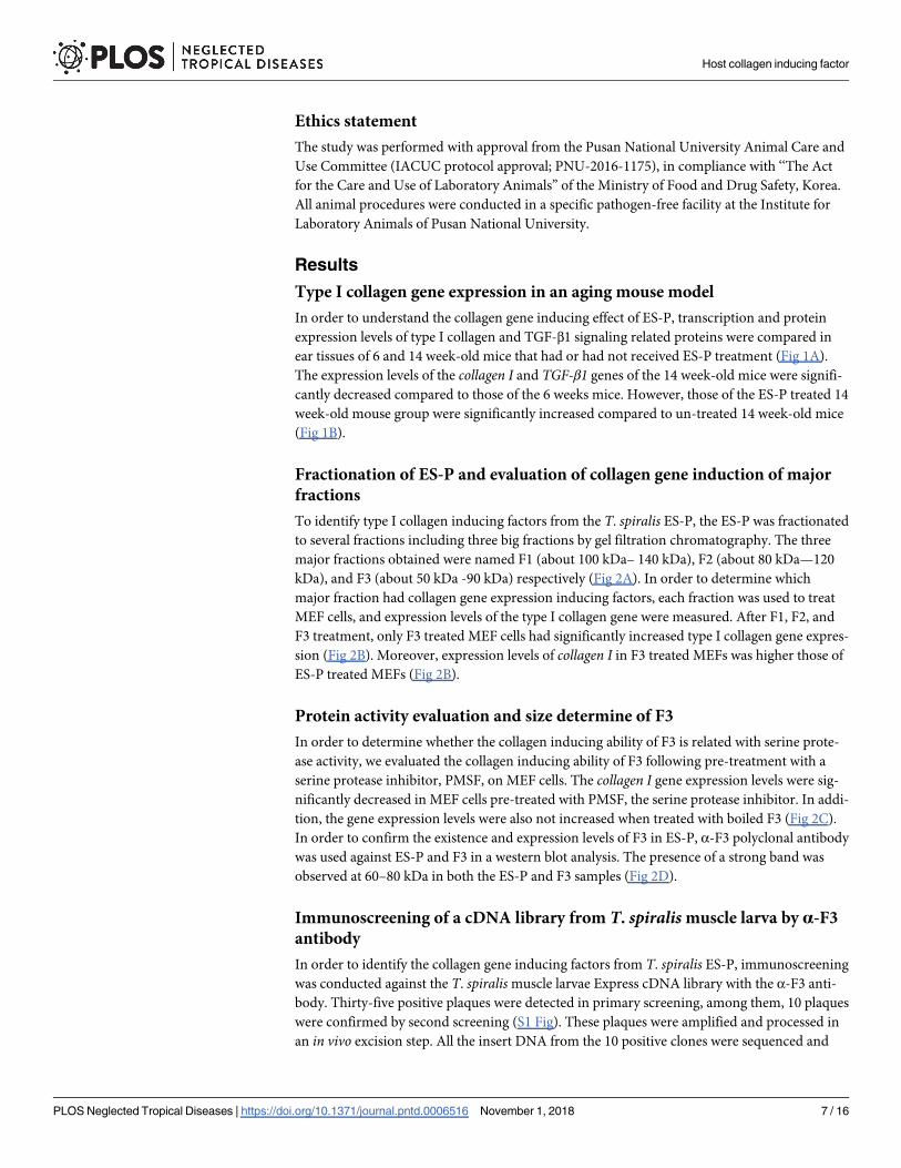
Fractionation of ES-P and evaluation of collagen gene induction of major
fractions
To identify type I collagen inducing factors from the T. spiralis ES-P, the ES-P was fractionated
to several fractions including three big fractions by gel filtration chromatography. The three
major fractions obtained were named F1 (about 100 kDa– 140 kDa), F2 (about 80 kDa—120
kDa), and F3 (about 50 kDa -90 kDa) respectively (Fig 2A). In order to determine which
major fraction had collagen gene expression inducing factors, each fraction was used to treat
MEF cells, and expression levels of the type I collagen gene were measured. After F1, F2, and
F3 treatment, only F3 treated MEF cells had significantly increased type I collagen gene expres-
sion (Fig 2B). Moreover, expression levels of collagen I in F3 treated MEFs was higher those of
ES-P treated MEFs (Fig 2B).
Protein activity evaluation and size determine of F3
In order to determine whether the collagen inducing ability of F3 is related with serine prote-
ase activity, we evaluated the collagen inducing ability of F3 following pre-treatment with a
serine protease inhibitor, PMSF, on MEF cells. The collagen I gene expression levels were sig-
nificantly decreased in MEF cells pre-treated with PMSF, the serine protease inhibitor. In addi-
tion, the gene expression levels were also not increased when treated with boiled F3 (Fig 2C).
In order to confirm the existence and expression levels of F3 in ES-P, α-F3 polyclonal antibody
was used against ES-P and F3 in a western blot analysis. The presence of a strong band was
observed at 60–80 kDa in both the ES-P and F3 samples (Fig 2D).
Immunoscreening of a cDNA library from T. spiralis muscle larva by α-F3
antibody
In order to identify the collagen gene inducing factors from T. spiralis ES-P, immunoscreening
was conducted against the T. spiralis muscle larvae Express cDNA library with the α-F3 anti-
body. Thirty-five positive plaques were detected in primary screening, among them, 10 plaques
were confirmed by second screening (S1 Fig). These plaques were amplified and processed in
an in vivo excision step. All the insert DNA from the 10 positive clones were sequenced and
Host collagen inducing factor
PLOS Neglected Tropical Diseases | https://doi.org/10.1371/journal.pntd.0006516 November 1, 2018 7 / 16
their amino acid sequences were determined. Two insert DNA fragments (TS 15–1 and TS
15–2) were similar to a putative trypsin of T. spiralis with 90% identity. Another clone (TS 15–
3) was matched to a nuclear receptor-binding protein of T. spiralis with 35% of identity.
Another clone (TS-16-1) was matched to a putative BTB/POZ domain protein of T. spiraliswith 63% identity. The remaining 6 insert clones were not matched with any previously
known genes.
Molecular characterization of TS 15–1Collagen inducing factors in ES-P and F3 had serine protease activity and were measured to be
about 60–72 kDa in size. After evaluation of the size and serine protease activity of positive
clone matched genes, the TS 15–1 gene was selected for downstream identification of the colla-
gen inducing factor. The TS 15–1 fragment was 2,004 bp long and encoded a 667 amino acid
protein, and the molecular weight and pI was calculated as 71.6 kDa and 8.83. The deduced TS
15–1 protein has two conserved catalytic motifs, an N-terminal Tryp_SPc domain (TS 15-1n)
and a C-terminal Tryp_SPc domain (TS 15-1c) (Fig 3A). The TS 15-1n and TS 15-1c peptides
Fig 2. Fractionation of the ES-P by chromatography and type I collagen expression analysis. (A) Chromatograhic profile of ES-P
conducted by gel filtration chromatography. (B) Type I collagen gene expression levels were compared after ES-P or fractionation
treatment. The collagen gene expression levels of each group were normalized with medium group. (Medium; cell culture medium,
ES-P; T. spiralis ES proteins, F1; F1 fraction, F2; F2 fraction, F3; F3 fraction). (C) To confirm the collagen inducing ability was
related with protease activity. (Boiled ES-P; treatment with T. spiralis ES protein boiled, boiled-F3; treatment with F3 boiled for 10
min at 90˚C, PMSF; only PMSF treatment, ES-P+PMSF; T. spiralis ES proteins and PMSF treatment, F3+PMSF; F3 fraction and
PMSF treatment). (D) Western blot analysis of ES-P and F3 with polyclonal α-F3 antibody. (�; P< 0.05, ��; P< 0.01, these were
representative results from three independent experiments).
https://doi.org/10.1371/journal.pntd.0006516.g002
Host collagen inducing factor
PLOS Neglected Tropical Diseases | https://doi.org/10.1371/journal.pntd.0006516 November 1, 2018 8 / 16
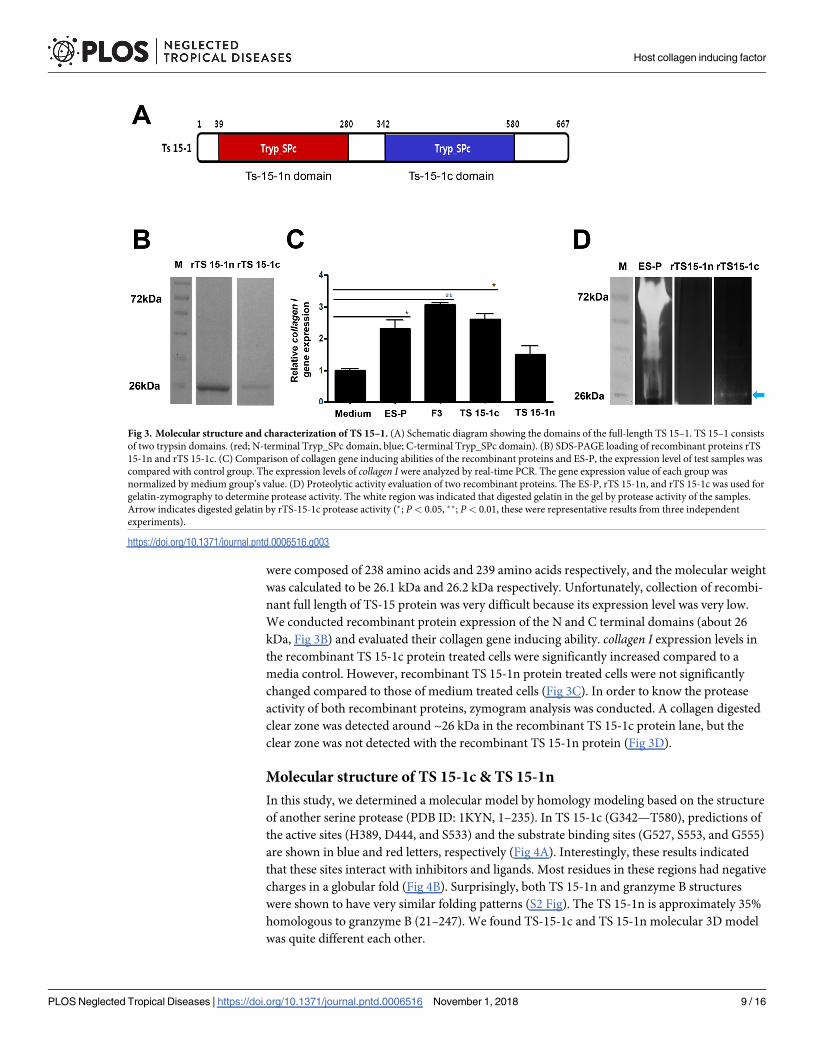
were composed of 238 amino acids and 239 amino acids respectively, and the molecular weight
was calculated to be 26.1 kDa and 26.2 kDa respectively. Unfortunately, collection of recombi-
nant full length of TS-15 protein was very difficult because its expression level was very low.
We conducted recombinant protein expression of the N and C terminal domains (about 26
kDa, Fig 3B) and evaluated their collagen gene inducing ability. collagen I expression levels in
the recombinant TS 15-1c protein treated cells were significantly increased compared to a
media control. However, recombinant TS 15-1n protein treated cells were not significantly
changed compared to those of medium treated cells (Fig 3C). In order to know the protease
activity of both recombinant proteins, zymogram analysis was conducted. A collagen digested
clear zone was detected around ~26 kDa in the recombinant TS 15-1c protein lane, but the
clear zone was not detected with the recombinant TS 15-1n protein (Fig 3D).
Molecular structure of TS 15-1c & TS 15-1n
In this study, we determined a molecular model by homology modeling based on the structure
of another serine protease (PDB ID: 1KYN, 1–235). In TS 15-1c (G342—T580), predictions of
the active sites (H389, D444, and S533) and the substrate binding sites (G527, S553, and G555)
are shown in blue and red letters, respectively (Fig 4A). Interestingly, these results indicated
that these sites interact with inhibitors and ligands. Most residues in these regions had negative
charges in a globular fold (Fig 4B). Surprisingly, both TS 15-1n and granzyme B structures
were shown to have very similar folding patterns (S2 Fig). The TS 15-1n is approximately 35%
homologous to granzyme B (21–247). We found TS-15-1c and TS 15-1n molecular 3D model
was quite different each other.
Fig 3. Molecular structure and characterization of TS 15–1. (A) Schematic diagram showing the domains of the full-length TS 15–1. TS 15–1 consists
of two trypsin domains. (red; N-terminal Tryp_SPc domain, blue; C-terminal Tryp_SPc domain). (B) SDS-PAGE loading of recombinant proteins rTS
15-1n and rTS 15-1c. (C) Comparison of collagen gene inducing abilities of the recombinant proteins and ES-P, the expression level of test samples was
compared with control group. The expression levels of collagen I were analyzed by real-time PCR. The gene expression value of each group was
normalized by medium group’s value. (D) Proteolytic activity evaluation of two recombinant proteins. The ES-P, rTS 15-1n, and rTS 15-1c was used for
gelatin-zymography to determine protease activity. The white region was indicated that digested gelatin in the gel by protease activity of the samples.
Arrow indicates digested gelatin by rTS-15-1c protease activity (�; P< 0.05, ��; P< 0.01, these were representative results from three independent
experiments).
https://doi.org/10.1371/journal.pntd.0006516.g003
Host collagen inducing factor
PLOS Neglected Tropical Diseases | https://doi.org/10.1371/journal.pntd.0006516 November 1, 2018 9 / 16
Expression and localization of TS-15 in the T. spiralis life cycle
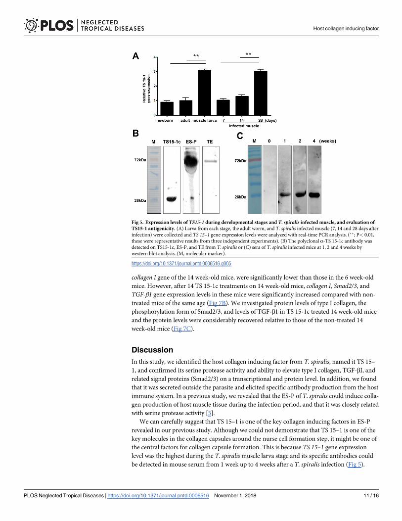
In order to determine when the Ts 15–1 mRNA was the most highly expressed in the T. spiralislife cycle, real-time PCR was performed on new born larvae, adult worms, muscle stage larvae
of T. spiralis, and during the T. spiralis infection period (at 0, 7, 14, 28 days after infection). As
the results show, the Ts 15–1 gene was the most highly expressed in muscle stage larvae, and its
expression is also highly elevated 28 days after infection (Fig 5A). In order to know whether TS
15–1 was secreted from parasites, an α-TS 15-1c antibody was produced and was reacted with
T. spiralis ES-P and total extract. The TS 15-1c antibody strongly reacted with proteins around
72 kDa in ES-P and slightly reacted with a total extract at the same size (Fig 5B). Furthermore,
to know whether TS 15-1c has antigenicity or not, T. spiralis infected mice sera (0, 1, 2, and 4
weeks after infection) were reacted with recombinant TS 15-1c protein (Fig 5C). rTS 15-1c
most strongly reacted with mouse serum collected 4 weeks after infection. To know where TS
15–1 is secreted in the parasite, α-TS 15-1c antibody was reacted against serial sections of the
T. spiralis infected muscle using immunohistochemical methods. α-TS 15-1c antibody strongly
reacted with only the ladder shapes structure around the esophagus in muscle stage larvae that
appear to be stichocytes (Fig 6).
Evaluation of the TS 15-1c protein type I collagen inducing ability
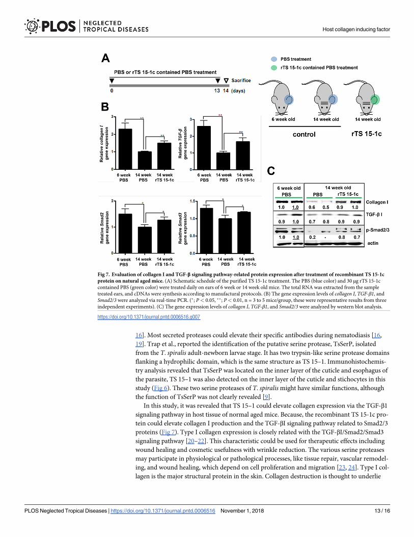
In order to know whether TS 15-1c had type I collagen elevating ability, we applied TS 15-1c
to the ear skin of 6 week- and 14 week-old mice and evaluated the expression levels of collagenI, Smad2/3, and TGF-β1 (Fig 7A). All of the tested genes’ expression levels, including the
Fig 4. Representation of the 3D structure of TS 15-1c and prediction of active sites. (A) The modeled structure of
TS 15-1c is shown as a Cα trace representation, and (B) a surface representation. The relative distribution of the
surface charge is shown with acidic regions in red, basic regions in blue and neutral regions in white. (C) Amino acid
sequence of the C-terminal domain of TS 15–1. Predicted active sites (H389, D444, and S533) and substrate binding
sites (G527, S553, and G555) are shown in blue and red, respectively.
https://doi.org/10.1371/journal.pntd.0006516.g004
Host collagen inducing factor
PLOS Neglected Tropical Diseases | https://doi.org/10.1371/journal.pntd.0006516 November 1, 2018 10 / 16
collagen I gene of the 14 week-old mice, were significantly lower than those in the 6 week-old
mice. However, after 14 TS 15-1c treatments on 14 week-old mice, collagen I, Smad2/3, and
TGF-β1 gene expression levels in these mice were significantly increased compared with non-
treated mice of the same age (Fig 7B). We investigated protein levels of type I collagen, the
phosphorylation form of Smad2/3, and levels of TGF-β1 in TS 15-1c treated 14 week-old mice
and the protein levels were considerably recovered relative to those of the non-treated 14
week-old mice (Fig 7C).
Discussion
In this study, we identified the host collagen inducing factor from T. spiralis, named it TS 15–
1, and confirmed its serine protease activity and ability to elevate type I collagen, TGF-βI, and
related signal proteins (Smad2/3) on a transcriptional and protein level. In addition, we found
that it was secreted outside the parasite and elicited specific antibody production from the host
immune system. In a previous study, we revealed that the ES-P of T. spiralis could induce colla-
gen production of host muscle tissue during the infection period, and that it was closely related
with serine protease activity [5].
We can carefully suggest that TS 15–1 is one of the key collagen inducing factors in ES-P
revealed in our previous study. Although we could not demonstrate that TS 15–1 is one of the
key molecules in the collagen capsules around the nurse cell formation step, it might be one of
the central factors for collagen capsule formation. This is because TS 15–1 gene expression
level was the highest during the T. spiralis muscle larva stage and its specific antibodies could
be detected in mouse serum from 1 week up to 4 weeks after a T. spiralis infection (Fig 5).
Fig 5. Expression levels of TS15-1 during developmental stages and T. spiralis infected muscle, and evaluation of
TS15-1 antigenicity. (A) Larva from each stage, the adult worm, and T. spiralis infected muscle (7, 14 and 28 days after
infection) were collected and TS 15–1 gene expression levels were analyzed with real-time PCR analysis. (��; P< 0.01,
these were representative results from three independent experiments). (B) The polyclonal α-TS 15-1c antibody was
detected on TS15-1c, ES-P, and TE from T. spiralis or (C) sera of T. spiralis infected mice at 1, 2 and 4 weeks by
western blot analysis. (M, molecular marker).
https://doi.org/10.1371/journal.pntd.0006516.g005
Host collagen inducing factor
PLOS Neglected Tropical Diseases | https://doi.org/10.1371/journal.pntd.0006516 November 1, 2018 11 / 16
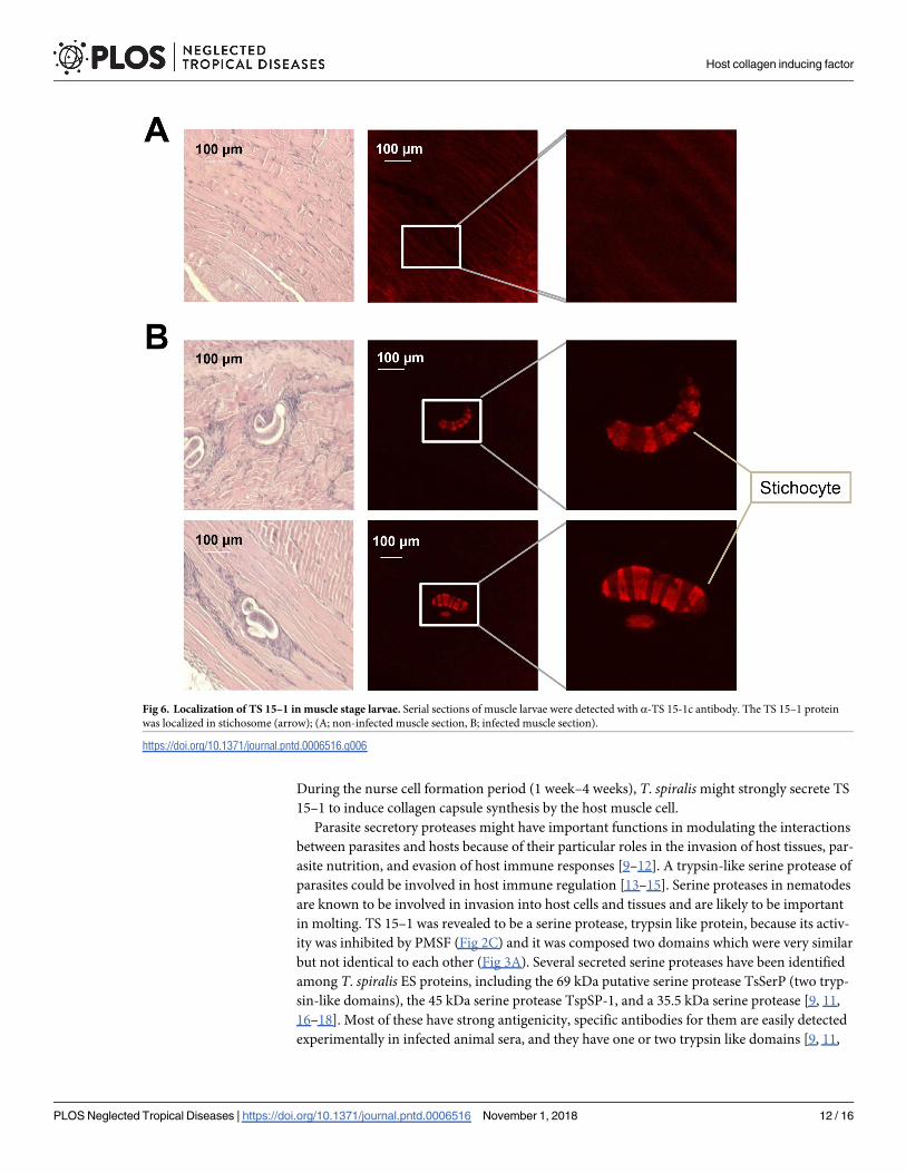
During the nurse cell formation period (1 week–4 weeks), T. spiralis might strongly secrete TS
15–1 to induce collagen capsule synthesis by the host muscle cell.
Parasite secretory proteases might have important functions in modulating the interactions
between parasites and hosts because of their particular roles in the invasion of host tissues, par-
asite nutrition, and evasion of host immune responses [9–12]. A trypsin-like serine protease of
parasites could be involved in host immune regulation [13–15]. Serine proteases in nematodes
are known to be involved in invasion into host cells and tissues and are likely to be important
in molting. TS 15–1 was revealed to be a serine protease, trypsin like protein, because its activ-
ity was inhibited by PMSF (Fig 2C) and it was composed two domains which were very similar
but not identical to each other (Fig 3A). Several secreted serine proteases have been identified
among T. spiralis ES proteins, including the 69 kDa putative serine protease TsSerP (two tryp-
sin-like domains), the 45 kDa serine protease TspSP-1, and a 35.5 kDa serine protease [9, 11,
16–18]. Most of these have strong antigenicity, specific antibodies for them are easily detected
experimentally in infected animal sera, and they have one or two trypsin like domains [9, 11,
Fig 6. Localization of TS 15–1 in muscle stage larvae. Serial sections of muscle larvae were detected with α-TS 15-1c antibody. The TS 15–1 protein
was localized in stichosome (arrow); (A; non-infected muscle section, B; infected muscle section).
https://doi.org/10.1371/journal.pntd.0006516.g006
Host collagen inducing factor
PLOS Neglected Tropical Diseases | https://doi.org/10.1371/journal.pntd.0006516 November 1, 2018 12 / 16
16]. Most secreted proteases could elevate their specific antibodies during nematodiasis [16,
19]. Trap et al., reported the identification of the putative serine protease, TsSerP, isolated
from the T. spiralis adult-newborn larvae stage. It has two trypsin-like serine protease domains
flanking a hydrophilic domain, which is the same structure as TS 15–1. Immunohistochemis-
try analysis revealed that TsSerP was located on the inner layer of the cuticle and esophagus of
the parasite, TS 15–1 was also detected on the inner layer of the cuticle and stichocytes in this
study (Fig 6). These two serine proteases of T. spiralis might have similar functions, although
the function of TsSerP was not clearly revealed [9].
In this study, it was revealed that TS 15–1 could elevate collagen expression via the TGF-β1
signaling pathway in host tissue of normal aged mice. Because, the recombinant TS 15-1c pro-
tein could elevate collagen I production and the TGF-βI signaling pathway related to Smad2/3
proteins (Fig 7). Type I collagen expression is closely related with the TGF-βI/Smad2/Smad3
signaling pathway [20–22]. This characteristic could be used for therapeutic effects including
wound healing and cosmetic usefulness with wrinkle reduction. The various serine proteases
may participate in physiological or pathological processes, like tissue repair, vascular remodel-
ing, and wound healing, which depend on cell proliferation and migration [23, 24]. Type I col-
lagen is the major structural protein in the skin. Collagen destruction is thought to underlie
Fig 7. Evaluation of collagen I and TGF-β signaling pathway-related protein expression after treatment of recombinant TS 15-1c
protein on natural aged mice. (A) Schematic schedule of the purified TS 15-1c treatment. The PBS (blue color) and 30 μg rTS 15-1c
contained PBS (green color) were treated daily on ears of 6 week or 14 week old mice. The total RNA was extracted from the sample
treated ears, and cDNAs were synthesis according to manufactural protocols. (B) The gene expression levels of collagen I, TGF-β1, and
Smad2/3 were analyzed via real-time PCR. (�; P< 0.05, ��; P< 0.01, n = 3 to 5 mice/group, these were representative results from three
independent experiments). (C) The gene expression levels of collagen I, TGF-β1, and Smad2/3 were analyzed by western blot analysis.
https://doi.org/10.1371/journal.pntd.0006516.g007
Host collagen inducing factor
PLOS Neglected Tropical Diseases | https://doi.org/10.1371/journal.pntd.0006516 November 1, 2018 13 / 16
the appearance of aged skin and changes resulting from chronic sun exposure [25]. Ultraviolet
irradiation from the sun has deleterious effects on human skin including cancer, photo-aging,
and intrinsic aging [26]. TGF-β/Smad pathway is the major regulator of collagen homeostasis
and plays a crucial role in dermal fibrosis [27, 28]. TGF-β is the most potent direct stimulator
of collagen production. Moreover, TGF-β is central to the process of wound healing and fibro-
sis formation in skin [29, 30]. It is well understood that activation of TGF-β signaling pathways
stimulus the Smad family downstream via phosphorylation. Wound healing is a well-orches-
trated process, where numerous factors are activated or inhibited in a sequence of steps [31].
Numerous signaling pathways are involved, among of them, the TGF-β1/Smad pathway is rep-
resentative and well known to participate in the wound healing process [31]. Hozzein et al.,
suggested that topical application of propolis would promote the wound healing process by
promoting TGF-β/Smad signaling, leading to increased expression of collagen type I [32]. The
gradual loss of collagen in skin with aging results in wrinkles and other signs of skin aging
[33]. The content of type I collagen, the major collagen in the skin and a marker of collagen
synthesis, is deceased by 68% in old skin versus young skin, and cultured young fibroblasts
synthesize more type I collagen than old cells [33]. In addition, a possible influence of collagen
membrane on extracellular matrix synthesis was addressed using analysis of TGF-β1 and
Smad2/3 complex [34, 35].
In conclusion, we identified a host collagen inducing factor from ES-P using immune
screening methods and demonstrated the molecular/genetic characteristics and function of TS
15-1c. Further study will be required to understand the detailed mechanisms for receptors in
the host cells, and to identify the minimal structure that can induce collagen for cosmetic and
medical purposes.
Supporting information
S1 Fig. Result of immunoscreening of T. spiralis cDNA library by α-F3 antibody. After T.
spiralis cDNA containing phages were mixed with E. coli, the plaques were incubated with NC
membranes for 4 hrs. The membranes were reacted with α-F3 antibody (1:500) as the primary
antibody and α-rat IgG antibody conjugated with HRP was reacted as secondary antibody.
After adding of 3,3’-diaminobenzidine (DAB), colored spots were compared with original
plates. Positive plaques were amplified and re-analyzed by the same method.
(PPTX)
S2 Fig. Representation of the 3D structure of TS 15-1n and prediction of active sites. The
modeled structure of TS 15-1n is shown as a surface representation (A), and a Cα trace represen-
tation (B). In TS-15-1n, predictions of the active sites (G227, S252, and H254) and the substrate
binding sites (R88, D142, and S233) are shown in blue and red letters, respectively. The relative
distribution of the surface charge is shown with acidic regions in red, basic regions in blue and
neutral regions in white. Amino acid sequence of the N-terminal domain of TS 15–1 (C).
(PPTX)
Author Contributions
Conceptualization: Mi Kyung Park, Hak Sun Yu.
Formal analysis: Hae-Jin Kim, So Young Park.
Funding acquisition: Hak Sun Yu.
Investigation: Mi Kyung Park, Shin Ae Kang, Hak Sun Yu.
Host collagen inducing factor
PLOS Neglected Tropical Diseases | https://doi.org/10.1371/journal.pntd.0006516 November 1, 2018 14 / 16
http://journals.plos.org/plosntds/article/asset?unique&id=info:doi/10.1371/journal.pntd.0006516.s001
Methodology: Se Bok Jang, Hak Sun Yu.
Software: Min Kyoung Cho.
Supervision: Hak Sun Yu.
Writing – original draft: Mi Kyung Park.
Writing – review & editing: Hak Sun Yu.
References
1. Dabrowska M, Skoneczny M, Zielinski Z, Rode W. (2008) Nurse cell of Trichinella spp. as a model of
long-term cell cycle arrest. Cell cycle (Georgetown, Tex. 7:2167–78.
2. Wu Z, Sofronic-Milosavljevic L, Nagano I, Takahashi Y. (2008) Trichinella spiralis: nurse cell formation
with emphasis on analogy to muscle cell repair. Parasites & vectors. 1:27.
3. Despommier DD. (1998) How Does Trichinella spiralis Make Itself at Home? Parasitology today (Per-
sonal ed. 14:318–23.
4. Shariati F, Perez-Arellano JL, Lopez-Aban J, Arefi M, Martinez-Fernandez AR, Muro A. (2009) Trichi-
nella: differential expression of angiogenic factors in macrophages stimulated with antigens from encap-
sulated and non-encapsulated species. Experimental parasitology. 123:347–53. https://doi.org/10.
1016/j.exppara.2009.08.016 PMID: 19723522
5. Park MK, Cho MK, Kang SA, Kim BY, Yu HS. (2016) The induction of the collagen capsule synthesis by
Trichinella spiralis is closely related to protease-activated receptor 2. Vet Parasitol. 230:56–61. https://
doi.org/10.1016/j.vetpar.2016.11.001 PMID: 27884442
6. Lee KH, Park HK, Jeong HJ, Park SK, Lee SJ, Choi SH, et al. (2008) Immunization of proteins from Tox-
ascaris leonina adult worm inhibits allergic specific Th2 response. Vet Parasitol. 156:216–25. https://
doi.org/10.1016/j.vetpar.2008.06.016 PMID: 18653284
7. Karsenty G, Park RW. (1995) Regulation of type I collagen genes expression. International reviews of
immunology. 12:177–85. PMID: 7650420
8. Amit M, Itskovitz-Eldor J. (2009) Embryonic stem cells: isolation, characterization and culture.
Advances in biochemical engineering/biotechnology. 114:173–84. https://doi.org/10.1007/10_2008_20
PMID: 19495683
9. Trap C, Fu B, Le Guerhier F, Liu M, Le Rhun D, Romand T, et al. (2006) Cloning and analysis of a cDNA
encoding a putative serine protease comprising two trypsin-like domains of Trichinella spiralis. Parasitol
Res. 98:288–94. https://doi.org/10.1007/s00436-005-0075-x PMID: 16341878
10. Fishelson Z, Amiri P, Friend DS, Marikovsky M, Petitt M, Newport G, et al. (1992) Schistosoma man-
soni: cell-specific expression and secretion of a serine protease during development of cercariae. Exp
Parasitol. 75:87–98. PMID: 1639166
11. Li X, Yao JP, Pan AH, Liu W, Hu XC, Wu ZD, et al. (2013) An antigenic recombinant serine protease
from Trichinella spiralis induces protective immunity in BALB/c mice. Parasitol Res. 112:3229–38.
https://doi.org/10.1007/s00436-013-3500-6 PMID: 23828191
12. McKerrow JH. (1989) Parasite proteases. Exp Parasitol. 68:111–5. PMID: 2645160
13. Balasubramanian N, Toubarro D, Simoes N. (2010) Biochemical study and in vitro insect immune sup-
pression by a trypsin-like secreted protease from the nematode Steinernema carpocapsae. Parasite
Immunol. 32:165–75. https://doi.org/10.1111/j.1365-3024.2009.01172.x PMID: 20398179
14. Nakhleh J, Christophides GK, Osta MA. (2017) The serine protease homolog CLIPA14 modulates the
intensity of the immune response in the mosquito Anopheles gambiae. J Biol Chem. 292:18217–26.
https://doi.org/10.1074/jbc.M117.797787 PMID: 28928218
15. Rodrigues J, Agrawal N, Sharma A, Malhotra P, Adak T, Chauhan VS, et al. (2007) Transcriptional anal-
ysis of an immune-responsive serine protease from Indian malarial vector, Anopheles culicifacies. BMC
Mol Biol. 8:33. https://doi.org/10.1186/1471-2199-8-33 PMID: 17502004
16. Wang B, Wang ZQ, Jin J, Ren HJ, Liu LN, Cui J. (2013) Cloning, expression and characterization of a
Trichinella spiralis serine protease gene encoding a 35.5 kDa protein. Exp Parasitol. 134:148–54.
https://doi.org/10.1016/j.exppara.2013.03.004 PMID: 23501807
17. Romaris F, North SJ, Gagliardo LF, Butcher BA, Ghosh K, Beiting DP, et al. (2002) A putative serine
protease among the excretory-secretory glycoproteins of L1 Trichinella spiralis. Mol Biochem Parasitol.
122:149–60. PMID: 12106869
Host collagen inducing factor
PLOS Neglected Tropical Diseases | https://doi.org/10.1371/journal.pntd.0006516 November 1, 2018 15 / 16
18. Cwiklinski K, Meskill D, Robinson MW, Pozio E, Appleton JA, Connolly B. (2009) Cloning and analysis
of a Trichinella pseudospiralis muscle larva secreted serine protease gene. Vet Parasitol. 159:268–71.
https://doi.org/10.1016/j.vetpar.2008.10.036 PMID: 19054614
19. Kennedy MW, Qureshi F. (1986) Stage-specific secreted antigens of the parasitic larval stages of the
nematode Ascaris. Immunology. 58:515–22. PMID: 3733151
20. Lu P, Wang S, Cai W, Sheng J. (2012) Role of TGF-beta1/Smad3 signaling pathway in secretion of
type I and III collagen by vascular smooth muscle cells of rats undergoing balloon injury. J Biomed Bio-
technol. 2012:965953. https://doi.org/10.1155/2012/965953 PMID: 23091366
21. Dangi-Garimella S, Strouch MJ, Grippo PJ, Bentrem DJ, Munshi HG. (2011) Collagen regulation of let-
7 in pancreatic cancer involves TGF-beta1-mediated membrane type 1-matrix metalloproteinase
expression. Oncogene. 30:1002–8. https://doi.org/10.1038/onc.2010.485 PMID: 21057545
22. Hirose T, Nakazato K, Song H, Ishii N. (2008) TGF-beta1 and TNF-alpha are involved in the transcrip-
tion of type I collagen alpha2 gene in soleus muscle atrophied by mechanical unloading. J Appl Physiol
(1985). 104:170–7.
23. Etscheid M, Beer N, Dodt J. (2005) The hyaluronan-binding protease upregulates ERK1/2 and PI3K/
Akt signalling pathways in fibroblasts and stimulates cell proliferation and migration. Cell Signal.
17:1486–94. https://doi.org/10.1016/j.cellsig.2005.03.007 PMID: 16153533
24. Ipaktchi K, Mattar A, Niederbichler AD, Hoesel LM, Vollmannshauser S, Hemmila MR, et al. (2007) Top-
ical p38 MAPK inhibition reduces bacterial growth in an in vivo burn wound model. Surgery. 142:86–93.
https://doi.org/10.1016/j.surg.2007.02.007 PMID: 17630004
25. Hwang KA, Yi BR, Choi KC. (2011) Molecular mechanisms and in vivo mouse models of skin aging
associated with dermal matrix alterations. Laboratory animal research. 27:1–8. https://doi.org/10.5625/
lar.2011.27.1.1 PMID: 21826153
26. Kohl E, Landthaler M, Szeimies RM. (2009) [Skin aging]. Der Hautarzt; Zeitschrift fur Dermatologie,
Venerologie, und verwandte Gebiete. 60:917–33; quiz 34. PMID: 19898765
27. Zhang GY, Li X, Yi CG, Pan H, He GD, Yu Q, et al. (2009) Angiotensin II activates connective tissue
growth factor and induces extracellular matrix changes involving Smad/activation and p38 mitogen-acti-
vated protein kinase signalling pathways in human dermal fibroblasts. Experimental dermatology.
18:947–53. https://doi.org/10.1111/j.1600-0625.2009.00880.x PMID: 19397700
28. Quan T, He T, Kang S, Voorhees JJ, Fisher GJ. (2004) Solar ultraviolet irradiation reduces collagen in
photoaged human skin by blocking transforming growth factor-beta type II receptor/Smad signaling.
The American journal of pathology. 165:741–51. PMID: 15331399
29. Crowe MJ, Doetschman T, Greenhalgh DG. (2000) Delayed wound healing in immunodeficient TGF-
beta 1 knockout mice. The Journal of investigative dermatology. 115:3–11. https://doi.org/10.1046/j.
1523-1747.2000.00010.x PMID: 10886500
30. Liu X, Hu H, Yin JQ. (2006) Therapeutic strategies against TGF-beta signaling pathway in hepatic fibro-
sis. Liver international: official journal of the International Association for the Study of the Liver. 26:8–
22.
31. Kasuya A, Tokura Y. (2014) Attempts to accelerate wound healing. J Dermatol Sci. 76:169–72. https://
doi.org/10.1016/j.jdermsci.2014.11.001 PMID: 25468357
32. Hozzein WN, Badr G, Al Ghamdi AA, Sayed A, Al-Waili NS, Garraud O. (2015) Topical application of
propolis enhances cutaneous wound healing by promoting TGF-beta/Smad-mediated collagen produc-
tion in a streptozotocin-induced type I diabetic mouse model. Cell Physiol Biochem. 37:940–54. https://
doi.org/10.1159/000430221 PMID: 26381245
33. Varani J, Dame MK, Rittie L, Fligiel SE, Kang S, Fisher GJ, et al. (2006) Decreased collagen production
in chronologically aged skin: roles of age-dependent alteration in fibroblast function and defective
mechanical stimulation. Am J Pathol. 168:1861–8. https://doi.org/10.2353/ajpath.2006.051302 PMID:
16723701
34. Derynck R. (1998) SMAD proteins and mammalian anatomy. Nature. 393:737–9. https://doi.org/10.
1038/31593 PMID: 9655387
35. Lakos G, Takagawa S, Chen SJ, Ferreira AM, Han G, Masuda K, et al. (2004) Targeted disruption of
TGF-beta/Smad3 signaling modulates skin fibrosis in a mouse model of scleroderma. The American
journal of pathology. 165:203–17. PMID: 15215176
Host collagen inducing factor
PLOS Neglected Tropical Diseases | https://doi.org/10.1371/journal.pntd.0006516 November 1, 2018 16 / 16
![Inducing Patent Infringement - Law Review...2005] Inducing Patent Infringement 229 understanding, inducing infringement is a natural outgrowth of the common law principle of respondeat](https://static.fdocuments.us/doc/165x107/5f9608d795a783197246401f/inducing-patent-infringement-law-review-2005-inducing-patent-infringement.jpg)