Identification and characterization of two new 5-keto-4 · PDF file ·...
10
RESEARCH ARTICLE Open Access Identification and characterization of two new 5-keto-4-deoxy-D-Glucarate Dehydratases/Decarboxylases André Pick 1 , Barbara Beer 1 , Risa Hemmi 2 , Rena Momma 2 , Jochen Schmid 1 , Kenji Miyamoto 2 and Volker Sieber 1* Abstract Background: Hexuronic acids such as D-galacturonic acid and D-glucuronic acid can be utilized via different pathways within the metabolism of microorganisms. One representative, the oxidative pathway, generates α-keto- glutarate as the direct link entering towards the citric acid cycle. The penultimate enzyme, keto-deoxy glucarate dehydratase/decarboxylase, catalyses the dehydration and decarboxylation of keto-deoxy glucarate to α-keto- glutarate semialdehyde. This enzymatic reaction can be tracked continuously by applying a pH-shift assay. Results: Two new keto-deoxy glucarate dehydratases/decarboxylases (EC 4.2.1.41) from Comamonas testosteroni KF-1 and Polaromonas naphthalenivorans CJ2 were identified and expressed in an active form using Escherichia coli ArcticExpress(DE3). Subsequent characterization concerning K m , k cat and thermal stability was conducted in comparison with the known keto-deoxy glucarate dehydratase/decarboxylase from Acinetobacter baylyi ADP1. The kinetic constants determined for A. baylyi were K m 1.0 mM, k cat 4.5 s -1 , for C. testosteroni K m 1.1 mM, k cat 3.1 s -1 , and for P. naphthalenivorans K m 1.1 mM, k cat 1.7 s -1 . The two new enzymes had a slightly lower catalytic activity (increased K m and a decreased k cat ) but showed a higher thermal stability than that of A. baylyi. The developed pH- shift assay, using potassium phosphate and bromothymol blue as the pH indicator, enables a direct measurement. The use of crude extracts did not interfere with the assay and was tested for wild-type landscapes for all three enzymes. Conclusions: By establishing a pH-shift assay, an easy measurement method for keto-deoxy glucarate dehydratase/ decarboxylase could be developed. It can be used for measurements of the purified enzymes or using crude extracts. Therefore, it is especially suitable as the method of choice within an engineering approach for further optimization of these enzymes. Keywords: Keto-deoxy-D-Glucarate, Acinetobacter baylyi, Comamonas testosteroni, Polaromonas naphthalenivorans, Dehydratase Background Renewable biogenic resources such as lignocellulosic hydrolysates, often referred to as second-generation feedstock, represent an increasingly important raw material for chemicals production. Complete exploit- ation of these substrates is still a challenging task due to their heterogeneous composition. Besides various hexoses and pentoses, which constitute the main fraction of the hydrolysates, sugar derivatives such as sugar acids accumulate. The latter include hexuronic acids such as D-galacturonic acid and D- glucuronic acid, which are mainly present when pectin-rich waste streams or plant xylans are utilized [1, 2]. Both acids are abundantly available in agricul- tural waste or forestry residues. In particular, plant pathogenic bacteria such as Pseudomonas syringae, Agrobacterium tumefaciens or Erwinia carotovora as well as Escherichia coli or Thermotoga maritima possess metabolic pathways for hexuronic acid utilization [3–7]. Up to now, three pathways have been identified for the utilization of hexuronic acids via isomerization, reduction or oxidation [8]. * Correspondence: [email protected] 1 Technical University of Munich, Straubing Center of Science, Chair of Chemistry of Biogenic Resources, Schulgasse 16, 94315 Straubing, Germany Full list of author information is available at the end of the article © The Author(s). 2016 Open Access This article is distributed under the terms of the Creative Commons Attribution 4.0 International License (http://creativecommons.org/licenses/by/4.0/), which permits unrestricted use, distribution, and reproduction in any medium, provided you give appropriate credit to the original author(s) and the source, provide a link to the Creative Commons license, and indicate if changes were made. The Creative Commons Public Domain Dedication waiver (http://creativecommons.org/publicdomain/zero/1.0/) applies to the data made available in this article, unless otherwise stated. Pick et al. BMC Biotechnology (2016) 16:80 DOI 10.1186/s12896-016-0308-3
Transcript of Identification and characterization of two new 5-keto-4 · PDF file ·...

RESEARCH ARTICLE Open Access
Identification and characterization of twonew 5-keto-4-deoxy-D-GlucarateDehydratases/DecarboxylasesAndré Pick1, Barbara Beer1, Risa Hemmi2, Rena Momma2, Jochen Schmid1, Kenji Miyamoto2 and Volker Sieber1*
Abstract
Background: Hexuronic acids such as D-galacturonic acid and D-glucuronic acid can be utilized via differentpathways within the metabolism of microorganisms. One representative, the oxidative pathway, generates α-keto-glutarate as the direct link entering towards the citric acid cycle. The penultimate enzyme, keto-deoxy glucaratedehydratase/decarboxylase, catalyses the dehydration and decarboxylation of keto-deoxy glucarate to α-keto-glutarate semialdehyde. This enzymatic reaction can be tracked continuously by applying a pH-shift assay.
Results: Two new keto-deoxy glucarate dehydratases/decarboxylases (EC 4.2.1.41) from Comamonas testosteroniKF-1 and Polaromonas naphthalenivorans CJ2 were identified and expressed in an active form using Escherichiacoli ArcticExpress(DE3). Subsequent characterization concerning Km, kcat and thermal stability was conducted incomparison with the known keto-deoxy glucarate dehydratase/decarboxylase from Acinetobacter baylyi ADP1.The kinetic constants determined for A. baylyi were Km 1.0 mM, kcat 4.5 s−1, for C. testosteroni Km 1.1 mM, kcat 3.1 s−1,and for P. naphthalenivorans Km 1.1 mM, kcat 1.7 s−1. The two new enzymes had a slightly lower catalytic activity(increased Km and a decreased kcat) but showed a higher thermal stability than that of A. baylyi. The developed pH-shift assay, using potassium phosphate and bromothymol blue as the pH indicator, enables a direct measurement.The use of crude extracts did not interfere with the assay and was tested for wild-type landscapes for all threeenzymes.
Conclusions: By establishing a pH-shift assay, an easy measurement method for keto-deoxy glucarate dehydratase/decarboxylase could be developed. It can be used for measurements of the purified enzymes or using crudeextracts. Therefore, it is especially suitable as the method of choice within an engineering approach for furtheroptimization of these enzymes.
Keywords: Keto-deoxy-D-Glucarate, Acinetobacter baylyi, Comamonas testosteroni, Polaromonas naphthalenivorans,Dehydratase
BackgroundRenewable biogenic resources such as lignocellulosichydrolysates, often referred to as second-generationfeedstock, represent an increasingly important rawmaterial for chemicals production. Complete exploit-ation of these substrates is still a challenging taskdue to their heterogeneous composition. Besidesvarious hexoses and pentoses, which constitute themain fraction of the hydrolysates, sugar derivatives
such as sugar acids accumulate. The latter includehexuronic acids such as D-galacturonic acid and D-glucuronic acid, which are mainly present whenpectin-rich waste streams or plant xylans are utilized[1, 2]. Both acids are abundantly available in agricul-tural waste or forestry residues. In particular, plantpathogenic bacteria such as Pseudomonas syringae,Agrobacterium tumefaciens or Erwinia carotovora aswell as Escherichia coli or Thermotoga maritimapossess metabolic pathways for hexuronic acidutilization [3–7]. Up to now, three pathways havebeen identified for the utilization of hexuronic acidsvia isomerization, reduction or oxidation [8].
* Correspondence: [email protected] University of Munich, Straubing Center of Science, Chair ofChemistry of Biogenic Resources, Schulgasse 16, 94315 Straubing, GermanyFull list of author information is available at the end of the article
© The Author(s). 2016 Open Access This article is distributed under the terms of the Creative Commons Attribution 4.0International License (http://creativecommons.org/licenses/by/4.0/), which permits unrestricted use, distribution, andreproduction in any medium, provided you give appropriate credit to the original author(s) and the source, provide a link tothe Creative Commons license, and indicate if changes were made. The Creative Commons Public Domain Dedication waiver(http://creativecommons.org/publicdomain/zero/1.0/) applies to the data made available in this article, unless otherwise stated.
Pick et al. BMC Biotechnology (2016) 16:80 DOI 10.1186/s12896-016-0308-3

The oxidative pathway comprises four enzymaticsteps (Fig. 1), generating α-keto-glutarate as the directlink entering the citric acid cycle [8]. The first oxida-tive step is catalysed by uronate dehydrogenase, whichproduces an aldaric acid lactone that hydrolyses spon-taneously [9, 10]. Several uronate dehydrogenases ofdifferent origins have been described [11–14].The subsequent two steps are catalysed by the en-
zymes glucarate dehydratase and keto-deoxy glucaratedehydratase/decarboxylase (KdgD). Both enzymes are re-sponsible for the defunctionalisation of glucarate. First,glucarate dehydratase removes water, leading to keto-deoxy glucarate, which is the substrate for KdgD; this inturn catalyses the dehydration and decarboxylation intoα-keto-glutarate semialdehyde [15]. In the final step, α-keto-glutarate semialdehyde dehydrogenase oxidizes thesemialdehyde to α-keto-glutarate [16]. The glucaratedehydratase belongs to the mechanistically diverse eno-lase superfamily, which is known to catalyse at least 14different reactions [17]. Within this superfamily, gluca-rate dehydratase is assigned to the mandelate racemasesubgroup [18]. The reaction mechanism and proteinstructure of several members have been studied in detail[19, 20]. The bifunctional enzyme KdgD belongs to theclass I aldolase family and is further sub-grouped intothe N-acetylneuraminate lyase superfamily [21]. Onlylittle attention has been devoted towards this enzymeeven though it catalyses a very interesting reaction. Just re-cently, the crystal structure for KdgD from A. tumefacienswas solved [22] in parallel with investigations to gain adeeper understanding of the catalytic mechanism, whichled to the identification of catalytically relevant aminoacids [23].For thorough characterization, easy monitoring of the
enzymatic reaction is one of the main challenges.Neither the substrate nor the product can be detectedphotometrically; moreover, no cofactor is involved inthe catalytic reaction. Therefore, all studies performedup to now have used a coupled enzyme assay with α-keto-glutarate semialdehyde dehydrogenase, followingthe formation of NAD(P)H at 340 nm [15]. However,the reaction catalysed by KdgD is well suited to estab-lish a direct method for measuring enzymatic activity.
The release of CO2 from a carboxylate leads to the con-sumption of protons and an increase in pH, which intheory can be monitored by a pH indicator and noadditional enzyme is necessary to detect the reaction.Colorimetric assays based on a pH indicator systemhave been successfully used to monitor several enzymaticreactions, e.g. hydrolysis of esters, transfer of sugars, phos-phate or nucleotides, as well as decarboxylation of aminoacids [24–30].Here, we report the identification and characterization
of two novel KdgDs from Comamonas testosteroni KF-1(Ct) and Polaromonas naphthalenivorans CJ2 (Pn). Forbetter evaluation and validation, an already known KdgDfrom Acinetobacter baylyi ADP1 (Ab) was used as the ref-erence. A first characterization and comparison was doneby developing an easy and direct measurement methodbased on a pH indicator system using bromothymol blue(BTB) as the indicator and potassium phosphate as thebuffer. The assay could be easily adopted to allow mea-surements in crude cell extracts and therefore will be veryuseful for screening approaches.
MethodsReagentsD-Saccharic acid potassium salt (glucarate), magnesiumchloride heptahydrate and BTB sodium salt were pur-chased from Sigma Aldrich (Seelze, Germany). Restrictionenzyme BsaI, alkaline phosphatase, Phusion™ polymerase,T4 ligase and T4 polynucleotide kinase were purchasedfrom New England Biolabs (Frankfurt, Germany). Taqpolymerase was obtained from Rapidozym (Berlin,Germany). Oligonucleotides were synthesized by ThermoFisher Scientific (Waltham, MA, USA). DNaseI was ob-tained from Applichem (Darmstadt, Germany). All otherchemicals were purchased from Carl Roth (Karlsruhe,Germany) or Merck (Darmstadt, Germany) and wereused without further purification. All columns used forprotein purification were from GE Healthcare (Munich,Germany).
Sequence selection and comparisonThe publicly available protein sequence of the 5-keto-4-D-deoxyglucarate dehydratase/decarboxylase of A. baylyi
Fig. 1 Oxidative Pathway. Schematic representation of the oxidative pathway for conversion of uronic acids using D-glucuronate as starting substrate
Pick et al. BMC Biotechnology (2016) 16:80 Page 2 of 10

ADP1 was used as the query sequence for BLAST analysis(blastp) for the identification of potential candidates [31].Candidate proteins belonging to another species with amaximum identity of 70 % were chosen. In the next step,to verify the possible occurrence of the oxidative pathwayfor D-glucuronate and D-glucarate conversion, the occur-rence of the upstream enzyme D-glucarate dehydratasewas confirmed by screening the genome sequence of P.naphthalenivorans CJ2 (NC_008781.1) and C. testosteroniKF-1 (AAUJ02000001.1). Identification of both enzymeswas the final criterion for selection.Four protein sequences encoding for KdgD were aligned
using the web-based program T-COFFEE. 5-keto-4-D-deoxyglucarate dehydratase/decarboxylase of A. baylyiADP1, the enzyme from A. tumefaciens C58, whose struc-ture was recently determined, were chosen as references[23]. Based on the BLAST results, the two enzyme candi-dates from P. naphthalenivorans CJ2 (WP_011800997.1)and C. testosteroni KF-1 (WP_003059546.1) were chosen.
Strains and plasmidsThe following strains were used in this study: E. coliXL1-Blue (Stratagene), E. coli BL21(DE3) (Novagen) andE. coli ArcticExpress(DE3) (Agilent Technologies). TheDNA sequences for the corresponding genes of keto-deoxy-D-glucarate dehydratase/decarboxylase (kdgD)from A. baylyi (ADP1) (protein sequence GenBank™WP_004930673.1), which is identical to A. baylyiDSM 14961 (protein sequence GenBank™ ENV53020.1),from C. testosteroni KF-1 (protein sequence GenBank™WP_003059546.1) and from P. naphthalenivorans (pro-tein sequence GenBank™ WP_011800997.1) were synthe-sized with optimized codon usage for expression in E. coli(Additional file 1: Figures S1-S3) (Life Technologies,Regensburg, Germany). The following primers wereused for amplification: F-kdgD-A.b.- CAGCAAGGTCTCACATATGGATGCCCTGGAACTG, R-kdgD-A.b.-CTGCGG-ACCCAGGGTTG, F-kdgD-C.t.-CAGCAAGGTCTCACATATGACACCGCAGG-ATCTGAAAG, R-kdgD-C.t.-xCTGCGGACCCAGTTTATCAATC, F-kdgD-P.n.-CAGCAAGGTCTCACATATGAATCCGCAGGATCTGAAAAC, R-kdgD-P.n.- CTGCGGACCCAGGCTTTTAATC.The restriction enzyme recognition site for BsaI is under-lined and the start codon is marked in bold. The reverseprimers were phosphorylated using T4 polynucleotidekinase according to the supplier’s manual. PCR prod-ucts were digested with BsaI and cloned into pCBR,pCBRHisN and pCBRHisC, which are derivatives ofpET28a (Novagen). The cloning strategy of all pET28derivatives is described by Guterl et al. [32]. Ligationof the PCR products and the following transformationled to the plasmids pCBR-kdgD-A.b., pCBR-kdgD-C.t.,pCBR-kdgD-P.n., pCBRHisN-kdgD-A.b., pCBRHisN-kdgD-C.t., pCBRHisN-kdgD-P.n., pCBRHisC-kdgD-A.b.,
pCBRHisC-kdgD-C.t., and pCBRHisC-kdgD-P.n. Multipli-cation of the plasmids was performed by E. coli XL1 Blue(Stratagene) in Luria–Bertani medium containing 30 μg/mlkanamycin. E. coli BL21(DE3) or E. coli ArcticExpress(DE3)were used for expression.
Overexpression and FPLC purificationProtein expression was performed with two different E. coliexpression strains depending on the target enzyme. E. coliBL21(DE3) [pET28a-NH-kdgD-A.b.] was cultivated with aslightly modified protocol described by Aghaie et al. [15].The preculture was incubated in 4 ml of Terrific brothmedium supplemented with 1 M sorbitol and 5 mM beta-ine with 100 μg/ml kanamycin at 37 °C overnight on a ro-tary shaker (180 rpm). The expression culture consisted ofthe same media and was inoculated with a 1:100 dilutionof the preculture. Incubation was performed at 37 °C untilan OD600 of 2 was reached. Protein expression wasinduced with the addition of IPTG to a final concentrationof 0.5 mM followed by incubation for 21 h at 16 °C. ForE. coli ArcticExpress(DE3) [pET28a-NH-kdgD-C.t. orpET28a-CH-kdgD-P.n.] the preculture was cultivated inLuria–Bertani media with 100 μg/ml kanamycin and15 μg/ml gentamycin over night at 37 °C. The expres-sion culture consisted of autoinduction media withboth antibiotics and was inoculated with a 1:100 dilu-tion of the preculture [33]. Incubation was performedfor 3 h at 37 °C followed by the second step at 12 °C for45 h. Afterwards, cells were harvested by centrifugationand washed one time with 50 mM sodium phosphate buf-fer (pH 8.0) and frozen at −20 °C or resuspended in abinding buffer (50 mM potassium phosphate, pH 8.0,20 mM imidazol, 500 mM NaCl and 10 % glycerol). Crudeextracts were prepared using a Basic-Z cell disrupter (IULConstant Systems) and the subsequent addition of MgCl2to a final concentration of 2.5 mM in combination withDNaseI (10 μg/ml), followed by an incubation for 20 minat room temperature for successful DNA degradation.The insoluble fraction of the lysate was removed bycentrifugation at 20,000 rpm for 20 min at 4 °C. Thesupernatant was filtered through a 0.45 μm syringefilter and applied to an IMAC affinity resin column,5 ml HisTrapTM FF, equilibrated with the bindingbuffer using the ÄKTA purifier system. The enzymewas washed with 20 ml of binding buffer and elutedwith 50 mM potassium phosphate buffer (pH 8.0,500 mM imidazol, 500 mM NaCl and 10 % glycerol).Aliquots of each eluted fraction were subjected to12 % SDS-PAGE. The fractions containing the elutedprotein were pooled and the protein was desaltedusing a HiPrepTM 26/10 desalting column, whichwas preliminary equilibrated with 50 mM Tris-HCl(pH 7.5) or 5 mM sodium phosphate buffer (pH 7.0).
Pick et al. BMC Biotechnology (2016) 16:80 Page 3 of 10
Protein concentrations were determined using a Brad-ford assay (Roti®-nanoquant, Carl Roth).
Enzyme expression in 96-deep well scaleFor all three enzymes, an expression in the 96-deep wellscale was performed. Therefore, electrocompetent E. coliArcticExpress(DE3) cells were transformed with the corre-sponding plasmid. Single clones were picked using theCP7200 Colony Picker (Norgren Systems) and transferredto 96-deep well plates filled with 1.2 ml autoinduction media[33] by a MicroFlo Select dispenser (Bio-Tek Instruments).After incubation (36 h, 37 °C at 1,000 rpm), further process-ing was done manually. First, 100 μl of cell culture wastransferred into a 96-well plate (U-shaped bottom) and har-vested by centrifugation (4,570 rpm, 10 min at RT) whilethe supernatant was discarded. The frozen pellets (1 h at−20 °C) were thawed at room temperature for one hour toimprove cell lysis. Lysis was continued by the addition of30 μl lysis buffer (3 h, 1,000 rpm, 37 °C) containing 2 mMKPi, pH 7.0, 2 mM MgCl2, 10 μg/ml DNaseI, 100 μg/mllysozyme. Next, 120 μl buffer (2 mM KPi, pH 7.0) was addedfollowed by centrifugation (3,000 rpm, 15 min at RT). Forthe photometric measurement, 20 μl of the crude extractwas transferred to a 96-well plate (F-shaped bottom) andthe reaction was started by adding 180 μl master mix to givea final volume of 200 μl (2.5 mM KPi, pH 7.0, 2 mM MgCl2,25 μg/ml BTB and 5 mM keto-deoxy-D-glucarate). Themeasurements were carried out for 60 min at 2-min inter-vals. Depending on the enzyme, different time windowswere used for the activity calculation.
Substrate preparation5-keto-4-deoxy-D-glucarate is not commercially avail-able. Therefore, it was prepared using an enzymatic con-version of D-glucarate. For that, a 250–500 mM solutionof D-glucarate containing 2 mM MgCl2 was prepared.Potassium glucarate is not completely soluble at thisconcentration, and the pH value was around 4.5. ThepH was shifted to 8.0 by adding potassium hydroxide. Asample was taken as zero-point control and the reactionwas started by the addition of D-glucarate dehydratase(Beer et al., manuscript in preparation). Using HPLC, asample was analysed at regular intervals. After full con-version, the reaction was stopped by removing the en-zyme by filtration (spin filters, 10 kDa MWCO, modifiedPES; VWR, Darmstadt, Germany). In the last step, thepH was adjusted to 7.0 using HCl.
HPLC analysisD-glucarate, 5-keto-4-D-deoxyglucarate and α-ketoglutaratesemialdehyde were separated by HPLC, using an Ultimate-3000 HPLC system (Dionex, Idstein, Germany), equippedwith an autosampler (WPS 3000TRS), a column compart-ment (TCC3000RS) and a diode array detector (DAD
3000RS). The column Metrosep A Supp10–250/40 column(250 mm, particle size 4.6 mm; Metrohm, Filderstadt,Germany) at 65 °C was used for separation by isocratic elu-tion with 30 mM ammonium bicarbonate (pH 10.4) as themobile phase at 0.2 mL min−1. Samples were diluted inwater, filtered (10 kDa MWCO, modified PES; VWR,Darmstadt, Germany) and 10 μL of the samples was appliedon the column. Data was analysed with Dionex Chromelionsoftware.
Determination of Δε617 for BTB and Q factor calculationThe extinction coefficient difference Δε617 was determinedexperimentally. The protonated form of BTB (0–100 μg/mL) was measured in different potassium phosphate bufferconcentrations (2.5–10 mM) at pH 5.5. In addition, thedeprotonated form was measured at pH 8.0 under identicalconditions. For both measurements, the concentrationmultiplied by the pathlength ((mol/L) (cm)) was plottedagainst the absorbance and the slope was determined. Δε617was calculated by subtracting the value of the protonatedBTB from the deprotonated BTB.After determination of Δε617, the buffer factor Q factor,
a constant relating absorbance change and reaction ratefor a given buffer/indicator system [30, 34, 35], was calcu-lated for different buffer and indicator concentrations byusing equation (1). CB and CIn are the total molar concen-trations of the buffer and the indicator, respectively, and lrepresents the path length.
Q ¼ CB=CIn
Δε617�lð1Þ
Colorimetric assayFor direct detection of KdgD activity, a colorimetric assayin a 96-well microplate format was developed in a Multis-kan® spectrum spectrophotometer (Thermo Fisher Scien-tific). The total reaction volume was 200 μl and consistedof 2.5 mM potassium phosphate buffer (pH 7.0), 2 mMMgCl2, 25 μg/ml BTB and the substrate at 37 °C. Everymeasurement was conducted at least three times. Additionof the enzyme solution initiated the measurement. Enzymeconcentration for each KdgD varied and corresponded to asuitable signal over time. One unit of enzyme activity wasdefined as the amount of protein that converts 1 μmol ofsubstrate/min at 37 °C. Calculation of the enzyme velocitywas performed using equation (2), where dA/dt is the rateof absorbance change, VR and VE represent the reactionvolume and the enzyme volume, cE is the enzyme concen-tration and D is a measure of the dilution factor for theenzyme solution. The enzyme concentration allowed use ofan 8–10 min time window for a linear slope. Substrateconversion was always below 10 % for each concentrationduring the kinetic measurements.
Pick et al. BMC Biotechnology (2016) 16:80 Page 4 of 10

U=mg ¼dAdt � Q� VR � D
VE � CEð2Þ
Enzyme characterizationEach enzyme was investigated concerning Km and kcat.The substrate concentration for the kinetic measure-ments was in the range 0.05–20 mM. The other condi-tions remained the same as was described in theprevious section for the colorimetric assay. Calculationof the Michaelis-Menten kinetic parameters was done byfitting the data to the Michaelis-Menten equation (3)
v ¼ vmax � s½ �Km � s½ � ð3Þ
using Sigma–Plot 11.0 (Systat Software). The Michaelis-Menten equation consists of the following terms: v is the
reaction rate (μmol/min/mg), Vmax is the maximum re-action rate (μmol/min/mg), [S] is the varying substrateconcentration (mM), and Km is the Michaelis-Mentenconstant (mM).The enzyme stability of the variants was investi-
gated for storage at 8 °C (refrigerator) and in thecontext of cryo-conservation and reuse. Enzymestock solutions in 50 mM Tris-HCl (pH 7.5) werestored without additional cryo-protectants, such asglycerol, at −20 °C.Enzyme stability was investigated using two different
temperatures: 37 °C and 65 °C. Therefore, aliquots of eachenzyme for each measuring point with a volume of 100 μlwere incubated in a water bath. The enzymes were in-cubated using a 5 mM KPi buffer (pH 7.0) at a concen-tration of 0.13 mg/ml. Therefore, the storage buffer50 mM Tris-HCl (pH 7.5) was exchanged with theÄKTA purifier system using a HiPrepTM 26/10
Fig. 2 Sequence alignment. Multiple sequence alignment of known 5-keto-4-deoxy-D-glucarate dehydratases/decarboxylases using clustal omega[46]. The secondary structures are shown above with thick bars representing α-helices and arrows representing β-sheets. Coloured residues representconserved residues of the active centre involved in the specific substrate recognition. The lysine residue (light grey) forms the intermediate Schiff basewith the substrate. The sequence identity for all three dehydratases/decarboxylases obtained by referring to KdgDAt as the standard and performingpairwise alignment with EMBOSS Needle [40] are as follows: KdgDAb = 47 %, KdgDCt = 49 %, KdgDPn = 52 %
Pick et al. BMC Biotechnology (2016) 16:80 Page 5 of 10
desalting column. In case of KdgDAb, an additionalbuffer system of 10 mM NH4HCO3 (pH 7.9) was used.For the measurements, 5 mM substrate was used. Thehalf-life for each enzyme at the incubation temperatureof 65 °C was calculated according to Rogers andBommarius [36].
Results and discussionSelection of KdgDsUntil now, only a few KdgD enzymes are described in theliterature [37–39]. Recently, the complete oxidative path-way for A. baylyi ADP1 was elucidated and the enzymesinvolved were recombinantly expressed in E. coli [15].Therefore, the selection of the novel KdgD candidates wasperformed by a BLAST analysis based on the amino acidsequence of the known enzyme derived from A. baylyiADP1 identified by Aghaie et al. [15]. Two candidateswere chosen, showing less than or equal to 70 % identitytoward A. baylyi ADP1 based on a pairwise alignment per-formed by EMBOSS Needle [40], P. naphthalenivoransCJ2 (69.6 %) and C. testosteroni KF-1 (67.7 %) (Fig. 2).With an eightfold (βα) barrel structure, these enzymesshare a ubiquitously found motif that is capable of catalys-ing many different reactions [41]. As a member of the N-acetylneuraminate lyase superfamily, KdgD exhibits aconserved lysine residue (Fig. 2, light grey) that formsthe Schiff base essential for the enzymatic reaction atthe end of the sixth β-sheet. Furthermore, a tyrosineresidue located at the end of the fifth β-sheet (Fig. 3,dark grey) is also conserved and catalyses the depro-tonation of the β-carbon after the Schiff base had beenformed. In the next step, the hydroxyl group at thefourth carbon atom is protonated and subsequently
released as a water molecule; this is mediated by aserine positioned at the C-terminus of the eighth β-sheet. A glycine and threonine (Fig. 3, dark grey) atthe end of the second β-sheet coordinate the C6-carb-oxylate group. The conserved residues described byTaberman et al. can be found in all investigatedenzymes [23].
Heterologous expressionThe codon-optimized kdgD genes of A. baylyi, C. testos-teroni and P. naphthalenivorans were heterologouslyexpressed in different E. coli expression strains. Untaggedversions as well as His-tagged versions (oH =without His-Tag, CH =C-terminal His-Tag and NH=N-terminal His-Tag) were constructed for all genes of interest. Using E.coli BL21(DE3) with autoinduction media resulted in in-clusion bodies for all three enzymes in every His-tag aswell as without His-tag version. Therefore, the expres-sion using slightly modified conditions that was de-scribed by Aghaie et al. was used [15]. Terrific brothmedium containing 1 M sorbitol and 5 mM betainewas used for the expression starting at 37 °C until A600reached ≥1.5. After the addition of IPTG to a final con-centration of 1 mM, cultivation was continued at 16 °Cfor 20 h. Under these conditions, a soluble expressionof the NHKdgDAb (hereafter, referred as KdgDAb),oHKdgDAb and oHKdgDCt was possible. Using anArcticExpress(DE3) E. coli expression strain in combin-ation with autoinduction media enabled the expressionof NHKdgDCt (KdgDCt) and CHKdgDPn (KdgDPn) ina soluble form. The distribution between the insolubleand the soluble proteins varied from almost completesolubility of NHKdgDAb to an 80:20 ratio for
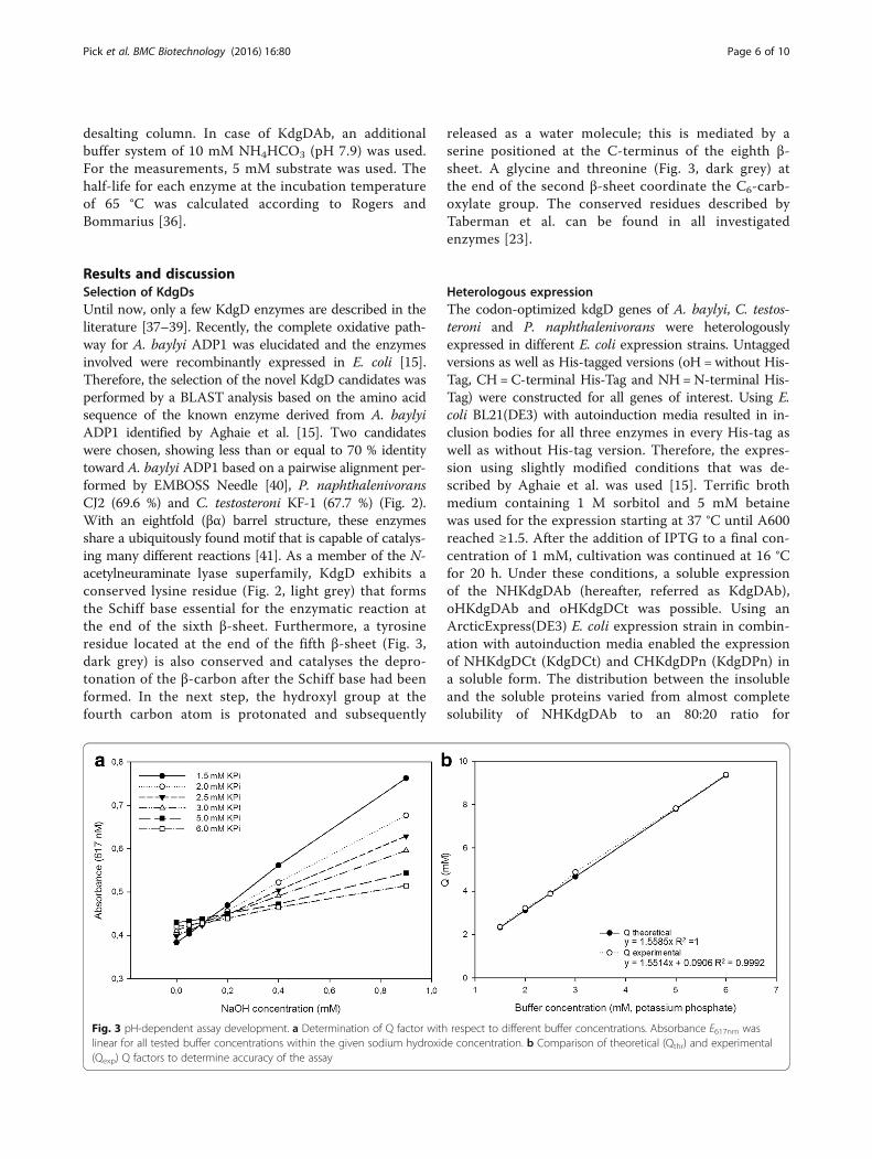
Fig. 3 pH-dependent assay development. a Determination of Q factor with respect to different buffer concentrations. Absorbance E617nm waslinear for all tested buffer concentrations within the given sodium hydroxide concentration. b Comparison of theoretical (Qthr) and experimental(Qexp) Q factors to determine accuracy of the assay
Pick et al. BMC Biotechnology (2016) 16:80 Page 6 of 10
NHKdgDCt and CHKdgDPn (data not shown). Elutedproteins appeared as a single band on SDS polyacryl-amide gels and no further bands indicating co-elutionof chaperonins of E. coli ArcticExpress(DE3) expressionwere detected. This is notwithstanding the publicationof several reports that mention co-elution as a problemfor successful enzyme purification [42, 43]. Enzyme ac-tivities of the different KdgDs were stable after storageat 8 °C in desalting buffer (50 mM Tris-HCl, pH 7.5)for at least 14 days. Independent stable long-term stor-age, preserving the catalytic activity of the enzymes,(four month) at −20 °C and −80 °C was realized with-out cryo-protectants.
Assay validationA reliable and sensitive pH-shift assay requires the pKaof the indicator and the buffer to be very similar to allowa direct correlation between the colour change and thechanging concentration of hydrogen ions within theassay solution. The pKa of the phosphate buffer is 7.2and that of the BTB lies between 7.1 and 7.3, resultingin a suitable combination.The absorbance spectra of protonated and deprotonated
BTB were determined and the maximum difference in ex-tinction coefficient was observed at 617 nm, which com-pares well with the wavelength reported in the literature[44]. The large difference in the extinction coefficient
between protonated and deprotonated BTB (Δε61728101 M−1 cm−1) results in a low Q value (Eq. 1), and thisin turn ensures a high sensitivity (dA/dt) of the assay. Fora final validation, the buffer factor (Q) was calculated anddetermined experimentally to guarantee the reliability ofthe assay. Q was experimentally determined (Qexp) by test-ing several buffer concentrations and titrations of sodiumhydroxide in the range of 0–1 mM (Fig. 3a). The recipro-cal of the slopes directly correlates to Q, and the theoret-ical Q (Qthr) value for each buffer concentration wascalculated using equation (1). Buffer concentration wasplotted against Qthr and Qexp to check the extent of thecorrelation (Fig. 3b). Although a concentration of 1.5 mMresulted in almost the same Qthr and Qexp, a higher bufferconcentration of 2.5 mM was chosen. The reasons for thisare an increased robustness of the assay and better pH sta-bility in the initial phase of the measurement. The Q valueat this buffer concentration was still sufficiently low toguarantee high sensitivity.
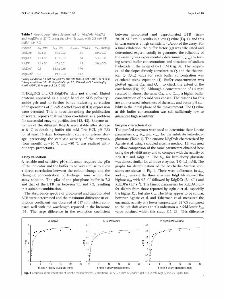
Enzyme characterizationThe purified enzymes were used to determine their kineticparameters kcat, Km and vmax for the substrate keto-deoxyglucarate (Table 1). The enzyme KdgDAb characterized byAghaie et al. using a coupled enzyme method [15] was usedto allow comparison of the same parameters obtained hereusing the pH-shift assay and to compare with the activity ofKdgDCt and KdgDPn. The Km for keto-deoxy glucaratewas almost similar for all three enzymes (1.0–1.1 mM). Thegraphs for determination of the Michaelis–Menten con-stants are shown in Fig. 4. There were differences in kcat,and vmax among the three enzymes. KdgDAb showed thehighest kcat with 4.5 s−1 followed by KdgDCt (3.1 s−1) andKdgDPn (1.7 s−1). The kinetic parameters for KdgDAb dif-fer slightly from those reported by Aghaie et al., especiallythe higher Km, but also kcat. The latter appear to be similar,however Aghaie et al. and Taberman et al. measured theenzymatic activity at a lower temperature (22 °C) comparedto the pH-shift assay (37 °C) indication a 2-fold lower kcatvalue obtained within this study [15, 23]. This difference
Fig. 4 Graphical representations of kinetic measurements. Conditions: 37 °C, 2.5 mM KPi buffer (pH 7.0), 2 mM MgCl2 and 25 μg/ml BTB
Table 1 Kinetic parameters determined for KdgDAb, KdgDCtand KdgDPn at 37 °C using the pH-shift assay with 2.5 mM KPibuffer (pH 7.0)
Enzyme Km [mM] kcat [1/s] kcat/Km [1/mM s] vmax [U/mg]
KdgDAb 1.0 ± 0.1 4.5 ± 0.02 4.5 8.0 ± 0.27
KdgDCt 1.1 ± 0.1 3.1 ± 0.02 2.8 5.3 ± 0.11
KdgDPn 1.1 ± 0.1 1.7 ± 0.01 1.5 3.0 ± 0.06
KdgDAta 0.5 8.83 ± 0.16 17.0
KdgDAbb 0.2 3.9 ± 0.04 19.2a Assay conditions: 50 mM NaPi pH 7.5, 100 mM NaCl, 4 mM NADP+, 22 °C [23]b Assay conditions: 50 mM Hepes/NaOH pH 7.5, 100 mM NaCl, 5 mM MgCl2,4 mM NADP+, 10 % glycerol, 22 °C [15]
Pick et al. BMC Biotechnology (2016) 16:80 Page 7 of 10
might be explained by the differing buffer conditions duringthe kinetic parameter measurement. The different buffersystem as well as the reduced buffer concentration withoutany stabilizers (glycerol, DTT or NaCl) in combination witha slight decrease in the pH value might be responsible forthis observation. Taberman et al. described the KdgD of A.tumefaciens and identified the pH optimum to be in therange 7.5–8.0 [23]. For this measurement, a phosphate buf-fer (pH 7.5) with additional NaCl was used and a Km of0.5 mM was reported. The enzyme showed only 65 % activ-ity at pH 7.0 compared with the activity at pH 8.0 (12 U/mg) using a phosphate buffer. Application of the pH-shiftassay system using BTB/phosphate buffer at a higher pH isnot suitable.In addition, the stability as a function of temperature
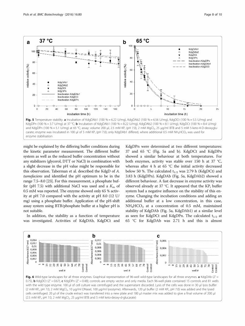
was investigated. Activities of KdgDAb, KdgDCt and
KdgDPn were determined at two different temperatures:37 and 65 °C (Fig. 5a and b). KdgDCt and KdgDPnshowed a similar behaviour at both temperatures. Forboth enzymes, activity was stable over 150 h at 37 °C,whereas after 4 h at 65 °C the initial activity decreasedbelow 50 %. The calculated t1/2 was 2.79 h (KdgDCt) and3.83 h (KdgDPn). KdgDAb (Fig. 5a, KdgDAb2) showed adifferent behaviour. A fast decrease in enzyme activity wasobserved already at 37 °C. It appeared that the KPi buffersystem had a negative influence on the stability of this en-zyme. Changing the incubation conditions and adding anadditional buffer at a low concentration, in this case,NH4HCO3 at a concentration of 0.5 mM, maintainedstability of KdgDAb (Fig. 5a, KdgDAb1) at a similar levelas seen for KdgDCt and KdgDPn. The calculated t1/2 at65 °C for KdgDAb was 2.71 h and this is almost
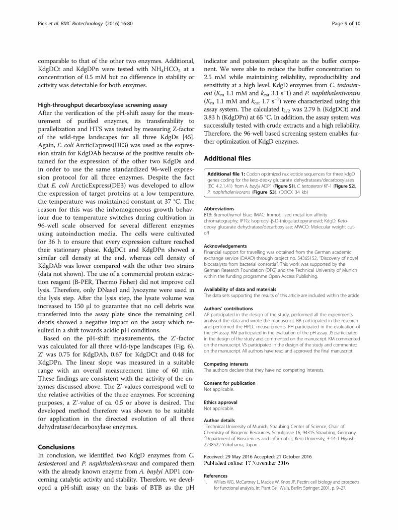
Fig. 6 Wild-type landscapes for all three enzymes. Graphical representation of 96-well wild-type landscapes for all three enzymes. a KdgDAb (Z’ =0.75), b KdgDCt (Z’ = 0.67), c KdgDPn (Z’ = 0.48); controls are empty vector and only media. Each 96-well plate contained 15 controls and 81 wellswith the wild type enzyme. 100 μl of cell culture was centrifuged and the supernatant discarded. Lysis of the cells was done in 30 μl lysis buffer(2 mM KPi, pH 7.0, 2 mM MgCl2, 10 μg/ml DNaseI, 100 μg/ml lysozyme). Afterwards, 120 μl buffer (2 mM KPi, pH 7.0) was added and the lysedcells centrifuged. 20 μl of the crude extract was transferred into a new plate and 180 μl master mix was added to give a final volume of 200 μl(2.5 mM KPi, pH 7.0, 2 mM MgCl2, 25 μg/ml BTB and 5 mM keto-deoxy-d-glucarate)
Fig. 5 Temperature stability. a Incubation of KdgDAb1 (100 % = 6.22 U/mg), KdgDAb2 (100 % = 6.56 U/mg), KdgDCt (100 % = 5.5 U/mg) andKdgDPn (100 % = 3.7 U/mg) at 37 °C; b Incubation of KdgDAb1 (100 % = 8.22 U/mg), KdgDAb2 (100 % = 8.1 U/mg), KdgDCt (100 % = 8.4 U/mg)and KdgDPn (100 % = 5.1 U/mg) at 65 °C; assay: volume 200 μl, 2.5 mM KPi (pH 7.0), 2 mM MgCl2, 25 μg/ml BTB and 5 mM 5-keto-4-D-deoxyglu-carate; enzyme was incubated in 100 μl of 5 mM KPi (pH 7.0); only KdgDAb1 differed, where additional 0.5 mM NH4HCO3 was used forenzyme stabilisation
Pick et al. BMC Biotechnology (2016) 16:80 Page 8 of 10
comparable to that of the other two enzymes. Additional,KdgDCt and KdgDPn were tested with NH4HCO3 at aconcentration of 0.5 mM but no difference in stability oractivity was detectable for both enzymes.
High-throughput decarboxylase screening assayAfter the verification of the pH-shift assay for the meas-urement of purified enzymes, its transferability toparallelization and HTS was tested by measuring Z-factorof the wild-type landscapes for all three KdgDs [45].Again, E. coli ArcticExpress(DE3) was used as the expres-sion strain for KdgDAb because of the positive results ob-tained for the expression of the other two KdgDs andin order to use the same standardized 96-well expres-sion protocol for all three enzymes. Despite the factthat E. coli ArcticExpress(DE3) was developed to allowthe expression of target proteins at a low temperature,the temperature was maintained constant at 37 °C. Thereason for this was the inhomogeneous growth behav-iour due to temperature switches during cultivation in96-well scale observed for several different enzymesusing autoinduction media. The cells were cultivatedfor 36 h to ensure that every expression culture reachedtheir stationary phase. KdgDCt and KdgDPn showed asimilar cell density at the end, whereas cell density ofKdgDAb was lower compared with the other two strains(data not shown). The use of a commercial protein extrac-tion reagent (B-PER, Thermo Fisher) did not improve celllysis. Therefore, only DNaseI and lysozyme were used inthe lysis step. After the lysis step, the lysate volume wasincreased to 150 μl to guarantee that no cell debris wastransferred into the assay plate since the remaining celldebris showed a negative impact on the assay which re-sulted in a shift towards acidic pH conditions.Based on the pH-shift measurements, the Z’-factor
was calculated for all three wild-type landscapes (Fig. 6).Z’ was 0.75 for KdgDAb, 0.67 for KdgDCt and 0.48 forKdgDPn. The linear slope was measured in a suitablerange with an overall measurement time of 60 min.These findings are consistent with the activity of the en-zymes discussed above. The Z’-values correspond well tothe relative activities of the three enzymes. For screeningpurposes, a Z’-value of ca. 0.5 or above is desired. Thedeveloped method therefore was shown to be suitablefor application in the directed evolution of all threedehydratase/decarboxylase enzymes.
ConclusionsIn conclusion, we identified two KdgD enzymes from C.testosteroni and P. naphthalenivorans and compared themwith the already known enzyme from A. baylyi ADP1 con-cerning catalytic activity and stability. Therefore, we devel-oped a pH-shift assay on the basis of BTB as the pH
indicator and potassium phosphate as the buffer compo-nent. We were able to reduce the buffer concentration to2.5 mM while maintaining reliability, reproducibility andsensitivity at a high level. KdgD enzymes from C. testoster-oni (Km 1.1 mM and kcat 3.1 s−1) and P. naphthalenivorans(Km 1.1 mM and kcat 1.7 s−1) were characterized using thisassay system. The calculated t1/2 was 2.79 h (KdgDCt) and3.83 h (KdgDPn) at 65 °C. In addition, the assay system wassuccessfully tested with crude extracts and a high reliability.Therefore, the 96-well based screening system enables fur-ther optimization of KdgD enzymes.
Additional files
Additional file 1: Codon optimized nucleotide sequences for three kdgDgenes coding for the keto-deoxy glucarate dehydratases/decarboxylases(EC 4.2.1.41) from A. baylyi ADP1 (Figure S1), C. testosteroni KF-1 (Figure S2),P. naphthalenivorans (Figure S3). (DOCX 34 kb)
AbbreviationsBTB: Bromothymol blue; IMAC: Immobilized metal ion affinitychromatography; IPTG: Isopropyl-β-D-thiogalactopyranosid; KdgD: Keto-deoxy glucarate dehydratase/decarboxylase; MWCO: Molecular weight cut-off
AcknowledgementsFinancial support for travelling was obtained from the German academicexchange service (DAAD) through project no. 54365152, “Discovery of novelbiocatalysts from bacterial consortia”. This work was supported by theGerman Research Foundation (DFG) and the Technical University of Munichwithin the funding programme Open Access Publishing.
Availability of data and materialsThe data sets supporting the results of this article are included within the article.
Authors’ contributionsAP participated in the design of the study, performed all the experiments,analysed the data and wrote the manuscript. BB participated in the researchand performed the HPLC measurements. RH participated in the evaluation ofthe pH assay. RM participated in the evaluation of the pH assay. JS participatedin the design of the study and commented on the manuscript. KM commentedon the manuscript. VS participated in the design of the study and commentedon the manuscript. All authors have read and approved the final manuscript.
Competing interestsThe authors declare that they have no competing interests.
Consent for publicationNot applicable.
Ethics approvalNot applicable.
Author details1Technical University of Munich, Straubing Center of Science, Chair ofChemistry of Biogenic Resources, Schulgasse 16, 94315 Straubing, Germany.2Department of Biosciences and Informatics, Keio University, 3-14-1 Hiyoshi,2238522 Yokohama, Japan.
Received: 29 May 2016 Accepted: 21 October 2016
References1. Willats WG, McCartney L, Mackie W, Knox JP. Pectin: cell biology and prospects
for functional analysis. In: Plant Cell Walls. Berlin: Springer; 2001. p. 9–27.
Pick et al. BMC Biotechnology (2016) 16:80 Page 9 of 10
2. Ebringerova A, Heinze T. Xylan and xylan derivatives–biopolymers withvaluable properties, 1. Naturally occurring xylans structures, isolationprocedures and properties. Macromol Rapid Commun. 2000;21(9):542–56.
3. Adams E, Rosso G. α-ketoglutaric semialdehyde dehydrogenase of pseudomonasproperties of the purified enzyme induced by hydroxyproline and of theglucarate-induced and constitutive enzymes. J Biol Chem. 1967;242(8):1802–14.
4. Chang YF, Feingold DS. D-Glucaric Acid and Galactaric Acid Catabolism byAgrobacterium Tumefaciens. J Bacteriol. 1970;102(1):85–96.
5. Ashwell G, Wahba AJ, Hickman J. A new pathway of uronic acidmetabolism. Biochim Biophys Acta. 1958;30(1):186–7.
6. Rodionova IA, Scott DA, Grishin NV, Osterman AL, Rodionov DA.Tagaturonate–fructuronate epimerase UxaE, a novel enzyme in thehexuronate catabolic network in Thermotoga maritima. Environ Microbiol.2012;14(11):2920–34.
7. Van Gijsegem F, Toussaint A. In vivo cloning of Erwinia carotovora genesinvolved in the catabolism of hexuronates. J Bacteriol. 1983;154(3):1227–35.
8. Richard P, Hilditch S. D-galacturonic acid catabolism in microorganisms and itsbiotechnological relevance. Appl Microbiol Biotechnol. 2009;82(4):597–604.
9. Boer H, Maaheimo H, Koivula A, Penttilä M, Richard P. Identification inAgrobacterium tumefaciens of the D-galacturonic acid dehydrogenase gene.Appl Microbiol Biotechnol. 2010;86(3):901–9.
10. Bouvier JT, Groninger-Poe FP, Vetting M, Almo SC, Gerlt JA. Galactaro δ-lactoneisomerase: lactone isomerization by a member of the amidohydrolasesuperfamily. Biochemistry. 2014;53(4):614–6.
11. Yoon S-H, Moon TS, Iranpour P, Lanza AM, Prather KJ. Cloning andcharacterization of uronate dehydrogenases from two Pseudomonads andAgrobacterium tumefaciens strain C58. J Bacteriol. 2009;191(5):1565–73.
12. Wagschal K, Jordan DB, Lee CC, Younger A, Braker JD, Chan VJ. Biochemicalcharacterization of uronate dehydrogenases from three Pseudomonads,Chromohalobacter salixigens, and Polaromonas naphthalenivorans. EnzymeMicrob Technol. 2015;69:62–8.
13. Parkkinen T, Boer H, Janis J, Andberg M, Penttila M, Koivula A, Rouvinen J.Crystal structure of uronate dehydrogenase from Agrobacterium tumefaciens.J Biol Chem. 2011;286(31):27294–300.
14. Pick A, Schmid J, Sieber V. Characterization of uronate dehydrogenasescatalysing the initial step in an oxidative pathway. J Microbial Biotechnol.2015;8(4):633–43.
15. Aghaie A, Lechaplais C, Sirven P, Tricot S, Besnard-Gonnet M, Muselet D, deBerardinis V, Kreimeyer A, Gyapay G, Salanoubat M, et al. New insights into thealternative D-glucarate degradation pathway. J Biol Chem. 2008;283(23):15638–46.
16. Watanabe S, Kodaki T, Makino K. A Novel α-ketoglutaric semialdehydedehydrogenase: evolutionary insight into an alternative pathway of bacteriall-arabinose metabolism. J Biol Chem. 2006;281(39):28876–88.
17. Glasner ME, Gerlt JA, Babbitt PC. Evolution of enzyme superfamilies. CurrOpin Chem Biol. 2006;10(5):492–7.
18. Babbitt PC, Hasson MS, Wedekind JE, Palmer DR, Barrett WC, Reed GH,Rayment I, Ringe D, Kenyon GL, Gerlt JA. The enolase superfamily: a generalstrategy for enzyme-catalyzed abstraction of the α-protons of carboxylicacids. Biochemistry. 1996;35(51):16489–501.
19. Gulick AM, Hubbard BK, Gerlt JA, Rayment I. Evolution of enzymaticactivities in the enolase superfamily: crystallographic and mutagenesisstudies of the reaction catalyzed by d-glucarate dehydratase fromEscherichia coli. Biochemistry. 2000;39(16):4590–602.
20. Gulick AM, Palmer DR, Babbitt PC, Gerlt JA, Rayment I. Evolution of enzymaticactivities in the enolase superfamily: crystal structure of (D)-glucaratedehydratase from Pseudomonas putida. Biochemistry. 1998;37(41):14358–68.
21. Babbitt PC, Gerlt JA. Understanding enzyme superfamilies Chemistry as thefundamental determination in the evolution of new catalytic activities. J BiolChem. 1997;272(49):30591–4.
22. Taberman H, Andberg M, Parkkinen T, Richard P, Hakulinen N, Koivula A,Rouvinen J. Purification, crystallization and preliminary X-ray diffractionanalysis of a novel keto-deoxy-d-galactarate (KDG) dehydratase fromAgrobacterium tumefaciens. Acta Crystallogr Sect F. 2014;70(1):49–52.
23. Taberman H, Andberg M, Parkkinen T, Jänis J, Penttilä M, Hakulinen N,Koivula A, Rouvinen J. Structure and function of a decarboxylatingAgrobacterium tumefaciens Keto-deoxy-D-galactarate dehydratase.Biochemistry. 2014;53(51):8052–60.
24. Tang L, Li Y, Wang X. A high-throughput colorimetric assay for screeninghalohydrin dehalogenase saturation mutagenesis libraries. J Biotechnol.2010;147(3):164–8.
25. Persson M, Palcic MM. A high-throughput pH indicator assay forscreening glycosyltransferase saturation mutagenesis libraries. AnalBiochem. 2008;378(1):1–7.
26. Chapman E, Wong C-H. A pH sensitive colorometric assay for the high-Throughput screening of enzyme inhibitors and substrates: a case studyusing kinases. Bioorg Med Chem. 2002;10(3):551–5.
27. Rosenberg RM, Herreid RM, Piazza GJ, O'Leary MH. Indicator assay for aminoacid decarboxylases. Anal Biochem. 1989;181(1):59–65.
28. Yu K, Hu S, Huang J, Mei L-H. A high-throughput colorimetric assay tomeasure the activity of glutamate decarboxylase. Enzyme Microb Technol.2011;49(3):272–6.
29. He N, Yi D, Fessner W-D. Flexibility of substrate binding of Cytosine-5’-Monophosphate-N-Acetylneuraminate Synthetase (CMP-Sialate Synthetase)from Neisseria meningitidis: an enabling catalyst for the synthesis ofneo-sialoconjugates. Adv Synthesis Catalysis. 2011;353(13):2384–98.
30. Janes LE, Löwendahl AC, Kazlauskas RJ. Quantitative screening of hydrolaselibraries using pH indicators: identifying active and enantioselectivehydrolases. Chem Eur J. 1998;4(11):2324–31.
31. Altschul SF, Madden TL, Schäffer AA, Zhang J, Zhang Z, Miller W, Lipman DJ.Gapped BLAST and PSI-BLAST: a new generation of protein database searchprograms. Nucleic Acids Res. 1997;25(17):3389–402.
32. Guterl J-K, Garbe D, Carsten J, Steffler F, Sommer B, Reiße S, Philipp A, HaackM, Rühmann B, Koltermann A, et al. Cell-free metabolic engineering:production of chemicals by minimized reaction cascades. ChemSusChem.2012;5(11):2165–72.
33. Studier FW. Protein production by auto-induction in high-density shakingcultures. Protein Expr Purif. 2005;41(1):207–34.
34. Gibbons BH, Edsall JT. Rate of hydration of carbon dioxide and dehydrationof carbonic acid at 25. J Biol Chem. 1963;238(10):3502–7.
35. Khalifah RG. The carbon dioxide hydration activity of carbonic anhydrase I.Stop-flow kinetic studies on the native human isoenzymes B and C. J BiolChem. 1971;246(8):2561–73.
36. Rogers TA, Bommarius AS. Utilizing simple biochemical measurements topredict lifetime output of biocatalysts in continuous isothermal processes.Chem Eng Sci. 2010;65(6):2118–24.
37. Jeffcoat R, Hassall H, Dagley S. Purification and properties of D-4-deoxy-5-oxoglucarate hydro-lyase (decarboxylating). Biochem J. 1969;115(5):977–83.
38. Jeffcoat R, Hassall H, Dagley S. The metabolism of D-glucarate byPseudomonas acidovorans. Biochem J. 1969;115(5):969–76.
39. Sharma BS, Blumenthal HJ. Catabolism of d-Glucaric Acid to α-Ketoglutaratein Bacillus megaterium. J Bacteriol. 1973;116(3):1346–54.
40. Rice P, Longden I, Bleasby A. EMBOSS: the European molecular biologyopen software suite. Trends Genet. 2000;16(6):276–7.
41. Nagano N, Orengo CA, Thornton JM. One fold with many functions: theevolutionary relationships between TIM Barrel families based on theirsequences, structures and functions. J Mol Biol. 2002;321(5):741–65.
42. Lee K-H, Kim H-S, Jeong H-S, Lee Y-S. Chaperonin GroESL mediates theprotein folding of human liver mitochondrial aldehyde dehydrogenase inEscherichia coli. Biochem Biophys Res Commun. 2002;298(2):216–24.
43. Hartinger D, Heinl S, Schwartz HE, Grabherr R, Schatzmayr G, Haltrich D, MollW-D. Enhancement of solubility in Escherichia coli and purification of anaminotransferase from Sphingopyxis sp. MTA 144 for deamination ofhydrolyzed fumonisin B 1. Microb Cell Fact. 2010;9:62.
44. Banerjee A, Kaul P, Sharma R, Banerjee UC. A high-throughput amenablecolorimetric assay for enantioselective screening of nitrilase-producingmicroorganisms using pH sensitive indicators. J Biomol Screen. 2003;8(5):559–65.
45. Zhang J-H, Chung TD, Oldenburg KR. A simple statistical parameter for usein evaluation and validation of high throughput screening assays. J BiomolScreen. 1999;4(2):67–73.
46. Sievers F, Wilm A, Dineen D, Gibson TJ, Karplus K, Li W, Lopez R, McWilliamH, Remmert M, Soding J, et al. Fast, scalable generation of high-qualityprotein multiple sequence alignments using Clustal Omega. Mol Syst Biol.2011;7(1):539.
Pick et al. BMC Biotechnology (2016) 16:80 Page 10 of 10