
ICBMB 2017 International Conference on Biochemistry and...
53
Transcript of ICBMB 2017 International Conference on Biochemistry and...
ICBMB 2017
International Conference on Biochemistry and Molecular Biology
3-5 April 2017, Munich, Germany
SheratonMünchenArabellaparkHotel
Arabellastraße 5, 81925 München, Germany
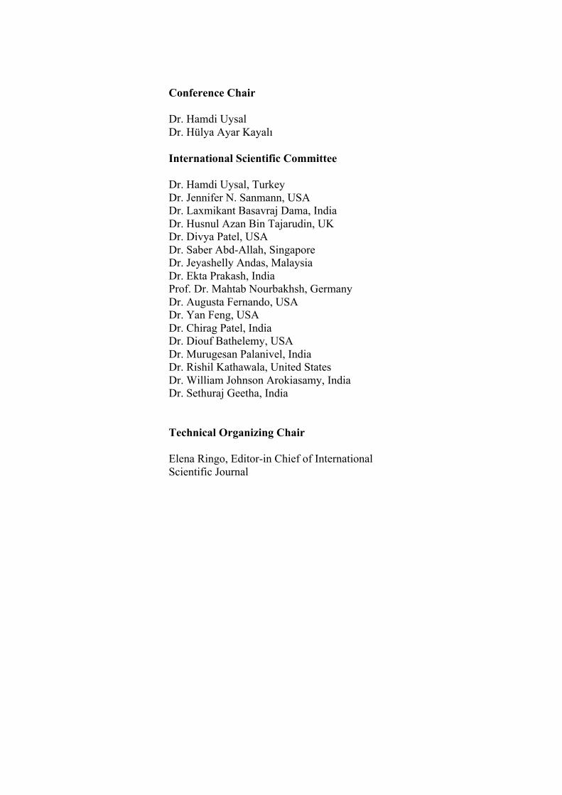
Conference Chair
Dr. Hamdi Uysal Dr. Hülya Ayar Kayalı
International Scientific Committee
Dr. Hamdi Uysal, Turkey Dr. Jennifer N. Sanmann, USA Dr. Laxmikant Basavraj Dama, India Dr. Husnul Azan Bin Tajarudin, UK Dr. Divya Patel, USA Dr. Saber Abd-Allah, Singapore Dr. Jeyashelly Andas, Malaysia Dr. Ekta Prakash, India Prof. Dr. Mahtab Nourbakhsh, Germany Dr. Augusta Fernando, USA Dr. Yan Feng, USA Dr. Chirag Patel, India Dr. Diouf Bathelemy, USA Dr. Murugesan Palanivel, India Dr. Rishil Kathawala, United States Dr. William Johnson Arokiasamy, India Dr. Sethuraj Geetha, India
Technical Organizing Chair
Elena Ringo, Editor-in Chief of International Scientific Journal
Protein Glycosylation in the Gastric Pathogen Helicobacter pylori
Hamdi Uysal
Department of Biochemistry, Faculty of Veterinary Medicine, Ankara University,
06110, Diskapi, Ankara, Turkey Abstract—Glycosylation was previously considered to be restricted to eukaryotes; however, through advances in analytical methods, there have been increasing reports of both O-linked and N-linked protein glycosylation pathways in bacteria, particularly amongst mucosal-associated pathogens. The purpose of this study was to detect the glycosylated proteins of Helicobacter pylori (H. pylori) separated by Sodium Dodecyl Sulfate Polyacrylamide Gel eEectrophoresis (SDS-PAGE) and Two Dimensional Gel Electrophoresis (2-DE). Protein analysis of the H. pylori strains was performed using denaturing 8% SDS-PAGE. H. pylori 26695 cell proteins were also separated by 2-DE into hundreds of spots. Separated proteins of H. pylori strains by SDSPAGE and 2-DE were transferred onto the Polyvinylidene Fluoride (PVDF) membrane by semi-dry blotting. Detection of glycosylated proteins of the protein bands or spots on blotted membranes were determined by overlay reactions with Digoxigenin (DIG)-Glycan Detection and Differentation kits (Roche Diagnostics, Germany). DIG-Glycan Differentiation Kit including peanut agglutinin (PNA) lectin was utilized to further characterize the glycosidic modifications. Analysis of protein bands on a blotted membrane with DIG Glycan Detection kit after SDS-PAGE analysis gave the general pattern of glycosylated proteins of H. pylori; interestingly PA4, PR20 and P12 strains of H. pylori gave the different patterns of protein glycosylation on 8% polyacrilamide gels. Moreover, a glycosylated protein band (~54 kDa) was also detected dominantly on the outer membrane part of H. pylori. 2-DE analysis of H. pylori proteins showed about twelve clear spots indicating the O-glycosidically linked carbohydrate chains (galactose–β(1-3)-N-acetylgalactosamine) determined by PNA lectin staining of the blotted membrane. Obtained results of this study and current studies on the protein glycosylation in the gastric pathogen H. Pylori will be discussed.
Effects of Fibronectins on Cell Morphology, Cell Attachment and Cell Spreading
Hamdi Uysal
Department of Biochemistry, Faculty of Veterinary Medicine, Ankara University, 06110, Diskapi, Ankara, Turkey
Abstract—Fibronectin (FN) is a multifunctional glycoproteins expressed by many different cell types (hepatocytes, fibroblasts, macrophages etc.). Fibronectin can be found as two major forms: plasma FN (pFN) and cellular FN (cFN). pFN is synthesized by hepatocytes and secreted into the blood plasma, where it circulates in a soluble form. cFN is an insoluble form that is secreted and assembled into dense complex fibril networks, affecting overall extracellular matrix (ECM)-cell interactions. In general, fibronectin contains 3-9% carbohydrate, depending on the tissue or cell origin. Cellular fibronectin from adult human skin fibroblasts has a low carbohydrate content, whereas plasma fibronectin has higher carbohydrate content especially sialic acid. But, plasma fibronectin has no fucose in comparison to cellular fibronectin. Cells can attach, spread, and migrate on a variety of extracellular matrix glycoproteins including fibronectin. These interactions occur through specific cell surface receptors. Cell adhesion receptors belong to a large superfamily of integrins. Many of the adhesive glycoproteins like fibronectin contain a common tripeptide sequence (RGD) which is recognised by cell surface integrins and helps bind cells to the ECM.
In this study, we have examined the effects of plasma and cellular fibronectins on cell morphology, cell attachment and spreading of fibroblast cells. Hence, cell culture plates were coated with different concentrations (1.5µg/cm2, 3µg/cm2 and 6µg/cm2) of purified cellular and commercial plasma fibronectins. Then, we observed the morphology and spreading of fibroblast cells on the light microscobe. We found that plasma and cellular fibronectins at different concentrations were equally active in promoting cell attachment. However, cellular fibronectin obtained from skin fibroblasts appeared more active in the promotion of cell spreading, proliferation and morphology of fibroblasts than plasma fibronectin. Obtained results demonstrated that cellular fibronectin behaves differently on cell morphology, profileration and spreading than plasma fibronectin. Significance of these results and some other functions of fibronectins will be discussed under the light of current literature.
The Association Between EpCAM, Claudins, and
Tetraspanins on Colorectal Cancer Progression
Hulya Ayar Kayali1,2 *, Deniz Erkan3 1 Dokuz Eylul University, Faculty of Science, Department of Chemistry, Izmir 35390, Turkey
2 Dokuz Eylul University, Izmir Biomedicine and Genome Center, Izmir 35340, Turkey
3Dokuz Eylul University, The Graduate School of Natural and Applied Sciences, Department
of Biotechnology, Izmir 35390, Turkey
Abstract—The changes in expression levels of cell adhesion and tight junction molecules, and formation of interactions between these molecules are effective in colorectal cancer development and metastasis. The aim of this study was to investigate the interactions between EpCAM, claudins and tetraspanins in colorectal cancer.
Normal colon cell line CCD-18Co, primary colorectal cancer cell line Caco-2 and metastatic colorectal cancer cell lines DLD-1 and SW620 were used. Protein levels of EpCAM, claudin-1, -3, -4, -7, CD9, CD82, CD151 and Tspan8 were determined by immunoblotting. EpCAM, claudin-4 and claudin-7 proteins were chosen for immunoprecipitation analyses according to the results of immunoblot analyses.
EpCAM, claudin-1, -4, -7 were not determined in CCD-18Co, but the levels of these molecules increased in colorectal cancer cell lines, except claudin-7 was not determined in SW620. The highest level of claudin-1 was determined in SW620. Claudin-3 was determined in CCD-18Co, but its levels increased in Caco-2, and especially in DLD-1 and SW620. CD9 levels were low in all cell lines, whereas CD82 levels were low in colorectal cancer cell lines compared to CCD-18Co. CD151 was not determined in any of cell lines. Tspan8 levels increased depending on carcinogenesis, except its level was even lower than that of CCD-18Co in DLD-1. EpCAM-claudin-7-Tspan8, claudin-4-claudin-3 and claudin-7-CD82 interactions were determined in Caco-2, and claudin-4-Tspan8 interaction was determined in DLD-1. However, EpCAM-Tspan8, claudin-4-claudin-3, claudin-4-Tspan8 interactions were also not determined in SW620 that does not express claudin-7. Our results showed that the levels of EpCAM, claudin-1, claudin-3, CD82 and Tspan8 and the interactions between these molecules are depended to the colorectal cancer stages.
Keywords—colorectal cancer, EpCAM, claudins, tetraspanin
The effect of a palm oil-enriched diet on caveolin-1β expression and reactive oxygen
species production in the Psammomys obesus aorta.
Mohamed El Fadel Ousmaal1,2*, M. Carmen Martínez3, Ramaroson Andriantsitohaina3,
Kahina Chabane1, Abderahim Gaceb3, Khen M'hamed Amine1, Ahsene Baz1
1Laboratory of Biology and Animal Physiology, ENS Kouba, Algiers, Algeria. 2Department of Nature and Life Sciences, Faculty of Science, University of Algiers 1,
Algiers, Algeria. 3INSERM U1063- Stress Oxydant et Pathologies Métaboliques, Université d’Angers, France
Abstract—Dyslipidemia is a lipoprotein metabolism disorder that has been directly linked to hyperglycemia, insulin resistance and cardiovascular disease. Psammomys obesus (P. obesus) is a naturally insulin-resistant animal that exhibits a tendency to develop diet-induced hyperglycemia and obesity. We hypothesized that a palm oil-enriched diet (PD) might prevent hyperglycemia and contribute to cav-1 overexpression and reactive oxygen species (ROS) production in P. obesus. Our study was designed to compare the effects of a palm oil-enriched diet with those of a standard diet (SD) on glucose and lipid metabolism, body weight gain and endothelial caveolin-1 (cav-1) protein expression in P. obesus. After 12 weeks of feeding with either the PD or SD, we observed that the PD caused a marked increase in lipid parameters, even in normoglycemic P. obesus. The PD group displayed significantly higher levels of total cholesterol (C), LDL-C, triglycerides (TG) and body weight gain than the SD group. Cav-1β protein expression and ROS production were increased in the vascular endothelium of PD-treated P. obesus. The results of our experimental study suggest that the PD exerts deleterious effects on the vascular system that are accompanied by pronounced dyslipidemia in normoglycemic P. obesus which may lead to cav-1β protein overexpression and ROS production in the vascular wall. Keywords—Dyslipidemia, palm oil-enriched diet, caveolin-1, vascular endothelium, Psammomys obesus.
Optimization of the culture medium of chitinolytic HE3 strain isolated from the marine environment:
improving the production of chitinase
Laribi – Habchi Hassiba1*, Ayeb Meriem2
1: Laboratory of Chemistry of Natural Substances and biomolecules, University of Blida 1 2: Laboratory of Biochemistry -Microbiology, Department of Engineering Processes, University of
Blida 1 E-mail : [email protected]
Abstract—Chitin, today is a renewable resource that offers better prospects in terms of reducing production costs by abundance and potentially lower prices to other substrates, despite the complexity of the transformation processes. Chitin is the important issues in research including biofuel. This research aims to improve conversion efficiencies of marine biomass (chitin) using enzymes capable of chitin degrading. Various microorganisms including various bacteria present biodegradation capacities of various organic molecules. This function of biodegradation is due to the variety of enzymes that they can synthesize as chitinases. These include biotechnology and environmental applications very interesting (as biopesticides in agriculture, medicine, industry etc ...).
Our study is the optimization of the production of chitinases chitinolytic and halotolerant bacterial strain isolated from the marine environment using experimental designs method to optimize the factors influencing the responses (rate of NAG products and bacterial growth). This strain was identified by classical taxonomic methods coupled to molecular biology (sequence of 16 S rRNA genes).
The best experimental results of the production of chitinase were 0.07g / l of NAG with good bacterial growth (absorbance of 0.465) using a concentration of 1.1g / l of chitin and only 0.25 g / l of sodium chloride. Which shows good agreement with the results expected theoretically (model). The analysis of the sequence obtained by alignment has linked this new strain HE3 to the species "Shewanella basaltis" with 98% similarity.
The study of effect of each factor on the two selected responses shows that the concentration of chitin presents the main factor for both responses. Les results suggest that the experimental model provided an efficient and economical method in the optimization of production chitinase.
Keywords: Chitinase, Shewanella basaltis., identification Experimental plan,
Degradation of chitin
Induction of senescence in cancer cells by 5'-Aza-2'-deoxycytidine: bioinformatics and
experimental insights to its targets Jayarani Putri*1,2, Yue Yu1,2, Kazuichi Sakamoto2, Nashi Widodo3,
Sunil C Kaul1, Renu Wadhwa1
1Drug Discovery & Assets Innovation Lab & DAILAB, National Institute of Advanced Industrial Science & Technology (AIST), Central 5-41, 1-1-1 Higashi, Tsukuba - 305
8565, Japan; 2Graduate School of Life & Environmental Sciences, University of Tsukuba, Ibaraki - 305 8572, Japan; 3Faculty of Mathematics and Natural Sciences,
Brawijaya University, Malang - 65145, Indonesia E-mail: [email protected]
Abstract—Epigenetic therapies are aiming to reverse epigenetic aberration
that occur in cancer to restore more normal epigenetic state.
Hypomethylating DNA drug, 5-Azacitidine (5-Aza-dC) suppressing
silenced gene during tumorigenesis leads to gene reactivation, growth arrest
and senescence. In this study, we conducted an analysis for new functions of
5-Aza-dC by applying the bio-chemo-informatics approach of FDA-
approved anticancer drugs which is desired to expand the list of multi-
module functioning drugs for cancer therapy. Bioactivity of 5-Aza-dC was
analyzed by Molinspiration and PASS online. The Protein Networks and
Biological Processes were analyzed by Biological Networks using Gene
Ontology tool, BINGO, based on BIOGRID database. Protein interaction
between 5-Aza-dC and targeted protein was performed using Autodoc Vina
integrated into Pyrx software. In vitro experiment was performed by
observing the expression of p53 and other related protein using cancer cells.
Bioinformatics analyses predicted that 5-Aza-dC functions as a p53 inducer,
radio-sensitizer, and inhibitor of several enzymes. It was predicted that
enzymes and proteins including POLA1, POLB, MDM2, and CXCR4 are
involved in the induction of DNA damage response and p53-HDM2-
p21signaling. We provide experimental evidence to the targeting of HDM2
by 5AZA-dC leading to activation of p53 pathway and growth of cells.
Furthermore, to our surprise we found that combinatorial treatment of
5AZA-dC with three anticancer drugs caused drug resistance. In order to
investigate its new regulatory targets experimentally, we performed loss-of-
function miRNA screening and found induction of miRNA-335 that targeted
CARF and several cell cycle regulatory proteins leading to growth
suppression in cancer cells. Taken together, we demonstrate that 5-Aza-dC-
induced senescence is a multi-module phenotype regulated not only by
proteins but also by noncoding miRNAs. Further studies are warranted to
dissect these mechanisms and establish 5-Aza-dC as an effective multi-
module anticancer reagent.
Keywords: 5-Aza-dC, bioinformatics, CARF, HDM2, miRNA-335, p53,
p21, senescence
Study of the Inhibitory E↵ect of Cleome Arabica
Extracts on Butyrylcholinesterase
Fatiha Seglab*
1, Mohamed Yousfi
1
1) Laboratoire des Sciences Fondamentales, Amar Telidji Laghouat University, Algria
This study focus on the evaluation of anti-oxidant activity of Cleome Arabicaleaves extract. Given the novelty of its study and valorization, this plant is ex-pected to have medicinal potential as high antioxidant and enzymatic inhibitory.The first approach in this study involves an extraction and a quantificationof the phenolic compounds of the Cleome Arabica Leaves extract which high-lighted the presence of some bioactive chemical groups. This was confirmed bya quantitative analysis based on the measurement of total phenolics, flavonoidsand condensed tannins content constructed in three liquid-liquid organic extrac-tions based on their polarity. Results show that for these bioactive sub-fractions(petroleum ether, dichloromethane and ethyl acetate), ethyl acetate is the bestextractor of flavonoids, while petroleum ether has the ability to extract moreof the terpene, and was explored further. In a second step we evaluate the an-tiradical potential of our extracts using five di↵erent methods and for di↵erentseasons. The antioxidant activity of ethyl acetate extracts was investigated us-ing five methods. phosphomolybdenum complex (PM-complex). DPPH, ABTS,CUPRAC and FRAP. Finally, we present our plan to study their inhibitory ef-fects on the butyrylcholinesterase enzyme, considered to play a major role inAlzheimer disease, due to its high activity in patients with Alzheimer’s disease.The antioxidant activity test shows that our phenolic extracts have good powerantioxidant compared to antioxidants taken as a reference. We also have noticeda positive correlation between the di↵erent tests. This work provides new ethnopharmacological and phytochemical knowledge about local plants in Laghouatregion (400km south of Algeria), and contributes to the study of the role ofnatural polyphenols in the regulation of oxidative stress and in the treatmentof neurodegenerative disorders.
* Poster presenter
CD9 and CD82 repress metastasis and chemotherapy resistance in ovarian cancer
Zehra Tavsan1,3, Hulya Ayar Kayali2,3*
e-mail: [email protected]; [email protected]*
1Department of Chemistry, The Graduate School of Natural and Applied Sciences, Dokuz Eylül University, İzmir, Turkey
2Division of Biochemistry, Department of Chemistry, Science Faculty, Dokuz Eylül University, İzmir, Turkey
3Dokuz Eylül University, International Biomedicine and Genome Institue, İzmir, Turkey
Abstract—Evolutionarily conserved membrane proteins, tetraspanins associate laterally with one another and to organize with other membrane proteins, most of which play a receptor role, or alternatively couple receptors to intracellular and intercellular processes, including signal transduction, cell proliferation, adhesion and migration, cell fusion and host-parasite interactions to form tetraspanin-enriched microdomains (TEMs). Many studies show that the expression of tetraspanins correlates with tumour stage, tumour type and patient outcome. But how most tetraspanins function is unclear. In our experiments, the protein expressions of CD9 and CD82 as well as the other partners of tetraspanins within TEM were determined by Western blot in in the cell lines which were isolated from primary ovarian tumours (A2780), metastasis ovarian tumors or ascites (OVCAR–3 and SKOV–3) and cisplatin-resistant A2780cis ovarian cancer cell line. Significantly, CD9 and CD82 levels decreased in A2780, OVCAR-3, SKOV-3 and A2780cis cell lines. The data correlated with the studies about tetraspanin CD9 and CD82 as tumour suppressors. In contrast, the other tetraspanin, CD151 expression increased in association with gaining the properties of metastasis and chemotherapy resistance. Many experiments also showed that CD151 is upregulated in various cancers, often in association with increased metastasis and/or poor prognosis. In a similar way, the partner molecules within TEM, EpCAM, claudin-1, -3, -4 and -7 levels increased. The significant alterations in tetraspanins and partner proteins suggested that these molecules may be good prognostic factor and worthy cancer targets in effective treatments.
Keywords: CD9, CD82, biomarker of ovarian cancer diagnosis.
MORFOLOGICAL CHANGES IN AMYGDALA AND HIPOTALAMIC NUCLEUS UNDER CONDITIONS OF THE DESTRUCTION
OF DORSAL AMYGDALOFUGAL WAY
R.M.Bagirova
Azerbaijan State Academy of Physical Culture and Sports, Department "Biomedical research"", Baku, Azerbaijan Republic, [email protected]
With the aim to study the role of amygdalo-hypothalamical connections in the mechanism of formation of hippocampal theta-rhythm at present work it has been done comparative analysis of participation of dorsal and ventral amygfalofugal ways in this process. The dorsal amygdalofugal way was coagulated by 15-25-sec-long 1.0 mA current passed through an electrode implanted into the precommissural region. Both recording of the electrical activity from the hippocampus and septum and collection of the samplings for morphological studies were performed 18-27 days after such a destruction. For histological studies, brains of the animals were fixed in Carnoy solution and dehydrated using a series of alcohols of increasing concentrations; the blocks including the structures under study were embedded in paraffin. Frontal 10-µm-thick slices were prepared with a microtome, stained with 0.1% cresyl violet solution, treated with alcohol, cleared in xylene, embedded in balsam, and examined under a light microscope. The objects under study were neurons and glial cells of the amygdalo and hypothalamus. In chronic experiments on rabbits it has been shown that destruction of dorsal amygdalofugal ways leads to full and petrsistent blockade of hippocampal theta-rhythm. Its restoration was not observed even in 6 months following excited injury. Coagulation of ventral amygdalofugal way leads to registration of unregular, polymorph, low amplitude, deformed activity combined both fast frequent oscillation and separate theta-waves in various parts of hippocamp and medial septal nucleus unlike the dorsal amygdalofugal way. However, full restoration of electrographic indices till background level was noted in 20-25 days. To elucidate the causes irreversible changes in different areas of the hippocampus and the medial nucleus of septum morphological studies were carried out in neurons and glial cells of bazolateral (AB), central (AC), lateral (AL) and cortical (ACO) nucleus of the amygdala and supraoptic (SO), ventromedial (VMH), lateral (AHL), medial mammilar (MM) nucleus of hypothalamus. Examination of the slices of the amygdalo (AB, AC, ACO, AL) and hypothalamic (SO, VMH, AHL, MM) nucleus of experimental animals after coagulation of the stria terminalis demonstrated that profound morphological changes were detected in neurons and glial cells of these structions. Morphological studies developed deep degenerative changes just lyzis of Nissel matter, swelling of apical dendrites, hyperchromatism of nuclei and decrease in the volume of the latter were typical findings, absence of tigroid matter in neurons and glial cells in different nucleus of hypothalamus and amygdala, under destruction of dorsal amygdalofugal tract. Neurons and glial cells are swelled. One of the factors which modulates the excitability of neurons in septo-hippocampal system is supposed may be disturbance of hypothalamo-hypophysial neurosecretory system under the influence of destruction of amygdala-hypothalamic relations. Keywords: hippocampal theta rhythm, dorsal and ventral amygdalofugal pathway, amygdala, hypothalamus, morphological changes.
Evaluation of an electrochemiluminescence immunoassay and an enzyme-linked fluorescent assay for detection of anti-cytomegalovirus IgM and anti-toxoplasma IgM
antibodies in pregnant women
[1*] BLERTA LAZE, [2] ARTA LUGAJ
[1*] Department of Biology, Faculty of Technical Sciences, University “Ismail Qemali”, Vlora, Albania
[2] Department of Biology, Faculty of Technical Sciences, University “Ismail Qemali”,
Vlora, Albania
e-mail: [1*] [email protected], [2] [email protected] Abstract—Nowdays, it is noticed an increase in morbidity from infectious factors, among which the principal ones are viral, bacterial and parasitic infections. This is quite sensitive in pregnant women, whose infections, especially in the first trimester of pregnancy cause malformation of the fetus that is being formed. This is more complicated in cases of Toxoplasma gondii and Cytomegalovirus because of cross reactions of their antibodies against similar antigenic epitopes.For this reason the aim of this study was the detection of gestational Cytomegalovirus and Toxoplasma gondii infections. Cytomegalovirus (CMV) is a herpes virus transmitted by intimate contact with infected excretions such as saliva, urine, cervical and vaginal excretions, semen, breast milk and blood. Toxoplasma gondii is a parasitic protozoa which can be transmited by eating infected meat or from mother to fetus during the first trimester of pregnancy. Because diagnosis of maternal infections solely depends on serology, routine tests with high sensitivity and specificity are required. Medical diagnostic is working to determine the most sensitive techniques for the detection of anti-cytomegalovirus IgM and anti-toxoplasma IgM antibodies, in the framework of which is developed this scientific work. This study compares an electrochemiluminescence immunoassay (ECL, applied in COBAS 6000 instrument) with an enzyme-linked fluorescent assay (ELFA, applied in MINI-VIDAS instrument) for detection of anti-cytomegalovirus IgM and anti-toxoplasma IgM antibodies in pregnant women. 400 pregnant women were involved in the study and serum samples were analyzed with both techniques. Sensitivity and specificity were evaluated and ECL immunoassay resulted with high sensitivity and specificity (98% - 100%), while ELFA immunoassay resulted with lower sensitivity and specificity (89,4% - 98,6%). The evaluation of the results showed a good concordance between the two immunoassays, but at the same time a better performance of ECL immunoassay as a first-line screening method to detect gestational Cytomegalovirus and Toxoplasma gondii infections. Anyway, for diagnostic purposes, the results should always be assessed in conjuction with the patient’s medical history and other clinical examinations.
Keywords: Cytomegalovirus, Toxoplasma gondii, Electrochemiluminescence, enzyme-linked fluorescent assay, sensitivity, specificity.
Detection of Crimean-Congo Hemorrhagic Fever Virus CCHFV-Specific IgG Antibodies using Enzyme-Linked Immunosorbent Assay ELISA in Sheep,
Albania
*ARTA LUGAJ1 BLERTA LAZE2 ISOLDE SCHUSTER3 MARC MERTENS3 MARTIN GROSCHUP3 KRISTAQ BERXHOLI4
1-2 Department of Biology, Faculty of Technical Sciences, “Ismail Qemali” University, Vlora, Albania
3 Institute of Novel and Emerging Infectious Diseases, Friedrich-Loeffler-Institut, Federal Research Institute, Greifswald, Germany
4 Department of Veterinary Public Health, Agricultural University of Tirana, Albania
Abstract—Crimean-Congo hemorrhagic fever (CCHF) is a tick borne disease named for the causative agent, Crimean-Congo hemorrhagic fever virus (CCHFV), which is a member of the genus Nairovirus (family Bunyaviridae). CCHF virus circulates in nature in an enzootic tick-vertebrate-tick cycle. Migrating birds and livestock transferred from endemic to non-endemic areas may carry large numbers of infected ticks thus spreading the CCHF virus into novel areas. As the antibody prevalence in animals is a good indicator for the presence or absence of the virus in a region, seroepidemiological studies can be used for the definition of risk areas for CCHFV. The aim of this study was to examine the distribution of CCHFV among sheep in different districts of Albania. This survey was carried out in 2013. Blood samples were taken from the jugular vein of 29 sheep in Kolonje-Erseke, 7 sheep in Pogradec-Buzaisht, 13 sheep in Korce-Shigjitas, 15 sheep in Korce-Libonik, 9 sheep in Lezhe-Ishull-Shengjin, 9 sheep in Lezhe-Torovice, 10 sheep in Lezhe-Koljak and 10 sheep in Lezhe-Ishull-Lezhe. A total of 102 samples were immediately taken to the laboratory and their serum separated by centrifugation with 3500 rpm in 10 minutes. The sera were kept in the Faculty of Veterinary Medicine, Agricultural University of Tirana, at -20°C until analysis. They were tested with an immunological methods using a CCHF animal IgG enzyme-linked immunosorbent assay (ELISA) kit at Friedrich-Loeffler-Institute (FLI), Greifswald, Germany. Through this technique it was possible to identify CCHFV-specific IgG antibodies in serum samples of infected animals. The results showed a high level of CCHF infection, respectively with a total prevalence of 42.2% in sheep. This study confirms the exposure of sheep to CCHF infection in Albania and identifies potential risk factors associated with the disease. It is recommended a better knowledge and awareness of the disease, in general population, especially in high-risk groups and particularly among health-care workers.
Abstract—CCHFV, Nairovirus, Bunyaviridae, Sheep, Indirect ELISA
Zymography-assisted identification and purification of a
lipolytic EF-Tu from the rumen metagenomic library
Mthanda, V.*, Tlou, M.G. 1
University of Johannesburg, Biochemistry department, Kingsway Campus, P. O. Box 524,
Auckland Park, 2006, Johannesburg, RSA
The rumen is a foregut of ruminant animals and serves as a bioreactor to process ingested
feed. It is reported to play a major role in regulation of lipid metabolism of ruminant animals,
as a result, it can serve as a good source for microbe-encoded (novel) lipolytic enzymes. The
aim of this study was to screen the rumen fosmid library for lipase-positive clones, identify
and biochemically characterize the enzyme(s) involved. The library was screened using the
glyceryl tributyrate plate assay, a positive clone was selected, cultured in LB broth and the
supernatant assayed for lipase activity using the p-nitrophenyl butyrate (p-NB)
spectrophotometric assay. Furthermore, the culture supernatant was analysed by renatured
SDS-PAGE and subjected to zymography with 4-methyllumbelliferyl butyrate, a lipase-
positive protein band (~40 kDa) was identified. The protein band was excised from the gel,
analysed using peptide mass fingerprinting (PMF) and data analysis revealed that the protein
shared high sequence similarity to the elongation factor TU from E. coli. The protein was
purified to near homogeneity using a 3 step purification strategy (Ammonium sulphate
precipitation, hydrophobic interaction and anion-exchange chromatography) and the
biochemical characterization using the p-NB assay revealed that the enzyme is optimally
active at pH 7.5 and 60 ºC. Elongation factor Tu is a GTPase enzyme, as a result, it was also
assayed for this activity using the GTPase-Glo Assay kit and the enzyme was found to be
positive for this activity. The data recorded in this study indicates that zymography can be
used as an alternative for the identification of lipolytic proteins from metagenomics libraries.
However, the identification of the EF-Tu protein with lipolytic activity further solidifies the
challenges that have been identified by other researchers regarding the poor specificity
associated with a number of assays used to screen for lipase activity.
Investigation of antimicrobial and anticoagulant effects of trypsin inhibitor from Caesalpinia
ferrea var. cearensis Rosenegger, P.F.*1; Bernardes, L.G.2; Oliveira, M.D. 2; Machado, A.M. 2; Machado, A.R.S.R. 2;
Pando, S.C. 2
1 University of Vienna, Vienna, Austria. 2 Universidade Federal de Mato Grosso do Sul – UFMS, Campus de Três Lagoas, MS, Brazil.
Abstract—The species belonging to the Fabaceae family are abundant in the Brazilian region. They have great relevance because they are a source of plant proteins and for its diversity of biomolecules with industrial potential and pharmaceutical applications. Among these are inhibitors, which can act in the regulation of endogenous proteinases and in the defense of plants against the attack of insects and microorganisms. In this study, was reported the purification steps, partial characterization, in vitro effects of CfTI against pathogenic microorganisms, and haemostasis tests. Caesalpinia ferrea Trypsin Inhibitor (CfTI) was purified and partially characterized by standardized protocols for others inhibitors. Total protein was determined according to Bradford and the values were 4.7 mg/mL and 1.4 mg/mL in the crude extract and fraction eluted from affinity chromatography, respectively. CfTI reduced 96% on trypsin activity at 0.25 µg but did not inhibit chymotrypsin. Additionally, the inhibitor kept 85% of its activity up to 60 oC and about 90% in pH from 2 to 9. The electrophoresis on SDS-PAGE revealed only one band with molecular mass of approximately 18 kDa. CfTI prolonged PT and aPTT, suggesting in vitro anticoagulant effect. CfTI was also tested with strain of pathogenic microorganisms, including bacteria and yeast, however there was no growth inhibition. The data suggests that CfTI belongs to the Kunitz family with potential anticoagulant effect.
Altered expression profile of glycolytic enzymes during testicular ischemia reperfusion injury is
associated with the p53/TIGAR pathway: effect of fructose 1,6-diphosphate
May Al-Maghrebi1* and Waleed M. Renno2
1Department of Biochemistry, 2Department of Anatomy, Faculty of Medicine, Kuwait University, Jabriyah, Kuwait.
Background. Testicular ischemia reperfusion injury (tIRI) is considered the mechanism underlying the pathology of testicular torsion and detorsion. Left untreated, tIRI can induce testis dysfunction, damage to spermatogenesis and possible infertility. In this study, we aimed to assess the activities and expression of glycolytic enzymes (GEs) in the testis and their possible modulation during tIRI. The effect of fructose 1,6 diphosphate (FDP), a glycolytic intermediate, on tIRI was also investigated. Methods. Male Sprague-Dawley rats were divided into three groups: sham, unilateral tIRI, and tIRI + FDP (2 mg/kg). tIRI was induced by occlusion of the testicular artery for 1 h followed by 4 h of reperfusion. FDP was injected peritoneally 30 min prior to reperfusion. Histological and biochemical analyses were used to assess damage to spermatogenesis, activities of major GEs, and energy and oxidative stress markers. The relative mRNA expression of GEs was evaluated by real-time PCR. ELISA and immunohistochemistry were used to evaluate the expression of p53 and TP53-induced glycolysis and apoptosis regulator (TIGAR). Results. Histological analysis revealed tIRI-induced spermatogenic damage as represented by a significant decrease in the Johnsen biopsy score. In addition, tIRI reduced the activities of hexokinase 1, phosphofructokinase-1, glyceraldehyde 3- phosphate dehydrogenase, and lactate dehydrogenase C. However, mRNA expression downregulation was detected only for hexokinase 1, phosphoglycerate kinase 2, and lactate dehydrogenase C. ATP and NADPH depletion was also induced by tIRI and was accompanied by an increased Malondialdehyde concentration, reduced glutathione level, and reduced superoxide dismutase and catalase enzyme activities. The immunoexpression of p53 and TIGAR was markedly increased after tIRI. The above tIRI-induced alterations were attenuated by FDP treatment. Discussion. Our findings indicate that tIRI-induced spermatogenic damage is associated with dysregulation of GE activity and gene expression, which were associated with activation of the TIGAR/p53 pathway. FDP treatment had a beneficial effect on alleviating the damaging effects of tIRI. This study further emphasizes the importance of metabolic regulation for proper spermatogenesis.
Analysis of circulating microRNAs in women with preeclampsia: Predicting microRNAs as early markers of the disease
Deeba S. Jairajpuri1,*, Wassim Y. Almawi1.
1Department of Medical Biochemistry, Arabian Gulf University, Manama, Bahrain
Introduction: Preeclampsia (PE) is a critical gestational condition that threatens the life of both mother and child. The pathogenesis of PE is incompletely understood and placenta is considered to play a key role in the disease. One of the most serious aspects of PE hampering both clinical management and scientific understanding is that there are, as yet no early warning signs or risk markers. The discovery of microRNAs (miRNAs) offers potential fertile ground for developing such markers as their detection in peripheral fluids could lead to minimally invasive risk assessment.
Study Objective: To identify differentially expressed plasma miRNAs in preeclamptic pregnancies compared with normal pregnancies, in order to elucidate their possible role in diagnosis.
Methodology: Pregnant women (22-24 week gestation) were diagnosed to have severe PE if they either had severe hypertension (systolic blood pressure of ≥160 mmHg and/or diastolic blood pressure of ≥ 110 mmHg on at least 2 occasions 6 hours apart) plus mild proteinuria OR mild hypertension plus severe proteinuria (≥2g/24 hr or ≥2+ by dipstick). Plasma purified from whole blood (10 severe PE patients and a control group of equal number of age and term matched normal pregnant women) was subjected to microarray analysis with Qiagen Pathway-Focused miScript miRNA PCR Array System.
Results: Among a total of 86 miRNAs analyzed, the presence of 10 differentially expressed miRNAs in PE patients as compared to controls (p < 0.01) was seen. Out of these 10, 7 miRNAs were seen to be upregulated (miR-20a-5p, miR-181a, miR-210, miR-195-5p, miR-1233, miR-574-5p, miR-29a) and 3 were found to be downregulated (miR-320c, miR-15b, miR-223). Gene ontology analysis of the target genes revealed enrichment for specific biological process categories i.e. regulation of vascular endothelial growth factor A/hypoxia-inducible factor 1 alpha subunit (miR-20a-5p), embryonic development/PPAR signaling(miR-181a), Hypoxia/Hydroxysteroid (17-beta) dehydrogenase I (miR-210), expression of endothelial nitric oxide synthase (miR-195-5p), renal carcinoma (miR-1233), cell cycle (miR-574-5p), liver fibrosis/diabetes (miR-29a), angiopoietin 2 (miR-320c), oncosuppressors (miR-15b/-223).
Conclusion: The differential expressed miRNAs profile observed in PE patients strongly suggest that these circulating miRNAs may play critical roles in the pathogenesis of preeclampsia and one or more of them may become potential markers for diagnosis of preeclampsia.
The effect of mangostin compounds on the inhibition of histone deacetylase
Sakdiphong Punpai1*, Sirinun Nilwarangkoon1, Sunit Suksamran2 Kiattawee Choowongkomon3 and Wanlaya Tanechpongtamb1
1Department of Biochemistry, Faculty of Medicine, Srinakharinwirot University, 10110 2Department of Chemistry, Faculty of Science, Srinakharinwirot University, 10110 3Department of Biochemistry, Faculty of Science, Kasetsart University, 10900 *Email: [email protected]
Abstract—Screening for antitumor activity of natural compounds has received much attention nowadays as an alternative approach for cancer treatment. To this aim, Mangostin compounds (α-mangostin, β-mangostin, ɤ-mangostin and 6-methoxy-α- mangostin) extracted from the pericarp of Mangosteen fruit were selected and tested for their anticancer activity. The inhibition of histone deacetylase (HDAC) was selected as drug target mechanism due to the blockage of this enzyme would enhance genetic expression of several related genes, especially apoptotic genes that induce cancer cell death. The HDAC screening assay was firstly performed to observe the possibility of compounds to function as HDAC inhibitor (HDACi). The results showed that mangostin compounds exhibited strong inhibition activity (>80% inhibition) comparing to the standard HDACi, trichostatin A (TSA). To evaluate further on the binding of mangostin compounds with the pocket active site of HDAC, in silico docking analysis was used and program GOLD 5.3.0 was selected for analysis. The results demonstrated that the structure of mangostin compounds are fitted into the binding pocket of HDAC2, 7 and 8 and binds to zinc ion as well as amino acid residues of the catalytic center. Hence, from this preliminary data it is showing that mangostin compounds are possibly potential HDACis and may act as anticancer agents. Keywords: Mangostin, anticancer, HDAC inhibitor
The effect of terrein to induce oxidative stress in breast cancer cells
Wanlaya Tanechpongtamb1*, Suntaree Chawsoun1, Faongchat Jarintanam 2, Suchada
Jongrungruangchok3 and Supinya Pongsunk 4
Abstract—Breast cancer is presently reported as the most cancer diagnosed in women worldwide. Although, new therapeutic methods are developed but it seems to be not fully effective yet. Hence, new anticancer drugs that react specifically to eradicate cancer cells are needed to develop to increase patient survival. In this work, we selected the natural compound terrein, a fungal metabolite, extracted from Aspergillus terreus tested for oxidative stress induction in triple negative breast cancer cell line (MDA-MB-231). MTT assay was performed in order to observe the cytotoxicity of terrein in breast cancer cell model. The result showed that terrein was cytotoxic with IC50 at 0.09 mM. The effect of compound to the level of reactive oxygen species (ROS) was further determined using cell permeable fluorescent probe DCFDA-DA. The data demonstrated that ROS was increased in a dose-dependent manner. Furthermore, the level of glutathione (GSH) also determined in order to observe the effect of terrein to the antioxidant system by using glutathione colorimetric assay kit. The result exhibited that terrein caused a reduction in the level of glutathione with dose- and time-dependent. Hence, from this data it is supported that terrein is an interesting compound to develop as anticancer agent for breast cancer treatment. Keywords— terrein, oxidative stress, breast cancer
1 Department of Biochemistry, Faculty of Medicine, Srinakharinwirot University, 10110 2 Faculty of Medical Technology, Rangsit University, 12000
3 Department of Pharmaceutical Chemistry, Faculty of Pharmacy, Rangsit University, Pathum Thani, 12000 4 Department of Microbiology, Faculty of Medicine, Srinakharinwirot University, 10110
Cutinolytic activity from the phytopathogen
Pseudomonas syringae pv. maculicola
G Mezoh1, V Mavumengwana2, MH Serepa-Dlamini2, LA Piater1, MG Tlou1
Abstract - Cutinases are enzymes that catalyse the cleavage of the plant polyester, cutin.
Pseudomonas syringae pv. maculicola (Psm) is a phytopathogenic bacteria that infects crucifers.
It grows epiphytically on the leaf surface prior to entry into the plant via the stomata and wounds,
and multiplies in the intracellular spaces leading to disease development. The genome of Psm
comprises a multitude of sequences annotated as lipases/esterases and a preliminary screening
for cutinolytic activity using a polycaprolactone plate assay was positive for this bacterium. As
a result, we hypothesised that some of the sequence(s) could represent cutinolytic enzyme(s) that
can aid cutin hydrolysis, a trait that could contribute to the epiphytic fitness if maintained during
plant leaf colonisation. Therefore, the aim of the study was to perform an in vitro characterisation
of cutinase production by Psm and to identify the enzyme(s) involved. Psm was cultured in
King’s B medium with cutin (2% w/v) as an inducer as well as uninduced conditions. Cutinase
production was monitored using the p-nitrophenyl butyrate (p-NB) hydrolase and
polycaprolactone (PCL) depolymerase assays. Enzyme production was only observed under
induced culture conditions and reached a maximum at the stationary phase of growth (40 – 100
h) with both the pNB and PCL assay, respectively. A zymogram of the extracellular fraction was
performed using 4-methylumbelliferyl butyrate as substrate, and yielded a UV luminescent
protein band which was subjected to peptide mass fingerprinting analysis. Similarity searches
with the peptide fragments resulted in a hit corresponding to a 30 kD putative lipase from Psm.
Keywords— Cutinase, peptide mass fingerprinting Pseudomonas syringae, zymography
1Department of Biochemistry, University
of Johannesburg, C2 Lab, 4th floor,
Auckland Park, 2006
2Department of Biotechnology and Food
Technology, University of Johannesburg,
John Orr Building, Doornfontein, 2094
PREVALENCE, ANTIMICROBIAL RESISTANCE AND SEROTYPE DISTRIBUTION OF LISTERIA MONOCYTOGENES ISOLATED
FROM RAW MILK AND DAIRY PRODUCTS
TAHSIN ONUR KEVENK1and GOKNUR TERZI GULEL2
1Veterinary Control Central Research Institute, Ankara Turkey
2Faculty of Veterinary Medicine, Department of Food Hygiene and Technology, Ondokuz Mayıs University, Samsun, Turkey
ABSTRACT
The objectives of study were to assess presence of Listeria monocytogenes, perform
serotyping and investigate antibiotic resistance in raw milk and dairy products. A total of 210
milk and dairy products including white (n = 20) and kashar cheese (n = 20), ice cream (n =
20), butter (n = 20), cokelek (n = 10), kuymak (n = 10) and farm cheese (n = 10) were
obtained from Samsun, Turkey. All samples were analyzed using an immunomagnetic
separation-based culture technique and strains of L. monocytogenes were confirmed by
presence of hlyA and iap genes by polymerase chain reaction (PCR). L. monocytogenes was
identified in 5 of 100 (5%) milk samples, serotyped as 4b and 1/2b, and in 9 of 110 (8.2%)
dairy products, serotyped as 1/2a, 1/2b and 1/2c. However, L. monocytogenes was not
identified from butter, kashar and ice cream samples. The antibiotic susceptibility against
ampicillin, amoxicillin/clavulanic acid, erythromycin, chloramphenicol, penicillin G,
oxytetracycline, tetracycline and vancomycin was assessed by disc diffusion method. It was
found that 15.3% of isolates were resistant to at least one drug and 36.5% were multidrug
resistant. Among isolates, resistance to tetracycline was most commonly encountered
(34.6%), followed by resistance to chloramphenicol (25%) and penicillin G (23%). In
conclusion, our data also indicate that consuming raw and unpasteurised milk and dairy
products could pose a risk of listeriosis in humans.
Acknowledgments: This study was supported by Ondokuz Mayıs University, Samsun,
Turkey, Scientific Research Project Programs (Project No: PYO.VET -1904.11.010) and this
article was part of a PhD thesis.
Keywords: Listeria Monocytogenes, milk and dairy products, serotype, antimicrobial
resistance
Microbiology Water Quality of Vlora Bay, Albania Aurora Bakaj1, Mirela Lika (Çekani) 2, Aida Rapi3
[email protected], [email protected], [email protected]
Abstract—The aim of this study is to determine the hygienic quality of coastal waters in Vlora seacoast. Is performed, a bacteriological and a chemical study from March to September for two consecutive years on the seacoast of Vlora. Water samples were taken from 7 stations evenly distributed on this coast line. Total coliform, fecal coliform and fecal streptococci were estimated using MPN method, while environmental parameters like temperature, pH, ammonia, phosphate, nitrite and nitrate where estimated using standard methods. Our data show that during 2016, in general, bacterial indicators were decreased compared to 2015 and August was the month with the highest concentration of fecal bacteria in most of the sampling stations. This could be due to the high number of people visiting the beaches in the coast line during summer time. High concentration of fecal bacteria was associated with high concentration of nitrite and ammonia. We observed an improvement in the quality of seawater of Vlora bay. Keywords: fecal coliform, fecal streptococci, environmental parameters, bacterial indicators, chemical indicators.
Improvement of thermal properties of carboxylesterases by protein domain shuffling
Ndiitwani T, Tlou MG
University of Johannesburg, Biochemistry department, P. O. BOX 524, Kingsway campus, Auckland Park, Johannesburg, 2006.
Abstract—Carboxylesterase (CESTs) are α/β hydrolases that catalyse the hydrolysis and synthesis of ester bonds. They have attracted considerable attention because of their potential applications in various industries such as food, pharmaceutical, detergents, textile and cosmetic industries. However, the properties of native enzymes do not always meet the requirements for industrial applications, which has prompted studies aimed at improving enzyme properties through protein engineering. In this study, we report on the construction of 2 chimeric enzymes by PCR-aided shuffling of the C- and the N-terminal domains from the Bacillus pumilus and B. licheniformis CESTs (parent enzymes) and the kinetic and structural characterization. Upon construction, the chimeric genes were expressed in Escherichia coli, purified to homogeneity, and characterised with respect to kinetic (thermal activity and stability) and structural properties (Circular dichroism spectroscopy) The results of chimeric enzyme characterization showed that chimera 2 displayed thermo-stability and –activity profiles that were significantly different to that of the parent enzymes. Chimera 2 displayed a temperature optimum of 60°C when compared to the parental enzymes whose temperature optimum ranged between 45 and 50°C. Chimera 2 was also observed to be stable at 80°C, with a half-life of 120 min. The improvement in the thermal properties of chimera 2 was further confirmed by the circular dichroism and fluorescence spectroscopy data which showed a modification in the secondary and tertiary structure of the protein. The thermal properties of chimera 2 reported here show that the protein domain shuffling can be used as a method for improving the kinetic properties of enzymes.
Keywords—Chimeric carboxylesterase, domain shuffling, CD and fluorescence spectroscopy, Thermostability
Phytochemical Investigation and Biological Activities of aerial and root parts of Thymus haussknechtii endemic to Turkey
Mehmet Boğa1*, Ercan Çınar2
1Dicle University, Faculty of Pharmacy, Diyarbakır, TURKEY 2Batman University, Faculty of Science&Letters, Department of Chemistry, Batman, TURKEY
*E-mail: [email protected]
The genus Thymus L. is a member of Lamiaceae family and represented by 318 species in the world, 40 species in Turkey and 18 of them are endemic for Turkey (45%) (1). In Turkish folk medicine it is used for its antihelmintic, palliative, stomachic, lowering blood circulation effects, and for protection of mouth and teeth health (2). Among the aromatic plants which belong to the Lamiaceae family, essential oils and extracts of the genus Thymus have attracted the attention of researcher due to the its high antimicrobial and antioxidant effects compared to other plants (1). Essential oils are natural, volatile, complex compounds known for their antibacterial, antifungal, antiviral, antioxidant and medicinal properties (3). In this study, determination of phenolic compounds with LC-MS/MS, essential oils with GC-MS analyses, total phenolic-flavonoid contents, antioxidant and antialzheimer activities of aerial and root parts of the endemic Thymus haussknechtii were aimed. The major components of the essential oil were identified as carvacrol (28.2%), camphor (12.3%) and b-caryophyllene (11.5%). Ethanol extract of root part of T. haussknechtii.is rich in total phenolic (pyrocatechol equivalent) and flavonoid (quercetin equivalent) content with 96.58±0.86 µg PEs/mg, 41.53 µg QEs/mg values, respectively. Ethanol extract of root part of T. haussknechtii showed better antioxidant activity with the value of IC50:17.25±0.22 µg/mL than BHT, used as standard in DPPH free radical scavenging activity. In ABTS cation radical scavenging method, the same extract demonstrated quite strong activity (IC50:7.89±0.27 µg/mL). The root extract showed better cupric reducing antioxidant activty (CUPRAC) than a-tocopherol, used as standard. None of extracts showed antiacetylcholinesterase activity; the root extract represented antibutyrylcholinesterase activity with 34.06±0.68 % inhibition at 200 µg/mL concentration. As a conclusion, further investigation could be carried out for the determination of responsible compounds related to the biological activities.
Keywords: Thymus haussknechtii, Phenolics, Essential oils, Antioxidant, Antialzheimer Acknowledgements: The research was funded by grant : ECZACILIK.15.003, Dicle University Scientific Research Projects Coordinator References
1. Ertaş, A., Boğa, M., Yilmaz, M.A., Yesil, Y., Tel, G., Temel, H., Haşimi, N., Gazioğlu, I., Özturk, M., Uğurlu, P. (2015). Industrial Crops and Products, 67: 336-345
2. Baytop T. Therapy with medicinal plants in Turkey. University of İstanbul Press. İstanbul; 1984.
3. Boubakera, H., Karima, H., El Hamdaoui, A., Msanda, F., Leach, D., Bombarda, I., Vanloot, P., Abbad, A., Boudyach, E.H., Ait Ben Aoumar A. (2016). Industrial Crops and Products, 86: 95-101
Potential effect of sucrose or fructose syrup on developmental programming of CD36 mediated fatty acid accumulation in the liver
Betul Kisioglu 1*, Reyhan Nergiz-Unal 1
1 Department of Nutrition and Dietetics, Faculty of Health Sciences, Hacettepe University, Ankara, Turkey
INTRODUCTION: High fatty acid accumulation in the liver is associated with many chronic diseases. The fatty acid transporter CD36 has been known to mediate free fatty acid (NEFA) uptake through the cell membrane. Furthermore, whether different types of added sugars have a similar metabolic impact on the developmental programming of lipogenesis and its underlying mechanisms has not yet been studied. Therefore this study planned to investigate the potential effect of sucrose or high fructose corn syrup (HFCS) on developmental programming of CD36 mediated fatty acid accumulation in the liver.
METHODS: This study was carried out on Sprague Dawley strain female rats. After a two-week wash-out period, the rats were randomly divided into three groups. The control group received plain water through the experimental period. The other two groups received water including sucrose or high fructose corn syrup (0.2 g/mL (20% w/v)) for 12 weeks. All groups received a standard chow diet. After mating, the dietary manipulation continued during pregnancy and lactation. At the end of the lactation period, blood and liver samples were isolated from pups (n=142). The animals were immediately sacrificed under anesthesia. Blood plasma CD36, NEFA and liver triglyceride levels were analyzed with ELISA/colorimetric methods.
RESULTS: Plasma NEFA concentrations among the HFCS, sucrose, and control groups in pups were found 1.1±0.3 µM, 0.8±0.1 µM and 0.7±0.1 µM respectively. Plasma CD36 concentrations in the HFCS, sucrose and control groups in pups were found 53.2±1.8 ng/mL, 48.3±2.4 ng/mL and 47.9±1.5 ng/mL respectively. The difference between plasma CD36 in the groups HFCS and control was found significant (p<0.05). Liver triglyceride concentrations in the HFCS, sucrose and control pup groups were determined 109.8±32.5 mg/dL, 7.8±1.5 mg/dL and 5.4±0.7 mg/dL respectively (p<0.05). The difference between liver triglyceride in HFCS-sucrose (p<0.05) and HFCS-control (p<0.01) groups were significant.
CONCLUSION: High consumption of HFCS as a part of the maternal diet may lead to liver fat accumulation through increased plasma NEFA and CD36 concentrations. Consequently, maternal fructose syrup intake might lead to developmental programming of lipogenesis related chronic diseases after birth.
Keywords: HFCS, sucrose, CD36, NEFA, liver triglyceride, developmental programming
Acknowledgements: Supported by Hacettepe University Scientific Research Projects Coordination Unit (Project Number: THD20155528), Ankara, Turkey.
Corresponding to: [email protected]; [email protected]
Is there a role of free or bound fructose on circulating leptin or ghrelin induced appetite regulation in developmental programming?
Betul Kisioglu 1*, Reyhan Nergiz-Unal 1
1 Department of Nutrition and Dietetics, Faculty of Health Sciences, Hacettepe University, Ankara, Turkey
INTRODUCTION: High fructose corn syrup (HFCS) consumption has been increased rapidly throughout the world. Fructose induced appetite dysregulation is thought to be a new mechanism of the development of obesity. In this context, circulating appetite peptides leptin and ghrelin have been widely studied. However the difference between HFCS (free fructose) and sucrose (bound fructose) on developmental programming of appetite signals is limited. The purpose of this study was to find out the effects of HFCS or sucrose on the peptide mediated appetite regulation pathway in obesity.
METODS: This study was carried out on Sprague Dawley strain female rats. After a two-week wash-out period, the rats were randomly divided into three groups. The control group received plain water through the experimental period. Water including sucrose or HFCS (0.2 g/mL (20% w/v)) was administered for 12 weeks. All groups received standard chow diet. After mating, the dietary manipulation continued during pregnancy and lactation. At the end of the lactation period, blood samples were isolated from pups (n=142) under anesthesia. The animals were immediately sacrificed. Body weights of the pups were measured during the study. Plasma leptin and ghrelin levels were analyzed by ELISA/colorimetric methods with a microplate reader.
RESULTS: Plasma leptin concentrations among HFCS, sucrose and control groups in pups were found 3.5±0.4 ng/mL, 4.8±0.6 ng/mL and 5.5±0.8 ng/mL respectively (p<0.05). The difference between plasma leptin in the groups HFCS and sucrose was found significant (p<0.05). Plasma ghrelin concentrations among HFCS, sucrose and control groups in pups were found 273.0±3.9 pg/mL, 269.9±2.6 pg/mL and 263.1±3.6 pg/mL respectively (p<0.05). Body weights of the groups HFCS, sucrose and control in pups were found 20.7±1.2 g, 16.9±1.3 g and 14.1±1.0 g respectively (p<0.05). The difference between body weights in the groups HFCS and control was found significant (p<0.01).
CONCLUSION: Fructose consumption in the maternal diet leads to body weight increase in the pups by decreasing plasma leptin and increasing plasma ghrelin. Here the consumption of HFCS rather than sucrose has a higher effect on body weight change. Consequently, free fructose as in HFCS when consumed through the maternal diet might contribute to the programming of obesity in the fetus’ later life through the disruption of leptin and ghrelin in appetite regulation.
Keywords: HFCS, sucrose, leptin, ghrelin, developmental programming, appetite
Acknowledgements: Supported by Hacettepe University Scientific Research Projects Coordination Unit (Project Number: THD20155528), Ankara, Turkey.
Corresponding to: [email protected]; [email protected]
Evaluation of in vitro anti-diabetic activity of selected herbal teas extracted with different
methods. F.Kubra Sayin*, Mehmet Altıncam
Department of Nutrition and Dietetics. Faculty of Health Sciences. Necmettin Erbakan University.
ABSTRACT
Background: One of the therapeutic approaches for decreasing postprandial hyperglycemia is to delay digestion of carbohydrates by the inhibition of hydrolyzing enzymes, alpha-amylase and alpha-glycosidase, in the digestive tract. For finding biologically active alpha-glycosidase inhibitors several plant extracts and phytochemicals have been evaluated. Frequently used herbal teas for weight management and for anti-hyperglycemic effects in Turkey can have potent of hydrolyzing enzyme inhibitory activity.
Aim: Therefore the aim of this study is to test the alpha-glycosidase and alpha-amylase inhibitory effects of six herbal tea that consumed for weight management in Turkey and to compare their effects when extracted with different methods.
Methods: Leaves of Crataegus monogya (hawthorne), Vaccinium myrtillus (blueberry), Morus Nigra (black mullberry), roots of Asparagus officinalis (asparagus), red flower of Trifolium pratense (red clover) and Plantago lanceolate (plantago) leaves were collected from local herbal markets and used for the preparation of extracts. Plants extracted with ethanol and water. Inhibition of α-glucosidase activity was determined spectrophotometrically and the results were expressed as % inhibition of enzyme activity.
Results: The alpha-amylase inhibition assay showed that the ethanolic extracts of Crataegus monogya, Vaccinium myrtillus and Trifolium pratense exhibited considerably alpha amylase inhibition activity (%43, %38 and %33 respectively). Out of six ethanolic plant extracts Crataegus monogya and Morus Nigra exhibited better alpha-glycosidase inhibition activity (%67 and %56 respectively). The ethanolic extract of Morus nigra leaves exhibited the strongest inhibitory effect on alpha-glycosidase (%67) while its tea (%28) exhibited weaker effect.
Conclusions: The results obtained indicated that the extracts of Morus nigra and Crataegus monogya could be good sources for further studies as anti-hyperglycemic agents.
Keywords: alpha-glycosidase inhibition, alpha- amylase inhibition, herbal tea, anti-diabetes, plants.
Some Biological Activies of Newly Synthesized Amino Acid Derivates
Nesrin Hasimi
Department of Nutrition and Dietetics, School of Health, Batman University, 72060 Batman, TURKEY
[email protected]; [email protected].
Abstract—In the present work, the antimicrobial, antioxidant and anti-Alzheimer activities of newly synthesized amino acid derivatives were determined. The antimicrobial activity was evaluated according to inhibition zone diameter and MIC value against Gram positive, Gram negative bacteria and yeast. The antioxidant activity was determined by β-carotene-linoleic acid, DPPH free radical scavenging, ABTS cation radical and CUPRAC methods. Anti-Alzheimer activity was determined according to acetyl- and butyryl- cholinesterase enzyme inhibitions. Compounds exhibited moderate and weak antimicrobial activity against tested microorganisms. The highest activity was recorded against E. coli, S. aureus by the same compound with 15 mm inhibition zone diameter (moderate activity) and 45µg/mL MIC value (against E. coli). The antioxidant properties of the compounds found to be weak in β-carotene-linoleic acid, DPPH free radical and ABTS cation radical scavenging methods. In β-carotene-linoleic acid and ABTS cation radical scavenging methods, the IC50 value found to be higher than 1000 µg/mL. In DPPH free radical scavenging method, the IC50 value of the highest activity was determined to be 458 µg/mL. On the other hand, compounds exhibited strong activity which was same as or better than positive controls in CUPRAC method. Most of the compounds inhibited the acetyl- and butyryl- cholinesterase enzymes higher than 50% inhibition ratio, which was found to be close to positive controls. The highest inhibition of acetyl- and butyryl- cholinesterase enzymes was 57% and a 76% inhibition ratio, respectively. Galantamine used as positive control inhibited 84% of the butyryl- cholinesterase enzyme. 3 of the 6 compounds synthesized showed close activity (74-76%) to galantamine on the butyryl- cholinesterase enzyme ınhibition.
Investigation of Age Related Effects of Absence Seizures on Brain Tissue: an FT-IR Study
Kübra Dağcı1, Gül İlbay2 and Sevgi Türker-Kaya1*
1Department of Biology, Faculty of Arts and Sciences, Kocaeli University, 41380, Kocaeli/TURKEY 2Department of Physiology, Faculty of Medicine, Kocaeli University, 41380, Kocaeli/TURKEY
*[email protected]; [email protected]
Abstract—Epilepsies are a heterogeneous group of several different causes and complexes whose common feature is the recurrence of seizures, which are brief episodes of involuntary movement that may involve a part of the body or the entire body1. As being the commonest neurological condition affecting people of all ages, worldwide, there is no definite cure for epilepsy2. To improve new diagnosis and treatment strategies the clinical and experimental studies have been continued, however, epileptic seizures remain as uncontrolled in at least 30% of all the cases in spite of reducing severity of seizures. To overcome this, epilepsy research has historically focused on the molecular, anatomical and physiological changes involved in its development and in the initiation of seizures3-5. With aiming to provide contribution to these studies, in the current study, we have examined brains of two months and six month old WAG/Rij rats, accepted as model of childhood absence epilepsy6, by using Fourier transform infrared (FT-IR) spectroscopy. In the scope of the present work, we have investigated the changes in content of lipids and proteins, and membrane fluidity, order and packing parameters. To achieve this, we have monitored frequency and wavenumber values of CH2 asymmetric, C=O, olefinic=CH, amide I and PO2 asymmetric stretching modes as performed in our previous studies3-4. According to the results, in six months old group, there was significant decrease a significant decrease in unsaturated, saturated lipids, cholesterol esters, phospholipids and triacylgylcerols. In addition, the data related with the ratio of CH2/olefinic=CH areas may suggest a decrement in saturated to unsaturated lipid ratio. A reduction in amide I band area and lipid/protein ratio revealed a decrease in protein amount. Moreover, no alteration in membrane order, differences in membrane packing and prominent decrement in membrane fluidity may reveal alterations in the structure and function of brain tissue membranes. Also, based on the spectral variations of samples two groups were successfully discriminated by cluster analysis. The corresponding results may provide a molecular perspective to understand the age effect on incidence of absence seizures.
References
1 B. Wang, L. Meng, Epilepsy Research, 2016, 126, 62-69. 2 G. Niso, S. Carrasco, M. Gudín, F. Maestú, F. Del-Pozo, Neuroimage Clin., 2015, 8, 503-515. 3 S. Nam, K. Cho, Neuropsychiatric Disorders and Epigenetics, 2017, Pages 233–260. 4 S. Turker, G. İlbay, M. Severcan, Analytical Chemistry, 2014, 86, 1365-1403. 5 S. Turker, G. İlbay, M. Severcan, BBA-Biomembranes, 2014, 42,124-129. 6 G. van Luijtelaar, M. Zobeiri, Curr. Med. Chem., 2014, 21, 704-721.
Cytological Effects of Coumarin on Mitosis
of Lens Culinaris Medik
ABSTRACT
The cytological effects of coumarin (2H-1-benzopyran-2-one) on Lens culinaris Medik. cv. Sultan in terms of cell response, mitotic index, mitotic abnormalities and chromosome aberrations. Effective concentration values were calculated according to a probit model, which is a type of regression where the dependent variable can only take two values, for example alive or death, after treatment for 72 h. The effective concentration (EC50) value was determined via probit analyses (approximately 278 µM) and then adjusted to 300 µM. The roots of bulbs were treated with the following concentrations depending on the root growth inhibition test: 300 µM (EC50), 600 µM (2X EC50) and control group with Hoagland. Germination percentages of L.culinaris roots decreased with increasing coumarin concentrations. Cytological observations demonstrated that the mitotic frequency in root meristematic cells decreased and that abnormality frequency also decreased in parallel to the increase in concentrations for coumarin. The obtained results indicated that this active ingredient had the ability to cause a reduction in the seed germination percentage in the number of different phases of mitosis and increased chromosomal aberrations in L.culinaris meristematic cells Keywords; L.culinaris, coumarin, cytological effects, chromosomal abnormalities, mitotic index.
bKocaeli University, Faculty of Sciences and
Letters, Department of Biology, Kocaeli,
Turkey.
aKocaeli University, Kocaeli Health Sciences
Vocational School, Kocaeli, Turkey;
Özlem Aksoy b Burcu Yüksel ab
β-estradiol ameliorates Cu-ascorbate induced oxidative stress in hepatic mitochondria in in vitro system: A new dimension in antioxidant chemistry
Arnab Kumar Ghosh1*, Sanatan Mishra1,2 ,Bharati Bhattacharjee1, Nabanita Das3,Nirajan Ghosal1 , Arijit Pal4, Sib Sankar Roy3,Aindrila Chattopadhyay2, and Debasish
Bandyopadhyay1#*
Abstract—Estra 1,3,5 triene-3,17β-diol (β-E) is a chemical form of estrogen ,that is responsible for regulating the activity of female reproductive system. There are some growing evidences about its antioxidant activity since few years of 21st century. Here we have identified this compound at the ethyl acetate partitioned fraction of aqueous bark extract of Terminalia arjuna and in our present work an attempt has been made to elucidate the mechanism of antioxidant potential of aqueous solution of β-E in chemically defined system and in goat liver mitochondria using Cu-ascorbate as an in vitro oxidative stress generating system. In our study incubation of goat hepatic mitochondria with Cu-ascorbate at pH 7.4 and 370C the significant elevation in the level of lipid peroxidation, protein carbonylation and a concomitant decrease in reduced glutathione content compared to control, that is indicating towards the generation of reactive oxygen species(ROS). These findings were also confirmed by significant rise in dityrosine content and a simultaneous decline in tryptophan level and NADH autofluorescence level ( a major hallmark to determine the mitochondrial redox strategy),All of these parameters were protected in a dose dependent manner from being altered upon co-incubation increasing concentrations of β-E. Moreover, incubation of Cu-ascorbate with mitochondria resulted also increase in the activities of antioxidant enzyme like Mn-superoxide dismutase(Mn-SOD) and prooxidant enzyme like xanthine oxidase(XO) and a parallel decrease in the activities of Kreb’s cycle enzymes and electron transport chain(ETC) linked enzymes(which are closely linked with ATP synthesis),which were protected from being changed dose dependently when increasing concentrations of β-E were co-incubated with liver mitochondria. Furthermore, increasing concentrations of β-E in presence of Cu-ascorbate also protected the mitochondrial DNA from Cu-ascorbate induced damage, that was confirmed by both agarose gel electrophoresis and DAPI staining. Finally western blot analysis of cytochrome C also showed that β-E also prevented the Cu-ascorbate mediated alteration in mitochondrial permeability which is a major important parameter of viability of mitochondria. From these above results it can be concluded that β-E possesses a significant antioxidant potential which may have a novel therapeutic relevance in oxidative stress induced biochemical disorders.
1*Oxidative Stress and Free Radical Biology Laboratory, Department of Physiology,
University of Calcutta, University College of Science and Technology, 92, APC Road,
Kolkata 700 009, INDIA. #Principal Investigator, Centre with Potential for
Excellence in Particular Area (CPEPA), University of Calcutta, University College of Science and Technology, 92, APC Road,
Kolkata 700 009, INDIA
2Department of Physiology, Vidyasagar College, Kolkata 700 006, INDIA
3 Cell biology and Physiology division, Indian Institute of Chemical
Biology,4,S.C.Mullick Road, Kolkata 700032,INDIA
4 DBT-IPLS section, University Colleges of Science, Technology and
Agriculture,35,Ballygunge Circular Road, Kolkata 700019, INDIA
achieved CHR by the end of 3 months. (p=0.10).It was also observed that the mean THR (time to
CHR) in low expression group was higher than in high expression group.(p=0.046)It was seen
that while all 15 patients with high expression had an optimal response , only 13.33 % (n=2)
patients with low expression had it. In the low expression group, 40% patients had treatment
failure(n=6)while remaining 23.33 % (n=7)were categorized as warning. (p=0.000)(ELN 2013
guidelines)
Conclusions: Hence it was concluded that high expression of hOCT1 gene leads to early
achievement of CHR. In case of molecular response, it was observed that high expression of
hOCT1 gene was significantly associated with achievement of on optimal response to imatinib.
These findings emphasize that knowledge of pretherapeutic level of hOCT1 could be a useful
marker to predict imatinib therapy outcome in CML patients, and the prospects of personalized
therapy in such patients.
Breast Cancer Tumor Suppressor ATM Controls Expression of HIF-1α and TRIM29 Response to Hypoxia
Muzaffer Dukel*1, Kevin D. Brown2
1Department of Biology, Mehmet Akif Ersoy University, Burdur, TURKEY
(E-mail: [email protected]) 2Department of Biochemistry and Molecular Biology, University of Florida College of Medicine, Gainesville,
Florida, USA. (E-mail: [email protected])
Abstract
We initially uncovered the TRIM29 gene in an expression microarray screen for genes that were downregulated in breast cancer lines with knocked down (RNAi) ATM expression. To confirm these findings, we knocked down ATM in two breast cancer cell lines that express TRIM29 (SKBr3 and MDA-MB-468) and an immortalized human mammary epithelial cell line (HMEC). Reduced ATM function has been linked to breast cancer risk and the TRIM29 protein is an emerging breast cancer tumor suppressor. We observed that diminished ATM expression resulted in a sharp drop in TRIM29 transcript abundance. Consistent with the established role that TRIM29 plays in restricting TWIST1 expression, we observed in each line that ATM knockdown resulted in increased TWIST1 transcript abundance. ATM is critical in the activation of cellular response to DNA DSBs. As TRIM29 expression is controlled in an ATM-dependent manner we sought to determine if genotoxic stress impacts TRIM29 expression. We measured no change in TRIM29 expression 18 hr after g-radiation exposure. ATM is activated in response to hypoxia but that ATM activation under these conditions occurred independent of the MRN complex. To test the effects of low oxygen conditions on TRIM29 abundance we cultured SKBr3, MDA-MB-468, and HMEC cells in a 1.0% O2 atmosphere for 18 hr. Our findings indicated a sharp increase in TRIM29 transcript abundance in cells cultured under hypoxic conditions. Consistent with activation of a robust hypoxic response we observed increased levels of HIF1α as well as the hypoxia-inducible CAIX transcript. To test HIF-1α effect on TRIM29 gene we knocked down HIF1α in two breast cancer cell lines that express TRIM29. Following the discovery that TRIM29 expression is upregulated in response to hypoxia, we sought to understand the signaling responsible for this response. we examined hypoxic response in ATM knockdown SKBr3 and MDA-MB-468 cells. When immunoblotting was used to judge protein abundance in ATM knockdown SKBr3 and MDA-MB-468 lines, we observed either abrogated or notably blunted upregulation of TRIM29 in response to hypoxia. we also observed limited increases in HIF1α protein in these ATM knockdown lines in response to hypoxia. To assess a role for HIF1α in the hypoxia induced TRIM29 upregulation we knocked down HIF1αin SKBr3 and MDA-MB-468 cells and HIF1α knockdown blunted the hypoxia-induced upregulation of TRIM29 in SKBr3 and MDA-MB-468 relative to controls.
Here we show that, in cultured breast tumor and non-tumorigenic mammary epithelial cells,
TRIM29 is upregulated in response to hypoxic stress but not DNA damage. Hypoxia-induced upregulation of TRIM29 is dependent upon ATM and HIF1α, and occurs through increased transcription of the TRIM29 gene. This study establishes TRIM29 as a hypoxia-induced tumor suppressor gene and provides a novel molecular mechanism for ATM dependent breast cancer suppression. Acknowledgements: This study was supported by a research fellowship from the Scientific and Technological Research Council of Turkey (TUBITAK).
Keywords: Breast cancer, ATM, TRIM29, HIF-1α
Effects of Some Flavanoids on the Cell Survival and Epithelial Mesenchymal Transition in Normal and Colon Cancer Cell Lines Muzaffer Dukel 1*, Hülya Ayar KAYALI2 1Department of Biology, Mehmet Akif Ersoy University, Burdur, TURKEY (E-mail: [email protected]) 2Inernational Izmir Biomedicine and Genom Institute, Dokuz Eylul University, İzmir, TURKEY (E-mail: [email protected] corresponding author) Abstract Colon cancer is the second most common type of cancer in men and the third in women. Fighting deaths caused by colon cancer and other cancer diseases is one of the most important problems of the scientific world and many new researches are being carried out every year for this purpose. Flavonoids are plant compounds that they present in human diet comprise many polyphenolic secondary metabolites and have different effect on normal and cancer cell lines. The epithelial to mesenchymal transition (EMT) is a critical in cancer metastasis and it’s well known that suppression EMT during cancer treatment is vital. Moreover, cancer chemoprevention, by taking natural dietary can reverse or prevent carcinogenic progression, and it has become an appealing strategy to combat increasing cases of cancers worldwide. This study aimed to show that in vitro effects of catechins, epicatechin and naringenin on colon cancer cell survival and epithelial mesenchymal transition. Cell culture and flavanoids treatment, immunoblot and immunoprecipitation (IP) analysis, qRT-PCR, and MTT had been performed during this study. In this study, three adenocarcinoma (CaCo2, DLD-1 and SW620) and 1 normal colon epithelial cell (CCD-18Co) cultured. To investigate effects of selected flavanoids on cell survival we treated to both normal and cancer cells via different concentration of these flavanoids and measured MTT data following 24 hours treatment. Then we decided to which concentration of flavanoids treatment has utilizable impact on colon cancer cells. To examine EMT we analyzed alteration in expression of mesenchymal markers (vimentin), decreased expression of epithelial markers (E-cadherin and EpCAM) via immunoblotting and qRT-PCR. We also tested those connections of our target genes on cell surface during flavanoids treatment via IP and confocal microscopy. We found out that catechins, epicatechin and naringenin reduce proliferation and induce cell death in colon cancer cell lines, and also MTT data indicated that these flavanoids have intensive effect on cell survival by even small concentrations. Furthermore these flavanoids induce epithelial to mesenchymal transition via alteration in EMT marker genes. We detected sharp changes EMT markers genes by western blot and qRT-PCR analysis. Acknowledgements: This study was supported by Dokuz Eylul University Department of Scientific Research Projects.
Keratinase production by Caldicoprobacter algeriensis, exhibiting outstanding hide
dehairing abilities
*Amel BOUANANE DARENFED1 , Khelifa BOUACEM 1 , Bassam JAOUADI 2 Marie-Laure FARDEAU 3 and Bernard OLLIVIER 3
1.Laboratory of Cellular and Molecular Biology (LCMB), Microbiology Team, Faculty of Biological Sciences, University of Sciences and Technology of Houari Boumediene (USTHB), Bab Ezzouar, 16111 Algiers, Algeria 2.Laboratory of Microbial Biotechnology and Engineering Enzymes (LMBEE), Centre of Biotechnology of Sfax (CBS), University of Sfax, 3018, Tunisia 3.Aix Marseille Université, IRD, Université de Toulon, CNRS, Mediterranean Institute of Oceanography (MIO), UM 110, 163 Avenue of Luminy, 13288 Marseille Cedex 9, France
Abstract—Considering the attractive properties of keratinases and the promising opportunities that they might open for the development of efficient and eco-friendly leather manufacturing processes, the present work aimed to study the purification and biochemical characterization of a novel keratinase (KERCA) from the thermophilic anaerobic bacterium C. algeriensis strain TH7C1T, isolated from a hydrothermal hot spring in Guelma (Algeria). The maximum keratinase activity recorded after 24-h of incubation at 50 °C was 21000 U/ml. The enzyme was purified by ammonium sulfate precipitation-dialysis and heat treatment (2 h at 50 °C) followed by UNO Q-6 FPLC anion exchange chromatography, and submitted to biochemical characterization assays. Matrix assisted laser desorption ionization-time of flight mass spectrometry (MALDI-TOF/MS) analysis revealed that the purified enzyme was a monomer with a molecular mass of 33246.10 Da. The sequence of the 23 N-terminal residues of KERCA showed high homology with those of bacterial keratinases. Optimal activity was achieved at pH 7 and 50 °C. The enzyme was completely inhibited by PMSF and DFP, which suggests that it belongs to the serine keratinase family. These properties make KERCA a potential promising and eco-friendly alternative to the conventional chemicals used for the dehairing of goat, sheep, and bovine hides in the leather processing industry. Keywords—Caldicoprobacter algeriensis, hot spring Algeria, Keratinase, Leather processing industry.
Co-Expression of TRPV1 and TLR4 in Sensory Neurons of a Cancer-Induced
Neuropathic Pain Model
Ahmad Maqboul1,2 (*), Bakheet Elsadek2, Shaaban Mousa1, Michael Schäfer1 (1) Department of Anesthesiology and Intensive Care Medicine, Charité–Universitätsmedizin Berlin, Campuses Mitte and Virchow-Klinikum, Berlin, Germany (2) Department of Biochemistry, College of Pharmacy, Al-Azhar University, Asyût, Egypt ABSTRACT: TRPV1, transient receptor potential vanilloid 1, is believed to be the thermosensitive receptor in several pain-provoking diseases. In cancer-induced neuropathy, it represents, with other family members of receptors, a milestone in measuring nociceptive potential. In our study, we developed a novel cancer-producing pain model in rat peripheral nerves. This model, for a compatibility issue, utilized a very fast growing non-metastasizing anaplastic tumor (AT-1) cells, and the RT1av1-haplotype major histocompatibility complex of Copenhagen rats (COP/CrCrl). AT-1 cells were inoculated in rat sciatic nerves (SN) and tissues of both the SNs and the dorsal root ganglia (DRG) were collected at time intervals of 0 days (control), 3, 7, 14 and 21 days (cancer). We measured protein immunofluorescence (confocal microscope) and electrophoretic tendency (Western blotting) in DRG neurons. In addition, we quantified mRNA transcripts of TRPV1 and TLR4, a pattern-recognition “innate immunity” receptor, also in DRGs. Capsazepine, a TRPV1 antagonist, was used for studying its blockade effect. We determined the hypersensitivity to noxious heat stimuli in healthy, cancer-producing, and capsazepine-antagonized neuropathic pain conditions. TRPV1 immunofluorescence micrographs showed an increase at 3 then 7 days and declined at 21 days, while TLR4 was up-regulated at 7 days and decreased at 14 then 21 days. They have almost similar potentiation profiles in such a severe inflammatory condition. The only difference is that TRPV1 might have a more sensitivity as indicated by an earlier threshold and a late fade. TRPV1 immunoblots verified the same pattern of protein expression. TRPV1’s mRNA quantification showed no change (could be attributed to post-transcriptional modifications). TLR4’s mRNA at 7 days was, however, 4.5 folds higher than control (normalized to 18S). Both receptors, TRPV1 and TLR4, were expressed on the same cells of DRG sensory neurons. This co-expression may reveal a functional relationship as confirmed by former findings. Injection of the TRPV1 antagonist, capsazepine, significantly reversed the induced hyperalgesia when compared to that of control group.
EFFECT OF QUERCETIN ON PHYSICOCHEMICAL AND ELECTRICAL PROPERTIES OF MODEL LIPID MEMBRANES AND HUMAN GLIOBLASTOMA CELLS
Monika Naumowicz1, Joanna Kotyńska1, Marcin A. Kruszewski1, Magdalena Kusaczuk2, Rafał Krętowski2, Marzanna Cechowska-Pasko,2, Aneta D. Petelska1
1Institute of Chemistry, University of Bialystok, Ciolkowskiego 1K St., 15-443 Bialystok, Poland
2Department of Pharmaceutical Biochemistry, Medical University of Bialystok, Mickiewicza 2a, 15-222 Bialystok, Poland
e-mail address: [email protected]
Quercetin is the most abundant of the flavonoids. The compound has several positive activities at the level of cells and the whole organism. Quercetin has been reported to exhibit antioxidative, anti-inflammatory, antiviral and anticarcinogenic effects[1]. The flavonoid regulates proliferation and apoptosis, but its action is not fully understood. At low concentrations, it can stimulate the proliferation of cells which is great promise in neurodegenerative diseases. Higher concentrations cause starting the process of apoptosis, resulting in the removal of abnormal cells. Due to the common occurrence of quercetin in food products (e.g. onions, apples, broccoli, berries) it is supplied to the body every day. Average daily intake varies from 3 to 70 mg. The presence of flavonoids in the diet translates into significantly rarer occurrence of civilization diseases[2].
The interactions between bioactive compounds and cell membranes both normal and pathologically changed are essential to understanding the processes occurring in the cells. However, these interactions are very difficult to study because of the complexity of biological membranes structure. Therefore, the use of mono- and bilayers is particularly favorable because they are the relatively simple physical models to describing these interactions.
The influence of quercetin on phosphatidylcholine model membranes was studied. The research methods: microelectrophoresis (liposomes), Langmuir method (monolayers), electrochemical impedance spectroscopy (spherical bilayers) were used. Physicochemical and electrical properties (surface charge density, surface area per single molecule, electrical resistance and electrical capacitance) of lipid membranes were examined. It was observed that incorporation of quercetin into phosphatidylcholine membrane results in changes of all determined parameters. The data obtained for model membranes were compared with data obtained for cell membranes. The effect of quercetin on human glioblastoma cells (line LN-18) was examined. Surface charge densities of the cell membranes as a function of pH were determined. In the course of the experiment, it was observed that treatment of human glioblastoma cells with quercetin cause decrease of the negative surface charge at high pH values as well as of the positive surface charge at low pH, compared to untreated cells.
Since new modalities of anticancer drugs are required to advance the treatment of malignant gliomas, examine the role of quercetin and its effect on the properties of both model membranes and cell membranes may be important for further research on quercetin as a potential drug in glioblastoma therapy.
[1] Aguirre L., Arias N., Macarulla M.T., Gracia A., Portillo M.P., Beneficial effects of quercetin on obesity and diabetes., Open Nutraceuticals Journal (2011) 4, 189–198. [2] Jakubowicz-Gil J., Quercetin in anticancer therapy., Advances in Cell Biology (2012) 39, 199-216.
The pro-apoptotic effect of the second generation proteasome inhibitor – Carfilzomib on LN-229 glioblastoma cell line
Magdalena Kusaczuk1*, Rafał Krętowski1, Monika Naumowicz2, Marcin Kruszewski2, Joanna Kotyńska2, Marzanna Cechowska-Pasko1
1 Department of Pharmaceutical Biochemistry, Medical University of Bialystok, Mickiewicza 2A, 15-222, Bialystok, Poland
2Institute of Chemistry, University of Bialystok, Ciolkowskiego 1K St., 15-443 Bialystok, Poland
OBJECTIVES:
Carfilzomib (CFZ) is a novel second-generation proteasome inhibitor that irreversibly inhibits the 26S proteasome and is highly specific in suppressing the chymotrypsin-like activity. CFZ is an epoxyketone that shows in vitro an in vivo activity against hematological malignancies and certain solid tumors, such as head and neck cancer. Glioblastoma multiforme (GBM) is the most aggressive and lethal tumor of the central nervous system, to date offering limited therapeutic strategies. CFZ-based treatment is drawing increasing attention, but still little is known about its activity against GBM cells. Therefore, this study was focused on the examination of the effect of CFZ treatment on viability and apoptosis of LN-229 cells.
METHODS:
The effect of 24h and 48h exposure of LN-229 cells to CFZ at different concentrations (0 – 100 nM) was investigated. The MTT test was performed to evaluate the viability of CFZ-treated cells. The Annexin V/propidium iodade staining followed by flow cytometry analysis, was used to determine the apoptosis ratio (FACSCanto flow cytometer). To confirm the apoptotic cell death of the GBM cells, additional assessment of the activities of caspases 3/7 and caspase 9 were evaluated by the luminescent assay (Promega).
RESULTS:
The exposure of LN-229 cells to CFZ in different concentrations resulted in evident dose- and time-dependent reduction of the cell viability. Also, a strong pro-apoptotic effect of the CFZ treatment at the chosen dosages (25, 50 and 100 nM) was detected in flow cytometry analysis. A significantly increased activities of the effector apoptotic enzymes caspases 3/7 were noticed after 24h and 48h of CFZ treatment. However, markedly more pronounced increase in caspases 3/7 activities was visible after 48h of incubation (nearly 3- and 4-fold increase in 50 and 100 nM concentrations, respectively). Accordingly, caspase 9 activity showed relevant increase in CFZ-treated LN-229 cells in comparison to control.
CONCLUSIONS:
Our findings demonstrate that CFZ treatment induces apoptotic cell death in glioblastoma cells. The exposure of LN-229 cells to CFZ, increases the activity of caspase 3/7. Additionally, caspase 9 activity is significantly enhanced, suggesting the involvement of the mitochondrial apoptotic pathway in CFZ-treated cells. Our preliminary data suggest that CFZ may possibly be used as a cytostatic agent in brain anticancer therapy. However, more detailed analysis of its effects is still required to comprehensively solve the nature of this inhibitor in glioblastoma cells.
Apoptosis induction by silica nanoparticles in human glioblastoma
LBC-3 cell line
Rafał Krętowski1*, Magdalena Kusaczuk1, Monika Naumowicz2, Joanna Kotyńska2, Marcin
Kruszewski2, Anna Galicka3, Marzanna Cechowska-Pasko1
1 Department of Pharmaceutical Biochemistry, Medical University of Bialystok, Mickiewicza 2A, 15-222, Bialystok, Poland
2Institute of Chemistry, University of Bialystok, Ciolkowskiego 1K St., 15-443 Bialystok, Poland
3Department of Medical Chemistry, Medical University of Bialystok, Mickiewicza 2A, 15-222, Bialystok, Poland
Abstract
Silica nanoparticles (SiNPs) are one of the most commonly used nanomaterials in
various medical applications. However, possible mechanisms of the toxicity caused by SiNPs
remain unclear. The study presented here, provides novel information on molecular and
cellular effects of 5-15 nm SiNPs in glioblastoma LBC-3 cells.
The effect of 24 and 48 h exposure of LBC-3 cells to spherical porous SiNPs at
different concentrations was investigated. The MTT assay was performed to evaluate the
viability of SiNPs-treated cells. Flow cytometry analysis was used to determine reactive
oxygen species (ROS) level, as well as apoptosis and necrosis ratio. Relative expression
levels of key pro-apoptotic genes such as Bim, Bax, Puma, and Noxa were evaluated using
RT-qPCR method.
It has been demonstrated, that the exposure of LBC-3 cells to SiNPs leads to
decrease of these cells viability after 24 and 48 h of incubation. The growth-inhibitory effect
was followed by the induction of oxidative stress, which was evidenced by an increased
intracellular level of ROS and subsequent apoptosis and necrosis of LBC-3 cells. RT-qPCR
results showed, that the mRNA levels of pro-apoptotic genes: Bim, Bax, Puma, Noxa were
significantly up-regulated in examined cells.
This research indicates that SiNPs induced cytotoxicity, apoptosis and necrosis of
LBC-3 cell line was probably mediated via ROS generation and oxidative stress progression.
Therefore, it is worthwhile to comprehensively elucidate all possible molecular mechanisms,
activated by SiNPs treatment, to use it successfully as a potential therapeutic agent for
glioblastoma multiforme therapy.
In-silico analysis of exonic and intronic splicing enhancers within hereditary cancer
predisposition genes
Lucian Negura*1, Andrei Chicos1, Anca Negura2
1) Oncogenetics Department, Gr.T.Popa University of Medicine and Pharmacy, Iasi, Romania
2) Biology Department, Alexandru Ioan Cuza University, Iasi, Romania
* Correspondence to [email protected]
Hereditary predisposition to breast and ovarian cancer (HBOC) is mainly due to deleterious mutations in BRCA genes, while mutations in mismatch repair (MMR) genes (MSH2, MLH1, MSH6) predispose to hereditary non-polyposic colorectal cancer (HNPCC) or Lynch syndrome. Mutation detection in these genes is part of routine oncogenetic diagnostic. However, up to 50% of detected variants are of unclear clinical significance, known as unclassified sequence variants (UVs). The magnitude of risk in mutation carriers is variable, depending on environmental factors and genetic variants known as risk modifiers. Therefore, carriers of the same deleterious mutation within the same family will develop different form of cancer at different ages. Understanding the role of risk modifiers variants is essential for oncogenetic follow-up of high risk families. Most of UVs are silent mononucleotide substitutions (SNPs), not definitely deleterious or benign. Although not affecting the protein sequence, some of them are affecting sequences known as exonic or intronic splicing enhancers (ESE/ISE), either by destroying or creating additional sites. There is strong possibility for at least some of them to affect alternative splicing of mRNA, which could interfere with global cell biology, given both BRCA and MMR genes present dozens of exons and several transcript variants. Here we present several in-silico analysis (ESE_Finder, Rescue_ESE, Splice_Site_Finder, Max_Ent_Scan, NN_Splice, Gene_Splicer, Human_Splicing_Finder) of ESE/ISE sites caused by UVs, good candidates for genetic risk modifiers in families where very few or none segregation analysis can be performed. For BRCA1, c.427G>C is a polymorphic variant slightly modifying 2 ESE sites. The variant was observed in association with in-frame deletion BRCA2 c.8248_8251delAGA, in one family member only. c.1206G>T was observed in an early-onset ovarian cancer patient, in-trans with benign c.4956G>A and c.4837A>G. Although predicted benign, is the only variant generating an important ESE site within exon 11 (score 4.44).
c.3454G>A and BRCA2 c.4258G>T are both destroying an ESE site (sore 2.79, respectively 2.78), being the only UVs identified in high risk HBOC families. c.4644G>A is a UV generating an ESE within exon 15, but is a variant observed only in affected family members carrying deleterious c.342_343delTC, and not in unaffected carriers, which can be considered surprising. c.5019G>A is generating 2 distinct ESE sites in the close vicinity of exon 22 splicing donor site (scores 2.86 and 3.00), being the only UV observed in a high risk HBOC family. For BRCA2, c.425+67A>C and c.426-89T>C are very interesting variants observed on the same haplotype at the ends of intron 5, affecting altogether 5 ISE sites in a very young ovarian cancer patient. MSH2 c.211+9C>G and c.2006-6T>C are intronic UVs affecting important ISE sites, either by creation or destruction in Lynch syndrome patients. The same situation was observed for MSH6 c.3802-40C>T variant. All the examples are issued from Romanian casuistic of cancer and are highlighting the importance of taking in account ESE/ISE modifications, either for understanding a multifactorial predisposition to the disease in the absence of deleterious mutations, or as an explanation to risk variability in carriers of pathogenic mutations. Acknowledgement: Research Grant PN-II-RU-TE-2014-4-2257, UEFISCSDI.
HIV-1 protease dimer stability and enzyme kinetics studied by high-pressure methods
Marek Ingr1,2,*, Laetitia Palmade3, Dominique Chevalier-Lucia3, Josef Hrnčiřík1, Taťána Majerová4, Eva Kutálková1 and Reinhard Lange3
1Tomas Bata University in Zlín, Faculty of Technology, Department of Physics and Materials Engineering, nám. T. G. Masaryka 5555, 76001 Zlín, Czech Republic 2Charles University in Prague, Faculty of Science, Department of Biochemistry, Hlavova 2030, 12843 Prague 2, Czech Republic 3Université Montpellier, INRA UMR IATE, Biochimie et Technologie Alimentaires, cc023, Place Eugene Bataillon, 34095 Montpellier Cedex 05, France 4Gilead Sciences and IOCB Research Center, Institute of Organic Chemistry and Biochemistry of Academy of Sciences of the Czech Republic, v.v.i., Flemingovo nam. 2, 166 10 Prague 6, Czech Republic *Corresponding author: tel.: +420 576031417, e-mail: [email protected].
Abstract
High-pressure methods have become a useful tool of biomolecular research during the last decades. Application of high pressure to biomolecules allows us to study various structural changes that are hardly observable at the atmospheric pressure. It is, therefore, possible to observe processes like protein unfolding, dissociation of oligomeric proteins or protein and its non-protein ligand, change of the rate of enzymatic reactions, etc., and to deduce the values of their thermodynamics and kinetics parameters at atmospheric pressure. HIV-1 protease is a proteolytic enzyme of HIV virus active as a homodimer of two subunits of 99 amino acids each. This enzyme was intensively studied in the past in many points of view, including its structure, enzyme kinetics and dimer dissociation, often within various pharmaceutical and drug-design research projects. Due to the high dimer stability, determination of the dissociation constant Kd is rather a tricky problem that provided different, often rather inconsistent, results in the past. As high pressure supports the dimer dissociation, it is quite a helpful instrument to study this phenomenon. We have determined the dissociation constant Kd of HIV-1 protease by the method based on the measurement of the intrinsic tryptophan fluorescence under varying
pressure. The atmospheric-pressure value of Kd was determined from the dependence of the inflection point of the measured curves on the pressure, together with the volume change of dimer dissociation DVd. The determined value of Kd = 0.92 µM compares reasonably with other results obtained by conventional methods and DVd = –32.5 ml mol-1 is consistent with similar systems studied under high pressure. Molecular-dynamics simulation of the enzyme at varying pressures revealed that the volume change originates from lower density of water molecules inside the hydrophobic active-site cavity of dimer and causes an approximately linear decrease of the pKd value throughout a substantial part of the pressure interval. High-pressure fluorescence assays also revealed that the dissociated molecules of HIV-1 protease unfold above the pressure of 250 MPa, while an inhibitor-stabilized dimer does not, which confirms the hypothesis of higher stability of the dimer in comparison with monomer. In order to describe the influence of the dimer-monomer equilibrium on both the stability and catalytic activity of the enzyme, a comparison was carried out between the wt-HIV-1 protease and its analog consisting of the covalently linked monomers that is unable to dissociate. At this enzyme, the unfolding occurs only at pressures above 400 MPa. The rate of the catalyzed substrate cleavage is comparable at the atmospheric pressure for both the enzymes, but decreases more rapidly for the wt enzyme than for the covalently linked dimer. These observations are coherent with the stabilizing effect of dimer formation. In addition, the pressure dependence enables the separation of the influence of dimer dissociation from the pressure effects on the kinetics constants, Km and kcat, and allows us to determine their pressure dependencies and the atmospheric-pressure value of Kd in a more sensitive way.
Microbiological analysis on mesophilic batch digesters treating animal slurries: A metagenomic approach
Spyridon Achinasa,*, Vasileios Achinasb, Gerrit Jan Willem Euverinka
aEngineering and Technology Institute, University of Groningen, Nijenborgh 4, 9747 AG Groningen, Netherlands bUnion of Agricultural Co-operatives of Monofatsi, Asimi 700 16 Crete, Greece
Abstract The past two decades has seen the rapid expansion of ‘omics’ oriented research, examining the abundance of the vast array of genes (metagenomics), RNA molecules (meta-transcriptomics), proteins (meta-proteomics) and small molecule metabolites (meta/community metabolomics) present in a wide range of different environments. Cow dung and sheep manure are being used in agricultural activities as they have a significant role in plant growth promotion and plant protection. They are also being used in bioenergy generation. Although there are several opportunities in the biogas sector, there are however challenges that cannot be ignored and barriers that have to be overcome. Cow and sheep slurries make up a source of a wide consortium of microorganisms which play a vital role in the biological decomposition of organic matter through the anaerobic digestion. In this study qPCR was performed in order to investigate the microbial structure community and its influence in biogas production yield. Samples of 5 mL (from the first and last day) were frozen in -20 oC until DNA extraction was conducted. DNA was isolated from the samples using the FastDNA SPIN Kit for soil (MP Biomedicals) according to the protocol provided by the Kit. DNA was stored -20 oC after the isolation until qPCR method was performed. Eight separate primer and probe sets were used for isolation of the 16S rRNA gene sequences of the following methanogens: Methanobacteriales, Methanococcales, Methanomicrobiales, and Methanosarcinales, and the families Methanosarcinaceae and Methanosaetaceae, as well as of the total bacteria and archaea. Considering the aforementioned facts, as well as the high availability of agro waste, the use of cow dung and sheep manure as substrates for anaerobic digestion represents an option for large-scale applications. From the experiment, it can be concluded that cow dung-based microorganisms make up the predominant factor for biogas production. *Correspondence address: Engineering and Technology Institute, University of Groningen, Nijenborgh 4, 9747 AG Groningen, Netherlands. E-mail address: [email protected] Tel.: +31 50 363 4422
Yersinia pseudotuberculosis OppA protein analysis to determine structural motifs involved in chaperone-like activity
Elena Escobar Garduño1*, Thomas Scior2, Beatriz E. Baca1, Lucía Soto Urzua1 and Luis Javier Martínez-Morales1.
Centro de Investigaciones en Ciencias Microbiológicas Instituto de Ciencias1, Facultad de Ciencias Químicas2. Benemérita Universidad Autónoma de Puebla. Edif. IC-11, C.U.
Av. Sn. Claudio y 24 sur Col. Sn. Manuel, C.P.72570, Puebla, Pue. Mex. E-mail: [email protected].
Corresponding author: [email protected]
The Yersinia pseudotuberculosis YPIII peptide binding protein OppA is the substrate binding protein (SBP) from the oligopeptide transporter (Opp). Some SBP have chaperone-like activity, one of which is maltose-binding-protein. It has been determined that OppA protein has chaperone-like activity as well [1]. Our goal is to disrupt the postulated refolding function of OppA by substituting only very few of its amino acids by Site Directed Mutagenesis (SDM), and test the OppA-mutants chaperone-like activity by subsequent biochemical studies. In order to choose the amino acids to be substituted, we use analogy protein modeling approach [2] and compared OppA protein (PDB: 3TCH and 3TCG) and HdeA (PDB: 1DJ8) from Escherichia coli with OppA (PDB:2Z23) from Yersinia pestis using SwissPDBViewer and Discovery Studio by Accelrys programs. The amino acids replaced were arginine forty-one, aspartic acid forty-two, and arginine four hundred nineteen and tyrosine four hundred twenty. Renaturation assay: The HtrA, OppA, OppAR41A:D42A, OppAR419G:Y420G and OppAR41A:D42A/R419G:Y420 proteins were purified by Ni-NTA. Testing of chaperone activity was carried out under the following conditions: one unit of α-glucosidase was denatured with urea 3.75 M, 20oC, after 40 minutes were added the chaperones: albumin 1.15 µM (used as negative control), OppA 34 µM and HtrA 21 µM (positive control), in a final volume of 500 µL, and incubated for 90 minutes at room temperature and subsequently added 2 µg maltose, after 90 minutes glucose concentration was measured by glucose oxidase method. The tests were performed by triplicate. The rate of recovery of activity in renaturation buffer KH2PO4 50mM and KCl 200 mM was 35%, with albumin 49.5%, 93% with HtrA, 83% with OppA, 61.3% with OppAR41A:D42A, 78.8% with OppAR419G:Y420G and with OppAR41A:D42A/R419G:Y420 60%. With these data we conclude that OppA protein has chaperone-like activity on α-glucosidase (we are testing another substrates LDH), and amino acids arginine 41 and aspartic acid 42 are part of a structural motif related to the contact surface between denatured α-glucosidase and OppA protein. References [1] G. Richarme. Chaperone Properties of the Bacterial Periplasmic Substrate-binding Proteins. J. Biol. Chem. 272 (1997), 25:15607-15612. [2] Scior T, Paiz-Candia B. Predicting a double mutant in the twilight zone of low homology modeling for the skeletal muscle voltage-gated sodium channel subunit beta-1 (Nav1.4 β1). Comput Struct Biotechnol J. 2015 Mar 27;13:229-40.
Specificity of release from biocompatible microcapsules with ovomucoid integrated
into the shell
Elena Popova, Peter Beltyukov, Victor Kuznetsov, Sergey Petunov, Andrey Radilov
Research Institute of Hygiene, Occupational Pathology and Human Ecology Federal Biomedical Agency of Russia
St. Petersburg, Russia e-mail: [email protected]
Abstract—The possibility of control of model substance release from microcapsules made of bovine serum albumin (BSA) and tannic acid (TA) by introducing ovomucoid into their shell was investigated. Since tannic acid, bovine serum albumin and ovomucoid are natural compounds, the systems should be non-toxic, biodegragable and biocompatible. Polymer microcapsules are often used for drug and biological molecules delivery (e.g., DNA, peptides, and polysaccharides). It was obtained that stable complexes consisting of tannic acid, BSA and ovomucoid formed and structures of these complexes were investigated. This system can be used in future for oral delivery of different proteins and peptides.
Keywords—layer-by-layer; tannic acid; release; ovomucoid; scanning electron microscope
SG2NA: An intrinsic regulator to maintain Endoplasmic Reticulum homeostasis.
Buddhi Prakash Jain*1&2, Shyamal K Goswami1.
School of Life Sciences, Jawaharlal Nehru University, New Mehrauli Road, New Delhi-110067, India1. Department of Zoology, School of Life Sciences, Mahatma Gandhi Central
University Bihar, Motihari-845401, India2 (Present address).
SG2NA was originally reported as a nuclear autoantigen whose expression is augmented during S to G2 phase of cell cycle. SG2NA belongs to a three member Striatin subfamily of WD-40 repeat superfamily. It has multiple protein-protein interaction domains that are involved in the assembly of supra-molecular signaling complexes. Earlier we had demonstrated that there are at least five variants of SG2NA, (87, 82,78, 52,and 35 kDas) generated by alternative splicing, intron retention and RNA editing. There is highly versatile and dynamic mode of regulation of SG2NA with potential implications in tissue development. In order to shed light on the possible role of SG2NA, total proteome analysis was performed in SG2NA downregulated NIH3T3 cells. Some ER stress markers were identified among the proteins that were modulated after SG2NA knockdown which indicates that there exists an imbalance in ER functioning due to loss of SG2NA. Treatment of Thapsigargin and Tunicamycin (ER stress inducers), resulted in increased expression of SG2NA in normal as well as SG2NA deprived NIH3T3 cells. The increased level of SG2NA was primarily in the mitochondria and the microsomes. Mouse injected with thapsigargin also had increase in SG2NA in the liver but not in the brain. Cell death and FACS analysis under ER stressors suggest that SG2NA might play a crucial role in survival of cells against ER stress as the number of apoptotic cells in SG2NA deprived cells were high as compared to their normal counterpart. Cell cycle analysis suggested that while loss of SG2NA reduces the level of cyclin D1 and retains a population of cells in the G1 phase, concurrent ER stress facilitates their exit from G1 and transverse through subsequent phases with concomitant cell death. Thus SG2NA is a component of intrinsic regulatory pathways that maintains ER homeostasis. As ER stress is associated with various human diseases, novel targets of ER stress can be used for therapeutic implications.
InvitroanalysisoftheC-terminaldomainofDrosophilamelanogasterMethoprenetolerantprotein
MartaKolonko1,MaciejKozak2,AndrzejOżyhar1,BeataGreb-Markiewicz1
1DepartmentofBiochemistry,FacultyofChemistry,WroclawUniversityofTechnology,Wroclaw,Poland
2JointLaboratoryforSAXSstudies,FacultyofPhysics,AdamMickiewiczUniversity,Umultowska85,61–614,Poznań,Poland
Methoprenetolerantprotein(Met)hasbeenrecentlydefinedasthelongtimewantedJHreceptor.This protein fulfils significant role on the cross of the 20-hydroxyecdysone and juvenile hormonesignallingpathways,importantfordevelopmentandmaturationofinsects.BioinformaticanalysishasshownthatMetbelongstothebHLH-PAStranscriptionfactorsfamily.TypicallyinsuchproteinsbHLHdomainsareresponsibleforDNAbindingandproteindimerization,whilePASdomainsareessentialfor selection of the dimerization partner and ensure specificity of target gene activation. In silicoanalysis revealed also possible presence in Met structure disordered fragments, mainly in the C-terminal part (MetC). These disordered fragments might be crucial for Met functions, since C-terminal regions of bHLH-PAS proteins are frequently responsible for regulation of the proteinactivityandalsoactivityofcomplexesformedbythisprotein.WehypothesizethatMetCcouldbeanintrinsicallydisorderedregion(IDR)ofMet.
ThesequenceofMetCisnothomologicaltoanysequencedepositedinPDBandwasnotstructurallycharacterizedbefore. Inthisstudywepresentresultsofanumberof invitrostudies involvingsize-exclusionchromatography(SEC),analyticalultracentrifugation(AUC)andsmallangleX-rayscattering(SAXS), performed to determine MetC structure and verify if MetC belongs to the group ofintrinsically disordered proteins (IDPs). Additionally, SAXS data were used for the low-resolutionstructuremodellingwithDAMMIFprogram(averagedwithDAMAVERprogram).Finallytheensembleoptimization method (EOM) was used to compare averaged theoretical scattering intensity withexperimentalSAXSdata.
AllresultswerecompatibleanddemonstratedthatC-terminalpartofMetisintrinsicallydisordered.Additionally SAXS experiments revealedMetC propensity for folding. The final averaged structureobtainedwithSAXS,clearlyshowsextendedconformationofMetC,withpossiblepresenceofsomeresidualstructures.Theextendedshapeandlongunfoldedregionscontributetohighflexibilityanddynamicsofproteins.WesuggestthattherangeofconformationthatdisorderedC-terminusofMetcanhave, isessential for itsactivity.Moreover short structurecanbeprobablyexplainedbyMetCinteractions with specific partners. All these properties are characteristic for proteins acting asbiologicalswitchbetweendifferentsignallingpathways.
This work was supported byWroclaw Centre of Biotechnology, programme The Leading NationalResearchCentre(KNOW)foryears2014-2018andpartiallysupportedbyastatutoryactivitysubsidyfrom thePolishMinistryof ScienceandHigherEducation for the FacultyofChemistryofWroclawUniversityofTechnology.
Effects of Indole-3-Acetic Acid on Hemocytes of Achoria grisella Fabr.
(Lepidoptera: Pyralidae)
Dilek Çelik, Rabia Özbek*, Fevzi Uçkan
Department of Biology, Faculty of Science and Literature, Kocaeli University, 41380, Kocaeli, TÜRKİYE
Abstract–Indole-3-acetic acid (IAA) is one of the most important natural auxin which affects plant growth and development. Authors have suggested that PGRs can be used instead of pesticides in Integrated Pest Management programs. However, the effects of these agents on different organisms still need to be clarified. The smaller wax moth, Achoria grisella Fabr. (Lepidoptera: Pyralidae) is ubiquitous pest of honey bee colonies and can be used as a model organism in toxicological studies. In order to provide a more complete understanding of physiological impacts of IAA on insects, we investigated the cytotoxic effects of variable doses (2 to 1,000 ppm) of IAA on hemocytes of Achoria grisella Fabr. (Lepidoptera: Pyralidae) larvae. Laboratory colonies of the smaller wax moth, A. grisella were established from adults reared at 25 ± 5°C, 60 ± 5% RH, and with a photoperiod of 12: 12 (L:D) h in our laboratory in Kocaeli University, Türkiye. Last stage larvae of A. grisella were used to demonstrate total and differential hemocyte counts, and also mitotic and apoptotic indices of hemocytes in A. grisella larvae with and without IAA application. The results revealed that addition of IAA in diet of A. grisella larvae resulted in an increase in the total hemocyte counts at all doses tested. The percentage of plasmatocytes decreased but granulocytes increased at 2, 5, 100, 200 and 1,000 ppm. Nevertheless, the application of IAA did not alter mitotic indices, the percentage of spherulocytes, prohemocytes and oenocytoids. The percentage of living cells decreased at all treated doses compared to control interrelated with the elevated ratio of early apoptotic hemocytes at 5, 10, 50, 100 and 200 ppm. Significant reductions were observed at 2, 10, 50 and 100 ppm in the ratio of necrotic hemocytes, and at 10,100 and 200 ppm in the late apoptotic cells in IAA-treated A. grisella larvae. Our findings demonstrate that IAA exhibits detrimental effects on the hemocytes of A. grisella larvae that are the main components of insect immunity.
Keywords: Achoria grisella, Apoptosis, Cytotoxic, Hemocyte, Indole-3-acetic acid, Mitosis
Indole-3-Aacetic Acid Induced Alterations in Antioxidant Enzyme Activity and Lipid Peroxidation of Pimpla
turionellae (L.) (Hymenoptera: Ichneumonidae)
Dilara Özyılmaz1, Rabia Özbek1*, Fevzi Uçkan1, Hülya Altuntaş2
1 Department of Biology, Faculty of Science and Literature, Kocaeli University, Kocaeli, 41380, TÜRKİYE
2 Department of Biology, Faculty of Science, Anadolu University, Eskişehir, 26470, TÜRKİYE
Abstract– Indole-3-acetic acid (IAA) is widely used in agriculture to promoting plant growth and development. However, their negatively impacts on nontarget organism still needs to be clarified. Several pollutants/toxins trigger production of reactive oxygen species (ROS). When ROS is not sufficiently reduced by enzymes such as superoxide dismutase (SOD), catalase (CAT), and glutathione S-transferases (GSTs), they cause damage to the organism by reacting with macromolecules of biological importance, such as lipids, proteins, and DNA, eventually leading to cell death. We aims to investigate the effects of various doses (0, 50, 500, 1,000, 5,000 and 10,000 ppm) of IAA on antioxidant enzymes activity and lipid peroxidation in solitary idiobiont pupal endoparasitoid Pimpla turionellae. P. turionellae is utilized for biological control of a number of lepidopteran pest species involving Galleria mellonella L. (Lepidoptera: Pyralidae). Upon paralyzation by the wasp species at the time of oviposition, the paralyzed host provides food and a living space for larval parasitoids. P. turionellae were reared on IAA treated and untreated pupae of the host, G. mellonella in cages. Adults of parasitoids were fed a 30% (wt: vol) honey solution. The SOD activity was determined using commercial available assay kits. Absorbance was read in a microtiter plate and determined at 450nm using xanthine and xanthine oxidase systems. The GSTs activity was determined with 1-chloro-2, 4-dinitrobenzene and reduced glutathione as substrates. The assay was carried out in a 96-well microtiter plate and absorbance was measured continuously at 340nm for 5 min. The CAT activity was determined by measuring the decrease in absorbance over a 3-min period at 240 nm due to hydrogen peroxide decomposition. Malondialdehyde (MDA) is as indicator of lipid peroxidation and was determined using commercial available assay kits. MDA-Thiobarbituric acid (TBA) adduct formed by the reaction of MDA and TBA under the high temperature and acidic conditions is measured at 540 nm. Treatment with IAA resulted in an increase lipid peroxidation and altering CAT, SOD and GST activity of P. turionella. The SOD activity were decreased dose depended. However, CAT and GST activity were increased at low doses and decreased at high doses of IAA. As a consequence, IAA treatment in diet of host G. mellonella leads to decreasing in antioxidant defence system of endoparasitoid P. turionellae.
Keywords: Catalase, Pimpla turionellae, Glutathione S-transferase, Indole-3-acetic acid, Lipid peroxidation, Superoxide Dismutase.
Comparison property geometry,electron charge and vibrational of Heterocyclic's hydantoin and
thiohydantoin with statistic Lazhar Bouchlaleg*
Group of Computational and Pharmaceutical Chemistry, LMCE Laboratory, University of Biskra, Faculty of Sciences, Department of Chemistry, 07000 Biskra, Algeria
Laboratory Ecodesign and Earthquake Engineering in Construction Innovation,, Mechanical Department, Faculty of Technology, University of Batna,05000,Batna,Algeria
Abstract—A comparative study was made the best from molecular to medicine geometry
optimisation , vibrtional frequencies , Hydantoin and thiohydantoin are measured, we have
been calculated and performed by using the molecular mechanics, Quantum mechanic
method (Parametric method Density Functional Theory developed by Becke, Lee, Yang,
and Parr method DFT/B3LYP method) basis set in order to obtain optimized geometrical
parameters are in good agreement with experimental values and allow and can explain
more or less the optical activity. Comparison by statistical regression of the obtained
fundamental vibrational frequencies of hydantoin result by DFT/B3LYP (6-311G++ (d,
p)) method, are in a close agreement with the experimental data. Detailed vibrational wave
number shifts and vibrational mode analyses were reported and can explain the biological
activities.