IBD in Different Age Groups - Dr. Falk Pharma · IBD in Different Age Groups March 27 –28, 2009...
206
Abstracts Poster Abstracts Falk Symposium 168 IBD in Different Age Groups March 27 – 28, 2009 Hotel Meliá Castilla Capitán Haya, 43 28020 Madrid Spain
Transcript of IBD in Different Age Groups - Dr. Falk Pharma · IBD in Different Age Groups March 27 –28, 2009...
AbstractsPoster AbstractsPoster Abstracts
Falk Symposium 168
IBD in Different Age Groups
March 27 – 28, 2009Hotel Meliá CastillaCapitán Haya, 4328020 MadridSpain

www.falkfoundation.com
© 2009 Falk Foundation e.V.All rights reserved.
FALK FOUNDATION e.V.Leinenweberstr. 579108 FreiburgGermany

Abstracts of Invited LecturesPoster Abstracts
Falk Symposium 168
IBD IN DIFFERENT AGE GROUPS
Madrid (Spain)March 27 – 28, 2009
Scientific Organization:M. Gassull, Badalona (Spain)A. Levine, Holon (Israel)A. López San Román, Madrid (Spain)G. Rogler, Zurich (Switzerland)


3
CONTENTS
Page
Session I
Insights from epidemiology
Chair:M. Gassull, BadalonaM. Sans Cuffi, Barcelona
Epidemiology of IBD – Is there a shift towards younger ages?C.P. Braegger, D. Rogler, M. Friedt, Zurich; P. Ballabeni,V. Pittet, Lausanne 19
Disease behavior in adult patients – Are there predictors forstricture or fistula formation?I. Dotan, Tel Aviv 20 – 21
Pediatric IBD – Is it different?A. Levine, Tel Aviv 22 – 23
Environmental factors affecting IBD – Have we made progress?P.L. Lakatos, Budapest 24
Chair:J.-P. Hugot, ParisF.M. Ruemmele, Paris
State-of-the-Art Lecture ISusceptibility genes and overall pathogenesis of inflammatorybowel disease – Where do we stand?C. Fiocchi, Cleveland 25 – 26

4
Session II
Pathophysiology of IBD and age of onset
Chair:C. Fiocchi, ClevelandG. Rogler, Zurich
Genetic determinants of pediatric IBD – Is age of onsetgenetically determined?S. Kugathasan, Atlanta 29
The epithelial barrier – Is it impaired in older ages?A. Sturm, Berlin 30 – 31
The link between autophagy and innate immunityV. Deretic, Albuquerque 32 – 33
The microbiotica in IBD in different age groupsS. Cucchiara, Rome 34
Session III
Differences in the diagnostic procedures
Chair:S. Danese, RozzanoM. Zeitz, Berlin
What role do serological markers play in IBD –Pediatric and adult dataM.C. Dubinsky, Los Angeles 37
Controversies in use of diagnostic proceduresB. Vucelic, Zagreb 38 – 40
The risk of radiation and choice of imagingH. Herfarth, Chapel Hill 41 – 42
Non-invasive monitoring of mucosal healing in IBD –The role of bowel ultrasoundF. Parente, S. Greco, Lecco 43– 44

5
Session IV
Management of Crohn’s disease
Chair:A. Levine, Tel AvivG. Veereman-Wauters, Antwerp
State-of-the-Art Lecture IIRisk/benefit strategies must be employed in themanagement of pediatric Crohn’s diseaseJ.S. Hyams, Hartford 47
Enteral nutrition as primary therapy in pediatricCrohn’s diseaseR. Heuschkel, Cambridge 48 – 49
Top-down therapy – Is the evidence strong enough?E. Domènech, Badalona 50
Immunomodulation with methotrexate – Underusedand undervalued?F.M. Ruemmele, Paris 51 – 52
Partial enteral nutrition as a maintenance therapy?P. Lionetti, Florence 53 – 55
Session V
Management of ulcerative colitis
Chair:A. López San Román, MadridF. Gomollón Garcia, Zaragoza
Treatment of severe ulcerative colitisS.R. Vavricka, Zurich 59
Severe pediatric ulcerative colitisD. Turner, Jerusalem 60
Treatment of ulcerative colitis in the elderlyA.N. Ananthakrishnan, D.G. Binion, Pittsburgh 61 – 72
Surgery in ulcerative colitis – Indication and timingJ.D. Söderholm, Linköping 73 – 74

6
Session VI
Tailored therapy for specific subgroups?
Chair:B.G. Feagan, London (Ontario)V. Motilva, Sevilla
Management of IBD during pregnancyA.U. Dignass, Frankfurt 77
Mild-to-moderate Crohn’s disease – Still room forstep-up therapies? – Adult perspectivesS. Bar-Meir, Tel Hashomer 78 – 79
Anti-TNF non-responders – Therapeutic strategiesE. Louis, Liège 80
Early IBD: Different treatment response to specific orall medications?J.F. Markowitz, New Hyde Park (NY) 81
State-of-the-Art Lecture IIIAfter TNF: Next generation biologicsD.K. Podolsky, Dallas 82 – 83
Session VII
Specific management issues
Chair:G. Rogler, ZurichB. Vucelic, Zagreb
How do we manage vaccinations in patients with inflammatorybowel disease?M. Esteve, Barcelona 87
Non-colorectal malignancies in IBD – More than meets the eye?L. Beaugerie, Paris 88
CRC prevention in patients with ulcerative colitis –Are pediatricians doing enough? 89 – 90M.D. Rutter, Stockton-on-Tees
Transition from pediatric to adult health careJ.C. Escher, Rotterdam 91 – 92

7
Session VIII
Are we approaching solutions for unresolvedproblems?
Chair:E. Ricart, BarcelonaC.J. van der Woude, Rotterdam
Fistula treatment: The unresolved challengeP. Michetti, Lausanne 95
Drug monitoring in IBD: Helpful or dispensable?T. Bruns, A. Stallmach, Jena 96 – 97
Growth retardation in early onset IBD: Should we monitor andtreat these patients differently?A.M. Griffiths, Toronto 98 – 99
Prebiotics, probiotics and helminths: The “natural” solution?F. Guarner, Barcelona 100
List of Speakers, Moderators and Scientific Organizers 101 – 105


9
Poster Abstracts
1. The effect of infliximab treatment on angiogenic factors levels in patients withinflammatory bowel diseaseA. Algaba, P. Linares, I. Domínguez, I. De Pousa, F. Bermejo, J.P. Gisbert, P. Nos,J.L. Rodríguez (Madrid, Valencia, E)
2. Hepatitis B status and flare in Asian patients with inflammatory bowel diseasereceiving immunosuppression therapyV.J. Appleby, P. Southern, J. Healey, D. Patterson, S. Moreea (Bradford, GB)
3. The behavior of ulcerative colitis (UC) in elderly patientsA. Atanassova, I.A. Kotzev, B. Manevska (Varna, BG)
4. Role of upper GI endoscopy in the initial diagnosis of children with IBDC. Banciu, I. Simedrea, L. Marian, I. Romosan (Timisoara, RO)
5. Adverse effects of pharmacologic treatment in children with inflammatory boweldiseaseE. Barredo Valderrama, M. Tolín Hernani, G. Alvarez Calatayud, M. CrespoMedina, J.L. Morales, B. Huidobro Fernández, M. Hernando, C. Sánchez Sánchez(Madrid, E)
6. Glutathione peroxidase activity in ulcerative colitisG. Baskol, M. Baskol, F. Dogruel, A. Yurci, S. Gursoy, E. Torun (Kayseri, TR)
7. Adenosine deaminase and xanthine oxidase activities in ulcerative colitisM. Baskol, G. Baskol, A. Caniklioglu, F. Dogruel, A. Yurci, S. Gursoy, E. Torun(Kayseri, TR)
8. CARD15/NOD2 gene mutations and the onset Crohn's disease in Slovak patientsM. Batovsky, L. Bartosova (Bratislava, SK; Brno, CZ)
9. What factors influence adhesion to therapy in inflammatory bowel disease?F. Bermejo, A. López San Roman, A. Algaba, J.A. Carneros, M.P. Valer,S. García-Garzón, I. Guerra, S. Sánchez Prudencio, B. Piqueras, F. García-Durán,E. Tomás, J.C. Villa, J.L. Rodríguez (Fuenlabrada, Madrid, E)
10. Mercaptopurine rescue after azathioprine-induced liver injury in inflammatory boweldiseaseF. Bermejo, A. López San Roman, A. Algaba, M. Van Domselaar, J.P. Gisbert, J.A.Carneros, E. Garrido, M.P. Valer, M. Rodriguez-Gandía, B. Piqueras (Fuenlabrada,Madrid, E)
11. Folate and B12 vitamin deficiency in Crohn's disease: Prospective controlledanalysis of their prevalence and risk factorsF. Bermejo, A. Algaba, J.A. Carneros, B. Piqueras, M.P. Valer, S. SánchezPrudencio, F. García-Durán, E. Tomás, I. Guerra, S. García-Garzón, J.C. Villa,J.L. Rodríguez (Fuenlabrada, Madrid, E)

10
12. Infliximab versus parenteral methotrexate for maintenance and induction therapy inpediatric inflammatory bowel diseaseB. Bidmon-Fliegenschnee, B. Raubal, J. Pichler, W.-D. Huber (Vienna, A)
13. Long-term budesonide treatment of collagenous colitisO.K. Bonderup, J.B. Hansen, P.S. Teglbjaerg, J. Fallingborg(Randers, Aalborg, DK)
14. Idiopathic pancreatitis at or before inflammatory bowel disease is more frequent inpediatric patientsE. Broide, B. Weiss, I. Dotan, M. Wilschanski, B. Yerushalmi, A. Klar, A. Lerner,A. Levine (Zerifin, Tel Aviv, Jerusalem, Beer Sheva, Holon, IL)
15. Pregnancy and IBD patientsA. Chavoushian, H. Kadian, Z. Spassova, S. Stoinov, P. Penchev (Sofia, BG)
16. Biological agents in Greek children with inflammatory bowel diseaseG. Chouliaras, J. Panayiotou, M. Crini, C. Dimakou, F. Palamidou, E. Lagona,A. Kattamis, E. Roma-Giannikou (Athens, GR)
17. Comparative intestinal anti-inflammatory effects of different probiotics in the TNBSmodel of rat colitisM. Comalada, B. Arribas, E. Bailón, D. Camuesco, A. Zarzuelo, L. Perán,M.E. Rodríguez-Cabezas, A. Nieto, A. Concha, J. Gálvez (Granada, E)
18. Oral tacrolimus in steroid-refractory pediatric ulcerative colitisD. Comito, A. Famiani, V. Raffa, P. Rossi, R. Gallizzi, C. Romano (Messina, I)
19. Lymphoproliferative disorders diagnosed in an inflammatory bowel disease unitM. Van Domselaar, A. López San Roman, E. Garrido (Madrid, E)
20. Colorectal cancer incidence during 10 years surveillance among IBD patientsV. Draganov, L. Tankova, P. Penchev, C. Velikova (Sofia, BG)
21. Oral beclomethasone dipropionate in pediatric active ulcerative colitis:A comparison trial with mesalazineA. Famiani, C. Romano, D. Comito, P. Rossi, V. Raffa, W. Fries (Messina, I)
22. Interest use of pelvic magnetic resonance imaging in patients with Crohn's diseaseperineal fistulaeM. Fekih, S. Ouerdiane, K. Nouira, S. Matri, L. Kallel, J. Boubaker, A. Filali(Tunis, TN)
23. Epithelial barrier impairment in ulcerative colitis - Ultrastructural comparisonbetween young and elderlyO. Fratila, T. Ilias, C. Craciun, G. Avram, R. Mihaila(Oradea, Cluj-Napoca, Sibiu, RO)
11
24. The immunomodulatory properties of Escherichia coli Nissle 1917 are notrestricted to the gastrointestinal tractJ. Gálvez, M. Comalada, B. Arribas, M.E. Bailón, D. Camuesco, A. Zarzuelo,P. Utrilla, M.E. Rodríguez-Cabezas (Granada, E)
25. Infliximab in ulcerative colitis: Concomitant cytomegalovirus infection, life's quality,PPD positive have an influence on the treatment with infliximab?S. Gómez Senent, L. Adán Merino, E. Martín Arranz, M.D. Martín Arranz,C. Froílán Torres, J.M. Segura Cabral (Madrid, E)
26. Ulcerative colitis associated with aphthous stomatitisL.S. Gotia, L.M. Susan, S.R. Gotia, D. Verdes (Timisoara, RO)
27. Immune deficiency in infants' malnutrition associated with inflammatory boweldiseaseS.R. Gotia, M. Cucuruz, S.L. Gotia (Timisoara, RO)
28. Linoleic acid, a dietary n-6 polyunsaturated fatty acid, and the etiology of ulcerativecolitis - A European prospective cohort studyA.R. Hart (Norwich, GB)
29. Enteral nutrition vs. corticosteroids in pediatric patients with CD - Retrospectivecomparison studyI. Hojsak, Z. Misak, S. Abdovic, A. Jaklin-Kekez, O. Jadresin, S. Kolacek(Zagreb, HR)
30. Ulcerative colitis in elderly: Epidemiological, clinical, endoscopic andimmunohistochemical study in a population from Western RomaniaT. Ilias, O. Fratila, C. Craciun, G. Avram, R. Mihaila(Oradea, Cluj-Napoca, Sibiu, RO)
31. Incidence, phenotype and surgery in pediatric inflammatory bowel diseasepatients: Changes over a 44-year periodC. Jacobsen, A. Paerregaard, P. Munkholm, V. Wewer (Copenhagen, DK)
32. Impact of biliary and nutritional lack of phospholipids in a colitis mouse modelJ. Jahnel, T. Claudel, A. Baghdasaryan, D. Silbert, J. Gumhold, A. Fuchsbichler,C. Langner, M. Trauner (Graz, A)
33. Anorectal lesions in Crohn's disease: Are there age-specific differences?J. Jongen, A. Eberstein, J.-U. Bock, H.-G. Peleikis, V. Kahlke (Kiel, D)
34. Psychoemotional peculiarities in adolescents with inflammatory bowel diseasesL. Jorjoliani, E. Chkhartishvili, M. Tskhakaia, R. Karseladze, L. Saginadze(Tbilisi, GEO)
35. Prognostic value of intestinal bacterial flora andmucosal immunity during infancy inthe development of inflammatory bowel diseases in adultsL. Jorjoliani, R. Karseladze, L. Saginadze, T. Bigvava, T. Arakhamia,E. Chkhartishvili (Tbilisi, GEO)

12
36. Particularities of Crohn's disease in young patients compared to adultsL. Kallel, N. Nija, S. Matri, M. Fekih, N. Ben Mustapha, S. Karoui, J. Boubaker,A. Filali (Tunis, TN)
37. Fecal calprotectin remains stable in inactive Crohn's diease patients: Results of aprospective studyL. Kallel, N. Nija, M. Fekih, S. Matri, S. Karoui, M.M. Feki, J. Boubaker,N. Kaabachi, A. Filali (Tunis, TN)
38. Protease inhibitors genetic system phenotypes in inflammatory bowel diseaseR. Karseladze, L. Jorjoliani, L. Saginadze, E. Karseladze (Tbilisi, GEO)
39. Diagnoses of ulcerative colitis in dependence on the age of patientsM. Konecny, V. Prochazka, J. Ehrmann (Olomouc, CZ)
40. Autophagy 16-like 1 (ATG16L1) is associated with inflammatory bowel disease(IBD) in childrenM. Lacher, S. Schröpf, A. Jurik, D. von Schweinitz, A. Ballauff, P. Lohse,H. Baurecht, R. Kappler, S. Koletzko (Munich, Essen, D)
41. The role of aluminum in bacterial-induced colitis in young IL-10-deficient miceA. Lerner, S. Eruli, D. Perl, S. Moalem, D.B. Sachar, S. Tonkonogy, R.B. Sartor(Haifa, IL; Chapel Hill, New York, Raleigh, USA)
42. Implementation of a day care unit for inflammatory bowel disease: Patients'satisfactionM.D. Martín Arranz, E. Martín Arranz, S. Gómez Senent, L. Adán Merino,C. Froílán Torres, J.M. Segura Cabral (Madrid, E)
43. Risk factors associated with failing to enteral nutriton in pediatric Crohn's diseaseZ. Misak, I. Hojsak, S. Abdovic, A. Jaklin-Kekez, O. Jadresin, S. Kolacek(Zagreb, HR)
44. Clinical course of adult ulcerative colitis (UC): A Markov model analysis of themultinational population-based prospective cohort of the European CollaborativeStudy of Inflammatory Bowel disease (EC-IBD)S. Odes, H. Vardi, M. Friger, B. Moum, T. Bernklev, D. Esser, H. Waters,M.E. Elkjaer, R.W. Stockbrügger, E. Tsianos, P. Munkholm, E. Langholz(Beer Sheva, IL; Oslo, N; Leiden, Maastricht, NL; Malvern, USA; Copenhagen, DK;Ioannina, GR)
45. Risk factors of the low bone mineral density in patients with inflammatory boweldiseaseA. Pacurari, C. Banciu, C. Serban, L.M. Susan, I. Romosan (Timisoara, RO)
46. Symptoms of functional gastrointestinal disorders (FGID) in patients withinflammatory bowel disease (IBD)A. Pacurari, C. Banciu, M. Floare, C. Serban, I. Romosan (Timisoara, RO)
13
47. Neonatal colitis: Be aware of rare causes other than IBDA. Paerregaard, L.F. Hansen, U. Engel (Copenhagen, DK)
48. Pancreatic autoantibodies are associated with reactivity to microbial antibodies,penetrating disease behavior, perianal disease and extraintestinal manifestations,but not with NOD2/CARD15 or TLR4 genotype in a Hungarian inflammatory boweldisease cohortM. Papp, I. Altorjay, K. Palatka, J. Tumpek, L. Lakatos, A. Kovacs, T. Molnar,Z. Barta, W. Stocker, J. Papp, G. Veres, P.L. Lakatos, Hungarian IBD Study Group(Debrecen, Veszprem, Budapest, Szeged, H; Lübeck, D)
49. Mannan-binding lectin levels and deficiency in a large Hungarian cohort ofinflammatory bowel disease patientsM. Papp, I. Altorjay, K. Palatka, G. Farkas, J. Harsfalvi, L. Lakatos, K. Farkas,T. Molnar, A. Kovacs, G. Veres, J. Papp, P.L. Lakatos, Hungarian IBD StudyGroup (Debrecen, Szeged, Veszprem, Budapest, H)
50. Myocarditis and ulcerative colitis: An infrequent associationA. Ponferrada, M Lozano, S. Martín, A. Díaz, J.M. Alberdi, A. Barreiro, G. Carrión,Y. Real, M. Aldeguer (Madrid, E)
51. Activated beta1-integrin in normal colonic mucosa and in ulcerative colitis:Immunohistochemical studyA. Portyanko, J.V. Gorgun (Minsk, WR)
52. Genitointestinal fistulas on Crohn's disease. Clinical characteristics and responseto therapyG. De La Poza, F. Bermejo, A. López San Roman, M. Van Domselaar, A. Algaba,J. Die, J. Alvarez (Madrid, Fuenlabrada, E)
53. Significance of immunoserological markers in Crohn's disease (CD) according tothe age at diagnosisC. Preda, S.N. Turchina, M. Manuc, M. Ciocarlan, R. Iacob, R. Vadan,M. Diculescu (Bukarest, Bucharest, RO)
54. Epidemiological characteristics of inflammatory bowel disease in AlbaniaS. Prifti, B. Kraja, M. Sina, I. Akshia (Tirana, AL)
55. CD83 as a marker of mature dendritic cells increased in Crohn's diseaseA. Pryczynicz, K. Guzinska-Ustymowicz, J. Czyzewska, D. Cepowicz, A. Kemona(Bialystok, PL)
56. Renal amyloidosis complicating Crohn's disease: Report of three casesA. Quaz, L. Kallel, M. Fekih, S. Matri, J. Boubaker, A. Filali (Tunis, TN)
57. Are probiotics useful in the treatment of inflammatory bowel disease (IBD)L. Radu, A. Pacurari, B. Pacurari, I. Romosan (Timisoara, RO)

14
58. The effect of inflammatory bowel disease (IBD) during pregnancy on long-termhealth and illness in children of IBD patients - A multicenter Israeli studyS. Reif, A. Alper, D. Rachmilewitz, I. Dotan (Tel Aviv, IL)
59. The activation of iron regulatory protein 1 dominates iron homeostasis in inflamedintestinal epitheliumR. Reifen, O. Savion, A. Kammer, S. Moshe, Y. Bujanover, B. Weiss, E. Meyron-Holtz (Rehovot, Haifa, Tel Aviv; IL)
60. Characteristics of the patients with inflammatory bowel diseases in RomaniaE.C. Rezi, A. Fraticiu (Sibiu, RO)
61. Colonoscopy in elder versus younger patients - A retrospective study comparingthe risk of developing IBD and colorectal cancer at two different age groups ofpatients from Sibiu, RomaniaE.C. Rezi, A. Fraticiu (Sibiu, RO)
62. Correlation between ileocolonoscopic and video capsule examination in ankylosingspondylitisM. Rimbas, M. Marinescu, S. Caraiola, M. Parvu, C. Baicus, S. Bucurica,C. Tanasescu, M.R. Voiosu (Bucharest, RO)
63. Profile of Belgian pediatric Crohn's disease subjects (2): Snap shot at diagnosisM. Rogalidou, I. Hoffman, T. Mahler, S. Staelens, S. Van Biervliet, M. Scaillon,P. Bontems, I. Paquot, F. Bury, S. Colinet, W. Arts, B. Hauser, F. Smets, E. Sokal,P. Alliet, E. Janssens, O. Bauraind, I. Etienne, G. Veereman-Wauters(Antwerp, Leuven, Gent, Brussels, Liege, Genk, Hasselt, Ottignies, B)
64. Profile of Belgian pediatric Crohn's disease subjects (1): Demography andbackround of the first 100 patientsM. Rogalidou, I. Hoffman, T. Mahler, S. Staelens, S. Van Biervliet, M. Scaillon,P. Bontems, I. Paquot, F. Bury, S. Colinet, W. Arts, B. Hauser, F. Smets, E. Sokal,P. Alliet, E. Janssens, O. Bauraind, I. Etienne, G. Veereman-Wauters(Antwerp, Leuven, Gent, Brussels, Liege, Genk, Hasselt, Ottignies, B)
65. Methotrexate for pediatric IBD: Induction and maintenance of remission in aregional cohort studyP. Rogers, A. Tybulewicz, D. Hoole, J. Satsangi, P.M. Gillett, D.C. Wilson(Edinburgh, GB)
66. Early atherosclerosis in inflammatory bowel disease patientsC. Serban, L.M. Susan, A. Pacurari, C. Banciu, V.M. Ancusa, G. Savoiu,I. Romosan (Timisoara, RO)
67. A study of colorectal cancer in inflammatory bowel diseaseC. Serban, M. Munteanu, L.M. Susan, C. Banciu, A. Pacurari, R. Dumache,G. Savoiu, I. Romosan (Timisoara, RO)

15
68. Arachidonic acid increases ulcerative colitis risk - A prospective cohort study inEPIC-Denmark using biomarker dataP. de Silva, A. Olsen, A. Tjonneland, K. Overvad, E. Berg Schmidt, A.R. Hart(Norwich, GB; Copenhagen, Aarhus, Aalborg, DK)
69. Mucosal healing and complete regression of transmural inflammation with doublingthe third infliximab induction dose in refractory Crohn colitisM. Sladek, A. Swiat, I. Herman-Sucharska, S. Pieczarkowski, Z. Grzenda-Adamek,K. Fyderek (Cracow, PL)
70. A two-year longitudinal study of the anemia associated with inflammatory boweldiseaseL.M. Susan, C. Serban, C. Banciu, A. Pacurari, V.M. Ancusa, S.R. Gotia,I. Romosan (Timisoara, RO)
71. Predictive value of serologic markers in inflammatory bowel diseaseL.M. Susan, C. Serban, C. Banciu, A. Pacurari, V.M. Ancusa, G. Savoiu,I. Romosan (Timisoara, RO)
72. Fertility and outcomes of pregnancies fathered by male patients exposed tothiopurinesC. Teruel, A. López San Roman, C. Taxonera, A. Algaba, J.P. Gisbert, J. PérezCalle, M.D. Martín Arranz, M. Van Domselaar, J. Estellés , F. Bermejo, P. Linares,P. López Serrano (Madrid, Fuenlabrada, Alcorcón, E)
73. Experience of infliximab therapy for refractory ulcerative colitis in a district generalhospitalI. Tetlay, D. Sadigh, D. Hughes, R. Turner, J. O´Brien, G. Bray, M. McStay(High Wycombe, GB)
74. Ulcerative colitis in young patients - Epidemiological and clinical course in North-Eastern Romania countyE. Toader, L. Croitoru, O. Arhip, R. Mihaila (Iasi, Suceava, Botosani, Barlad, RO)
75. Perinuclear anti-neutrophil cytoplasmic antibodies in patients with inflammatorybowel disease and their first degree in North-Eastern Romania areasE. Toader, C. Durnea (Iasi, RO)
76. Extraintestinal manifestations in pediatric patients with inflammatory bowel diseaseM. Tolín Hernani, E. Barredo Valderrama, G. Alvarez Calatayud, M. CrespoMedina, J.L. Morales, B. Huidobro Fernández, M. Hernando, C. Sánchez Sánchez(Madrid, E)
77. Cytokine profile in autoimmune liver disease-inflammatory bowel diseases(overlap syndrome)E.A. Torres, O.S. Shifrin, V.B. Zolotatevsky, V.T. Ivashkin (Moscow, R)

16
78. Endoscopic diagnosis versus histopathologic diagnosis in inflammatory boweldisease (IBD)S.N. Turchina, C.M. Preda, G. Becheanu, M. Dumbrava, C. Gheorghe,M. Diculescu (Bucharest, RO)
79. A prospective multicenter study of outcomes and predictors of response in severepediatric ulcerative colitisD. Turner, D. Mack, K. Uusoue, J.S. Hyams, N. Leleiko, S.T. Leach, T.D. Walters,A.S. Day, W. Crandall, J.F. Markowitz, M. Silverberg, A.R. Otley, P. Mamula,A.M. Griffiths (Jerusalem, IL; Toronto, CDN)
80. Epidemiological risk factors for childhood onset inflammatory bowel disease inScotland: A case-control studyJ. Van Limbergen, H. Spiers, R. Farhadi, M.L. Wilson, R.K. Russell, G. Mahdi,J. Satsangi, D.C. Wilson (Edinburgh, Glasgow, Aberdeen, GB)
81. Childhood-onset versus adult-onset inflammatory bowel disease: PhenotypeJ. Van Limbergen, R.K. Russell, H.E. Drummond, M.C. Aldhous, N. Round,E.R. Nimmo, P.M. Gillett, P. McGrogan, L.T. Weaver, W.M. Bisset, G. Mahdi,I. Arnott, J. Satsangi, D.C. Wilson (Edinburgh, Glasgow, Aberdeen, GB)
82. Characteristics of new pediatric IBD patients enrolled in the Hungarian PediatricIBD Registry (HUPIR)G. Veres, P.L. Lakatos, M. Papp, Hungarian Pediatric IBD Registry Group(Budapest, Debrecen, H)
83. Response to medical treatment in patients with Crohn's disease: The role ofNOD2/CARD15 mutations, disease phenotype and age of diagnosisB. Weiss, O. Lebowitz, H. Fider, I. Maza, A. Levine, R. Shaoul, S. Reif,Y. Bujanover, A. Karban (Tel-Hashomer, Holon, Tel-Aviv, Haifa, IL)
84. Role of the antioxidative enzyme Prdx6 in inflammatory bowel disease (IBD)J. Zeitz, I. Frey-Wagner, E. Kresin, M. Fried, G. Rogler (Zurich, CH)

17
Session I
Insights from epidemiology


19
Epidemiology of IBD – Is there a shift towards younger ages?
Christian P. Braegger1, Pierluigi Ballabeni2, Daniela Rogler1, Valerie Pittet2,Michael Friedt1, and the Swiss IBD Cohort Study Group1Division of Gastroenterology and Nutrition, University Children’s Hospital Zurich,Switzerland; 2Institute of Social and Preventive Medicine, University of Lausanne andCentre Hospitalier Universitaire Vaudois, Lausanne, Switzerland
Increasing numbers of paediatric and adolescent patients with Crohn’s disease (CD)and ulcerative colitis (UC) are reported. To test the hypothesis that this observationmay be a consequence of a shift towards younger ages at first manifestation anddiagnosis during the last decades we analysed data of paediatric and adult patientsrecruited from the Swiss IBD cohort study (SIBDCS).
The SIBDCS is a population-based cohort in Switzerland, which is prospectivelycollecting data on a large sample of paediatric and adult IBD patients acrossSwitzerland through physician and patient questionnaires since 2006. Patients arerecruited by paediatric and adult gastroenterologists during a routine consultation orby mail invitation, and additionally by patient organizations. Inclusion criteria includediagnosis based on Lennard-Jones diagnostic criteria, confirmed by radiology,endoscopy or surgery. Diagnosis must be established at least four months prior toinclusion.
The patients of the cohort were stratified according to diagnosis CD, UC, andinderminate colitis (IC), as well as age at first manifestation and diagnosis related tocalendar year of first manifestation and diagnosis, respectively. Data were extractedfrom both Enrolment Physician Questionnaires and Patient EnrolmentQuestionnaires. Both questionnaires were analysed separately. Linear regressions ofage at first manifestation, respectively at diagnosis, on calendar year of firstmanifestation, respectively of diagnosis, were performed. Analyses were performedseparately for each diagnosis and each questionnaire.
All regression coefficients (slopes) for CD and UC were significantly positive, i.e. ageat first manifestation and age at diagnosis have increased with time (coefficientsranged between 0.20 and 0.57). Only the coefficients of the IC analyses werestatistically not significantly different from zero.
The results of the SIBDCS do not support the hypothesis that first manifestation anddiagnosis of both CD and UC patients in Switzerland occur today at younger ages. Inthe contrary, the results of the SIBDCS show that there is a significant trend for bothfirst manifestation and diagnosis occurring at older ages today compared to the lastdecades. The results for IC are statistically not significant, probably because of thelow number of patients. However, there is also in IC patients a trend towardsincreasing age parallel to higher calendar years.
We conclude that the observation of increasing numbers of paediatric and adolescentpatients with IBD is not caused by a trend towards younger ages. It might rather be aconsequence of increasing incidence of these conditions.
20
Disease behavior in adult patients – Are there predictors forstricture or fistula formation?
Iris Dotan, M.D.Head, IBD Center, Department of Gastroenterology and Liver Diseases, Tel AvivSourasky Medical Center, Tel Aviv, Israel
Disease phenotype predictors-why are they required?In the current era in IBD step-up vs. top-down therapeutic approaches for thetreatment of Crohn's disease (CD) are evaluated. As a consequence, we need to beable to differentiate between patients who will have more aggressive phenotypes tothose with potentially more benign CD course. The former would require closer followup but more importantly-might be the subgroup of patients to whom we would offerbiologic and immunomodulator therapy early on. This strategy is the only onecurrently known to prevent hospitalization and surgical intervention, specifically inpatients with fistulae.Patients with expected fibrostenotic disease phenotype require early identification aswell. The data regarding primary prevention of fibrostenosis are scarce; however, theassociation of biologic therapy with fewer surgeries might suggest that at least asubgroup of these patients would benefit from early, step-up therapeutic strategy.They might also benefit more from early immunomodulator therapy- as this wasshown to have a secondary (though modest) preventive effect. The patients withfibrostenotic phenotype are also candidates for the most needed but still practicallynon-existent anti-fibrotic therapies.In any case where patients are identified as having a higher chance to develop themore aggressive phenotypes-fibrostenotic and perforating, recommendation to avoidtriggers/accelerators of disease progression (smoking, NSAIDS use) should be keptrigorously.
What are the current and near-future tools for disease phenotype prediction?Until recently we based our attempts to predict disease phenotype mainly on clinicalcharacteristics. As would be the case with many clinical features-some of them arenot even predictors, but already manifestations of the condition we are trying topredict. Intervention at this stage might be too late for this patient.In addition to known demographic and clinical sophisticated predictors reported morerecently shall be described. These predictors belong to three major groups:serological markers, genetic markers, mucosal disease/healing.The major serologic markers used: anti Saccharomyces cerevisiae antibodies(ASCA), anti neutrophil cytoplasmic antibodies (ANCA), outer membrane porin C(OmpC), CBir1-flagellin, antibodies against I2 protein and the anti-glycan antibodies:anti laminaribioside carbohydrate (ALCA), anti chitobioside carbohydrate (ACCA) andanti mannobioside carbohydrate (AMCA) and their associations with penetrating andfibrostenotic disease shall be discussed.The associations of genetic polymorphisms such as CARD15 and TLR4 variants andtheir association with more aggressive disease phenotype will be described as well.Finally, the data supporting the relationship between inflamed, in contrast to healedintestinal mucosa and more aggressive disease course will be illustrated.

21
These predictors may be used in clinical practice and/or research in order to betterstratify CD prognosis. Thus they may be significant in our therapeutic decisions.Models for using these predictors would be presented.
Address for correspondence:
Iris Dotan, M.D.Head, IBD CenterDepartment of Gastroenterology and Liver DiseasesTel Aviv Sourasky Medical Center6 Weizmann StreetTel Aviv 64239, IsraelTel: +972-3-6947305Fax: +972-3-6974622E-Mail: [email protected]

22
Pediatric IBD – Is it different?
Arie LevinePediatric Gastroenterology & Nutrition Unit, Wolfson Medical Center, Tel AvivUniversity, Israel
The clinical manifestations of Crohn's disease (CD) and Ulcerative colitis (UC) arehighly variable, with significant diversity in phenotrypes of the diseases. This diversityin adults is manifested by differences in the location and distribution of the diseases,the natural history and outcomes. Patients may also differ by age of onset, and raisesthe question if age of onset dictates any difference in disease phenotype or outcome.Like adults, children and adolescents are prone to the same diverse array ofcomplications stemming from the disease and its therapy. However, recent evidenceindicated that pediatric onset of the disease may be associated with differentpresentations or behavior of the disease (1–4).
Ulcerative ColitisClear evidence exist at present to state that pediatric onset UC may be different thanadult onset UC. The primary difference in disease phenotype is extent of the disease.Approximately 60–70% of patients with pediatric onset of UC present with pancolitis,as opposed to approximately 20–30% in adults. Proctitis is an unusual manifestationof the disease. This finding has been replicated in large North American andEuropean registries. Rectal sparing also appears to be more common in pediatriconset disease than in adult onset UC. Clear evidence about the natural history of UCin children is problematic, and interpretation of the natural history is complicated bythe fact that it was collected recently during the era of biologics use in UC. However,a recent study demonstrate that about 80% of pediatric UC, had pancolitis, 80% hadmoderate to severe colitis, and 80% patients receive corticosteroids within 30 days pfdiagnosis. Of those receiving steroids, 45% were steroid dependent at 1 year.Colectomy was performed in 5% of patients within a year of diagnosis.
Crohn's diseaseEvidence exists that manifestations of Crohn's disease may be affected by age.However, as opposed to a pediatric versus adult cutoff seen in ulcerative colitis, thephenotype of Crohn's disease may be affected by an age gradient, rather than ageneral cutoff age. There is an inverse linear relationship between age and colonicCrohn's disease, the younger the patient, the more likely the patient is to havecolonic Crohn's. This inverse relationship is true through age 10. In addition, pediatricpatients are more likely to have upper gastrointestinal involvement than their adultpeers. Disease behavior in childhood onset, as assessed by the Montrealclassification, seems to be similar to adult onset disease, in that duration of diseaseis the most significant factor associated with stricture or fistula formation. Whilechildren with Crohn's disease may appear to be more responsive than adults toseveral types of therapies in trials, this may be due to a bias for earlier intervention,shorter duration of disease, and earlier immunomodulation use in pediatric CD.Lastly, growth retardation is a unique complication and management issue inpediatric onset disease, and may correlate with a poorer prognosis.
23
References:
1. Heyman MB, Kirschner BS, Gold BD, Ferry G, Baldassano R, Cohn SA et al.Children with early-onset inflammatory bowel disease (IBD): analysis of apediatric IBD consortium registry. J Pediatr 2005; 146: 35–40.
2. Markowitz, J, Hyams J , Mack D et al., Corticosteroid therapy in the age ofinfliximab: acute and 1-year outcomes in newly diagnosed children with Crohn'sdisease. Clin Gastroenterol Hepatol 2006; 4 (9): 1124–1129.
3. Hyams J, Markowitz J, Lerer T et al. The natural history of corticosteroid therapyfor Ulcerative colitis in children. Clin Gastroenterol Hepatol 2006; 4 (9):1118–1123.
4. Levine A, Kugathasan S, Annese V, et al. Pediatric onset Crohn's colitis ischaracterized by genotype-dependent age-related susceptibility. Inflamm BowelDis. 2007; 13 (12): 1509–1515.

24
Environmental factors affecting IBD – Have we made progress?
Peter Laszlo Lakatos1st Department of Medicine, Semmelweis University, Budapest, Hungary
The pathogenesis of IBD is only partially understood; various environmental and host(e.g. genetic-, epithelial-, immune and non-immune) factors are involved. The criticalrole for environmental factors is strongly supported by the recent worldwide trends inIBD epidemiology. The most consistent association so far identified is the associationbetween non-smoking and ulcerative colitis (UC) as well a between smoking andCrohn's disease (CD). A meta-analysis partially confirmed previous findings thatsmoking was found to be protective against ulcerative colitis and, after onset of thedisease, might improve its course, decreasing the need for colectomy. In contrast,smoking increases the risk of developing Crohn’s disease and aggravates its course.The role for passive smoking however is even more controversial. A recent meta-analysis suggests that there is not a strong association between childhood passivesmoke exposure and CD susceptibility. Furthermore, there was no evidence thatchildhood passive smoke exposure exerts a protective effect against UC. Theheterogeneity among the small number of studies limited the ability to drawconclusions about prenatal smoke exposure.
The history of IBD is dotted by cyclic reports on the isolation of specific infectiousagents responsible for CD or UC, while others reported on the absence of helminths.Several microorganisms, such as Mycobacterium paratuberculosis, Listeriamonocytogenes, Chlamydia trachomatis, Escherichia coli, Cytomegalovirus,Saccharomyces cerevisiae, and many more, have been proposed as having apotential etiologic role. The fascinating cold chain hypothesis on the role forrefrigeration is providing an even broader platform by linking dietary factors andmicrobial agents (psychrotrophic bacteria). Another theory has suggested abreakdown in the balance between putative species of ‘‘protective’’ versus ‘‘harmful’’intestinal bacteria - this concept has been termed ‘‘dysbiosis’’ resulting in decreasedbacterial diversity. In concordance, recent development in IBD genetics/immunologyidentified altered bacterial sensation of the commensal flora as one of thecornerstones of IBD pathogenesis.
Other factors such as oral contraceptive use, appendectomy, dietary factors (e.g.refined sugar, fat, fast food), perinatal events, childhood infections have also beenfound to be associated with both diseases but their role is more controversial.Nonetheless, there is no doubt that economic development, leading to improvedhygiene and other changes in lifestyle (“Westernized lifestyle”), may play a role in theincrease in IBD. Further studies are however needed to better understand theimportance of these and other environmental factors in the pathogenesis and duringthe course of IBD and to explore the interaction between environmental and hostfactors.

25
State-of-the-Art Lecture I
Susceptibility genes and overall pathogenesis of inflammatorybowel disease – Where do we stand?
Claudio FiocchiDepartment of Pathobiology, and Department of Gastroenterology and HepatologyThe Cleveland Clinic Foundation, Cleveland, Ohio, USA
For at least three decades the major components of inflammatory bowel diseasepathogenesis (IBD) have been identified and partially characterized, includingenvironmental changes, genetic susceptibility, the enteric commensal flora, and theimmune response. Each of these components influences the function and relativeinput of the others in the overall pathogenesis of Crohn's disease (CD) and ulcerativecolitis (UC). The amount of accumulated knowledge and progress achieved inunderstanding the exact role of the above four components in IBD pathogenesis hasbeen rather uneven. Studying the environmental changes that accompany theincreased incidence and prevalence of IBD worldwide has proven the most difficult,given the need to study large populations of patients and unaffected relativesprospectively over long periods of time. Consequently, knowledge in this area islimited and fairly speculative at the moment. The analysis of the enteric microbiota inIBD has also been unexpectedly challenging, considering that the intricatecomposition of gut flora in normal humans has yet to defined, that each personseems to carry an "individualized" sets of microorganisms, and that reportedvariations in the types of bacteria populating the IBD intestine have beeninconsistent, perhaps with the only exception of an increased number of E. coli. Themost investigated and best understood component of IBD pathogenesis is by far theintestinal immune response, both the innate and adaptive immune branches, that hasled to the development of novel anti-inflammatory therapeutic strategies - largelybased on anti-cytokine approaches – that are the direct result of an improvedunderstanding of how immunity and inflammation are regulated at the systemic andmucosal levels.
The investigation of possible genetic variations and their role in the pathogenesis ofIBD has gained a major boost from the development of techniques that allowperforming genome-wide associations (GWA) in large human populations in arelatively rapid – though still expensive – fashion. GWA association studies havereplicated and confirmed findings achieved with positional cloning strategies basedon linkage analysis followed by linkage disequilibrium mapping, like in the case ofNOD2 variants. More importantly, GWA have enabled variants at different loci to beassociated with particular diseases, and this has had a major impact on IBD, with thediscovery of more than 30 genetic variants associated with this condition. So far, themajority is associated with CD, or both CD and UC, although variants exclusivelyassociated with UC have been recently reported. The genetic variants identified byGWA have provided support for or confirmed suspected associations with innate andadaptive immune pathways, like for TLR4 and IL-23/IL-17, respectively, but alsodisclosed previously unsuspected potential pathogenic pathways related to handling
26
or disposing of bacteria – like the ATGL16L1- and IRGM-dependent autophagosomepathways, epithelial cell function – like the PTGER4-dependent pathway, apoptosis -like the TNFRSF6B-dependent pathway, and immune suppression – like the PTPN2-dependent pathway, in addition to several others.
The discovery of multiple new susceptibility genes in CD and UC is unquestionablycritical to the investigation of IBD pathogenesis, as it allows the pursuit of specificmolecular pathways that will undoubtedly provide novel information on howenvironmental and microbial factors modulate inflammation. On the other hand, thisdiscovery has also added a new and higher level of complexity to the study of themolecular and cellular mechanisms of IBD. First, the > 30 new associations explain< 30% of all IBD cases, suggesting that many more genetic variants wait to berecognized and, most crucially, that the impact of each variant on IBD pathogenesisis rather small. Second, the investigation of the consequences of each geneticvariant at the level of the biology of specific cell types creates enormous logisticproblems related to the innumerous experiments that should be carried out inhumans – to confirm that a particular pathway is indeed functionally altered, and tothe creation of brand new sets of knock-out, knock-in and transgenic animal modelsto carry out finer experimentations that are not feasible in humans. Third, eachgenetic variation alone may or may not have a direct impact on IBD pathogenesis,and may only be relevant if associated with other variations in gene-gene or gene-environment interaction systems. Finally, there is the issue of whether enoughresources – both human, logistic and financial - are currently available to perform allthat appears to be required to implement and coordinate the studies necessary tomeet the above challenges. The use of bioinformatics may solve or, at least, alleviatesome of these problems, but this approach is still at its infancy and largely untestedfor complex diseases such as IBD, and its use may generate hypotheses that stillneed to be tested in vitro and in vivo, and not just in silico.
In summary, while major advances in the field of genetics have generatefundamental new insights into potential mechanisms of IBD pathogenesis, major newchallenges have been created that result directly from such rapid progress. Thus,one can only hope that we are not a position now where the carriage has been put infront of the horse.

27
Session II
Pathophysiology of IBD and age of onset

29
Genetic determinants of pediatric IBD – Is age of onsetgenetically determined?
S. KugathasanProfessor of Pediatrics and Human Genetics, Emory University School of Medicine,Division of Pediatric Gastroenterology, Emory Children's Center, 2015, UppergateDrive, Room 248, Atlanta GA 30322, USA, Telephone: 404 727 4542, Telefax:404 727 4069
Inflammatory bowel diseases (IBD) are life long conditions with an onset that canoccur at any age, but peaking in the late teens and early twenties, the age wherechildhood transcends from puberty into adulthood. One of the most compellinghypothesis is that pediatric onset IBD is more likely to be influenced by geneticscompared to late onset as there is less time for environmental modifiers to haveinfluenced disease. While this question about differing age of onset among thechronic complex inflammatory disorders such as in IBD encourages debate, afundamental issue in IBD remains unanswered. Does early (pediatric) onset IBDrepresent the same disease process occurring in adults but merely at an earlier age(ie. age of onset is a random event) or is IBD in children have a very different etiologyand pathogenesis (hence different natural history) but just with the same clinicalpresentation as adults? Although no hard scientific evidence exists about differingetiology, pediatric onset IBD does ‘differ’ from adult IBD in many aspects. In fact,there is growing evidence from clinical observations, as well as, epidemiologic andnatural history studies that pediatric onset IBD represents a distinct disease withdifferences in disease type, disease location, disease behavior, genderpreponderance and genetically attributable risk compared to its ‘adult’ counterpart.We will examine the clinical genetics of pediatric IBD by demonstrating how pediatriconset IBD differs from adult disease including evidence from family and populationstudies, as well as highlighting differences in disease demographics and phenotype.The genome-wide association approach (GWAS) of performing broad, unbiasedscreening for the contribution of common genetic variation to disease susceptibility inadults has already rapidly identified strong evidence for many Crohn’s disease andulcerative colitis susceptibility loci. There was no effect of age of onset on thesenewly discovered susceptibility loci when adult IBD patients were studied. Using theconcept that stratifying early onset cases may identify new genes, we haveperformed and published the first pediatric GWAS IBD scan. Two novel susceptibilityloci have been discovered using over 1000 cases of pediatric onset IBD, theTNFRSF6B and PSMG1 genes. The gene TNFRSF6B, which encodes a decoyreceptor for the FasL pathway (DCR3), was found to increase the risk for pediatric-onset CD and UC. Our functional studies have suggested that this variant regulatesDCR3 protein abundance, lymphocyte JAK/STAT signaling, and serum cytokinelevels, and that this effect is most pronounced in patients with pancolitis phenotype ofUC or colon-only CD, the two pediatric specific phenotype that differs from adultonset IBD. Another larger pediatric GWAS scan involving nearly 3000 pediatric onsetIBD patients has been completed and further new gene discoveries are awaitingpublication. In addition, we will review the current knowledge of molecular genetics inpediatric IBD emphasizing the similarities and differences with respect to adult IBD.Lastly, we will highlight possible future directions of genetics in the field of pediatricIBD.
30
The epithelial barrier – Is it impaired in older ages?
Andreas SturmDepartment of Gastroenterology and Hepatology, Charité Universitätsmedizin Berlin,Campus Virchow-Klinikum; Augustenburger Platz 1, 13353 Berlin, Germany; E-Mail:[email protected]
The population over 65 years of age is increasing rapidly and malnutrition is a morecommon problem of elderly patients. The epithelial barrier is the surface of thedigestive tract which consists of epithelial cells that constitute an efficient physicalbarricade between the dietary and enteric flora pathogens found in the intestinallumen and the individuum, but also allow an exchange between nutrients and thesystemic circulation. Epithelial defense mechanism can be categorized into three keycomponents: pre-epithelial, epithelial and post-epithelial, the latter is represented bythe lamina propria. The pre-epithelial mucus barrier is composed of mucin associatedwith other proteins and lipids and forms a continuous gel into which a bicarbonate-rich fluid is secreted, maintaining a neutralizing pH at the epithelial surface.Phosphatidylcholine is the predominant surface bioactive phospholipid found withinthe gastrointestinal tract. Intestinal epithelial cells secrete mucins and glycocalyx,which contains membrane-anchored negatively charged mucin-like glycoproteins andhydrophobic phospholipids. The tight adherence of mucin to the apical surfaces ofepithelia is owed to the existence of the specific complex between mucinoligosaccharides and the mucin binding protein of the apical mucosal membrane.The hydrophobic lining of the luminal surface has an important functional role. Itprevents microorganisms to get into contact with and to adhere to the plasmamembrane. It furthermore protects the mucosal epithelium against chemical andmechanical injuries. Epithelial cells provide the second line of the mucosal defensesystem. Whereas in the upper digestive tract this layer consists of a stratifiedepithelium, the stomach, small, and large bowel are surfaced with a simple epitheliallayer sealed by tight junctions. When intact, the uptake of antigens, macro- andmicroorganism through this layer is restricted by luminal cell-surface structures. Themucosal surface epithelial cells are rapidly proliferating with a complete turnoverevery 24 to 96 hours. The proliferative compartment of epithelial cells is localized inthe crypt region and is segregated from a gradient of increasingly differentiatedepithelial cells present along the vertical axis of the functional villus compartment.
Although the GI tract epithelium has a remarkable capacity to rapidly resealsuperficial erosions by migration of epithelial cells, damage and impairment of theintestinal surface barrier are observed in the course of various diseases and mayresult in an increased penetration and absorption of toxic and immunogenic factorsinto the body leading to inflammation, uncontrolled immune response. Whereas it isclear, that in inflammatory bowel diseases such as Crohn’s Disease or ulcerativecolitis, the intestinal barrier function is impaired, no information is available aboutpossibly changed intestinal repair mechanism in the elderly. From a morphometricpoint of view, Lipski and coworkers (J Clin Pathol 1992) revealed that there is nosignificant correlation between age and areas of duodenal surface epithelium, cryptsand lamina propria, height of villi and surface epithelium, depths of crypts, crypt tovillus ratio, number of intraepithelial lymphocytes, duodenal architecture, enterocytesor brush borders. Underlying this finding, data from animal and human studies

31
suggest that the lipid digestion and absorption are in general well preserved in aging.However, regarding lipid absorption, results are contradictory. In animals, resultsshow a reduced gastric lipase and bile acid secretion, decreasing lipid solubilizationand thus decreasing lipid absorption. In humans, a study of only healthy agedshowed no correlation between age and 72 h fecal fat excretion. Other studies inhumans reported that absorption of fat may take longer in the elderly, and that post-prandial serum bile acid levels may be reduced with aging. The prolonged absorptionof fats in elderly may induce post-prandial satiety, reducing overall intake in theelderly. Concerning carbohydrates, D-xylose, a decreased absorption in ageinghumans and an age-associated decline in D-glucose absorption in mice has beendemonstrated. However, since D-xylose excretion is also dependent on renalfunction, when stratifying the results to the kidney function, the significance was lostin the different age groups. With regard to amino acids, the absorption of tyrosine,arginine and aspartic acid declines in senescent rodents, however, systematichuman studies are missing.
With focus on the intestinal epithelial barrier and its function, functional studies inelderly patients are lacking. Only in other organ systems, dysfunction of the choroidplexuses and the blood-cerebrospinal fluid barrier has been clearly associated toageing processes (Exp. Gerontology 2008). However, from a clinical point of view, animpaired blood flow, ischemic changes and an increased use of NSAIDs naturallycontribute to an impaired epithelial barrier in elderly patients, leading to increasedrisk for ulcers in those patients. Following a period of stress, like illness or injury, ithas been shown that elderly patients continued to underfeed themselves for10–15 days while younger patients increased their energy intake (Woudstra T andThomson ABR, Best Prat Res Clin Gastroenterol 2002). Elderly patients may havedecreased functional reserve of the intestine and may become undernourished morerapidly during acute hospitalizations, and may require an extended period ofintensive nutritional monitoring because of reduced adaptive responses.

32
The link between autophagy and innate immunity
Vojo DereticUniversity of New Mexico School of Medicine, Albuquerque, NM, USA
Autophagy is an evolutionarily conserved, ubiquitous biological process for cleaningthe eukaryotic cell’s interior whereby portions of the cytoplasm, including organellessuch as mitochondria, get sequestered by autophagic membranes for delivery tolysosomes and degradation (Deretic and Klionsky, 2008). Autophagy affects a widerange of fundamental biological processes and whole classes of human health anddisease states, including infectious or inflammatory diseases, cancer,neurodegeneration, diabetes, and aging (Mizushima et al., 2008). Autophagy hasbeen affirmed as an immune mechanism in recent years (Levine and Deretic, 2007;Schmid and Munz, 2007). The following principles have been uncovered: (i)Autophagy functions as an innate defense mechanism against intracellular microbes(Gutierrez et al., 2004). (ii) Autophagy is under control by pattern recognitionreceptors (PRR), e.g. Toll-like receptors (TLR), and autophagy acts as one of theimmunological output effectors of PRR (Delgado et al., 2009; Delgado et al., 2008).(iii) Autophagy is an immune effector of Th1/Th2 T cell response polarization -autophagy is activated by Th1 cytokines (which act in defense against intracellularpathogens) and is inhibited by Th2 cytokines (which render cells permissive tointracellular pathogens) (Harris et al., 2007). (iv) Autophagy has been implicated incentral immunological tolerance (Nedjic et al., 2008) and in chronic inflammatoryconditions such as Crohn’s disease (Cadwell et al., 2008; Saitoh et al., 2008; Xavierand Podolsky, 2007). (v) Autophagy is one of the effector functions associated withthe immunity regulated GTPases (IRG), which have been initially characterized ascell-autonomous defense but remained until very recently orphaned for themechanism of function. We have described (Gutierrez et al., 2004; Singh et al., 2006)a connection between autophagy and murine Irgm1 and human IRGM, nowrecognized as a Crohn’s disease risk locus (McCarroll et al., 2008; Parkes et al.,2007). In this presentation we will cover the above areas and present some of therecent finding regarding IRGM function.
References:
Cadwell, K., Liu, J.Y., Brown, S.L., Miyoshi, H., Loh, J., Lennerz, J.K., Kishi, C.,Kc, W., Carrero, J.A., Hunt, S., et al. (2008). A key role for autophagy and theautophagy gene Atg16L1 in mouse and human intestinal Paneth cells. Nature 456,259–263.
Delgado, M., Singh, S., De Haro, S., Master, S., Ponpuak, M., Dinkins, C.,Ornatowski, W., Vergne, I., and Deretic, V. (2009). Autophagy and patternrecognition receptors in innate immunity. Immunological Reviews 227, 189–202.
Delgado, M.A., Elmaoued, R.A., Davis, A.S., Kyei, G., and Deretic, V. (2008). Toll-like receptors control autophagy. EMBO J 27, 1110–1121.

33
Deretic, V., and Klionsky, D.J. (2008). How cells clean house. Scientific American298, 74–81.
Gutierrez, M.G., Master, S.S., Singh, S.B., Taylor, G.A., Colombo, M.I., andDeretic, V. (2004). Autophagy is a defense mechanism inhibiting BCG andMycobacterium tuberculosis survival in infected macrophages. Cell 119, 753–766.
Harris, J., De Haro, S.A., Master, S.S., Keane, J., Roberts, E.A., Delgado, M., andDeretic, V. (2007). T helper 2 cytokines inhibit autophagic control of intracellularMycobacterium tuberculosis. Immunity 27, 505–517.
Levine, B., and Deretic, V. (2007). Unveiling the roles of autophagy in innate andadaptive immunity. Nat Rev Immunol 7, 767–777.
McCarroll, S.A., Huett, A., Kuballa, P., Chilewski, S.D., Landry, A., Goyette, P.,Zody, M.C., Hall, J.L., Brant, S.R., Cho, J.H., et al. (2008). Deletion polymorphismupstream of IRGM associated with altered IRGM expression and Crohn's disease.Nature Genetics 40, 1107–1112.
Mizushima, N., Levine, B., Cuervo, A.M., and Klionsky, D.J. (2008). Autophagy fightsdisease through cellular self-digestion. Nature 451, 1069–1075.
Nedjic, J., Aichinger, M., Emmerich, J., Mizushima, N., and Klein, L. (2008).Autophagy in thymic epithelium shapes the T-cell repertoire and is essential fortolerance. Nature 455, 396–400.
Parkes, M., Barrett, J.C., Prescott, N.J., Tremelling, M., Anderson, C.A., Fisher, S.A.,Roberts, R.G., Nimmo, E.R., Cummings, F.R., Soars, D., et al. (2007). Sequencevariants in the autophagy gene IRGM and multiple other replicating loci contribute toCrohn's disease susceptibility. Nature Genetics 39, 830–832.
Saitoh, T., Fujita, N., Jang, M.H., Uematsu, S., Yang, B.G., Satoh, T., Omori, H.,Noda, T., Yamamoto, N., Komatsu, M., et al. (2008). Loss of the autophagy proteinAtg16L1 enhances endotoxin-induced IL-1beta production. Nature 456, 264–268.
Schmid, D., and Munz, C. (2007). Innate and adaptive immunity through autophagy.Immunity 27, 11–21.
Singh, S.B., Davis, A.S., Taylor, G.A., and Deretic, V. (2006). Human IRGM inducesautophagy to eliminate intracellular mycobacteria. Science 313, 1438–1441.
Xavier, R.J., and Podolsky, D.K. (2007). Unravelling the pathogenesis ofinflammatory bowel disease. Nature 448, 427–434.
34
The microbiotica in IBD in different age groups
Salvatore CucchiaraPediatric Gastroenterology & Liver Unit, Department of Pediatrics, SapienzaUniversity of Rome, Rome, Italy
Crohn's disease (CD) and ulcerative colitis (UC) are chronic, relapsing,immunologically mediated inflammatory bowel diseases (IBD) with unknownaetiologies. It now appears that commensal enteric bacteria, some with increasedvirulence, cause aggressive T cell responses and chronic inflammation in the settingof genetic polymorphisms that regulate mucosal barrier function, innate microbialkilling, and immune responses. Abnormal microbial composition and host-microbialinteractions in IBD have been elucidated in experimental rodent models, translationalresearch, clinical trials and research. No specific bacterial agents have beenidentified as potential factors triggering intestinal inflammation in IBD. Bacterial floradiffers between healthy people and IBD patients: the latter have higher amounts ofmucosa-attached bacteria, even in non-inflamed mucosa, than controls; furthermore,it has been shown that bacteria are from diverse genera and some of them havebeen identified in the epithelial layer. The role of Bacteroides spp. in IBD is stillunclear: these anaerobic bacteria have been shown to exhibit proinflammatoryproperties in IBD animal models, but a protective role and even a decrease in therelative proportion of the phylogenetic group have been postulated in other studies.Moreover, distinct adherent or invasive strains of Escherichia coli have been identifiedin the ileal mucosa of CD patients and the involvement of a new potentiallypathogenic group of adherent invasive E coli has been suggested. Recently, abreakdown in the balance between putative species of "protective" versus "harmful"intestinal bacteria ("dysbiosis"), has been postulated.
We have characterized mucosa-associated bacteria in colonoscopy biopsies ofileum, caecum and rectum in 12 CD patients, 7 UC, 6 indeterminate colitis (IC),10 ileal lymphonodular hyperplasia and 7 controls. Characterisation were carried outby conventional culture techniques for aerobic and facultative-anaerobic micro-organisms, and molecular analysis (16S rRNA-based amplification and RTpolymerase chain reaction) for the detection of anaerobic bacterial groups or species.A higher number of mucosa-associated aerobic and facultative-anaerobic bacteriawere found in specimens of IBD children than controls. An overall decrease in somebacterial species belonging to the normal anaerobic intestinal flora was suggested bymolecular approaches: i.e. occurrence of Bacteroides vulgatus was low in CD, UCand IC specimens. This was the first pediatric report investigating the intestinalmucosa-associated microflora in IBD. These results allow a better understanding ofchanges in mucosa-associated bacterial flora in these patients, showing either apredominance of some potentially harmful bacterial groups or a decrease inbeneficial bacterial species.
35
Session III
Differences in the diagnostic procedures


37
What role do serological markers play in IBD – Pediatric andadult data
M.C. DubinskyUniversity of California, Cedars-Sinai Medical Center, Pediatric IBD Center, LosAngeles, CA, USA
Immune responses were first investigated as tools to differentiate UC from CD giventhe specificity of ASCA for CD and pANCA for UC. Advances in the sensitivity of thetest characteristics lead to studies evaluating antibodies as diagnostic tools todifferentiate IBD from non-IBD. Although conflicting, studies do support the use ofthese markers, particularly in children, to guide clinicians in cases of diagnosticuncertainty. It has become clear, however, that immune responses may also haveperhaps a more important mechanistic implication in the pathogenesis of IBD.Immune reactivities, as measured by the serological expression of immuneresponses to specific bacteria, may be representative of the host gene luminalbacterial interaction characteristic of IBD. Moreover if these immune responsesrepresent the sum of a genetic and environmental predisposition to IBD, quantitativeand qualitative expression of these immune responses may serve as an immunologicrisk marker for IBD phenotypes. The initial immune-clinical phenotype studiesdemonstrated that although pANCA has been established as a UC-specific marker,approximately 25% of all CD patients also express pANCA. These CD patients aredescribed as “UC-like” and tend to have an uncomplicated disease course .Incontrast, higher ASCA levels were shown to be associated with earlier age ofdisease onset, both stricturing and internal penetrating disease behaviors and needfor small bowel surgery. Further reports have found that patients with CD who arepositive for ASCA IgA, IgG, or both, may define a subset of patients with Crohn’sdisease at increased risk for early surgery and more aggressive disease course.These studies also demonstrated that both the number of immune responses to thedifferent microbial antigens expressed by a given individual as well as the magnitude(titer level) of these immune responses correlated most significantly with thepresence of complicated CD phenotypes. Newer immune responses targeted againstbacterial antigens have been introduced and found to be associated with more rapiddisease progression among children and adult patients. The identification of thosepatients at greatest risk for rapid disease progression would be of great value instratifying patients into more or less aggressive treatment paradigms at the time ofdiagnosis.

38
Controversies in use of diagnostic procedures
Boris VucelicDivision of Gastroenterology and Hepatology, Department of Medicine, UniversityHospital Rebro, Zagreb, Croatia
Diagnosis of chronic inflammatory bowel disease (IBD) is based on history, physicalexamination, laboratory investigations, endoscopic findings, histology and radiologicfindings. Main treatment goals are rapid control of symptoms, induction of remission,maintenance of remission, reduction of surgeries and hospitalizations andachievement of normal quality of life. In order to achieve these goals, each patientmust be assessed in appropriate way to guide the treatment, with main elements ofassessment being determination of IBD phenotype, disease extension anddistribution (local activity of drugs), extraintestinal manifestations, disease behavior,disease severity and drug responsiveness. Main assessment tools are endoscopyand imaging procedures. Each element of diagnostic process cannot be looked atalone but it has to be incorporated in general clinical assessment, if possibly by usingdifferent indeces.
Endoscopy is essential for diagnosis of IBD. In ulcerative colitis (UC), endoscopy isused to confirm the diagnosis, to evaluate the extent of disease, to assess diseaseactivity, to evaluate disease unresponsive to therapy and to assess complications likestricture, dysplasia and cancer. Elements of analysis are vascular transparence, lightreflex, presence of erosions/ulcers and bleeding on contact or spontaneous bleeding,frequently reported as Baron endoscopic score (1). Baron score, however, is basedon rigid sigmoidoscopy, friability test is not standardised and it has a very wideinterobserver variation. Since endoscopic findings correlate reasonable well withclinical activity, they tend to be incorporated into indeces like Mayo score (2) which isa combination of clinical Truelove Witts index and endoscopic Baron scale.
Endoscopic features of Crohn's disease (CD) are partially or not involved rectum,asymmetric affection, “skip“ lesions, aphthous lesions, linear/serpiginous lesions,ulcerations within normal appearing mucosa, «cobblestone» appearance andpresence of fistulae and stenoses. In view of effects of biologic therapy, assessmentof mucosal healing becomes important but remains difficult and prognostic value hasto be shown in prospective studies. Endoscopic indeces used in clinical practice areCrohn’s Disease Endoscopic Index of Severity (CDEIS) (3) and Simple EndoscopicScore for CD (SES-CD) (4). CDEIS is reliable, reproducible and validated and ispresently gold standard for evaluation of endoscopic activity. However, it has poorcorrelation with clinical activity, it is time consuming, elaboration of the score requiresanalogue scale transformation, and therefore is unsuitable for everyday clinicalpractice. SES-CD is easier and faster to score and calculate than CDEIS and theresults are are reproducible and reliably correlate with clinical activity.
Imaging Techniques used in IBD are plain X-rays of the abdomen, contrast studies,ultrasonography, computed tomographic studies, magnetic resonance imaging andcapsule endoscopy. Complete opacification and regular distension of the entire smallbowel is essential for proper analysis of the bowel wall and 2 techniques used areCT-enterography and CT-enteroclysis. MRI advantages are multiplanar imaging, high

39
contrast resolution, rapid and multiple sequences, no irradiation (important in youngpatients and repeated investigations) and excellent contrast resolution (intestinalwall). However, only accepted indication of MRI in CD is perianal disease, while it isnot yet accepted as a routine test in luminal disease due to its cost, availability andduration of examination. Potential use of capsule endoscopy in CD is for initialdiagnosis in patients with suspected CD but normal prior studies (colonoscopy, EGD,radiological studies) and in microscopic colitis. Its use for evaluation of diseaseextension, monitoring response to therapy and detection of post-operativereccurence has to be defined.
Activity assessment of IBD is based on noninvasive indices (clinical parameters),invasive indices (include endoscopy), biochemical markers of activity and quality oflife measures. The main problem with assessment of activity using indices is the factthat they are combination of subjective and objective measurements(symptomatology, laboratory tests, endoscopic appearances, histology, radiology).The most commonly used activity indeces in CD are CDAI (5), Harvey-Bradshawindex (6) and Van Hees index (7). CDAI is a complex index, difficult to calculate indaily clinical practice, with predominance of subjective symptoms and sizable inter-observer variation (up to 100 points), with poor correlation with endoscopic andlaboratory data and with inadequate representation of EIMs, perianal disease andpost-operative recurrence. In addition, it is not suitable for measuring perianaldisease activity because these patients have low CDAI scores. Therefore, we have touse Perianal Disease Activity Index (8). Perianal disease should be assessed byMRI, proctosigmoidoscopy (to assess inflammatoryactivity), EUA (examination underanesthesia) which is a gold standard in hands of experienced surgeon and anorectalUS (requires expertise, sometimes difficult or impossible due to local complications).Fistulography is not recommended. There are numerous activity indeces used in UC,but no UC index can be clearly recommended. Most commonly used is Truelove-Witts index (9) with Powel-Tuck index (10) as reasonable alternative. Pouchinflammation should be assessed by the Pouchitis Disease Activity Index (11).
References:
1. Baron JH, et al. Br Med J 1964; 5375: 89.
2. Schroeder KW, et al. NEJM 1987; 317: 1625
3. GETAID. Gut 1989, 30: 983–989.
4. Daperno M, et al. Gastroenterology 2002; 122: A216.
5. Best, et al. Gastroenterology 1979; 77: 843.
6. Harvey RF, Bradshaw JM. Lancet 1980; 1: 514.
7. Van Hees, et al. Gut 1980; 21: 279–286.
8. Irvine, et al. Arch Surg 2002; 137: 774.
9. Truelove CS, Witts LJ. Br Med J 1955; 2: 1041.

40
10. Powell-Tuck, et al. Scand J Gastroenterol 1978; 13: 833.
11. Sandborn WJ, et al. Mayo Clin Proc 1994; 69: 409.
Address for correspondence:
Professor Boris Vucelic, MD, PhD, FRCP, FACG, FACPDivision of Gastroenterology and HepatologyDepartment of MedicineUniversity Hospital RebroKispaticeva 1210000 ZagrebCroatiaE-Mail: [email protected]

41
The risk of radiation and choice of imaging
Hans Herfarth, M.D., Ph.D.Associate Professor of Medicine, University of North Carolina, Chapel Hill, NC, USA
Radiological imaging especially of the small bowel plays an important role in thediagnosis and management of patients with inflammatory bowel diseases (IBD). Theradiographical examination of the small intestine with barium either as enteroclysis oras small bowel follow through are still the mainstays in small bowel imaging.However, abdominal CT or MRI, which has the advantage not utilizing ionizingradiation, or the technique of CT-enteroclysis or MR-enteroclysis are overallcomparable with regard to the sensitivity and specificity in detecting intestinalpathologies and have already replaced the conventional techniques in centersdedicated to the management of IBD. Additionally these cross sectional imagingtechniques provide in a sense a “one stop abdominal imaging workup”, the diagnosisof extraluminal disease manifestations or complications. Abdominal ultrasound isalso a non-invasive, inexpensive and widely available imaging technique. Inexperienced hands, the sensitivity and specificity of this method in detecting intestinalpathologies is probably comparable with the radiological methods.
Cancer risk associated with diagnostic procedures employing radiation such as CT orconventional x-ray studies has been receiving increasing attention over the lastyears. A recently published analysis of the increase in number of diagnostic CTexams performed in the US suggested that radiation exposure from CT studies alonemay be responsible for 1.5–2.0% of all cancers. The cumulative exposure to ionizingradiation may be a specific concern in patients with IBD, especially since the onset ofIBD happens often in adolescence. Several studies have demonstrated substantialexposure to radiation especially in patients with Crohn’s disease mainly caused byCT examinations of the abdomen. For that reason, imaging methods such as MRI orultrasound should be the preferable diagnostic methods especially in young IBDpatients. However, the drawbacks of MRI are limited availability and increased costscompared to CT. The diagnostic accuracy of abdominal ultrasound is clearly operatordependent, which limits the wide application of this method. Therefore, we need toidentify subsets of IBD patients, who are at greater risk of a significant lifetimeexposure to radiation and we need to develop low-radiation imaging protocols andfurther improve and facilitate the access to MRI imaging procedures.
References:
1. Brenner, D.J. and Hall, E.J. (2007). Computed tomography – an increasingsource of radiation exposure. N Engl J Med 357 (22): 2277–2284.
2. Desmond, A.N., O'Regan, K., et al. (2008). Crohn's disease: factors associatedwith exposure to high levels of diagnostic radiation. Gut 57 (11): 1524–1529.
3. Newnham, E., Hawkes, E., et al. (2007). Quantifying exposure to diagnosticmedical radiation in patients with inflammatory bowel disease: are wecontributing to malignancy? Aliment Pharmacol Ther 26 (7): 1019–1024.

42
4. Peloquin, J.M., Pardi, D.S., et al. (2008). Diagnostic ionizing radiation exposurein a population-based cohort of patients with inflammatory bowel disease. Am JGastroenterol 103 (8): 2015–2022.
5. Ron, E. (2003). Cancer risks from medical radiation. Health Phys 85 (1): 47–59.

43
Non-invasive monitoring of mucosal healing in IBD – The roleof bowel ultrasound
F. Parente1, S. Greco21Gastrointestinal Unit, A.Manzoni Hospital, Lecco & 2Division of Internal Medicine,Predabissi Hospital Melegnano, Italy
Monitoring of mucosal appearance during medical treatment could be considered asan important step in the therapeutic work-up of IBD patients due to the potentialprognostic role of mucosal healing in predicting disease outcome. Indeed, it has beenshown that macroscopic appearance of colonic mucosa at endoscopy is an accuratepredictor of the anatomical severity of both Crohn’s disease (CD) and ulcerativecolitis (UC) during severe disease flare-up1-3; in addition, recent data suggest thatmucosal healing, assessed with endoscopy, after short-term treatment withconventional or biological drugs seems to be associated with a better diseaseprognosis4-6. However, IBD patients are often reluctant to be re-endoscoped duringfollow-up because of the invasiveness of colonoscopy; therefore, there is need fornon-invasive surrogate markers of mucosal healing which could replace endoscopyin clinical practice.During recent years bowel US has become accepted as an important imagingprocedure in the diagnostic work-up and follow-up of IBD 7. This has been due to thetechnological advancement of US equipments that has greatly improved resolutioncapability with good cross-sectional imaging of the gut wall and display of thesurrounding mesentery, thus making possible to detect not only bowel wallinfiltrations but also peri-intestinal abnormalities. At present, bowel US is being usedas screening imaging technique in patients with clinically suspected IBD, where it ishighly accurate and well accepted by the patients, in assessing anatomical locationof lesions and their extent within the bowel at primary diagnosis and in the detectionof abdominal complications of CD such as strictures, abscesses and internalfistulae8.Another important and promising field of application of bowel US is the assessmentof IBD activity and the monitoring of response to medical therapy, especially in UC.Two studies of ours have recently addressed this matter: 1) in the first study,86 consecutive patients with already known UC admitted to our Centres forrecurrence of intestinal symptoms were studied with colonoscopy and bowel USbefore starting any new therapy. Severity of UC was graded 0–3 endoscopically,according to Baron score, for the various colonic segments (rectum, sigmoid,descending, transverse and ascending colon), and ultrasonographically, according tothe maximum colonic wall thickening and bowel vascularity measured at Doppler US.Considering endoscopy as the reference test for UC activity, segment-by-segmentanalysis revealed an high accuracy of bowel US in determining disease relapse:93%, 90%, 93%, 89% accuracy for the sigmoid, descending, transverse andascending colon, respectively. By contrast, accuracy of US was very poor fordetecting rectal recurrence (42%) due to the well known difficulty of US in exploringthe deep pelvis. ROC curve identified a colonic wall thickness > 5 mm as directlyassociated with the risk of having moderate to severe endoscopic UC flare-up(hazard ratio 9.05; 95% CI 8.16–39.1). This colonic thickness threshold correlatedalso significantly with the clinical activity (chi-square 26.7, p < 0.001). 2) In thesecond study, 74 out of 83 patients with severe UC undergoing i.v. high-dose

44
steroids regimen with favorable clinical response were recruited. All patientsunderwent baseline colonoscopy and bowel US and severity of disease graded 0–3according to Baron score and US score (see above), respectively. Steroid therapywas slowly tapered within 4 months and then patients were maintained onmesalazine 1.6–2.4 g/day with a 15 months follow-up by means of repeatedcolonoscopy and bowel US at 3, 9 and 15 months from entry. Consideringendoscopy as the reference test for good response to steroids, in the three visits weshowed high and consistent concordance between 0–I Baron scores and US scores(weighted K between 0.76 and 0.90). On logistic regression analysis, patients withsevere US scores (2–3) at 3 months, regardless of their clinical score, had a high riskof severe endoscopic activity at 15 months (OR 9.1; 95% CI: 2.5–33.5).Therefore, these results show that in expert hands bowel US is an accurate tool indiagnosing UC relapse as well as in determining the extension of colonicinvolvement; it may be therefore proposed as a non-invasive surrogate tocolonoscopy in order to assess macroscopic appearance of colonic mucosa (exceptfor the rectum) in UC patients whenever they refer recrudescence of intestinalsymptoms.
References:
1. Carbonnel F, Lavergine A, Lemann M et al. Colonoscopy of acute colitis. A safeand reliable tool for assessment of severity. Dig Dis Sci 1994; 39: 1550–1557.
2. Nahon S, Bouknik Y, Lavergne-Slove A. et al. Colonoscopy accurately predictsthe anatomical severity of colonic Crohn’s disease attacks: correlation withfindings from colectomy specimens. Am J Gastroenterol 2002; 97: 3102–3107.
3. Daperno M, Sostegni R, Scaglione N et al. Outcome of a conservative approachin severe ulcerative colitis. Dig Liver Dis 2004; 36: 21–24.
4. Daperno M, Sostegni R, Lavagna A The role of endoscopy in inflammatory boweldisease. Eur Rev Med Pharmacol Sci 2004; 8: 209–214.
5. Rutgeerts P, Sandborn W, Feagan B et al. Infliximab for induction andmaintenance therapy of ulcerative colitis. N Engl J Med 2005; 353: 2462–2476.
6. Meucci G, Fasoli R, Saibeni S et al. Prognostic significance of endoscopicremission in patients with active ulcerative colitis treated with oral and topicalmesalazine: preliminary results of a prospective multicenter study.Gastroenterology 2006; 130 (Suppl. 2): A-197.
7. Parente F, Greco S, Molteni M et al. Imaging of inflammatory bowel diseaseusing bowel ultrasound. Eur J Gastroenterol Hepatol 2005; 17: 283–291.
8. Parente F, Greco S, Molteni M et al. Modern imaging of Crohn’s disease usingbowel ultrasound. Inflamm Bowel Dis 2004; 10: 452–461.

45
Session IV
Management of Crohn’s disease


47
State-of-the-Art Lecture II
Risk/benefit strategies must be employed in the managementof pediatric Crohn’s disease
Jeffrey S. Hyams, M.D.Professor of Pediatrics, Head, Division of Digestive Diseases, Hepatology, andNutrition, Connecticut Children’s Medical Center, University of Connecticut School ofMedicine, Hartford, CT, USA
Clinicians caring for children with Crohn’s disease must consider the long-termimplications of therapeutic interventions and cumulative diagnostic studies in theirpatients whose disease duration will be measured in decades. Children diagnosedwith Crohn’s disease will have the longest duration in which “natural history” will beplayed out. At the present time there is convincing evidence of the increased severityof pediatric Crohn’s disease compared to its adult counterpart, its frequent co-morbidgrowth disturbances, and the frequent need for aggressive medical therapiesincluding immunomodulators and biological agents.
Currently the initial management of most children newly diagnosed with Crohn’sdisease involves enteral nutritional support, corticosteroids, and immunomodulators.While enteral nutritional support has found favor in some parts of the world, it is notwidely utilized in others, and less successful with severe colonic involvement andmay be difficult to administer for extended periods of time. Corticosteroids, whileinitially very helpful in decreasing signs and symptoms of disease, are occasionallyineffective, and frequently associated with a state of corticosteroid dependency.Corticosteroid therapy is generally ineffective in healing inflamed mucosa and canimpair growth. Mood disturbances, deleterious effects on bone, and cosmeticchanges limit its acceptability and it is has no maintenance efficacy. Budesonide isnot as effective as prednisone and also has no demonstrated long term efficacy.Immunomodulators (azathioprine, 6-mercaptopurine, methotrexate) have convinc-ingly been shown to be effective maintenance therapies with corticosteroid sparingeffects, but are of little value in the acutely ill newly diagnosed child. Concerns aboutopportunistic infections and malignancy give physicians, patients, and families pausewhenever their use is discussed. The past decade has witnessed the emergence ofbiological therapy with its impressive record of rapid efficacy and use as maintenancetherapy but controversies remain about who are the most suitable candidates, whenis the ideal time for initiation, which if any medications can be used concomitantly,and long-term safety.
Not to be forgotten in children with IBD are the frequent diagnostic studies involvingionizing radiation. Repeated barium studies, abdominal and pelvic CT scans, andmore recently CT enterography can be associated with a large cumulative radiationburden and convincing evidence exists that this exposure is associated withsignificantly increased lifetime risk of malignancy. In the setting of therapeuticinterventions that may predispose to cancer the clinician and family must considerthe risk/benefit of all interventions, while not losing sight of the known devastatingeffects of poorly treated disease.

48
Enteral nutrition as primary therapy in pediatric Crohn’sdisease
Rob Heuschkel, MBBS MRCPCHConsultant Paediatric Gastroenterologist, Department of PaediatricGastroenterology, Addenbrookes Hospital, Cambridge, UK
It is over 30 years ago that patients with Crohn’s disease first received nutritionalsupport as part of their disease management. It became clear quite quickly thatsimple nutritional restitution per se, in preparation for surgical intervention, led todisease remission in a number of patients.
Since that time exclusive enteral nutrition (EEN) has been used in both adults andchildren with Crohn’s disease as an alternative therapy to corticosteroids to achieve aclinical remission. Although optimising nutrition remains a high priority in themanagement of all children with inflammatory bowel disease, the use of nutrition as‘therapy’ is limited to acute Crohn’s disease. It is clear that to be an effectiveinduction agent, such nutritional therapy must be given as an ‘exclusive’ liquidformula.
It is in children that EEN has been used most widely and effectively used. Earlycohorts demonstrated that EEN was able to achieve clinical remission in a significantproportion of children. At least in part it is their compliance with the exclusive diet thatis crucial to its success, although shorter disease duration, milder diseasephenotypes and ileal disease distribution all appear to respond better to thistreatment modality.
Larger adult studies were carried out, yet subsequent meta-analyses demonstratedoral corticosteroids to be more efficacious in adult practice. Following thesepublications, few units have continued to use EEN as a first line therapy in adults withCrohn’s disease. Meta-analysis of randomized pediatric data did howeverdemonstrate at least equal efficacy to corticosteroids in children
Mucosal healing has been assessed endoscopically, by histology and bydemonstrating down-regulation of mucosal pro-inflammatory cytokines. Despiteimpressive results, the mechanism of action remains unclear.
Convincing data has been reported demonstrating very rapid reduction in pro-inflammatory cytokines and systemic inflammatory markers within days ofcommencing EEN. There must be a very rapid direct anti-inflammatory effect toachieve systemic changes within 3–5 days.
There is currently no robust data supporting nutritional therapy as a maintenancestrategy. However small case series and retrospective studies have documentedimproved growth rates and lower relapse rates in children using intermittent EEN orenteral supplements of at least 30% RDA 5 nights/week.

49
Given the minimal adverse effects, the direct anti-inflammatory effect and and thepotential benefits on growth, as well as its efficacy in achieving mucosal healing,EEN clearly remains an important first line therapy in children with acute Crohn’sdisease.
Address for correspondence:
Rob Heuschkel, MBBS MRCPCHConsultant Paediatric GastroenterologistDepartment of Paediatric GastroenterologyBox 267Addenbrookes HospitalHills RoadCambridge, CB2 0QQUnited Kingdom
50
Top-down therapy – Is the evidence strong enough?
Eugeni Domènech, M.D, Ph.D.Gastroenterology Department, Hospital Universitari Germans Trias i Pujol, Badalona,Catalonia, Spain
Epidemiologic studies demonstrated that more than 70% of CD patients will developdisease complications (stenosis, abscesses, fistulae) within the first 10 years fromdiagnosis. The management of such complications usually requires intestinalresection. Chronic or repeated use of steroids (and its collateral effects), persistentdisease activity, hospitalizations, and intestinal resections (and ostomies) clearlyreduce the patient’s quality of life. That’s why current therapeutic approaches in IBDare focused not only in controlling disease symptoms but also in preventingcomplications and improving patients’ quality of life.
The development of biological agents and their efficacy in both inducing rapidly andmaintaining long-term disease remission, raised the idea that these agents couldchange the natural history of CD. The study by D’Haens and coworkers in which a“top-down” strategy was compared to the conventional medical management of CDdemonstrated that a more intensive medical approach is better in terms of steroiduse and mucosal healing. In addition, it showed that, in recently diagnosed CD,almost 70% of patients will require immunomodulators to control disease activitywithin the first year. However, the study had some weaknesses in its design. First,the steroid regimen allowed a dose-increase in primary non-responders, leading toan unreasonable increase in cumulative steroid dose. Second, the “conventionalmanaged” group did not receive maintenance treatment. Data from clinical practicehave showed that early introduction of immunomodulators may be followed by a lessdisabling course of the disease. Given that CD is a chronic condition andcomplications will develop in more than one half of the patients within the first 5 yearsof disease, it seems reasonable that CD therapeutic strategies should always includeboth a good induction to remission and a long-term (and early introduced) treatmentto maintain it. From this point of view, the D’Haens’ study showed that infliximab is agood drug to induce remission and that early introduction of thiopurines is effective inmaintaining IFX-induced remission; by the other hand, although steroids may induceCD remission, early relapse in at least 70% of patients if maintenance therapy is notstarted. If we consider top-down therapy as early introduction of biologicals in CD,the evidence is not strong enough to recommend it as long as it has not beencompared to an appropriate strategy with steroids. Nevertheless, strong evidencesupports early introduction of thiopurines.

51
Immunomodulation with methotrexate – Underused andundervalued?
Prof. Dr. Dr. med. Frank M. RuemmeleUniversity Paris-Descartes, Faculty of Medicine, INSERM U793, Hôpital NeckerEnfants Malades, Paediatric Gastroenterology, Program for IntestinalImmunopathology and Inflammatory Bowel Diseases, Hôpital Necker Enfants-Malades, Paris, France
Immunosuppressive or immunomodulatory strategies are now integral part of thetreatment of patients with Crohn’s disease (CD). Since the precise molecular basis ofCD remains still unclear, different treatment strategies were tested in the past. Bestexperience was gained with drugs that are able to block or down-regulate theinflammatory cascade within the intestinal mucosa. There is good experimentalevidence that active induction of effector T cell apoptosis is a major anti-inflammatorymechanism, allowing to control the inflammatory reactions within the intestinalmucosa of patients. Two well-known drugs with pro-apoptotic effects on T cells areazathioprine and methotrexate (MTX).
Most clinical experience was gained over the last years with the purine analoguesazathioprine (AZA)/6-mercaptopurine (6-MP). These drugs are discussed as goldstandard for severe CD. Two recent Cochrane-based meta-analyses (five studiesincluding 319 adult CD patients in remission and eight studies analyzing adult CDpatients with active disease) clearly underlined the efficacy of AZA/6-MP to induce(after a treatment interval of at least 17 weeks) and to maintain remission. One singlerandomized placebo-controlled pediatric study confirmed the efficacy of AZA/6-MP inchildren with CD. However, for patients who do not tolerate or escape therapy with(AZA)/6-MP, there is a marked need for alternative immunosuppressive drugs. Tworecent retrospective studies indicate that MTX might be a good second line choice forAZA/6-MP failures or intolerant pediatric patients. The recent French multicenterstudy showed that MTX improved the clinical course in 49 of 61 CD patients with aclinical remission rate of 39%, 49% and 45% at 3,6 and 12 months, respectively.Follow-up over at least 24 months confirmed a sustained remission on MTX-monotherapy up to 40 months. However, close to 30% of patients experienced arelapse after 13±10 months of treatment in this analysis. Comparable results wereobserved in the study of Turner et al. with 42% of 60 children responding withsteroid-free remission at 6 and 12 months of MTX therapy. Success rates weresimilar in previously thiopurine-intolerant and refractory patients. These data are inkeeping with two placebo-controlled studies in adult CD patients: MTX was shown tobe successful in inducing and maintaining remission after steroid tapering in 39versus 19% of a total of 141 patients with steroid-resistant CD. In the second study,MTX' potential to maintain remission was about 65% at 40 weeks compared to 39%with placebo. Comparable trials in pediatric CD patients are still missing. ParenteralMTX application (sc or im) seems to be more efficacious in maintaining remissioncompared to oral MTX.
In summary, MTX seems to be an alternative drug to AZA/6-MP with reasonablegood results. Altogether, about 50% of patients respond and remain in prolongedremission under MTX monotherapy. In approx. 10% of patients, the short-time

52
toxicity of MTX leads to drug discontinuation, indicating an overall acceptabletolerance profile. MTX might be used as first line therapy. However, there is a greatneed to perform well-powered placebo-controlled trials as well as trials comparing theefficacy of AZA/6-MP to MTX before definitive recommendations can be given forpediatric CD.
References:
! Uhlen S, Belbouab R, Narebski K, Goulet O, Schmitz J, Cézard JP, Turck D,Ruemmele FM. Efficacy of methotrexate in pediatric Crohn's disease: a Frenchmulticenter study. Inflamm Bowel Dis. 2006; 12: 1053–1057.
! Feagan BG, Rochon J, Fedorak RN, Irvine EJ, Wild G, Sutherland L,Steinhart AH, Greenberg GR, Gillies R, Hopkins M, et al. Methotrexate for thetreatment of Crohn's disease. The North American Crohn's Study GroupInvestigators.N Engl J Med. 1995; 332: 292–297.
! Feagan BG, Fedorak RN, Irvine EJ, Wild G, Sutherland L, Steinhart AH,Greenberg GR, Koval J, Wong CJ, Hopkins M, Hanauer SB, McDonald JW.A comparison of methotrexate with placebo for the maintenance of remission inCrohn's disease. North American Crohn's Study Group Investigators. N Engl JMed. 2000; 342: 1627–1632.
! Lemann M, Zenjari T, Bouhnik Y, Cosnes J, Mesnard B, Rambaud JC,Modigliani R, Cortot A, Colombel JF. Methotrexate in Crohn's disease: long-termefficacy and toxicity. Am J Gastroenterol. 2000; 95: 1730–1734.
! Mack DR, Young R, Kaufman SS, Ramey L, Vanderhoof JA. Methotrexate inpatients with Crohn’s disease after 6-mercaptopurine. J Pediatr 1998; 132:830–835.
! Turner D, Grossman AB, Rosh J, Kugathasan S, Gilman AR, Baldassano R,Griffiths AM. Methotrexate following unsuccessful thiopurine therapy in pediatricCrohn's disease. Am J Gastroenterol. 2007; 102: 2804–2812.
Address for correspondence:
Prof. Dr. Dr. med. Frank M. RuemmeleUniversity Paris-Descartes, Faculty of Medicine, INSERM U793Hôpital Necker Enfants Malades,Paediatric GastroenterologyProgram for Intestinal Immunopathology and Inflammatory Bowel DiseasesHôpital Necker Enfants-Malades149, rue des SèvresF-75015 Paris, FranceE-Mail: [email protected]

53
Partial enteral nutrition as a maintenance therapy?
Paolo LionettiPediatric Gastroenterology Unit, University of Florence, Meyer Children’s Hospital,Florence, Italy
Exclusive Enteral Nutrition (EEN) is an effective primary therapy for pediatric Crohn’sdisease (CD). More than 30 years ago a group of surgeons discovered fortuitouslythat it was possible to induce disease remission in patients treated preoperativelywith an exclusive elemental formula that was used to improve nutritional status1.Since then EEN become an increasingly used alternative to corticosteroids and it isregarded as first-line therapy in many pediatric gastroenterology centres2,3. Liquiddiet both elemental and polymeric can induce disease remission, promote lineargrowth and have steroid sparing capacity in children and adolescents who are at riskof stunting. Several evaluations of the efficacy of EN vs. corticosteroids have beenreported. Although a meta-analysis in children suggests at least equal efficacy ofthese two therapies4 a systematic review on the existing efficacy data indicate thatcorticosteroid therapy is more effective5. However the beneficial effect on growth andnutritional status and the added benefit of lack of medication-induced side effectsfavor the usage of EN in the pediatric population. Elemental and polymeric diets areequally effective5,6. It has been suggested that that long-term enteral nutritionalsupplementation, in addition to unrestricted normal food may prolong remission andreduce number of relapses in patients with CD7,8. It has been shown retrospectivelythat providing supplementary enteral nutrition at night by nasogastric tube, with norestriction of normal diet, after successful induction of remission in active Crohn’sdisease by EEN, is associated with prolongation of remission and improved lineargrowth in children and adolescents with Crohn’s disease 9. In a recent study, patientswho received half of their total daily calorie requirements as elemental diet and theremaining half by normal diet had a significantly lower relapse rate compared topatients who received unrestricted normal diet (9 of 26 versus 16 of 25; OR 0.3,95% CI 0.09–0.94)10. In an other study, elemental and polymeric feeds (providingbetween 35 and 50% of patients’ pretrial calorie intake in addition to unrestrictednormal food) were equally effective for maintenance of remission and allowingwithdrawal of steroid therapy (8 of 19 versus 6 of 14; OR 0.97, 95% CI 0.24–3.92)11.In a recent review of the literature on the efficacy of partial enteral nutrition inmaintaining remission in CD authors conclude that available evidence suggests thatthis therapeutic strategy may be effective for maintenance of remission in CD. Whilstlarger studies are needed to confirm these findings, enteral nutritionalsupplementation could be considered as an alternative or as an adjunct tomaintenance drug therapy in Crohn’s disease12. In a selected group of children withCD by a molecular approach we were able to show profound modification of theintestinal microflora in all children following EEN. Modification of band distribution ofdifferent bacterial species was present also in children on partial EN when comparedwith band distribution before EN13. Our current approach in children with CD afterinduction of remission with an 8 week course of EEN with a polymeric formula is tocontinue to provide 40% of the daily caloric intake as polymeric liquid formula inaddition to an unrestricted diet.

54
References:
1. Voitk AJ, Echave V, Feller JH, et al. Experience with elemental diet in thetreatment of inflammatory bowel disease. Is this primary therapy? Arch Surg1973; 107: 329–333.
2. Walker-Smith JA. Management of growth failure in Crohn’s disease. Arch DisChild 1996; 75: 351–354.
3. Griffiths AM, Buller HB. Inflammatory Bowel Disease. In: Walker WA, Durie PR,Hamilton JR, Walker-Smith JA, Watkins JB, eds. Pediatric GastrointestinalDisease. Hamilton, Ontario: B.C. Decker Inc; 2000: 613–652.
4. Heuschkel RB, Menache CC, Megerian JT, et al. Enteral nutrition andcorticosteroids in the treatment of acute Crohn’s disease in children. J PediatrGastroenterol Nutr 2000; 31: 8–15.
5. Zachos M, Tondeur M, Griffiths AM. Enteral nutrition therapy for inducingremission of Crohn’s disease. Cochrane Database Syst Rev 2001; 3: CD000542.
6. Verma S, Brown S, Kirkwood B, Giaffer MH. Polymeric versus elemental diet asprimary treatment in active Crohn’s disease: a randomized, double-blind trial. AmJ Gastroenterol 2000; 95: 735–739.
7. Verma S, Kirkwood B, Brown S, Giaffer MH. Oral nutritional supplementation iseffective in the maintenance of remission in Crohn’s disease. Dig Liver Dis 2000;32 (9): 769–774.
8. Koga H, IidaM, Aoyagi K, Matsui T, Fujishima M. Long-term efficacy of lowresidue diet for the maintenance of remission in patients with Crohn’s disease.Nippon Shokakibyo Gakkai Zasshi 1993; 90 (11): 2882–2888.
9. Wilschanski M, Sherman P, Pencharz P, Davis L, Corey M, Griffiths A.Supplementary enteral nutrition maintains remission in paediatric Crohn’sdisease. Gut 1996; 38 (4): 543–548.
10. Takagi S, Utsunomiya K, Kuriyama S, Yokoyama H, Takahashi S, Iwabuchi M, etal. Effectiveness of an ’half elemental diet’ as main-tenance therapy for Crohn’sdisease: A randomized-controlled trial. Aliment Pharmacol Ther 2006; 24 (9):1333–1340.
11. Verma S, Holdsworth CD, Giaffer MH. Does adjuvant nutritional support diminishsteroid dependency in Crohn disease? Scand J Gastroenterol 2001; 36 (4):383–388.
12. Akobeng AK, Thomas AG. Enteral nutrition for maintenance of remission inCrohn’s disease. Cochrane Database Syst Rev 2007; 3: CD005984.

55
13. Lionetti P, Callegari ML, Ferrari S et al. Enteral nutrition and microflora inpediatric Crohn’s disease. Journal of Parenteral and Enteral Nutrition 2005; 29:S173–S175
Address for correspondence:
Prof. Paolo LionettiPediatric Gastroenterology UnitUniversity of FlorenceMeyer Children’s HospitalViale Pieraccini 2450139 FirenzeItalyE-Mail: [email protected]


57
Session V
Management of ulcerative colitis

59
Treatment of severe ulcerative colitis
Stephan R. VavrickaUniversitätsspital Zürich, Klinik für Gastroenterologie und Hepatologie, Zürich,Switzerland
The natural history of untreated severe ulcerative colitis shows a mortality of up to30% and is therefore considered to be a medical emergency. This mortality rate hasbeen reduced to < 1% in specialized centres at the present time. The managementrequires close collaboration between gastroenterologists and surgeons. In adultpatients, current evidence supports initial treatment with intravenous steroids.However, only 40% of patients show complete response after corticosteroid therapyand almost 30% come to colectomy. CMV colitis can mimic ulcerative colitis and isthought to be responsible for treatment failure in up to 10% of patients labelled assteroid-refractory. Therefore, sigmoidoscopy and biopsy should be performed as apart of the initial assessment of the patient to rule out this condition. Cyclosporin at2–4 mg/kg intravenously followed by 5 mg/kg orally still has a first place as salvagetherapy because of its short half-life and its established short-term efficacy in about70% of patients who fail steroids. The long-term benefit of this therapy remainsunsatisfactory as colectomy is often only delayed. Infliximab is the choice for thosepatients with a less severe colitis and less likelihood of urgent colectomy. This isbecause infliximab has a long duration of immunomodulation that is not desirableafter emergency colectomy. Tacrolimus has only been used in case series withsimilar results to cyclosporin. Surgery is still the definitive procedure for the treatmentof ulcerative colitis in adult patients, and its timing is of paramount importance.Morbidity of severe ulcerative colitis results from prolonged ineffective medicaltreatment and therefore a delay in surgical treatment should be avoided. The surgicalprocedure most commonly performed is a subtotal abdominal colectomy andileostomy, followed about 3 months later (when the patient is off steroids with animproved nutritional state) by completion proctectomy and the formation of an ileal-pouch anal anastomosis (IPAA).

60
Severe pediatric ulcerative colitis
Dan Turner, M.D., Ph.D.Shaare Zedek Medical Center, Jerusalem, Israel
It is estimated that approximately 15% of adults with ulcerative colitis (UC) willexperience at least one episode of severe exacerbation during their lifetime. Theprevalence of extensive colitis in pediatric onset UC is twice the rate found in adults.Therefore, it is not surprising that the admission rate for severe UC is higher inchildhood onset UC, reaching 28% by the age of 16 years. The optimal dose ofintravenous corticosteroids is yet to be determined, however it has been suggestedthat doses above 60mg of methylprednisolone in adults do not increase responserate. Extrapolating from adult data, the pediatric dosing should range between1–2 mg/kg up to 40–60 mg daily. Approximately one third of adults will fail steroidtherapy and require second line medical therapy or colectomy. In three of fourpublished retrospective studies, the rate of steroid refractoriness has been shown tobe higher among children. A recent large prospective multicenter study of128 children with severe UC indicates that the failure rate may be actually similar tothat reported in adults. Predictors of steroid refractoriness in children includenocturnal stools, rectal bleeding, abdominal pain and C-reactive protein, while feveris not as common as in adults. Radiographically, the upper range of colonic luminalwidth is 40 mm in children younger than 11 years versus 60 mm in older patients.Children with colonic width up to these ranges may still respond to steroid therapy.The recently developed pediatric UC activity index (PUCAI) can guide theintroduction of second line medical therapy or colectomy early during the admission.As per the “get set, go!” criteria, the PUCAI, determined at day 3 (> 45 points), shouldbe used to screen for patients likely to fail corticosteroids, and at day 5 (> 70 points)to dictate the introduction of second-line therapy. Infliximab is successful in inducingremission in 75% of steroid-refractory children, of those 70–75% will not requireescalation of therapy within 1-year. Tacrolimus may be similarly effective asinfliximab in children but should be used preferably as a bridging therapy toazathioprine. In toxic megacolon or in cases refractory to one salvage therapy, theoption of colectomy is preferred. Decision making regarding colectomy in childrenshould carefully consider the toxicity of medication consumed over many futureyears, the quality of life associated with either choice, body self image, and the highinfertility rate in females undergoing pouch procedure.

61
Treatment of ulcerative colitis in the elderly
Ashwin N. Ananthakrishnan, M.D., M.P.H., David G. Binion, M.D.Division of Gastroenterology and Hepatology, Medical College of Wisconsin,Milwaukee, WI, USADivision of Gastroenterology, Hepatology and Nutrition, University of PittsburghSchool of Medicine, Pittsburgh, PA, USA
AbstractUlcerative colitis has a bimodal age distribution, with the majority of patients beingdiagnosed between the second and fourth decades of life. However, a second peakin diagnosis occurs in older patients and an estimated 15% of patients present afterage 65. Caring for older UC patients who have either presented later in life or whohave carried an IBD diagnosis for multiple decades may pose additional challengesin management. Recent studies using nationwide administrative databases from theU.S. have demonstrated that older IBD patients are challenged by worse hospitaloutcomes. This pattern, seen for both UC and Crohn’s disease, demonstratedincreased rates of vascular complications (i.e. venous thrombosis), worse post-operative outcomes and increased rates of complicated and prolonged clinicalcourses compared to younger IBD patients. This article provides an overview ofcaring for elderly patients with UC, including diagnostic and therapeuticconsiderations.
I. UC demographics: The aging colitis populationThe percentage of IBD patients who meet criteria for geriatric age, being 65 years ofage or older, is not precisely defined. A review of the previously described MedicalCollege of Wisconsin’s IBD Center database demonstrated that approximately 11.8%of 382 UC patients were over the age of 65, while 9% of 916 Crohn’s diseasepatients were in the geriatric age group. Among the elderly UC patients, 16 out of 45(36%) were diagnosed at age ≥ 65, while 29 out of 83 (34.9%) elderly Crohn’sdisease patients presented at age ≥ 65.
Reviews from populations based samples of patients, including the OlmsteadCounty IBD Database, also demonstrated approximately 10% of their cohort beinggeriatric [1]. Analysis of the National hospital ambulatory medical care survey andNational Ambulatory Medical care survey (NHAMCS/NAMCS) maintained by theNational Center for Health Statistics (NCHS) revealed an increasing proportion ofambulatory visits for IBD (both UC and CD) being constituted by the elderlypopulation, suggesting rising burden of disease related to IBD in the elderly [2]. Inaddition, one quarter of U.S. hospital admissions related to IBD occurred in patientsolder than age 65.
II. The differential diagnosis of colitis in older patients: Infection, ischemia andSCAD
Ulcerative colitis typically presents with symptoms such as abdominal pain, fecalurgency and bloody diarrhea. However, these symptoms are not specific to UC andsimilar to the younger IBD patient, it is important to consider a variety of differentialdiagnoses.

62
Infectious colitisSimilar to younger IBD patients, the majority of cases of bloody diarrhea result frominfectious pathogens, with organisms associated with bacterial food poisoning (i.e.campylobacter, E. coli O157H7, salmonella, shigella) being most common. Entericinfection with E. coli 0157H7 will often produce more severe illness in elderlyindividuals and may be associated with renal failure. It is important to perform stoolstudies identify and distinguish this pathogen prior to initiating immunosuppressivetherapy appropriate for UC.
Clostridium difficileAn additional enteric pathogen which will mimic an IBD colitis flare which isparticularly relevant to the elderly UC population is Clostridium difficile (C. difficile)[3]. This spore forming anaerobe is classically associated with antibiotic exposureand disturbance in the enteric flora, which will lead to expansion of this organism.C. difficile has doubled in incidence in North America over the past 10 years. Thisinfection has classically been associated with patients from high risk environments,specifically long-term care facilities (i.e. nursing homes) and hospitals. Recentresearch suggests that IBD patients are at high risk of acquiring C. difficile infection,and an elderly IBD population may represent one of the highest risk individuals foracquiring this infection [4]. Stool analysis for the presence of C. difficile toxins A andB is the most frequent employed modality for diagnosis, but the sensitivity of thisassay is fairly low. In IBD patients infected with C. difficile in 2005, 54% of patientswere diagnosed on the initial stool analysis, and 4 stool samples were required toreach a diagnostic accuracy above 90% suggesting that repeat testing may beindicated in patients where there is a high index of suspicion for this infection [5]. IBDpatients with C. difficile rarely demonstrated pseudomembranes, which are found inapproximately half of C. difficile infected patients with no prior history of colitis. Themajority of IBD patients who contracted C. difficile had pre-existing diagnoses of UCor Crohn’s colitis, and 10% of patient presented with concomitant C. difficile infectionat the time of the IBD diagnosis. C. difficile superinfection will lead to an exacerbationof colitis, and over half of infected IBD patients will require hospitalization. Treatmentof the hospitalized IBD patient with C. difficile represents a high risk scenario, as highrates of colectomy have been identified in this setting. Use of oral vancomycin as theprimary antibiotic regimen targeting C. difficile has resulted in improved rates ofmedical treatment success for hospitalized IBD patients. Rapidly decreasing oralcorticosteroid dosing has also improved clinical outcomes, as the ability to mount anantibody response to toxin A is felt to be the critical mechanism for clearing aninfection. In severely ill patients, infliximab has been used in conjunction with oralvancomycin and lower dosages of corticosteroids with overall reduced rates ofcolectomy.
Ischemic colitisA particularly important differential diagnosis to be considered in the setting of colitisin the elderly is ischemic colitis. This is the most common vascular injury seen in thehuman gastrointestinal tract, occurring at rates varying from 5–44 cases per100,000 person-years [6]. Ischemic colitis is caused by compromised vascular flow tothe large bowel, and may range from mild transient injury to potentially life-threatening transmural infarction [7]. The classic findings of ischemic colitis includethe triad of acute abdominal pain, bloody stool and low blood pressure. The classicpatient with ischemic colitis in an elderly female older than age 65 [8, 9], but other

63
subsets of individuals can also manifest injury, with long-distance runners being oneexample [10], while drug induced ischemic colitis can also affect seemingly low riskindividuals (i.e. use of serotonin modulating agents in patients with irritable bowelsyndrome) [11, 12].
Multiple etiologies underlie ischemic colitis, but all share the mechanism ofdecreased perfusion of the large bowel. Key contributing mechanismsm includedecreased cardiac output, chronic renal failure [13], cardiac arrhythmia, shock[14, 15], arterial thrombosis, embolism, complications of surgery (i.e. failed re-implantation of the inferior mesenteric artery following abdominal aortic aneurysmrepair) [16], colonic obstruction with increased luminal pressure leading to impairedmucosal perfusion [12], hypercoagulability [17, 18], vasculitis [19], intra-abdominalinflammation or infection or complications of drugs [17, 20, 21].
The clinical presentation of ischemic colitis depends on the severity of vascular injuryand the anatomic segment affected by the ischemic insult [22]. The most commonlyaffected vascular territory involved with inferior mesenteric artery which suppliesblood to the left colon. Ischemic colitis involving the left colon will typically result inabdominal pain, increased bowel movements and rectal bleeding. Ischemic injury tothe right colon may manifest with a broader range of presentations including moresubtle abdominal symptoms, or conversely more dramatic symptoms in patients withtransmural injury to the thinner right colon wall. Mild right colonic ischemia maypresent with abdominal pain in the absence of bloody diarrhea or altered bowelfunction.
Physical findings of ischemic colitis are variable, but will frequently present with mildtenderness and/or abdominal distention. More severe injury will present withperitonitis, and this has been estimated to occur in up to 20% of patientsexperiencing ischemic colitis.
Diagnostic testing is often non-specific in ischemic colitis. In the majority of casesroutine laboratories (i.e. blood counts, creatinine phosphokinase, amylase, serumlactate and lactate dehydrogenase) are all normal. Severe ischemic colitis maydemonstrate an elevated white blood count and acidosis. Radiologic findings inischemic colitis are variable, paralleling the severity of the ischemic injury. Mostcommonly, non-specific radiographic findings are present, which will include boweldilation, ileus and mural thickening. Thumbprinting, the classic, pathognomonicradiographic finding in ischemic colitis is identified in only 20% cases. CT scanning ishelpful in the evaluation of ischemic colitis, as it will help to define the extent of thecolonic injury. The diagnosis of ischemic colitis is confirmed by colonoscopy andbiopsy, but the decision to evaluate the entire colon must be carefully weighed giventhe severity of injury and the propensity of the ischemic colon to perforate. Theendoscopic appearance of ischemic colitis will manifest a sequence of appearancesranging from friability, petechial hemorrhages and pale appearing mucosa early inthe disease. The later appearance of the evolving colitis may demonstrate ahemorrhagic and sloughing mucosa. The most severe ischemic colonic injurydemonstrates gangrene, with a bluish-black, dusky mucosal appearance.Histopathology will also demonstrate an evolving tissue injury, with the early processshowing mucosal hemorrhage, edema and tissue necrosis and later injury showingleukocytic infiltration, sloughing of the surface epithelium and mucosal ulceration.
64
During the resolution of ischemic colitis, histology will demonstrate repair withgranulation tissue and scarring which may ultimately lead to stricture formation.
Treatment of ischemic colitis is supportive, emphasizing dietary limitation andantibiotics which cover bowel flora [23]. Patients with moderate to severe ischemiccolitis, which will manifest radiographic changes, require hospitalization to facilitatebowel rest, administration of intravenous antibiotics and pain control. Restriction ofdiet is recommended as bacterial translocation can occur in the injured bowel, andavoiding the physiologic demand for increased enteric blood flow required duringdigestion may also limit further ischemic injury. Likewise, medications which mayadversely effect perfusion including non-steroidal anti-inflammatory drugs and aspirinshould be avoided during the acute period of bowel injury.
The majority of patients with moderate to severe ischemic colitis requiring hospitaladmission will recover within 48 hours. Complete resolution of ischemic colitis mayrequire multiple week for resolution to occur. Colectomy and diverting ileostomyshould be considered in patients who have failed to resolve over a two week timeperiod, or in the setting of impending sepsis. It is estimated that up to 20% of patientswith ischemic colitis will experience this protracted clinical course, and theseindividuals are at risk for the development of strictures should they finally resolve theinjury. The surgical management of ischemic colitis requires a conservativeapproach, favoring the use of diverting ostomies and avoiding creation ofanastomoses which may involve ischemic tissues, due to high rates of anastomoticfailure and intra-abdominal sepsis. Close surgical management is necessary inpatients with refractory disease, as overt gangrene can emerge in patients who fail tore-establish circulation, which will lead to perforation and high rates of peri-operativemortality.
Segmental colitis associated with diverticuli (SCAD)Diverticulosis is an extremely common clinical finding in Western countries, occurringin 5–10% of the population over age 45, and rising in incidence to 80% of individualsover age 85 [24]. Diverticuli are typically asymptomatic, but episodes of acutediverticulitis may affect up to 20% of patients during their lifetime. Acute diverticulitisis felt to be a microperforation of the bowel wall with associated peritonitis, occurringmost commonly where the vasa recta will penetrate across the bowel wall. Indistinction from this well characterized acute inflammatory complication, a form ofchronic inflammation involving a colonic segment involved with diverticuli has alsobeen described. Segmental colitis associated with diverticular disease (SCAD) wasinitially described in the 1980’s and 1990’s [25], and this typically resembles IBD atboth the endoscopic and histologic levels. SCAD will occur in individuals with anormal rectum and proximal colon [26], and will typically demonstrate a segment ofcolitis in a field of diverticuli. Patients presenting with SCAD are typically over age 60,are more frequently male and will present with symptoms ranging from painlesshematochezia to lower abdominal cramps or altered bowel habits. Occassionallypatients with SCAD will demonstrate fever, leukocytosis, nausea and/or weight loss.
Endoscopy is essential to establish a diagnosis of SCAD, and findings will typicallyinclude mucosal erythema, granularity and/or friability of the sigmoid colon withsparing of the rectum and more proximal colon. Histology will range from a chroniccolitis similar to IBD, to non-specific findings, often similar to mucosal prolapse.

65
Therapy of SCAD will initially follow the treatment approach for acute diverticulitis[27]. After ruling out potential infectious pathogens with stool testing, includinganalysis for the presence of Clostridium difficile toxin, SCAD patients are typicallytreated with a regimen of oral broad spectrum antibiotics for 7–10 days, which willinclude coverage for bowel flora. Following completion of this antibiotic regimen,long-term management of SCAD emphasizes treatment with oral fibersupplementation [28]. The majority of patients with SCAD respond to conservativetreatment. Patients who fail to respond to antibiotics can receive mesalaminecompounds as well as topical steroid enemas. There is a small subgroup of patientswith SCAD who develop more aggressive chronic inflammation, similar to Crohn’sdisease, manifesting with non-caseating granulomas and fissuring ulcers or fistulae.These individuals will often require segmental resection, and followup studiesevaluating SCAD patients for the subsequent 6–12 months have demonstrated thatthe majority of individuals do not progress to overt Crohn’s disease.
The etiopathogenesis of SCAD may represent an overlap with either classic IBD orpouchitis, the complication associated with increased mucosal bacterial counts inpatients who have undergone ileoanal pouch reconstruction following colectomy.SCAD patients share histologic findings with pouchitis and the initial treatment forboth of these conditions employs antibiotics targeting bowel flora. Bacterial stasis indiverticuli may represent a critical etiologic component for the development ofdiverticular colitis, which drives the development of chronic bowel inflammation in thisform of IBD.
III. Treatment of UC in elderly patientsMedical treatment optionsElderly UC patients are candidates for treatment with all of the therapeutic optionsavailable for younger IBD patients. Following induction with corticosteroids, elderlyUC patients with mild UC can receive maintenance therapy with agents such as the5-ASA compounds [29, 30]. Disease of moderate severity may requireimmunosuppressive drugs such as azathioprine and 6-mercaptopurine [31], withsevere, or steroid-refractory disease requiring the most potent class of maintenancetreatment, the biologic agent infliximab [32].
All of the major trials for 5ASA compounds as well as infliximab did include elderlyUC patients in their study populations, with no specific upper limit for age restrictionamongst elderly patients for the majority of the 5ASA trials as well as infliximab(www.clinicaltrials.gov). However, none of these trials have been specificallyanalyzed to compare the rates of response and remission in elderly patients withthose in younger IBD patients. Also importantly, adverse effects of these agents havealso not been evaluated specifically in the elderly IBD population. All of the majortrials for 5ASA compounds as well as infliximab included elderly UC patients in theirstudy populations, with no specific upper limit for age restriction amongst elderlypatients for the majority of the 5ASA trials as well as infliximab (www.clinicaltrial.gov).Data regarding the use of azathioprine and 6 mercaptopurine for the treatment of UCis limited [31], and did not specifically evaluate response rates in elderly patients.Case reports from tertiary referral centers demonstrated clinical efficacy in elderlypatients for immunosuppressive agents, and use of these agents should beconsidered in elderly UC patients.
66
An important consideration in the management of elderly UC patients, is the loss ofphysical reserve which accompanies aging. Elderly patients who present with severecolitis flare should have consideration for earlier inpatient management. The rationalefor this more aggressive strategy is multiple and includes both pragmatic andtherapeutic implications. The ability to handle fecal urgency and multiple nocturnaltrips to the bathroom, which are hallmark features of UC flare, may not be readilyhandled by an elderly individual without assistance. As discussed in the next section,elderly patients may not be able to tolerate the stress of colitis flare and this maytranslate into worse clinical outcomes. The ability to rapidly induce remission withmore potent agents (i.e. intravenous steroids, initiation of biologic therapy withinfliximab) compared with a course of oral corticosteroids, can often be more readilyinitiated in the inpatient setting. Our IBD center recommends immediate assessmentof tuberculosis exposure status at the time of admission with either the placement ofa PPD skin test or sending the in vitro interferon gamma release assay (i.e.Quantiferon Gold TB), so that patients not exhibiting a rapid clinical response can beinitiated on infliximab [33]. Our center has emphasized the use of infliximab as anoutpatient treatment strategy for refractory UC patients, so the extension of thisstrategy for early in-hospital initiation follows this paradigm [34].
Hospitalization outcomesUse of all of these treatment modalities for elderly UC patients who are experiencingsignificant flare is an even more important issue when one considers thathospitalized older IBD patients are at increased risk for worse clinical outcome.Ananthakrishnan and colleagues used the Nationwide Inpatient Sample, anadministrative database of approximately 1000 short stay hospitals in the U.S., whichis maintained by the Healthcare Cost Utilization Project (HCUP) to determine theimpact of age on IBD inpatient outcome [2]. In 2004, there were 105,423 IBDadmissions, of which 25% occurred in patients who were over the age of 65. Amajority of the elderly IBD patients carried a UC diagnosis.
When the patient populations were adjusted for the presence of co-morbidity, itbecame apparent that age is an independent predictor of mortality in IBD inpatientadmissions. The oldest patients had the highest mortality, even when adjusting forco-morbidities. Factors which were associated with mortality in the oldest cohort ofIBD patients included malnutrition and the requirement for bowel surgery during theinpatient stay, while female sex was found to be associated with diminished mortality.In UC, elderly age was predictive of mortality with an odds ratio of 2.84 (1.43–5.63).
Surgical outcomes for elderly UC patientsUC patients who have fail to respond to medical treatment will require surgicalmanagement with colectomy with either permanent ileostomy or pouch reconstructivesurgery. The concern regarding elderly UC patients failing medical therapy, is theiroverall lack of reserve, and the impact of co-morbid illness which may worsen theirsurgical outcome. Two studies have evaluated surgical outcomes in elderly UCpatients. Almogy reviewed the surgical experience in elderly UC patients at the Mt.Sinai Hospital in New York, and identified 113 consecutive UC patients whounderwent surgery between 1960 and 1999 and were age 65 years or older [35]. Theauthors found that the indication for colectomy changed during the four decadesevaluated, as toxic megacolon was significantly decreased in the more recent timeperiod and surgery for dysplasia has replaced adenocarcinoma as the leading reason

67
for colectomy in elderly UC patients. These authors found that surgery associatedadverse outcomes decreased significantly from 50% of cases during the 1960–1984time period (13% deaths, 37% major complications) to 27% in the most recent timeperiod evaluated (1994–1999; with 3% deaths, 24% major complications). Theseauthors further defined that male sex, albumin level ≤ 2.8 g/dl and need for urgentsurgery were independent predictors of poor outcome.
Page and colleagues evaluated factors affecting surgical risk in elderly IBD patients,using a case-control approach comparing surgical outcome in patients 60 years orolder compared with younger individuals using multivariate models [36]. The datasetincluded 30 IBD patients age 60 or older and 75 IBD patients younger than age 60.These authors found that elderly IBD patients had an increased rate of postoperativecomplications along with an increased length of hospital stay and increasedoperating room time. The effect of elderly age persisted after adjustment forcomorbidity and use of immunosuppressive therapy. A similar longer duration ofpost-operative stay was found in the study using a nationwide sample byAnanthakrishnan et al. who also identified a higher incidence of post-operativecardiovascular (OR 2.26, 95% CI 1.13–4.54) and pulmonary (OR 1.66, 95% CI1.12–2.48) complications in the elderly. As such, it is important to accurate riskstratify elderly patients who may be particularly at a high-risk for these complicationsand institute early appropriate interventions to minimize the occurence of thesecomplications.
UC patients who have undergone colectomy may be candidates for ileoanalreconstruction procedures. The ability to successfully rebuild a pouch-reservoir forfecal continence and take down of the end-ileostomy has represented an importantadvance in the surgical options available for patients with severe/refractory UCrequiring total colectomy. However, the prevalence of anorectal dysfunction in theaging population may make the outcome of these procedures less successful.Delaney and colleagues from the Cleveland Clinic Foundation prospectivelyevaluated the effect of age on surgical results, functional outcome and quality of lifeafter ileal pouch anal anastomosis (IPAA) [37]. Among 1895 patients who underwentthe reconstructive procedure, 42 patients were older than age 65. Patients wereassessed 1, 3, 5 and 10 years after surgery. The majority of elderly pouch patientshad UC (81%). Within 1 year of surgery, elderly UC patients were experiencing onaverage 1.5 nocturnal bowel movements and 6 daily bowel movements. Nocturnalseepage was described in 44% of elderly patients, and 44% described never beingincontinent. These results were slightly worse than those in younger cohorts ofpatients, but these differences did not achieve statistical significance. When elderlyUC patients were asked to describe whether having undergone the ileoanal pouchanastomosis had an impact on their quality of life, 28% said that social restrictionswere imposed as a result of surgery, 33% had experienced sexual restrictions, whichwas significantly higher than in younger age groups. On the positive side, 89% ofelderly UC patients stated that they would opt to undergo the pouch reconstructionagain, and 96% would recommend the surgery to others. These authors concludedthat ileo-anal pouch anastomosis could be safely performed with acceptable resultsin elderly UC patients.

68
It is important for physicians and surgeons involved in the management of theseelderly IBD patients to discuss the risk-benefits of these procedures with the patientsprior to surgery so that a fully informed decision may be taken prior to reconstructivesurgery. Patients with pre-existing diagnosis of anorectal dysfunction or incontinencemay not be candidates for such reconstructive surgery and may have better functionand quality of life outcomes with permanent ileostomy.
IV. Special scenarios relevant to the elderly UC populationThrombotic complications in elderly UC patients requiring hospitalizationIBD is now recognized as a hypercoagulable state [38]. One of the highest risk timeperiod for clotting complications to occur is during inpatient IBD management.Medical admissions for UC are typically focused on the management of severecolitis, where patients experience multiple clinical factors which predispose toclotting. Among these are dehydration, thrombocytosis, and increased inflammatoryactivity, all of which may contribute to activation of clotting cascades. Surgicalmanagement is also associated with increased rates of clotting for all of the abovelisted reasons, as well as the activation of tissue factor which occurs during theoperation itself and decreased mobility in the post-operative period.
In a recent review of clotting complications in IBD patients, Nguyen et al.demonstrated that elderly IBD patients were at significantly increased risk ofdeveloping venous clotting complications which rose in a linear relationship to age[39]. Using the Nationwide Inpatient Sample dataset, elderly UC patientsdemonstrated the highest rates of venous thromboembolism, which occurred in 25%or more of the hospitalizations in patients who were 50 years and older.Approximately 1/3 of the UC patients who were older than age 80 experienced avenous clotting complication during a hospitalization in the year 2004. These highrates of clotting complications suggest that prophylaxis strategies using heparincompounds should be considered in elderly UC patients during hospitalization formedical or surgical management.
Co-morbid illnessesElderly UC patients are more likely to have co-morbid illness compared to those of ayounger age group. In the study of the Nationwide Inpatient Sample byAnanthakrishnan et al., 9.6% of elderly IBD patients had 3 or more comorbidconditions compared to 2.6% of the < 65 year old IBD patients [2]. In concert with thiswas the finding that fewer elderly IBD patients had no comorbid conditions (46%)compared with younger IBD patients (79%). As such, it is important to take intoaccount the effect of both disease and therapy on underlying co-morbid illness inthese patients. Long-term use of corticosteroids may be associated with worsening ofdiabetes control. Steroid use may also be associated with fluid retention, which maybe significant in patients with underlying hypertension, congestive heart failure orrenal disease. Elderly patients may also have underlying ocular problems such asglaucoma which may be worsened by steroids. Osteoporosis and osteopenia areprevalent in the older population making it important to consider preventative andtreatment measures with use of calcium, vitamin D, and bisphosphonates asindicated. Elderly patients may also be at a higher risk for infectious complicationswith steroids and the immunosuppressive agents such as azathioprine or infliximab,though such analysis has not been specificially performed from large treatment

69
registries. Elderly patients may also have a lower reserve to withstand complicationsfrom disease such as blood loss and may merit from more active therapy than theyounger patient.
Drug interactions, polypharmacy and medication adherenceIn line with the higher co-morbid burden in the elderly, it is important to consider thefact that these patients are more likely to be on multiple medications raising concernsfor both drug interactions and medication adherence [40]. Higher pill burden hasbeen associated with nonadherence. Elderly patients with underlying cognitivedefects or with limited functional ability may be especially prone to non-adherencewith regimens involving multiple pills spread out over varying times of the day.Visiting nurse programs or other social support mechanisms may be important in thiscohort to ensure adherence with therapy and optimal outcomes.
Preservation of functional independence and quality of lifeA particularly important issue in the management of the elderly UC patient is theimportance of preserving quality of life and functional independence. Multiplenocturnal bowel movements or frequency and urgency of bowel movements may beparticularly disabling in this population which is also likely to have impaired mobility.Cautious use of antimotility agents may be warranted in this cohort to maximizecontinence and maintain independence in activities of daily living. According toestimates from the HCUP-NIS, less than 1% of patients aged 18–45 years with aprimary discharge diagnosis of UC or Crohn’s required discharged to a nursing homeor rehab facility compared to 15.7% of those aged 65–84 years and 35.7% aged85+ years [2]. Similarly the proportion requiring home health care on discharge was4.7% for those aged 18–44 years compared to 12.6% for those between the ages of65–84 years.
V. Directions for future researchThere are several areas in the management of the elderly UC patients that meritfuture investigation. It is not yet known if the response rates to treatments as well thesusceptibility to adverse effects are different between elderly and younger adult UCpatients. The impact of disease activity as well as treatment on quality of lifeoutcomes in the elderly also merits special attention.
VI. ConclusionsElderly UC patients constitute an increasing proportion of patients with IBD. Althoughthey share many similarities with younger IBD patients in regards to treatmentoptions, elderly patients are at risk for worse outcomes related to medical or surgicalhospitalizations if they become severely ill. This implies that elderly UC patients haveless reserve, and may not tolerate the stress of colitis flare, as well as physiologicchallenges of abdominal surgery. Elderly UC patients who are hospitalized are atmarkedly increased risk for venous thrombotic complications. For all of thesereasons, elderly UC patients warrant an aggressive medical approach to achieve andmaintain remission. Elderly UC patients are at risk of ischemic and infectiouscomplications, including C. difficile colitis, which may complicate their underlying IBD.Future studies are warranted to define the optimal treatment algorithms for theoptimal elderly UC population.

70
VII. Bibliography
1. Jess, T., et al., Survival and cause specific mortality in patients with inflammatorybowel disease: a long term outcome study in Olmsted County, Minnesota,1940–2004. Gut 2006; 55 (9): 1248–1254.
2. Ananthakrishnan, A.N., E.L. McGinley, and D.G. Binion, Inflammatory boweldisease in the elderly is associated with worse outcomes: a national study ofhospitalizations. Inflamm Bowel Dis 2009; 15 (2): 182–189.
3. Issa, M., A.N. Ananthakrishnan, and D.G. Binion, Clostridium difficile andinflammatory bowel disease. Inflamm Bowel Dis 2008; 14 (10): 1432–1442.
4. Ananthakrishnan, A.N., E.L. McGinley, and D.G. Binion, Excess hospitalisationburden associated with Clostridium difficile in patients with inflammatory boweldisease. Gut 2008; 57 (2): 205–210.
5. Issa, M., et al., Impact of Clostridium difficile on inflammatory bowel disease. ClinGastroenterol Hepatol 2007; 5 (3): 345–351.
6. Gandhi, S.K., et al., Ischemic colitis. Dis Colon Rectum 1996; 39 (1): 88–100.
7. Guttormson, N.L. and M.P. Bubrick, Mortality from ischemic colitis. Dis ColonRectum 1989; 32 (6): 469–472.
8. Binns, J.C. and P. Isaacson, Age-related changes in the colonic blood supply:their relevance to ischaemic colitis. Gut 1978; 19 (5): 384–390.
9. Brandt, L.J., S.J. Boley, and S. Mitsudo, Clinical characteristics and naturalhistory of colitis in the elderly. Am J Gastroenterol 1982; 77 (6): 382–386.
10. Lucas, W. and P.C. Schroy, 3rd, Reversible ischemic colitis in a high enduranceathlete. Am J Gastroenterol 1998; 93 (11): 2231–2234.
11. Friedel, D., R. Thomas, and R.S. Fisher, Ischemic colitis during treatment withalosetron. Gastroenterology 2001; 120 (2): 557–560.
12. Chang, L., et al., Incidence of ischemic colitis and serious complications ofconstipation among patients using alosetron: systematic review of clinical trialsand post-marketing surveillance data. Am J Gastroenterol 2006; 101 (5):1069–1079.
13. Flobert, C., et al., Right colonic involvement is associated with severe forms ofischemic colitis and occurs frequently in patients with chronic renal failurerequiring hemodialysis. Am J Gastroenterol 2000; 95 (1): 195–198.
14. Lambert, M., R. de Peyer, and A.F. Muller, Reversible ischemic colitis afterintravenous vasopressin therapy. JAMA 1982; 247 (5): 666–667.
15. Byrd, R.L., M.W. Cunningham, and L.I. Goldman, Nonocclusive ischemic colitissecondary to hemorrhagic shock. Dis Colon Rectum 1987; 30 (2): 116–118.
16. Seeger, J.M., et al., Routine reimplantation of patent inferior mesenteric arterieslimits colon infarction after aortic reconstruction. J Vasc Surg 1992; 15 (4):635–641.

71
17. Mann, D.E., Jr., et al., Ischemic colitis and acquired resistance to activatedprotein C in a woman using oral contraceptives. Am J Gastroenterol 1998;93 (10): 1960–1962.
18. Yee, N.S., D.T. Guerry, and G.R. Lichtenstein, Ischemic colitis associated withfactor V Leiden mutation. Ann Intern Med 2000; 132 (7): 595–596.
19. Kistin, M.G., M.M. Kaplan, and J.T. Harrington, Diffuse ischemic colitisassociated with systemic lupus erythematosus - response to subtotal colectomy.Gastroenterology 1978; 75 (6): 1147–1151.
20. Niazi, M., et al., Spectrum of ischemic colitis in cocaine users. Dig Dis Sci 1997;42 (7): 1537–1541.
21. Charles, J.A., et al., Ischemic colitis associated with naratriptan and oralcontraceptive use. Headache 2005; 45 (4): 386–389.
22. Scharff, J.R., et al., Ischemic colitis: spectrum of disease and outcome. Surgery2003; 134 (4): 624–629; discussion 629–630.
23. Medina, C., et al., Outcome of patients with ischemic colitis: review of fifty-threecases. Dis Colon Rectum 2004;. 47 (2): 180–184.
24. Ferzoco, L.B., V. Raptopoulos, and W. Silen, Acute diverticulitis. N Engl J Med1998; 338 (21): 1521–1526.
25. Peppercorn, M.A., Drug-responsive chronic segmental colitis associated withdiverticula: a clinical syndrome in the elderly. Am J Gastroenterol 1992; 87 (5):609–612.
26. Harpaz, N. and D.B. Sachar, Segmental colitis associated with diverticulardisease and other IBD look-alikes. J Clin Gastroenterol 2006; 40 (Suppl. 3):S132–S135.
27. Lamps, L.W. and W.L. Knapple, Diverticular disease-associated segmentalcolitis. Clin Gastroenterol Hepatol 2007; 5 (1): 27–31.
28. Makapugay, L.M. and P.J. Dean, Diverticular disease-associated chronic colitis.Am J Surg Pathol 1996; 20 (1): 94–102.
29. Schroeder, K.W., W.J. Tremaine, and D.M. Ilstrup, Coated oral 5-aminosalicylicacid therapy for mildly to moderately active ulcerative colitis. A randomized study.N Engl J Med 1987; 317 (26): 1625–1629.
30. Levine, D.S., et al., A randomized, double blind, dose-response comparison ofbalsalazide (6.75 g), balsalazide (2.25 g), and mesalamine (2.4 g) in thetreatment of active, mild-to-moderate ulcerative colitis. Am J Gastroenterol 2002;97 (6): 1398–1407.
31. Jewell, D.P. and S.C. Truelove, Azathioprine in ulcerative colitis: final report oncontrolled therapeutic trial. Br Med J 1974; 4 (5945): 627–630.
32. Rutgeerts, P., et al., Infliximab for induction and maintenance therapy forulcerative colitis. N Engl J Med 2005; 353 (23): 2462–2476.

72
33. Schoepfer, A.M., et al., Comparison of interferon-gamma release assay versustuberculin skin test for tuberculosis screening in inflammatory bowel disease. AmJ Gastroenterol 2008; 103 (11): 2799–2806.
34. Jarnerot, G., et al., Infliximab as rescue therapy in severe to moderately severeulcerative colitis: a randomized, placebo-controlled study. Gastroenterology2005; 128 (7): 1805–1811.
35. Almogy, G., et al., Surgery for ulcerative colitis in elderly persons: changes inindications for surgery and outcome over time. Arch Surg 2001; 136 (12):1396–1400.
36. Page, M.J., et al., Factors affecting surgical risk in elderly patients withinflammatory bowel disease. J Gastrointest Surg 2002; 6 (4): 606–613.
37. Delaney, C.P., et al., Prospective, age-related analysis of surgical results,functional outcome, and quality of life after ileal pouch-anal anastomosis. AnnSurg 2003; 238 (2): 221–228.
38. Hatoum, O.A. and D.G. Binion, The vasculature and inflammatory bowel disease:contribution to pathogenesis and clinical pathology. Inflamm Bowel Dis 2005;11 (3): 304–313.
39. Nguyen, G.C. and J. Sam, Rising prevalence of venous thromboembolism and itsimpact on mortality among hospitalized inflammatory bowel disease patients. AmJ Gastroenterol 2008; 103 (9): 2272–2280.
40. Cross, R.K., K.T. Wilson, and D.G. Binion, Polypharmacy and Crohn's disease.Aliment Pharmacol Ther 2005; 21 (10): 1211–1216.
Address for correspondence:
David G. Binion, M.D.Co-Director, Inflammatory Bowel Disease CenterDirector, Translational Inflammatory Bowel Disease ResearchVisiting Professor of MedicineDivision of Gastroenterology, Hepatology and NutritionUniversity of Pittsburgh Medical Center Presbyterian HospitalUniversity of Pittsburgh School of MedicineMezzanine Level – C Wing200 Lothrop StreetPittsburgh, PA 15216E-Mail: [email protected]
73
Surgery in ulcerative colitis – Indication and timing
Johan D. Söderholm, M.D. Ph.D.Professor of Surgery, Linköping University Hospital, Linköping, Sweden
The main indications for surgery in ulcerative colitis remain: 1. fulminant or acutecolitis not responding to medical therapy; 2. chronic continuous inflammation;3. dysplasia and/or cancer; 4. reconstruction after previous surgery.
1. In acute colitis a close collaboration between the medical gastroenterologist andthe colorectal surgeon is pertinent. The surgeon should be informed in every case ofsevere colitis admitted to the hospital, and the patient should also at an early stagebe informed that colectomy is an alternative should the medical treatment fail. To beable to make the correct decision the colorectal surgeon must be consulted at anydeterioration and/or before rescue treatment with anti-TNF is started. The absoluteindications for surgery are toxic megacolon, perforation, and severe colorectalbleeding. In addition, surgery should always be considered upon deterioration duringmedical therapy. The recommended operation in acute colitis is colectomy andileostomy, with the rectum left in situ. A proposal of an algorithm for acute colitis willbe presented.Reconstruction should be done approximately 6 months from primary surgery, whenthe patient is back in a good general condition. Currently, many cases of severeacute colitis have been treated with high-dose steroids combined with rescue therapywith infliximab. It must be remembered that there are studies suggesting anincreased risk of postoperative complications in pouch surgery for at least 4 monthsafter rescue therapy.
2. In chronic continuous colitis, with long-term steroid therapy, the conditions forhealing are poor. The patient should be in an optimized nutritional state and thesteroid dose kept at a minimum. Because of the compromised healing, a stagedprocedure is preferred also in these cases, i.e. primary surgery with colectomy andileostomy, leaving the rectum intact to consider the options of reconstruction at alater stage.
3. In cases with dysplasia, surgery should be done after carefully verifying thedysplasia since these patients often have little symptoms from their colitis, andproctocolectomy with ileal pouch-anal anastomosis is a procedure with significantrisks of postoperative morbidity and lowering of quality of life. The proctocolectomyshould in these cases generally include total mesorectal excision. Mucosectomy ofthe rectal mucosa with a hand-sewn anastomosis of the pouch to the dentate line isoften recommended. However, this procedure does not guarantee removal of allmucosa. Therefore the double-stapled technique, leaving a rectal cuff, is acceptablebecause of its better functional results. The remaining rectal cuff should, however, beno more than 1–2 cm above the dentate line to minimize the mucosal surface at riskof malignant transformation. These patients should be kept under surveillance.
4. Ileal pouch-anal anastomosis (IPAA) is the standard bowel reconstruction inulcerative colitis. The options to IPAA, ileorectal anastomosis (IRA) or remainingileostomy, should always be thoroughly discussed, considering the pros and cons in
74
each individual patient, before a choice is made. The IPAA offers the patients anunchanged body image with no stoma and a preserved anal route of defecation.However, function is often less than perfect, there is a considerable risk of pouchitis,and fertility and sexual function may be affected. The IRA is in select cases(especially in young females not having had their children) a temporary alternative,leaving the pelvis intact and with better anorectal function. To consider this option,however, the proctitis must be easily kept in remission; therefore cases havingsurgery for chronic continuous disease may not be ideal for IRA. Ileostomy is a validoption if the patient tolerates the stoma. The rectal remnant must be kept insurveillance as if an extensive colitis; if this is not possible, the rectum should beremoved.
In conclusion, surgery for ulcerative colitis should be seen as complimentary tomedical treatment and will, when used in the right situations, prevent complications,improve the patients’ quality of life and occasionally be life saving.

75
Session VI
Tailored therapy for specific subgroups?


77
Management of IBD during pregnancy
Axel U. DignassDepartment of Medicine I – Gastroenterology, Hepatology, Oncology and Nutrition,Markus Hospital, Goethe University, Frankfurt, Germany
Crohn’s disease (CD) and ulcerative colitis (UC) have a high prevalence in youngerpatients with child-bearing potential. Thus, uncertainties and a number of questionsregarding medical treatment before and during pregnancy and the nursing periodexist in IBD patients. Several studies have demonstrated that most pregnancies inwomen with IBD will develop normal, if the patient is in remission or has minordisease activity at the time of conception. In contrast, the frequency of normalpregnancies is significantly reduced and the frequency of adverse outcomes likepreterm birth or miscarriage is increased, when conception occurs in phases withactive IBD. Therefore it is generally recommended to women with IBD to conceive ata time with minor disease activity or in remission.
Because of the observation that adverse outcomes of pregnancy are observed morefrequently in IBD patients with active IBD women with IBD, who plan to becomepregnant or are pregnant, should be treated adequately for active disease. Currentlyit is widely accepted that the treatment of IBD with corticosteroids and 5-ASAderivatives does not increase the risk of malformations or adverse outcomes inpregnant IBD patients. However, a slight increase in the number of pre-termdeliveries or reduced birth weight is observed. Therefore, corticosteroids and 5-ASAderivatives can be used for the treatment of pregnant patients with active IBD and,when necessary, also to maintain remission during pregnancy. Treatment of activeIBD with corticosteroids and 5-ASA derivatives during pregnancy does notsignificantly differ from treatment considerations in non-pregnant women. Morerecently, it has also been appreciated that azathioprine and 6-MP and presumablyalso infliximab and other TNF-alpha blocker can be safely used during pregnancy inIBD, as no significant increase of malformations, miscarriages or adverse pregnancyoutcomes are observed. There is no medical indication for a medical termination of apregnancy, if women become pregnant on these medications. Information oncyclosporine and tacrolimus during pregnancy is more scarce, but it may becontinued or started in some situations if clinically needed. Methotrexate iscontraindicated, as this drug is known to significantly increase the risk ofmalformations and spontaneous abortion.
Patients, who wish to nurse their babies, may be treated with steroids and 5-ASAderivatives without a significantly increased risk for the newborn. Immuno-suppressants should not be used in nursing women or children should be switched tobottle feeding, because most immunosuppressants are insufficiently metabolized bythe newborn because of immature liver function and may cause significant adverseevents in the new-born.
78
Mild-to-moderate Crohn’s disease – Still room for step-uptherapies? – Adult perspectives
Simon Bar-Meir, M.D.Department of Gastroenterology, Chaim Sheba Medical Center, Tel Hashomer, Israel
Step-up therapy refers to the classic therapeutic approach resulting in progressiveincrease of therapies with the increasing severity of the disease. This approach hasbeen recently challenged by the top-down strategy, where biologicals and thiopurinesare used as first line therapy.
Several arguments exist against the top-down therapy. First, data from a largepopulation-based study performed in Copenhagen County, showed that 80% of CDpatients maintain high disease activity only in the first year after diagnosis. Thispercentage decreases dramatically after the first year, with about 55% of CD patientsare in stable remission at any given year later on. Prospective studies from theGETAID, including patients in remission after a corticosteroid treatment; theprobability of remission off corticosteroid therapy was 60% at 12 months and 53% at18 months. Taken together, these data indicate that the indiscriminate use ofinfliximab as first-line therapy would represent an overtreatment for most of CDpatients. One should keep in mind that Infliximab is not an innocent medication andthe incidence of serious infections during infliximab therapy is increased compare toa control group (4–4.2 vs. 8.2–8.3%, respectively),in particular the risk of activetuberculosis.
ECCO Statement is therefore that budesonide 9 mg daily is the preferred treatmentin mild-to-moderate CD patients. The benefit of mesalazine is limited. At this stage5-ASA should be considered clinically no more effective than placebo for active ilealor colonic CD. Antibiotics cannot be recommended unless septic complications aresuspected. No treatment is an option for some patients with mild symptoms.Budesonide 9 mg daily is favored because it is superior to both placebo (OR 2.85,95% CI: 1.67–4.87) and 5-ASA 4 g/day (OR 2.8, 95% CI: 1.50–5.20) and achievesremission in 51–60% over 8–10 weeks. Budesonide is preferred to prednisolone formild active CD because it is associated with fewer side effects. Active mild colonicCD may be treated with sulfasalazine and if needed with systemic corticosteroids aswell. Topical treatment should be considered for distal disease. Any patient who hasan early relapse is best started on an immunomodulator.
Two important trials (The national cooperative Crohn’s disease study and theEuropean co-operative Crohn’s disease study) established corticosteroids aseffective therapy for inducing remission in CD. Remission is achieved in 60–83% ofthe patients.
A Cochrane review of the efficacy of azathioprine and 6-mercaptopurine for inducingremission in active CD showed a benefit for thiopurine therapy compared withplacebo with an odds ratio of 2.36 (95% CI: 1.57–3.53). This equates to an NNT offive and a number needed to harm (NNH) of 14. Methotrexate is another effectivemedication that has been confirmed in a systematic review. In a controlled study,

79
more of the MTX treated group was able to withdraw corticosteroids and enterremission compared with placebo (39% vs. 19%; p = 0.025).
If remission has been achieved with systemic corticosteroids, azathioprine should beconsidered. For patients in remission on 5-ASA cessation of treatment may beconsidered after two years of full remission. For patients with extensive colitis, longterm treatment is an option as this may reduce the risk of colon cancer, although thisis still unproved in Crohn’s disease. For patients in remission on azathioprine asmaintenance treatment, cessation may be considered after four years of fullremission, but a small treatment benefit persists even after six years.
In conclusion, the natural course of most patients with CD is relatively mild and thereis a room for step-up therapy. The efficacy of most medications is similar to theefficacy of infliximab but with less adverse effects. Infliximab should be reserved onlyfor patients where other therapies failed.

80
Anti-TNF non-responders – Therapeutic strategies
Edouard LouisDepartment of Gastroenterology, CHU Liège, Belgium
The absence of response to an anti-TNF treatment can be encountered either as aprimary or secondary non-response.
The primary non-response affects 20–40% of the Inflammatory Bowel Diseasepatients in uncontrolled series and controlled trials, respectively. Best results havebeen obtained by an association of anti-TNF, immunosuppressant and steroids forthe induction. Main reasons for non-response are: inflammation not responding toanti-TNF, stricturing or infectious complications (intrabdominal or perianal abscess inCrohn’s disease (CD), clostridium difficile or CMV colitis in both CD and ulcerativecolitis (UC)), absence of significant inflammation with functional symptoms. Theprimary non-response can thus be minimized by a pre-treatment assessmentincluding biological test of inflammation and if needed endoscopic and/or medicalimaging procedure. According to the mechanism of non-response and the inductiontreatment used, the therapeutic strategy can be either the use of another anti-TNF,another biologics, immunosuppressant or steroids, surgery or symptomatictreatment. In severe UC, infliximab (the only anti-TNF with proven efficacy in UC)failure usually leads to surgery and, for safety reasons, a subsequent cyclosporinetrial is not recommended.The secondary non-response, or loss of response, can be seen in 10–40% of thepatients per year of treatment. The main reasons are low anti-TNF trough levels dueto either antibody formation or accelerated clearance of the anti-TNF, thedevelopment of a stricturing or infectious complication, or the development of aninflammatory pathway escaping anti-TNF treatment. A proper assessment of thepatient is useful to define the appropriate treatment strategy. It may include biologicaltest for inflammation, endoscopic and/or medical imaging procedures and, althoughnot widely available, anti-TNF trough levels measurement. If a complication hasdeveloped, the treatment is usually surgical, instrumental or antibiotics. If not, theanti-TNF can be optimized by either increasing the frequency of administration orincreasing the dosage. If an absence of sustained response occurs despiteoptimization, a switch in anti-TNF treatment can be attempted. Depending on thetherapeutic history of the patients, immunosuppressant or other biologics (whenavailable) may also be an option. Steroids may be used as rescue therapy. In UC,the strategy will depend on the severity of the flare, but surgery will often be the bestoption.

81
Early IBD: Different treatment response to specific or allmedications?
J.F. MarkowitzSchneider Children’s Hospital, Pediatric Gastroenterology, New Hyde Park, NY, USA
Data suggest that the medications prescribed for the treatment of inflammatory boweldisease may be more commonly efficacious in children than adults. While this mayfrequently be true, care must be exercised in evaluating the data, as differences indisease duration, drug dose and concomitant therapy can cloud comparisonsbetween studies. In active Crohn disease (CD), corticosteroids acutely induce aresponse in 84–89% of children and 80–84% of adults. Prolonged response at 1 yearmay, however, be better in children (50–61%) than adults (32–44%), althoughdifferences in duration of CD among the various studies’ subjects and the proportionof subjects receiving concomitant immunomodulators probably explain much of thesedifferences. CD remission rates with thiopurines appear higher in children, whetherassessed at 6 months (85% vs. 31%) or 15–18 months (81% vs. 42%), but theduration of CD in the different study populations probably influences the outcomes ofthese reports as well. Similarly, remission of CD 1 year following initiation ofinfliximab also appears higher in children than adults (56% [REACH] vs. 28%[ACCENT I]). These differences may be due to the shorter duration of CD in thechildren in REACH (mean 1.9 yrs) compared to the adults in ACCENT I (median7.5 yrs), as subanalyses of the major trials of all of the anti-TNF antibodies revealdiminishing rates of response with longer duration of disease. In addition, theapparent enhanced response in children to infliximab can also be due to differencesin the rates of concomitant immunomodulator therapy, as 99% of children in REACHreceived concomitant immunomodulators compared to only 27% of the adults inACCENT I. Recent data from the SONIC trial appear to confirm this speculation, asthe 6 month rate of remission in the adults receiving both infliximab and azathioprine(57%) appears comparable to the 6 month remission rate in REACH (60%).Conclusion: Understanding the reasons behind the observed differences in treatmentresponses between children and adults has implications for the treatment of patientsof all ages. Children are an important model of early IBD, presenting a largelyinflammatory population with little fibrosing disease or complicating social behaviorssuch as cigarette smoking. Adult trials with corticosteroids, thiopurines and biologicsall reveal lower responses than seen in pediatric trials, until correction is made for thegenerally longer duration of disease characteristic of the adult populations studied,and the effect of concomitant therapies. As a much higher proportion of children thanadults receive potent immunomodulators early in their course, the pediatricexperience lends weight to the argument that some form of “top-down” therapy offersthe best option to maximize remission rates in all patients with IBD.

82
State-of-the-Art Lecture III
After TNF: Next generation biologics
Daniel K. Podolsky, M.D.President, Professor of Medicine and Doris and Bryan Wildenthal Distinguished Chairin Medical Science, University of Texas Southwestern Medical Center, Dallas, TexasUSA
Even as further refinements of anti-TNF continue to emerge, new biologics targetingalternative mechanisms are progressing through clinical development programs andoffer the opportunity for categorically new therapeutics in the years ahead. Several ofthese target regulatory cytokines which occupy important nodal positions in theintricate pathways mediating immune and inflammatory injury. A number of agentswhich antagonize the IL-12/23 axis appear to offer promise. One targets the p40subunit which is common to both IL-12 and IL-23 and the other (ustekimumab) isselective in recognizing only the subunit unique to IL-23. In each instance, there wassignificant response among patients who received active agent in clinical trials.Future clinical trials may focus on an agent which is directed to the downstreamcytokine, a strategic IL-17 which has been found to be effective in preclinical murinemodels. The apparent efficacy of these anti-cytokine agents has served as a stimulusfor development of therapeutics that inhibits the signaling pathway common to theiraction. Thus, a JAK3 inhibitor has already been found to have a potent activity inrheumatoid arthritis which may predict activity in inflammatory bowel disease. Finally,despite earlier disappointing results from the systemic administration of IL-10, a keydown-regulatory cytokine, recent evaluation of local delivery of IL-10 to the mucosathrough secretion by engineered lactobacillus suggests that this biologic which down-regulates many immune pathways may yet offer therapeutic leverage.
In addition to the focus of new biologic development on additional components of thecytokine network, the biologics target mechanisms of recruitment of various key cellpopulations to mucosa involved in inflammatory bowel disease. Natalizumab isalready approved for clinical use and targets α4 with proven efficacy in Crohn’sdisease. A more specific antibody designated finds α4β7 (also known as MAdCAM)trials has had efficacy in ulcerative colitis and probable efficacy in Crohn’s disease.Finally, an antisense oligonucleotide that selectively prevents expression of ICAM-1,another integrin involved in recruitment of varied cell populations has potential,though as yet unproven, effectiveness in patients with distal colitis.
Efforts continue to exploit increasing understanding of the mechanisms necessary forT-cell activation, and most especially co-stimulatory molecules to intervene inimmune related injury. A chimerin in protein encompassing CTLA4 and animmunoglobulin tail (abatacept) has yielded promising results. Another mechanisticstrategy to intervene with recruitment of key leukocytes to sites of disease activityhas focused on members of the chemokine family that appear to be especially criticalto the intestinal mucosa. These include CCR5 and CCR9 that appear to play aparticularly critical role. Although initial development efforts for the former were

83
equivocal, further studies to be designed may yet provide evidence of efficacy. Insummary, the expanding knowledge of mechanisms that contribute to thepathogenesis of inflammatory bowel diseases has yield a wealth of new potentialtargets and the results of the variety of agents currently being developed offerspromise for a rich mix of next generation biologics.


85
Session VII
Specific management issues


87
How do we manage vaccinations in patients with inflammatorybowel disease?
Maria EsteveHospital Universitari Mutua de Terrassa, Barcelona, Spain
The mortality in Inflammatory Bowel Disease (IBD) has been reported similar orslightly increased as compared to that of the general population. However, deathsrelated to infectious and parasitic diseases have been repeatedly reported in clinicaltrials, open series and registries. The IBD patients are exposed to the sameinfections affecting the community, added to opportunistic infectious related to theimmunosuppression. Some of these infectious diseases may be prevented by theappropriate use of a vaccination program. Thus, vaccination status should beassessed at IBD diagnosis, and from time to time, and vaccination should be updatedto every patient as soon as possible, since deaths due to preventable diseasesshould never occur.
Present recommendations include vaccination for influenza (annually), forpneumococcal disease with the 23-valent strain (every five years), for hepatitis Bvirus (in patients with no detectable hepatitis B surface antibodies), combinedvaccination against tetanus, diphtheria and inactivated poliomyelitis (every tenyears). The role of human papillomavirus vaccine preventing cervical dysplasia andneoplasia in IBD women taking immunosuppressives are at present unknown.
In patients lacking varicella immunisation, specific vaccination should be considered.Nevertheless it should be taken into account that varicella vaccine contains liveattenuated virus that cannot be administered in patients taking immunosuppressives.The same consideration should be kept in mind for patients travelling to endemicareas for yellow fever.
Finally, IBD patients on immunosuppressives may have an altered response tovaccine immunisation. Decreased response has been reported for hepatits B andpneumoccocal vaccination. In those cases, testing for serological responses tovaccine should be performed and booster doses may be required.

88
Non-colorectal malignancies in IBD – More than meets the eye?
Prof. Laurent Beaugerie, M.D., Ph.D.Department of Gastroenterology, Hôpital Saint-Antoine, Paris, France
In patients with Crohn’s disease, the risk of small bowel adenocarcinoma is 20 to 40higher than the low background risk of the general population. In the subset ofpatients with longstanding small bowel lesions, the absolute risk of small boweladenocarcinoma exceeds 1 per 100 patient-years after 25 years of follow-up andbecomes equivalent to the risk of colorectal cancer. Growing evidence suggests thatthe pathogenesis of small bowel adenocarcinoma arising in inflammatory lesions ofCrohn’s disease is similar to that of colorectal cancer complicating chronic colonicinflammation (inflammation-dysplasia-cancer sequence). However, contrasting withthe established endoscopic detection of colonic advanced neoplasias in patients withlongstanding extensive colitis, there is no consensus at this time how to face theexcess-risk of small bowel adenocarcinoma in patients at high risk. There are nospecific clinical or imaging alert signs and endoscopic surveillance of the totality ofthe inflamed small bowel mucosa would suppose to perform repeated enteroscopies,with the potential limiting factor of stenosis. Very preliminary data suggest thatchemoprevention with salicylates could be an alternative way for reducing the risk.
Data from referral centres and from the CESAME cohort suggest that intestinallymphomas may arise in the chronically inflamed segments in patients with IBD.Regarding non-intestinal lymphroliferative disorders (LD), it is now established thatIBD patients treated with thiopurines have an excess risk of LD, exhibiting in mostcases pathological features of LD associated with immunosuppression, including thefrequent presence of EBV in neoplastic tissues. There is growing evidence thattreatment with thiopurines is responsible by itself for this excess risk. IBD patientsreceiving immunomodulators, especially young men, are also at risk (0.4 for 10,000patient-years in the CESAME study) for developing fatal early post-mononucleosisLD, like in Purtilo’s syndrome, may be in association with a background geneticsusceptibility. Finally, patients receiving thiopurines and/or TNF-inhibitors are at riskfor developing fatal hepatosplenic T cell lymphomas, but this risk is low (no case inthe CESAME study). Whether patients receiving a monotherapy with methotrexateand/or TNF inhibitors are at increased risk of LD is not known.
Concordant data suggest that women receiving immunosuppressive therapy are atincreased risk for developing uterine cervix dysplasia and require closer surveillance.But it is not established whether the risk of uterine cervix cancer and basal andsquamous cell skin cancers (that may be associated with chronic HPV infection) isincreased in patients receiving immunomodulators.

89
CRC prevention in patients with ulcerative colitis – Arepediatricians doing enough?
Matt D. RutterUniversity Hospital of North Tees, Hardwick, Stockton-on-Tees, UK
There is an increased risk of colorectal cancer in both ulcerative colitis & Crohn’scolitis. Sadly, when cancers do occur, they tend to develop at a younger age –approximately 10 years earlier than in sporadic colorectal cancer. Suboptimalmanagement of inflammatory bowel disease in childhood will adversely affect apatient’s lifetime risk of cancer. Therefore it is imperative to establish an individual’srisk and to optimize their treatment at an early stage, in order to minimize their futurecancer risk.
Established risk factors for cancer in inflammatory bowel disease include diseaseextent (1), disease duration (2), family history of colorectal cancer (3) and co-existence of primary sclerosing cholangitis (4). Recently, the severity of inflammationhas also been shown to be an independent risk factor (5). Whether early age of onsetof colitis is an independent risk factor for cancer is controversial: some studiessuggest it is (1), others do not (6). Potential confounders include the fact that patientswho develop their inflammatory bowel disease in childhood tend to have more severedisease, and that younger patients have more potential life years ahead of them inwhich to develop cancer.
Recent studies have raised the possibility of chemoprevention in colitis-associatedcancer, particularly with 5-ASA drugs, which may have direct anti-neoplastic effectsbeyond their anti-inflammatory properties (7). Establishing patients on appropriatedrugs is important not just for disease control, but for cancer prevention as well.
My talk will review the current literature on colitic cancer risk and provide theaudience with a practical guide on how to minimize the lifetime cancer risk of a youngpatient with inflammatory bowel disease.
References:
(1) Ekbom A, Helmick C, Zack M, Adami HO. Ulcerative colitis and colorectal cancer.A population-based study. N Engl J Med 1990; 323 (18): 1228–1233.
(2) Eaden JA, Abrams KR, Mayberry JF. The risk of colorectal cancer in ulcerativecolitis: a meta-analysis. Gut 2001; 48 (4): 526–535.
(3) Askling J, Dickman PW, Karlen P, Brostrom O, Lapidus A, Lofberg R et al. FamilyHistory as a Risk Factor for Colorectal Cancer in Inflammatory Bowel Disease.Gastroenterology 2001; 120 (6): 1356–1362.
(4) Broome U, Lofberg R, Veress B, Eriksson LS. Primary sclerosing cholangitis andulcerative colitis: evidence for increased neoplastic potential. Hepatology 1995;22 (5): 1404–1408.

90
(5) Rutter M, Saunders B, Wilkinson K, Rumbles S, Schofield G, Kamm M et al.Severity of inflammation is a risk factor for colorectal neoplasia in ulcerativecolitis. Gastroenterology 2004; 126 (2): 451–459.
(6) Greenstein AJ, Sachar DB, Smith H, Pucillo A, Papatestas AE, Kreel I et al.Cancer in universal and left-sided ulcerative colitis: factors determining risk.Gastroenterology 1979; 77 (2): 290–294.
(7) Velayos FS, Terdiman JP, Walsh JM. Effect of 5-aminosalicylate use oncolorectal cancer and dysplasia risk: a systematic review and metaanalysis ofobservational studies. Am J Gastroenterol 2005; 100 (6): 1345–1353.
91
Transition from pediatric to adult health care
Johanna C. Escher, M.D., Ph.D.ErasmusMC-Sophia Children’s Hospital, Rotterdam, The Netherlands
Until the age of 16 or 18, it is the pediatric gastroenterologist who will take care ofchildren and adolescents with inflammatory bowel disease (IBD). The two uniqueaspects of pediatric IBD, growth and pubertal development are closely monitored bythe pediatric gastroenterologist. Parents are often closely involved in decisionmaking, endoscopy is performed under general anaesthesia and adolescents withthis disease are used to getting ample attention from their doctor and nursing staff.All this changes after a patient moves to the adult department at the age between 16and 18.
The GI specialist has many more patients and a clear focus on long-term side effectsof medication, risk of malignity, and concomitant need for regular endoscopicsurveillance (without anaesthesia), while the presence of parents is often felt asbothersome. Transition is the process where a patient is moving from pediatric healthcare to the adult caregivers, and transfer is the actual moment of moving.Understandably, adjustment problems sometimes arise if a patient is “handed over”to the adult GI doctor by abrupt transfer. A transitional outpatient clinic (TC), whereadolescent IBD patients are seen by both pediatric and adult health care givers, isthe ideal setting to overcome these differences. One goal of the TC is to increase theadolescent’s knowledge of his/her disease, as this will encourage independence fromthe parents. Another goal of the TC is to coach the adolescent towards a higher levelof self-efficacy and thereby readiness for transfer. A key figure in the transitionprocess is a IBD-specialised nurse, to coordinate the outpatient clinic, be availablefor patients and parents by mobile phone and email and to detect and discussspecific age-related issues with the adolescent patients.
In the ErasmusMC-Sophia, we have started a TC in 2006 for IBD patients betweenage 14 and 18 years. The TC is located in the adult department, and patients areseen together by both the pediatric and the adult gastroenterologist at first visit, andonce yearly therafter. At all other visits (at least 4 per year), the pediatricgastroenterologist sees the patients alone. In addition to this, the IBD nurse hasscheduled appointments with all patients.
In a recent pilot study, we tested a self-efficacy questionnaire among 50 patients(mean age 15.6 years) and a mirror-version in 40 parents. Self-efficacy is a person’sbelief in his/her capability to organize and execute actions required to deal withprospective situations. For example, if a patient is convinced he capable of talking tohis doctor about his disease, self-efficacy is high in this matter. The items on the self-efficacy questionnaire showed good to excellent internal consistency, asdemonstrated by Cronbach’s alphas of 0.85, 0.88 and 0.92 for questions relating toindependent behavior during outpatient clinic visit, self-efficacy in treatment, andability to discuss the disease in social environment, respectively.
When patients, parents and doctors were asked to score general self-efficacy on avisual analogue scale (0 to 100%), no significant differences were found: a mean

92
score of 66%, 71%, and 66% was given by patients, parents, and doctors,respectively.
Good correlation was found between both the number of visits to the IBD transitionclinic and the age of the patient and readiness of the adolescent (r = 0.37; p = 0.02,and r = 0.45; p = 0.005, respectively) for transition. Gender differences were notseen, except for knowledge about disease, which was better in boys.
These results suggest that the self-efficacy questionnaire is a new and valuable toolthat deserves further validation by assessment of quality and efficacy of IBDtransition programs. Second, the results indicate that our IBD transition program iseffective in increasing readiness for transfer to the adult caregivers in adolescent IBDpatients.

93
Session VIII
Are we approaching solutions forunresolved problems?


95
Fistula treatment: The unresolved challenge
Pierre Michetti, M.D.Division of Gastroenterology and Hepatology, Centre Hospitalier UniversitaireVaudois, Lausanne, Switzerland
Fistulas are common complications in Crohn's disease, as their cumulativeprevalence reaches two third of the patients on the long term. Fistulas worsen theoverall patient prognosis, with permanent sphincter and perineal tissue destruction aswell as professional and personal disabilities. The importance of healing thesefistulas has been less well appreciated than mucosal healing for luminal disease.Management should not left to any specialty alone, but requires an optimalcombination of surgery, infection control, and immunosuppression. Outcome oftherapy beyond fistula drainage is unclear and the means of assessing healing overa long time period poorly characterized. Recent studies suggested that a substantialproportion of patients can achieve fistula healing with surgical and medical therapies.However, studies that measure the benefit of integrated approaches, of earlyintervention and of precise healing assessment are still missing. Such information isparticularly needed in this subset of sick patients that undergo substantial physicaland emotional distress because of pain, discharge, incontinence, perineal and genitaldisfigurement. The advent of adequate pelvic imaging, improved surgical outcomes,and potent biological therapies make it timely to develop best-managementstrategies and appropriateness of care criteria.

96
Drug monitoring in IBD: Helpful or dispensable?
Tony Bruns and Andreas StallmachKlinik für Innere Medizin II, Universitätsklinikum Jena, Germany
Lately, conventional immunosuppressants have not received the same amount ofattention as biological drugs in inflammatory bowel disease (IBD). But, because oftheir efficacy, safety profile, and relatively low cost they remain the backbone of IBDtherapy.
The thiopurine drugs azathioprine (AZA) and 6-mercaptopurine (6-MP) are a well-established, effective treatment of Crohn's disease (CD) and of ulcerative colitis(UC). Both AZA and 6-MP are pro-drugs that undergo extensive metabolism:mercaptopurine is metabolized by thiopurine S-methyltransferase (TPMT), as well asby the hypoxanthine phosphoribosyltransferase (HPRT), the xanthine oxidase (XOD),and the inosine triphosphate pyrophosphatase (ITPAse). Thiopurine-related adversedrug reactions are frequent, ranging from 5% up to 40%, in a dose-dependent as wellas in a dose-independent manner. Pharmacogenetically guided dosing andmetabolite monitoring have therefore been investigated for optimization of individualtherapy. Although 23 genetic variants of TPMT have been described, approximately80% of patients present with a normal TPMT activity. Individuals with a low enzymeactivity (10% of patients) or a loss of function (0.45%) are at a high risk of developingsevere haematotoxicity due to accumulation of 6-thioguanine nucleotides (6-TGN)and methylated metabolites if standard dosing is performed. Based on cost-benefitanalyses, assessment of TPMT enzyme activity is recommended prior to thiopurinetherapy in patients with IBD. However, a normal TPMT activity does not exclude theoccurrence of a severe haematotoxicity. Furthermore, polymorphisms in ITPAse maypredict AZA-intolerance, such as flu-like symptoms, rash, or pancreatitis.
A strong positive correlation between clinical response and high 6-TGN levels(threshold: 230–260 pmol/8 x 108 red blood cells) has been shown by a meta-analysis of 12 studies (OR 3.3). At a standard dose of 2 mg/kg AZA patients with areduced TPMT activity (threshold: 35 pmol/h/mg/Hb) were more likely to achieveclinical response. Remarkably, the use of standard doses regimens with 6-MP(≥ 1 mg/kg) achieves sufficient 6-TGN levels in only 37% of treated patients.However, at present therapeutic drug monitoring of 6-TGN levels is onlyrecommended to estimate patients’ adherence and as a guide to dose escalation innon-responders.
The application of the folate-analogue methotrexate (MTX) to achieve and maintainremission in IBD is complicated by its unpredictabe efficacy and toxicity. Serum MTXlevels are of no diagnostic value because of its serum half-time of 5-8 hours. MTX isactively transported into cells and polyglutamated by the enzyme folyl polyglutamatesynthase (FPGS). In rheumatoid arthritis (RA) intracellular levels of MTXpolyglutamates (MTX-PG) in red blood cells (RBC) and polymorphonuclear cellshave been shown to correlate with clinical efficacy. Interestingly, a pilot study ofMTX-PG levels in patients with Crohn’s disease demonstrated a correlation of higherMTX-PG4/5 levels with a worse disease activity and more frequent adverse effects.Furthermore, numerous variants in MTX transporter genes, in the folate metabolism

97
pathway, and the adenosine pathway have been reported to be associated with MTXefficacy and toxicity in RA. Drug monitoring in MTX therapy for IBD is notrecommended because of a lack of valid data in IBD, the lack of standardizedmethods, and its high costs. In clinical practice toxicity monitoring is performed bysurveillance of pancytopenia and hepatotoxicity.
The calcineurin inhibitors Cyclosporine (CsA) and tacrolimus (FK506) are effectivetherapeutic options in selected patients with severe UC. Calcineurin inhibitor therapyrequires therapeutic monitoring because of highly inter- and intraindividually variablepharmacokinetics and a narrow therapeutic range. CsA and FK506 metabolisms aremainly controlled by the cytochrome P-450 (CYP) isoenzymes 3A4/3A5 and themultidrug resistance-1 (MDR-1) p-glycoprotein, which underlie a variety of genepolymorphisms. Continuous CsA application of 2 mg/kg/d is as efficacious in severeUC as doses of 4 mg/kg, results in lower whole blood levels (230 instead of300–400 ng/ml), and is potentially less toxic. Especially parenteral applicationrequires frequent monitoring of drug levels, liver function, and creatinine, since it isfrequently associated with adverse events although the majority of these are minorand respond to dose adjustment. Besides dose-dependent toxiticity, a dose-dependent efficacy has been described for tacrolimus in a controlled trial in chronicactive UC patients: Whereas 38% clinically improved at through levels of 5–10 ng/mlwhole blood, the improvement rate was 68% at levels of 10–15 ng/ml. Although notall severe adverse events can be clearly attributed to CsA or tacrolimus use alone, itshigh incidence suggests that vigorous monitoring by experienced clinicians at tertiarycare centers may be required.

98
Growth retardation in early onset IBD: Should we monitor andtreat these patients differently?
Anne M. Griffiths, M.D., FRCPCUniversity of Toronto, The Hospital for Sick Children, Department ofGastroenterology, Toronto, Canada
Unique to pediatric patients with IBD is the potential for growth impairment as acomplication of the disease and/or its treatment. This is particularly true for Crohndisease (CD), where linear growth often slows prior to recognition of disease.Prevention and management of impaired growth require an understanding of itsetiology.
Pathophysiology of growth impairmentChronic undernutrition (related primarily to inadequate intake) and the effects of pro-inflammatory cytokines released from inflamed intestine are the two major andinterrelated factors contributing to growth delay in CD. They do so via interferencewith the growth hormone (GH) – insulin growth factor 1 (IGF-1) axis, and additionally,in the case of cytokines, via direct effects on growing bones. Inflammatory mediators,moreover, potentiate the puberty-delaying effects of undernutrition via alterations ingonadotropin releasing hormone secretion patterns. Injudicious use of chroniccorticosteroid therapy compounds the disease-related causes of growth impairmentin CD, creating a state of functional GH deficiency.
There are clearly genetic influences on stature of all children. Whetherpolymorphisms in IBD susceptibility genes or common polymorphisms, which altercytokine expression, influence the effects of IBD on growth has been explored but isyet to be conclusively demonstrated.
Monitoring of growthAn appreciation of normal growth is a pre-requisite for accurate recognition ofimpaired growth. At the time of diagnosis earlier (pre-illness) standard deviationscores (SDS) for height should be obtained and compared with height SDS atdiagnosis, so that the impact of disease on growth can be fully appreciated. Thegreater the deficit prior to recognition of IBD, the greater is the demand forcatch-up growth. Following diagnosis, height velocity should be regularly monitoredand its adequacy for age and pubertal stage assessed. Restoration and maintenanceof a child’s pre-illness growth pattern indicate success of therapy. Timelyidentification of impaired growth allows adjustment of therapy before the deficitbecomes greater.
Management of growth impairmentIn the management or prevention of growth impairment attention needs to focus ontreatment of inflammatory disease using the most appropriate pharmacologic,nutritional or surgical intervention. Current treatment regimens limit use ofcorticosteroids, via optimization of immunomodulatory drugs, use of enteral nutritionin Crohn disease, and, if necessary, surgery for ulcerative colitis and for intestinalcomplications of localized Crohn disease. Biologic agents with the potential formucosal healing hold promise of growth enhancement even among patients, whose

99
growth with previously available therapies remained compromised. For all therapies,there is a window of opportunity, which must be taken advantage of before puberty istoo advanced.

100
Prebiotics, probiotics and helminths: The “natural” solution?
Francisco GuarnerDigestive System Research Unit, University Hospital Vall d’Hebron, Barcelona, Spain
The precise aetiologies of the chronic inflammatory bowel diseases remain to beelucidated and therefore, the available medical therapies can only control up to someextent the eruptions of disease activity, but fail completely regarding to theeradication or permanent cure of such diseases. However, the pathophysiologicalmechanisms that generate chronic inflammatory lesions in IBD have been unveiled atleast in part. Abnormal communication between gut microbial communities and themucosal immune system is being incriminated as the core defect leading to intestinalinjury in genetically susceptible individuals. Currently, the most widely acceptedhypothesis argues that altered composition of gut microbiota and/or derangedepithelial barrier function elicit pathologic responses from the mucosal immunesystem.
Experimental evidence indicates that induction and regulation of the immune systemoccurs primarily in gut-associated lymphoid tissues and the gut-draining mesentericlymph nodes. The gut is the major site for induction of regulatory T cells, whichsecrete immunoregulatory cytokines such as IL-10 and TGF-beta and can regulateboth Th1 and Th2 responses. Recent findings suggest that some gut commensals,including lactobacilli, bifidobacteria and helminths, play a major role in the inductionof regulatory T cells in gut lymphoid follicles. Such T-cell mediated regulatorypathways are essential homeostatic mechanisms by which the host can tolerate themassive burden of innocuous antigens within the gut without responding throughinflammation.
Prebiotics, probiotics and helminths can be used to influence the composition of gutmicrobiota, their metabolic activities and their interactions with the mucosal immunesystem. In vitro and animal studies have shown that such interventions are able toincrease production of immune-regulatory cytokines by Peyer’s patch lymphocytesand increase mucosal secretion of IgA.
The mechanistic studies suggest a role of prebiotics, probiotics or helminths in theprevention of immune-inflammatory disorders. In recent years, the therapeuticmanipulation of gut microecology has attracted high expectation as a strategic areafor the control and prevention of IBD. The evidence for the use of probiotics orprebiotics is strongest in the case of pouchitis. In addition, one probiotic strainappears to be equivalent to mesalazine in maintaining remission of ulcerative colitis.However, studies of probiotics in Crohn´s disease have been disappointing, and theCochrane systematic review concluded that currently there is no evidence to suggestthat probiotics are beneficial for Crohn’s disease. Further research is needed tooptimize the use of probiotics or prebiotics for these indications.

101
List of Speakers, Moderators and Scientific Organizers
Prof. Dr. S. Bar-MeirChaim Sheba Medical CenterDepartment of Gastroenterology2 Sheba Road52 621 Tel HashomerIsrael
Dr. L. BeaugerieHôpital Saint AntoineDepartment of Gastroenterology184, rue du Faubourg St.-Antoine75571 ParisFrance
D.G. Binion, M.D.Associate Professor of MedicineDivision of Gastroenterology,Hepatology and NutritionUniversity of Pittsburgh Medical CenterPresbyterian HospitalUniversity of Pittsburgh School ofMedicineMezzanine Level – C Wing200 Lothrop StreetPittsburgh, PA 15216USA
Prof. Dr. C.P. BraeggerUniversität ZürichKinderspitalGastroenterologie und ErnährungSteinwiesstr. 758032 ZürichSwitzerland
Prof. Dr. S. CucchiaraUniversità di RomaPoliclinico Umberto IGastroenterologia PediatricaViale Regina Elena, 32400161 RomaItaly
Dr. S. DaneseIstituto Clinico HumanitasIRCCS in GastroenterologyVia Manzoni, 5620089 RozzanoItaly
V. Deretic, Ph.D.University of New MexicoSchool of MedicineMolecular Genetics & Microbiology,MSC 08 4660900 Camino de Salud, NEAlbuquerque, NM 87131USA
Prof. Dr. A.U. DignassInnere Medizin IMarkus-KrankenhausWilhelm-Epstein-Str. 260431 FrankfurtGermany
Dr. E. DomènechHospital Universitari GermansTrias i PujolDepartment of GastroenterologyCarretera del Canyet s/n08916 BadalonaSpain
Dr. I. DotanTel Aviv Medical CenterIchilov HospitalDepartment of Gastroenterology6, Weizman Street64239 Tel AvivIsrael

102
M.C. Dubinsky, M.D.Assistant Prof. of PediatricsUniv. of California, Los AngelesCedars-Sinai Medical CenterPediatric IBD Center8635 West Third StreetLos Angeles, CA 90048USA
Dr. J.C. EscherErasmus Medical CenterLaboratory of Gastroenterology& HepatologyGravendijkwal 2303015 CE RotterdamThe Netherlands
Dr. M. EsteveHospital UniversitariMutua de TerrassaPlaza Dr. Robert 508221 Terrassa/BarcelonaSpain
Dr. B.G. FeaganUniversity of Western OntarioRobarts Research InstituteLCTRG100 Perth DriveLondon, ON N6A 5K8Canada
C. Fiocchi, M.D.Professor of MedicineThe Cleveland Clinic FoundationPathobiology / NC 209500 Euclid AvenueCleveland, OH 44195USA
Prof. Dr. M. GassullGermans Trias i Pujol FoundationHealth Sciences Research Inst.Carretera de Can RutiCami de les Escoles, s/n08916 BadalonaSpain
Dr. F. Gomollón GarciaHospital Clinico UniversitarioLosano BlesaServicio de GastroenterologiaAvenida San Juan Bosco 1550009 ZaragozaSpain
Dr. A.M. GriffithsUniversity of TorontoThe Hospital for Sick ChildrenDepartment of GastroenterologyRoom 1448555 University AvenueToronto, ON M5G 1X8Canada
Dr. F. GuarnerHospital General Vall d'HebronServicio de Patologia DigestivaPaseo Vall d'Hebron 11908035 BarcelonaSpain
H. Herfarth, M.D.Associate Professor of MedicineUniversity of North CarolinaGastroenterology & Hepatology4151 Bioinformatics Bldg.130 Mason Farm RoadChapel Hill, NC 27599-7080USA
Dr. R. HeuschkelUniversity of CambridgeAddenbrookes HospitalPediatric GastroenterologyHills RoadCambridge, CB2 0QQGreat Britain
Dr. J.-P. HugotHôpital Robert DebréINSERM U 84348, bd. Sérurier75019 ParisFrance
103
J.S. Hyams, M.D.Professor of PediatricsConneticut Children'sMedical CenterDepartment of Gastroenterology282 Washington StreetHartford, CT 06106USA
S. Kugathasan, M.D.Associate Professor of PediatricsEmory UniversitySchool of MedicineDepartment of Pediatrics201 Dowman DriveAtlanta, GA 30322USA
Prof. Dr. P.L. LakatosSemmelweis UniversityMedical SchoolI Department of MedicineKoranyi u. 2/a1083 BudapestHungary
Dr. A. LevineE. Wolfson Medical CenterPediatric Gastroenterology& NutritionP.O. Box 558 100 HolonIsrael
Dr. P. LionettiOspedale Pediatrico MeyerDepartment of PediatricsViale Pieraccini, 2450139 FlorenzItaly
Dr. A. López San RománHospital UniversitarioRamon y CajalServicio de GastroenterologiaPlanta-1, izq.Carretera Colmenar KM 9,10028034 MadridSpain
Dr. E. LouisC.H.U. Sart TilmanGastro-enterologie4000 LiègeBelgium
J.F. Markowitz, M.D.Schneider Children's HospitalPediatric Gastroenterology269-01 76th AvenueNew Hyde Park, NY 11040USA
Prof. Dr. P. MichettiC.H.U.V.Département de Médecine InterneDivision de la GastroentérologieRue du Bugnon 461011 LausanneSwitzerland
Dr. V. MotilvaUniversidad de SevillaFaculty of PharmacyLaboratory of PharmacologyAvda. Dr. Fedriani, s/n41009 SevillaSpain
Dr. F. ParenteA. Manzoni HospitalGastroenterology UnitVia dell'Eremo 9-1122040 LeccoItaly
D.K. Podolsky, M.D.Professor of MedicineUniversity of TexasSouthwestern Medical Center5323 Harry Hines Blvd.Dallas, TX 75390-9046USA

104
Dr. E. RicartHospital Clinico y ProvincialUniversidad de BarcelonaDepartment of GastroenterologyVillarroel 17008036 BarcelonaSpain
Prof. Dr. Dr. G. RoglerUniversitätsspital ZürichKlinik für Gastroenterologie& HepatologieRämistr. 1008091 ZürichSwitzerland
Prof. Dr. Dr. F.M. RuemmeleUniversity Paris-DescartesFaculty of MedicineINSERM U793Paediatric GastroenterologyHôpital Necker Enfants-Malades149, rue des Sèvres75015 ParisFrance
Dr. M.D. RutterUniversity Hospital of North TeesTees Bowel CancerScreening CenterHardwick RoadStockton-on-Tees, TS 19 8PEGreat Britain
Dr. M. Sans CuffiHospital Clinico y ProvincialUniversidad de BarcelonaInflammatory Bowel Disease UnitVillarroel 17008036 BarcelonaSpain
Prof. Dr. J.D. SöderholmLinköping UniversityUniversity HospitalDepartment of SurgeryColorectal Surgery Unit581 85 LinköpingSweden
Prof. Dr. A. StallmachKlinik für Innere Medizin IIKlinikum der Friedrich-Schiller-Universität JenaErlanger Allee 10107747 JenaGermany
PD Dr. A. SturmHepatologie/GastroenterologieCharité – UniversitätsmedizinCampus Virchow-Klinikum (CVK)Augustenburger Platz 113353 BerlinGermany
Dr. D. TurnerShaare Zedek Medical CenterDepartment of PediatricsP.O. Box 323591 031 JerusalemIsrael
Dr. S.R. VavrickaUniversitätsspital ZürichKlinik für Gastroenterologie &HepatologieRämistr. 1008091 ZürichSwitzerland
Dr. G. Veereman-WautersQueen Paola Children's HospitalPediatric Gastroenterology & NutritionLindendreef 12020 AntwerpenBelgium
Prof. Dr. B. VucelicUniversity Hospital RebroDiv. of GastroenterologyDept. of MedicineKispaticeva 1210000 ZagrebCroatia

105
Dr. C.J. van der WoudeErasmus Medical CenterAfd. GastroenterologieDr. Molewaterplein 403015 GD RotterdamThe Netherlands
Prof. Dr. M. ZeitzGastroenterologieCharité – UniversitätsmedizinCampus Benjamin Franklin (CBF)Hindenburgdamm 3012203 BerlinGermany


POSTER ABSTRACTS
Poster Numbers 1 – 84
Author Index to Poster Abstracts


1
The effect of infliximab treatment on angiogenic factors levelsin patients with inflammatory bowel disease
A. Algaba1, P.M. Linares2, I. Domínguez2, I. De Pousa2, F. Bermejo1, J.P. Gisbert2,P. Nos3, J.L. Rodríguez1Department of Gastroenterology; 1Hospital Universitario de Fuenlabrada, Madrid;2Hospital Universitario de La Princesa, Madrid; 3Hospital La Fe, Valencia, Spain
Introduction: Infliximab (IFX) is a chimeric monoclonal antibody against TNFαeffective in the treatment of Inflammatory Bowel Disease (IBD). Effectiveness of thisdrug could be related to the modification of different angiogenic proteins, such assome members of Vascular Endothelial Growth Factor gene family (VEGF andPlGF), angiopoietins (Ang-1 and Ang-2) and their receptor (Tie-2). Different studiesreveal an increase of VEGF serum concentrations in patients with Crohn’s Disease(CD) in comparison with healthy controls. There is only a survey in which adecrement of the VEGF serum concentrations has been observed in the patients withCD after IFX infusion. Our aim was to compare the concentrations of theseangiogenic proteins in patients with IBD and healthy controls and to analyze theirmodifications during the treatment with IFX.
Methods: Prospective and case-control study in 30 healthy controls and 12 patientswith IBD that initiate treatment with IFX in induction + maintenance therapy. Oneserum sample was obtained from each control and 4 from each patient with IBDcoinciding with the previous moments to the first 4 doses of IFX (week 0, 2, 6, and14). VEGF, PlGF, Ang1, Ang2 and Tie2 serum concentrations were determined byELISA.
Results: Mean age in controls was 43 ± 13 years, 50% men. Of the 12 patients withIBD, 11 were diagnosed as having CD and 1 had Ulcerative Colitis; mean age in thisgroup was 35.4 ± 8 years, 75% women, 50% smokers and 50% in concomitanttreatment with corticoids and azathioprine. In all cases there was response to IFXtreatment (CDAI/Truelove-Witts). Patients with IBD had significantly higherconcentrations of Ang-2 (p = 0.001) and their receptor, Tie-2 (p = 0.000) thancontrols. No significant differences were found for the rest of proteins. In patients withIBD a tendency to lower values of Ang-1 than those of the controls was observed.Concentrations of VEGF, PlGF, Ang-1, Ang-2 and Tie-2, were unchanged duringtreatment with IFX.
VEGF(pg/ml)
PlGF(ng/ml)
Ang-1(ng/ml)
Ang-2(ng/ml)
Tie-2(ng/ml)
HealthyControls
335 ± 118 23 ± 9 67 ± 23 3.9 ± 2.0 22 ± 7
Pretreatment
333 ± 206 21 ± 10 56 ± 17 8.7 ± 5.6* 80 ± 46*
Week 2 231 ± 159 19 ± 7 55 ± 19 8.1 ± 6.5 73 ± 47Week 6 284 ± 206 17 ± 6 49 ± 19 8.0 ± 4.7 74 ± 47Week 14 321 ± 221 18 ± 6 45 ± 17* 8.1 ± 4.4 70 ± 41* Significant values (p < 0.05) respect to healthy controls.

Discussion/Conclusion:1. There are significant differences of Ang2 and Tie2 serum concentrations
between patients with IBD and healthy controls.2. Effectiveness of the treatment with IFX does not seem to be related to
modifications in the concentrations of the angiogenic factors, VEGF, PlGF,Ang-1, Ang2 and Tie2, although a larger sample size might be required toconfirm it.

2
Hepatitis B status and flare in Asian patients with inflammatorybowel disease receiving immunosuppression therapy
V.J. Appleby, P. Southern, J. Healey, D. Patterson, S. MoreeaBradford Hospitals Foundation Trust, Duckworth Lane, Bradford BD9 6RJ, UK
Background: Bradford has a significant ethnic minority population originating fromSouth Asia where the prevalence of Hepatitis B (HBV) may be 3–4%. It is recognisedthat HBV can flare during immunosuppressive therapy but data in patients with IBD issparse.
Aims: To establish the incidence of HBV flare in our Asian IBD patients treated withimmunomodulators (azathioprine, methotrexate, 6 mercaptopurine [6MP], infliximab).Secondary outcomes are analysis of characteristics of our Asian patients with IBD,HBV prevalence and outcome of HBV flare.
Methods: Our IBD database and the hospital’s computerised results service wereanalysed to obtain the following data: demographics, ethnicity, immunosuppressiontreatment, ALT levels and hepatitis B serology/virology.
Results: There were 1243 patients on the IBD database: 603 (48.5%) males (M) and639 (51.5%) females (F). 290 patients (23%) were of Asian origin – 167 M, 123 F.35 (22 F, 13 M) Asians (12%) had a HBsAg test and 2 patients (2 M, 0 F) were HBVpositive, prevalence rate 5.7%. There were 58 Asians on immunosuppressiontherapy (53 on azathioprine, 5 6MP) – this represented 20% of all Asians and 4.7%of the total population. 26/58 (50%) had an ALT flare. 6 of the 23 patients (23%, 4 M,2 F) were tested for HBV and 1 male patient was found to have HBV (3.8% ofpatients with an ALT rise), This patient was eAg negative with viral breakthrough andwas treated successfully with Lamivudine.
Discussion: The prevalence of IBD in Asians (23%) reflects the ethnic mix of theregion. Our figures suggest a HBV prevalence rate of 5.7% in the local Asianpopulation with IBD. This work lends further weight to the calls to screen patients forviral hepatitis prior to initiation of immunomodulator therapy.
3
The behavior of ulcerative colitis (UC) in elderly patients
A. Atanassova, I. Kotzev, B. Manevska*Department of Gastroenterology, *Department of Pathology, Medical University ofVarna, Bulgaria
Introduction: The aim of the current study was to evaluate the behavior of UC inelderly patients.
Methods: From 2000 to 2007 forty nine elderly patients with UC aged from 60 to 79at the diagnosis from total number of 528 UC patients were investigated. Clinical,endoscopical, morphological, ultrasonnographical, biochemical data were analyzed.
Results: The age of the diagnosis was from 60–69 year – 29/49 pts; 70–79 year in20/49 pts.The disease extent at the diagnosis was proctitis (E1) – 6/49pts, left-sidedcolitis (E2) – 20/49 pts, extensive colitis (E3) – 23/49 pts, post inflammatory (pseudo)polyps at the diagnosis – 17/49 pts. Typical manifestations were bleeding 47/49 pts,abdominal pain 40/49 pts, diarrhea 36/49 pts, weight loss 42/49 pts, hypochromicanemia 43/49 pts. Forty UC patients had mild (S1) disease with intermittentsymptoms and the relapses in this group were one per year and all patientsresponded to the treatment. Nine patients (all with E3 and post inflammatory polyps)presented more than two relapses per year and the UC was moderate (S2).
Discussion/Conclusion: Most elderly UC patients had mild (S1) disease withintermittent symptoms and well responded to the therapy. To evaluate if the realbehavior of UC elderly patients are differed from the average UC patients, we needmore studies.

4
Role of upper GI endoscopy in the initial diagnosis of childrenwith IBD
C. Banciu*, I. Simedrea**, Loredana Marian*, I. Romosan**IV. Department of Internal Medicine, **Department of Pediatric Gastroenterology,University of Medicine and Pharmacy, Timisoara, Romania
Introduction: Initial differentiation between Crohn’s disease (CD), ulcerative colitis(UC) and indeterminate colitis (IC) in children can be difficult. Previous studiessuggest that esophagogastroduodenoscopy (EGD) and biopsy prelevation areimportant in the initial diagnosis of IBD.
Aims: This prospective study wants to confirm the importance of EGD in the initialdiagnosis of IBD in children, comparing with other diagnostic methods, such asbarium meal (BM), barium enema (BE) and colonoscopy, respectively.
Material and methods: 31 children pts with suspected IBD underwent EGD andbiopsies were taken from esophagus, stomach and duodenum. They also underwent,beside laboratory tests, one of other diagnostic tools (BM, BE or colonoscopy withbiopsy).
Results: In 17 children upper GI modifications were observed (ulcerations ofesophagus and duodenum, cobblestoning of the duodenum, nodularity of thestomach) and CD was confirmed by histology in 15 of these pts (inflammation in twoor all three regions in 11 pts and granulomas in 4 pts) and also by another from theabove mentioned methods. In 2 pts with CD no histologic alterations at EGD wereobserved. Nine children were diagnosed with UC, from which 6 pts had upper GIchanges at EGD (mild erythema of esophagus and stomach) and histology (increaseof inflammatory cells in esophagus/duodenum and mild gastritis). Three ptspresented IC, only one child had an erythema at EGD and mild duodenitis athistology.
Conclusions: EGD is a useful and simple procedure in detecting CD and UC inchildren (in our study 88% and 66% positivity, respectively) and should be used asan obligatory diagnostic method in the work-up of IBD.

5
Adverse effects of pharmacologic treatment in children withinflammatory bowel disease
E. Barredo Valderrama, M. Tolín Hernani, G. Alvarez Calatayud, M. Crespo Medina,J.L. Morales, B. Huidobro Fernández, M. Hernando, C. Sánchez SánchezHospital Gregorio Marañón, Madrid, Spain
Introduction: The objective of our study was to describe the frequency of adverseeffects during pharmacologic treatment in children with inflammatory bowel disease(IBD) in our hospital in the last five years.
Methods: Retrospective descriptive study including 20 patients with IBD followed inthe pediatric gastroenterology service of our hospital in the last 5 years. We describethe adverse effects that appeared in some patients during IBD treatment.
Results: We found adverse reactions in 35% of followed patients. The frequency ofside effects was: 2 adverse effects to 6-mercaptopurine (pancreatitis and dermatitis),3 to mesalazine (toxic hepatitis, fotosensibility and severe acne). 2 to glucocorticoids(diabetes mellitus that requires insulin treatment and vertebral flatten due toosteoporosis).
Discussion/Conclusion: Although adverse effects to drugs are less frequent inchildren, these could be more severe than in adults. For this reason, this must beconsidered when we initiate a therapy in these patients.

6
Glutathione peroxidase activity in ulcerative colitis
Gulden Baskol1, Mevlut Baskol2, Fatma Dogruel2, Alper Yurci2, Sebnem Gursoy2,Edip Torun21Erciyes University School of Medicine, Department of Biochemistry, Kayseri, Turkey2Erciyes University School of Medicine, Department of Gastroenterology, Kayseri,Turkey
Introduction: The etiology and pathogenesis of chronic inflammatory bowel diseasesare still poorly understood. Recently, the role of oxidative stress and antioxidantenzymes come into prominence in the pathogenesis of ulcerative colitis. GlutathionePeroxidase (GSH-Px) (EC 1.11.1.9) enzymes belong to a family of selenoproteinswhose function is to catalyze the reduction of various peroxides. There is limited dataconcerning about antioxidant enzyme glutathione peroxidase in ulcerative colitis. Weaimed to evaluate serum GSH-Px activity as an important antioxidant in patients withulcerative colitis.
Methods: A total of 42 patients with ulcerative colitis and 42 age-and-gender-matched healthy subjects were enrolled in the study as a control group. GSH-Pxactivity in serum was measured according to the method of Paglia and Valentine.Enzyme activity was determined from the oxidation of reduced NADPH in thepresence of H2O2 used as substrate. The decrease in concentration of NADPH wasmonitored and recorded at 340 nm in a mixture containing reducted glutathione andglutathione reductase. Enzymes units were defined as the number of micromoles ofNADPH oxidized per minute. Results were defined as U/L.
Results: Serum GSH-Px activities of the patients group were significantly higher thanthe control group (p < 0.032).
Discussion/Conclusion: A high level of GSH-Px, which is response againstoxidative stress, indicates the increase of free radicals in ulcerative colitis. Thereforeeffective antioxidant therapy to inhibit oxidative stress is necessary and agents toincrease antioxidant enzyme may be a therapeutic option in ulcerative colitis.

7
Adenosine deaminase and xanthine oxidase activities inulcerative colitis
Mevlut Baskol2, Gulden Baskol1, Aysen Caniklioglu1, Fatma Dogruel2,Alper Yurci2, Sebnem Gursoy2, Edip Torun21Erciyes University School of Medicine, Department of Biochemistry, Kayseri, Turkey2Erciyes University School of Medicine, Department of Gastroenterology, Kayseri,Turkey
Introduction: Oxidative stress is an important pathophysiological mechanism inulcerative colitis. ADA is an enzyme catalyzing the deamination reaction fromadenosine to inosine. Adenosine deaminase (ADA) and xanthine oxidase (XO) areenzymes that play a role in the metabolism of the adenine nucleotides. ADA is aregulatory enzyme in this pathway. The enzyme is widely distributed in humantissues, especially high in T lymphocytes. XO, (EC 1.2.3.2) catalyzes the conversionreactions of hypoxanthine to xanthine to uric acid, the last reactions in the purinemetabolism, with by product of toxic superoxide radical (O2-). In this regard, it is a keyenzyme between purine and free radical metabolism We aimed to evaluate serumXO activity (as a generator of reactive oxygen species) and serum ADA activity (as aT lmphocytes activation marker) in patients with ulcerative colitis.
Methods: A total of 31 patients with ulcerative colitis and 20 age-and-gender-matched healthy subjects were enrolled in the study as a control group. ADA activityin serum was measured according to the method of Guisti and Galanti. Results weredefined as U/mL. Serum XO activity was measured by the method of Prajda andWeber, where the activity is measured by determination of uric acid from xanthine.Results were defined as U/mL.
Results: Serum ADA and XO activities of the patients group were significantly higherthan the control group (p < 0.001).
Discussion/Conclusion: ROS generated enzymatically by xanthine oxidase in thepresence of purine enhanced release of ADA activity. Therefore, inhibition of bothADA and XO activities may have a role in understanding the pathogenesis ofulcerative colitis and contribute to development of new drugs.

8
CARD15/NOD2 gene mutations and the onset Crohn’s diseasein Slovak patients
Marian Batovsky+, Ladislava Bartosova+++IIIrd Med. Clin., Derer’s Fac. Hospital, Bratislava, Slovakia++Department of Human Pharmacology and Toxicology, University of Veterinary andPharmaceutical Sciences, Brno, Czech Republic
Introduction: Interest in genetically determined aethiology of Crohn’s disease (CD)has been growing particularly since the discovery of CARD15/NOD2 gene mutations,which are evidently involved in the development of the disease. Clinicalgastroenterologists have focused also on the consequence of being aCARD15/NOD2 gene mutations carrier with respect to the early onset CD (under40 years).
Methods: We evaluated the frequency of CARD15/NOD2 gene mutations in asample of 31 CD patients with effort to confirm the assumed lower age of CDinception in carriers of this gene mutations. The PCR-RFLP method was used fordetection of selected polymorphisms (R702W, G908R, 1007fs). The differences inobserved onset CD were statistically analyzed by means of chi-square and Fisher’sexact test with assessment of rm quotient.
Results: CARD15/NOD2 gene mutations were observed in 16/31 of CD patients (in4 patients two gene mutations). Early onset CD under 40 years occurred in15/16 patients with CARD15/NOD2 gene mutations and in 12/15 patients withoutmutations of this gene. The link between CARD15/NOD2 gene mutations carriersand the supposedly lower age at onset CD was not confirmed (chi-square testp = 0.254, Fisher’s exact test p = 0.275, rm = 0.254).
Conclusion: The affirmation that CD often occurs under age of 40 years in patientswith CARD15/NOD2 gene mutations was not confirmed in our set of patients.

9
What factors influence adhesion to therapy in inflammatorybowel disease?
F. Bermejo, A. López-Sanromán*, A. Algaba, J.A. Carneros, M.P. Valer,S. García-Garzón, I. Guerra, S. Sánchez-Prudencio, B. Piqueras, F. García-Durán,E. Tomás, J.C. Villa, J.L. Rodríguez-AgullóDepartments of Gastroenterology, Hospital Universitario de Fuenlabrada, *HospitalUniversitario Ramón y Cajal, Madrid, Spain
Introduction: Inflammatory bowel disease (IBD) is a clinical condition associatedwith a high risk of deficient adhesion to therapy. Our aim was to analyze the degreeof adhesion to treatment in a specialized IBD Clinic, and to study which factors couldinfluence it.
Methods: A total of 107 consecutive patients were included during a three-monthperiod. With previous consent and in all privacy, patients filled up an anonymoussurvey with demographic data, data concerning the disease and therapy, self-appliedadhesion declaration and self-medication.
Results: Mean age was 41.3 ± 11 years, 60% were women. The number of yearssince IBD diagnosis was 8 ± 7; 64% were Crohn’s disease (71% inactive), 36%ulcerative colitis (70% inactive). A 66% was treated with aminosalicylates, 51% withimmunosuppressors, 8% with glucocorticoids. A 66% needed an IBD-related hospitaladmission in the past, and 17% any IBD-related surgical procedure. A 69% (95% CI:60–77%) showed some type of non-adhesion. A 66% (57–75%) acknowledged domedegree of involuntary non-adhesion: either forgetting to take their dose (63%) orbeing careless about having taken it (27%). A 16% (9–22%) showed some kind ofvoluntary non-adhesion: interrupting the therapy when feeling better (13%) or whenfeeling worse (6%). A 25% (17–33%) forgot at least a dose a week (mean weeklynumber of forgotten doses 1.6), and the most frequent cause was to be away formhome when they were supposed to take the medication. This was more frequentunder mesalazine therapy (30%) than with azathioprine (17%) (p = n.s.). Amultivariate analysis identified as risk factors for a lower adhesion the dosing in threeor more takes a day (OR 3; 95% CI: 1.1–8.4; p = 0.03) and feeling little informedabout their disease (OR 4.9; 95% CI: 1.1-23.8; p = 0.04). On the other hand,immunomodulator therapy was a predictive factor for better adhesion (OR 0.29; 95%CI: 0.11–0.74; p = 0.01). The concordance between patient recall and clinical recordswas complete in 86%, whereas in 10.3% the patients did not accurately rememberthe dose and in 3.7% there was confusion about the drug taken. A 9% acknowledgedself-medication during flares.
Discussion/Conclusion: In our setting, adhesion to therapy in IBD patients is notsatisfactory. Patients treated with immunosuppressors have better adhesion.Optimizing the information on the disease and giving the medication in one or twodaily doses could enhance therapeutic adhesion.
10
Mercaptopurine rescue after azathioprine-induced liver injuryin inflammatory bowel disease
F. Bermejo1, A. López-Sanromán2, A. Algaba1, M. Van Domselaar2, J.P. Gisbert3,J.A. Carneros1, E. Garrido2, M.P. Valer1, M. Rodríguez-Gandía2, B. Piqueras11Hospital Universitario de Fuenlabrada, 2Hospital Universitario Ramón y Cajal,3Hospital Universitario de La Princesa, Madrid, Spain
Introduction: Thiopurine immunomodulators are widely used in the management ofinflammatory bowel disease (IBD). Liver toxicity arises in approximately 3% ofpatients, and may result in treatment discontinuation. The non-enzymatic reactionconverting azathioprine (AZA) into mercaptopurine (MP) gives rise to imidazolederivatives that can be partially responsible for AZA-induced liver injury. Ourobjective was to describe the tolerance to MP in patients with previous AZA-relatedliver injury, demanding AZA discontinuation.
Methods: Retrospective description of 30 patients with IBD (14 Crohn’s,16 ulcerative colitis), in which AZA therapy was interrupted due to the appearance ofliver injury, and in which MP was started as alternative therapy, at a dose of1–1.5 mg/kg.
Results: Mean age was 50 ± 15 yrs; 63% were male; 20% were smokers. Theindication for thiopurine therapy was steroid dependence (63.3%), maintenance aftercyclosporine or infliximab indicated for steroid-refractoriness (16.7%), fistulousdisease (13.3%) and postsurgical prophylaxis (6.7%). Mean AZA dose was 2.2 ±0.4 mg/kg/d. Mean time of AZA exposure when liver injury was first detected was3 months (range 0.5–11 mo). The type of AZA-related liver injury was cytolitic in33%, cholestatic in 40% and mixed in 27%. The mean ALT value in cytolytic injurywas 175 U/mL (normal < 40); mean GGT and alkaline phosphatase levels incholestatic injury were respectively 301 U/L (normal < 85) and 194 U/L (normal< 135). In 40% of cases, liver injury was first approached with a reduction of AZAdose, and in 50% N-acetylcysteine had been prescribed. After a mean time of2.5 months (range 0–11), the therapy was switched to MP at a mean dose of 1.3 ±0.3 mg/kg/d. In 86.7% of cases (95% CI: 69–96%), MP was tolerated withnormalization of liver tests and without further liver injury; of these, 76.7% (95% CI:57–90%) tolerated full MP doses, and 10% (95% CI: 2–26%) tolerated lower doses.In a further 13.3% (95% CI: 3–30%), liver injury reappeared (two cases of cholestasisand one of mixed injury), one to three months after the onset of MP exposure.
Discussion/Conclusion: The administration of MP is an excellent alternative in IBDpatients with AZA-related liver injury, before thiopurine immunomodulators aredefinitely discarded.
11
Folate and B12 vitamin deficiency in Crohn’s disease:Prospective controlled analysis of their prevalence and riskfactors
F. Bermejo, A. Algaba, J.A. Carneros, B. Piqueras, M.P. Valer, S. Sánchez-Prudencio, F. García-Durán, E. Tomás, I. Guerra, S. García-Garzón, J.C. Villa,J.L. Rodríguez-AgullóDepartment of Gastroenterology, Hospital Universitario de Fuenlabrada, Madrid,Spain
Aim: Crohn’s Disease (CD) commonly involves small bowel, which is the site ofvitamin B12 and folic acid absorption. Our aim was to define prospectively theprevalence of B12 and folate deficiency in patients with CD and to identify predictivefactors associated with such abnormalities.
Methods: One-year prospective study of 98 patients with CD. Deficiency of B12 andfolate was defined as levels < 200 pg/ml and < 3 ng/ml, respectively. We analyzedseveral possible predictive factors: small bowel resection, CD location, diseaseactivity (Harvey-Bradshaw index), duration of CD, gender, age, smoking and need formaintenance treatment. Controls were patients with a diagnosis of Ulcerative Colitis(UC) evaluated at our centre (n = 61). Patients were excluded if they were receivingsupplemental B12 and/or folate or they had a known cause of vitamin deficiency otherthan CD.
Results: Mean age was 36.6 ± 12 years, 57% female, 53% smokers. The prevalenceof B12 deficiency in patient with CD was 17.7% (95% CI 10.6–26.8%) compared with3.3% (95% CI 0.4–11.5%) in patients with UC (p = 0.007). With regard to folatedeficiency, the prevalence in patients with CD was 15.6% (95% CI 9–24%) comparedwith 4.9% (95% CI 1–13%) in control group (p = 0.004). Seventeen percent patientswith B12 and/or folate deficiency had macrocytic anaemia (95% CI 3.8–43%). Onunivariate analysis, ileal involvement (L1 or L3 Montreal’s classification, p = 0.006);CD activity (Harvey-Bradshaw index > 2, p = 0.001) and disease duration (long-standing CD, p = 0.004) were factors associated to B12 vitamin deficiency.Multivariate study revealed that ileal involvement (OR 9.86; 95% CI 1.2–80.8; p =0.003) and CD activity (OR 5.37; 95% CI 1.6–17.7; p = 0.006) were risk factorsassociated with deficient B12 concentrations. Only CD activity was significantassociation with folate deficiency (OR 3.1; 95% CI 1.1–9.8; p = 0.03).
Discussion/Conclusion: A significant proportion of patients with CD suffer from B12vitamin and/or folate deficiency, and associated anaemia. These findings reinforcethe attitude of monitoring these vitamins in the regular blood tests that are performedon patients. Patients with higher risk of deficiency of one or these vitamins are thosewith active disease, as well as those with ileal involvement (only for vitamin B12deficiency).

12
Infliximab versus parenteral methotrexate for maintenance andinduction therapy in pediatric inflammatory bowel disease
Bettina Bidmon-Fliegenschnee1, M.D., Barbara Raubal2, M.D., Judith Pichler1, M.D.,Wolf- Dietrich Huber1, M.D.1Division of Pediatric Gastroenterology, Hepatology and Nutrition, Department ofPediatrics, Medical University, Vienna, Austria2Division of Pediatric Cardiology, Department of Pediatrics, Medical University,Vienna, Austria
Introduction: Infliximab and methotrexate are effective agents for induction andmaintenance of remission in adult and pediatric Crohn’s disease patients who showintolerance or inadequate response to conventional therapy. At present no studyexists that compares the efficiency and savety of methotrexate to infliximab inpediatric Crohn’s disease.
Methods: We therefore analyzed the clinical response to and safety of episodicinfliximab versus parenteral methotrexate treatment in pediatric subjects with activeCrohn’s disease. We reviewed a total of 32 patients of our pediatric gastroenterologiccenter who received infliximab infusions or subcutaneously applicated methotrexateinjections between 2002 and 2008.Clinical outcome, height, weight, daily steroid intake and safety data were recordedbefore therapy initiation, after 2–3 months and 9–12 months. Hematocrit, C-reactiveprotein, alpha-1-acid glycoprotein, ferritin and platelet count were used as secondaryobjective parameters.
Results: 14 patients with a mean age of 13.94 (SD ± 3.32) entered the infliximabtreatment group. 18 patients aged 11.93 years (SD ± 3.11) were in the methotrexatetreatment group. 6 patients (42.85%) responded to infliximab therapy after2–3 months, and 5 children (35.71%) achieved full clinical remission. 2 children(11.11%) responded to subcutaneous methotrexate application after 2–3 months,5 children (27.7%) experienced relapse and 6 (33.3%) achieved full clinicalremission. 6 from 8 patients (75%) were in clinical remission after 9–12 months ofinfliximab treatment compared to 2 from 6 children (33.3%) in the methotrexategroup. 69.14% from the initial steroid dosage could be saved in the infliximabtreatment group. Steroid dosage per kg bodyweight increased at 9–12 months in themethotrexate treatment group. Body mass index increased during the whole studyperiod in chidren who were treated with infliximab infusions.
Discussion/Conclusion: Our data confirm previous studies concering the efficiencyand safety of methotrexate and infliximab treatment in pediatric patients with activeand therapy refractory Crohn’s disease.

13
Long-term budesonide treatment of collagenous colitis
O.K. Bonderup, J.B. Hansen, P.S. Teglbjaerg, J.F. FallingborgDepartment of Medicine, Randers Hospital, Randers; Department ofGastroenterology, Aalborg Hospital and Institute of Pathology, Aalborg HospitalDenmark
Introduction: Budesonide is a highly effective and a well tolerated treatment ofcollagenous colitis. However there is a high risk of relapse of clinical symptoms afterstopping 6–8 weeks of treatment. The effect of long-term treatment and the optimalduration of treatment are not established.
Methods: The aim was to study the effect of long-term treatment of collagenouscolitis with budesonide. The primary outcome was the effect of 24 weeks budesonidemaintenance treatment in keeping patients in remission. Secondary outcomes werethe rate of relapse and the time to relapse after stopping treatment. Patients inremission after 6 weeks treatment with budesonide 9 mg/day were randomized tomaintenance treatment with budesonide 6 mg/day or placebo for 24 weeks. Thepatients were controlled until relapse up to 24 weeks after stopping maintenancetreatment. Rate of relapse at 24 and 48 weeks among the two groups analyzed byFischers exact test. Kaplan-Meier estimates and Mantel-Haenszel Logrank test wereused for analysis for the length of time to relapse after discontinuation in the twogroups.
Results: Thirty-four patients in remission after 6 weeks open treatment withbudesonide 9 mg/day were randomized. Seventeen patients were randomized tomaintenance treatment with budesonide 6 mg/day and 17 to placebo. Two patients(one in each group) discontinued medication due to adverse events. The numbers ofpatients in remission after 24 weeks were 14/17 in the budesonide group and 2/17 inthe placebo group. At week forty-eighth 15/17 in the placebo group and 13/17 in thebudesonide group had relapsed (NS).The median time to relapse in the budesonidegroup was 207 days compared to 45 days in the placebo group (p < 0.02).
Discussion/Conclusion: Budesonide is an effective for maintenance treatment ofcollagenous colitis. However, the risk of relapse is unchanged after stopping24 weeks of maintenance treatment.

14
Idiopathic pancreatitis at or before inflammatory bowel diseaseis more frequent in pediatric patients
E. Broide, B. Weiss, I. Dotan, M. Wilschanski, B. Yerushalmi, A. Klar, A. Lerner,A. LevineAssaf Harofeh Medical Center, Zerifin; Safra Children Hospital, Sheba MedicalCenter, Tel-Hashomer; Soraski Medical Center, Tel-Aviv; Hadassah Medical Center,Jerusalem; Soroka Medical Center, Beer Sheva; Bikur Holim Medical Center,Jerusalem and Wolfson Medical Center, Holon, Israel
Acute pancreatitis (AP) is a rare manifestation of inflammatory bowel disease (IBD),usually associated with the course of the disease. There are only few reports of APpresented before the diagnosis of IBD.
Aims: To characterize IBD patients where AP preceded the onset of IBD, and tocompare disease presentation and prevalence between children and adults.
Patients and methods: Pediatric and adult patients, with AP as the first symptom ofIBD were identified retrospectively (10 years, 6 University hospitals). We revieweddemographic, clinical and laboratoty data as well as imaging methods used fordiagnosis, number episodes of pancreatitis and time between onset of first episode ofpancreatitis in relation to onset of IBD (weeks).
Results: AP presenting prior to IBD was found in 10/460 IBD pediatric patients(2.17%) with a mean age of 13 (range 3–19 years), compared to only 2/3500 adults(0.06%). Eight children had colonic disease (4 Crohn's colitis, and 4 UC –3 pancolitis). Mean amylase level was 1419; range 100–1370. Three children (30%)had mild elevated transaminases. Median time between onset of first episode ofpancreatitis in relation to onset of IBD was 12 weeks (range 1–156) (in adultsrange 24–156 weeks). Pancreatitis is most commonly presented with abdominalpain.
Conclusion: AP presenting prior to IBD is more frequent among the pediatric IBDpopulation in comparison to adults. Both in children and adults it was more commonin patients with colitis than small bowel disease suggesting that AP is anextraintestinal manifestation of IBD.

15
Pregnancy and IBD patients
A. Chavoushian*, H. Kadian***, Z. Spassova**, S. Stoinov, P. Penchev****Tokuda Hospital, Sofia, Bulgaria**University Hospital "Sv. Iv. Rilski"***University Hospital "Tsaritsa Ioanna"
We monitored 12 couples, where one of the spouses had IBD. The outcome ofpregnancy in 10 females was 12 births – 13 babies (one mother gave birth to twins).One pregnancy was ended by spontaneous abortion in the 10th gestation week(Crohn’s disease). One pregnancy was ended by surgical operation of extrauterinalpregnancy (ulcerative colitis).Of the nine couples with ulcerative colitis in 6 cases the UC patient was the motherand in 3 cases the patient was the father. Out of the total of 11 pregnancies oneended with surgical operation of extrauterinal pregnancy, one ended with birth oftwins, 2 women gave birth twice and five women gave birth once. Nine of the womengave birth per vias naturalis and one gave birth by caesarean section.At the time of conception the UC was in remission in 5 couples (no medication), inactive stage in 6 couples, where 4 of the patients were given medication (1 withImuran, 4 with mesalazine, 1 was without medication.Of the 3 couples with CD, in 2 couples the patient was the father and in 1 couple thepatient was the mother, who had a spontaneous abortion in the 12th week ofgestation. The other two patients – one was treated with Imuran, the other one waswithout treatment. The pregnancies ended with birth of healthy babies per viasnaturalis (full-term baby).
Our interest is focused on:F.M.K., 28 years of age, became pregnant soon after colectomy with ileostomy dueto severe persistent ulcerative colitis with 9-year period of sterility. She gave birth pervias naturalis of a full-term healthy baby.A.S.O., 32 years of age, with severe ulcerative pancolitis, Hashimoto tyreoiditis,antiphospholipid syndrome which appeared in the course of the second pregnancywith brain stroke. She gave birth of a full-term healthy baby.I.L.L., 27 years of age, with ulcerative colitis and primary sclerosing cholangitis, shegave birth of a normal healthy baby.Two of the babies were of fathers treated with Imuran. One of the babies grew up tobe a healthy woman, now of 28 years of age, and gave birth to a healthy baby6 months ago.
Conclusions:1. Long-term monitoring is required (decades!) in order to evaluate the effect of
medication on the development of the fetus.2. Research and evidence based supportive information is of vital importance for a
well-informed decision of the mother-to-be in respect of her pregnancy.

16
Biological agents in Greek children with inflammatory boweldisease
G. Chouliaras, J. Panayiotou, M. Crini, C. Dimakou, F. Palamidou, E. Lagona,A. Kattamis, E. Roma-Giannikou1st Department of Paediatrics, University of Athens, Greece
Background: The availability of biological agents has contributed significantly inachieving and retaining remission in adults as well as pediatric patients withinflammatory bowel disease (IBD) during the last 10 years.
Purpose: This study aimed at recording and evaluating pediatric data regarding theefficacy and side effects of infliximab which has been the anti-TNFα agent mainlyused in our Department since 2003.
Patients and methods: All IBD patients who had received one or more infusions ofinfliximab until 30/09/2008 were retrospectively included. Clinical and laboratory datafor each patient were extracted out of Hospital records which are updated in astandard basis.
Results: A total of 28 patients (43% males) were included and the majority (71%)had Crohn’s disease. Non-responsiveness to previous treatments was the onlyreason for initiating anti-TNFα therapy. Mean age at the first infusion was 10.9 ± 2.9(range: 3.9–15.4) while the mean disease duration at the introduction of infliximabwas 2.5 ± 2.6 years. The number of infusions per patient ranged from 1–19 (median:7). Previous treatments included steroids (100% of patients), azathioprine or 6-MP(96.5%), cyclosporine (28.6%) and methotrexate (7.1%). Rate of remission forpatients having received > 3 infusions was 80%. Administration of infliximab wasdiscontinued in 9 out of 28 patients (32%) mainly due to adverse reactions (4 out of9) and lack of maintaining remission (4 out of 9).
Conclusions: Infliximab is the ultimate therapeutic option for IBD patientsunresponsive to standard treatment in our department. Results, so far, have beensatisfactory although discontinuation due to allergic reactions has proven to be animportant issue. Long-term follow up will allow safer conclusions regarding efficacyand safety of infliximab in pediatric IBD patients.

17
Comparative intestinal anti-inflammatory effects of differentprobiotics in the TNBS model of rat colitis
M. Comalada1, B. Arribas1, E. Bailón1, D. Camuesco1, A. Zarzuelo1, L. Perán1,M.E. Rodríguez-Cabezas1, A. Nieto2, A. Concha2, J. Gálvez11CIBER-EHD, Department of Pharmacology, University of Granada, Granada, Spain;2CIBER-EHD, Department of Pathology, Hospital Universitario “Virgen de lasNieves”, Granada, Spain
Introduction: The use of probiotics in the treatment of IBD may be of special interestin children and elderly since they are considered as an effective and safe therapy.However, several studies have revealed that not all probiotics show the sameproperties as intestinal anti-inflammatory agents. In consequence, it is interesting tocompare different probiotics in the same experimental model. This study assays thepreventative effects of different probiotics (Lactobacillus salivarius ssp. salivarius,L. reuteri, L. fermentum, Bifidobacterium longum and B. bifidum) in the trinitro-benzenesulphonic acid (TNBS) model of rat colitis.
Methods: Rats were randomly assigned to seven groups (n = 10); two of them (non-colitic and control groups) did not receive probiotic treatment and the rest wereadministered each probiotic orally (5 x 10⁸ CFU suspended in 0.5 mL of skimmedmilk), daily for three weeks. Two weeks after starting the treatment, colitis wasinduced by intracolonic administration of TNBS (10 mg). All rats were sacrificed oneweek after colitis induction, and the colonic damage was evaluated histologically andbiochemically: colonic myeloperoxidase (MPO) activity and TNFα.
Results: All probiotics assayed showed intestinal anti-inflammatory effects.Macroscopically they significantly reduced the extension of the colonic damaged areaand the weight/length ratio, except of B. longum. These beneficial effects wereconfirmed microscopically. Biochemically, all probiotics decreased MPO activity, thusrevealing a reduction of the neutrophil infiltration in the inflamed tissue. When colonicTNFα production was evaluated, all probiotics, except B. longum were able tosignificantly decrease the production of this cytokine in comparison with controlgroup, showing the highest efficacy L. salivarius ssp salivarius.
Discussion/Conclusion: All the probiotics assayed have shown intestinal anti-inflammatory activity in the TNBS model of rat colitis, although each probiotic showstheir own anti-inflammatory profile. Among them, the three lactobacilli have shown toameliorate all the inflammatory parameters studied.

18
Oral tacrolimus in steroid-refractory pediatric ulcerative colitis
D. Comito, A. Famiani, V. Raffa, P. Rossi, R. Gallizzi, C. RomanoDepartment of Pediatrics, University of Messina, Italy
Introduction: cyclosporine and tacrolimus are used in the treatment of solid organtransplantation and prophylaxis of organ rejection in allogenic transplantation.Bousvaros et al has reported the results obtained in a multicentre trial with oraltacrolimus in moderate-severe pediatric ulcerative colitis (UC) and Crohn’s disease(CD) not responding to conventional therapy.
Aim: Evaluate the efficacy of oral tacrolimus in moderate-severe steroid-refractoryUC.
Methods: it is erolled 6 patients (pts, 6–10 yrs old) with a new onset of moderate-severe UC (PUCAI score at admission 60 ± 12). Each patient had failed induction-therapy with steroids (2 mg/k/d). Mayo Score was 2–3 at admission and pancolitisextension (Montreal: E3) was present in all patients. Was started oral tacrolimus0.1 mg/kg/dose twice daily with plasma levels of 7–10 ng/ml. All patients receivedprophylaxis for Pneumocystis carinii pneumonia with trimethoprim-sulfamethoxazole.Patients who responded after 2 weeks with improvement of the PUCAI score,continued to receive tacrolimus for 3 months and if clinical remission was obtained,they were shifted to azathioprine for maintaining remission.
Results: a significant clinical (PUCAI < 10) and Mayo score (0–1) improvement wasobtained in 5/6 patients (90%) after 2 weeks. No significative adverse effects werereported.
Discussion/Conclusion: Our findings demonstrate that low-dose oral tacrolimus canbe considered as an alternative therapy to infliximab or intravenous cyclosporine inmoderate steroid refractory CU. It could be considered a bridge-therapy for inducingshort-medium term remission and to begin AZA. Randomized and controlled trialsare still necessary to confirm these results.

19
Lymphoproliferative disorders diagnosed in an inflammatorybowel disease unit
Manuel Van Domselaar, Antonio López San Román, Elena GarridoGastroenterology Department, Hospital Ramón y Cajal, Madrid, Spain
Introduction: The relationship between inflammatory bowel disease (IBD) andlymphoproliferative disorders (LD) has been previously reported. Our aims were todescribe the clinical characteristics and incidence of LD in an IBD unit.
Methods: All the clinical records of patients with ulcerative colitis (UC) or Crohn'sdisease (CD) followed-up in a tertiary center were reviewed.
Results: We identified 6 cases of lymphoma in 841 IBD patients with a mean follow-up time of 8.6 years (range: 1–53).Four were males, 4 had been diagnosed with UC, 5 were B-cell non-Hodgkinlymphomas and 4 were colorectal lymphomas. Mean time from IBD to lymphomadiagnosis was 5.45 years (range: 0–20). Mean age at lymphoma diagnosis was56.5 years (range: 41–76). Three cases were associated to Epstein-Barr virus (EBV)infection. Four patients had been treated with thiopurines, and 3 of them also withinfliximab. All the EBV-positive patients had been treated with combined treatment(biologicals and thiopurines). Estimated incidence of LD was 79.2/100,000/year.After a mean follow-up of 32.3 months (range: 7–57) after the last treatment for theLD, all patients are in remission.
Discussion/Conclusion: Our findings suggest a possible relationship betweencombined treatment with biologicals and thiopurines and the development of EBV-associated LD. The incidence rate of LD was much higher than the expected for thegeneral population.

20
Colorectal cancer incidence during 10 years surveillanceamong IBD patients
Vladimir Draganov, M.D.; L. Tankova, M.D. Ph.D.*; P.l. Penchev, M.D.*; C. Velikova,M.D.National Cancer Centre, Sofia, Bulgaria*University Hospital “Tzaritza Ioanna”, Sofia
Introduction: The colorectal cancer (CRC) incidence among IBD patients is higherthan other population and depends to extent of disease, duration of disease andactive inflammation. Years of continuous treatment with 5-ASA provide a protectiveeffect against development of colorectal cancer. Number of studies havedemonstrated no protective effect from 5-ASA. Risk of CRC in IBD patients with flatlow-grade dysplasia (LGD) or LGD with dysplasia-associated lesion or mass (DALM)and high-grade dysplasia (HGD) remains controversial.
Methods: From 1998 to 2008 years in prospective study we evaluate 108 IBDpatients – 32 Crohn’s disease (CD) patients (average duration of disease 12.3 years)and 76 ulcerative colitis (UC) patients (average duration of disease 16.8 years). Weperformed colonoscopy at time of diagnosis, eight years after the onset of diseaseand surveillance colonoscopy – every year without dysplasia and every three or sixmonths if dysplasia was found. Patients received permanent treatment – Imuran® forCD and at least 2.0 g. 5-ASA daily for UC.
Results: 486 colonoscopies were performed to evaluate IBD patients. No dysplasiaor CRC among CD patients were found. Between UC patients – 4 were with flat LGD,3 were with DALM and 1 patient with HGD. Extension of colitis was E3 in all thesepatients, average duration of the disease 15.1 years in non-aggressive course.During follow up from 1.3 to 3.5 years they received maintenance treatment 3.0 g/d5-ASA and no CRC was detected. In 3 other UC patients, we found CRC. One ofthem was with two synchronous cancer lesions and two more DALM lesions. Theother two were with cancer lesion, several adenomas and no DALM. Cancer patientswere with extended (E3) and aggressive disease. Duration of disease was 10.8 yearsand in first years they didn’t receive 5-ASA treatment.
Discussion/Conclusion: Incidence of CRC in our IBD patients is lower than otherpublished data. No progression to CRC was found in LGD and DALM lesions amongpatients who achieve remission lasting years. DALM lesions and HGD is notindication for proctocolectomy in non-aggressive course of disease. Treatment with5-ASA for the time of disease provide a protective effect against development ofCRC.
21
Oral beclomethasone dipropionate in pediatric active ulcerativecolitis: A comparison trial with mesalazine
Annalisa Famiani, Claudio Romano, Donatella Comito, Paolo Rossi, Vanessa Raffa,Walter Fries*Department of Pediatrics, University of Messina, *Department of Internal Medicine,University of Messina, Italy
Introduction: Beclomethasone diproprionate (BDP) is a glucocorticoid used forinflammatory bowel disease (IBD) in the form of gastroresistant and enteric-coatedcapsules. Aim of the study is to evaluate efficacy of oral BDP in inducing clinical andendoscopic remission in children with mild to moderate moderate active ulcerativecolitis (UC).
Methods: Thirty patients (pts) with mildly or moderately active UC (pancolitis or left-sided colitis), newly diagnosed or in clinical relapse, were enrolled in an open-labelled, randomized, head to head study. Group 1 (n = 15) received oral BDP(5 mg/day) for 8 weeks, followed by maintenance therapy with oral mesalazine(5-ASA); Group 2 (n = 15) received therapy with oral 5-ASA (80 mg/kg/day). Clinicaland endoscopic scores were assessed at 12 weeks, together with a final clinicalassessment after one year follow-up.
Results: In Group 1, 12 pts (80%) had clinical remission compared with only 5 pts(33%) of group 2 after 12 weeks treatment (p < 0.025) but the difference, statisticalsignificant, was already present at 4 weeks (pediatric ulcerative colitis activity index,PUCAI score ≤ 10). Colonoscopy, performed at 12 weeks, showed in 11 pts fromGroup 1 (73%) a significantly improved score, while patients from Group 2, treatedwith only 5-ASA showed no significant improvement (p < 0.025). Erythrocytesedimentation rate (ESR) was significantly reduced (p < 0.025 or less) with bothtreatments, whereas C-reactive protein dropped significantly (p < 0.02) only inGroup 1.
Discussion/Conclusion: Oral BDP was well tolerated and significantly moreeffective than mesalazine in inducing clinical remission and endoscopic improvementin pediatric moderate UC.

22
Interest use of pelvic magnetic resonance imaging in patientswith Crohn’s disease perineal fistulae
M. Fekih, S. Ouerdiane, K. Nouira, S. Matri, L. Kallel, J. Boubaker, A. FilaliLa Rabta Hospital, Tunis, Tunesia
Anoperineal lesions occurs in 25% of patients with Crohn's disease and are oftenresistant to conventional treatment including antibiotics and seton drainage. Theinflximab is currently the most effective treatment for anoperineal fistulas. However,exhaustive exploration should be performed before its use to detect deep abscesses.
Purpose: To study the frequency of asymptomatic collections, under-diagnosed byexamining, in patients with anoperineal lesions.
Materials and methods: This is a retrospective study including all patients withanoperineal lesions of Crohn's disease treated with infliximab with a record pre-treatment including a pelvic magnetic resonance imaging (MRI).
Results: We included 10 patients, 5 men and 5 women aged between 34 and46 years. The average time of occurrence of perineal fistula from the start of thedisease was 5 years (0–13 years). On perineal examination, the number of fistularanged between 1 and 5 and there was no clinical evidence of a pelvic abscess. Allpatients were explored by pelvic magnetic resonance imaging. A deep abscess wasdiagnosed in six of the ten patients. The collection average size was 2.4 cm. Allpatients had a seton drainage and a control magnetic resonance imaging. beforestarting the treatment with infliximab.
Conclusion: MRI should precede systematically infliximab treatment. This exam canaccurately identify deep and unsuspected abscesses in order to avoid seriouscomplications such as sepsis or gangrene of the perineum. This method seems to beefficacious and does not expose the patient to X-rays.

23
Epithelial barrier impairment in ulcerative colitis –Ultrastructural comparison between young and elderly
O. Fratila1, Tiberia Ilias2, C. Craciun3, Gratiela Avram1, Romeo Mihaila41University of Oradea, Romania; 2Emergency Clinical County Hospital, Oradea,Romania; 3Electron Microscopy Center, “Babes-Bolyai“ University, Cluj-Napoca;4“Lucian Blaga” University, Sibiu, Romania
Introduction: Several studies suggested that ulcerative colitis (UC) follows a moreaggressive pattern in elderly.
Aim: To assess possible differences in ultrastructural changes of the colonicepithelial barrier in patients with UC in whom the diagnosis was made after the age of60 compared to patients diagnosed before the age of 40.
Methods: The older group consisted of 20 patients (12 men: 8 women, aged 65 ±3.1 years) and the younger group consisted of 19 patients (10 men: 9 women, aged28 ± 10.9 years). Rectal biopsies were taken during endoscopy from both groups.The biopsies were contrasted with acetate-uranyl and lead-citrate and studied with aJEM-1010 electron microscope.
Results: In general, both groups expressed similar ultrastructural epithelial changes:rarefaction of goblet cells, depletion of microvilli, destruction of tight junctions,vacuolized cytoplasm, alterations of endoplasmic reticulum, mitochondria, Golgicomplexes – resulting in a drastic decrease of mucus formation. The most importantdifference observed was that dysplasia (enlarged, irregularly shaped nuclei, cellstratification, enlargement of intercellular spaces, luminal surfaces of the cellsfrequently eroded) was more frequent in younger patients (4:1).
Discussion/Conclusion: In our study, the epithelial barrier in elderly didn’t show anysupplementary impairment which might dictate a different course of UC, except forthe aforementioned dysplasia which is probably due to longer duration of the diseasein young patients. Therefore the ultrastructural insight provided by our researchsuggests that in elderly UC runs on a rather similar morphological pattern to that inyounger patients.

24
The immunomodulatory properties of Escherichia coli Nissle1917 are not restricted to the gastrointestinal tract
J. Gálvez, M. Comalada, B. Arribas, M.E. Bailón, D. Camuesco, A. Zarzuelo,P. Utrilla, M.E. Rodríguez-CabezasCIBER-EHD, Department of Pharmacology, University of Granada, Granada, Spain
Introduction: Probiotics may constitute an attractive alternative to standard therapydue to its reputed safety, especially in children and the elderly. Escherichia coliNissle 1917 (EcN) has shown beneficial effects in ulcerative colitis. It also exertssystemic immunomodulatory properties, which may be very interesting given the highprevalence of extraintestinal diseases in IBD patients. This study investigates if EcNshows beneficial effects when an altered systemic immune response occurs.
Methods: EcN was administered to rats or mice for two weeks in two experimentalmodels: the trinitrobenzenesulphonic acid (TNBS) model of rat colitis and thelipopolysaccharide (LPS) model of septic shock in mice. The inflammatory status wasevaluated both macroscopically and biochemically one week after in the TNBS modelor 24 hours after in the LPS. Splenocytes were obtained from mice and stimulatedwith concanavalin A or LPS to activate T or B cells to evaluate cytokine production(IL-2, IL-5 and IL-10) and IgG secretion.
Results: EcN showed anti-inflammatory effects in both models. In the TNBS model,the probiotic improved the altered histology of the intestine, and it significantlyreduced colonic MPO activity (60%) and TNFα production (50%), when comparedwith control rats. When the probiotic was assayed in LPS model, it significantlyprevented the increase in spleen weight induced by septic shock. Both lung MPOactivity and colonic iNOS expression was significantly reduced with the probiotic. Theincreased production of IL-2 and IL-5 in spleen T-cells were inhibited in the probiotictreated mice, and it ameliorated the reduction in IL-10 induced by LPS. The probioticpromoted the downregulation of IgG release from splenocyte-derived B cells.
Discussion/Conclusion: The intestinal anti-inflammatory effects of EcN can beassociated to beneficial effects on the systemic altered immune response,ameliorating the extraintestinal manifestations that may occur in IBD patients, with aspecial relevance in children and elderly.

25
Infliximab in ulcerative colitis: Concomitant cytomegalovirusinfection, life’s quality, PPD positive have an influence on thetreatment with infliximab?
S. Gómez Senent, L. Adán Merino, E. Martín Arranz, M.D. Martín Arranz,C. Froílán Torres, J.M. Segura CabralHospital Universitario La Paz, Madrid, Spain
Aim: Results of randomized controlled trials showing efficacy of infliximab inulcerative colitis (UC) must be confirmed in clinical practice. Our aim was to describethe patients treated with infliximab of the Hospital La Paz of Madrid, looking forclinical predictors of response.
Methods: Retrospective survey of all UC patients treated with infliximab in HospitalLa Paz, Madrid followed up for at least 3 months.
Results: 15 UC patients were included (40% males, mean age 37.9 years), with amean follow up of 18.7 months (range 3–30) and a total number of 148 infliximabinfusions. Infliximab was prescribed for steroid-resistance in 46.7% of the patients,and for steroid-dependence in 53.3%. Response rate was, 80% at the 2nd week,73.3% at the 6th week and 57% at the 8th week. 100% of the patients were treatedwith concomitant thiopurinic therapy. Four patients did not response: one of themgave up the treatment in the second session, other began with adalimumab, and thethird patient is being treated with adalimumab and the last one withleucocytoapheresis. 66.7% of patients were S2 in Montreal classification, 26.7%were S3 and 6.7% S1 previous of treatment with infliximab. 26.7% of patients hadcytomegalovirus infection. All of them response at infliximab after treatment withgancyclovir. All patients filled up a cuestionary of life’s quality before and afterinduction therapy.100% had improved their life’s quality after treatment. 13.3% of thefifteen patients had PPD positive, these patients were treated with isoniazida duringone month before treatment with infliximab, this treatment did not change theresponse rate at infliximab. Age, gender, disease duration, indication for infliximab,disease severity, C-reactive protein, smoking habit did not influence on efficacy.Extent of the disease was the only predictive factor: patients with pancolitis hadbetter response than those left sided colitis (p = 0.02). We had not adverse events.
Conclusion: Infliximab is safe for UC. Cytomegalovirus infection doesn’t get worsethe response rate at infliximab. Extent of the disease was the only predictive factorfor clinical response.

26
Ulcerative colitis associated with aphthous stomatitis
Smaranda Laura Gotia1, Lelia Susan2, Smaranda Rodica Gotia1, Doina Verdes31Department of Physiology, 2IV Department of Internal Medicine, 3Department of Celland Molecular Biology, University of Medicine and Pharmacy "Victor Babes",Timisoara, Romania
Introduction: The aim of our study was to investigate ulcerative colitis (UC) patientswith afthous stomatitis correlated with some aspects of non-specific defence fromblood and saliva.
Methods: There were examinated 20 patients with UC, which presented chronicdiarrhea, abdominal pain, bloating and nausea. From venous blood samples wereperformed: erythrocyte sedimentation rate (ESR), leucocytes count and formula,C-reactive protein (CRP). Leucocytes phagocytosis was estimated by nitroblue-tetrazolium dye reduction test (NBT%) from blood and saliva.
Results: Physical examination revealed extraintestinal manifestations in 52% of UCcases: fever, arthritis, spondilitis, and lesions on oral mucosa (aphtous stomatitis,ulcerative lesions, leukoplakia, granulomatous tongue). Accelerated ESR (35 ±5 mm/h), elevated CRP levels (68.5 ± 2.5 mg/L) were correlated with leucocytosis(12,500 ± 720/microliter), decreased serum albumin (3.2 ± 0.2 g/dl). Phagocyticactivity from blood was normal. Leukocyte number from saliva (760 ± 41.8/microliter),the viability of salivary cells (76 ± 4.17%), the phagocytic activity of salivaryleukocytes were decreased (NBT = 2.6 ± 0.54%). The number of the epithelial cellswas increased (1340 ± 89.4/microliter).
Discussion/Conclusion: These modifications can produce defective mucosalintegrity, the decrease of local mechanisms of mucosal defence and can explain thepresence and chronic evolution of ulcerative lesions in oral cavity, in UC patients.

27
Immune deficiency in infants’ malnutrition associated withinflammatory bowel disease
Smaranda Rodica Gotia1, Maria Cucuruz2, Smaranda Laura Gotia11Department of Physiology and 2Louis Turcanu Pediatric Hospital, University ofMedicine and Pharmacy “Victor Babes”, Timisoara, Romania
Introduction: Protein-energy malnutrition (PEM) is a nutrient (protein, energy)deficiency state.The aim of our study was to investigate PEM induced by inflammatory boweldiseases (IBD) in infants correlated with immune modification.
Methods: There were investigated 10 healthy and 34 infants aged 5–15 months,diagnosed with IBD and malnutrition (PEM – 18 cases, energy malnutrition –9 patients, and protein malnutrition – 7 cases), with decreased ponderal index,admitted in Louis Turcanu Pediatric Hospital, Timisoara. Serum immunoglobulinslevels, lymphocytes subsets, and phagocytosis by nitrobluetetrazolium dye reductionassay (NBT%) were perfomed.
Results: Patients with PEM demonstrated elevated levels of IgG, IgM, IgA, andreduced IgD. Total T and B lymphocytes were decreased, but T helper lymphocyteswere increased. NBT test (4.2 ± 0.25%) was significantly lower (p < 0.01) than thevalues of control group (11.5 ± 1.45%). PEM is one of the most frequent causes ofsecondary immune deficiency states. In the malnourished organism alterations eitherin cellular or humoral immune mechanisms increased the susceptibility to infections,especially diarrheal disease.
Discussion/Conclusion: PEM induced profound immunodeficiency, characterizedmainly by decreased cell-mediated immunity, and non-specific immunity. Treatmentand monitoring IBD evolution, refeeding such patients and the restoration of theirnutritional status lead to improvement in all immune responses.

28
Linoleic acid, a dietary n-6 polyunsaturated fatty acid, and theetiology of ulcerative colitis – A European prospective cohortstudy
Andrew R. Hart for the “IBD in EPIC Study” InvestigatorsCo-ordinating Centre, University of East Anglia, Norwich, UK
Introduction: Dietary linoleic acid, an n-6 polyunsaturated fatty acid, is metabolisedto arachidonic acid, a component of colonocyte membranes. Metabolites ofarachidonic acid have pro-inflammatory properties and are increased in the mucosaof patients with ulcerative colitis. The aim of this investigation was to conduct the firstprospective cohort study investigating if a high dietary intake of linoleic acidincreases the risk of developing incident ulcerative colitis.
Methods: Dietary data from food frequency questionnaires were available for203,193 healthy men and women aged 30–74 years, resident in the UK, Sweden,Denmark, Germany or Italy and participating in a prospective cohort study (EPIC –European Prospective Investigation into Cancer). These participants were followedup for the diagnosis of ulcerative colitis. Each case was matched with four controlsand the risk of disease calculated by quartile of intake of linoleic acid adjusted forgender, age, smoking, total energy intake and centre.
Results: A total of 126 participants developed ulcerative colitis (47% women) after amedian follow-up of 4.0 years (range 1.7–11.3 years). The highest quartile of intakeof linoleic acid was associated with an increased risk of ulcerative colitis (OR = 2.49,95% CI: 1.23–5.07, p = 0.01) with a significant trend across quartiles (OR for trend =1.32, 95% CI: 1.04–1.66, p = 0.02).
Discussion/Conclusion: The data support a role for dietary linoleic acid in theetiology of ulcerative colitis. Approximately 30% of cases could be attributed todietary intakes higher than the lowest quartile of linoleic acid intake.
29
Enteral nutrition vs. corticosteroids in pediatric patients withCD – Retrospective comparison study
Iva Hojsak, Zrinjka Mišak, Slaven Abdović, Alemka Jaklin Kekez, Oleg Jadrešin,Sanja KolačekReferral Centre for Paediatric Gastroenterology and Nutrition, Children’s HospitalZagreb, Klaićeva 16, Zagreb, Croatia
Introduction: There are still controversies for optimal induction remission treatment(enteral nutrition vs. corticosteroids) in patients with Crohn’s Disease (CD). The aimof this study was to investigate duration of remission (time until first relapse) inpatients treated with enteral nutrition (EN) comparing to children treated withcorticosteroids.
Methods: Data of all newly diagnosed CD patients (n = 47) treated in Children’sHospital Zagreb from 1997 to 2007 were retrospectively analyzed (27 male and20 female, age range 9–17.8 years). As a first line therapy, for remission induction,EN was introduced in 30 (63.8% of all treated patients) patients vrs 16 patients(34.04%) treated primarily with corticosteroids. EN (polymeric formula) was given for5–6 weeks. Corticosteroids were introduced in the dose 1 mg/kg for 4 weeks, andafter that dose was downregulated. All patients received mesalamine or azathiprinewith the aim to keep the patient in remission.
Results: From 30 patients who were treated with EN, 21 patients (70%) establishedremission which lasted from 4 months to 5 years (mean 17.57 months). All patientswho failed EN were treated with corticosteroids. In corticosteroid group all patientsestablished remission which lasted from 3 to 12 months (mean 6 months) (p < 0.01).There was no statistical difference between EN and corticosteroid groups regardingage at diagnosis and duration of illness, on the other hand, majority of patientstreated with EN had ileal or ileocoecal disease (71.4%) comparing corticosteroidgroup in which 81.25% had ileocolonic and 18.75% colonic disease.
Discussion/Conclusion: Our study showed that all patients with corticosteroidsachieved remission comparing to 70% of patients treated with EN. Nevertheless,patients treated with EN established statistically significant longer remission thanpatients treated with corticosteroids. All results have to be taken with percussion,because it was retrospective study and the number of patients was rather small.

30
Ulcerative colitis in elderly: Epidemiological, clinical,endoscopic and immunohistochemical study in a populationfrom Western Romania
Tiberia Ilias1, O. Fratila2, C. Craciun3, Gratiela Avram2, Romeo Mihaila41Emergency Clinical County Hospital, Oradea, Romania; 2University of Oradea,Romania; 3Electron Microscopy Center, “Babes-Bolyai“ University, Cluj-Napoca4“Lucian Blaga” University, Sibiu, Romania
Introduction: Previous studies suggested a more aggressive clinical behavior ofulcerative colitis (UC) in elderly and a higher risk of developing cancer.
Aim: to determine characteristics of UC in patients diagnosed after the age of 60 andto check for possible associated dysplasia.
Methods: Our study was conducted in Oradea Clinical County Hospital between1st January 2003–31st December 2007. The epidemiological, clinical, endoscopicfeatures of all patients with UC diagnosed after 60 years (n = 44) were comparedwith those diagnosed before 40 years (n = 50). Paraffin-embedded biopsies from16 elderly and 14 young patients with longstanding UC from the aforementionedgroup were retrieved from the Pathology Department. Immunohistochemistry wasperformed with antibodies against p53 and staining was evaluated semiquantitativelywith an expression of more than 20% of tumor/epithelial cell nuclei in non-tumorspecimens considered positive.
Results: Sex prevalence (male/female ratio = 1/1), extent of disease, (elderly/young-proctitis in 18/19 (40.9%), left sided colitis in 20/22 (45.5%) and subtotal or pancolitisin 6/9 cases) in elderly were similar to those observed in young. No significantdifferences between the two groups were found concerning the severity and outcomeof the initial episode. p53 immunoassay showed: in elderly 3 patients presented highgrade dysplasia and 2 patients had low grade dysplasia compared to the young inwhich 4 patients had HGD and 5 had LGD.
Discussion/Conclusion: UC in Western-Romanian elderly runs a rather similarcourse to that in younger patients. The malignant potential of UC is partiallyconfirmed by a high prevalence of p53 expression. p53 immunohistochemicaldocumented dysplasia is higher in young people probably due to longer duration ofUC.
31
Incidence, phenotype and surgery in pediatric inflammatorybowel disease patients: Changes over a 44-year period
C. Jacobsen1, A. Paerregaard1, P. Munkholm2, V. Wewer11Hvidovre University Hospital, Copenhagen, Denmark2Herlev University Hospital, Copenhagen, Denmark
Aim: To describe the development in incidence, disease localisation, disease activityand surgery in two Danish pediatric IBD population based cohorts from 1962–1987(period I) and 1998–2006 (period II) in Copenhagen County.
Material and methods: Incident IBD patients < 15 years were included. The medianbackground population (< 15 years) in period I was 135,235 and in period II 117,216.Incidence rates were calculated per 100,000 person-years under the age of 15.Disease localisation was classified according to the Montreal classification for UC: E1= proctitis (disease limited to the rectum), E2 = left-sided disease (disease distal tothe splenic flexure) and E3 = Extensive disease (proximal to the splenic flexure) andfor CD into small bowel only, large bowel only and small + large bowel. Diseaseactivity and need of surgery (colectomy for UC and bowel resections for CD) wereassessed during the first year after the year of diagnosis. Disease activity wasdefined as symptoms at any time leading to a change in medication or surgery.
Results: A total of 119 IBD patients (77 UC and 42 CD) were included in the twoperiods. Incidence rates were 2.0 and 0.2 for UC and CD, respectively, in period Iand 1.6 and 3.1 in period II (incidence rate ratios (period II: period I) of 0.81 (95% CI:0.5–1.4) and 15.6 (95% CI: 7.5–32.7) for UC and CD, respectively). A significantincrease in UC patients with extensive disease E3 (46.7% and 94.1%, p < 0.001) anda decrease in patients with proctitis E1 (28.3% and 0%, p = 0.017) were seen whencomparing results from periods II with period I.No significant difference in disease localisation was found in CD patients, whencomparing the two periods.Disease activity during first year after the year of diagnosis was present in 62% and22% of UC and CD patients, respectively in period I vs. 47% and 67% in period II(p > 0.05). Nine UC patients (15%) had a colectomy performed in period I comparedto none in period II (p = 0.13). No significant difference in the use of surgery wasfound for CD patients.
Conclusion: A 15-fold increase in incidence of CD was seen over the last 45 years.Despite more extensive disease in UC patients in period II, we found a similardisease activity within the first year in both periods.

32
Impact of biliary and nutritional lack of phospholipids in acolitis mouse model
Joerg Jahnel1, Thierry Claudel1, Anna Baghdasaryan1, Dagmar Silbert1,Judith Gumhold1, Andrea Fuchsbichler2, Cord Langner2, and Michael Trauner11Laboratory of Experimental and Molecular Hepatology, Division of Gastroenterologyand Hepatology, Department of Internal Medicine, Medical University Graz, Graz,Austria; 2Institut of Pathology, Medical University Graz, Graz, Austria
Introduction: Patients with ulcerative colitis (UC) have low mucus levels ofphospholipids (PL); conversely PL therapy has a positive effect on UC. Phospholipidhomeostasis requires exogenous PL supply and biliary PL secretion by the hepaticPL floppase Mdr2. We hypothesized, that reduced biliary PL concentration anddecreased choline content in nutrition could reduce intestinal PL levels and facilitatecolitis genesis.
Methods: DSS colitis was induced in mice lacking biliary PL secretion (Mdr2-/-) undercholine deficient diet (CDD). Control groups included Mdr2+/+ and Mdr2-/- mice withoutDSS induction or/and CDD. Histological scoring, serum biochemistry, bile flow andcomposition were determined.
Results: Mdr2-/- mice did not develop spontaneously UC, but after 4 weeks of CDD amild colonic inflammation was observed. DSS colitis in Mdr2-/- mice led to similardegree of mucosal damage compared to wild-type and liver histology, serumbiochemistry, bile flow (2.5 µl/g/min) and biliary output of bile acids and PL wereunchanged. CDD in Mdr2-/- mice did not influence the extent of DSS colitis butlowered the bile flow (1.2 µl/g/min).
Summary/Conclusion: Depletion of biliary PL and PL precursors from nutrition didnot influence the degree of chemically-induced colitis in mice. Therefore, PL involvedin the pathogenesis of colitis originates rather from ileal and jejunal enterocytessecretion than from bile or diet. Finally, PL were found to not have positive propertiesin our models in contrast to human, either because of species specific differences inPL metabolism or due to different disease course induced by DSS.

33
Anorectal lesions in Crohn’s disease: Are there age-specificdifferences?
Johannes Jongen, Anne Eberstein, Jens-Uwe Bock, Hans-Günter Peleikis,Volker KahlkeProctological Office Kiel, Kiel and Department of Surgical Proctology, Park-Klinik,Kiel, Germany
Introduction: To investigate specific differences between two age groups of patientswith anorectal Crohn’s disease (CD).
Methods: All patients with anorectal CD, who were treated in our office/departmentfrom 2002 until 2007 were studied in regard of time of the diagnosis of CD, previousanorectal and abdominal surgery, anorectal lesions, number and type of necessary,anorectal operations.
Results: 170 patients (103 females) with CD were under the age of fifty at the time offirst presentation (group A). In 45 of these patients (26%) CD was diagnosed by theanorectal examination.36 patients (23 females) with CD were 50 years or older at the time of firstpresentation (group B). In 12 of these patients (33%) CD was diagnosed by anorectalexamination.Findings and diagnoses (group A vs. B): previous anorectal surgery: 57 (34%) vs. 12(33%), previous abdominal surgery: 42 (25%) vs. 12 (33%), previous both anorectaland abdominal surgery: 18 (11%) vs. 4 (11%), NO anorectal lesions: 6 (4%) vs. 12(33%), proctitis 57 (34%) vs. 9 (25%), anal fissure 97 (57%) vs. 19 (53%), abscess(106 (62%) vs. 12 (3%), fistula 124 (73%) vs. 21 (58%), stoma for abdominal reasons9 (5%) vs. 1 (3%), stoma for anorectal reasons 10 (6%) vs. 1 (3%), number ofanorectal operations 1 (1–14) vs. 1 (1–4).Statically younger patients had significantly more abscesses than older patients.Regarding all other studied parameters there were no significant differences betweenthe groups.
Discussion/Conclusion: Younger patients with CD have more anorectal abscessesthan older patients with CD.

34
Psychoemotional peculiarities in adolescents withinflammatory bowel diseases
Liana Jorjoliani, Ekaterine Chkhartishvili, Mariam Tskhakaia, Rusudan Karseladze,Lali SaginadzeTbilisi State University, Faculty of Medicine, GeorgiaInstitute of Pediatrics, Tbilisi, Georgia
Introduction: Inflammatory bowel diseases (IBD) is a well known multietiologicdisease with complex psychosomatic symptoms. Disease formation significantlydepends on psychological factors as well as motor disturbances in different parts ofintestinal tract. During IBD psychical disorders are in 70–90%. In some casesmanifestations of the gastrointestinal disorders are on the basis of preliminarypsychical pathology. On the other hand, more than 50% of patients respond to stressby gastrointestinal and psychopathological symptoms.
Objective: Assessment of individual-psychological peculiarities, anxiety and stress inadolescents.
Methods: Under observation were IBD 45 patients and 20 controls, aged11–16 years. Psychoemotional and individual-characteristic data were studied on thebasis of Lusher color test, health self-control test and Rieder stress degree test.
Results: In 58.7% of patients evaluated their health as not satisfied, bad in 38%,good in 3.3% of cases. 81% of adolescents revealed moderate degree of stress and19% high degree of stress. Anxiety data (63.21 ± 4.89) was rather high, which limitedpersonal relationship. Adolescents reveal fear, depression, fear of loosing self-confidence. Parasympathetic influence was dominated and physical activity wasrather low.
Discussion/Conclusion: Anxiety, hypochondric mood, limited self-confidence aremain signs in IBD adolescents, which worsen disease duration, increases stress,diminishes physical activity.

35
Prognostic value of intestinal bacterial flora and mucosalimmunity during infancy in the development of inflammatorybowel diseases in adults
L. Jorjoliani1, R. Karseladze1, L. Saginadze2, T. Bigvava1, T. Arakhamia2,E. Chkhartishvili21Tbilisi State University, Tbilisi, Georgia2Institute of Pediatrics, Tbilisi, Georgia
Introduction: Intestinal microbial and topical immune environment may have somesignificant influence on inflammatory bowel disease development in future.
Objectives: To determine: 1. the influence of bottle feeding on intestinal microecology and mucosal immune defense. 2. the influence of intestinal micro ecologyand mucosal immune barrier data on adolescent inflammatory disease development.
Methods: Intestinal microbial flora was determined by microbial culture analysis. Inthe fecal filtrate secretory Immunoglobulin A (sIgA) was measured by gel radialimmune diffusion method. Serum total Immunoglobulin E by radioimmunology testPRIST.
Results: Acid adaptive formula activates intestinal wall mucosal topical immunity andoptimal micro ecology status formation, therefore, lowers degree of hypersensitivityduring infancy. This was confirmed in main group (80 under 12 months of agechildren were fed with acid adaptive formula) by increase of SIgA level from140.2 mg/100 g fecal mass) to 254.2 mg/100 g fecal mass) in comparison withcontrol group (40 under 12 months of age children who got ordinary formula(158.5 mg/100 g fecal mass – 172 mg/100 g fecal mass). Intestinal microbial culturerevealed tendency bifid- and lactobacteries intensive growth, decreased growth ofpathogenic flora and balanced of dysbiosis in intestinal flora. Observation covered1993–1995 yy. period. Total IgE content was low in main group in comparison withcontrol group. After 10 years period 2004–2007 study showed that in children whohave had revealed diminished fecal filtrate SIgA content and intestinal flora dysbiosis,in 46,4% of cases developed different intestinal inflammatory diseases.
Conclusions: 1. In infancy low SIgA content has a predictive value for intestinal wallmucosal topical immunity status. 2. Misbalanced intestinal microecology and lack ofmucosal immunity, which mainly is determined by the nutritional pattern can be a riskfactor for intestinal inflammatory process formation in adults.

36
Particularities of Crohn’s disease in young patients comparedto adults
Lamia Kallel, Narjess Nija, Samira Matri, Monia Fekih, Nadia Ben Mustapha,Sami Karoui, Jalel Boubaker, Azza FilaliDepartment of Gastroenterology A, Rabta Hospital, Tunis, Tunisia
Introduction: Crohn’s disease generally affects adult patients but can occur, thoughless frequently, in children and teenagers. The aim of the study was to determine theepidemiological, clinical and therapeutic features of Crohn’s disease in patients ofless than 18 years old compared with adults.
Methods: A retrospective study was performed including all patients hospitalized inour unit between 1997 and 2007. Patients were separated in two groups dependingon their age at disease onset, young patients (0–17 years old) and adult patients(18 years old or more). We examined the epidemiological, clinical and therapeuticfeatures of the disease in both groups.
Results: Among the 332 patients included, 54 (16.26%) were younger than 18 yearsold and 277 (83.74%) had more than 18 years old at diagnosis. The mean follow-uptime was 5.46 yr in the younger group and 4.7 yr in the older group (p = 0.2). Familialform of Crohn’s disease was more frequent in young patients compared to adults(9.3% vs. 2.2%; p = 0.008). The frequency of ileal, colonic and ileocolonic diseasesubtypes didn't differ significantly between both groups and upper digestivelocalization was as frequent in young patients as in adults (7.4% vs. 5.4% p = 0.56).No difference was noted regarding the natural course of the disease (inflammatory,fistulizing or stenosing disease). In addition, a severe acute colitis occurred in 11.1%of the young patients compared to 12.4% of the older group (p = 0.79). In contrast,immunosuppressive drugs prescription was significantly more frequent in youngpatients (59.3%) compared to adults (33.9%) (p < 0.001). No significant differencewas noted regarding the frequency of bowel resection when comparing both groups(38.9% vs. 34.7% in young and old patients respectively, p = 0.55).
Discussion/Conclusion: Compared to adults, young patients with Crohn’s diseasehad higher familial form frequency and were more often prescribed immuno-suppressive drugs in our study.

37
Fecal calprotectin remains stable in inactive Crohn's diseasepatients: Results of a prospective study
Lamia Kallel1, Narjess Nijaa1, Monia Fekih1, Samira Matri1, Sami Karoui1,Moncef Feki2, Jalel Boubaker1, Naziha Kaabachi2, Azza Filali11Department of Gastroenterology A, Rabta Hospital, Tunis, Tunisia, 2Department ofBiochemistry, Rabta Hospital, Tunis, Tunisia
Introduction: Fecal calprotectin is a reliable marker of the intestinal inflammation inboth ulcerative colitis (UC) and Crohn’s disease (CD) patients. A recent study hadalready proven that fecal calprotectin level remains stable in UC with inactive diseaseduring 12 months of follow-up. No such study was done in inactive Crohn’s diseasepatients. The aim of the study was to assess fecal calprotectin level variation duringlong-term follow-up among inactive Crohn’s disease patients.
Methods: We included patients with clinically quiescent colonic Crohn’s diseasepatients. Remission was obtained at least 6 months before inclusion. Patients werefollowed for 12 months. Those who developed clinical relapse during this period wereexcluded as well as patients who were receiving steroids or sequential anti-TNFtreatment. A stool sample was collected at inclusion and was repeated every3 months until 12 months. Fecal calprotectin concentration was determined by ELISAtechnique (Calprest test).
Results: Twenty-eight (28) CD patients were included, 9 men and 19 women, with amean age of 36 years old (17–66 years), in remission since a mean period of31 months (2–192 months). Among these patients, 16 (57%) were receivingazathioprine. Mean calprotectin concentration was 206 ug/g at inclusion, and197 ug/g, 188 ug/g, 203 ug/g and 223 ug/g respectively at 3, 6, 9 and 12 months offollow-up. No statistical difference was noted between these concentrations.Interclass correlation coefficient study showed good and very good correlationsbetween the different calprotectin values. Fecal calprotectin levels were notcorrelated with patient’s features or with azathioprine use.
Discussion/Conclusion: Our study had shown that fecal calprotectin level remainsstable in patients with clinically quiescent colonic Crohn’s disease over a period of12 months.
38
Protease inhibitors genetic system phenotypes in inflammatorybowel disease
Rusudan Karseladze1, Lia Jorjoliani1, Lali Saginadze2, Eka Karseladze11Tbilisi State University, Georgia2Institute of Pediatrics, Tbilisi, Georgia
Introduction: Inflammatory bowel diseases (IBD) are multifactorial diseases.Hereditary predisposition plays the most important role in the developing of disease,but it is not everything recognizable. The epithelial cell layer expresses many endo-and exopeptidases at its apical side, colocalizing with proteins and peptides derivedfrom food and microflora.
Methods: Phenotyping of PI-genes was performed in polyacrylamide gel usingisoelectric focusing method.
Results: PI-genetic system gens were studied by phenotyping of 53 probands withIBD, 6–16 years of age and 115 healthy individuals. The patients wereretrospectively divided into two groups: one with total colitis and another with distalcolitis. In patients with total colitis significant increase was revealed in pathologicalhomozygote phenotypes in comparison with control. PI-S allele rate (Px = 0.143,Px = 0.014; p < 0.001), PI-Z allele rate exceeded appropriate index 10 times in thecontrol group (Px = 0.046, Px = 0.005; p < 0.0001). In the blood plasma of carriers ofthe PI- was observed only 15% of the inhibitor normal content i.e. 0.3 g/l (norm –2.0 g/l), but in the PI-S allele variant -58–60% i.e. 1.2 g/l (norm – 2,0 g/l). Patientswith distal disease, whereas no difference in comparison with control.
Discussion/Conclusion: Study of interrelationship of the PI genetic system and IBD,particularly it clarifies the intimate mechanisms of Inflammatory bowel diseasespathogenesis. It is concluded that the Z-homozygote deficit phenotype in patientswith severe total colitis is mainly due to a consumption caused by complex formationwith proteases, as in patients with distal colitis.Correct and timely diagnosis, favorable working environment and appropriate geneticconsultation will protect the health of PI-deficiency patients and prolong their lifespan.

39
Diagnoses of ulcerative colitis in dependence on the age ofpatients
M. Konečný, V. Procházka, J. EhrmannIInd Medical Clinic, Faculty Hospital, Olomouc, Czech Republic
Introduction: Ulcerative Colitis (UC) belongs to inflammatory bowel diseases thatmanifest mostly in younger patients. However the age of patients at the time of thediagnosis varies; the bimodal age distribution of the first finding of UC is described inthe third or fifth decades of patients’ lives.
Methods: In 1998–2007, we admitted 128 patients with a new finding of UC. The setof patients was divided into age groups according to decades of patient’s age whenUC was diagnosed, and then the occurrence of patients in a particular age decadewas observed. We monitored the need of surgical treatment at the time of the firstattack of UC.
Results: In more than a half of patients UC occurs in the third age decade. In theabove-mentioned age group of 76 patients (59.4%), of which 44 were men (57.9%)and 32 women (42.1%), proctocolectomy was carried out during the first attack in5 patients (6.6%). In the fifth age decade, UC was found in 19 patients (14.8%),12 men (63.2%) and 7 women (36.8%), and proctocolectomy had to be carried outduring the first attack only in one patient (5.3%). The results achieved in particularage groups were mutually compared and none of found differences was statisticallyimportant.
Discussion/Conclusion: In absolute majority, patients with UC were diagnosed untilthe thirtieth year of age, with increasing age of patients UC is found more often inmen, the need of surgical treatment during the first attack of UC does not varythroughout the age decades.
40
Autophagy 16-like 1 (ATG16L1) is associated with inflammatorybowel disease (IBD) in children
M. Lacher1, S. Schröpf1, A. Jurik1, D. von Schweinitz1, A. Ballauff2, P. Lohse3,H. Baurecht4, R. Kappler1, S. Koletzko51Department of Pediatric Surgery, Research Laboratories, University of Munich,Lindwurmstr. 2a, D-80337 Munich, Germany2Division of Pediatric Gastroenterology, Universitätsklinikum Essen, University ofDuisburg-Essen, Hufelandstr. 55, D-45147 Essen, Germany3Institute of Clinical Chemistry, University of Munich, Marchioninistr. 15, D-81377Munich, Germany4Institute for Medical Statistics and Epidemiology, Technical University of Munich,Ismaningerstr. 22, D-81675 Munich, Germany5Department of Pediatrics, Division of Pediatric Gastroenterology, University ofMunich, Lindwurmstr. 4, D-80337 Munich, Germany
Introduction: Genome-wide association studies have described a variant within theautophagy 16-like gene ATG16L1 (rs2241880) to be associated with Crohn’s diseasein independent IBD cohorts. This locus has been evaluated in three pediatric IBDcohorts with conflicting results. Therefore, the aim of this study was to investigate theassociation of rs2241880 in a German pediatric IBD cohort and to describe thetranscriptional activity of ATG16L1 in colonic tissues.
Methods: Subjects: 232 German Caucasian children with IBD (152 CD, 80 UC) and253 ethnically matched healthy adult blood donors. Determination of mRNAexpression of ATG16L1 in large bowel biopsies of selected IBD patients and controlsusing real-time PCR. Investigation of the rs2241880 polymorphism of the ATG16L1gene using a pre-designed TaqMan® SNP Genotyping Assay (Applied Biosystems,Foster City, CA, USA). To verify potential interactions of the T300A variant with theNOD2/CARD15 gene we also tested for epistasis with NOD2/CARD15.
Results: Genotyping for the rs2241880 polymorphism exhibited significantdifferences in the genotypes between CD patients and controls (p < 0.01), whereasthere were no differences between UC patients and controls in genotype or allelefrequencies. We could not demonstrate an epistatic interaction between theNOD2/CARD15 gene and the A300T variant of the ATG16L1 gene. The geneexpression analysis did not reveal a characteristical overexpression or down-regulation of ATG16L1 in CD and UC patients compared to unaffected controls.
Discussion/Conclusion: We have confirmed the association between CD and thers2241880 polymorphism in ATG16L1 in a pediatric cohort of German Caucasianchildren. We could not demonstrate an epistatic interaction between theNOD2/CARD15 gene and the rs2241880 polymorphism. These results mirror thefindings in adult patients that even in the absence of NOD2/CARD15 mutations theATG16L1 variation is a risk factor in pediatric CD thereby supporting a role forautophagy in the pathogenesis of this disease.

41
The role of aluminum in bacterial-induced colitis in youngIL-10-deficient mice
A. Lerner1, S. Eruli1, D. Perl2, S. Moalem2, D. Sachar2, S. Tonkonogy3, R.B. Sartor11University of North Carolina, Chapel Hill, NC, 3College of Veterinary Medicine, NorthCarolina State University, Raleigh, NC, 2Mount Sinai School of Medicine, NY, USA
Aluminum (Al) is a common immunogenic environmental compound. Since loss oftolerance to commensal intestinal bacteria is involved in the pathogenesis of IBD, wepostulate that dietary Al increases luminal bacterial virulence and/or enhancesmucosal immune responses to enteric bacteria. Therefore, the capacity of Alpreloaded bacteria to enhance immune responses in vitro and the in vivo effect ofdietary Al on immune- mediated young murine colitis, were explored.
Methods: Immune responses were studied by 3[H] Thymidine incorporation, gamma-interferon by Elisa on young IL-10 KO colitic mouse splenocytes stimulated bymurine E. coli grown in increasing Al concentrations. Young 15 IL-10 KO germ freemice colonized with specific pathogen free enteric microbiota, fed a low Al, andexposed to 3 different Al concentrations: low (0.03 uM), middle (5 uM) and high(500 uM) Al L-lactate added to their drinking water. Colitis was measured by blindedhistological scores (0–4+) and IL-12 by ELISA.
Results: Bacterial growth was suppressed slightly by high Al concentrations in themedia. Lower Al concentrations (150–200 uM) stimulated, while higherconcentrations inhibited in vitro T cell proliferation and IFN secretion by splenocytes.In vivo Al feeding worsened colitis, with increased proximal colon histological scoresaccompanying higher Al intake (2.0 ± 0.2 low vs.3.8 ± 0.2 high, p < 0.05). ColonicIL-12 secretion by colonic strip cultures increased with higher Al intake (3.5 ± 1.6 vs.4.9 ± 0.4 ng/ml).
Conclusions: Al stimulates immune responses to enteric bacteria and enhancesbacterial- induced experimental colitis in young IL-10 KO mice. Environmental Almight be an inducer of Crohn's disease.

42
Implementation of a day care unit for inflammatory boweldisease: Patients’ satisfaction
M.D. Martín Arranz, E. Martín Arranz, S. Gomez Senent, L. Adán Merino,C. Froilan Torres, J.M. Segura CabralGastroenterology Department, La Paz University Hospital, Madrid, Spain
Introduction: Patients with IBD require continued attention, with multiple admissionsdue to activity periods of their illness and to receive treatments, with the followingbother and loss of productivity. In order to reduce these admissions a Day Care Unitfor IBD was set in our hospital during year 2007, to administer biological treatmentsand other techniques related to the disease on an outpatient basis, sitting andthrough the application of a clinical protocol followed by a specialized nurse.Previously these treatments took place during ordinary hospital admission in amedical ward.
Methods: All 50 patients with inflammatory bowel disease receiving biologicaltreatment filled in a satisfaction survey form before the first and after severaltreatment sessions in the Day Care Unit (DCU). Seven patients refused to answer ordid not fulfill it. The form included also the presence of problems in DCU, medicalvisit or recommendations at home. Three patients reported minor complaints, relatedwith the need for more information. Cuantitative variables were compared using Chisquare test and cualitative variables with t student.
Results: In the following tables we show the analyzed variables and the resultsobtained.
Age (years) 36.9 (18–71)
Gender (male) 20 (46.5%)Study level Elementary (21%); Secondary (42%);
College (37%)Previous admissions 86%Knowledge of physician’s name 97.7Knowledge of nurse’s name 93%
Data are presented as mean (range) or percentage.Satisfaction with different items scored from 1 to 5 with an analogical visual scale.

Variables are expressed as mean ± SD.
Discussion/Conclusion: The Day Unit system has improved significantly importantoutcomes as the number of hospital admissions, adverse reactions attention, andperception of assistance by patients.
Before DCU After DCU PAssistance by sanitary personal 2.30 ± 0.50 4.63 ± 0.49 0.02Medical Care 4.58 ± 0.46 4.60 ± 0.54 0.82Nursery Care 2.60 ± 0.90 4.70 ± 0.46 0.03
3.56 ± 0.96 4.36 ± 0.81 0.05Recommendations 3.87 ± 0.81 4.43 ± 0.70 0.06Citations 3.66 ± 0.58 4.49 ± 0.59 0.05Waiting time 2.87 ± 0.90 4.51 ± 0.67 0.03Biologic treatment information. 0.90 ± 0.84 4.36 ± 0.79 0.01Biologic treatment informedconsent.
0.90 ± 0.58 4.48 ± 0.67 0.01
Global satisfaction score(1–10 score)
6.70 ± 0.87 8.90 ± 0.10 0.04
Hospital admissions (6 months) 1.40 ± 0.08 0.20 ± 0.02 0.05

43
Risk factors associated with failing to enteral nutrition inpediatric Crohn’s disease
Zrinjka Mišak, Iva Hojsak, Slaven Abdović, Alemka Jaklin Kekez, Oleg Jadrešin,Sanja KolačekReferral Centre for Paediatric Gastroenterology and Nutrition, Children’s HospitalZagreb, Klaićeva 16, Zagreb, Croatia
Introduction: Treatment for remission induction in patients with Crohn’s disease(CD) is still a subject for debate. However, it seems that enteral nutrition (EN) may befront line therapy in children with CD, nevertheless, it has been proven that EN is noteffective in all patients with CD. The aim of this study was to investigate the riskfactors for failing EN in pediatric patients with CD.
Methods: Data of all newly diagnosed CD patients (n = 47) treated in Children’sHospital Zagreb from 1997 to 2007 were retrospectively analyzed (27 male and20 female, age range 9–17.8 years). For remission induction, EN was introduced in30 (63.8% of all treated patients) patients and was given for 5–6 weeks. At the sametime mesalamine or azathiprine were introduced with the aim to keep the patient inremission.
Results: In 21 patients (70%) treated with EN, remission was established and lastfrom 4 months to 5 years ( mean 17.6 months). We found that all patients (n = 9) whofailed EN had ileocolonic disease comparing to EN successful group which majorityhad ileocecal disease 61.9% (13). As well, 33.33% (3) of patients who failed EN hadstricture or stenosis comparing to 9.52% (2) in EN successful group. Patients whofailed EN had less perianal disease (22.22%) and upper GI involvement (11.11%)comparing to EN successful group (52.38% and 28.57%, respectively). There was nodifference for duration of illness (mean 6 months) and the age at diagnosis (mean13.8 years) for both groups.
Discussion/Conclusion: Although the number of patients in this study was rathersmall it seems that patients who had predominantly colonic disease fail on enteralnutrition more often, on the other hand perianal disease and involment of upper GItract seems to respond very good to EN.

44
Clinical course of adult ulcerative colitis (UC): A Markov modelanalysis of the multinational population-based prospectivecohort of the European Collaborative Study of InflammatoryBowel Disease (EC-IBD)
Selwyn Odes1, Hillel Vardi2, Michael Friger2, Bjorn Moum3, Tomm Bernklev4,Dirk Esser5, Heidi Waters6, Margarita Elkjaer7, Reinhold Stockbrugger8,Epameinondas Tsianos9, Pia Munkholm7, Ebbe Langholz101Gastroenterology and Hepatology Department, Soroka Medical Center and BenGurion University, Beer Sheva, Israel; 2Epidemiology Department, Ben GurionUniversity, Beer Sheva, Israel; 3Gastroenterology Unit, Aker University Hospital andFaculty of Medicine, University of Oslo, Trondheimsveien, Oslo, Norway; 4MedicalDepartment, Gastroenterology Unit, Aker University Hospital, Oslo, Norway;5Centocor BV, Leiden, The Netherlands; 6Centocor Ortho Biotech Services, Malvern,PA, USA; 7Gastroenterology Department, Herlev University Hospital, Copenhagen,Denmark; 8Gastroenterology and Hepatology, University Hospital Maastricht,Maastricht, The Netherlands; 9Division of Internal Medicine, University of Ioannina,Ioannina, Greece; 10Gentofte Hospital, Copenhagen, Denmark
Introduction: Forecasting the long-term course of UC is problematic, given itswaxing-waning behavior and multiple consequences. We determined clinicaloutcome by a Markov model, a stochastic process well-suited to outcomes analysisin complex disease.
Methods: EC-IBD incepted European and Israeli patients aged > 15 yearsprospectively during 1991–1993. Patients tracked over 10 years were populated into7 transition states classified according to therapy, and a death-state. Transitionstates, in 3-month cycles, were: remission, mild, steroid-responsive, steroid-dependent, steroid-refractory, surgery, post-surgical.
Results: 896 patients (age 41.9 ± 17.0 years, 53% male, 25,093 patient-cycles)spent most of the course as remission or mild states. However, initially 20% andthereafter 10% of patient-cycles were spent in steroid or surgery states. Patients ininitial remission/mild state remained there with probability 0.900, and few entered asteroid state or surgery. Steroid-responsive patients transited to less severe stateswith probability 0.7290. Patients initially in steroid-dependent, steroid-refractory orsurgery states had high probabilities of worsening or requiring surgery or dying(Table). Probabilities of transition to further surgery (pouch removal) were 0.0408 and0.0085 respectively after initial surgery (colectomy) or post-surgical remission.

SUBSEQUENT STATEINITIAL STATEsteroid-responsive
steroid-dependent
steroid-refractory
surgery death
steroid-responsive 0.1944 0.0000 0.0673 0.0056 0.0037steroid-dependent 0.0000 0.4509 0.0491 0.0000 0.0000steroid-refractory 0.0015 0.0083 0.8270 0.0197 0.0053surgery 0.2551 0.0306 0.0816 0.0408 0.0204
Discussion/Conclusion: While much of the course of UC was mild, an appreciablenumber of cycles were spent in steroid therapy or surgery, and pouchitis wasfrequent. Wider use of biologic therapy may alter this course.

45
Risk factors of the low bone mineral density in patients withinflammatory bowel disease
Alina Pacurari, C. Banciu, Corina Serban, Lelia Susan, I. RomosanUniversity of Medicine and Pharmacy “V. Babes” Timisoara, IVth Medical Clinic,Timisoara, Romania
Introduction: Patients with inflammatory bowel disease (IBD) are at increased risk ofdeveloping osteopenia and osteoporosis. The aim of the study was to investigate theprevalence of decreased bone density (BMD) and related risk factors in IBD patients.
Methods: The study was carried out on a group of 23 patients diagnosed with IBD,admitted in the IVth Medical Clinic of the University of Medicine and Pharmacy. Wemeasured in all patients the bone mineral density, expressed by the T-score andblood samples were obtained to measure biochemical markers.
Results: In the studied group 16 (69.6%) patients had ulcerative colitis (UC) and7 (30.4%) had Crohn’s disease (CD). From all patients included in the study, 21.8%had normal bone mineral density, 60.8% had osteopenia and 17.4% hadosteoporosis. Osteopenia was more pronounced at the femoral neck (69.6%)followed by the ultra-distal radius lumbar spine (L1–L4). There were no significantdifferences between men and women or between patients with UC or CD. Weperformed a correlation and regression analysis for determining the risk factors of lowBMD in IBD patients. The analysis revealed that the T score was predicted by age(p < 0.0001), chronic corticosteroid use (p < 0.002), body mass index (BMI)(p < 0.005), hypocalcemia (p < 0.005) and smoking (p < 0.009).
Discussion/Conclusion: Osteopenia and osteoporosis is highly prevalent in patientswith IBD. Corticosteroid use, age, smoking, and BMI are predictive factors for lowbone density.

46
Symptoms of functional gastrointestinal disorders (FGID) inpatients with inflammatory bowel disease (IBD)
Alina Păcurari1, C. Banciu1, Mihaela Floare2, Corina Serban1, I. Romoan11University of Medicine and Pharmacy “V. Babe” Timioara, IVth Medical Clinic2„Tibiscus” University Timioara, Department of Psychology, Timisoara, Romania
Introduction: Psychological comorbidities like anxiety and depression are frequentpresent in IBD patients there for symptoms of functional gastrointestinal disorders arehighly prevalent in those patients. The aim of this study was to determine whetherthere is any inter-relationship between the presence and number of FGIDs andpatients’ quality of life or psychological status.
Methods: The study was carried out on a group of 41 patients diagnosed with IBD(39% had ulcerative colitis, 24.4% Crohn’s disease and 36.6% not differentiatedIBD), admitted in the IVth Medical Clinic of the University of Medicine and Pharmacy“V. Babes” Timisoara. The patients were aged between 31–62 years. We applied inall patients a psychological test for personality (E.P.Q.), and a stress test in order toevaluate the psychological status and the quality of life. Disease activity andfunctional symptoms according to Rome III criteria were also collected.
Results: Of all patients, 70.7% had functional bowel symptoms (Rome III criteria),and 43.9% had other then bowel related functional symptoms. Anxiety was present in61% of patients and depression was present in just 19.5% of patients. We observedthat anxiety was more often present in women then in men and the prevalence washigher in older patients.
Discussion/Conclusion: Functional gastrointestinal disorders are highly prevalent inIBD patients. Patients with no FGID had significantly better physical quality of lifethan those with functional symptoms.

47
Neonatal colitis: Be aware of rare causes other than IBD
A. Paerregaard, L. Hansen, U. Engel, V. WewerHvidovre University Hospital Copenhagen, Denmark
Background: Colitis in the neonate may have a variety of causes. Hypohidroticectodermal dysplasia (HED) is a rare inherited disorder that may be associated withhypothyroidism and lung disease. Little attention has been paid to its association withneonatal colitis.
Patients: We describe 3 neonates with HED and severe neonatal colitis. They wereall of ethnic Danish origin: 1 male (no siblings) and 2 females (sisters, no othersiblings). Pregnancies and deliveries were uncomplicated. All infants experiencedfailure to thrive, bloody stools and endoscopic evidence of moderate-severepancolitis. Histologic examination of biopsies disclosed acute and chronicinflammation without evidence of inflammatory bowel disease or allergic colitis. Stoolexaminations for pathogens were negative. All infants required parenteral nutritionand enteral nutrition with hydrolysed formulas and they all gradually recovered over4–6 months with no later evidence of colonic inflammation at endoscopy. Theseinfants have been followed-up for 3, 10 and 10 years. Incontinentia pigmenti hasbeen diagnosed in all 3, hypothyroidism in 2 and severe lung disease in 1.
Conclusion: Neonatal colitis may occur together with HED as part of an inheriteddisorder. In such cases the acronym ANOTHER may be used (Alopecia, Naildystrophy, Ophtalmic complications, Thyroid dysfunction, Hypohydrosis, Entero-pathy, and Respiratory tract infections). The intestinal inflammation is not related toinflammatory bowel disease and its prognosis seems to be favorable.
48
Pancreatic autoantibodies are associated with reactivity tomicrobial antibodies, penetrating disease behavior, perianaldisease and extraintestinal manifestations, but not withNOD2/CARD15 or TLR4 genotype in a Hungarian inflammatorybowel disease cohort
Maria Papp1, Istvan Altorjay1, Karoly Palatka1, Judit Tumpek2, Laszlo Lakatos3,Agota Kovacs4, Tamas Molnar5, Zsolt Barta6, Winfried Stocker7, Janos Papp8,Gabor Veres9, the Hungarian IBD Study Group, Peter Laszlo Lakatos812nd Department of Medicine, University of Debrecen, Debrecen, Hungary;2Laboratory of Clinical Immunology, University of Debrecen; 31st Department ofMedicine, Csolnoky F. County Hospital, Veszprem, Hungary; 41st Department ofMedicine, Peterfi Hospital, Budapest, Hungary; 51st Department of Medicine,University of Szeged, Szeged, Hungary; 63rd Department of Medicine, University ofDebrecen; 7Euroimmun Medizinische Labordiagnostika AG, Lübeck, Germany; 81stDepartment of Medicine, Semmelweis University, Budapest, Hungary; 91stDepartment of Pediatrics, Semmelweis University, Budapest, Hungary
Introduction: Pancreatic (PAB) and goblet cell autoantibodies (GAB) are specific forCrohn’s disease (CD) and ulcerative colitis (UC), but the sensitivity alone is low.Conventional antibodies and carbohydrates (glycans) are associated with diseasephenotype and may be of diagnostic importance in inflammatory bowel diseases(IBD). Our aim was to determine the accuracy of PAB and GAB autoantibodies aswell as to study relevant phenotype-serotype associations.
Methods: A Hungarian study cohort of 1092 subjects; including 689 well-characterized, unrelated IBD patients (CD: 579, m/f: 274/305, duration: 7.9 ±11.2 yrs; UC: 110, m/f: 53/57, duration: 8.9 ± 9.8 years), 139 celiac patients,100 healthy and 64 non-IBD gastrointestinal controls were investigated. Sera wereassayed for PAB-GAB IgA/IgG, anti-Omp, ASCA and anti-glycans. TLR4 andNOD2/CARD15 was tested by PCR-RFLP. Detailed clinical phenotypes weredetermined.
Results: The prevalence of PAB was significantly more frequent in CD (41.1%) vs.UC (22.7%), celiac (22.3%), and controls (8% and 4.6%, p < 0.01 for each), whileGAB detection was poor in all groups except UC (15.4%). In CD, the combination ofPAB and/or anti-glycans/ASCA increased the sensitivity to 72% and 59% for isolatedcolonic disease. PAB was associated to gylcans (OR: 1.74, p = 0.002), ASCAIgG/IgA (OR: 1.75, p = 0.002), Omp (OR: 1.86, p = 0.001) as well as perforating,perianal disease, arthritis, ocular and cutaneous manifestations (p = 0.002–0.032). Incontrast, PAB and GAB antibodies were not associated with NOD2/CARD15 orTLR4, response to medical therapy or need for surgery. No associations were foundin UC.
Discussion/Conclusion: PAB autoantibodies in combination with ASCA or anti-glycan antibodies increase the sensitivity for detecting CD, especially isolated colonicCD. Antibody response to PAB was associated with complicated disease phenotypeand extraintestinal manifestations in this Eastern European IBD cohort.
49
Mannan-binding lectin levels and deficiency in a largeHungarian cohort of inflammatory bowel disease patients
Maria Papp1, Istvan Altorjay1, Karoly Palatka1, Gyula Farkas1, Jolan Harsfalvi2,Laszlo Lakatos3, Klaudia Farkas4, Tamas Molnar4, Agota Kovacs5, Gabor Veres6,Janos Papp7, the Hungarian IBD Study Group, Peter Laszlo Lakatos712nd Department of Medicine, University of Debrecen, Debrecen, Hungary; 2ClinicalResearch Center, University of Debrecen; 31st Department of Medicine, Csolnoky F.County Hospital, Veszprem, Hungary; 41st Department of Medicine, University ofSzeged, Szeged, Hungary; 51st Department of Medicine, Peterfi Hospital, Budapest,Hungary; 61st Department of Pediatrics, Semmelweis University, Budapest, Hungary;71st Department of Medicine, Semmelweis University, Budapest, Hungary
Introduction: Mannan-binding lectin (MBL) is a pattern-recognition molecule which isable to recognize carbohydrate sequences present on microbial surface andstimulate the immune system via the lectin pathway of complement activation.Mutations in the MBL gene have been suggested to protect against ulcerative colitis(UC) susceptibility and MBL deficiency was associated to the presence of antibodiesagainst Saccharomyces cerevisiae (ASCA) in Crohn’s disease (CD) and healthyrelatives of the patients. We aimed to investigate the association between MBL levelsand serological markers as well as to study relevant phenotype-serotypeassociations in patients with IBD.
Patients and methods: A Hungarian cohort of 1385 subject; including 990 well-characterized, unrelated IBD patients (CD: 740, male/female: 337/403; age: 36.7[SD: 12.7] years; mean duration 8.7 [7.6] yrs); UC: 250, m/f: 114/136; age: 42.9[14.4] yrs; mean duration 11.2 [9.2]) yrs), 296 healthy controls (HC, m/f: 122/174, age45.1 [15.5] yrs) and 99 non-IBD gastrointestinal controls (GK, m/f: 36/63) wereincluded. Sera were assayed for MBL, antimicrobial antibodies (anti-Omp, ASCA andanti-glycan antibodies) by ELISA and antineutrophil cytoplasmic antibodies (ANCA)by indirect immunofluorescence. TLR4 and NOD2/CARD15 was tested by PCR-RFLP. Detailed clinical phenotypes were determined.
Results: Mean MBL level was not significantly different between IBD (CD: 1298 [SD:1296] ng/ml; UC: 1109 [1337] ng/ml) and either of the control groups (HC: 1404[1380]) ng/ml; GK: 1250 [1224] ng/ml), as well as the prevalence of absolute MBLdeficiency (< 100 ng/ml) was not different (CD: 15.0%, UC: 18.4%, HC: 13.8%, GK:12.1%). The presence of low MBL level (< 500 ng/ml) was not associated toASCAIgG/IgA, anti-Omp, anti-glycan antibodies, ANCA, or a combination of thesemarkers in IBD. There was also no association with CRP, CDAI or NOD2/CARD15status in CD. In contrast, low MBL level or complete deficiency was more common inCD patients without a TLR4 variant (13.0% vs. 5.3%, OR: 2.64, 95% CI: 1.18–5.90).In addition, there was no association with gender, familial disease, smoking statusdisease duration, location, behavior, perianal disease, extraintestinal manifestations,steroid use/refractory disease, azathioprine or biological use or need for surgery ineither CD or UC.

Conclusion: In contrast to previous reports we did not find an association betweenMBL deficiency and serological marker positivity. In addition, MBL was not predictivefor clinical phenotype in IBD, but MBL deficiency was associated to TLR4 genotype.
50
Myocarditis and ulcerative colitis: An infrequent association
A. Ponferrada, M. Lozano, S. Martín, A. Díaz, J.M. Alberdi, A. Barreiro, G. Carrión,Y. Real, M. AldeguerGastroenterology Department, Hospital Infanta Leonor, Madrid, Spain
Introduction: Association between myocarditis and inflammatory bowel disease(IBD) is infrequent.
Clinical case: A 36-year-old male with a severe ulcerative colitis relapse (UC). Hehad no known allergies, and his home medications mesalamine 2 g twice daily sincetwo months due to a first mild relapsed. At hospital admission was treated withmethylprednisolone 60 mg i.v. At fifth day presented a clinical syndrome with chestpain, elevated cardiac enzymes, and depressed left ventricular systolic dysfunction(left ventricular ejection fraction 35%) noted on echocardiogram. On the intensivecase unit's admission, mesalamine was discontinued. Without another medication, heexperienced rapid improvement of symptoms and a normalization of the troponinlevels at fifth day. Fifteen days later, the patient's left ventricular ejection fractionincreased at 55% and course of UC improves with disappearance of diarrhoea,rectorragia and fever. Azatioprin 150 mg/day was introduced for prevention of newrelapses. Now, one month later, he is without symptoms and he is completing a slowcorticosteroid taper.
Discussion: Extraintestinal manifestations are common complications ofinflammatory bowel disease (IBD) whereas the association of cardiac disease withIBD is rarely reported. A Danish study in 15,572 patients with IBD, show a incidencerate ratio of 8.3 for Crohn's disease and 2.6 for ulcerative colitis, but the incidencerate was very low (only six had myocarditis -0.03%-). The use of mesalamine isassociated with myocarditis in less than ten cases in the literature. According to theNaranjo probability scale, it is probable that this episode of myocarditis was due tomesalamine. There are less than ten cases of this association in the literature and itseems to be a hypersensitivity reaction.
References:
1. Sørensen HT, Fonager KM. Myocarditis and inflammatory bowel disease. A 16-year Danish nationwide cohort study. Dan Med Bull. 1997 Sep; 44 (4): 442–444.
2. Naranjo CA, Busto U, Sellers EM, et al. A method for estimating the probability ofadverse drug reactions. Clin Pharmacol Ther. 1981; 30: 239–245
3. Stelts S, Taylor MH, Nappi J, Van Bakel AB. Mesalamine-associatedhypersensitivity myocarditis in ulcerative colitis. Ann Pharmacother. 2008 Jun; 42(6): 904–905.

51
Activated beta1-integrin in normal colonic mucosa and inulcerative colitis: Immunohistochemical study
A. Portyanko1, J. Gorgun21Belarusian State Medical University, 2Belarusian Medical Academy of PostgraduateEducation, Minsk, Belarus
Introduction: Activated beta1-integrins are typically found in wound regions. HUTS-4antibody recognizes specific epitopes on the common beta1 subunit of integrinswhose expression correlates with the ligand binding activity of these molecules.Since beta1-integrins play an important role in cell migration we hypothesized thatthe expression of activated beta1-integrin on epithelial and immune cells should bechanged in ulcerative colitis (UC).
Methods: Immunohistochemical staining with HUTS-4 antibody of 5 normal and15 biopsy samples with active UC was performed.
Results: In normal samples positive staining of superficial epithelium was revealed,while there was no staining of lamina propria cells or cryptal epithelium. In 12 casesof UC there were negative regions in superficial epithelium and in 2 cases allsuperficial cells were negative. Positive staining of cryptal epithelium was found in6 cases and positive cells in lamina propria – in 11 cases of UC.
Discussion/Conclusion: Activated beta1-integrin is normaly present in superficialcolonic epithelium. In UC the expression of this molecule is changed. In the majorityof studied cases downregulation of activated beta1-integrin in the superficial colonicepithelium was revealed (80%), as well as upregulation in crypts (40%). Thepresence of inflammatory cells in lamina propria with activated beta1-integrin wasfound not in all cases (73%). We believe that this heterogeneity of activated beta1-integrin expression in UC could be associated with different prognosis of clinicalcourse. Further investigations are needed to clarify the role of this molecule in UC.

52
Genitointestinal fistulas in Crohn’s disease. Clinicalcharacteristics and response to therapy
Gema De La Poza1, Fernando Bermejo2, Antonio López San Román1, Manuel VanDomselaar1, Alicia Algaba2, Javier Die1, Jesús Alvarez 11Hospital Ramón y Cajal; 2Hospital de Fuenlabrada, Madrid, Spain
Introduction: Genitointestinal fistulas (GIF) in Crohn’s disease (CD) are a difficultproblem that probably has not warranted enough attention. Our aim was to analyzethe experience of two IBD Units in the management of GIF.
Methods: Patients diagnosed with GIF were identified through the databases of2 IBD units. Clinical records were reviewed.
Results: Twelve patients with GIF, 3.42% of 350 female Crohn’s patients, wereidentified. The main type of fistula was recto-vaginal (66.7%). Predominantsymptoms were vaginal expulsion of gas (41.7%) and faeces (66.7%).In one case no specific therapy was indicated for the GIF. Antibiotics were given infour cases (100% of temporary responses). Thiopurines were given in 10 cases (10%of complete closures, 30% of transient closures, 50% of partial responses and 10%of non-response). Infliximab was administered in four cases (2 cases of partialresponse and 2 of non-response). Additionally, one patient received adalimumab,one certolizumab and another one tacrolimus (100% non-response). Three patients(25%) were operated, successfully in all cases (two reinterventions). Despite thisvariety of therapies, fistulae are still active in 9 cases (75%).
Conclusion: GIF are fortunately rare in women with CD, but condition importantanatomical and functional changes. Therapeutic approach is confusing and scarcelyeffective.
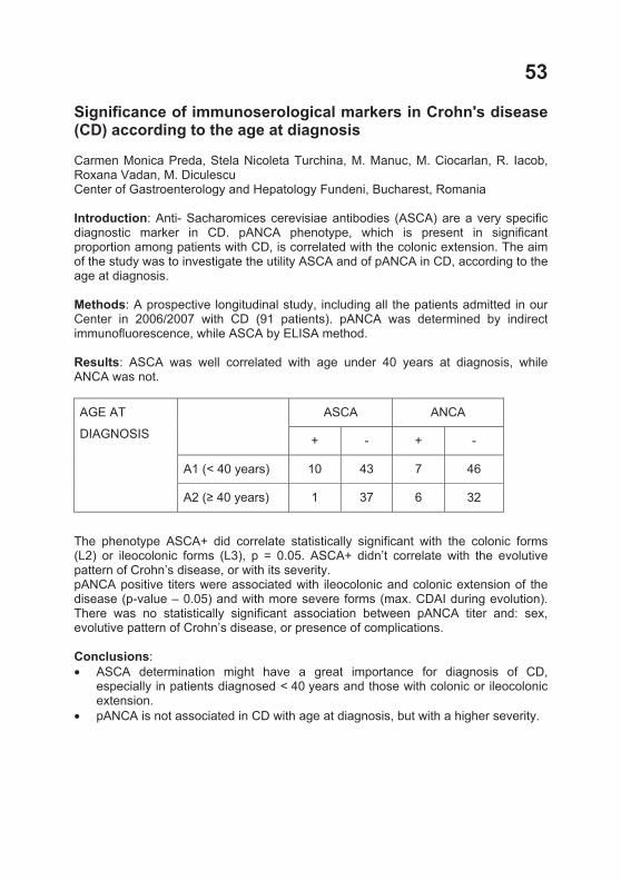
53
Significance of immunoserological markers in Crohn's disease(CD) according to the age at diagnosis
Carmen Monica Preda, Stela Nicoleta Turchina, M. Manuc, M. Ciocarlan, R. Iacob,Roxana Vadan, M. DiculescuCenter of Gastroenterology and Hepatology Fundeni, Bucharest, Romania
Introduction: Anti- Sacharomices cerevisiae antibodies (ASCA) are a very specificdiagnostic marker in CD. pANCA phenotype, which is present in significantproportion among patients with CD, is correlated with the colonic extension. The aimof the study was to investigate the utility ASCA and of pANCA in CD, according to theage at diagnosis.
Methods: A prospective longitudinal study, including all the patients admitted in ourCenter in 2006/2007 with CD (91 patients). pANCA was determined by indirectimmunofluorescence, while ASCA by ELISA method.
Results: ASCA was well correlated with age under 40 years at diagnosis, whileANCA was not.
ASCA ANCA
+ - + -
A1 (< 40 years) 10 43 7 46
AGE AT
DIAGNOSIS
A2 (≥ 40 years) 1 37 6 32
The phenotype ASCA+ did correlate statistically significant with the colonic forms(L2) or ileocolonic forms (L3), p = 0.05. ASCA+ didn’t correlate with the evolutivepattern of Crohn’s disease, or with its severity.pANCA positive titers were associated with ileocolonic and colonic extension of thedisease (p-value – 0.05) and with more severe forms (max. CDAI during evolution).There was no statistically significant association between pANCA titer and: sex,evolutive pattern of Crohn’s disease, or presence of complications.
Conclusions:• ASCA determination might have a great importance for diagnosis of CD,
especially in patients diagnosed < 40 years and those with colonic or ileocolonicextension.
• pANCA is not associated in CD with age at diagnosis, but with a higher severity.

54
Epidemiological characteristics of inflammatory bowel diseasein Albania
Skerdi Prifti1, Bledar Kraja1, Marsela Sina1, Ilir Akshia21University Clinic of Gastrohepatology, University Hospital Centre Mother Teresa,Tirana, Albania2Service of Statistics, University Hospital Centre Mother Teresa, Tirana, Albania
Introduction: Inflammatory bowel disease (IBD) including ulcerative colitis (UC) andCrohn’s disease (CD) occur with different frequencies around the world. Thecountries reporting the highest estimates of UC are North America, UK, North Europeand Scandinavia. The diagnosis of IBD can be made at any age with predominancein females. Our aim was to provide epidemiological data of IBD in Albanian patients.
Methods: A total of 110 patients diagnosed and hospitalized in the Department ofGastrohepatology, University Hospital Centre Mother Teresa, Tirana, during2005–2008, were included in the study. We analyzed age, sex and UC/CD ratio.
Results: Of the total of 110 IBD patients 104 (94.5%) were diagnosed as UC andonly 6 (5.5%) were diagnosed as CD. Therefore the UC/CD ratio was over 17/1. Themean age of the IBD patients at the time of diagnosis was 44.6 ± 10.7 years. Patientswith CD were significantly younger at the time of diagnosis (27.3 ± 2.1 vs. 45.1 ± 7.8,p < 0.01). Important female predominance was observed in UC patients (female/maleratio was 1.4).
Discussion/Conclusion: Albanian population, similarly with other South EastEuropean countries, represents a relatively low incidence of IBD. This is reflectedmainly on regard of CD which seems to be very rare in comparison with CU. Wefound higher prevalence of IBD in females. The age at the time of diagnosis of CU issimilar to other European countries, while age of onset of CD is significantly young.

55
CD83 as a marker of mature dendritic cells increased inCrohn’s disease
Anna Pryczynicz1, Katarzyna Guzinska-Ustymowicz1, Jolanta Czyzewska2,Dariusz Cepowicz3, Andrzej Kemona2Department of General Pathomorphology1, Department of Clinical LaboratoryDiagnostics2, Department of General Surgery and Gastroenterology3, MedicalUniversity of Białystok, Poland
Background/Aim: The aim of this study was to evaluated numbers of maturedendritic cells in Crohn’s disease.
Methods and results: For the study all patients underwent surgical intervention dueto failure of medical treatment. 12 patients with Crohn’s disease and 10 patients as acontrol samples were obtained.At least six files in each tissue were counted for each specimen at a magnification ofx400. CD83 expression was found in 90% patients with Crohn’s disease.The increasing of number of dendritic cells (CD83) was observed especially inheavily inflamed tissue. In the control samples CD83 expression we observed inabout 10% patients.
Conclusion: Our results demonstrate that Crohn’s disease may lead to increasednumbers of mature dendritic cells.
Address for correspondence:
Anna PryczyniczDepartment of General PathomorphologyUl.Waszyngtona 1315-089 BialystokPolandE-Mail: [email protected]

56
Renal amyloidosis complicating Crohn's disease: Report ofthree cases
A. Ouaz, L. Kallel, M. Fekih, S. Matri, J. Boubaker, A. FilaliLa Rabta Hospital, Tunis, Tunesia
Secondary amyloidosis is caused by the extracellular store of the fragment AA of thecirculatory protein in serum amyloid-A. It can complicate diseases such as familymediterranean fever, rheumatoid arthritis or Crohn's disease. Renal amyloidosis is arare but a serious complication of Crohn's disease. About 70 documented cases ofsecondary amyloidosis associated with inflammatory bowel disease have beenreported. We report three additionnel patients who have developed renal amyloidosisassociated with Crohn's disease (CD).
Case 1: Mr GS, 49-year-old is followed since 1990 for an ileo colic CD associatedwith ankylosing spondylitis. On June 91, the patient was hospitalized for explorationof a biological inflammatory syndrome (VS = 50 mm, fibraemia = 7.9 g/l) while thepatient did was asymptomatic regarding the intestine. No etiology for theinflammatory syndrome has been found and during the hospitalization, the patientdeveloped a nephrotic syndrome (proteinuria of 24 hours = 4.7 g/l). He had a renalbiopsy that showed a renal amyloidosis. The patient was treated with colchicine,spironolactone and Lasilix®. Since 2001, the patient is in terminal renal failure andhemodialysis sessions.
Case 2: Mr MS, 35 years is followed for Crohn's disease with perineal lesions treatedby antibiotics and seton drainage. In August 2007, he developed edema around theeyes, feet face and hands. In Urinary analysis a 24 hour urinary proteinmeasurement showed proteinuria at 7 g. Blood analysis showed Hypoalbuminemia(7 g/l) hypoprotidemia (50 g/l) without renal failure. Hypothyroidism and adrenalinsufficiency were also diagnosed. A lip biopsy confirmed the diagnosis ofamyloidosis. The patient was treated by Colchicine®, Aldactone®, hydrocortisone,L-thyroxine and azathioprine (for perineal lesions). The evolution was marked by theimprovement of the nephrotic syndrome.
Case 3: Mr MA, 31-year-old is followed for ileocolic CD since 1998, associated withankylosing spondylitis. In July 2002, the patient developed edema of the lower limbsassociated with inflammatory syndrome. The biology showed a nephritic syndrome.The kidney biopsy confirmed the diagnosis of secondary renal amyloidosis and thepatient was put under colchicine.
Conclusion: Amyloidosis is a rare complication during Crohn's disease. Theassociation CD – amyloidosis is a major cause of morbidity and mortality. Theabsence of any predictor of the occurrence of this complication leads us to be vigilantwith patients with Crohn's disease especially in the presence of a persistent infectionlike perineal fistulae.

57
Are probiotics useful in the treatment of the inflammatorybowel disease (IBD)
Luiza Radu1, Alina Pacurari1, B. Pacurari2, I. Romoan11University of Medicine and Pharmacy “V. Babe” Timioara, IVth Medical Clinic2Technical University, Timioara, Romania
Introduction: Probiotics are used in the therapy of inflammatory bowel disease. Oneof the pathogenic hypothesis of the IBD is the imbalance in the normal gut flora andthe excessive immune response. Probiotics like, lactobacilli species andbifidobacteria species, play a role thru the manipulation of the microflora of the gutand the stimulation of the immune system. The aim of this study is to evaluate theefficacy of probiotics in the treatment of IBD.
Methods: The study was performed in the IVth Medical Clinic of the University ofMedicine and Pharmacy “V. Babes” Timisoara, on a group of 42 patients diagnosedwith IBD. We divided the group in two subgroups. The first group received in theirdiet probiotics, and the second group followed the prescribed diet without probioticsfor a 6 months period. All of the patients also received a standard sulphasalazinetreatment.
Results: During the 6 months period, in the first group 85.7% of the patientsremained in remission, 14.3% of the patients had an acute episode. In the secondgroup, the patients with persistent remission were 57.1%, the rest of them had anacute episode of IBD. No differences between men and women or between UC andCD were observed.
Discussion/Conclusion: The adjuvant treatment with probiotics in IBD seams to beeffective in patients with UC and CD.
58
The effect of inflammatory bowel disease (IBD) duringpregnancy on long-term health and illness in children of IBDpatients – A multicenter Israeli study
Shimon Reif, Arik Alper, Daniel Rachmilewitz, Iris DotanDepartment of Gastroenterology and Pediatric Gastroenterology, Tel Aviv SouraskyMedical Center, Tel Aviv, Israel
Background: Several studies describe the effect of IBD during pregnancy onperinatal outcomes. However, limited data exists regarding the long-term effect ofmaternal IBD on the childs' health and development.
Aim: To investigate long-term morbidity or developmental defects in children of IBDmothers.
Methods: Questionnaires containing IBD mothers and child medical details andchildren’s developmental data were filled and compared to matched controls.
Results: A hundred and forty six IBD mothers (93 Crohn's disease, 53 ulcerativecolitis) were compared to 70 controls. IBD mothers had 385 children (age 16 ±10.8 years, 52.8% born after IBD diagnosis) that were compared to 144 children (age13 ± 9 years) of the control group. Mean mother age: 43 + 10 vs. 39 + 8.3 years inIBD vs. controls. Disease duration: 10.8 ± 7.5 years. A Third of the patients had IBDexacerbation, mostly (45%) during the first trimester. IBD patients had morespontaneous abortions (0.68 ± 1.2 vs. 0.33 ± 0.6 in controls, p = 0.01). Birth weightswere significantly lower in IBD mothers vs. controls' offspring: 3.13 ± 0.6 vs. 3.27 ±0.45 kg, p = 0.05. Importantly, this trend was found also in adolescence where IBDmothers offspring were significantly shorter and lighter compared to controls'offspring (1.28 ± 0.41 vs. 1.47 ± 0.33 m, 29.3 ± 21 vs. 39 ± 24 kg, p < 0.01). IBD instudy group offspring was diagnosed in 3% (p < 0.05 vs. controls) whereas atopicdermatitis was more frequent in controls' offspring (11 vs. 5%, p < 0.05). First-yearintercurrent infections were more frequent in study group offspring (p = 0.001 vs.controls). In contrast, wheezy bronchitis was more common in offspring controls(p < 0.05). More learning difficulties and attention deficit disorders (5 vs. 0.8% and4.6 vs. 0.8%, respectively, p < 0.05), and gross neurologic abnormalities (4.4 vs.0.7%), but not autism, dyslexia or abdominal pain were detected in the study groupoffspring.
Conclusions: IBD has significant and diverse short and long-term effects on thehealth and development of IBD mothers' offspring.

59
The activation of iron regulatory protein 1 dominates ironhomeostasis in inflamed intestinal epithelium
Ram Reifen, Orly Savion, Adi Kammer, Shirly Moshe, Yoram Bujanover, Batia Weiss,Esther Meyron-HoltzThe School of Nutritional Sciences The Hebrew University of Jerusalem, Rehovot,E-Mail: [email protected]; Laboratory for Molecular Nutrition, Faculty ofBiotechnology and Food Engineering, Technion, Technion City, Haifa, E-Mail:[email protected] and Pediatric Gastroenterology Shiba Medical Center, Tel-Aviv, Israel
Introduction: In IBD, anemia is a common problem with prevalence ranging between17% and 50%. It is related with a higher score of disease activity, loss of weight,impaired physical activity and a poor growth in children. The causes are thought toinclude, a chronic blood loss from the colon and intestine, reduced absorption of ironand, a suppression of erythropoietin production. Iron, which is one of the mainstaytherapies of anemia in IBD is highly reactive and has the capacity to accept anddonate electrons readily. Since patients with IBD have increased production ofoxygen reactive species, there is some concern that oral iron supplementation mayexacerbate inflammation and tissue damage by hydroxyl radicals formed fromhydrogen peroxide via the Fenton reaction. There is evidence that ironsupplementation induces inflammation in animal models of colitis. Also, iron-chelators have been shown to ameliorate oxidative stress and inflammation asshown in colitic rats and colonic biopsies from patients with ulcerative colitis. Therecent report of CD patients having elevated blood-hepcidin levels and decreasedintestinal transepithelial iron import suggest a contribution of hepcidin to nutritionaliron uptake in AI and CD. Elevated hepcidin levels reduce the level of the ironexporter ferroportin, which affects two central locations of the iron cycle: 1. Iron fromdegraded erythrocytes will be retained in the RE system and will re-enter the blood ata slow pace. 2. Dietary iron imported to the enterocyte may never reach the blood,due to low levels of iron exporter. Intravenous iron administration in combination witherythropoietin has been shown to improve the anemia of CD patients, possiblybecause this treatment eludes both locations of iron retention. Iron regulatoryproteins (IRP) are cytosolic proteins, that modulate the expression of proteinsinvolved in the transport and storage of iron, through binding to their m-RNAtemplates during iron deficiency. IRP binding generates upregulation of the ironimporter transferrin receptor 1 (TfR) and downregulation of the iron storage proteinferritin and possibly the iron exporter ferroportin. At physiologic O2 conditions IRP2 isthe dominant regulator as IRP1 sensing depends on the stability of its iron sulfurcluster and physiologic O2 concentration is too low for this cluster to act as a sensor.
Methods and results: Both, tissues obtained from CD patients and an in-vitro modelof inflammation were used. We compared the IRP1 and IRP2 RNA binding activityfrom biopsies of CD patients to non-inflamed controls and found IRP1-RNA bindingactivity elevated in patients. In agreement, levels of ferritin were decreased. We wereable to mimic this IRP1 mediated misregulation of iron homeostasis in Caco-2 cells(intestinal epithelial cell-line) by creating an inflammation condition using an NO

donor. We found that inflammation induced IRP-1 activation causes elevatedtransferrin receptor and decreased ferroportin levels suggesting iron accumulation inthe inflamed tissue. This phenomenon was more significant in cells grown at3% oxygen then 21% oxygen.
Conclusion: These results support our model of an inflammation induced imbalanceof iron homeostasis, mediated by IRP1 activation. Caco-2 cells are a promisingcandidate for a cell model for iron homeostasis during inflammation, supplyingadditional evidence that the preliminary observation in human biopsies of IRP1activation in CD patients is the mechanism for reactive iron accumulation andacceleration of tissue damage by reactive iron in CD.

60
Characteristics of the patients with inflammatory boweldiseases in Romania
E.C. Rezi, A. FraticiuDepartment of Gastroenterology, Emergency Clinical County Hospital, Sibiu,Romania
Introduction: The aim of this study was to establish the prevalence andcharacteristics of the patients with inflammatory bowel diseases (IBD) in ourgeographical area.
Methods: 911 colonoscopies and rectosigmoidiscopies which were performed duringthe last 4 years in the Gastroenterological Department from the Clinical CountyHospital from Sibiu, Romania.
Results: From the total number of 911 patients, 1.53% patients were diagnosed withIBD (21.42% of them with Crohn’s disease and 78.58% with ulcerative colitis). Themedium age of the patients with IBD was 55.92 ± 13.35 years. The genderdistribution was 57.14% women and 42.86% men. 42.86% were from rural areas,while 57.14% live in urban areas. 54.54% from the patients with ulcerative colitis hadproctosigmoiditis and 45.46% had pancolitis. 3 patients with ulcerative colitis werediagnosed in an active stage (moderate to servere disease). 64.28% of the patientsreceived maintenance therapy with mesalazine (1.5 g/day orally or suppositories). Asassociated disorders, we have found out that 21.42% of the patients with IBD hadskin lesions, one patient presented arthritic complaines and one patient had psychicdisorders. 2 patients were obese, three patients were hypertensive and one patienthad diabetes mellitus. Only a patient with ulcerative colitis developed high-gradedysplasia.
Discussion/Conclusion: Inflammatory bowel diseases have a low incidence inRomania; ulcerative colitis is three times more frequently than Crohn’s disease. IBDhave a higher incidence in women and in urban areas. The risk for IBD relatedcolorectal cancer is low in our area, possibly due to the optimized treatment in thelast decade.

61
Colonoscopy in elder versus younger patients –A retrospective study comparing the risk of developing IBDand colorectal cancer at two different age groups of patientsfrom Sibiu, Romania
E.C. Rezi, A. FraticiuDepartment of Gastroenterology, Emergency Clinical County Hospital, Sibiu,Romania
Introduction: Colonoscopy is currently the best diagnostic modality for evaluatingcolonic diseases like inflammatory bowel diseases (IBD) and colorectal cancer (CRC)but studies of its use in the very elderly are limited.
Methods: We have retrospectively analyzed all colonoscopies andrectosigmoidiscopies which were performed during the last 4 years in theGastrenterological Department from the Clinical County Hospital from Sibiu,Romania. The aim of this study was to establish the prevalence of IBD and CRC atelderly people (over 70 years of age) compared with those less than 70 years of age.The results were statistically analyzed using the relative risk (RR).
Results: The medium age for the whole group was 61.31 ± 14.16 years. From thetotal number of 911 patients, 1.53% patients were diagnosed with IBD (21.42% withCrohn’s disease and 78.58% with ulcerative colitis) and at 9.33% there was acolorectal cancer found. The studied lot was divided into 2 groups: a group formed by318 patients over 70 years, and a group formed by 593 patients which were less than70 years of age. The gender distribution was equally in both groups. The relative riskof developing a colorectal cancer at the patients older than 70, compared with thoseless than 70 was 1.73. The relative risk of finding an inflammatory bowel disease atolder patients compared with younger ones was 0.5, so this group had a lower risk ofIBD.
Discussion/Conclusion: In our area, inflammatory bowel diseases have a lowincidence; ulcerative colitis is three times more frequently than Crohn’s disease. Theinflammatory bowel disease is two times more likely to appear in the youngerpatients than in the elder ones. In contrast, the colorectal cancer is almost two timesmore frequently in elderly patients.

62
Correlation between ileocolonoscopic and video capsuleexamination in ankylosing spondylitis
M. Rimba1,2; M. Marinescu1,2, S. Caraiola1,4, M. Pârvu3, C. Băicu4, S. Bucurică1,2,C. Tănăsescu1,4, M.R. Voiosu1,21“Carol Davila” University of Medicine and Pharmacy, Bucharest, Romania2Gastroenterology Department, Colentina Clinical Hospital, Bucharest, Romania3Rheumatology Department, Colentina Clinical Hospital, Bucharest, Romania4Department of Internal Medicine, Colentina Clinical Hospital, Bucharest, RomaniaN.B. This research is financed by a Romanian governmental programme (contract_UEFISCSU/Idei No. 320/01.10.2007; NCT00768950)
Introduction: An intriguing relationship exists between gut and synovium in patientswith spondyloarthropathy.In ankylosing spondylitis (AS), macroscopic signs of gut inflammation have beenfound in 29% to 49% of cases, as seen by ileocolonoscopy.Ileocolonoscopic findings have prognostic importance:− the presence of gut inflammation is related to a more severe clinical andradiological expression of AS;
− 20% of AS patients with an initial subclinical chronic gut inflammation developclinical-overt IBD.
Methods: 10 patients with AS (as classified using the modified New York criteria)and no signs of inflammatory gut involvement were investigated using colonoscopywith intubation of the terminal ileum followed by video capsule examination; thepresence of macroscopic small bowel inflammatory lesions was recorded.
Results: Of the 10 patients, 7 were found to have macroscopic inflammatory lesionsin the small bowel at video capsule examination. Of these, only 2 were found to havemacroscopic lesions of the terminal ileum at ileocolonoscopy; the ileoscopy wasnormal in the other 8 patients.
Discussion/Conclusion: There's a weak correlation between lesions found atileocolonoscopy and video capsule examination in patients with AS. It is probablethat performing only ileocolonoscopy, an inflammatory subclinical involvement of thesmall bowel is overlooked in more than half of the patients.Taking into consideration the fact that abnormalities in the gut could have majorconsequences in the AS patients, and that early treatment of the gut inflammationcould prevent disease evolution when the appropriate drugs become available, smallbowel video capsule examination could become necessary for the bettermanagement of these patients.

63
Profile of Belgian pediatric Crohn’s disease subjects (2): Snapshot at diagnosis
M. Rogalidou1, I. Hoffman2, T. Mahler1, S. Staelens1, S. Van Biervliet3, M. Scaillon4,P. Bontems4, I. Paquot5, F. Bury5, S. Collinet5, W. Arts6, B. Hauser7, F. Smets8,E. Sokal8, P. Alliet9, E. Janssens9, O. Bauraind10, I. Etienne11, G. Veereman1Pediatric Gastroenterology Departments of 1Queen Paola Children’s Hospital,Antwerp, 2UZ Gasthuisberg, Leuven, 3UZ Gent, 4University Children’s Hospital QueenFabiola, Brussels, 5CHC Clinique de l Esperance, Liege, 6ZOL, Genk, 7UZ VUB,Brussels, 8UCL St. Luc, Brussels, 9Virga Jesse Hospital, Hasselt, 10Clinique St.Pierre, Ottignies, 11CHR de la Citadelle, Liege, Belgium
Introduction: Recent reports on pediatric IBD patients show that early onset Crohn’sdisease (CD) is characterized by a phenotype with extensive, severe andcomplicated course1,2.
Methods: The IBD working group of Bespghan recently established a registry ofBelgian pediatric CD. We report data on the first 100 patients at the time ofdiagnosis.
Results: Mean age at diagnosis was 11.7 ± 2.7 yrs corresponding with mean boneage 11.6 ± 3.3 yrs. The mean z-score for height was -1.1 ± 1.3. PCDAI was retrievedfrom 58 charts: 3 scored less than 10, 18 had mild to moderate disease (10-30) and37 rated severe (> 30). The PCDAI at diagnosis was correlated negatively with birthweight, (p = 0.0473) and birth height (p = 0.0407) not with age at presentation orsymptom duration. Diagnosis was based on endoscopy and histology in 87.7% (inaccordance with Porto criteria3), 97% had (ileo)colonoscopy, 68.7% OGD, 1%sigmoidoscopy. Imaging studies: 61.6% of patients had abdominal US, 25.3% smallbowel enteroclysis, 14.1% CT and 9.1% MRI. White cell scan was used in 2% andsmall bowel capsule in none. Based on endoscopic exploration the most frequentlyinvolved intestinal area was the ileocolon (52%). Histological diagnosis was possiblein 72.7% patients undergoing OGD.
Discussion/Conclusion: In conclusion: At diagnosis children presented withextensive involvement of the GI tract but the small bowel remains mostly unexplored.The severity of disease at presentation may be related to intrauterine growth. Datacollection on disease progression and therapy is being conducted.
Bespghan: Belgian Society for Pediatric Gastroenterology Hepatology and Nutrition

References:
1. Vernier-Massouille G., et al. Natural history of pediatric Crohn's disease: Apopulation-based cohort study. Gastroenterology. 2008 Oct; 135 (4): 1106–1113.
2. Van Limbergen J., et al. Definition of phenotypic characteristics of childhood-onset inflammatory bowel disease. Gastroenterology. 2008 Oct; 135 (4):1114–1122.(Comment: Kugathasana S., Cohenb S. Searching for new clues in inflammatorybowel disease: Tell tales from pediatric IBD natural history studies.Gastroenterology. 2008 Oct; 135 (4):1038–1041).
3. IBD Working Group of the European Society for Paediatric Gastroenterology,Hepatology and Nutrition. Inflammatory bowel disease in children andadolescents: recommendations for diagnosis--the Porto criteria. J. Pediatr.Gastroenterol. Nutr. J Pediatr Gastroenterol Nutr. 2005 Jul; 41 (1): 1–7
64
Profile of Belgian pediatric Crohn’s disease subjects (1):Demography and background of the first 100 patients
M. Rogalidou1, I. Hoffman2, T. Mahler1, S. Staelens1, S. Van Biervliet3, M. Scaillon4,P. Bontems4, I. Paquot5, F. Bury5, S. Colinet5, W. Arts6, B. Hauser7, F. Smets8,E. Sokal8, P. Alliet9, E. Janssens9, O. Bauraind10, I. Etienne11, G. Veereman1Pediatric Gastroenterology Departments of 1Queen Paola Children’s Hospital,Antwerp, 2UZ Gasthuisberg, Leuven, 3UZ Gent, 4University Children’s Hospital QueenFabiola, Brussels, 5CHC Clinique de l Esperance, Liege, 6ZOL, Genk, 7UZ VUB,Brussels, 8UCL St. Luc, Brussels, 9Virga Jesse Hospital, Hasselt, 10Clinique St.Pierre, Ottignies, 11CHR de la Citadelle, Liege, Belgium
Introduction: Epidemiological observations seek to identify factors and parametersthat may influence the evolution and prognosis of CD.
Methods: The IBD working group of Bespghan recently established a registry ofBelgian pediatric CD with ulterior goal to study demographical data, treatment anddisease course over 5 years.
Results: We report on the background and history of the first 100 inclusions. Thegroup counts 60 boys and 40 girls, 92 are Caucasian. Birth history reveals that 85%were delivered by vaginal route, mean gestational age was 38.4 wks ± 2.3, and19 patients were premature. The mean birth weight was 3.3 kg ± 0.6 and height49.8 cm ± 3.0. Data on neonatal feeding are available from 77 infants: 67.6% werebreastfed for a mean duration of 11.4 wks. The majority (60%) of formula used wascow’s milk based. There was a suspicion of food allergy in 20.9% infants (33.3% tocow’s milk). In their prior history 18.5% needed dietary restrictions and 38.5%underwent surgery. Family history was positive for IBD (CD and Ulcerative colitis) in52.3% and a 1st degree relative in 15.4% or a 2nd degree relative in 27.7% with CD.Family history for autoimmune diseases (exc diabetes & hepatitis) was positive in49.2%.
Discussion/Conclusion: In conclusion: in the Belgian pediatric CD population priordisease history appears unremarkable. Positive family history for IBD & autoimmunediseases is common. Further study will compare phenotypes with disease courseand factors that may influence severity and prognosis.
Bespghan: Belgian Society for Pediatric Gastroenterology Hepatology and Nutrition

65
Methotrexate for pediatric IBD: Induction and maintenance ofremission in a regional cohort study
P. Rogers1, A. Tybulewicz1, D. Hoole1, J. Satsangi2, P.M. Gillett1, D.C. Wilson31Paediatric Gastroenterology, Royal Hospital for Sick Children, Edinburgh, UnitedKingdom2Adult Gastroenterology, Western General Hospital, Edinburgh, United Kingdom3Child, Life and Health, University of Edinburgh, Edinburgh, United Kingdom
Background and aim: There is limited data on the use of Methotrexate (MTX) forinduction and maintenance of remission in children with Crohn’s Disease (CD). Wereviewed our experience of induction and maintenance of remission.
Methods: A cohort study of 244 patients diagnosed with early-onset (< 18 years ofage) IBD and managed in a UK regional centre 1/8/97–31/12/07 and treated withMTX (16 week induction course of s/c MTX (15 mg/m2) followed by the equivalentoral dose). PCDAI was used to measure disease activity during treatment; PCDAI≤ 15 indicated remission and a score > 30 indicated severe disease.
Results: 122 with CD were treated with AZA/6-MP; 53 children received a MTXinduction course. These 53 patients had received MTX at 14.0 yrs (6.8–17.9) years.The median PCDAI at commencement was 35 (IQR 20–37.5). 41 (78%) patientsentered remission at 11 (3–16) weeks. 2 of 41 had recalcitrant OFG, so 39 withluminal CD had a median (range) duration of follow-up of 85 (26–310) weeks.20 (51%) maintained long-term remission with no relapse; 7 (18%) had a singlerelapse of which 6 (15%) re-entered remission, and 12 (31%) had > 1 relapses. Themedian (range) time to first relapse was 39 (20–181) weeks. On MTX treatment, 24(46%) had nausea/vomiting, 18 (34%) had raised ALT, 1 had shingles and none hadbone marrow toxicity.
Conclusions: MTX appears to be effective in inducing (78%) and maintaining (51%)remission in patients who have failed to remit on AZA, and, appears to be welltolerated and safe.

66
Early atherosclerosis in inflammatory bowel disease patients
Corina Serban1, Lelia Susan1, Alina Pacurari1, C. Banciu1, Versavia Ancusa2,Germaine Savoiu1, Ioan Romosan11UMF, Timisoara, Romania2UPT, Timisoara, Romania
Introduction: Patients with inflammatory bowel disease (IBD) have an increased riskof thrombotic complications. Arterial and venous system may be involved. Moreover,mesenteric microvascular thrombosis has been hypothesised as a contributing factorin the pathogenesis of IBD. The main objective of this study was to evaluate thepresence of atherosclerosis in a group of 78 IBD patients without the classicalcardiovascular risk factors comparative with a group of 70 control subjects.
Methods: Patients aged > 45 years, with a history of cardiovascular disease andknown risk factors for atherosclerosis were excluded from the study. All the patientsunderwent complete clinical and paraclinical exams. We measured inflammatorymarkers (erythrocyte sedimentation rate, fibrinogen, C-reactive protein) in allsubjects. Intima-media thickness (IMT) was measured by carotid ultrasonographyproximal to the carotid bifurcation over both right and left common carotid arteries.
Results: Inflammatory markers (erythrocyte sedimentation rate, fibrinogen,C-reactive protein) were significantly elevated in IBD patients compared withcontrols. Common carotid artery intima-media thickness was significantly higher ininflammatory bowel disease patients (0.65 ± 0.15 mm) compared with controls(0.51 ± 0.06 mm).
Discussion/Conclusion: The vasculature plays a central role in human IBD andmay contribute to pathogenesis through thrombotic, ischemic, or inflammatorymechanisms. The patients with IBD may have an increased risk of earlyatherosclerosis that healthy control, a finding that requires further study.Inflammatory markers can be indicators of disease severity or activity of IBD.

67
A study of colorectal cancer in inflammatory bowel disease
Corina Serban1, Madalin Munteanu2, Lelia Susan1, C. Banciu1, Alina Pacurari1,Raluca Dumache1, Germaine Savoiu1, Ioan Romosan11UMF, Timisoara, Romania2Clinical County Hospital, Timisoara, Romania
Introduction: Inflammatory bowel disease (IBD) characterized by conditions such asulcerative colitis and Chron's disease, increases the risk of developing colorectalcancer (ulcerative colitis increases risk more than Chron's disease). The mainobjective of this study was to analyze colorectal cancer in IBD in a group of patientshospitalised between January 2004 and January 2008 in Medical Clinic CFTimisoara.
Methods: We investigated 152 patients with IBD (40 women and 112 men, agedbetween 45 ± 30 years). The diagnosis of IBD was based on the combination ofclinical, endoscopic and histologic findings.
Results: Mean age at the time of diagnosis of cancer was 46 years. 5 patients haddocumented IBD for less than eight years before their cancer diagnosis. 40% ofthese patients had the colonoscopy to investigate changes in IBD symptoms or signsand the other 50% underwent endoscopy as routine surveillance. For 10% of thepatients, operated for worsening symptoms, the carcinoma was detected in thepathological specimen. Most of the patients did not have a preoperative diagnosis ofdyplasia. The tumors were: mucinous (23%), multicentric (17%) and 60% werelocated distal to the splenic flexure.
Discussion/Conclusion: The key risk factors that can contribute to colorectal cancerin people with IBD include: having IBD for 10 years or longer; having IBD involvingthe whole colon (pancolitis); having primary sclerosing cholangitis; and having afamily history of colorectal cancer. For patients with IBD, the main way to detectcolorectal cancer is through regular colonoscopies every year or two.

68
Arachidonic acid increases ulcerative colitis risk – Aprospective cohort study in EPIC-Denmark using biomarkerdata
P.S.A. de Silva*1, A. Olsen2, A. Tjonneland2, K. Overvad3, E. Berg Schmidt4,A.R. Hart51Department of Gastroenterology, Norfolk and Norwich University Hospital, Norwich,United Kingdom, 2Institute of Cancer Epidemiology, Danish Cancer Society,Copenhagen, 3Departments of Cardiology and Clinical Epidemiology, AarhusUniversity Hospital, 4Department of Cardiology, Aalborg Hospital, Aalborg, Denmark,5School of Medicine, University of East Anglia, Norwich, United Kingdom
Introduction: Arachidonic acid (AA), an n-6 polyunsaturated fatty acid, is acomponent of the phospholipid bilayer of colonocyte membranes and is metabolizedto pro-inflammatory eicosanoids PGE2 and LTB4. The latter are found in increasedconcentrations in the mucosa of patients with ulcerative colitis (UC). We aimed tomeasure for the first time, in a prospective cohort investigation, if increased AAconcentration in adipose tissue biopsies increases risk of developing incident UC.
Methods: 57,053 men and women, aged 50–64 years, were recruited in the EPIC-Denmark Study (European Prospective Investigation in to Cancer) between1993–1997. Adipose tissue biopsies were taken at recruitment and AA fatty acidconstituents measured. The cohort was monitored upto June 2004 to identifyindividuals who developed UC. Each case had 4 age and gender matched controls,and AA levels divided into quartiles. Unconditional logistic regression was used tocalculate univariate odds ratios for AA levels and multivariate analysis performedadjusted for cigarette smoking and marine n-3 PUFAs in adipose tissue.
Results: 34 subjects (15 w, 19 m) developed incident physician confirmed UC.Median age at diagnosis 58.8 years, (53.3–71.1 years). Median time betweenrecruitment and diagnosis 3.7 years (1.7–7.1 years). The highest adiposeconcentration of AA was associated with an odds ratio for UC of 12.3 (95% CI2.6–57.8, p = 0.002) with a statistically significant trend across quartiles (OR for trend= 1.9, 95% CI 1.3–2.8, p = 0.001). Adjustment for co-variates did not affect themagnitude of the findings.
Discussion/Conclusion: Initially well participants, who have higher adipose tissueconcentrations of AA, which represent dietary intake, have a significantly higher riskof developing UC compared to those with lower evels. This supports previous datafrom dietary questionnaire-based studies showing n-6 PUFAs may be involved in UCetiology. Lowering dietary intake of AA may prevent the UC development.

69
Mucosal healing and complete regression of transmuralinflammation with doubling the third infliximab induction dosein refractory Crohn colitis
Małgorzata Sładek, Agnieszka Świat, Izabela Herman-Sucharska*,Stanisław Pieczarkowski, Zofia Grzenda-Adamek, Krzysztof FyderekDepartment of Pediatrics, Gastroenterology and Nutrition, Jagiellonian UniversitySchool of Medicine, *Medical Diagnostic Center – Voxal, Krakow, Poland
Introduction: The recommended infliximab (IFX) initial dosage in Crohn disease.(CD) is 5 mg/kg body weight in an induction regiment at 0, 2, and 6 weeks. Primarynon-response to IFX can be determinate after the second dose and patients notresponding to the initial two infusions should be discontinued from further IFXtherapy. IFX dose intensification is currently recommended only for the lost of theresponse to maintenance therapy.
Methods: We report the case of 15-years-old girl with fulminant steroid refractoryCrohn colitis who was successfully treated with the third induction IFX doseincreased up to 10 mg/kg body weight.An otherwise healthy 15-year-old girl presented with abdominal pain and intensebloody diarrhea for one week was diagnosed according to Porto criteria withmoderate Crohn disease (A1, L2 + L4, B1). Pediatric Crohn Disease Activity Index(PCDAI) was 35. Because of the lack of the response to metyprednisolon (2 mg/kg)and worsening disease activity (PCDAI 51) IFX was introduced at recommendedschedule. After the second IFX dose, despite clinical response intense blood lostpersisted. On control colonoscopy severe mucosal lesions was found and transmuralcolon inflammation was present on MRI scan. Then, the third induction dose wasincreased up to 10 mg/kg body weight.
Results: Doubling the third IFX induction dose resulted in the rapid mucosal healingand complete regression of transmural inflammation and clinical remission (PCDAI< 10), thereby avoiding colectomy.
Discussion/Conclusion: This is the first report of the successful IFX induction doseintensification in CD and the first report of the IFX induction dose intensification inrefractory Crohn colitis. Modification of the induction remission strategy by doublingthe third dose may be considered in case of not full response, before concerningpatient as primary no responder.

70
A two-year longitudinal study of the anemia associated withinflammatory bowel disease
Lelia Susan1, Corina Serban1, C. Banciu1, Alina Pacurari1, V. Ancusa2, SmarandaGotia1, Ioan Romosan11UMF Timisoara, Romania2UPT Timisoara, Romania
Introduction: Anemia affects many patients with inflammatory bowel disease (IBD).A lot of factors contribute to anemia in patients with IBD, including blood loss,inadequate intake/absorbtion and the underlying inflammatory disease process. Theaim of this study was to analyze the association between anemia and IBD in patientswith IBD, from Clinical Medical CF Timisoara in a two years period.
Methods: From October 2006 to October 2008, we investigated 78 IBD patients,aged between 45 ± 15 years. By all, 48 of the patients (61.53%) were diagnosed withulcerative colitis (UC) and 30 of them with Crohn’s disease (38.46%). All the patientsunderwent complete clinical and paraclinical tests. The values of Ht, the number ofreticulocytes, the Hb erytrocytes indices, transferring saturation (TS), the totalcapacity of iron bounding (CTLF) and the ferritin were determined in all patients.
Results: 24 patients with ulcerative colitis (50%) and 8 patients with Crohn’s disease(26.66%) had anemia (Hb lower than 13.5 g/dl in men and lower than 11.5 g/dl inwomen). The mean Hb value was 8.1 ± 1.4 and the mean value of Ht was 26 ± 3.3 inthe patients with anemia. The number of reticulocytes and the values of CTLF,ferritin, TS were decreased in patients with anemia and IBD.
Discussion/Conclusion: Our study suggested that there is a relationship betweenanemia, disease severity and quality of life in patients with IBD. The therapy witherythropoietin is useful in treating the anemia associated with Crohn’s disease andulcerative colitis.

71
Predictive value of serologic markers in inflammatory boweldisease
Lelia Susan1, Corina Serban1, C. Banciu1, Alina Pacurari1, V. Ancusa2,Germaine Savoiu1, Ioan Romosan11UMF Timisoara, Romania2UPT Timisoara, Romania
Introduction: Perinuclear antineutrophil cytoplasmic antibodies (pANCA) and anti-Saccharomyces cerevisiae antibodies (ASCA) are proposed to be specific markersfor ulcerative colitis (UC) and Crohn's disease (CD). Their prevalence in patients withinflammatory bowel disease (IBD), however, it is unclear. We studied the prevalenceof these serologic markers in a group of IBD patients,
Methods: 126 patients with IBD (UC, n = 88 and CD, n = 38) hospitalised betweenOctober 2005 until October 2008 in Clinical Medical CF Timisoara participated in thisstudy. All the patients underwent complete clinical, paraclinical, haematological,imagistic and histological exams.
Results: 10 of the patients with CD (26.31%) were ASCA-positive and 27 of UCpatients were pANCA-positive (30.68%). Positive ASCA was more frequent in CDpatients with stricturing or penetrating complications than in those with inflammatorybehavior at diagnosis. Moreover, the presence of ASCA was associated with an atleast twice higher risk of evolving more severe disease behavior during follow-up. InUC, pANCA expression was related to female gender and the use of azathioprineand in CD, to colon-limited disease and age ≥ 45 years at diagnosis.
Discussion/Conclusion: The prevalence of ASCA in CD and pANCA in UC appearsmarkedly lower than in referral-based populations. Even with the low prevalence, ourstudy gives further support to the role of ASCA and pANCA as markers forinflammatory bowel disease.

72
Fertility and outcomes of pregnancies fathered by malepatients exposed to thiopurines
Carlos Teruel1, Antonio López San Román1, Carlos Taxonera2, Alicia Algaba3,Javier Gisbert4, José Pérez Calle5, María Martín Arranz6, Manuel Van Domselaar1,Jesús Estellés2, Fernando Bermejo3, Pablo Linares4, Pilar López Serrano51Hospital Ramón y Cajal; 2Hospital Clínico San Carlos; 3Hospital de Fuenlabrada;4Hospital de la Princesa; 5Fundación Hospital Alcorcón; 6Hospital La Paz, Madrid,Spain
Introduction: Immunomodulators are widely used as maintenance treatment ofinflammatory bowel disease (IBD). Data regarding their safety in the course ofpregnancy when fathers are exposed at the time of conception are limited. Our aimwas to evaluate outcomes of pregnancies in which fathers had been takingmercaptopurine or azathioprine at the time of conception.
Methods: A series of male patients followed in IBD clinics in the Community ofMadrid, Spain, was studied. Any exposure to thiopurines during the 3 monthspreceding conception was considered. Controls consisted of pregnancies fathered bymen with IBD who had never taken thiopurines or had abandoned them more than3 months before conception.
Results: There were 44 pregnancies in the group exposed to thiopurines(mercaptopurine 8, azathioprine 36) and 74 in the control group. There were nosignificant differences regarding negative pregnancy outcomes (preterm delivery,abortion, low weight at birth, malformations) and no infant malignancies wereregistered. The proportion of patients who needed more than a year to achieve apregnancy was also similar, although higher in the thiopurine exposed group.
Conclusion: Our data suggest that paternal exposure to thiopurines does notinfluence pregnancy outcomes or male fertility.

73
Experience of infliximab therapy for refractory ulcerative colitisin a district general hospital
I. Tetlay, D. Sadigh, D. Hughes, R. Turner, J. O’Brien, G. Bray, M. McStaySouthend University Hospital, High Wycombe, UK
Introduction: Tumour necrosis factor-α (TNF-α) is an important cytokine involved inthe pathogenesis of inflammatory bowel disease. The benefits of monoclonalantibody to TNF-α, infliximab, in Crohn’s disease are established. We investigated itsefficacy in ulcerative colitis (UC).
Methods: Since 2005, 23 patients (aged 14–83 years) with UC refractory totreatment with standard combination therapy have been treated with infliximab inSouthend Hospital. The patients’ notes were reviewed.
Results: Of 23 patients, 16/23 (70%) had moderately active, and 7/23 (30%) hadseverely active UC requiring intravenous steroids.In patients with moderately active UC: 6/16 (36%) remain in remission on 8-weeklymaintenance infusions (5 mg/kg) [median follow-up 24 months (range 7–28)];3/16 (19%) achieved remission on induction therapy and remain in remission onazathioprine/6 mercaptopurine [median follow-up 24 months (range 12–26)];6/16 (38%) achieved remission with induction therapy but relapsed after a mediantime of 6 months (range 3–16) requiring colectomy; 1/16 (6%) had secondary loss ofresponse after 18 months of maintenance infusion requiring colectomy.In patients with severely active UC: 3/7 (43%) achieved remission after inductiontherapy and remain in remission on azathioprine/6 mercaptoprine [median follow-up24 months (range 6–24)]; 2/7 (29%) achieved remission with induction therapy butsubsequently relapsed (mean duration 12 months) requiring colectomy; 2/7 (29%) ofpatients had no response to infliximab and underwent colectomy.No side-effects were reported. The 2 patients with severe UC who did not respond toinfliximab died from post-surgical complications within 30 days.
Discussion/Conclusion: Our experience suggests acceptable response rates,colectomy rates and side-effect profile of treatment with infliximab for moderate andsevere UC.
74
Ulcerative colitis in young patients – Epidemiological andclinical course in North-Eastern Romania county
Elena Toader1, Liliana Croitoru2, Oana Arhip3, Rodica Mihăilă41“Gr.T. Popa” University of Medicine and Pharmacy Iai, Institute of Gastroenterologyand Hepatology Iai, Romania2District Hospital, Suceava, Romania3District Hospital, Botosani, Romania4District Hospital, Barlad, Romania
Background: Ulcerative colitis (UC) represents a distinct entity of InflammatoryBowel Disease (IBD) with unknown etiology which affects young people. UC isubicvitar disease with an incidence rate that varies greatly worldwide.
Introduction: The aim of the present study was to evaluate the frequency andclinical course of UC in young patients from North-Eastern (NE) Romania county.
Methods: There were identified 746 UC cases according the diagnosis criteria forUC during 1988–2007. In 485 cases (65%) the onset of the disease was made in theyoung patients (< 50 years).
Results: UC incidence in the young patients from NE Romania county during1988–2007 was 2.04/105 inhabitants. Extraintestinal manifestations were noted in13% patients and family history of UC was found in 1.4% of cases. In our regionp-ANCA were found in 18% patients with UC. Extension of disease as proctitis werefound in 25.3% of cases, lift-sided colitis in 22.6% cases and pancolitis in 22.6%cases. Severity of the first attacks evaluated according to Truelove and Witts criteriawas mild in 38.2% cases, moderate in 40.7% cases and severe in 21.1% cases.
Discussion/Conclusion: The low incidence of UC in NE Romania(2.04/105 inhabitants) with high frequency in young patients confirms theepidemiological trend of the disease in this part of Europe. Socio-demographiccharacteristics, clinical features, disease extent and clinical course of UC are thesame in low- as well as high-incidence area.

75
Perinuclear anti-neutrophil cytoplasmic antibodies in patientswith inflammatory bowel disease and their first degree inNorth-Eastern Romania areas
Elena Toader1, Cecilia Durnea21“Gr.T. Popa” University of Medicine and Pharmacy, Institute of Gastroenterology andHepatology, Iai, Romania2Institute of Public Health, Immunology Department, Iasi, Romania
Background: Perinuclear anti-neutrophil cytoplasmic antibodies (p-ANCA) are morecommon in patients with ulcerative colitis (UC) than in patients with Crohn’s disease(CD), but its prevalence depends on the population being studied and the methodemployed for its detection.
Introduction: The aim of the present study is to determinate the prevalence ofp-ANCA in inflammatory bowel disease (IBD) patients and their first degree in NERomania area
Methods: In this study, we investigated the prevalence of p-ANCA as detected byELISA, in the sera of 66 patients with IBD (44 with UC and 22 with CD) and 22 firstdegree relatives. We also correlated the presence of this antibody with diseaseactivity and extent, extraintestinal complications and therapy. 26 healthy individualscomprised the control group.
Results: p-ANCA was detected in 19% of the patients with IBD. 12 of 44 patientswith UC were positive for p-ANCA and only one of 22 patients with CD was p-ANCApositive. Of the 12 p-ANCA positive UC patients 2 cases were found to haveproctosigmoiditis, 6 cases had left-sided colitis and 4 cases had pancolitis. Therewas no correlation between the presence of this antibody and any of the studiedclinical variables. No person of the first degree and control group presented positivetest.
Discussion/Conclusion: The prevalence of p-ANCA in NE Romania patients withIBD (19%) is lower than that in the Western population. The negativity of p-ANCA inall first degree relatives of NE Romania IBD patients should be further elucidated

76
Extraintestinal manifestations in pediatric patients withinflammatory bowel disease
M. Tolín Hernani, E. Barredo Valderrama, G. Alvarez Calatayud, M. Crespo Medina,J.L. Morales, B. Huidobro Fernández, M. Hernando, C. Sánchez SánchezHospital Gregorio Marañón, Madrid, Spain
Introduction: The objective of our study was to describe the frequency ofdevelopment of extraintestinal manifestations (EIM) in pediatric patients withinflammatory bowel disease (IBD) in our hospital in last 20 years.
Methods: Retrospective descriptive study including 50 patients followed in pediatricgastroenterology service with IBD (18 ulcerative colitis [UC], 32 Crohn’s disease[CD]) and the presentation of EIM.
Results: EIM appeared in 30% (15 children) of followed patients. There weredifferences comparing EIM between UC and CD: 16% of patients with UC presentEIM and 37% of children with CD. We found the next EIM in our patients: Articularmanifestations 33%: artralgias (n = 4: UC 2/CD 3), sacroileitis (n = 1: CD).Dermatologic manifestations 13%: erythema nodosum (n = 2: 1 UC/1 CD).Ophthamologic manifestations 13%: uveitis (n = 2: CD). Hepatobiliary/pancreaticmanifestations 13%: sclerosing cholangitis (n = 1: CD); autoimmune pancreatitis(n = 1: CD). Endocrinologic manifestations 13%: hyperparatiroidism (n = 1: UC),hypotiroidism (n = 1: CD). Other manifestations 13%: idiopathic thrombocytopenicpurpura (ITP) (n = 1: CD), Wolff-Parkinson-White: (n = 1: CD).
Discussion/Conclusion: Articular disease was the most prevalent of extraintestinalmanifestations of IBD. Systemic manifestations are more frequent in patients withCrohn’s disease than in those with the diagnosis of ulcerative colitis in children.
77
Сytokine profile in autoimmune liver disease-inflammatorybowel diseases (overlap-syndrome)
Е.А. Torres, О. S. Shifrin, V.B. Zolotatevsky, V.T. IvashkinDepartment of Propaedeutics of Internal Diseases, Gastroenterology andHepatology, Moscow Medical Academy I.M. Sechenov, Moscow, Russia
Сytokine serum profile in autoimmune liver diseases and chronic inflammatorycolonopathies may help clarify overlap-syndrome pathogenesis.
Objectives: To determine the serum level of principal pro- (IL-12, TNFα и INFγ) andanti-inflammatory (IL-4, IL-10) cytokines in autoimmune liver disease (primary biliarycirrhosis) and inflammatory bowel disease (ulcerative colitis)-overlap-syndrome.Methods: Main group consisted of 20 patients with the mean age of 55.25 ±11.8 years. Overlap-syndrome in this group was diagnosed based on the clinicalsigns and symptoms as well as laboratory results. In 15 of these 20 patientsmorphology study was performed and results confirming the overlap-syndrome werereceived. Control group consisted of 10 nearly healthy individuals, main group – of20 patients, 10 out of them suffer primary biliary cirrhosis (group 1), and the rest(10 patients) – primary biliary cirrhosis + ulcerative colitis (group 2).
Results: IL-4 (7.78 ± 1.79 pg/ml) and IL-10 (83.96 ± 25.75 pg/ml) level showed nosignificant difference between overlap-syndrome group and the control group(р = 0.56, р = 0.49) and group 1 (р = 0.81, р = 0.93). IL-12 (180.51 ± 62.3 pg/ml),TNFα (22.5 ± 4.3 pg/ml) and IFNγ (251.5 ± 214.6 pg/ml) level in the group 2 wassignificantly higher than in control group (р = 0.01, р = 0.002, р = 0.015). In thegroup 1 IL-12 level was lower (р = 0.002) than in the group with overlap-syndrome,and TNFα и IFNγ level didn’t show significant difference with the this group (р = 0.36,р = 0.38).
Discussion/Conclusion: Increased secretion of pro-inflammatory cytokines (IL-12,TNFα, IFNγ) in overlap-syndrome is likely to be accounted for by the immune cell(T-lymphocytes, monocytes/macrophages) activation.

78
Endoscopic diagnosis versus histopathologic diagnosis ininflammatory bowel disease (IBD)
Stela Nicoleta Turchina, Carmen Monica Preda, G. Becheanu, Mona Dumbrava,C. Gheorghe, M. DiculescuCenter of Gastroenterology and Hepatology Fundeni, Bucharest, Romania
Introduction: We consider that the endoscopy is the most important diagnostic toolin IBD. The aim was to correlate the endoscopic description with histopathology.
Material and methods: We performed a retrospective study, including 170 patientswith active IBD (endoscopic diagnosis), examined during 2000–2003 in ourDepartment. There were 3 groups of patients:Group CD: 65 patients with an endoscopic description suggestive of Crohn’s disease.Group UC: 99 cases with an endoscopy suggestive of ulcerative colitis.Group IC: 6 patients with non-specific description (indeterminate colitis).All patients included performed colonoscopy, with multiple mucosal biopsies.
Results: on the description of an expert histopathologist, we found 6 cathegories ofbiopsies: suggestive of UC, suggestive of CD, non-specific (chronic activeinflammatory infiltrate without any specificity), suggestive of acute microbial colitis(AMC), other diagnosis (other), normal. This description was applied to the 3 patientsgroups: see table.
Type of biopsyPatientsgroup
UC CD Non-specific
AMC Other Normal
CD 5 12 38 6 0 4
UC 68 0 23 1 1 6IC 3 0 3 0 0 0
Conclusions:- in patients with endoscopy suggestive of CD, biopsy findings are most often non-specific (58.5%), while in 7.7% suggests UC and in 9.23% AMC.
- in patients with endoscopic ulcerative colitis, the biopsy is concordant with theendoscopy in 68.7%, only 23.2% have non-specific findings.
- cases with endoscopic indeterminate colitis have bioptic findings of UC in 50%,while in 50% are non-specific.
- mucosal biopsies seem more important for differential diagnosis, rather than forpositive diagnosis.
79
A prospective multicenter study of outcomes and predictors ofresponse in severe pediatric ulcerative colitis
D. Turner, D. Mack, K. Uusoue, J. Hyams, N. Leleiko, S.T. Leach, T.D. Walters,A.S. Day, W. Crandall, J. Markowitz, M. Silverberg, A.R. Otley, P. Mamula,A.M. GriffithsSickKids Hospital, University of Toronto, Canada (coordinating site)
Aim: In the first prospective study of hospitalized children with severe UC, we aimedto assess short and long-term outcomes and identify clinical and biological predictorsof non-response to intravenous corticosteroids (IVCS).
Methods: 128 children (47% males; 12.9 ± 3.9 years) admitted for severe UC (85%extensive; 52% new onset; disease duration 14 months [IQR 4–32]) were enrolledfrom 10 pediatric IBD centers. Patients were managed according to the localpractice. Clinical data and the Pediatric UC Activity Index (PUCAI) were recorded ondays 1, 3, 5, at discharge and initiation of additional therapy. Radiographic andlaboratory variables, including day-3 fecal calprotectin, lactoferrin and S100A12 wereascertained. Predictors of response were analyzed using univariate and multivariateanalyses, survival analysis and diagnostic utility approach. Follow-up data wereprospectively obtained 1-year after hospital discharge.
Results: 37 (29%; 95% CI 22–37%) children failed IVCS and received, within 12 ±7 days, cyclosporine (n = 1, 3%), colectomy (n = 3, 8%) or infliximab (n = 33, 89%).Of the latter, 25 (76%) responded and 8 (24%) proceeded to colectomy, bringingcumulative colectomy rate by discharge to 9%. Several predictors were associatedwith IVCS failure, but the best multivariable model included rectal bleeding (OR 2.3[1.2–4.1]), abdominal pain (OR 1.8 [1.1–3.3]) and number of nocturnal stools (OR 1.6[1.2–2.2]) at the 3rd day of treatment. The PUCAI most strongly predicted responsewhen compared with Travis (Oxford), Seo and the Fulminant Colitis Index, ESR,albumin and CRP (AUC of ROC curve 0.82 [95% CI 0.72–0.92]; p < 0.001; vs. allothers with AUC < 0.77). The 3 fecal inflammatory markers were very high in all128 patients but were not useful in predicting response to medical therapy (AUC 0.65[0.5–0.81]. Aiming for sensitivity on day 3, PUCAI > 45 screened for patients likely tofail IVCS (NPV = 94%, PPV = 43%; p < 0.001). Aiming for specificity on day 5,PUCAI score > 70 optimally guided implementation of 2nd line therapy (PPV = 100%,NPV = 87%; p < 0.001). The day 3 PUCAI score, predicted response up to 1 yearpost discharge (p < 0.001; log rank test of time to 2nd line therapy). In 1 year follow-up, 10 of the 91 primary IVCS responders received infliximab or colectomy and7/25 infliximab-responders underwent colectomy (cumulative 1 year colectomy rate18%).
Conclusions: The PUCAI, calculated on days 3 and 5 of steroid therapy, can identifypatients requiring 2nd line therapy. Infliximab therapy is successful in inducingremission in steroid-refractory UC. Remission, once achieved with or withoutinfliximab, can usually be sustained to 1 year.

80
Epidemiological risk factors for childhood onset inflammatorybowel disease in Scotland: A case-control study
J. Van Limbergen1,2,5, H. Spiers1,5, R. Farhadi1,5, M.L. Wilson5, R.K. Russell3,G. Mahdi4, J. Satsangi2, D.C. Wilson1,51Paediatric Gastroenterology & Nutrition, Royal Hospital for Sick Children, Edinburgh;2GI Unit, Molecular Medicine Centre, Western General Hospital, University ofEdinburgh; 3Paediatric Gastroenterology, Yorkhill Hospital, Glasgow; 4PaediatricGastroenterology, Royal Aberdeen Children’s Hospital, Aberdeen; 5Child Life andHealth, University of Edinburgh, UK
Introduction: Inflammatory bowel disease (IBD)-incidence is increasing amongchildren in Scotland. We investigated the influence of several environmental factorsin IBD pathogenesis.
Methods: 126 paediatric IBD cases (diagnosed < 17 years of age; median age atdiagnosis 9.8 years [Q1–Q3: 7.5–11.7]) were matched with one control subject byage, sex and geographical location (socio-economic status). Children with IBD andtheir parents were interviewed face-to-face to obtain data on breastfeeding,immunisation history, surgical and medical history and family history (FH). Controldata was obtained via postal questionnaire. Unifactorial analyses (χ2) andmultifactorial analysis (binary logistic regression) were performed.
Results: FH of IBD was associated with IBD (OR 3.34; 95% [1.70–6.56], p = 0.0003).History of asthma, eczema and food allergy (OR 2.48 [1.28–4.78], p = 0.005, OR2.83 [1.50–5.36], p = 0.001 and OR 2.98 [1.04–8.54], p = 0.03, respectively) showassociation with paediatric IBD and CD. There were no differences between casesand controls regarding breastfeeding (52% vs. 55%, p = 0.71). Multivariate analysisimplicated FH of IBD, bowel cancer and history of asthma and eczema (p < 0.001,0.033, 0.045 and 0.015, respectively). In contrast with unifactorial analysis, noassociation of IBD with parental smoking during pregnancy, at birth and currently wasfound in the multifactorial analysis, irrespective of FH of IBD.
Discussion/Conclusion: We have shown an association between atopy andpaediatric IBD but none with breastfeeding practices. FH of IBD/bowel cancer isassociated with IBD. Our epidemiological data are noteworthy in the context of theincreasing number of genetic associations related to common inflammatory pathwaysidentified in IBD and cancer.
81
Childhood-onset versus adult-onset inflammatory boweldisease: Phenotype
Johan Van Limbergen1,2,6, Richard K. Russell3, Hazel E. Drummond1, MarianC. Aldhous1, Nicola K. Round1,2, Elaine R. Nimmo1, Peter M. Gillett2,Paraic McGrogan3, Lawrence T. Weaver4, W. Michael Bisset5, Gamal Mahdi5, IanD. Arnott1, Jack Satsangi1, David C. Wilson2,61Gastrointestinal Unit, Molecular Medicine Centre, Western General Hospital,University of Edinburgh, Edinburgh, United Kingdom2Department of Paediatric Gastroenterology and Nutrition, Royal Hospital for SickChildren, Edinburgh, United Kingdom3Department of Paediatric Gastroenterology, Yorkhill Hospital, Glasgow, UnitedKingdom4Department of Child Health, University of Glasgow, Glasgow, United Kingdom5Department of Paediatric Gastroenterology, Royal Aberdeen Children’s Hospital,Aberdeen, United Kingdom6Child Life and Health, University of Edinburgh, Edinburgh, United Kingdom
Introduction: Childhood-onset inflammatory bowel disease (IBD) might beetiologically different from adult-onset IBD. We analyzed disease phenotype/progression of childhood-onset disease and compared them with adult-onset IBD.
Methods: Anatomical location and behavior were assessed in 416 patients withchildhood-onset (276 CD/99 UC/41 IBDU diagnosed before 17th birthday) and1297 patients with adult-onset (596 CD/701 UC) IBD using the Montrealclassification.
Results: At diagnosis in children, Crohn’s disease (CD) involved small bowel andcolon in 51%, colon in 36%, and ileum in 6%; the upper GI tract was also affected in51%. In 39%, the anatomical extent increased within 2 years. Behavioralcharacteristics progressed: 24% developed structuring/penetrating complicationswithin 4 years (versus 9% at diagnosis; p < 0.0001, OR 3.32 [1.86–5.92]).Compared with adults, childhood-onset disease was characterized by a ‘panenteric’phenotype (ileocolonic plus upper GI) (43% versus 3%; p < 0.0001, OR 23.36[13.45–40.59]) with less isolated ileal (2% versus 31%; p < 0.0001, OR 0.06[0.03–0.12]) or colonic disease (15% versus 36%; p < 0.0001, OR 0.31 [0.21–0.46]).Ulcerative colitis (UC) was extensive in 82% of the children at diagnosis, versus 48%of adults (p < 0.0001, OR 5.08 [2.73–9.45]); 46% of the children progressed todevelop extensive colitis during follow-up.46% of children with CD and 35% with UC required immunomodulatory therapywithin 12 months of diagnosis. The median time to first surgery was longer inchildhood-onset than adult-onset patients with CD (13.7 versus 7.8 years, p < 0.001);the reverse was true for UC.
Discussion/Conclusion: Childhood-onset IBD is characterized by extensiveintestinal involvement and rapid early progression.

82
Characteristcs of new pediatric IBD patients enrolled in theHungarian Pediatric IBD Registry (HUPIR)
G. Veres1, P. Lakatos2, M. Papp3, Hungarian Pediatric IBD Registry (HUPIR) Group1Ist Department of Paediatrics, Budapest, Hungary2Ist Department of Medicine, Budapest, Hungary3Department of Gastroenterology, Debrecen, Hungary
Background and aims: On behalf of the of Hungarian Pediatric GastroenterologySociety prospective registry of pediatric inflammatory bowel disease (IBD) waslaunched from the 1st of January 2007 with the cooperation of 27 institutes (clinics,hospitals, outpatient departments) ensuring the coverage of the whole country. Thesurvey data has been forwarded online to the center of the European Pediatric IBDRegistry (Rotterdam). In recent epidemiological studies from Europe and NorthAmerica the reported incidence of childhood Crohn disease (CD) is 3–4 cases per100,000 per year, and of ulcerative colitis (UC) 2–3 cases per 100,000 per year. Sofar the incidence of pediatric IBD in Hungary is unknown.
Methods: The participating institutes are requested to fill out a questionnaire(78 parameters) about every newly diagnosed IBD patients younger than 18 years.The questionnaire is about epidemiological and antropometrical data, mainsymptoms, diagnostic procedures (endoscopy, CT, MRI), and the detailed results ofhistological and imaging procedures.
Results: Between 01.01.2007 and 30.10.2008 233 newly diagnosed cases of IBDwere prospectively identified: 145 cases of CD, 72 cases of UC and 16 cases ofindeterminate colitis. As a result the incidence of childhood IBD was 6.13 cases per100,000, with the incidence of CD being 4.07 cases per 100,000 per year, theincidence of UC 1.67 cases per 100,000 per year and the incidence of indeterminatecolitis 0.40 cases per 100,000 per year. The mean age at diagnosis was 13 years(range: 1.5–18 year). There was a male preponderance in CD, in contrast, sex ratioin UC patients were equal. Positive family history of IBD was registered in 7.8% ofpatients and 9.2% of patients with CD were reported to have a fistula. Ileoscopy ratewas only 48% technical problem was the most common reason for the lack of ilealintubation. Oesophago-gastro-duodenoscopy was performed in 50% of all cases. In35% were MRI or CT scan made for the detailed verification of the disease.
Conclusions: The incidences reported in the first 22 months of HUPIR are similar tothe European and North American data. The dominance of CD proved to be alsoconsistent with other studies. Almost 10% of the patients with CD had fistula. Ilealintubation and oesophago-gastro-duodenoscopy were performed in the half of thecases, and this rate should be improved in the future.

83
Response to medical treatment in patients with Crohn'sdisease: The role of NOD2/CARD15 mutations, diseasephenotype and age of diagnosis
B. Weiss1, O. Lebowitz1, H. Fider2, I. Maza3, A. Levine4, R. Shaoul5, S. Reif6,Y. Bujanover1, A. Karban31Division of Pediatric Gastroenterology and Nutrition, Safra Children's Hospital,Sheba Medical Center, Tel-Hashomer, and Sackler Faculty of Medicine, Tel-AvivUniversity, Tel-Aviv, Israel2Department of Gastroenterology, Sheba Medical Center, Tel-Hashomer, andSackler Faculty of Medicine, Tel-Aviv University, Tel-Aviv, Israel3Department of Gastroenterology, Rambam Medical Center, and Rappaport Facultyof Medicine, Technion, Haifa, Israel4Division of Pediatric Gastroenterology and Nutrition, Wolfson Medical Center, Holon,and Sackler Faculty of Medicine, Tel-Aviv University, Tel-Aviv, Israel5Division of Pediatric Gastroenterology and Nutrition, Bnei-Zion Medical Center, andRappaport Faculty of Medicine, Technion, Haifa, Israel6Division of Pediatric Gastroenterology and Nutrition, Dana Children's Hospital,Souraski Medical Center, and Sackler Faculty of Medicine, Tel-Aviv University, Tel-Aviv, Israel
Introduction: Medical therapeutic options of Crohn's disease are diverse, however, itis unclear which factors affect the outcome of the medical treatment, and whydifferent patients respond differently to the same treatment. To examine the effects ofdisease characteristics and NOD2/CARD15 gene mutations on the outcome of themedical treatment with steroids, immunomodulators and infliximab.
Methods: A retrospective analysis of 199 medical records of Crohn's diseasepatients, children and adults, from several medical institutions in Israel. Patients weretreated with steroids, immunomodulators (6-mercaptopurine or azathioprine) orinfliximab. Each of the participants has a previous evaluation for the presence of oneof three mutations in the NOD2/CARD15 gene (G908R, R702W, fs1007).Demographic, genotype and phenotype details were reviwed. Remission or responseto medical therapy was assessed using the Harvey- Bradshaw score.
Results: CD-associated NOD2/CARD15 mutations were not related to the rate ofsteroids dependency, or clinical response to AZA/6-MP and infliximab. Steroiddependency was associated with colonic involvement: 33/127 (26%) patients withcolonic disease were steroid dependent, compared with 7/72 (9.7%) patients withisolated small bowel disease (ISB), (p = 0.009). ISB was mildly associated with abetter remission/response to AZA/6-MP treatment. Disease behavior and age ofdiagnosis were not related to response to therapy.
Discussion/Conclusion: Response to treatment with systemic steroids, AZA/6-MPand infliximab are not related to NOD2/CARD15 mutations, age of diagnosis anddisease behavior. Patients with colonic disease have higher rates of steroiddependency.

84
Role of the antioxidative enzyme Prdx6 in inflammatory boweldisease (IBD)
Jonas Zeitz, Isabelle Frey-Wagner, Esther Kresin, Michael Fried, Gerhard RoglerDivision of Gastroenterology and Hepatology, University Hospital, Zurich,Switzerland, E-Mail: [email protected]
Background: Inflamed tissue of patients suffering from IBD shows high infiltration ofmacrophages and neutrophils. Oxidative burst of these cells exerts oxidative stressand contributes to tissue damage and subsequently increased epithelial permeability.The antioxidative enzyme Peroxiredoxin 6 (Prdx6) has been shown to be highlyexpressed in the intestinal epithelium and in macrophages.
Methods: Acute or chronic colitis was induced by administration of 2% dextransodium sulfate (DSS) with the drinking water in Prdx6 knockout mice and transgenicmice overexpressing Prdx6. Oxidative tissue damage was determined by Westernblot of derivatized protein carbonyl units. Cytokine secretion by mesenteriallymphocytes (ML) was determined in a multiplex immunoassay.
Results: Prdx6 protein expression was decreased by acute and chronic colitis inwildtype and in Prdx6 overexpressing animals. Prdx6 knockout animals displayedlower histological scores in acute as well as in chronic DSS colitis. Prdx6overexpressing animals showed a higher histological score in the proximal colonafter induction of chronic DSS colitis. In the chronic colitis model increased secretionof proinflammatory (TNF-α, IL-6) as well as antiinflammatory (IL-10, IL-2, IL-4, IL-5)cytokines by ML could be observed in Prdx6 knockout animals in comparison towildtype animals whereas lower levels of these cytokines were secreted by MLisolated from Prdx6 overexpressing animals.
Conclusion: Our data suggest that effective defense against bacterial translocationby reactive oxygen species might ameliorate the course of DSS-colitis as Prdx6knockout animals, in which less neutralization of ROS occurs, displayed lowerhistological scores in acute as well as in chronic colitis.

Author Index to Poster Abstracts(Name - Poster Number)
Abdovic, S. 29, 43Adán Merino, L. 25, 42Akshia, I. 54Alberdi, J.M. 50Aldeguer, M. 50Aldhous, M.C. 81Algaba, A. 1, 9, 10, 11, 52,
72Alliet, P. 63, 64Alper, A. 58Altorjay, I. 48, 49Alvarez, J. 52Alvarez Calatayud, G. 5, 76Ancusa, V.M. 66, 70, 71Appleby, V.J. 2Arakhamia, T. 35Arhip, O. 74Arnott, I. 81Arribas, B. 17, 24Arts, W. 63, 64Atanassova, A. 3Avram, G. 23, 30
Baghdasaryan, A. 32Baicus, C. 62Bailon, E. 17, 24Ballauff, A. 40Banciu, C. 4, 45, 46, 66, 67,
70, 71Barredo Valderrama,E.
5, 76
Barreiro, A. 50Barta, Z. 48Bartosova, L. 8Baskol, G. 6, 7Baskol, M. 6, 7Batovsky, M. 8Bauraind, O. 63, 64Baurecht, H. 40Becheanu, G. 78Ben Mustapha, N. 36Berg Schmidt, E. 68Bermejo, F. 1, 9, 10, 11, 52,
72Bernklev, T. 44
Bidmon-Fliegenschnee, B.
12
Biervliet, S. van 63, 64Bigvava, T. 35Bisset, M. 81Bock, J.-U. 33Bonderup, O.K. 13Bontems, P. 63, 64Boubaker, J. 22, 36, 37, 56Bray, G. 73Broide, E. 14Bucurica, S. 62Bujanover, Y. 59, 83Bury, F. 63, 64
Camuesco, D. 17, 24Caniklioglu, A. 7Caraiola, S. 62Carneros, J.A. 9, 10, 11Carrión, G. 50Cepowicz, D. 55Chavoushian, A. 15Chkhartishvili, E. 34, 35Chouliaras, G. 16Ciocarlan, M. 53Claudel, T. 32Colinet, S. 63, 64Comalada, M. 17, 24Comito, D. 18, 21Concha, A. 17Craciun, C. 23, 30Crandall, W. 79Crespo Medina, M. 5, 76Crini, M. 16Croitoru, L. 74Cucuruz, M. 27Czyzewska, J. 55
Day, A.S. 79De Pousa, I. 1Díaz, A. 50Diculescu, M. 53, 78Die, J. 52Dimakou, C. 16Dogruel, F. 6, 7

Domínguez, I. 1Domselaar, M. Van 10, 19, 52, 72Dotan, I. 14, 58Draganov, V. 20Drummond, H.E. 81Dumache, R. 67Dumbrava, M. 78Durnea, C. 75
Eberstein, A. 33Ehrmann, J. 39Elkjaer, M.E. 44Engel, U. 47Eruli, S. 41Esser, D. 44Estellés , J. 72Etienne, I. 63, 64
Fallingborg, J. 13Famiani, A. 18, 21Farhadi, R. 80Farkas, G. 49Farkas, K. 49Feki, M.M. 37Fekih, M. 22, 36, 37, 56Fider, H. 83Filali, A. 22, 36, 37, 56Floare, M. 46Fraticiu, A. 60, 61Fratila, O. 23, 30Frey-Wagner, I. 84Fried, M. 84Fries, W. 21Friger, M. 44Froílán Torres, C. 25, 42Fuchsbichler, A. 32Fyderek, K. 69
Gallizzi, R. 18Galvez, J. 17, 24García-Durán, F. 9, 11García-Garzón, S. 9, 11Garrido, E. 10, 19Gheorghe, C. 78Gillett, P.M. 65, 81Gisbert, J.P. 1, 10, 72Gómez Senent, S. 25, 42Gorgun, J.V. 51Gotia, L.S. 26, 27Gotia, S.R. 26, 27, 70Griffiths, A.M. 79
Grzenda-Adamek, Z. 69Guerra, I. 9, 11Gumhold, J. 32Gursoy, S. 6, 7Guzinska-Ustymowicz, K.
55
Hansen, J.B. 13Hansen, L.F. 47Harsfalvi, J. 49Hart, A.R. 28, 68Hauser, B. 63, 64Healey, J. 2Herman-Sucharska, I. 69Hernando, M. 5, 76Hoffman, I. 63, 64Hojsak, I. 29, 43Hoole, D. 65Huber, W.-D. 12Hughes, D. 73Huidobro Fernández,B.
5, 76
Hyams, J.S. 79
Iacob, R. 53Ilias, T. 23, 30Ivashkin, V.T. 77
Jacobsen, C. 31Jadresin, O. 29, 43Jahnel, J. 32Jaklin-Kekez, A. 29, 43Janssens, E. 63, 64Jongen, J. 33Jorjoliani, L. 34, 35, 38Jurik, A. 40
Kaabachi, N. 37Kadian, H. 15Kahlke, V. 33Kallel, L. 22, 36, 37, 56Kammer, A. 59Kappler, R. 40Karban, A. 83Karoui, S. 36, 37Karseladze, E. 38Karseladze, R. 34, 35, 38Kattamis, A. 16Kemona, A. 55Klar, A. 14Kolacek, S. 29, 43

Koletzko, S. 40Konecny, M. 39Kotzev, I.A. 3Kovacs, A. 48, 49Kraja, B. 54Kresin, E. 84
Lacher, M. 40Lagona, E. 16Lakatos, L. 48, 49Lakatos, P.L. 48, 49, 82Langholz, E. 44Langner, C. 32Leach, S.T. 79Lebowitz, O. 83Leleiko, N. 79Lerner, A. 14, 41Levine, A. 14, 83Limbergen van, J. 80, 81Linares, P. 1, 72Lohse, P. 40López SanRoman, A.
9, 10,19, 52, 72
López Serrano, P. 72Lozano, M 50
Mack, D. 79Mahdi, G. 80, 81Mahler, T. 63, 64Mamula, P. 79Manevska, B. 3Manuc, M. 53Marian, L. 4Marinescu, M. 62Markowitz, J.F. 79Martín, S. 50Martín Arranz, E. 25, 42Martín Arranz, M.D. 25, 42, 72Matri, S. 22, 36, 37, 56Maza, I. 83McGrogan, P. 81McStay, M. 73Meyron-Holtz, E. 59Mihaila, R. 23, 30Mihaila, R. 74Misak, Z. 29, 43Moalem, S. 41Molnar, T. 48, 49Morales, J.L. 5, 76Moreea, S. 2Moshe, S. 59
Moum, B. 44Munkholm, P. 31, 44Munteanu, M. 67
Nieto, A. 17Nijaa, N. 36, 37Nimmo, E.R. 81Nos, P. 1Nouira, K. 22
O´Brien, J. 73Odes, H.S. 44Olsen, A. 68Otley, A.R. 79Ouerdiane, S. 22Overvad, K. 68
Pacurari, A. 45, 46, 57, 66,67, 70, 71
Pacurari, B. 57Paerregaard, A. 31, 47Palamidou, F. 16Palatka, K. 48, 49Panayiotou, J. 16Papp, J. 48, 49Papp, M. 48, 49, 82Paquot, I. 63, 64Parvu, M. 62Patterson, D. 2Peleikis, H. 33Penchev, P. 15, 20Peran, L. 17Pérez Calle, J. 72Perl, D. 41Pichler, J. 12Pieczarkowski, S. 69Piqueras, B. 9, 10, 11Ponferrada, A. 50Portyanko, A. 51Poza, G. de la 52Preda, C. 53, 78Prifti, S. 54Prochazka, V. 39Pryczynicz, A. 55
Quaz, A. 56
Rachmilewitz, D. 58Radu, L. 57Raffa, V. 18, 21Raubal, B. 12

Real, Y. 50Reif, S. 58, 83Reifen, R. 59Rezi, E.C. 60, 61Rimbas, M. 62Rodríguez, J.L. 1, 9, 11Rodriguez-Cabezas,M.E.
17, 24
Rodriguez-Gandía,M.
10
Rogalidou, M. 63, 64Rogers, P. 65Rogler, G. 84Roma-Giannikou, E. 16Romano, C. 18, 21Romosan, I. 4, 45, 46, 57, 66,
67, 70, 71Rossi, P. 18, 21Round, N. 81Russell, R.I. 80, 81
Sachar, D.B. 41Sadigh, D. 73Saginadze, L. 34, 35, 38Sánchez Prudencio,S
9, 11
Sánchez Sánchez, C. 5, 76Sartor, R.B. 41Satsangi, J. 65, 80, 81Savion, O. 59Savoiu, G. 66, 67, 71Scaillon, M. 63, 64Schröpf, S. 40Schweinitz, D. von 40Segura Cabral, J.M. 25, 42Serban, C. 45, 46, 66, 67,
70, 71Shaoul, R. 83Shifrin, O. 77Silbert, D. 32Silva, P. de 68Silverberg, M. 79Simedrea, I. 4Sina, M 54Sladek, M. 69Smets, F. 63, 64Sokal, E. 63, 64Southern, P. 2Spassova, Z. 15Spiers, H. 80Staelens, S. 63, 64
Stockbrügger, R.W. 44Stocker, W. 48Stoinov, S. 15Susan, L.M. 26, 45, 66, 67,
70, 71Swiat, A. 69
Tanasescu, C. 62Tankova, L. 20Taxonera, C. 72Teglbjaerg, P.S. 13Teruel, C. 72Tetlay, I. 73Tjonneland, A. 68Toader, E. 74, 75Tolín Hernani, M. 5, 76Tomás, E. 9, 11Tonkonogy, S. 41Torres, E.A. 77Torun, E. 6, 7Trauner, M. 32Tsianos, E. 44Tskhakaia, M. 34Tumpek, J. 48Turchina, S.N. 53, 78Turner, D. 79Turner, R. 73Tybulewicz, A. 65
Utrilla, P. 24Uusoue, K. 79
Vadan, R. 53Valer, M.P. 9, 10, 11Van Limbergen, J. 80, 81Vardi, H. 44Veereman-Wauters,G.
63, 64
Velikova, C. 20Verdes, D. 26Veres, G. 48, 49, 82Villa, J.C. 9, 11Voiosu, R. 62
Walters, T.D. 79Waters, H. 44Weaver, L.T. 81Weiss, B. 14, 59, 83Wewer, V. 31, 47Wilschanski, M. 14Wilson, D.C. 65, 80, 81

Wilson, M.L. 80
Yerushalmi, B. 14Yurci, A. 6, 7
Zarzuelo, A. 17, 24Zeitz, J. 84Zolotarevsky, V.B. 77
Innovative Drugsfor bowel and liver diseases
Modern formulations and specially designed delivery systemsensure targeted release of the active drug
Scientific Dialoguein the interest of therapeutic progress
More than 200 Falk Symposia and Workshops,attended by some 100,000 participants from over 100 countries since 1967
Over 13,000 continuing medical education seminars,attended by more than one million physicians and patients in Germany alone
Comprehensive literature service with more than 200 publicationsfor healthcare professionals as well as patients and their families
www.falkfoundation.com www.drfalkpharma.comLeinenweberstr. 5 79108 Freiburg Germany Tel +49 (0)761/1514-0 Fax +49 (0)761/1514-321 Mail [email protected]