IB Biology SL Curriculum (Notes)
134
Core TOPIC 1: Statistical analysis 1.1.1 State that error bars are a graphical representation of the variability of data. Error bars can be used to show either the range of the data or the standard deviation. 1.1.2 Calculation of mean and the standard deviation of the sample data. Entering data into list and calculating 1-var-statisticcs to find mean and standard dev. 1.1.3 State that the term standard deviation is used to summarize the spread of values around the mean, and that 68% of the values fall within one standard deviation of the mean. For normally distributed data, about 68% of all values lie within ±1 standard deviation (s or σ) of the mean. This rises to about 95% for ±2 standard deviations. 1.1.4 Explain how the standard deviation is useful for comparing the means and the spread of data between two or more samples. A sample with a small standard deviation o Narrow variation (less error/less uncertainty) A sample with large standard deviation o Wider variation (more error/more uncertainty) Can be used to determine if a single measurement lies outside the normal data range 1.1.5 Deduce the significance of the difference between two sets of data using calculated values for t and the appropriate tables. t-test is the statistical comparison of two means o If you carry out a statistical significance test, such as the t-test, the result is a P value , where P is the probability that there is no difference between the two samples. When there is no difference between two samples o Small difference will give a higher P value o No true difference between the two samples o If P > 0.05 you can conclude that the result is not significant (the two samples are not significantly different) When there is a difference between two samples o Large difference in results gives a lower P value o Makes you suspect there is a difference (assuming you have a good sample size) o If P < 0.05 you say the result is statistically significant Page 1 of 134
-
Upload
shane-constantinou -
Category
Documents
-
view
2.385 -
download
2
Transcript of IB Biology SL Curriculum (Notes)
CoreTOPIC 1: Statistical analysis1.1.1 State that error bars are a graphical representation of the variability of data.
Error bars can be used to show either the range of the data or the standard deviation.1.1.2 Calculation of mean and the standard deviation of the sample data.
Entering data into list and calculating 1-var-statisticcs to find mean and standard dev.1.1.3 State that the term standard deviation is used to summarize the spread of values around the
mean, and that 68% of the values fall within one standard deviation of the mean. For normally distributed data, about 68% of all values lie within ±1 standard deviation (s or
σ) of the mean. This rises to about 95% for ±2 standard deviations.1.1.4 Explain how the standard deviation is useful for comparing the means and the spread of data
between two or more samples. A sample with a small standard deviation
o Narrow variation (less error/less uncertainty) A sample with large standard deviation
o Wider variation (more error/more uncertainty) Can be used to determine if a single measurement lies outside the normal data range
1.1.5 Deduce the significance of the difference between two sets of data using calculated values for t and the appropriate tables.
t-test is the statistical comparison of two meanso If you carry out a statistical significance test, such as the t-test, the result is a P value,
where P is the probability that there is no difference between the two samples. When there is no difference between two samples
o Small difference will give a higher P valueo No true difference between the two sampleso If P > 0.05 you can conclude that the result is not significant (the two samples are
not significantly different) When there is a difference between two samples
o Large difference in results gives a lower P valueo Makes you suspect there is a difference (assuming you have a good sample size)o If P < 0.05 you say the result is statistically significanto If P < 0.01 you say the result is highly significant and you can be more confident you
have found a true effect1.1.6 Explain that the existence of a correlation does not establish that there is a causal relationship
between two variables.
TOPIC 2: Cells2.1 CELL THEORY
1.1.7 Outline the cell theory.1) All cells come from pre-existing cells
What about the first cell?2) All living things are made of cells
Bones? What are they made of?3) Cells are the smallest unit of life
All life processes happen in cells1.1.8 Discuss the evidence for the cell theory.
Skeletal Muscle Cells
Page 1 of 111

Muscle cells are unusualo Multiple nucleio Large cells
So, is it a cell?
Plasmodesmata – Connections between plant cells
Xylem – Dead, but functions
Phloem – They don’t have a nucleus (among other things) it’s just a tube
Fungal hyphae
Also have many nuclei and is large
1.1.9 State that unicellular organisms carry out all the functions of life.
State:means to give a specific name, value or other brief answer without explanation or calculation.
These organisms are able to carry out all the processes which are characteristic of living things such as:
a. metabolism which includes respiration the synthesis of ATP. b. response to a change in the environment c. homeostasis the maintenance and regulation of internal cell conditions. d. growth which for a unicellular organism means an increase in cell size and volume. e. reproduction which for the unicellular organism is largely asexual through cell division to form a clone.
f. nutrition which means either the synthesis of organic molecules or the absorption of organic matter.
1.1.10 Compare the relative sizes of molecules, cell membrane thickness, viruses, bacteria, organelles and cells, using the appropriate SI unit.
Compare: means to Give an account of similarities and differences between two (or more) items, referring to both (all) of them throughout.
We depend on the microscope for our observation of cellular structures. Observations of this type are for the most part dependable but we must consider the introduction of 'artifacts' by those processes that prepare the material for microscopy. These artifacts are a consequence of specimen dehydration, contrast enhancement (staining), radiation and microscope function. These artifacts can lead to image or data distortions and misinterpretation.
Relative sizes:1. molecules (1nm). 2. cell membrane
Page 2 of 111

thickness (10nm).3. virus (100nm).4. bacteria (1um).5. organelles (less 10um).6. cells (<100 um).7. generally plant cells are larger than animal cells.
nm= nanometer (10-9m) um= micrometer (10-6m)
Molecules of Biological significance are around 1 nm in size where as the cell membrane is about ten times thicker at 10nm.
Whereas a virus is ten times larger again at around 100nm. Whereas a bacteria is ten times larger again at around 1 um. Whereas a eukaryotic animal cell is is ten time larger again at around 10 um. Whereas a eukaryotic plant cell is ten times larger again at around 100 um.
1.1.11 Calculate the linear magnification of drawings and the actual size of specimens in images of known magnification.
Magnification = measured length of the image /measured length of the specimen
Length of the actual specimen = length on the image/ magnification ( e.g. rose leaf = image length 4.2cm/ magnification 0.82 = 5cm real length
1.1.12 Explain the importance of the surface area to volume ration as a factor limiting cell size.
As the size of a structure increases the surface area to volume ratio decreases. Reasoning: This can be seen by performing some simple calculations concerning
different-sized organisms.
The rate of exchange of substances therefore depends on the organism's surface area that is in contact with the surroundings.
Reason: as organisms get bigger their volume and surface area both get bigger, but not by the same amount. The volume increases as the cube but the area of the surface only increases by the square.
Conclusions: As the organism gets bigger its surface area : volume ratio decreases This rule is a limiting factor for cell size. As the cell gets bigger the ratio decreases If the ratio decreases the rate of exchange decreases
Example: gas exchange of oxygen for respiration.
A cell which respires aerobically demands oxygen for the process.
Page 3 of 111

Oxygen is obtained form the surrounding environment such as water or blood (depends on the cell).
Oxygen diffuses across the cell membrane. More membrane more diffusion (Surface area= increases by the 2). Bigger cell (Volume = increases by the 3). However the ratio of surface area2 : volume 3 is decreasing Therefore the volume of oxygen obtained for each unit of cell volume is
actually decreasing Cells must not get too big because they cannot obtain sufficient oxygen to
satisfy the demands of the cell.
Why cells are small (reasoning):
Size as a limiting Factors for cell because: A big cell needs more oxygen than a little cell Big cells need to have more oxygen diffusion across the cell membrane. But the big cell has relatively small surface area compared to its volume
i.e. the surface area: volume ratio is small. What ever other benefits a cell might gain from being big, it cannot
become larger than is limited by the rate of gas exchange. This reasoning can be applied to nutrients and to waste, anything that is
exchanged across the cell surface. Try preparing a reason why size is a limiting factor for: Obtaining nutrient (glucose) Excretion of waste molecules ( urea, ammonia, carbon dioxide).
1.1.13 State that multicellular organisms show emergent properties. What are emergent properties?
o Cell -> Tissue -> Organ -> Organ System -> Animalo “The whole is greater than the composition of its parts”
Ex. Heart is made up of cells only, but if you just have heart cells (sum of its parts) it won’t do anything. But if the whole heart is there it will perform the function of pumping blood (the whole is greater)
1.1.14 Explain that cells in multicellular organisms differentiate to carry out specialized functions by expressing some of their genes but not others.Differentiate: Cells become specialized (structures, functions)
Ex. Stem cells from a foetus differentiate to become organs, bones etc.
Gene expression: every cell has all your DNA
1.1.15 State that stem cells retain the capacity to divide and have the ability to differentiate along different pathways.
A stem cell retains the capacity to divide and has the ability to differentiate along different pathways.
A stem cell is able to divide but has not yet expressed genes to specialise to a particular function. Under the right conditions stem cells can be induced to express particular genes and differentiate into a particular type of cell.
Stem cells can be obtained from a variety of different places including the blastocyte. Adults still possess stem cells in some organs but much less so than a child. Even the placenta can be a useful source of stem cells.
Ex. EmbryosAdult stem cells – Skin, bone marrow
1.1.16 Outline one therapeutic use of stem cells.
Page 4 of 111

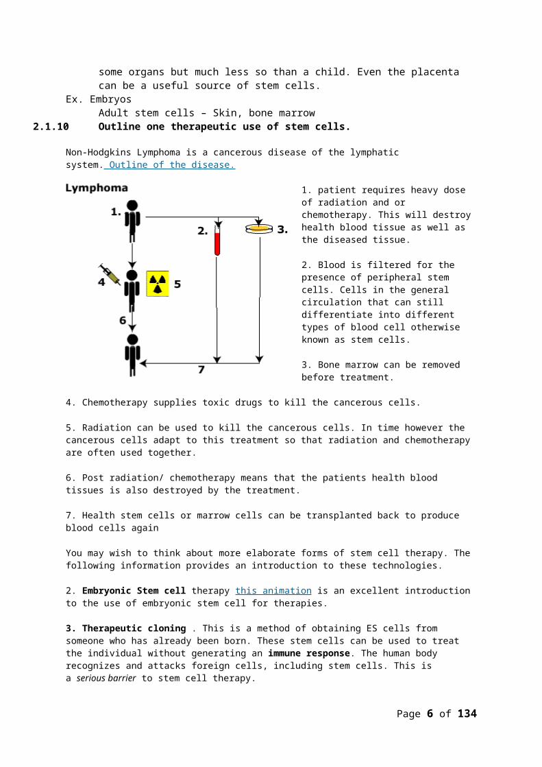
Non-Hodgkins Lymphoma is a cancerous disease of the lymphatic system. Outline of the disease.
1. patient requires heavy dose of radiation and or chemotherapy. This will destroy health blood tissue as well as the diseased tissue.
2. Blood is filtered for the presence of peripheral stem cells. Cells in the general circulation that can still differentiate into different types of blood cell otherwise known as stem cells.
3. Bone marrow can be removed before treatment.
4. Chemotherapy supplies toxic drugs to kill the cancerous cells.
5. Radiation can be used to kill the cancerous cells. In time however the cancerous cells adapt to this treatment so that radiation and chemotherapy are often used together.
6. Post radiation/ chemotherapy means that the patients health blood tissues is also destroyed by the treatment.
7. Health stem cells or marrow cells can be transplanted back to produce blood cells again
You may wish to think about more elaborate forms of stem cell therapy. The following information provides an introduction to these technologies.
2. Embryonic Stem cell therapy this animation is an excellent introduction to the use of embryonic stem cell for therapies.
3. Therapeutic cloning . This is a method of obtaining ES cells from someone who has already been born. These stem cells can be used to treat the individual without generating an immune response. The human body recognizes and attacks foreign cells, including stem cells. This is a serious barrier to stem cell therapy.
The process of therapeutic cloning is shown in this diagram. It begins by taking a somatic (body) cell from the individual. The somatic cell is fused with an egg that has had its nucleus removed. The resulting cell is genetically identical to the individual because it contains the DNA from the individual’s somatic cell. The new cell behaves like a fertilized egg and develops into a blastocyst. ES cells can be harvested from the blastocyst and grown in culture. These ES cells could be used to treat the individual without encountering resistance from his or her immune system.
Notice that we do not not refer to this type of blastocyst as an embryo. This is because, technically speaking, an embryo is the result of the union of an egg and a sperm, which has not happened in this case. ¨
Page 5 of 111
1. The patient requires the replacement of some diseased tissue. First we obtain a health cell from the same patient.
2. At the same time we require a human egg cell. This is mainly as the cell retains the tendency to divide unlike the sample tissue from the patient.
3. The nucleus is removed from the egg and discarded. The cell body itself is retained.
4. The nucleus of the patients cell is removed and retained. The cell body of the patients cell is discarded.
5. The nucleus from the patients cell is transferred to the enucleated cell body.
6. The cells then stimulated to divide forming a clone.
7. The cell mass forms a blastocyst.
8. The inner cell mass becomes a source of totipotent stem cells. Totipotent means they are capable of being stimulated to become one of any type of cell.
9. Cells are stimulated using differentiation factors to become the type of cell required for therapy.
10. Therapy would require the transfer of the new healthy cell to the patient. In therapeutic
cloning these cells have the same immune system identity as the patient therefore there is not immune rejection problem.
It is important that this technique is not confused with embryonic stem cell cultures or with reproductive cloning.
2.2 PROKARYOTIC CELLS
1.1.17 Draw and label a diagram of the ultrastructure of Escherichia coli (E. coli) as an example of a prokaryote.
Page 6 of 111

The general size of a prokaryotic cell is about 1-2 um. Note the absence of membrane bound organelles There is no true nucleus with a nuclear membrane The ribosome's are smaller than eukaryotic cells The slime capsule is used as a means of attachment to a surface Only flagellate bacteria have the flagellum Plasmids are very small circular pieces of DNA that maybe transferred from one bacteria to
another.
1.1.18 Annotate the diagram from 2.2.1 with the functions of each named structure.
Cell Wall:
Made of a murein (not cellulose), which is a glycoprotein or peptidoglycan (i.e. a protein/carbohydrate complex). There are two kinds of bacterial cell wall, which are identified by the Gram Stain technique when observed under the microscope. Gram positive bacteria stain purple, while Gram negative bacteria stain pink. The technique is still used today to identify and classify bacteria. We now know that the different staining is due to two types of cell wall
Plasma membrane:
Controls the entry and exit of substances, pumping some of them in by active transport.
Cytoplasm:
Contains all the enzymes needed for all metabolic reactions, since there are no organelles.
Ribosome:
The smaller (70 S) type are all free in the cytoplasm, not attached to membranes (like RER). They are used in protein synthesis which is part of gene expression.
Nucleoid:
Is the region of the cytoplasm that contains DNA. It is not surrounded by a nuclear membrane. DNA is always a closed loop (i.e. a circular), and not associated with any proteins to form chromatin.
Flagella:
These long thread like attachments are generally considered to be for movement. They have an internal protein structure that allows the flagella to be actively moved as a form of propulsion. The presence of flagella tends to be associated with the pathogenicity of the bacterium. The flagella is about 20nm in diameter. This structure should not be confused with the eUkaryotic flagella seen in protoctista.
Pilli:
These thread like projections are usually more numerous than the flagella. They are associated with different types of attachment. In some cases they are involved in the transfer of DNA in a process called conjugation or alternatively as a means of preventing phagocytosis.
Page 7 of 111

Slime Capsule:
A thick polysaccharide layer outside of the cell wall, like the glycocalyx of eukaryotes. Used for sticking cells together, as a food reserve, as protection against desiccation and chemicals, and as protection against phagocytosis. In some species the capsules of many cells in a colony fuse together forming a mass of sticky cells called a biofilm. Dental plaque is an example of a biofilm.
Plasmids:
Extra-nucleoid DNA of up to 400 kilobase pairs. Plasmids can self-replicate particularly before binary fission.
They are associated with conjunction which is horizontal gene transfer. It is normal to find at least one anti-biotic resistance gene within a plasmid. This
should not be confused with medical phenomena but rather is an ecological response to other antibacterial compounds produced by other microbes. Commonly fungi will produce anti-bacterial compounds which will prevent the bacteria replicating and competing with the bacteria for a resource.
Conjugation
Direct contact between bacterial cells in which plasmid DNA is transferred between a donor cell and a recipient cell.
There is no equal contribution to this process, no fertilisation and no zygote formation. It cannot therefore be regarded as sexual reproduction.
1.1.19 Identify structures from 2.2.1 in electron micrographs of E. coli. 1. Note the double membrane of this E. coli .This feature means that the cells do not retain the dark blue stain used in microscopy. They are therefore known as Gram-negative this contrast with Gram-positive single membrane bacteria.2. There is some evidence in the image of pilli which are the surrounding light grey masses.3. In the cytoplasm of the bacterium there are no visible organelles which is consistent with how we expect a prokaryote cell to appear.4. The nucleoid region is not seen well in this particular image but is clearer in the next image.
1.1.20 State that prokaryotic cells divide by binary fission.
Prokaryotic cells divide by binary fission. This is an asexual method of reproduction in
which a 'parental' cell divides into two smaller but equally sized cells.
Page 8 of 111
The cells are genetically identical and form the basis of a reproductive clone.
a little extra information for the interested reader.
The process of binary fission takes place in four stage:
(a). Reproduction signal: The cell receives a signal, of internal or external origin that initiates the cell division.
E.coli replicates about once every 40 minutes when incubated at 37o C. If however we increase the concentration of carbohydrate nutrients that the cell is supplied with then the division time can be reduced to 20 minutes. There is a suggestion here that an external signal (nutrient concentration) is acting as the reproductive signal.
(b). Replication of DNA: bacterial cells have a single condensed loop of DNA. This is copied by a process known as semi-conservative replication to produce two copies of the DNA molecule one for each of the daughter cells
The replication begins at a single point (ori)on the loop of DNA. The process proceeds around the loop until two loop have been produced, each a copy of the original. The process finishes at a single point on the loop of DNA called the ter position.
(c). Segregation of DNA: One DNA loop will be provided for each of the daughter cells.
As the new loops form the ori site becomes attached to some contractile proteins that pull the two ori sites, and therefore the loops, to opposite ends of the cell. This is an active process that requires the bacteria to use energy for the segregation.
(d). Cytokinesis: Cell separation.
This occurs once the DNA loop replication and segregation is complete. The DNA completes a process of condensing whilst the plasma membrane begins to form a 'waist' or constriction in the middle of the cell. As the plasma membrane begins to pinch and constrict the membrane fuses and seals with additional new membrane also being
formed.
2.3 EUKARYOTIC CELLS1.1.21 Draw and label a diagram of the
ultrastructure of a liver cell as an example of an animal cell.
N:Nucleus
PM: plasma membrane
Page 9 of 111

M: mitochondria
rER: Rough endoplasmic reticulum
GA: Golgi apparatus
L: Lysosome
MV: Microvilli
1.1.22 Annotate the diagram from 2.3.1 with the functions of each named structure.
Nucleus: This is the largest of the organelles. The nucleus contains the chromosomes which during interphase are to be found the nucleolus.
The nucleus has a double membrane with pores(NP).
The nucleus controls the cells functions through the expression of genes.
Some cells are multi nucleated such as the muscle fibre
Plasma membrane: controls which substances can enter and exit a cell. It is a fluid structure that can radically change shape. see 2.4
The membrane is a double layer of water repellant molecules.
Receptors in the outer surface detect signals to the cell and relay these to the interior.
The membrane has pores that run through the water repellant layer called channel
proteins.
Mitochondria: location of aerobic respiration and a majot synthesis of ATP region.
Double membrane organelle. Inner membrane has folds called
cristae. This is the site of oxidative phosphorylation.
Centre of the structure is called the matrix and is the location of the Krebs cycle.
Page 10 of 111

Oxygen is consumed in the synthesis of ATP on the inner membrane
The more active a cell the greater the number of mitochondria.
Rough endoplasmic reticulum (rER): protein synthesis and packaging into vesicles.
rER form a network of tubules with a maze like structure.
In general these run away from the nucleus The 'rough' on the reticulum is caused by the
presence of ribosomes. Proteins made here are secreted out of the
cell
Ribosomes: the free ribosome produces proteins for internal use within the cell.
Golgi apparatus: modification of proteins prior to secretion.
proteins for secretion are modified
possible addition of carbohydrate or lipid components to protein
packaged into vesicles for secretion
Lysozyme:
Vesicles in the above diagram that have formed on the golgi apparatus. Containing hydrolytic enzymes. Functions include the digestion of old organelles, engulfed bacteria and viruses.
1.1.23 Identify structures from 2.3.1 in electron micrographs of liver cells.
Nucleus:
In an electron micrograph the nucleus will be the largest of the organelles.
Page 11 of 111

In this image there is a dark stained region called the nucleolus which is the location of the DNA.
The membrane has pores which allow the entry of cell signal molecules, nucleotides and the exit of mRNA.
Generally the nucleus appears spherical however there are cells in which the nucleus has more unusual shape such as the multi-lobbed white blood cells.
Plasma membrane:
This image shows the junction between two liver cells. The image has been manipulated for clarity to see the two adjoining plasma membranes.
Notice the mitochondria to the left and the rER to the right of the membranes.
Mitochondria:
This micrograph of a mitochondria shows:
Double outer membrane Folded inner membrane called the
cristae. Matrix of the mitochondria
These features are common to all mitochondria. Notice the rER above the mitochondria for scale and the dark
granules of glycogen below the organelle.
Endoplasmic reticulum (rER).
The rER runs vertical in the image. Note the dark spots which are the ribosomes.
A cell with a great deal of rER is producing proteins for secretion outside of the cell.
The network of endoplasmic tubules allows proteins to be moved around within the cytoplasm before final packaging and secretion.
Page 12 of 111

Golgi apparatus:
The golgi apparatus in the diagram forms a stack of membrane envelopes on top of each other.
Vesicles containing proteins fuse with the structure.
The proteins are modified inside the apparatus usually with the addition of non-protein substances.
Lysosome:
simple membrane bound vesicle containing hydrolytic enzymes produced in the golgi apparatus. used to digest engulfed bacteria or viruses or old organelles used to digest macromolecules hydrolytic enzymes are retained within the vesicle membrane to prevent
autodigestion of the cell.
1.1.24 Compare prokaryotic and eukaryotic cells.Prokaryotic Cells Eukaryotic Cells
All prokaryotic cells have cell walls Have no lysosomes Have a nucleoid instead of a nucleus,
nothing surrounding it DNA flows freely Cell membrane Only bacteria No mitochondria No internal membranes Naked DNA Small and simple All cells have a cell membrane
Only some have a cell wall Contain lysosomes Nucleus has pores DNA enclosed in the nucleus Have a Golgi apparatus Have an endoplasmic reticulum Cell membrane Animal, plant, fungi, protists (Amoeba,
paramecium) DNA with proteins attached All cells have a cell membrane
1.1.25 State three differences between plant and animal cells.Plant Cells Animal Cells
Cell wall Vacuole (large fluid filled sac) No lysosomes Chloroplasts that contain chlorophyll Photosynthesis
No cell wall Fluid filled sacs (vesicles) Lysosomes No chloroplasts
1.1.26 Outline two roles of extracellular componentsAnimal cells:
ECM = Extracellular matrix ECM influences shape, orientation and polarity, movement, metabolism, and differentiation ECM is made and oriented by the cells
o Take two general forms: Interstitial Matrix
Page 13 of 111

3D gel that surrounds cells that fills space Basement Membrane
Mesh-like sheet formed at the base of epithelial tissues Cellular organizer
o When cells are put on a basement membrane the differentiate
o They do not grow unless properly anchored to the matrix ECM functions in support, adhesion and movement
o Gel surrounds cells made of glycoproteins called interstitial matrix Basement membranes are sheet formed around tissues
o Causes cell to organize (movement) and differentiate
Plant Cells
Cell wall: (Outside the cell)
1) Cellulose micro fibril pass through the plasma membrane to add to the thickness of the cell wall
2) The wall is then able to support the plasma membrane so that it can prevent a lot of water from entering the cell, protecting it from bursting
- Maintains cell shape- Holds up the whole plant against gravity
2.4 MEMBRANES1.1.27 Draw and label a diagram to show the structure of membranes.
Page 14 of 111

1.1.28 Explain how the hydrophobic and hydrophilic properties of phospholipids help to maintain the structure of cell membranes.
This model of the bilayer's has the proteins removed for clarity.
The 'head's have large phosphate groups, thus they are hydrophilic (attract water) or polar. These section are suited to the large water content of the tissue fluid and cytoplasm on opposite sides of the membrane.
The fatty acid tails are non-charged, hydrophobic meaning they repel water. This creates a barrier between the internal and external 'water'
Page 15 of 111

environments of the cell. The 'tails' effectively create a barrier to the movement of charged molecules
The individual phospholipids are attracted through their charges and this gives some stability. They can however move around in this plane
The stability of the phospholipid can be increased by the presence of cholesterol molecules.
1.1.29 List the functions of membrane proteins.
1.1.30 Define diffusion and osmosis.
Diffusion: passive movement of particles from a region of high concentration to a region of low concentration.
Osmosis is the passive movement of water molecules from a regions of lower solute concentration to a region of higher solute concentration
1.1.31 Explain passive transport across membranes by simple diffusion and facilitated diffusion.
The passive movement implies that there is no expenditure of energy in moving the molecules from one side of the membrane to the other:
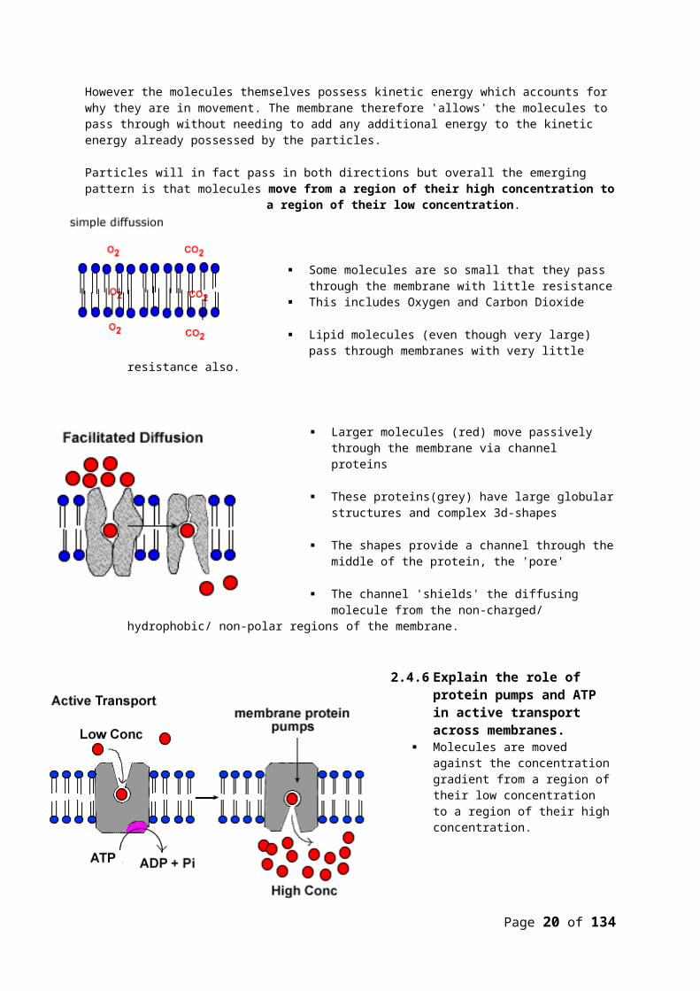
However the molecules themselves possess kinetic energy which accounts for why they are in movement. The membrane therefore 'allows' the molecules to pass through without needing to add any additional energy to the kinetic energy already possessed by the particles.
Particles will in fact pass in both directions but overall the emerging pattern is that molecules move from a region of their high concentration to a region of their
low concentration.
Some molecules are so small that they pass through the membrane with little resistance
This includes Oxygen and Carbon Dioxide
Lipid molecules (even though very large) pass through membranes with very little resistance also.
Page 16 of 111
Larger molecules (red) move passively through the membrane via channel proteins
These proteins(grey) have large globular structures and complex 3d-shapes
The shapes provide a channel through the middle of the protein, the 'pore'
The channel 'shields' the diffusing molecule from the non-charged/ hydrophobic/ non-polar regions of the membrane.
1.1.32 Explain the role of protein pumps and ATP in active transport across membranes.
Molecules are moved against the concentration gradient from a region of their low concentration to a region of their high concentration.
Active mean that the membrane protein 'pump' requires energy (ATP) to function
The source of energy is ATP is produced in cell respiration
Transported molecules enter the carrier protein in the membrane. The energy causes a shape change in the protein that allows it to move the
molecule to the other side of the membrane.
The sodium-potassium, pump that creates electro-chemical gradient across the cell membrane of all cells.
Cells are -ve charged on the inside relative to the outside.
This pump is modified in the nerve cell to create some of the electrochemical phenomena seen in nerve cells.
1.1.33 Explain how vesicles are used to transport materials within a cell between the rough
endoplasmic reticulum, Golgi apparatus and plasma membrane.
Page 17 of 111

1. Protein is already synthesised and present in the rER.2. The protein is moved through the rER and modified.3. A spherical vesicle is formed form the end of the rER with the protein inside.4. The vesicle migrates to the golgi apparatus.5. Vesicle and golgi membranes fuse. The protein is released into the lumen of the golgi
apparatus.6. The golgi modifies the protein further by adding lipid or polysaccharides to the
protein.7. A new vesicle is formed from golgi membrane which then breaks away. The vesicles
migrates to the plasma membrane.8. The vesicle migrates to the plasma membrane fuses and secretes content its
contents out of the cell. A process called exocytosis.
1.1.34 Describe how the fluidity of the membrane allows it to change shape, break and re-form during endocytosis and exocytosis.
a) Exocytosis: vesicle membrane fuses with the plasma membrane.
b) Endocytosis:a vesicle is formed by the infolding of the plasma membrane
In each of the cases above the membranes are able to form and break without loss of the continuity of the plasma membranes. The process is very similar to the childhood game of playing with bubbles of detergent. Bubbles are produced then they can be
watched readily joining together or splitting apart.
Membrane fluidity:
(a) The phospholipid molecules can change places in the horizontal plane. This creates the so called fluid property of the membrane.
(b) Molecule exchange in the vertical plane does not occur. This maintains the integrity of the membrane.
(c) Cholesterol embedded in the membrane reduces its fluidity.
Page 18 of 111

2.5 CELL DIVISION1.1.35 Outline the stages in the cell cycle, including interphase (G1, S, G2) mitosis and cytokinesis.
Interphase (grey) is the longest phase which itself occurs in three stages. G1 The cell performs its normal differentiated function. Protein synthesis/
mitochondria replication/ chloroplast replication. S DNA replication. At this point the mass of DNA in the cell has doubled. G2 Preparation for cell division Phases of mitosis (see 2.5.4) Cytokinesis: division of the cytoplasm to form two daughter cells.
1.1.36 State that tumours (cancers) are the result of uncontrolled cell division and that these can occur in any organ or tissue.
Tumours (cancers) are a cell mass formed as a result of uncontrolled cell division. They can occur in any tissue. Ex. Stomach cancer
1.1.37 State that interphase is an active period in the life of a cell when man metabolic reactions occur, including protein synthesis, DNA replication and an increase in the number of mitochondria and/or chloroplasts.
The cell specialises to a particular function in a process called differentiation. Through gene expression and protein synthesis there is a specialisation of cell structure and
function. During this interphase the cell carries out this specialist function. The length of the interphase varies from one type of cell to another. G1 follows cytokinesis. The cell is involved in the synthesis of various proteins which allow
the cell to specialise. S-phase involves the replication of DNA molecules which takes place prior to the phases of
mitosis.
Page 19 of 111
G2 preparation for the phases of mitosis which involves the replication of mitochondria and in the case of plants, the chloroplast.
1.1.38 Describe the events that occur in the four phases of mitosis (prophase, metaphase, anaphase and telophase).
Super coiling: Eukaryotic DNA is combined with histone proteins and non-histone proteins to form chromatin. The method of folding of chromatin is specific to each chromosome leaving genes in predictable positions and a distinctive overall chromosome shape. The human cell has a DNA length of about 1.8 m this has to be packed into a nucleus which has only a 5 um diameter. This packaging process requires up to a X 15,000 reduction. This super coiling makes the structure so dense that it can be see with a light microscope during the phases of mitosis.
In this sequence only one chromosome is illustrated so that we can more clearly follow the process. In a human a complete diagram would have 46 chromosomes each replicating and condensing and separating.
a)The cell membrane is intact during this the interphase. The chromosomes cannot be seen during G1,S and G2.
b) G1,Within the nucleus, genes on the chromosome are being expressed to carry out normal cell function (interphase). Remember you cannot see chromosomes at this stage. The diagram has a 'see's through' the nuclear membrane so you can see inside. In reality it would look just like cell a).
c) S-phase in which DNA replication occurs and the
chromosomes are copied. The copies called sister chromatids are held together by a protein to form the centromere. It is still not possible to see this happen with an intact cell.
d) Early Prophase in which the sister chromatids have condensed by super coiling. Note the formation of the spindle microtubules and their attachment to centrioles. The nuclear membrane will now break down to reveal sister chromatids. The internal arrangements of chromosomes can now be seen with a light microscope.
e) Metaphase the chromosomes arranged on the equator of the cell each attached to a spindle microtubule at the centromere
f) Anaphase: The spindle microtubules contract and pull apart the sister chromatids one to each pole of the cell. The centromere splits allowing the sister chromatids to be separate.
g) Telophase: at each pole there are separate groups of the replicated chromosomes the spindles is degenerating
Page 20 of 111

h) Cytokinesis: the cell membrane begins to separate, dividing the cell into two new cells. The nuclear membrane is reforming around each cell.
i) Two daughter cells are formed. They are genetically identical to each other and in effect the basis of a clone. (see 2.5.6)
Notice that cell a) begins with one chromosome and that by step h) there are two cells each with a copy of that chromosome.
As suggested by cell theory, all cells have come from other cells.
1.1.39 Explain how mitosis produces two genetically identical nuclei.
The process of cell division produces genetically identical daughter cells.
Conservation of chromosome number. The chromosome number in each of the daughter cells is the same as that of the original parental cell
During the S-phase, each chromosome is copied exactly. The two copies of each chromosome are held together by a protein structure called a centromere.
Therefore just prior to the beginning of the phases of mitosis there is actually double the number of chromosomes present in a cell.
Each chromosome in this state is represented by a pair of sister chromatids. These give the now classic cross image of the DNA (see image below)
This pair of sister chromatids image was taken during one of the phases of mitosis.
The two sister chromatids are held together at the centromere
The arms of the chromatids are visible because of a condensation of the molecule called super coiling.
This condenses the molecule some x 15,000 times of its original length The pairs of sister
chromatids is a non-random organisation. The position of genes is predicable within the structure seen here. Also there is a unique shape to each of the chromosomes.
Mitosis makes sure that each cell obtains a copy of each of the chromosomes in the parental cell.
However, it is the process of DNA replication during the S-phase that actually copies each DNA molecules to make mitosis possible.
1.1.40 State the growth, embryonic development, tissue repair and asexual reproduction involve mitosis.
Page 21 of 111

Growth: multicellular organisms increase their size through growth. This growth involves increasing the number of cells through mitosis. These cells will differentiate and specialise their function.
Embryonic development is when the fertilised egg cell (zygote) divides to form the multicellular organism. Each cell in the organisms is identical (genetically) to all the other cells. However, each cell will express only a few of its genes to determine its overall specialisms, a process called differentiation. In this way a stem cell may become a muscle, or it may become a nerve cell or any one of the many different kinds of cells found in a complex multicellular organism. The best book about this process for the interested reader is
Tissue Repair: As tissues are damaged they can recover through replacing damaged or dead cells. This is easily observed in a skin wound. More complex organ regeneration can occur in some species of amphibian.
Asexual Reproduction: The production of offspring from a single parent using mitosis. The offspring are therefore genetically identical to each other and to their “parent”- in other words they are clones. Asexual reproduction is very common in nature, and in addition we humans have developed some new, artificial methods. Bacteria DO NOT asexually reproduce by mitosis but rather by a process called Binary Fission.
Topic 3: The Chemistry of life3.1 CHEMICAL ELEMENTS AND WATER
1.1.41 State that the most frequently occurring chemical elements in living things are carbon, hydrogen, oxygen and nitrogen.
Carbon C Hydrogen H Oxygen O Nitrogen N
1.1.42 State that a variety of other elements are needed by living organisms, including sulphur, calcium, phosphorus, iron and sodium.
Sulphur Calcium Phosphorus Iron Sodium
1.1.43 State one role for each of the elements mentioned in 3.1.2. Sulfur (S): Needed to make two of the twenty amino acids that proteins contain. Calcium (Ca): Acts as a messenger, binding to calmodulin and other proteins that regulate
processes inside cells including transcription (needed to send a message from the nerve to a muscle).
Phosphorus (P): Part of the phosphate groups in ATP and DNA molecules. Iron (Fe): Needed to make cytochromes – proteins used for electron transport during
aerobic cell respiration. Sodium (Na): Pumped into the cytoplasm to raise the solute concentration and cause water
to enter by osmosis and are also responsible for nerve impulses.1.1.44 Draw and label a diagram showing the structure of water molecules to show their polarity and
hydrogen bond formation.
Page 22 of 111

This image of water show the covalent bonds between oxygen and two hydrogen atoms.
The nuclei of oxygen is significantly larger and greater charge (+8) than the hydrogen nuclei (+1).
Consequently the electron pair in the covalent bond is found 'closer' to the oxygen than the hydrogen nuclei.
This creates a polar molecule in which the oxygen carries and additional small negative dipole and each hydrogen a small positive dipole.
1.1.45 Outline the thermal, cohesive and solvent properties of water.1. Thermal
Heat capacity Boiling point
o 100⁰C Evaporation Water needs large amounts of energy to increase its temperature
o The bonds need to break down before they begin to heat up (hydrogen bonds) all bonds must break first.
2. Cohesive Binding of two water molecules Stick together because of the hydrogen bonding
3. Solvents Water can take apart anything that’s ionic, polar, or charged
o Ex. Glucose, sodium ions, enzymes1.1.46 Explain the relationship between the properties of water and its users in living organisms as a
coolant, medium for metabolic reactions and transport medium.1. Why is water a good medium for transport?
Water is used as a transport medium in the xylum of plants Water has hydrogen bonds and can go upwards a tree (cohesive) Solvent properties means dissolving of substances and can be carried around in
blood and the sap of plants Heat can travel in blood
o Warm/cold blooded2. How can water be used as a coolant?
Sweating (perspiration = animals, transpiration = plants) Through evaporation the area cools down
3. How can water be used as a medium for metabolic reactions? Because it’s a solvent, the chemicals needed for the reactions are already dissolved
in it Mostly liquid on Earth
3.2 CARBOHYDRATES, LIPIDS AND PROTEINS
3.1.1 Distinguish between organic and inorganic compounds.Organic Inorganic
Carbohydrates WaterLipids OxygenProteins Potassium (K), Iron (Fe), Sodium (Na)Nucleic acids CO2
- Contains carbon - Does not contain Carbon(Except CO2)
Page 23 of 111

Organic compounds are based on carbon and are found in living things. There are a number of exceptions includinghydrogen carbonate (HCO3
- ), carbon dioxide (CO2 )and Carbon monoxide (CO). Inorganic compounds are by default all the molecules other than those in the
category above.
3.1.2 Identify amino acids, glucose, ribose and fatty acids from diagrams showing their structure.Amino Acids
Glucose
Ribose
Fatty acids
Page 24 of 111

3.1.3 List three examples each of monosaccharides, disaccharides and polysaccharides.
3.1.4 State one function of glucose, lactose and glycogen in animals, and of fructose, sucrose and cellulose in plants.
3.1.5 Outline the role of condensation and hydrolysis in the relationships between monosaccharides, disaccharides and polysaccharides: between fatty acids, glycerol and triglycerides: and between amino acids and polypeptides.
Polymer: consisting of large molecules made up of a linked series of repeated simple molecules called monomers
Monomers: simple molecular units
Model of polymerisation through condensation reaction.
(1) Dimers
a) Two monomers are bonded together to form a dimer.
Page 25 of 111

b) Water (H + OH) are removed to form water.
c) The dimer can be split by hydrolysis but needs water adding
(2) Polymerisation
a) In this example six monomers are joined together
b) Polymers normally form more complex shapes than suggested in this model
c) The polymer can be 'digested' back to monomers by hydrolysis reaction
Formation of a disaccharide
a) Two molecule of glucose will polymerise to form maltose
b) The condensation reaction will take place between C1 of the first glucose and C4 of the second glucose.
c) A condensation reaction takes place between the glucose 1 (-OH on C1) and Glucose(-H on C4).
d) The bond formed is a covalent bond between C1 -O-C4 , called a 1, 4 glycosidic bond.
e) The disaccharide molecule formed is called Maltose which like glucose is a reducing sugar.
f) Hydrolysis; The diagram can be reversed so that the disaccharide can be split into two glucose monosaccharides.
g) Hydrolysis is the type of reaction catalysed by the digestive enzymes.
Laboratory Hydrolysis
In the lab you can hydrolyse maltose and other disaccharides to their monomers by gentle warming the disaccharide in a dilute Hydrochloric acid.
The test for sucrose has the initial step of acidifying and very gently warming sucrose with an acid before carrying out the Benedicts test.
Sucrose gives a negative benedicts test. However after hydrolysis to glucose and fructose both these sugars give a positive test with Benedicts reagent.
Formation of a polysaccharide
Page 26 of 111

The above chain of glucose molecules represent the polysaccharide formed by many glucose monomers joining together to form this polysaccharide called amylose.
The molecule to the left represents the helical structure of the polypeptide, amylose.
Amylose is a polymer of glucose.
Intramolecular hydrogen bonding causes the chain molecule to twist into a helical shape.
Amylose is one of two molecules found in starch, the other being a branching polymer of glucose (below)called amylopectin.
Amylopectin
Starch: Starch is composed of two polysaccharides, Amylose and amylopectin
Starch is metabolically un-reactive and insoluble and hence an excellent storage carbohydrate.
Formation of a dipeptide and a polypeptide
a) Two amino acid monomers of glycine aligned to form a peptides bond by condensation reaction.
b) The peptide bond can form between the carboxyl group of the first amino acid and the amino group of the second amino acid.
Page 27 of 111

d) H-OH or water is removed in the reaction hence the term condensation reaction.
d) Dipeptide is formed (naming system not required) with the characteristic -C-N- bond between the two monomers.
e) Notice that in the dipeptide there is still an amino group at one end and a carboxylic group at the other end.
f) The above pattern is true of all polypeptides and known as the amino terminal and carboxyl terminal of the polypeptide.
Polypeptide chains do not remain as linear (straight) chains. Instead they fold up into the complex yet specific shapes of the protein as seen in this image. The different types of shapes are not required for SL but are covered in section 7. 5.1
The shape of a protein is determined by intra-molecular hydrogen bonding and some covalent bonding between R groups (-S-S-, disulphide bridges).
Polypeptides can be hydrolysed in the same way as polysaccharides with by incubating with acids. Naturally polypeptides are digested by a group of enzymes called Peptidases which hydrolyse the chain into amino acids.
Formation of a triglyceride:
Chemically all fats and oils are triglycerides (simple lipids). Fats are those lipids which are solid state at 20oC. Oils are those lipids which are liquid at 20oC. Oils with unsaturated fatty acids have bends in their tail structure which reduces the density of the molecule and lowers its melting point. Oil also tend to have short fatty acid tails. Conversely fats tend to have longer fatty acids with saturated bonds. This makes their structure densely packed and raises the melting point.
The formation of a triglyceride or any lipid is not a polymerisation like the previous examples.
Instead three fatty acids chains (usually of different length) are bonded to the molecule glycerol.
Ester bonds (-O-) are formed between an -OH group on the glycerol molecule and the carboxylic acid group (-COOH) of the fatty acid.
Page 28 of 111

The triglyceride formed is insoluble. (Hydrophobic). Fatty acid tails can vary in length and may contain unsaturated bonds Animals fats have saturated fatty acids which are straight molecules and very
compact. This is gives them a higher melting point than the plant oils Plant oils have unsaturated and polyunsaturated fatty acid chains that tend to
branch and make the molecule less dense and with a lower melting point.
Phospholipids are the principle molecule in the cell membrane they form the 'bilayer' that is the cell membrane.
Phospholipid structure:
Very similar to the triglyceride except one fatty acid chain is replaced by a polar phosphate group. The molecule is in two parts
a) Polar hydrophilic phosphate heads.
b) 2 Non polar hydrophobic tails
This diagram is a short hand version of the phospholipid molecule.
It illustrates the negatively charged hydrophilic head and the hydrophobic tails
Often additional groups are attached to the negative head such as Choline, Serine or Inositol
3.1.6 State three functions of lipids.
3.1.7 Compare the use of carbohydrates and lipids in energy storage.
Page 29 of 111

3.3 DNA STRUCTURE
3.1.8 Outline DNA nucleotide structure in terms of sugar (deoxyribose), base and phosphate.
Sugar is deoxyribose which differs from ribose in having one less oxygen on carbon 2.
Phosphate is the PO4-3 group.
Bases are nitrogen based ring structures of which there are 4 different kinds.
3.1.9 State the names of the four bases in DNA Adenine Guanine Cytosine Thymine
3.1.10 Outline how DNA nucleotides are linked together by covalent bonds into a single strand. Two DNA nucleotides can be linked together by a covalent bond between the sugar of one
nucleotide and the phosphate of another. More nucleotides can be added to form a single strand.
Page 30 of 111

Page 31 of 111
Covalent bond

Covalent bonds link two phosphates togethero They’re strong and don’t break aparto Radioactivity breaks down these structures
3.1.11 Explain how DNA double helix is formed using complementary base pairing and hydrogen bonds.
Page 32 of 111
Covalent bond
Covalent bond

Bases pair up:o A - To C - Go (Complementary base pairings)
These base pairs stick together by hydrogen bondso These bonds are weak and easily broken
3.1.12 Draw and label a simple diagram of the molecular structure of DNA.
Molecule is DNA The whole piece is a chromosome One gene is a piece of DNA that codes for one protein
o That piece is RNA 46 chromosomes in a nucleus
Page 33 of 111
Hydrogen bond

o On these chromosomes, we have about 25,000 genes in a nucleus In every cell of your body
o Exception of red blood cells that don’t have nuclei
3.4 DNA REPLICATION
3.1.13 Explain DNA replication in terms of unwinding the double helix and separation of the strands by helicase, followed by formation of the new complementary strands by DNA polymerase.
One molecule of double stranded DNA copies itself to make two new molecules of DNA Old strands are templates for making new complementary strands
o Strand A and Strand B separateo Strand A forms a template for the new strand B
Vice-versa Steps in the process
o Helicase splits the DNA molecule by breaking hydrogen bonds between the base pairs
o DNA polymerase adds new complementary nucleotides to the original template strands. DNA polymerase also builds covalent bonds between adjacent nucleotides
3.1.14 Explain the significance of complementary base pairing in the conservation of the base sequence of DNA.
The significance of the mechanism outlined above is that the DNA molecule is copied precisely from one cell generation to the next.
In a unicellular organism this means that the total genome is successfully copied into each new generation.
In the multi-cellular organism all cells contain an exact copy of the total genome (even though not fully expressed).
Genes (base sequences) are faithfully passed from one generation to the next. The genes (base sequences) which the reader possess have been passed from
generation to generation until they arrived in you now. With minor and rare modification the base sequences copied by DNA replication and successfully passed on through sexual reproduction. Your base sequences have been copied for thousands of years.
A-T, C-G Adenine pairs with Thymine, Cytosine pairs with Guanine
3.1.15 State that DNA replication is semi-conservative. This is semi-conservative replication
o For each new molecule of DNA, there is one old strand conserved, and one new strand produced
Page 34 of 111

3.5 TRANSCRIPTION & TRANSLATION
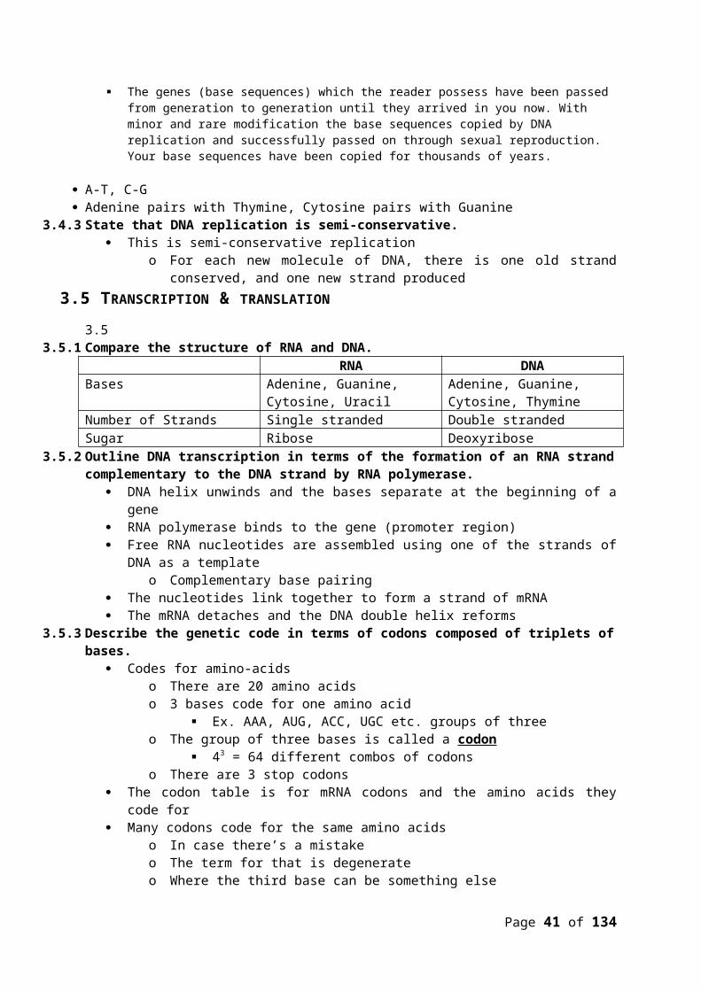
3.1.16 Compare the structure of RNA and DNA.RNA DNA
Bases Adenine, Guanine, Cytosine, Uracil
Adenine, Guanine, Cytosine, Thymine
Number of Strands Single stranded Double strandedSugar Ribose Deoxyribose
3.1.17 Outline DNA transcription in terms of the formation of an RNA strand complementary to the DNA strand by RNA polymerase.
DNA helix unwinds and the bases separate at the beginning of a gene RNA polymerase binds to the gene (promoter region) Free RNA nucleotides are assembled using one of the strands of DNA as a template
o Complementary base pairing The nucleotides link together to form a strand of mRNA The mRNA detaches and the DNA double helix reforms
3.1.18 Describe the genetic code in terms of codons composed of triplets of bases. Codes for amino-acids
o There are 20 amino acidso 3 bases code for one amino acid
Ex. AAA, AUG, ACC, UGC etc. groups of threeo The group of three bases is called a codon
43 = 64 different combos of codonso There are 3 stop codons
The codon table is for mRNA codons and the amino acids they code for Many codons code for the same amino acids
o In case there’s a mistakeo The term for that is degenerateo Where the third base can be something else
3.1.19 Explain the process of translation, leading to polypeptide formation.1) Steps of translation
1. mRNA binds to the small subunit of a ribosome2. Each tRNA has a triplet of bases called an anticodon and carries an amino acid
corresponding to this anticodon3. Two tRNA molecules bind to the ribosome
Their anticodons are complementary to the bases on the mRNA codonsForm hydrogen bonds
4. The two amino acids carried by the tRNA molecules form a peptide bond. They stay attached to the second tRNA. The 1st tRNA leaves
5. The ribosome moves along the mRNA to the next codonComplementary tRNA molecules attach, continue building the growing polypeptide.Finally, a stop codon is reached.The mRNA, ribosome and tRNA molecules detach
6. The polypeptide is complete3.1.20 Discuss the relationship between one gene and one polypeptide.
2) Genes determined the amino acid sequence of proteins (polypeptides)1. One gene/one polypeptide rule2. Polypeptide: a chain of amino acids that make a protein3. One gene codes for one polypeptide (protein) in the cytoplasm
3) Exceptions1. Some genes code for tRNA or mRNA not DNA
Page 35 of 111

i. Then it doesn’t go anywhere, it doesn’t become a protein2. Some proteins are made of more than one polypeptide chain so it takes more than
one gene to make them4) Some sequences of DNA are the “switches” to turn genes on or off
3.6 ENZYMES
3.1.21 Define enzyme and active site.
Enzyme: A substance produced by a living organism that acts as a catalyst to bring about a specific biochemical reaction.
Active site: A region on an enzyme that binds to a protein or other substance during a reaction.
3.1.22 Explain enzyme-substrate specificity.
a) Large globular protein enzyme
b) Active Site where the substrate combines to the enzyme
c)Substrate which fits the active site
d) Activated complex. The substrate is weakened to allow the reaction.
e)Unchanged enzyme/ re-used at low concentrations
f) Product of the reaction
Page 36 of 111

other keypoints from the hypothesis:
The active site is often composed of open loops of polar amino acids on the exterior of the enzyme molecule.
Enzyme specificity is due to the complementary shape of the active site and the substrate.
Enzymes work at low concentrations because they are unaffected by the reaction and can return for more substrate.
3.1.23 Explain the effects of temperature, pH and substrate concentration on enzyme activity.
Effect of temperature on the rate of an enzyme catalysed reaction:
(a) As the temperature increases enzyme stability decreases. The kinetic energy of the enzyme atoms increases causing vibrations in the enzyme molecule that lead to the hydrogen bonds to breaking, shape changes in the active site.
(b) As the temperature increases the kinetic energy of the substrate and enzyme molecules also increases. Therefore more collisions of the substrate with the active site and the formation of activated complex's and product. The rate
of reaction is increasing.
(c) The optimal temperature (X) is the highest rate of reaction. Compromise between decreasing enzyme stability and kinetic energy of the reactants.
(d) Higher temperature increases the kinetic energy of the enzyme atoms so much that they break bonds, change shape of the active site.
The main diagram is often simplified to this diagram which still shows the three key stages in the reaction.
Page 37 of 111

The effect of pH on the rate of an enzyme catalysed reaction:
pH also affects the rate of reaction of an enzyme catalysed reaction.
At the optimal pH (a) or (b) the maximum rate of reaction is achieved.
Above or below the optimal pH the rate decreases.
The change in rate is because bonds are
made and broken which change the shape of the active site and therefore decrease the rate of reaction.
The two enzyme shown in the image illustrate the fact that different enzymes can have very different optimal pH.
e.g. Blue curve = pepsin (a)= pH3, Red curve =salivary amylase (b)= pH 7.2
Effect of substrate concentration on the rate of an enzyme catalysed reaction:
(a) As the substate concentration is increased the rate of reaction increases.
There are more collisions between the substrate and the enzyme such that more activated complex's are formed and therefore product per unit time.
(b) Further increases in substrate also increase the rate but proportionately less than previously.
The number of occupied active site is increasing and there is competition for the active site.
(c) The rate is constant.
The enzyme active site is fully saturated with substrate such that adding more substrate does not increase the rate of reaction. The enzymes molecules are fully occupied converting substrate to product and any substrate must await a free active site before conversion to product.
3.1.24 Define denaturation.
Page 38 of 111

Denaturation is a structural change in a protein that results in the loss (usually permanent) of its biological properties.
Temperature:(see section 3.6.3)
Temperature rises cause the average kinetic energy of the enzyme atoms to increase.
This vibration breaks the weakest bonds first, which in the enzyme are the hydrogen bonds.
The breaking of bonds, changes the shape of the enzyme. Change the shape of the enzyme changes the shape of the active site. Change the shape of the active site prevents substrate from entering. The rate of reaction reduces or stops.
pH: (see section 3.6.3)
At pH lower than the optimal pH the concentration of H+ in the solution will be higher than normal.
The hydrogens will tend to be attracted to electronegative regions of the enzyme protein.
Bonds are formed or changed as a consequence of the additional H+ which changes the shape of the enzyme molecule.
Changes in shape, change the active site shape. Changes in active site shape reduces the ability of the substrate to bind with the
active site. This reduces the rate of reaction that changes substrate to product. The rate of reaction reduces. For pH values above the optimum breaks bonds in the same way and have the
same reductions in the rate of reaction
3.1.25 Explain the use of lactase in the production of lactose-free milk.
Lactose is a disaccharide (glucose + Galactose) milk sugar Around 90% of all humans show some kind of lactose intolerance. People who are lactose intolerant can drink milk if it is lactose free. Lactase is an enzyme extracted from yeast that can digest the milk sugar to
glucose and galactose.
Enzyme Immobolisation:
It is possible to make the process more efficient by immobilising the lactose on a recoverable surface such as alginate.
First the Lactase is immobilised in alginate beads.
Next the beads are placed in a container over which milk can be passed.
Page 39 of 111

The milk is collected and re-circulated (pump) to convert any remaining lactose to glucose and galactose.
The circulation is maintained until all lactose has been converted. This model of an industrial process allow the lactase to be recovered and re-used
(cheaper). Efficient conversion of lactose to glucose and galactose. High % lactose conversion is achieved. All these factors reduce cost particularly on the downstream processing and
purification.
3.7 CELL RESPIRATION
3.1.26 Define cell respiration.Cell respiration: Cell respiration is the controlled release of energy from organic compounds in cells to form ATP.
ATP or Adenosine triphosphates is the molecule which directly fuels the majority of biological reactions.
Everyday each person will hydrolyse (reduce) 1025 ATP molecules to ADP. The ADP is reduced back to ATP using the free energy from the oxidation of organic
molecules.3.1.27 State that, in cell respiration, glucose in the cytoplasm is broken down by glycolysis into pyruvate,
with a small yield of ATP.
Location: Cytoplasm Process: Glycolysis Substrate: Glucose Products: 2 Pyruvate and a small amount of ATP Glycolysis does not use oxygen.
3.1.28 Explain that, during anaerobic cell respiration, pyruvate can be converted in the cytoplasm into lactate, or ethanol and carbon dioxide, with no further yield of ATP.
Anaerobic respiration is the oxidation of organic compounds without oxygen. It is less efficient than aerobic respiration (with oxygen). There are different types of anaerobic respiration. Here we will compare anaerobic
respiration in yeast and humans.
Humans anaerobic respiration:
Location: cytoplasm Substrate: Glucose Product: lactic acid (lactate) + ATP Note: lactic anaerobic respiration
supplements aerobic respiration in the production of ATP. Both aerobic
and anaerobic respiration can take place in the human cell at the same time.
Page 40 of 111
Yeast anaerobic respiration:
Location: cytoplasm
Substrate: Glucose Product: Ethanol + carbon dioxide +
ATP This is the end point for this
fermentation reaction. Ethanol and CO2are both excreted with no further metabolism of the energy stored in the ethanol (very inefficient)
Note: The glucose molecule has been hydrolysed further than in human respiration. Some organisms are totally anaerobic others can switch between anaerobic and aerobic.
Exercise and anaerobic respiration :
Human lactic anaerobic respiration is a process that supplements the production of ATP. The lactic pathway is so inefficient that under normal circumstances it cannot produce enough energy to support human systems. In describing the lactic pathway it is often suggested that sprinters 'do not breath during the 100m sprint' (they do, just watch any video) and they only produce ATP for running from the lactic pathway. This is a ms-representation of a complex response to the demand for ATP. It is far better to consider that anaerobic respiration in humans supplements (adds to) the aerobic production of ATP.
Anaerobic respiration:
Fermentation respiration in yeast yields two useful products from a human perspective. The carbon dioxide can be used in a variety industrial processes the best known of which is to raise bread. Many Brewers of alcohol will bottle the CO2 for use in the 'carbonation' of other drink products.
The alcohol itself is of course the basis of many industries such as beer brewing. In more recent time the use of fermentation products is being used as an alternative source of fuel such as is the case in fuel for automobiles.
Page 41 of 111

3.1.29 Explain that, during aerobic cell respiration, pyruvate can be broken down in the mitochondrion into carbon dioxide and water with a large yield of ATP.
Location: Mitochondria
Substrate: Pyruvate
Products: ATP, Carbon dioxide, water and heat.
The production of ATP in the aerobic pathway is much greater than in either glycolysis or the anaerobic alternatives. The oxygen breathed in during ventilation is sent form the lung into the blood and then transported to the cell. The oxygen diffuses into the cell and then into the mitochondria for aerobic respiration.
Cellular respiration:
This diagram is a summary of the complete aerobic pathway.
The by-product carbon dioxide is excreted and of course the heat produced is important in thermoregulation.
Summary of human cellular respiration :
(a) Glucose transported to the cell diffuses into the cytoplasm. Glucose is the initial substrate for respiration.
Page 42 of 111

(b) Glycolysis in which glucose with six carbons is broken down into two Pyruvate each with 3 carbons. This yields a small amount of ATP.
(c) Anaerobic respiration in which lactic acid is produced, oxidation from glucose yields a small amount of ATP.
Remember that anaerobic respiration will occur at the same time as aerobic respiration to provided more energy.
(d) Aerobic respiration in which pyruvate is broken down, oxidised, further in the mitochondria where a lot of ATP is produced.
(e) Oxygen is required for step (d)to be completed. This is transported to the cell on the haemoglobin found inside red blood cells.
(f) carbon dioxide is produced as waste from aerobic respiration it diffuses into the blood and is transported to the lungs where it is excreted in exhaled air.
3.8 PHOTOSYNTHESIS
3.1.30 State the photosynthesis involves the conversion of light energy into chemical energy.
Location: chloroplast or prokaryotic equivalent.
Reaction: Traps light energy (photons) and converts it into chemical energy.
Organisms: Prokaryotic and Eukaryotic
Substrate: Inorganic CO2 and H2O
Products: Organic compounds (sugars) and O2
Environments: Aquatic environments with light, terrestrial environments with light. There are even extremophiles that can photosynthesis at some extreme
latitudes and altitudes. At extreme high temperatures we see photosynthesis in geothermal active regions.
3.1.31 State that light from the Sun is composed of a range of wavelengths (colours).
Light form the sun is composed of a range of wavelengths (colours).
The visible spectrum to the left illustrates the wavelengths and associated colour of light.
Combined together these wavelengths give the 'white' light we associate with full sunlight.
Page 43 of 111

The shortest wavelengths are the 'blues' which have more energy. The longer wavelengths are the 'reds' which have less energy.
3.1.32 State that chlorophyll is the main photosynthetic pigment.
Chlorophyll is the main photosynthetic pigment. This is where light energy is trapped and turned into chemical energy.
The head of the molecule is polar and composed of a ring structure. At the heart of this ring structure is the inorganic ion magnesium. This is the light trapping region of the chlorophyll molecule.
The tail of the molecule is non polar and embeds itself in membranes in the chloroplast.
There are other pigments, reds, yellows and browns but these are only usually seen in the experimental chromatography or if you have been lucky enough to witness the autumnal colours of deciduous trees in a temperate climate.
3.1.33 Outline the differences in absorption of red, blue and green light by chlorophyll.
The details of this image are not important and need not be learnt for the SL course.
The 'peaks' show which wavelength of light are being absorbed.
Look at the x-axis for colours of light absorbed at the 'peaks'.
The main colour of light absorbed by chlorophyll is red and blue.
The main colour reflected (not absorbed) is green.
Hence why so many plants are seen as green, the light is reflected from the chlorophyll to your eye.
3.1.34 State that light energy is used to produce ATP, and to split water molecules (photolysis) to form oxygen and hydrogen
(a) Light is absorbed by chlorophyll molecules (green) on membranes inside the chloroplast.
Page 44 of 111

This is the light trapping stage in which photons of light are absorbed by the chlorophyll and turned into chemical energy (electrons).
(b) The chemical energy (electrons) is trapped in making ATP.
Photolysis(c):
Water used in photosynthesis is split which provides: hydrogen for the formation of organic molecules. (C6H12O6) oxygen gas is given off.
3.1.35 State that ATP and hydrogen (derived from the photolysis of water) are used to fix carbon dioxide to make organic molecules.
H+ from the splitting of water are combined with carbon dioxide to form organic compounds like sugar.
Bonds are formed between the carbon, hydrogen and oxygen using the energy from ATP (which came form the sun).
C, H, O are enough to form lipids and carbohydrates.
With a Nitrogen source amino acids and therefore proteins can be made.
Plants have this remarkable ability to manufactory all their own organic molecules and by
definition all the basic organic molecules required by all life forms.
3.1.36 Explain that the rate of photosynthesis can be measured directly by the production of oxygen or the uptake of carbon dioxide, or indirectly by an increase in biomass.
Processes like photosynthesis and respiration can be measured by either:
Depletion of substrate. Accumulation of products
Investigation: Photosynthesis: Carbon dioxide + water ----> Organic molecule + Oxygen
The rate of photosynthesis can therefore be measured by:
Depletion of substrate which includes measuring how much carbon dioxide has been used or how much water is used.
Accumulation of product which might include measuring how much oxygen is produced or organic molecules (biomass) produced.
Page 45 of 111

In this simple experiment the accumulation of oxygen is measure of rate of reaction.
Independent variable: Light Intensity or wavelength of light.
Dependent variable O2 vol against time
Method the collection of gas over water.
Specimen: Pond weed
The above set up represents a typical school laboratory experiment. Perhaps on a preparatory course for IB Biology you carried out this experiment. It is normal to count the bubbles per minute but it is possible to be more rigorous than this in determining and quantifying your dependent values. Spend some time revising the diagram, make modifications to improve the collection of valid and reliable data.
Directly:1. Measure O2 gas (bubbles) released from a water plant
a. Count bubbles in a certain amount of timeb. Find the total volume released over a certain amount of time
2. Measure the uptake of CO2
a. More CO2 = more acidic (water, in the case of the water plant)b. Measure the pH change in water as a plant photosynthesizes
i. pH should increase
Indirectly1. Measure growth of the plant over time
a. Dry biomassi. Take water out
3.1.37 Outline the effects of temperature, light intensity and carbon dioxide concentration on the rate of photosynthesis.
The effect of temperature on the rate of photosynthesis:
Photosynthesis is a biological reaction and like all other such reactions there are steps that require the presence of enzymes.
Temperature as we have already met is a change in the average kinetic energy of the particle.
Page 46 of 111

The graph the left should look familiar as this is the same one covered in the section on the effect of temperature on the rate of an enzyme catalysed reaction.
(a) Increasing rate of photosynthesis as the kinetic energy of reactants increases.
(b) Maximum rate of reaction of photosynthesis at the 'optimal' temperature.
(c) Decrease in rate of photosynthesis as the enzymes become unstable and denature.
The effect of carbon dioxide concentration on the rate of photosynthesis:
Carbon dioxide is one of the reactants of the reaction so this graph is very much like the effect of substrate on the rate of reaction.
(a) O2 is used up as the plant is not photosynthesising but only respiring.
(b) As the concentration of the carbon dioxide (substrate) increases the rate of reaction increases.
(c) The atmospheric levels of carbon dioxide and the associate rate photosynthesis.
(d) Maximum rate of photosynthesis (see section e).
(e) The is a range of values for different plants reaching their saturation level with carbon dioxide. One the saturation level has been reached there is no further increase in the rate of photosynthesis.
The effect of light intensity on the rate of reaction.
Light energy absorbed by chlorophyll is converted to ATP and H+ see section 3.8.5.
At very low light levels (a) the plant will be respiring only not photosynthesising.
As the light intensity increases then the rate of photosynthesis increases.
At high light intensities the rate becomes constant, even with further increases in light intensity there are no increases in the rate.
Page 47 of 111

The plant is unable to harvest the light at these high intensities and indeed the chlorophyll system can be damaged by very intense light levels.
Topic 4: Genetics4.1 CHROMOSOMES, GENES, ALLELES, AND MUTATIONS
3.1.38 State that eukaryote chromosomes are made of DNA and proteins. The actual chromosome is made of DNA and proteins
3.1.39 Define gene, allele and genome.Gene: A heritable factor that controls a specific characteristic.
Gene: Eye colouro Allele: blue or brown etc.
Allele: One specific form of a gene, differing from other alleles by one or few bases only and occupying the same gene locus as other alleles of the gene.
Locus: The particular position of an allele on a chromosome. (Location)Genome: The whole of the genetic information of an organism.
3.1.40 Define gene mutation.Gene mutation: a mistake in the sequence of nucleotides in one gene.
3.1.41 Explain the consequence of a base substitution mutation in relation to the processes of transcription and translation, using the example of sickle-cell anaemia.
Replacement of one base pair with another in a geneo Normal DNA: ATTACGGACC
Base Substitution: ATTAGGGACC This could result in:
o No change to the amino acid No effect
o A different amino acid coded for, but no change in the protein No effect
o A different amino acid that leads to a different protein Can lead to no effect Can be a bad change (disease) Or an advantageous change
Natural selection Sickle-cell anaemia
o This is caused by one base substitution mutation GAG becomes GTG in the DNA Glutamic acid is changed to valine
o Normal haemoglobin (HbA) is replaced by sickle-cell haemoglobin (HbS)o At low oxygen, the HbS becomes crystallized changing the shape of the red blood
cell.o Red blood cells are sickle-shaped and get stuck in tiny capillaries
Sickle-cell alleleo Normal HbA
Sickle HbS
o Normal genes: HbA HbA (Can die from malaria)Sickle cell disease: HbS HbS (Sickle cell disease)Heterozygous: HbA HbS (Survive malaria)
o Sickle-cell disease protects from malaria (the reason it’s still around) Same thing happens with thalassemia in Cyprus
Page 48 of 111

4.2. MEIOSIS4.1.1 State that meiosis is a reduction division of a diploid nucleus to form haploid nuclei.
Meiosis is a reduction division of a diploid nucleus to form haploid nucleiMitosis:
In meiosis: n = 2 (haploid)
o 2 for egg, 2 for sperm To make an embryo
o With 2n = 4 (diploid)4.1.2 Define homologous chromosomes.
The same chromosomes, one from mother, one from father Same genes, same loci not necessarily the same alleles The chromosomes come in (homologous) pairs
4.1.3 Outline the process of meiosis, including pairing of homologous chromosomes and crossing over, followed by two divisions, which results in four haploid cells.
Page 49 of 111
2n = 4Diploid cell – full set of
2 Diploid nuclei identical to parent cell and each other

4.1.4 Explain that non-disjunction can lead to changes in chromosome number, illustrated by reference to Down syndrome (trisomy 21).
Non-disjunctiono This is an error that can occur during meiosiso Chromosomes do not separate properly during anaphase (I or II)o This results in a gamete with either an extra chromosome or a missing chromosomeo If the gamete is fertilized: The individual will have 45 or 47 chromosomes instead of
the normal 46 Down syndrome is trisomy 21 (three #21 chromosomes)
4.1.5 State that, in karyotyping, chromosomes are arranged in pairs according to their size and structure.
Chromosomes are arranged in pairs according to their size and structure
Page 50 of 111
Crossing over:Segments of one chromosome in a
homologous pair can switch places with a
segment from
Haploid cells (only 2)
Produces 4 genetically
Meiosis II:Sister
chromatids
Meiosis I:Homologous
pairs separate

4.1.6 State that karyotyping is performed using cells collected by chorionic villus sampling or amniocentesis, for pre-natal diagnosis of chromosome abnormalities.
Karyotyping is performed using cells collected by chorionic villus sampling or amniocentesis, for pre-natal diagnosis of chromosome abnormalities
4.1.7 Analyse a human karyotype to determine gender and whether non-disjunction has occurred.Karyotyping can be carried out when:
Chromosomes from the metaphase are available. Appropriate staining techniques are used to reveal characteristic banding
patterns. Count the number of chromosomes. Size of the sister chromatids can be compared to find the homologous pairs Position of centromeres. Human Karyotyping exercise.
The human karyotype has already been organised for you. Try to spot the abnormal condition and then try to identify its name and the symptoms of the condition.
4.3. THEORETICAL GENETICS4.1.8 Define genotype, phenotype, dominant allele, recessive allele, codominant alleles, locus,
homozygous, heterozygous, carrier and test cross.
Page 51 of 111

Genotype: The alleles of an organism. (Tt, tt)Phenotype: The characteristics of an organism. (tall, short)Dominant allele: An allele that has the same effect on the phenotype whether it is present in the homozygous or heterozygous state.Recessive alleles: An allele that only has an effect on the phenotype when present in the homozygous state.Locus: The particular position on homologous chromosomes of a gene.Homozygous: Having two identical alleles of a gene. (TT or tt)Heterozygous: Having two different alleles of a gene. (Tt)Carrier: An individual that has one copy of a recessive allele that causes a genetic disease in individuals that are homozygous for this allele.Test Cross: Testing a suspected heterozygote by crossing it with a known homozygous recessive (the term backcross is no longer used)
4.1.9 Determine the genotypes and phenotypes of the offspring of a monohybrid cross using a Punnett grid.
H hH HH Hhh Hh hhGenotypes: 1 HH 2 Hh 1 hhPhenotypes: 3 hairy 1 bald
4.1.10 State that some genes have more than two alleles (multiple alleles). Some genes have more than two alleles (multiple alleles)
o That’s why it’s near impossible to get two people identically the same4.1.11 Describe ABO blood groups as an example of codominance and multiple alleles.
Co-dominance: pairs of alleles that both affect the phenotype of a heterozygote For example:
o H = hairyB = baldHB = kind of hairy
Blood types Alleles(Genotypes)
Phenotypes
A IA IA or IAi A antigens on cells(B antibodies)
B IB IB or IBi B antigens on cells(A antibodies)
O ii No markers on RBCs(A + B antibodies)(Universal donor)
AB IA IB A + B antigens on cells(No antibodies)(Universal recipient)
IA = Type A (dominant)IB = Type B (dominant)i = Type O (recessive)
Page 52 of 111

Type O x Type AB
IA IB
i IAi IBii IAi IBi
Genotype ratio: 2 IAi 2 IBiPhenotype ratio: 2 type A 2 type B
To get all four:
IA iIB IA IB IBii IAi ii
4.1.12 Explain how the sex chromosomes control gender by referring to the inheritance of X and Y chromosomes in humans.
Females have two X chromosomes Males have 1 X and 1 Y
X YX XX XYX XX XY
Genotypes: 2 XX2 XY
Phenotypes:2 females2 males
4.1.13 State that some genes are present on the X chromosome and absent from the shorter Y chromosome in humans.
Some genes are present on the X chromosome and absent from the shorter Y chromosome4.1.14 Define sex linkage.
Sex-linked: Any genes located on the sex chromosomes4.1.15 Describe the inheritance of colour blindness and haemophilia as examples of sex linkage.
Colour blindness and haemophilia are carried on the X chromosomeso They are recessive disorders
XB = normalXb = color blind (missing red photoreceptors on the retina)
XH = normalXh = haemophilia (missing blood clotting factor VIII
XhY = father with haemophilia (males cannot carry it, they either have it or they don’t)XHXH = normal mother
XH XH
Xh XH Xh XH Xh
Page 53 of 111
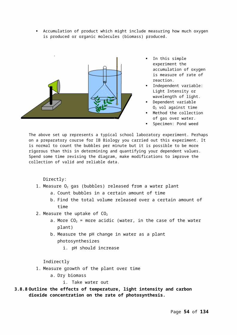
Y XHY XHY2 XH Xh = 2 carrier females2 XHY = 2 normal males
XH Xh
XH XH XH XH Xh
Y XHY XhY1 XH XH = 1 normal female1 XH Xh = 1 carrier female1 XH Y = 1 normal male1 Xh Y = 1 male with haemophilia
In order for a female to get haemophilia:
XH Xh
Xh XH Xh Xh Xh
Y XHY XhY4.1.16 State that a human female can be homozygous or heterozygous with respect to sex-linked genes.
A human female can be homozygous or heterozygous with respect to sex-linked genes4.1.17 Explain that female can be homozygous or heterozygous with respect to sex-linked genes.
Heterozygous: XH Xh (Carrier)Homozygous: XH XH or Xh Xh
4.1.18 Predict the genotypic and phenotypic ratios of offspring of monohybrid crosses involving any of the above patterns of inheritance.
4.1.19 Deduce the genotypes and phenotypes of individuals in pedigree charts.
Recessive Parents don’t have the trait, but offspring do
Dominant If the offspring have it, one parent must have it
Page 54 of 111
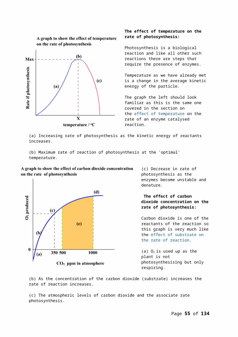
Co-dominance If there are three physical traits
Sex-linked When more males have the trait
4.4 GENETIC ENGINEERING AND BIOTECHNOLOGY4.1.20 Outline the use of polymerase chain reaction (PCR) to copy and amplify minute quantities of DNA.
PCR = Polymerase Chain Reaction (pg. 163 tiger)o Purpose is to make lots of DNA from a small sample
We use a machine to copy small amounts of DNAo This is called amplifyo Piece of bone, hair sample, skin sample etc.
4.1.21 State that, in gel electrophoresis, fragments of DNA move in an electric field and are separated according to their size.
Gel electrophoresiso Fragments of DNA move in an electric field and are separated according to size
4.1.22 State that gel electrophoresis of DNA is used in DNA profiling. Gel electrophoresis of DNA is used in DNA profiling
4.1.23 Describe the application of DNA profiling to determine paternity and also in forensic investigations.
Compare DNA samples from crime scenes with samples from suspects, victims, etc.o For criminal investigationso To see if someone is related (for example, paternity suites)o Identifying remains (like the UN missing persons)
4.1.24 Analyse DNA profiles to draw conclusions about paternity of forensic investigations.
4.1.25 Outline three outcomes of the sequencing of the complete human genome. Sequencing of the entire human genome leads to the production of gene probes to detect
specific genetic diseases Develop pharmaceuticals based on DNA sequences Better understanding of human development Technology
4.1.26 State that, when genes are transferred between species, the amino acid sequence of polypeptides translated from them is unchanged because the genetic code is universal.
When genes are transferred between species, the amino acid sequence of polypeptides translated from them is unchanged because the genetic code is universal
4.1.27 Outline a basic technique used for gene transfer involving plasmids, a host cell (bacterium, yeast or other cell), restriction enzymes (endonucleases) and DNA ligase.
The basic technique to transfer genes is called:o Gene transfero Genetic engineeringo Genetic modificationo Genetic recombination
Results in: Genetically modified organisms (GMOs)o Recombinant DNA
We need:o Restriction enzymes (endonucleases)
Enzymes that cut DNA in certain sequences. They leave staggered edges on the ends of the DNA called “sticky ends”
o Bacterial plasmids
Page 55 of 111

Small circle of extra DNA in a bacterial cello Host organism
Usually bacteria because they are easy to culture and they take in DNA easily
Steps1. Isolate two kinds of DNA: the human gene of interest and a bacterial plasmid.2. Treat plasmid and human DNA with the same restriction enzyme. They will have
complementary sticky ends.3. Mix the human DNA and plasmids together. They will base pair with their sticky ends.4. Add DNA ligase to form covalent bonds between sugar and phosphate.5. Put the recombinant plasmid into a host organism. Bacterial cells should absorb the
plasmids.6. Culture the genetically modified bacteria to produce large colonies that make the human
protein.4.1.28 State two examples of the current uses of genetically modified crops or animals.
High salt tolerance tomato plants “Golden rice” – Rice with more vitamin A Bt maize (or bt corn) – Insect repellent crops, a gene from bacteria is transferred into corn
making the corn produce a toxin that kills insects Factor IX in sheep milk – a human blood clotting gene is inserted into sheep so they produce
the factor IX in their milk4.1.29 Discuss the potential benefits and possible harmful effects of one example of genetic modification.
Benefits of Bt Maizeo Less pesticide useo Economical, less land area used to grow the same amounto Higher crop yields to reduce world hunger
Possible harmful effects of Bt Maizeo Humans may be harmed by the Bt toxino Kills all insects (predators or “good” species)o Genes could spread to wild plants making them “superweeds”o Corn could spread to nearby fields outcompeting wild species
4.1.30 Define clone. A group of genetically identical organisms Or a group of cells derived from a single parent.
4.1.31 Outline a technique for cloning using differentiated animal cells. Plants can be cloned by cutting off a branch and putting it in water and it grows roots and
then planting it Steps
1. Isolate a donor cell from the animal that you want to clone2. Remove and discard the nucleus from another female’s egg cell3. Transfer the nucleus from the somatic cell (step 1) into the enucleated egg cell4. Stimulate cell division5. Implant the embryo into the surrogate mother (or any mother)
4.1.32 Discuss the ethical issues of therapeutic cloning in humans. Cloning stem cells could be harmful to embryos which are potential lives Humans should not decide whether embryos live or die Religions usually disagree with changing DNA Possible harmful effects that we don’t foresee
Topic 5: Ecology and evolution5.1 COMMUNITIES AND ECOSYSTEMS
Page 56 of 111

4.1.33 Define species, habitat, population, community, ecosystem and ecology.
4.1.34Distinguish between autotroph and heterotroph.
4.1.35 Distinguish between consumers, detritivores and saprotrophs.
4.1.36 Describe what is meant by a food chain, giving three examples, each with at least three linkages (four organisms).
Food chains show a simple linear flow of' who eats who' and therefore the energy and matter flowing through the links in the chain
Carrot plant ---> Carrot fly ---> Flycatcher -----> Sparrow hawk
1. The carrot fly consumes the carrot plant.
2. Some of the carrot plant molecules are assimilated by the fly for growth of the carrot fly and others are metabolised in fly respiration.
3. The carrot fly is consumed by the flycatcher.
4. Some of the molecules of the carrot fly are assimilated by the fly for growth of the flycatcher and others are metabolised in flycatcher respiration.
Page 57 of 111

5. In turn the flycatcher is consumed by the sparrow hawk.
6. Some of the molecules of the flycatcher are assimilated for the growth of the sparrow hawk and others are metabolised in hawk respiration.
Bushgrass---> Impala ---> Cheetah----> Lion
buckwheat ---> Gopher ---> Gopher snake ----> Red Tailed Kite
As you view each food chain try to focus on the processes in which energy and matter are transferred along the chain from one organism to the next.
Consider at each stage how much of the available energy in the food is actually captured by the consumer.
What kind of processes will create losses from one link to the next. Consider the question: Why are big, scary predator so rare?
4.1.37 Describe what is meant by a food web.
The food web is a diagram that shows how food chains are linked together into more complex feeding relationships
The food web has a number of advantages over a food chains including:
Shows the much more complex interactions between species within a community/ ecosystem
More than one producer supporting a community
Page 58 of 111

A single producer being a food source for a number of primary consumers That a consumer may have a number of different food sources on the same or
different trophic levels That a consumer can be an omnivore, feeding as a primary consumer and as a
consumer at higher trophic levels
There are certain problems in drawing a complete food web as this would in most cases require a very complex study and identification of species. For this reason, food webs often reflect the interests of its author. The author will detail the species of interest by name but group other less interesting/ important species into larger family. order groups.
4.1.38 Define trophic level
The trophic level of an organism defines the feeding relationship of that organism to other organisms in a food.
In a food web a consumer can occupy a number of different trophic levels depending on which organism is the prey.
4.1.39 Deduce the trophic level of organisms in a food chain and a food web.
Using the simple food web to the left.
Determine the trophic level of each organism in
the diagram using the table in 5.1.6 Check your assessment by placing the mouse-over the diagram. The red numbers provide the correct answers.
4.1.40 Construct a food web containing up to 10 organisms, using appropriate information.
4.1.41 State that light is the initial energy source for almost all communities. To maintain food chains, food webs, communities and all their interactions
requires energy.
Sunlight is the source of this energy for most communities both aquatic and terrestrial.
Page 59 of 111

The principle trap of sunlight energy is the protein molecule chlorophyll found in the chloroplasts of producers cells
4.1.42 Explain the energy flow in a food chain.
a) Not all solar energy will come into contact with chlorophyll and will therefore not be trapped in the synthesis of organic compounds during photosynthesis
b) Photosynthesis in which light energy is trapped by producers.
c) Consumers feeding and passing on energy in the food molecules.
d) Loss of energy as heat from respiration
e) death and the consumption of dead organisms by detritivores. Or as food not assimilated because of incomplete digestion.
Energy Loss
loss of energy in undigested food which will then be used by saprophytes/ decomposers
loss of heat energy in the reactions of respiration ultimately all energy will be lost has heat
4.1.43 State that energy transformations are never 100% efficient. The transfer of energy from one trophic level to the next is inefficient Approx 10-20 % of the energy on one trophic level will be assimilated at the next
higher trophic level4.1.44 Explain reasons for the shape of pyramids of energy.
This model shows the typical loss of energy from solar radiation through the various trophic levels.
Note how this causes a tapering of the model
Page 60 of 111

The volume of one layer is 10% of the layer below.
It is this loss of energy which in part makes food chains relatively short. In extreme environments like the arctic the initial trapping of energy by producers
is low. Thus the food chains are short. In a tropic rainforest the trapping of energy is more efficient and therefore food
chains are longer, webs are more complex.
This is a more typical pyramid of energy. Note that the initial solar energy is not shown.
The narrowing shape illustrates the gradual loss of energy progressing along the links of a food chain to
higher tropic levels (see above for detail).
The base of this pyramid would have a scale = energy/ area/unit time e.g. kJ m-
2 yr-1
Unlike pyramids of number (of organisms) a pyramid of energy cannot invert due to the second law of thermodynamics,'energy cannot be created nor destroyed' .
4.1.45 Explain that energy enters and leaves ecosystems, but nutrients must be recycled.
(a) Energy flows: this diagram is a simple version of the pyramids of energy. At each trophic level energy is lost as heat. At the top of the pyramid of energy it tapers to a point showing how all energy is ultimately radiated to space as heat.
(b) Matter cycles: new matter is not created, no new carbon, hydrogen or oxygen. Producers (autotrophs) take inorganic molecules and convert them to organic compounds. Consumers feed at different trophic levels taking in organic matter and using it for their own growth. This cycling of matter is the subject of the carbon, nitrogen and water cycle.
4.1.46 State that saprotrophic bacteria and fungi (decomposers) recycle nutrients.
Saprotrophic bacteria and fungi recycle the nutrient (organic molecules) of dead organisms.
Page 61 of 111

Decomposition is a complex process and serves many functions, including the formation of soil, the recycling of nutrients stored in the organic materials, and the reduction of high energy carbon compounds.
Decomposition is a biological process begins with the secretion
of extra-cellular digestive enzymes These enzymes are produced by the saprophytic bacteria and fungi They secrete the enzymes onto the dead organism The enzymes hydrolyse the biological molecules of which the dead organism is
composed The hydrolysed molecules are soluble and will then be absorbed by the fungi or
the bacteria Organic molecules are oxidised to release carbon dioxide back to the atmosphere Organic molecule are oxidised to release nitrogen in form of nitrate, nitrite and
ammonium. The oxidation of these organic compounds produces energy for the saprophyte
but returns the various forms of matter to the abiotic environment.
5.2. THE GREENHOUSE EFFECT5.1.1 Draw and label a diagram of the carbon cycle to show the processes involved.
5.1.2 Analyse the changes in concentration of atmospheric carbon dioxide using historical records.
The trends in atmospheric gases are studied as indicators of potential climate change.
Major gases studied include carbon dioxide, methane and oxides of nitrogen which are collectively called the greenhouse gases (5.2.3).
Page 62 of 111
Fossilization
Feeding
Fossilization
Death
Death
Photosynthesis
(CO2 - > O2)
Respiration
(O2 -> CO2)Respiration
(O2 -> CO2)
Plants
(Organic Molecules)
Animals
(Organic Molecules)
Fossil FuelsAtmospheric CO2Decomposers

Atmospheric carbon dioxide has been monitored at Mauna loa atmospheric laboratory on Hawaii since 1958. There are now other laboratories around the world which are adding to the database of carbon dioxide levels in the atmosphere.
The graph below illustrates what is known as the Mauna Loa effect or the Keeling Curve
Carbon dioxide is released unevenly around the world which in part is due to the distribution of vegetation. The collective data therefore allows us to see what happened after there is a mixing of the atmospheric carbon dioxide.
The analysis of carbon dioxide trends is complex and is affected by a number of factors and assumptions.
However the basic trend is an increase in atmospheric carbon dioxide levels.
Longer term estimates of global CO2levels have been determined by a variety of sources including Isotopic analysis of gases trapped in
ancient ice cores.
Bubbles of atmospheric gases are trapped within the ice formed thousands of years ago. Taking cores of the ice and then analysing the gases allows the CO2levels to be determined.
Direct instrument measurements of surface temperature are only reliable back to approx 1850. Prior to that estimates based on estimates from proxy methods. It is necessary to average the various estimates form proxy methods. It
is also possible to calibrate such proxy estimations by comparing the last 150 years of direct instrumental measurements against the proxy estimates for the same period. These adjustment factors can the be applied to historical estimates of temperature to provide a more reliable value.
Students may like to access: Historical trends in carbon dioxide concentrations and temperature, on a geological and recent time scale and carry out a critical analysis of the presented data trends. Hint....look at the scales.
5.1.3 Explain the relationship between rises in concentrations of atmospheric carbon dioxide, methane and oxides of nitrogen and the enhanced greenhouse effect.
Page 63 of 111

Students should note:Greenhouse effect is a natural process that creates moderate temperatures on earth to which life has adapted. The earth has relatively little carbon dioxide in its atmosphere compared to a planet like venus which has an atmosphere of CO2 x 200, 000 times greater
and a surface temperature nearly twenty times higher than earth.Enhanced greenhouse effect is the concern that the activities of human's may be increasing the levels of carbon dioxide and other 'greenhouse gases' such as methane and oxides of nitrogen in the atmosphere. That this may lead to increased global temperatures and
climate change. a) Short wave solar radiation (light)b) light penetrates the atmosphere and passes through the molecules of the atmospherec) Absorption by the ground and conversion to long wave infrared radiation (heat)d)This warms the planete) Some infrared is lost to space as heatf) Atmospheric gases particularly water vapour, carbon dioxide, methane and CFC'sg) Greenhouse gases absorb infra-red radiation and scatter this rather than letting it escape to space. In effect this traps the heat energy.h) Some light reflects off the outer surface of the atmosphere and never entersNote that if this 'greenhouse' effect did not exist the average global temperature would be -17 C. The enhanced greenhouse effect.
Increase in carbon dioxide and other greenhouse gases (methane, oxides of nitrogen) will increase the particles(f) in the above diagram.
Therefore more infra-red will be absorbed, scattered and retained as heat. The average global temperatures will rise. Some models suggest as much as 40C in the next
50 years.
Page 64 of 111

An enhanced greenhouse effect is predicted to cause global climate changes. This is often referred to as global warming but whilst the average global temperatures may rise the local effects may vary widely.
5.1.4 Outline the precautionary principle.
'If the effects of human-induced change would be large, perhaps catastrophic, those responsible for the change must prove that it will not do harm before proceeding.'
William Clifford in his 1879 essay "The Ethics of Belief", contains the famous principle: "it is wrong always, everywhere, and for anyone, to believe anything upon insufficient evidence."
In this context Clifford would have argued that its is wrong (immoral) to believe in climate change or the causes of climate change until there is sufficient evidence to to support this view.
Cliffords argument is an extension of the 'Burden of proof' principle in which (in context) it is up to those who claim that there is climate change( due to human causes) to prove that this is the case. That there is no need to respond to request for action to reduce human impact until that case has been proven.
However the 'Precautionary principle' suggests that the obligation actually falls on those accused of causing climate change (or enhanced greenhouse effect) to show that their actions are not causing damage. If we wait until it is proven that humans are causing climate change it will be too late to reduce the impacts. Is it better to respond now as a precaution even if in the long term it turns out that the case cannot be made.
The use of the precautionary principle in risk assessment is covered in this policy document form the UK Health and Safety Executive.
This wiki article is a good summary of the precautionary principle.
5.1.5 Evaluate the precautionary principle as a justification for strong action in response to the threats posed by the enhanced greenhouse effect.
Modern Skepticism has a number of tools that allow the critical examination of phenomena. These tools are often used in the natural sciences to examine the quality of evidence which is provided in support of a hypothesis.
Such tools include:
Burden of Proof; in which the obligation is on those who make claims about the causes of phenomena to provide the evidence before other might change their understanding or behaviour.
Occams Razor; in which a phenomena is always explained with reference to the simplest possible explanation.
Sagans Balance; which is the principle that extraordinary claims require extraordinary evidence.
Authors such as William Clifford (more recently Jonathan Adler) argue that it is unethical to believe in a phenomena for which the evidence is poor.
The Precautionary principle reverses the argument of the 'Burden of Proof'. The precautionary principle argues that those responsible for an effect (global warning, climate change, enhanced greenhouse effect) have the obligation to show that what they are doing has causes no harm.
Page 65 of 111

In context of the enhanced greenhouse effect then:
Skeptic approach: The 'burden of proof' lies with those claiming that harm is caused by those responsible for the enhanced greenhouse effect to provide evidence that this is in fact the case.
I.e. the environmentalists need to provide conclusive evidence that the actions of the 'polluters' are causing harm to the environment.
Precautionary Principle: Those allegedly responsible for causing the effects of the enhanced greenhouse effect are required to demonstrate that their actions do not cause harm.
This responsibility would then fall on a wide spectrum or Nation Government, Industries, Communities and individuals to demonstrate that their actions cause no harm.
This includes the actions of every individual including you, the reader!
Climate is a complex phenomena with many emergent properties often based on time frames beyond the human experience. This makes the exact predictions of location and timing it difficult. It is however possible to hypothesis about the general effects.
increased frequency and intensity of droughts; flooding as a result of higher rainfalls, increased snowmelts, and rising sea levels;
This interactive site allows maps you to determine the rise in sea level and the effects this will have on coastlines. (Brilliant !).
declines in food production; increased disease; infectious pathogens ar generally reduced by cold
temperatures (winter). warmer temperature will allow pathogens to survive better and indeed for diseases to extend their distribution. Current evidence suggest that both malaria and dengue fever are currently extending their distribution.
more extreme weather; and loss of biodiversity.
5.1.6 Outline the consequences of a global temperature rise on arctic ecosystems.- Loss of habitat (polar bears)- Increased success of pest species (mosquitoes = malaria)
o Including pathogens- Increased decomposition of detritus previously trapped in the permafrost (more
decomposition = more CO2)- Expansion of range of habitats for temperate species- Changing prey can affect higher trophic levels
5.3 POPULATIONS5.1.7 Outline how population size is affected by natality, immigration, mortality and emigration.
Mortality = death rate increases, population decreasesNatality = birth rate increases, population increasesImmigration = coming in to population, immigration increases, population increasesEmigration = leaving the population increases, population decreases
If natality and immigration is greater than mortality and emigration the population increases.N + I > M + E => Population increases
M + N > I + E =? UNKNOWN rate is needed5.1.8 Draw and label a graph showing a sigmoid (S-shaped) population growth curve.
Page 66 of 111

5.1.9 Explain the reasons for the exponential growth phase, the plateau phase and the transitional phase between these two phases.Exponential Growth Phase
Natality > Mortality Population increases at an increasing rate Most offspring survive and reproduce Plenty of resources Each generation produces more than the last
Transitional Phase Natality > Mortality Rate of increase slows down Resources are becoming limited
Plateau Phase Natality = Mortality Reached carrying capacity
o The maximum number the environment can support Resources are limited
o Food, shelter, water, space etc.5.1.10 List three factors that set limits to population increase.
Competition for resources Accumulation of toxic by-products in waste Increase in disease/parasites Increase in predators
5.4 EVOLUTION5.1.11 Define evolution.
Evolution: The cumulative change in the heritable characteristics of a population.Evolution may occur by the process of natural selection. (Darwin-Wallace theory)
1) All populations show variation. No two individuals are the same.Variation is inherited (genetic).
2) Populations tend to produce more offspring than the environment can support.3) There is a struggle for survival. Individuals compete for resources or to avoid predation.
The individuals that are most suited for the environment are more likely to survive and reproduce. (Natural selection)
5.1.12 Outline the evidence for evolution provided by the fossil record, selective breeding of domesticated animals and homologous structures.
“The fossils show evidence not proof evolution.”
Page 67 of 111
P
T
E
1) Fossil record Preserved remains of organisms Evidence not proof Under sedimentary rocks A chronological collection of life’s remains during the passage of time It does not give evidence for how evolution occurs
2) Selective breeding of domesticated animals Human chose traits in wild animals most suitable for humans Breeding animals with the favourable traits over and over Shows that selection can cause evolution Not natural selection, it is artificial selection
3) Homologous Structures Structures that look superficially different and perform a different function but have
basic similaritiesi. Example: Pentadactyl limb
ii. Come from a common ancestor Very similar bones so they must have come from a common ancestor and changed
in response to different lifestyles Embryos – all vertebrate embryos look similar at certain stages
i. Evidence for a common ancestor5.1.13 State that populations tend to produce more offspring than the environment can support.
Population growth produces more offspring than the carrying capacity of an environment can support.
5.1.14 Explain that the consequence of the potential overproduction of offspring is a struggle for survival.
The population produces more offspring than the carrying capacity of the environment can support
Offspring/population compete for limited resources (Intraspecific competition) Some individuals have characteristic (or combination ) that give them a competitive
advantage. These individuals are consequently 'fitter' in terms of freedom from disease, food availability
etc. These individuals are more likely to successfully reproduce (offspring survive) Through inheritance of the genes for these advantageous characteristics the frequency of
these characteristics become greater in the next generation. The alleles for the advantageous characteristic becomes more frequent in the population
Page 68 of 111

5.1.15 State that the members of a species show variation.
Populations of a species show variation. Variation means differences in phenotypes.
Variation shows two basic patterns.
This type of variation is called discontinuous.
There are distinct classes of individual
e.g. Blood groups of a human population
Discontinuous variation usually indicates the condition is controlled by one to two genes.
This type of variation is called continuous variation with no distinct classes but a complete range of the characteristic
e.g. Height of a trees in a forest
Continuous variation like this normally indicates a polygenic condition or multiple alleles
5.1.16 Explain how sexual reproduction promotes variation in a species.
Asexual and sexual populations both experience mutation which increases the variation within the members of a population. However sexually reproducing populations also experience significant additional sources of variation.
The sources of genetic variation in a populations :
Meiosis and the independent assortment of chromosomes creates 2n new combinations of chromosome in the next generation n = haploid number of chromosomes
Random fertilisation increases the variation in the population to 22n again where n = haploid number of chromosomes
The number of different genetic variations is increased further by cross-over in meiosis by an estimated 23 in addition to the two above.
5.1.17 Explain how natural selection leads to evolution.Natural selection is a process (not a thing) which requires:
Production of variation. (the random part) the actual selection (non-random)
When a population evolves there is a cumulative change in the heritable characteristics of the population.Natural selection can act on a population without speciation occurring.In effect the genetic profile of the population is adapting to changes in local conditions.Every phase in the process of evolution is affected by variation and by selection.
Page 69 of 111

5.1.18 Explain two examples of evolution in response to environmental change; one must be antibiotic resistance in bacteria.
Page 70 of 111
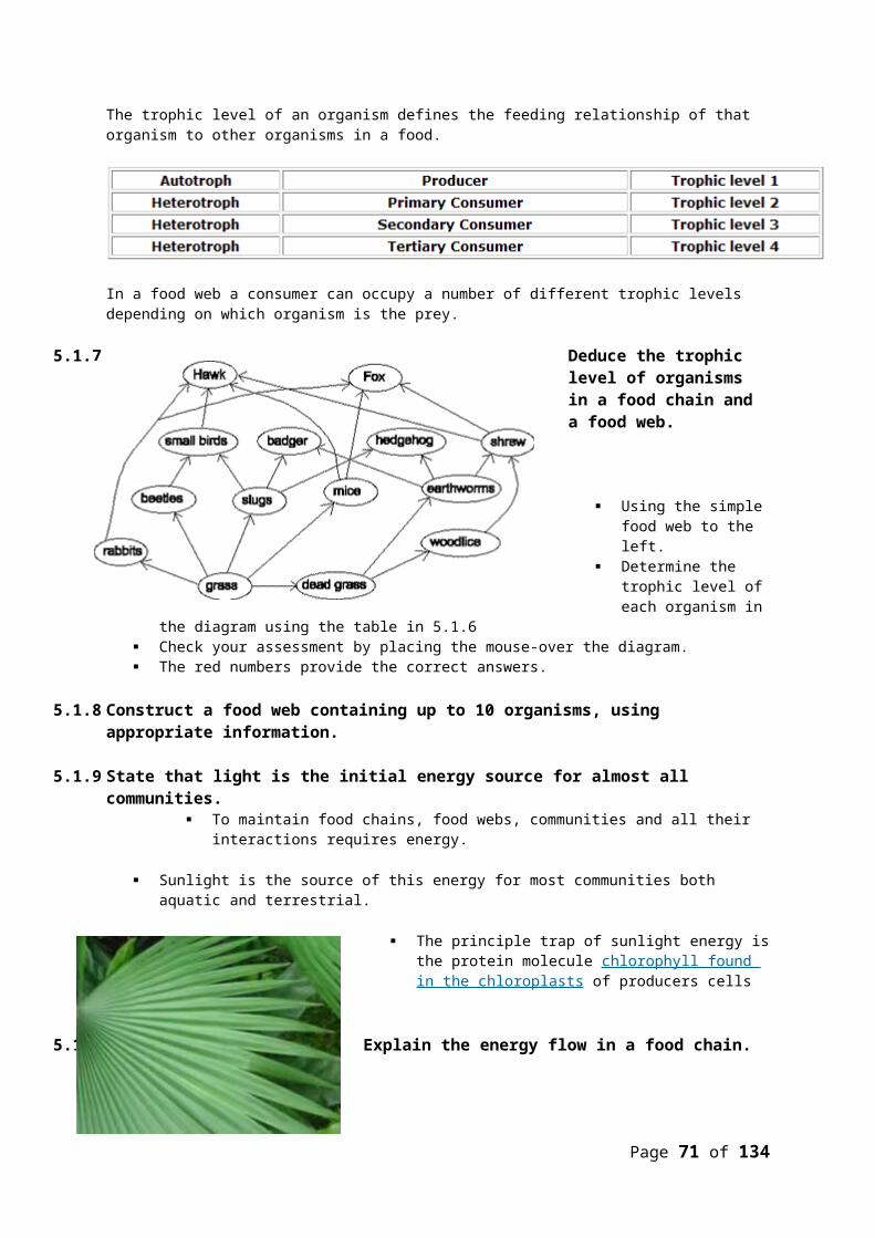
Example 1: Staphylococcus aureus
This bacteria is associated with a variety of conditions including skin and lung infections. As an example of evolution it can be shown that the population of S. aureus has diverged into two forms.
The bacteria is in two forms;
Methicillin-resistant Staphylococcus aureus (MRSA) also known as the epidemic MRSA against which Methicillin antibiotic has no effect.
Methicillin Susceptible Staphylococcus aureus (MSSA). This form is still contained by the use of the antibiotic Methicillin.
This image shows the increase in the increased frequency of MRSA from samples in USA hospitals
The genome for S. aureus was completed at the Sanger Institute and published in June 2004.
Comparison of the genomes for the two forms shows significant genetic differences between MRSA and MSSA.
How MRSA evolved:
Antibiotics selectively kill susceptible forms of the bacteria. The antibiotic is the selection pressure on the population of Staphylococcus
aureus. Random mutation with the species produces a resistance gene at low frequency
in the population The gene can be transferred via plasmids to other bacteria Frequent use of the antibiotic puts MRSA at a selective advantage to survive and
MSSA at a selective disadvantage MRSA therefore survives the antibiotic to reproduce. The descendants of MRSA will also carry the resistance gene The resistance gene increases in frequency or there is a process of cumulative
change in the heritable characteristics (resistance gene) in the population The species has evolved into two new forms
Currently this organisms is the subject of much concern amongst Health professionals particularly in the UK and USA. . This concern stems from the evolution of a new antibiotic resistant form of the species (eMRSA-16 or MRSA252). Figures from the USA indicate that some 51% of infections are contracted in hospital itself and 31% are contracted from within the community.
Page 71 of 111

Example 2 New Zealand Kaka
1. Proto-kaka isolated from ancestral parrot family by the Tazmin sea formation 100 my ago.
2. Mountains form in New zealand (southern alps) creating alpine environment about 50 my ago.
Proto-kaka diverges under selection pressures to form the alpine kea adapted to the mountain environment and the lowland kaka.
3. 0.5 M years ago New Zealand splits into two Islands. The Lowland Kaka diverges into two reproductively isolated populations of the North and South Island Kaka.
5.5 CLASSIFICATION5.1.19 Outline the binomial system of nomenclature.
Binomial nomenclature – devised by Linnaeus Every spears has a two-part name:
o First name = genuso Second name = species
Homo Sapeins
Genus: Closely related speciesSpecies: Usually describes them in Latin
The whole name must be underlined (or Italicized) First letter capital Space between first and second name Can be abbreviated: H. Sapiens
5.1.20 List seven levels in the hierarchy of taxa—kingdom, phylum, class, order, family, genus and species—using an example from two different kingdoms for each level.Example 1: Humans
Animalia Kingdom KinkyChordata Phylum PervertsMamalia Class ComePrimates Orders Over
Page 72 of 111

Hominidae Family ForHomo Genus GroupSapiens Species Sex
Kingdom: 5 Kingdoms, Animals, plants, fungus, protoctista, BacteriaPhylum: About 20 phyla
More examples:Grey Wolf Date PalmAnimalia PlantaeChordata Angiospermophyta
Mammalia MonocotyledoneaeCarnivora PalmalesCanidae Aecaceae
Canis PhoenixLupus Dactylifera
The species would be written as:Canis Lupus Phoenix Dactylifer
5.1.21 Distinguish between the following phyla of plants, using simple external recognition features: bryophyta, filicinophyta, coniferophyta and angiospermophyta.
Bryophyta Filicinophyta Coniferophyta AngiospermophytaNo roots (rhizoids to anchor them, moss)
Roots Roots
Simple leaves and stems, no veins
Leaves (divided) Conifers (cones) – waxy narrow needle like leaves
Leaves
Woody stems StemSpores on the end of stalk
Reproductive sports: Sporangia (sori)
Reproductive cones Reproduction: make flower, pollen from one to other with inset or wind
Can grow up to 20 m Vascular system (veins: tracheids)
Vascular bundlesWaxy cuticleCan grow up to 100 m
Moss Ferns Flowers5.1.22 Distinguish between the following phyla of animals, using simple external recognition features:
porifera, cnidaria, platyhelminthes, annelida, mollusca and arthropoda.Phylum Mouth/Anus Symmetry Skeleton Other Features
Porifera(Sponges)
None None Internal spicules Pores in the surface for filler feeding
Cnidaria(Jellyfish, sea anemones, coral)
Mouth Radial Soft, but coral make CaCO3
Tentacles around mouth with stinging cells
Platyhelminthes(Flatworms, tapeworms)
Mouth Bilateral Soft (none) Flat, no blood system
Mollusca(Snails, octopus, shells, squid)
Mouth, anus Bilateral Shell made of CaCO3
Mantle that secrets the shell, feed with a radula
Page 73 of 111

Annelida(Bristle worms, earth worm, leeches)
Mouth, anus Bilateral Internal cavity with fluid under pressure
Divided by rings/segmentsBristles
Arthropoda(Insects, spiders, crustaceans, scorpions, shrimp, crabs, lobster)
Mouth, anus Bilateral External plates made of chitin (there’s nothing on earth that can digest chitin)
Segmented bodies with joints between segmentsJointed legs
5.1.23 Apply and design a key for a group of up to eight organisms.
Topic 6: Human health and physiology6.1 DIGESTIONS
5.1.24 Explain why digestion of large food molecules is essential. Most food molecules are large polymers and insoluble They must first be digested to smaller soluble molecules before they can be absorbed into
the blood
5.1.25 Explain the need for enzymes in digestion. Enzymes are biological catalysts that increase the rate of reaction Digestive enzymes are secreted into the lumen of the gut Digestive enzyme increase the rate of reaction of the hydrolysis of insoluble food molecules
to soluble end products Digestive enzymes increase the rate of reaction at body temperature This image illustrates the reduction in activation energy that is achieved by the use of an
enzyme
Page 74 of 111

Notice that the normal reaction requires a higher activation energy which would correspond to a high body temperature. This is usually not possible in living organisms.
The enzyme-catalysed reaction has a lower activation energy. This lower activation energy would correspond to body temperature but is only possible in the presence of an enzyme
5.1.26 State the source, substrate, products and optimum pH conditions for one amylase, one protease and one lipase.
Enzymes Sources Substrate Product Optimum pHSalivary amylase Salivary glands
(mouth)Starch Maltose 7
(Neutral)Pepsin Stomach Protein Smaller
polypeptides2
(Basic-Acid)Pancreatic lipase Pancreas Lipids Fatty acids &
glycerol7
(Neutral)5.1.27 Draw and label a diagram of the digestive system.
Page 75 of 111

5.1.28 Outline the function of the stomach, small intestine and large intestine.1. Stomach:
The stomach stores the food from a meal and begins protein digestion. (a) Lumen of the stomach which stores the food from a meal(b) Gastric pits from which mucus , enzymes and acid are secreted(c) Mucus secreting cells. Mucus protects the surface of the stomach from auto-digestion(d) Parietal cells that produce HCL which kills microorganism that enter the digestive system (food & tracheal mucus). This also converts inactive pepsinogen to active pepsin(e) Chief cells: produces pepsinogen a protease enzyme
2. Small IntestineIn the small intestine digestion is completed.
The products of digestion are absorbed into the blood stream. (a) Villus which increase the surface area for absorption of the products of digestion(b) Microvilli border of the epithelial cell increases the surface are for absorption.(c) Lacteals are connect to the lymphatic system for the transport of lipids.(d) In the wall of the small intestine are the blood vessels to transport absorbed products to the general circulation, There are also the muscle to maintain peristalsis
Page 76 of 111

3. Large Intestine or colon:The colon is responsible for the reabsorption of water from the gut. (a) The lumen of the colon(b) The mucus producing goblet cells(b) Muscular walls to maintain peristalsis
5.1.29 Distinguish between absorption and assimilation.Insoluble food molecules are digested to soluble products in the lumen of the gut.Absorption:
1. The soluble products are first taken up by various mechanisms into the epithelial cells that line the gut.
2. These epithelial cells then load the various absorbed molecules into the blood stream.Assimilation:
1. The soluble products of digestion are then transported to the various tissues by the circulatory system.
2. The cells of the tissues then absorb the molecules for use within this tissues
5.1.30 Explain how the structure of the villus is related to its role in absorption and transport of the products of digestion.The structure of the villus increases the surface are for the absorption of digested food molecules.
(a) Folds increase SA:VOL ration by X 3
Page 77 of 111
(b) Villi project into the lumen of the gut increasing the surface area by X 10(c) Microvilli are outward folds of the plasma membrane increasing the surface area another X10 This sequence of light microscope and electron micrograph images show the same sequence as the diagram above.
Blood supply in the villus which absorb the end products of digestion from the epithelial cells The lacteals (green) that receive the lipoproteins before transporting them to the circulatory system. Muscular walls that maintain the movement of chyme by peristalsis.
6.2 THE TRANSPORT SYSTEM6.1.1 Draw and label a diagram of the heart showing the four chambers, associated blood vessels,
valves and the route of blood through the heart.
Each atrium fills with blood from the veins.
Page 78 of 111

Ventricles are filled with blood from their respective atria. Blood exits the ventricles (and heart) in arteries. The red arrows suggest oxygenated blood Blue arrows represent deoxygenated blood. Blood enters the heart under lower pressure in veins, it exists the heart in arteries under high
pressure
Heart Valves Vales maintain a one way flow of blood. Atrio-ventricular vales open to let blood flow from atria to the ventricles. The atrio-ventricular valves close to prevent a back flow of blood into the atria. Semi-lunar valves open to allow high pressure blood to pulse into the arteries. Semi-lunar valves close to prevent black flow of blood into the ventricles form arteries. The left atrio-ventricular valve is also known as the bicuspid valve. The right atrio-ventricular valve is also known as the tricuspid valve.
Pressure of blood to the left is greater than pressure to the rightValve flaps (cusps) pushed open.Blood flows to the right.
The pressure on the right is greater than the pressure on the left.Cusps pushed closed.Back flow stops
Page 79 of 111

6.1.2 State that the coronary arteries supply heart muscle with oxygen and nutrients.
The heart has its own blood vessels.
Blood passing through the chambers of the heart does not provide nutrient or oxygen to the heart muscle cells.
Coronary arteries are branches of the aorta which provide the heart muscle with a supply of oxygen and nutrient.
The coronary arteries branch and spread through the heart muscle supplying the individual muscles cells.
6.1.3 Explain the action of the heart in terms of collecting blood, pumping blood, and opening and closing of valves.
Diastole
The heart muscle is relaxed this is called diastole. There is no pressure in the heart chambers. Blood tries to flow back into the heart but closes the semi-lunar valves.
Diastole
Both atria fill with blood returning to the heart in the veins. The right atria fills with blood returning in the vena cava from
the body tissues (deoxygenated). The atrio-ventricular valves are still closed and the atria fill up. When the pressure in the atria is greater than the pressure in
the ventricles the atrio-ventricular valves will open.
Late Diastole
Page 80 of 111

In this diagram the heart is still relaxed (diastole). The pressure of blood returning to the heart and filling the atria is now high enough to open the atrio-
ventricular valves. The pressure in the atria is greater than the pressure in the ventricles. Atrio-ventricular valves open Ventricles begin to fill with blood.
Atrial systole
Both atria contract together (see control of heart rate) The muscles of the atria contract. volume of the atria reduces. Pressure of blood increases Blood flow into the ventricle, filling this chamber and causing
the ventricle wall to stretch.
Ventricular Systole
The ventricle contracts (systole) The pressure increases in the ventricle The atrio-ventricular valve closes The pressure rises further Pressure in the ventricle is greater than the artery, semi-lunar valve
opens Blood pulses into the arteries
6.1.4 Outline the control of the heartbeat in terms of myogenic muscle contraction, the role of the pacemaker, nerves, the medulla of the brain and epinephrine (adrenaline).
Myogenic muscle contraction describes the way the heart generates its own impulse to contract. It does not require external nerve input.
In the wall of the right atrium there are a group of specialised cells(SAN).
Cells of the Sino-Atrial Node generate an impulse that can spread across the muscle cell of both atria (red pathway).
The impulse causes a contraction of both atria together.
The impulse cannot spread to the muscle cells of the ventricles.
The impulse is picked up by a sensory ending called the atrio-ventricular node (AVN).
Page 81 of 111

The atria have already contracted sending blood down into the ventricles.
The ventricles are stretched and full of blood.
(A) The impulse to contract (generated in the SAN) is picked up by the AVN .
(B) The impulse to 'contract' travels down the septum of the heart, insulated from ventricle muscle fibres
(C) The impulse emerges first at the apex of the heart. This causes this region to contract first.
(D) The impulse now emerges higher up causing this region to contract.
(E). This region contract last.
The effect is to spread the contraction from the apex upwards, pushing blood towards the semi-lunar valves.
Modification of myogenic contraction
The basic myogenic contraction can be accelerated or slowed by nerve input form the brain stem or medulla.
There are two nerves:
Decelerator nerve (parasympathetic) which decreases the rate of depolarisation at the SAN. Note that the synapse releases acetyl
choline. Accelerator nerve (sympathetic) which accelerates
the rate of depolarisation at the SAN. Synapse releases nor-adrenaline.
Epinephrine (adrenaline) and heart rate
Page 82 of 111

The hormone epinephrine is produced in the adrenal glands (an endocrine gland). The hormone travels through the blood to its target tissue, the sino-atrial node(SAN). Epinephrine increases the rate of depolarisation of the SAN. This accelerates heart rate. This reaction is associated with the 'fight or flight response'.
6.1.5 Explain the relationship between the structure and function of arteries, capillaries and veins.
a) Arteries have muscular walls and outer layer of collagen for support.The collagen resists the expansion due to the high pressure of blood.The muscle layer contracts on the pulse of blood maintaining pressure all the way to the tissues.b) Veins have carry blood under low pressure the lumen is wide to reduce the resistance to blood flow. c) Capillaries have only a single layer
of endothelium through which exchange can occur in the tissues.
General functions of arteries and veins.Arteries carry blood away from the heart under high pressure.Veins return blood to the heart under lower pressure.Capillaries are the site of exchange of blood with tissue fluid and cells.
6.1.6 State that blood is composed of plasma, erythrocytes, leucocytes (phagocytes and lymphocytes) and platelets.Blood is composed of:
Plasma Erythrocytes Leucocytes
Phagocytes Lymphocytes
6.1.7 State that the following are transported by the blood: nutrients, oxygen, carbon dioxide, hormones, antibodies, urea and heat.
The items listed above take advantage of the properties of water (as a solvent):
Page 83 of 111

Nutrients Carbon dioxide Hormones Antibodies Urea
Or its thermal properties: heat
Oxygen and carbon dioxide rely in the RBC for their transport6.3 DEFENCE AGAINST INFECTIOUS DISEASE
6.1.8 Define pathogen.Pathogen: an organism or virus that causes a disease.
Virus is not an organism but a pathogen Types
o Viruses Influenza, measles, chicken pox, rhino viruses, HIV
o Bacteria Staph infections, tuberculosis (Mycobacterium tuberculosis), tetanus,
syphilis You can take antibiotics to get rid of them
o Fungus Yeast infections, athletes foot
o Protozoans Malaria (Plasmodium sp.), yellow fever, African sleeping sickness
o Worms Flat worms, round worms
6.1.9 Explain why antibiotics are effective against bacteria but not against viruses. Bacterial cells and human cells carry out many different metabolic processes to survive Antibiotics block certain processes in bacterial cells without harming human cells.
o This allows the immune system to catch up Viruses use human cell pathways to reproduce
o These are not affected by antibiotics It is not possible to block these pathways without hurting you
6.1.10 Outline the role of skin and mucous membranes in defence against pathogens. 1st Layer
o Skin Nonspecific defence because it protects against everything Keratin is a tough protein that prevents entry unless skin is broken Oil glands produce fatty acids that have low pH to kill germs Lysozymes break down bacterial cell walls When your skin gets burned off the biggest concern is water loss
o Mucus membranes Mucus and tiny hairs (cilia) line your openings to trap pathogens and push
them out Tears in the eye kill pathogens Stomach and vagina are acidic Lysozymes are also present and break down bacterial cell walls
6.1.11 Outline how phagocytic leucocytes ingest pathogens in the blood and in body tissues. 2nd Line of Defence
o Internal nonspecific barrier Phagocytes are large white blood cells in the bloodstream that eat any
invaders
Page 84 of 111

They eat until they die and they become puss They can also squeeze out of the walls of capillaries and go to
infected or irritated tissues They protect you from everything that comes into your body (non-
specific) Those that smoke irritate the lungs which sends phagocytes to the
lungs and lots of mucus is created6.1.12 Distinguish between antigens and antibodies.
3rd Line of Defenceo Immune system
Specific white blood cells (lymphocytes) respond to specific pathogens (antigen) by producing specific proteins (antibodies) to destroy the pathogens
o Antigens: Any foreign substance that stimulates the production of antibodieso Antibodies: Globular proteins that recognize and bind to specific antigens causing
their destruction. They are produced by lymphocytes
Lymphocytes: One of the two white blood cells that specify in the immune system
6.1.13 Explain antibody production. Many different types of lymphocytes exist in our blood Each type recognizes one specific antigen and responds by dividing to form clones The clones then secrete specific antibodies to kill the antigen
o The bacteria makes antigens on their surface that acts as a flag for helper T cells A type of lymphocytes is helper T cell
It identifies the antigen it’s specified to fight against that is on the surface of a phagocyte and when it gets activated creating a cytotoxic T cell and a B cell
B lymphocyte cells produce antibodies to attack the bacteria therefore destroying the pathogen
B cells that are not used become memory B cells that makes your body immune to specific pathogens
Finally, suppressor T Cells stop the whole process so that you don’t continue to attack your body cells
6.1.14 Outline the effects of HIV on the immune system. (Avert.org) Pathogen: HIV Over time, there’s a reduction in the number of lymphocytes (helper T-cell counts) and the
inability to produce antibodieso Without helper t-cells you basically have no immune systemo Without antibodies you can’t fight off most infectionso You become vulnerable to pathogens
Weakens the body Eventually death
6.1.15 Discuss the cause, transmission and social implications of AIDS. Cause: Human Immunodeficiency Virus (HIV)
o Causes Acquired Immune Deficiency Syndrome (AIDS) Enters helper T-Cells Retro Virus
o It has RNAo It goes in and turns it’s RNA into DNA and becomes part of you
Page 85 of 111

Transmission:o Blood (transfused, needles)o Sexo Across the placenta, childbirth, breastfeeding
Social Implications:o Family suffers griefo Financial problems
Definitely not getting insuranceo Problems getting jobs, insurance, partners, and housingo Countries and economic problemso Treatment is expensive so some peoples can’t get ito Orphans and orphans with AIDS
6.4 GAS EXCHANGE6.1.16 Distinguish between ventilation, gas exchange and cell respiration.
1. Ventilation: The flow of air in and out of the alveoli is called ventilation and has two stages: inspiration
(or inhalation) and expiration (or exhalation). Lungs are not muscular and cannot ventilate themselves, but instead the whole thorax
moves and changes size, due to the action of two sets of muscles: the intercostal muscles and the diaphragm.
2. Gas Exchange:
This is the diffusion of gases (oxygen and carbon dioxide)There are two sites for gas exchange
(a)Alveoli: Oxygen diffuses into the blood from the alveoli and carbon dioxide diffuses from the blood into the alveoli
(b)Tissues: Oxygen diffuses from blood
into the cells and carbon dioxide diffuses from cells to the blood
3. Cell Respiration
Aerobic respiration uses oxygen in the mitochondria and produces carbon dioxide Anaerobic respiration does not use oxygen but still produces carbon dioxide
6.1.17 Explain the need for a ventilation system. A ventilation system is needed to maintain concentration gradients in the alveoli The steep concentration gradient across the respiratory surface is maintained in two ways:
by blood flow on one side and by air flow on the other side. The ventilation system replaces diffuses oxygen (keeping the concentration high) and removes carbon dioxide (keeping the concentration low).
This means oxygen can always diffuse down its concentration gradient from the air to the blood, while at the same time carbon dioxide can diffuse down its concentration gradient from the blood to the air.
6.1.18 Describe the features of alveoli that adapt them to gas exchange.
Page 86 of 111

Large surface area due to the combined spherical shape (600 million alveoli = 80 m2) Flattened epithelial cells of alveoli and close association with capillaries Short diffusion distance from alveoli to blood (0.5-1.0 um) Dense capillary network Moist surface for the solution of gases
6.1.19 Draw and label a diagram of the ventilation system, including trachea, lungs, bronchi, bronchioles and alveoli.
(a) Trachea(b) Cartilage ring support(c) Bronchi (plural) Bronchus (single)(d) Lung(e) Heart(f) Sternum(g) Rib cage(h) Bronchioles(j) Alveoli(k) Diaphragm
6.1.20 Explain the mechanism of ventilation of the lungs in terms of volume and pressure changes caused by the internal and external intercostal muscles, the diaphragm and abdominal muscles.
The diaphragm contracts and flattens downwards. The external intercostal muscles contract, pulling the ribs up and out This increases the volume of the thorax this increases the lung and alveoli volume This decreases the pressure of air in the alveoli below atmospheric (Boyle's law)
air flows in to equalise the pressure
Page 87 of 111

The diaphragm relaxes and curves upwards The external intercostal muscles relax, allowing the ribs to fall This decreases the volume of the thorax This decreases the lung and alveoli volume This increases the pressure of air in the alveoli above atmospheric (Boyle's law)air
flows out to equalise the pressure. The abdominal muscles contract, pushing the diaphragm upwards The internal intercostal muscles contract, pulling the ribs downward This gives a larger and faster expiration, used in exercise
6.5 NERVES, HORMONES AND HOMEOSTASIS6.1.21 State that the nervous system consists of the central nervous system (CNS) and peripheral nerves,
and is composed of cells called neurons that can carry rapid electrical impulses.
6.1.22 Draw and label a diagram of the structure of a motor neuron.
Page 88 of 111
Nervous System
Central Nervous System
(the control)
Brain Spinal Cord
Peripheral Nerves
All signals are conducted by neurons
Nerve - ropelike bundle of neurons

6.1.23 State that nerve impulses are conducted from receptors to the CNS by sensory neurons, within the CNS by relay neurons, and from the CNS to effectors by motor neurons.
6.1.24 Define resting potential and action potential (depolarization and repolarization). Inside there is always a negative charge as opposed to the positive outside
o Proteins are in the insideo Pumps constantlyo Pumps out Na+ (sodium) and pump in K (potassium)
Resting potentialo When the axon is not sending a nerve impulse, Na+ is being pumped out, K+ is being
pumped ino The membrane has a charge difference of -70 mV
Action potentialo The reversal and restoration of the charges across the membrane as a nerve impulse
moves along Depolarization
o Reversal Repolarization
Page 89 of 111
Sensory receptors
Central Nervous System
Brain + Spinal ChordCan be passed around your brain and spinal chord with relay neurons
Effectors Muscles, glands

o Restoration6.1.25 Explain how a nerve impulse passes along a non-myelinated neuron.
1. Resting potential: K+ and Na+ gates are closed2. Na+ diffuses in from another part of the neuron and causes the inside of the neuron to
become positive (depolarization). This makes the Na+ gate open. Na+ floods in by diffusion3. K+ gate opens slowly, K+ diffuses out making the cell negative again (repolarization)4. The K+ gate closes slowly, making the cell more negative than resting potential. So the action
potential cannot go backwards
6.1.26 Explain the principles of synaptic transmission. Nerve impulse crossing a synapse Synapse – gap between two neurons
o Where the charge passes over so you can feel paino Sends the message to your braino By using painkillers you can block this message from going
Pre-synaptic neuron sends a message to the post-synaptic neuron1. A nerve impulse reaches the end of the pre-synaptic neuron2. The pre-synaptic membrane is depolarized causing calcium channels to open and Ca2+ ions to
flow in3. Calcium causes synaptic vesicles to release neurotransmitters into the synaptic cleft by
exocytosis (fusing with the membrane)4. The neurotransmitters cross the synapse and bind to receptors on the post-synaptic
membrane5. Binding causes voltage-gated channels in the post-synaptic membrane to open6. Movement of Na+ can cause an action potential in the post-synaptic cell. The nerve impulse
continues on the other side of the synapse7. The neurotransmitters are quickly broken down or reabsorbed to prevent more action
potentials6.1.27 State that the endocrine system consists of glands that release hormones that are transported in
the blood. The endocrine system consists of glands that release hormones that are transported in the
blood
Page 90 of 111

o They go all over your bodyo Ex. Adrenaline etc.o But only to specific organs that have receptors for them
For example, FHS hormone goes all over the body but is only picked up by the ovaries or the testes
6.1.28 State that homeostasis involves maintaining the internal environment between limits, including blood pH, carbon dioxide concentration, blood glucose concentration, body temperature and water balance.
Homeostasis involves maintaining the internal environment between limitso Ex. Temperature, water balance, CO2 concentration, blood pH, blood glucose
6.1.29 Explain that homeostasis involves monitoring levels of variables and correcting changes in levels by negative feedback mechanisms.
Homeostasis is maintained by negative feedbacko Monitor levels of variables
You have a set point and constantly update ito Respond to the levels – bring them back to normal
6.1.30 Explain the control of body temperature, including the transfer of heat in blood, and the roles of the hypothalamus, sweat glands, skin arterioles and shivering.
The hypothalamus (brain) constantly measures the blood temperature and compares it to a set point of 37ᵒC
If the temperature is below 37ᵒC: (chilled)o Sweat glands stop releasing sweat so no heat loss by evaporationo Shivering: rapid muscle contractions produce heato Skin arterioles constrict keeping blood closer to the core. Less heat is lost by
radiation. If the temperature is above 37ᵒC: (overheating)
o Skeletal muscles are relaxed (no shivering)o Sweat glands release sweat – skin surface becomes damp. When water evaporates,
heat is carried awayo Skin arterioles dilate so blood flows close to the surface allowing heat loss by
radiation Temperature control is carried out by neurons (for the most part)
6.1.31 Explain the control of blood glucose concentration, including the roles of glucagon, insulin and α and β cells in the pancreatic islets.
Cells in the pancreas constantly monitor blood glucose level, comparing it to a set point (~90 mg/100 mg of blood)
If the level of glucose goes up (hyperglycaemia):o Β cells in the Islets of Langerhans of the pancreas produce insulino Insulin tells the liver and muscle cells to absorb glucose from the blood and convert
it to glycogeno Other cells use glucose for cell respirationo Excess glucose gets converted to fat
If the level of glucose goes down (hypoglycaemia)o α cells in the Islets of Langerhans of the pancreas produce glucagonso Glucagons tell the liver to break glycogen down into glucose and put it back into the
blood6.1.32 Distinguish between type I and type II diabetes.
Disease characterized by high blood glucose but not enough glucose in cells where it’s needed
Uncontrolled diabetes leads too Damage to kidneys, nerves, retina (eye)
Page 91 of 111

o Increased risk of cardiovascular diseaseo Poor wound healing (gangrene)
Type Io Β-cells of the pancreas do not produce enough insulino Most often in childreno Autoimmune – T-cells kill β-cellso Insulin can be injected to control this
Type II (90% of diabetics)o Body cell receptors do not respond properly to the insulino Can be controlled by dieto Often associated with genetic history, advanced age, obesity, lack of exercise and
certain ethnic groups6.6 REPRODUCTION
6.1.33 Draw and label diagrams of the adult male and female reproductive systems.
(What we need to know: Testes/testis/testicles, scrotum, epididymis, sperm duct, urinary bladder, seminal vesicles, prostate gland, urethra, erectile tissue, penis, and foreskin.)
(What we need to know: Ovaries, oviduct (fallopian tube), uterus, cervix, and vagina.)6.1.34 Outline the role of hormones in the menstrual cycle, including FSH (follicle stimulating hormone),
LH (luteinizing hormone), estrogen and progesterone.Two hormones are created in the brain (both men and women)
Page 92 of 111

LH and FSH Come from the Pituitary
Women have ovarian hormone levels in the blood Estrogen Progesterone
The egg The eggs are ready from the moment you’re born
o Unlike males who make them during puberty and then continue to The egg and sperm meet in the oviduct not the uterus
The menstrual cycle 28 day cycle An egg matures, comes out of the ovary and the uterine lining prepares for pregnancy Day 0≈5
o Menstruationo Uterine lining (endometrium) is shed
Day 6≈13o Follicle phase
Follicle: Big layer of cells around the eggo FSH coming from the pituitary gland stimulates the development of follicles in the
ovaryo The follicle cells secrete estrogen therefore FSH stimulates estrogen
Estrogen stimulates the repair and growth of the endometrium Estrogen inhibits FSH (negative feedback) Estrogen stimulates the production of LH (from the pituitary)
Day 14o Ovulationo LH peaks causing the egg to rupture out of the ovary (out of the follicle)o LH stimulates the follicle to become the corpus luteumo LH causes the follicle (corpus luteum) to secrete some estrogen and progesterone
Day 15≈28o Luteal phaseo Progesterone: thickens the endometrium preparing for an embryoo Progesterone inhibits FSH and LH (negative feedback)o Lack of LH leads to the inhibition of estrogen and progesterone (they would both
stop)o Falling progesterone levels cause the endometrium to shedo Low progesterone causes FSH to be released
6.1.35 Annotate a graph showing hormone levels in the menstrual cycle, illustrating the relationship between changes in hormone levels and ovulation, menstruation and thickening of the endometrium.
Page 93 of 111

6.1.36 List three roles of testosterone in males.- Pre-natal development of genitalia- Development of secondary sexual characteristics
o Facial hairo Bone and muscle growtho Voice deepening
- Maintenance of sex drive6.1.37 Outline the process of in vitro fertilization (IVF).
Fertility drugs are given to the female to make many eggs mature (follicles develop)o The entire menstrual cycle is disrupted for an entire month before it can be done
Eggs are extracted from the ovary with a syringe through the vaginal wall Sperm is collected from the male Eggs are fertilized by sperm in a petri dish
o Insemination 4 cell embryos are put into the uterus Wait for implantation Take extra progesterone Pregnancy testing
Page 94 of 111

6.1.38 Discuss the ethical issues associated with IVF.Arguments For:
Helps couples with fertility problems Prevent some genetic disorders
Arguments Against Against religion/unnatural Choosing embryos
o Gender, disease, certain characteristics) Allows some embryos to die Health risks
OptionsOption E: Neurobiology and behaviour
E1 STIMULUS AND RESPONSEE.1.1 Define the terms stimulus, response and reflex in the context of animal behaviour.
Stimulus : a change in the environment (internal or external) that is detected by a receptor and elicits a response
Response : a reaction to a stimulus Reflex : a rapid, unconscious response
E.1.2 Explain the role of receptors, sensory neurons, relay neurons, motor neurons, synapses and effectors in the response of animals to stimuli.
Animals respond to stimuli with a reflex Receptors
o Receive the stimuluso Generate a nerve impulse in sensory neurons
Sensory neurons
Page 95 of 111
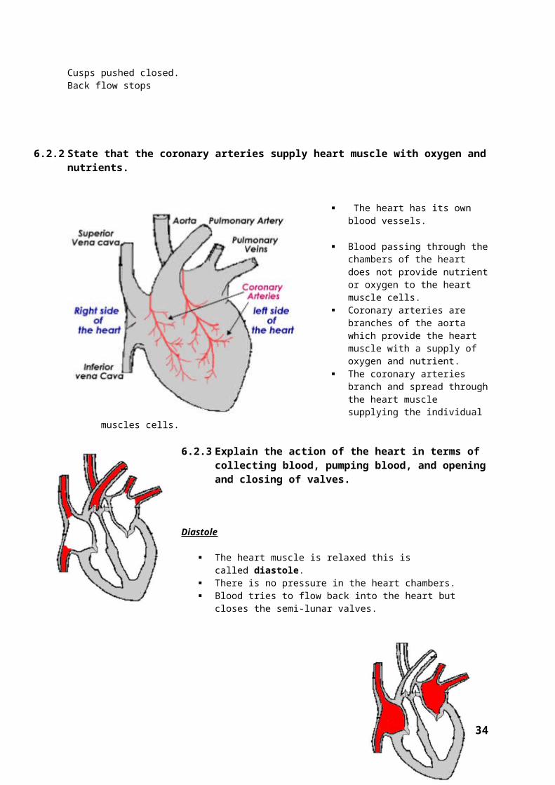
o Carry the impulse to the central nervous systemo The axon enters the spinal cord in the dorsal rooto Sends neurotransmitters to a relay neuron
Relay neuronso Located in the grey mattero Synapses with a motor neuron in the grey mattero Sends the impulse to the motor neuron
Motor neurono Located in the ventral root of the spinal cordo Carries the impulse to an effector
Effectorso Muscle or gland that carries out the response
Impulses can also be carried to the brain by relay neurons in the spinal cordE.1.3 Draw and label a diagram of a reflex arc for a pain withdrawal reflex, including the spinal cord and
its spinal nerves, the receptor cell, sensory neuron, relay neuron, motor neuron and effector. Pain Withdrawal Reflex
o Stimulus (pain) -> sensory receptors -> sensory neuron –(synapse) relay neuron-> motor neuron -> effectors(muscles) -> response (move your limb)
Sensory neuron and motor neuron are in the spinal cord (CNS)
Page 96 of 111

E.1.4 Explain how animal responses can be affected by natural selection, using two examples. Variations in behaviour can occur in populations Variations in behaviour can be selected by the environment The organism with behaviours that are best suited to the environment will be more likely to
survive and reproduce Example: European Blackcaps
o European black cap usually spend the spring and summer in Germany and migrate to Spain for the winter
o In the last 50 years, some have been migrating to the UK in the wintero These birds returned to Germany 10 days earlier than the birds in Spaino The earlier birds had a better choice of territory and laid more eggso An experiment shoed that the migration behaviour was genetico Eggs were reared artificially from the UK and Spanish birds (without parents)o No matter where the birds were raised, they flew in the direction that their parents
wento They are genetically programmed to respond to stimuli when they migrate, so they
fly in a particular direction Example: Sockeye Salmon
o There are two different populations of Sockeye Salmon in Lake Washington, USA (60 years, 13 generations)
o Lake Salmon Live in the lake Spawn on the beaches and lay their eggs in the sandy beach Males have heavy bodies for hiding in deep water They do not swim well in the fast river water
o Variations in the original population were selected for by the two different environments
o Now there are two genetically distinct populations River salmon
Live in the river Females bury the eggs deep in the river bottom so they are not
washed away Males have thinner, narrower bodies for moving in the current
E2 PERCEPTION OF STIMULIE.2.1 Outline the diversity of stimuli that can be detected by human sensory receptors, including
mechanoreceptors, chemoreceptors, thermoreceptors and photoreceptors. Mechanoreceptors
Page 97 of 111
Relay Dorsal Root
Ventral rootMotor
Sensory

o Stimulated by physical deformations (movement, sound, pressure, gravity)o (Hair cells in ear)
Photoreceptorso Detect lighto (Rod and cone cells in eye)o Located in the retina
Chemoreceptorso Detect certain chemicals or changes in concentrationo Inside and outside your body (ex. inside to say if you have too much CO2 in your
blood)o (Olfactory receptor cells in nose)o (Taste buds)
Thermoreceptorso Detect temperatureo (Hot and cold receptors in skin)
E.2.2 Label a diagram of the structure of the human eye.
E.2.3 Annotate a diagram of the retina to show the cell types and the direction in which light moves. Light hits a rod or cone cell (photoreceptors) An action potential is sent from the photoreceptor to a bipolar cell This cell synapses with a ganglion cell The ganglion cell axon goes through the optic nerve to the brain
Page 98 of 111
Choroid
Vitreous humour

E.2.4 Compare rod and cone cells.Rods Cones
Found all over the retina, not the fovea Found concentrated on foveaMore sensitive to dim light Less sensitive to dim lightAbsorb all wavelengths of light (monochrome vision)
Sensitive to red, green and blue light (give colour vision)
Groups of up to 200 pass impulses to the same neuron of the optic nerve (not very accurate)
Each cone cell has its own individual neuron of the optic nerve (greater visual acuity)
Fuzzy visionGrey tones
Good, colour vision but only in bright light
E.2.5 Explain the processing of visual stimuli, including edge enhancement and contralateral processing. Light enters through cornea, pupil, gets focused by the lens and this the retina Photo receptors turn the light energy into an action potential which goes through bipolar
cells and then ganglion cells to the optic nerve Contralateral Processing:
o Both retinas receive information from your left and right fields of visiono Left and right optic nerves cross in the optic chiasmao Neurons from both eyes carrying information from the left field of vision go to the
right side of the braino This allows the brain to perceive depth and distance
Edge Enhancement o Light sensitive receptors in the eye switch off their neighbouring receptorso This makes a nice contrast of black and white edges
E.2.6 Label a diagram of the ear. Know
o Pinna (Outer ear)o Eardrumo Bones of the middle ear
(Malleus, incus, stapes)
Page 99 of 111

o Oval windowo Round windowo Semi-circular canalso Auditory nerveo Cochlea
E.2.7 Explain how sound is perceived by the ear, including the roles of the eardrum, bones of the middle ear, oval and round windows, and the hair cells of the cochlea.
The pinna catches sound waves Sound waves hit the eardrum and cause it to vibrate The bones of the middle ear vibrate and amplify the vibrations about 20 times The last bone strikes the oval window causing it to vibrate The vibration is passed on to the fluid in the cochlea The fluid causes special hair cells to vibrate. These are receptors that release a
neurotransmitter across a synapse to the sensory neuron of the auditory nerve Hair cells are mechanoreceptors Auditory nerve carries nerve impulses to the brain Round window releases pressure so the fluid in the cochlea can vibrate
E3 INNATE AND LEARNED BEHAVIOURE.3.1 Distinguish between innate and learned behaviour.
Innate behaviouro Develops independently from the environment contexto Occurs in all members of the specieso Stereotyped response to a stimulus
Learned behaviouro Develops as a result of experienceo Memories
E.3.2 Design experiments to investigate innate behaviour in invertebrates, including either a taxis or a kinesis.
Taxiso Movement towards or away from a stimuluso Ex. Euglena swim toward light to photosynthesize (positive phototaxis = since the
euglena moves toward the stimulus)o Ex. Maggots move away from light (negative phototaxis)
Page 100 of 111
Oval windowRound window

Kinesiso Response to a non-directional stimulus in which the rate of movement depends on
the level or intensity of the stimulus, not the directiono Ex. Woodlice like humid, dark areas
Design investigations to test innate behaviouro Put woodlice in a controlled environmento Choose a stimulus that causes a responseo Ensure that other factors do not have an effecto Many trials
E.3.3 Analyse data from invertebrate behaviour experiments in terms of the effect on chances of survival and reproduction.
Innate behaviour helps organisms survive and reproduce Basic needs
E.3.4 Discuss how the process of learning can improve the chance of survival. Learn how to avoid predators/danger
o Ex. Baby geese learn to stay near their mother to avoid danger Can avoid poisoning Learn how to obtain food
o Ex. Bears can find a place where there’s shallow water and a lot of salmon Learn how to adapt to a changing environment Learning doesn’t work if you don’t survive
o Benefit of the innate behaviour Ex. If baby’s don’t cry when they need something (innate) then they could
dieE.3.5 Outline Pavlov’s experiments into conditioning of dogs.
A change in behaviour by associating one stimulus with another Two kinds of conditioning
o Operate conditioning: conditioning one stimulus with a reward or punishment Unconditioned stimulus: see or taste food Unconditioned response: make saliva Conditioned stimulus: ring a bell Conditioned response: make saliva
E.3.6 Outline the role of inheritance and learning in the development of birdsong in young birds. Combination of both innate and learned
o The birds have the ability to sing the song of their species but they have to hear it to sing it right
Birds are hatched with a crude template of the species’ song During their development, there is a memorization phase where the birds are quiet, listening
to other birds sing Motor-phase
o Baby bird starts practicingo He must hear himself to try and match his template to the adult
E4 NEUROTRANSMITTERS AND SYNAPSESE.4.1 State that some presynaptic neurons excite postsynaptic transmission and others inhibit
postsynaptic transmission. Some presynaptic neurons excite postsynaptic transmission and other inhibit postsynaptic
transmissionE.4.2 Explain how decision-making in the CNS can result from the interaction between the activities of
excitatory and inhibitory presynaptic neurons at synapses. A neuron sums up all the signals coming in If the sum is inhibitory no new action potential is produced in that neuron
Page 101 of 111

o For example, if there are more negatives than positives, then it stops the action potential
E.4.3 Explain how psychoactive drugs affect the brain and personality by either increasing or decreasing postsynaptic transmission.
Can increase or decrease synapse transmission Mimics the action of a neurotransmitter and binds to receptor Some drugs block the receptors on the postsynaptic membrane so no impulses are sent Increase the release of a neurotransmitter Some can stop the breakdown of neurotransmitters so they stay in the synapse longer
E.4.4 List three examples of excitatory and three examples of inhibitory psychoactive drugs. Excitatory
o Cocaineo Amphetamineso Nicotine
Inhibitoryo Benzodiazepineso Alcoholo Tetrahydrocannabinol (THC)
E.4.5 Explain the effects of THC and cocaine in terms of their action at synapses in the brain. Tetrahydrocannabinol (THC)
o Inhibits nerve impulseso Blocks the release of excitatory neurotransmitterso Binds to cannabinoid receptors
Cocaineo Stimulates transmission at adrenergic synapses (noradrenaline synapses)o Causes dopamine release and blocks the removal of dopamine from the synapseo Dopamine is a molecule of the reward pathway in the brain. It gives you a feeling of
pleasure.E.4.6 Discuss the causes of addiction, including genetic predisposition, social factors and dopamine
secretion. Causes of addiction
o Genetic predisposition Many studies have shown that if someone is addicted to something their
children are more likely to become addict Many studies show addiction runs in families They may have less dopamine receptors
o Social factors Family addiction, being around the addiction a lot Parenting skills Cultural/ethical values Mental health problems Peer pressure
o Dopamine secretion During addiction, dopamine receptors are constantly stimulated The brain can decrease the amount of receptors and the remaining can
become less sensitive
Option F: Microbes and biotechnologyF1 DIVERSITY OF MICROBES
F.1.1 Outline the classification of living organisms into three domains.
Page 102 of 111

Eubacteria Eukaryota Archea
F.1.2 Explain the reasons for the reclassification of living organisms into three domains.o There’s very small differenceso At the time they’re classifying them by what they’re made ofo They also evolve quickly
It’s hard to classify something that’s constantly changingo Scientists noticed differences in rRNA
That’s how they wanted to classify them 3 Domains as opposed to 5 kingdoms
o Everything that wasn’t made up of 1 cell (like bacteria) was classified as protozoans Protozoans
Fungi Plant Animal
o Old system was based on structural (what do they look like?) Now they’re classified by molecular (What are they made of?)
For example, rRNAF.1.3 Distinguish between the characteristics of the three domains.
Characteristic Eubacteria Archaea EukaryaHistones X Histone-like proteins ✔
Introns X Present in some DNA ✔
Size of ribosomes 70S 70S 80SStructure of cell membrane lipids
Unbranched hydrocarbons
Some branched hydrocarbons
Unbranched hydrocarbons
Structure of cell walls ✔ X XMembrane-bound organelles
X X ✔
- This supports the theory that Archaea were the first, and where eubacteria and eukarya evolved from
F.1.4 Outline the wide diversity of habitat in the Archae, as exemplified by methanogens, thermophiles and halophiles.Types of Archaea
- Methanogenso Us CO2 to make CH4 (methane)o O2 kills them (Strict anaerobes)o Live in guts of termites and cattleo Siberian tundrao Swamps, rice fieldso Large intestines of dogs, pigs, humans
- Thermophileso Heat loverso Sulphur hot springs (pH 1-5. 90ᵒC)o Hydrothermal vents in the ocean (105ᵒC)
- Halophileso Salt loverso Saltiest places: Dead Sea, Salt lakes
F.1.5 Outline the diversity of Eubacteria, including shape and cell wall structure.
Page 103 of 111

- Spheres (cocci)o They can be single, pairs, chains, clusterso (staphylococcus, streptococcus)o Like strep throato When cocci go into chains they’re called streptococcuso Staphyli: Grape
Clusters of cocci are called staphylococcus- Rods (bacilli)
o Single, chains (streptobacillus)o E-coli
- Helices (spirilla)o Spirals
- Commas (vibrio)o Comma shapeo Cholera
- Cell wall structure:o Gram positive
Bacteria with this cell wall stain purple with the Gram stain Have many layers of peptidoglycan (a polymer consisting of amino acids and
sugars) Layers are connected by amino acid bridges Antibiotics can kill it
o Gram negative Stain pink Much thinner cell wall Only 20% peptidoglycan Antibiotics can’t kill it Complex cell wall
Covered by an outer membraneo Cell membrane sandwich
F.1.6 State, with one example, that some bacteria form aggregates that show characteristics not seen in individual bacteria.
- Vibro fischerio Bacteriumo Found in seawater that can emit light (bioluminescence)
F.1.7 Compare the structure of the cell walls of Gram-positive and Gram-negative Eubacteria.Gram Positive Eubacteria Cell Wall Structure:
Gram Negative Eubacteria Cell Wall Structure:
Page 104 of 111

Revision- Gram positive
o Bacteria with this cell wall stain purple with the Gram staino Have many layers of peptidoglycan (a polymer consisting of amino acids and sugars)o Layers are connected by amino acid bridgeso Antibiotics can kill it
- Gram negativeo Stain pinko Much thinner cell wallo Only 20% peptidoglycano Antibiotics can’t kill ito Complex cell wall
Covered by an outer membrane Cell membrane sandwich
F.1.8 Outline the diversity of structure in viruses including: naked capsid versus enveloped capsid; DNA versus RNA; and single stranded versus double stranded DNA or RNA.
Viruses are biological structures that are organized but not cells They can only reproduce by hijacking the machinery of a host cell Viruses consist of nucleic acid surrounded by a protein coat called a capsin Viruses can have
o Capsids that are covered in a membranous bilayer derived from the membrane of the host cell they infect (enveloped) or naked capsids
o DNA or RNA Single stranded or double stranded
o Singled stranded or double stranded nucleic acids
HIV: Single stranded RNA with Envelope
Page 105 of 111

F.1.9 Outline the diversity of microscopic eukaryotes, as illustrated by Saccharomyces, Amoeba, Plasmodium, Paramecium, Euglena and Chlorella.Genus Saccharomyces
(Yeast)Amoeba Plasmodium Paramecium Euglena Chlorella
Mode of nutrition
Secrete digestive enzymes outside their cells and absorb the products of digestion back into the cell (extracellular digestion)Yeast ferment carbohydrates and gain energy from this
Uses pseudopodi to wrap around prey and trap themHeterotrophs which take their food into a food vacuole and digest it (Intracellular digestion)
HeterotrophFeeds on red blood cells by digesting the haemoglobin
Cialiated heterotrophDigestion is intracellularFood is taken in by the oral groove and passes to the gulletEventually it forms a food vacuole and is joined with a digestive vacuole to break down the food
Both autotropic and heterotrophicIn the absence of light, it can absorb food from outside the cell
Photosynthesis(Autotrophic)
Locomotion XDoesn’t move
Creates pseudopodia (false feet) out of its cytoplasm
Mosquitos carry the parasite and can infect humans when they bite.Inside the human body, the parasite can move with a gliding motion similar to a crawl
Swims backwards or forwards using cilia
Whipping motion of its long flagellum
XDoesn’t move
Cilia or flagella
X X X ✔ Flagella X
Cell wall ✔
Chitin in cell wallX X X X ✔
Chloroplasts X X X X ✔ ✔
F2 MICROBES AND THE ENVIRONMENTF.2.1 List the roles of microbes in ecosystems, including producers, nitrogen fixers and decomposers.
Nitrogen-fixing- Some can fix nitrogen gas and ammonia- Some bacteria convert nitrites to nitrates- Examples: Rhizobium, nitrobacter, nitrosomonas etc
Decomposers- Saprotrophic bacteria and fungi breakdown dead organic matter and release inorganic
molecules into the ecosystem
Page 106 of 111

Producers- Convert inorganic elements into organic- Photosynthesis- Chemosynthesis
o Use chemical energy to make ATPo Ex. Nitrogen
F.2.2 Draw and label a diagram of the nitrogen cycle.
(1) Bacteria in soil turn NH3 into NO2- by using O2 (Nitrosomonas)
(2) Bacteria in soil turn NO2- into NO3
- by using O2 (Nitrobacter)
Side notes:- Amino acids have Nitrogen
o Necessary for making proteins- Humans can only get it from food so someone has to get it out of the air
o It comes into the living part of the food chain in three ways “Nitrogen fixing by a non-living process, burning fossil fuels to produce
fertilizer (Haber Process)” Produces ammonia (NH3)
“Mutualistic nitrogen-fixing bacteria in rod modules of legumes (Rhizobium)” Produces ammonia (NH3)
Free living nitrogen fixing bacteria in soil (azotobacter) Produces ammonia (NH3)
o So they’re made into something dangerous (ammonia) but are then used to make something useful for us
Through nitrification you turn ammonia into nitrites (nitrosomonas) Nitrobacter make nitrites to nitrates
Page 107 of 111
Nitrification (1)
Nitrification (2)
Putrefaction – decomposers
releases nitrogen compounds
FEEDING
Active transport of nitrates into roots using ATP
Free living nitrogen fixing bacter in soil
Nitrogen fixing by a non-living
process
Burning fossil fuels to produce fertilizer (Haber Process)
Mutualistic nitrogen-fixing bacteria in root nodules of
legumes (Rhizobium)
<- Dentrification: Bacteria put nitrogen back into
atmosphere
FEEDING
FEEDING
FEEDING
FEEDING

Now that we have nitrates, a plant can pick them up They pick it up from the nitrates in soil Animals can eat them
o When plants and animals die or excrete the decomposers make ammonia out of it through putrefaction
Pseudomonas bacteria denitrificate the nitrates and put N2 back to the atmosphere
Farmers don’t want this, they want nitrobacter to put it into the food cycle
F.2.3 State the roles of Rhizobium, Azotobacter, Nitrosomonas, Nitrobacter and Pseudomonas denitrificans in the nitrogen cycle.Rhizobium – Nitrogen-fixing bacteria in root modules of legumes that put N2 from atmosphereAzotobacter – Free living nitrogen fixing bacteria in soil that put N2 from the atmosphere into the soil.Nitrosomonas – Turn ammonia into nitrites.Nitrobacter – Turn nitrites into nitrates.Pseudomonas denitrificans – Bacteria put nitrogen back into the atmosphere (produce N2 from nitrites)
F.2.4 Outline the conditions that favour denitrification and nitrification.Denitrification
- Anaerobic bacteria soil iso Little oxygeno Floodedo Too compact (no air spaces)o High nitrogen input
Nitrification- The two bacteria are aerobic- Soil should be
o Full of oxygeno Neutral pHo Warm temperature
F.2.5 Explain the consequences of releasing raw sewage and nitrate fertilizer into rivers.1. Pathogens in drinking or bathing waters
o Cholerao E. Colio Ritual bathing in Ganges can spread disease
2. Eutrophicationo Fertilizer is like raw sewage-full of nitrogen/phosphateso Algae grow really well in fertilizer – it makes an algae “bloom”o Algae get decomposed by bacteria which use up the oxygen in the water creating a
biochemical oxygen demand (BOD)o The water has little oxygen and the fish can die
F.2.6 Outline the role of saprotrophic bacteria in the treatment of sewage using trickling filter beds and reed bed systems.
Trickling filter systemo A rock bed up to 2 meters deep, is laid outo The rocks are colonized by a biofilm of aerobic bacteriao Sewage water is sprayed onto the rockso Spraying adds oxygen to the sewage to help the bacteria digest the sewage content
Reed bed systemo This is an artificial wetland used to treat waste water
Page 108 of 111

o The waste water provides the water and nutrients to the growing reedso The reeds extract the nitrogen from the waste watero Small animals and invertebrates extract particulateso Saprotrophic bacteria break down the organic wasteo This type of system can only handle small sewage flows and they need a lot of space
Saprotrophic bacteria digest all the organic waste in the sewageF.2.7 State that biomass can be used as raw material for the production of fuels such as methane and
ethanol. Biomass can be used as raw material for the production of fuels such as methane and
ethanol In this case, biomass: waste, human waste, manure
o Manureo Forest productso Agricultural products
F.2.8 Explain the principles involved in the generation of methane from biomass, including the conditions needed, organisms involved and the basic chemical reactions that occur.
Manure and cellulose are put into a digester without oxygeno Anaerobic bacteria convert the raw organic wastes into a mixture of organic acids,
alcohol, hydrogen and CO2
o Second set of bacteria convert acids and alcohol into acetate, CO2 and hydrogeno The last group of bacteria are methanogens (Archaea)
They convert CO2, Hydrogen and acetate into methane CO2 + 4H2 -> CH4 + 2H20
Carbon dioxide + Hydrogen gas -> Methane + 2 waters CH3COOH -> CH4 + CO2
Acetate -> Methane + Carbon dioxide Conditions must be kept constant inside the generator
o No free O2 (all bacteria are anaerobic)o 35ᵒC constant tempo pH not too acidic
F3 MICROBES AND BIOTECHNOLOGYF.3.1 State that reverse transcriptase catalyses the production of DNA from RNA.
Reverse transcriptase catalyses the production of DNA from RNAF.3.2 Explain how reverse transcriptase is used in molecular biology.
RNA and reverse transcriptase enter the host cell, injected by the virus Reverse transcriptase makes a DNA copy of itself DNA of virus injects into nucleus and integrates into the DNA of the host cell
F.3.3 Distinguish between somatic and germ line therapy. Somatic line therapy: only changing your body cells
o Cures the disease in the individual; however, it can still be passed to offspring Germ line therapy: treating of the gametes
o Germ-line therapy stops spread of genetic disease to offspring; however, individual remains afflicted
F.3.4 Outline the use of viral vectors in gene therapy. Viral vectors take out harmful genes and put the normal genes in the cells Retroviral therapy has more permanent change and work better SCIDS
o First successful example of gene therapy Replaced gene allows for the production of ADA
F.3.5 Discuss the risks of gene therapy.
Page 109 of 111

Viral vectors can trigger a cancer-causing gene Genes can be over-expressed and make too much protein Virus vector might place the new gene in the wrong location in the DNA molecule Might stimulate an immune reaction Virus vector might be transferred from person to person Does not always work, therefore raising the hopes of patients/families and then drop their
hopes Potential Alternative: Adenovirus do not incorporate themselves into the human genome
F4 MICROBES AND FOOD PRODUCTIONF.4.1 Explain the use of Saccharomyces in the production of beer, wine and bread.
Yeasts are one-celled fungi that reproduce by buddingo They reproduce quickly
Buddingo Grow a new cell on them and then it breaks off to make another oneo Sponges also do this
Breado Fermentation of sugar by yeasto This produces CO2 gas (air holes)o Baking in the oven, kills yeast
Ethanol produced in anaerobic production evaporates Wine
o A specific strain of Saccharomyces is added to crushed grapes and put into a fermentation tank
Different strain of saccharomyces than bread (but still yeast) The white on the grapes is yeast
o That’s why they just step on it then leave it to fermento Carbon dioxide escapes from the tank leaving ethanol behindo Saccharomyces can stand up to 11-15% alcohol and then it dies.
Fermenting stops Beer
o A grain (barley) is wetted to make it germinate They release enzymes turning starch into sugar (maltose)
This mixture is called malt Water is added
This is called worto Hops (flowers) are added to make it bitter
Hops and wort are boiled togethero This is cooled and yeast is added
It starts fermenting, and this strain of yeast can withstand 2-6% alcohol before dying.
o Filtered, pasteurized at 82ᵒC to kill any yeast leftF.4.2 Outline the production of soy sauce using Aspergillus oryzae.
Comes from fermented Soy Beans A fungus called Aspergillus breaks down the soybeans anaerobically How:
o Soy beans are soaked, boiled and drainedo Soy beans are mashed with toasted wheato Add the culture of Aspergillus oryzae which breaks down starch into sugaro Add salt and water and ferment for months (no oxygen)
Anaerobic respiration
Page 110 of 111

o Filter and pasteurizeF.4.3 Explain the use of acids and high salt or sugar concentrations in food preservation.
Food Preservation Acid:
o Low pH kills microbeso Vinegar is 5% acetic acid (pickling)
Sugar or Salto Sugar or salt added to food to make a hypertonic solutiono Bacterial or fugal cells get into the hypertonic environment. They lose water by
osmosis and die Examples
o Jams, jellies, marmalades use sugaro Cheeses like halloumi use salto Pickles and kapari use vinegar
F.4.4 Outline the symptoms, method of transmission and treatment of one named example of food poisoning.Salmonella: Bacteria that contaminates food and makes us sick
An example of food poisoningo When food gets contaminated with harmful microorganisms
Symptomso Vomitingo Diarrheao Abdominal crampso Fevero Can lead to Reiter’s syndrome
Painful urination, irritated eyes, arthritiso (4-7 days)
Transmissiono Salmonella is found in digestive tracks of humans and animals (so, poop)o Unwashed handso Undercooked food (contaminated)o Faeces from petso Uncooked eggs and poultry
Mayonnaise that sits out if it’s contaminatedo Uncooked food touching raw contaminated foods
Treatmento Drink fluids for dehydrationo Intravenous liquids (IV)o Antibiotics
Page 111 of 111