Observational Remote Procedures used at CEAMIG-REA – I77 C. Jacques.
Upload
truongxuyenCategory
view
213download
0
POSTGRAD. MED. J. (1963), 39, 176
BILIRUBIN METABOLISMBARBARA H. BILLING, Ph.D.
Department of Medicine, Royal Free Hospital, London, W.C.I
DURING the last ten years considerable advanceshave been made in our knowledge of the metabo-lism of bilirubin and thus in our understandingof the basic defects underlying jaundice, partic-ularly in the newborn and in some types offamilial hyperbilirubinaemia. These advancesare due mainly to the characterization of 'direct'bilirubin as an ester glucuronide of bilirubin.Many of the classical concepts of bilirubin meta-bolism can therefore now be explained in chemicalterms.An exciting new development has been the
production of radioactive bilirubin (Ostrow, Ham-maker and Schmid, I96I; Grodsky, Carbone,Fanska and Peng, 1962); this tool has enabled theexistence of an entero-hepatic circulation for bili-rubin to be demonstrated and will doubtless helpunravel many of the unsolved problems of bilirubinmetabolism.
Formation of bilirubinMost of the circulating bilirubin results from
the catabolism of haemoglobin in the reticulo-endothelial system, particularly in the bonemarrow, spleen and liver. The exact pathway ofhaemoglobin breakdown first to biliverdin andthen, after reduction, to bilirubin is still notclear. It is known that the administration of bothhamatin and protoporphyrin results in theformation of bilirubin but there is no conclusiveevidence that either of these compounds isinvolved in the normal catabolism of haemoglobin.In vitro studies have suggested that haemoglobinmay be degraded to bilirubin via choleglobin butits formation has not been demonstrated in vivo.The studies of Gray, Neuberger and Sneath
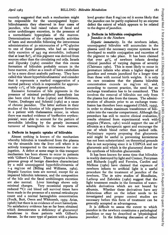
(I950), together with those of other investigators,have established that not all the bile pigmentformed results from the breakdown of the redcells at the end of their life span. They usedfaecal stercobilin as a measure of bile pigmentproduction and after the administration of 15Nglycine determined its specific activity at suitabletime intervals (Fig. i). The 15N stercobilin time-curve had two peaks; the first occurred immediatelyfollowing the glycine feeding while the second hada maximum about the I3oth day when the labelledred cells were breaking down. In the normal
S~. -006
0a2 4 -004I:/ i\ -°I
x.' ·- 0.02~0.I '
4.
20 40 60 60 100 120 140 160 1)Time (days)
FIG. I.-15N contents of haem in samples (full line) andof stercobilin (dotted line) at various times afteradministration of glycine (I2 g.) containing31.65% excess 15N to a normal subject (Gray andothers, I950).
subject the first peak is responsible for approxi-mately Io% of the pigment excreted but indiseases such as pernicious anemia, congenitalporphyria and thalassaemia 40-80% of the bilepigment excreted may be due to this fraction.It was originally thought that this 'early sterco-bilin' arose by direct synthesis from simpleprecursors such as aminolaevulic acid or porpho-bilinogen. It is now considered, however, to bemainly erythrohaemopoietic in origin since thisearly incorporation of labelled glycine into sterco-bilin is increased when erythrohaemopoiesis isstimulated. This erythrohaemopoietic stercobilinmay be derived from three sources, namely, theimmature red cells in the bone marrow (which aredegrading haemoglobin as well as synthesising it),from haem formed in excess of globin or fromhaem formed by the destruction of newly formedred cells as soon as they reach the peripheralcirculation. A very small amount of bilirubin couldtheoretically come from haem pigments such asmyoglobin or the cytochromes but there is noinformation regarding the role played by theseproteins in bile pigment formation. The possibilityof an hepatic origin of the early labelled sterco-bilin has been suggested by the work of Watson-James and Abbott (I961) who found an increase ina patient with aplastic anaemia.
copyright. on 15 A
ugust 2019 by guest. Protected by
http://pmj.bm
j.com/
Postgrad M
ed J: first published as 10.1136/pgmj.39.450.176 on 1 A
pril 1963. Dow
nloaded from

April 1963 BILLING: Bilirubin Metabolism I77
Bilirubin is a lipid-soluble, non-polar pigmentwhich gives an 'indirect' reaction in the presenceof alcohol in the van den Bergh test. Prior toexcretion in the bile, it is converted into a pigmentwhich is water-soluble at a physiological pH andgives a 'direct' van den Bergh reaction with diazo-tised sulphanilic acid. Using chromotographictechniques Cole and Lathe (1953) were able toshow that the properties of 'direct' bilirubin werenot due to a different type of protein attachmentbut to an alteration in the chemical structure of thepigment. By means of partition chromatography(Cole, Lathe and Billing, 1954) (Fig. 2) or paperchromatography (Giovanneti, Maggiore andVivaldi, I96I) it is possible to demonstrate that'direct' bilirubin consists of two components (pig-ments I and II). The most polar pigment (pigmentII) is the main pigment present in bile and togetherwith an intermediate pigment (pigment I) is foundin the icteric urine and sera of patients withobstructive jaundice, hepatitis and cirrhosis. Theextreme instability of these pigments has so farprevented their isolation in a pure form. Thestructure of pigment II has therefore beenestablished by characterizing the azo-pigmentformed in the van den Bergh reaction; thispigment (azo-pigment B) is considerably morepolar than azo-pigment A and can be separatedfrom it and other contaminants in diazotised bileby partition chromatography (Fig. 2), counter-current distribution (Billing, Cole and Lathe,1957) and paper chromatography (Schmid, I957).The application of the latter two techniques hasshown azo-pigment B to be the ester glucuronideof azo-pigment A. Since two molecules of azo-pigment A are known to be formed from onemolecule of bilirubin it has been concluded thatpigment II behaves similarly and is therefore anester diglucuronide of bilirubin, the glucuronicacid radicals being linked to the carboxyl groupsof the proprionic substituents of the molecule asindicated in Fig. 3. A similar conclusion wasreached by Talafant (1956) as the result of experi-ments involving the electrophoresis of bile.The structure of pigment I has been a more
controversial matter. Since both azo-pigmentA andazo-pigment B are formed from pigment I in thevan den Bergh reaction it seemed likely that thepigment is the monoglucuronide of bilirubin.However, if pigment I is eluted from a chromato-gram and then rechromatographed it has beenobserved that both bilirubin and pigment IIappear on a second chromatogram (Billing, Coleand Lathe, 1957; Nosslin, 1960). This observationtogether with other studies (Gregory, 1962;Weber, Schalm and Witmans, I963) gives supportto the hypothesis that pigment I is a labile equimo-lecular complex of bilirubin and bilirubin diglu-
BILE PIGMENTS AZO PIGMENTS
I-- ILIRUBINb
PIGMENT I * n
pH 6 pH pHCHCtl3-CCt -CH3, OH n-BUTANOL n-BUTANOL
FIG. 2.-Behaviour of bile pigments and their diazo-products on reverse phase partition chromatograms.(Nosslin, I960).
COOR, COOR,
I ICH, CH2I I
Me V Me CH, CH2 M Me V
HNJHAHN H NH H H HFIG. 3.-Structure of bile pigments (bilirubin, RI and
R2 = H; Pigment I, R1 = glucuronyl and R2 = H;pigment II, RI and R2 = glucuronyl; Me = methyland V = vinyl).
curonide, and further investigations are nowneeded to determine the factors involved in its.formation.
It is generally accepted that over 80% of thebilirubin in bile is normally present as the glucuro--nide. Examination of chromatograms and theobservation that mild alkali treatment does notconvert all the 'direct' bilirubin in icteric serumor bile into bilirubin indicate that other con-jugates may be present. Isselbacher and McCarthy(I959) by means of 35SO4 tracer studies have-demonstrated the presence of sulphate conjugatesof bilirubin in the bile of rats, cats and humans.These findings have been confirmed in the rat bySchoenfield, Bollman and Hoffman (1962) whodemonstrated that the sulphate conjugate ofbilirubin is associated with pigment II but notwith pigment I. Gregory and Watson (I962)have not, however, been able to demonstrate thepresence of bilirubin sulphate in the bile of dogs orhuman subjects and question the assumption ofIsselbacher and McCarthy that alkali-stable'direct' bilirubin is identical with bilirubinsulphate. At present there is no evidence thatdefective conjugation of bilirubin as a glucuronide
copyright. on 15 A
ugust 2019 by guest. Protected by
http://pmj.bm
j.com/
Postgrad M
ed J: first published as 10.1136/pgmj.39.450.176 on 1 A
pril 1963. Dow
nloaded from

178 POSTGRADUATE MEDICAL JOURNAL April I963
in man can be compensated for by an appreciableincrease in the excretion of bilirubin as a sulphateor some other conjugate.Biosynthesis of Bilirubin Glucuronide
It has been demonstrated that uridinediphosphate glucuronic acid (UDPGA), and notglucuronic acid, is the glucuronyl donor necessaryfor the formation of bilirubin glucuronide (Fig. 4).The conjugating enzyme is a glucuronyl trans-ferase (UDP-trans-glucuronylase) which is locatedin the microsomes; the evidence regarding thespecificity of this enzyme is equivocal. Both thekidney and gastrointestinal tract as well as theliver contain an enzyme system capable of con-jugating bilirubin. In the intact adult animal nosatisfactory evidence has, however, been presentedto indicate that conjugation of bilirubin takesplace in any organ other than the liver. Hepa-tectomy in experimental animals results in thegradual accumulation of both bilirubin and apigment which gives a 'direct' van den Berghreaction and behaves chromatographically likepigment I. This pigment has been identified inthe dog as bilirubin monoglucuronide (Schoen-field, Grindley, Foulk and Bollman, 1961) but thesite of its formation under these abnormal condi-tions has not been established.The ability of the foetal and newborn liver to
form bilirubin glucuronide is markedly reduced,due mainly to a deficiency of glucuronyl transferaseactivity rather than UDPGA. In the rat foetusthe glucuronyl trransferase activity of the gastricmucosa is twice that of the adult liver (Stevensonand Dutton, I962). The possibility that thegastric mucosa might be an important meansof removing bilirubin in the fcetus hasbeen considered but seems unlikely in thelight of recent studies using 14C bilirubin inthe guinea-pig foetus. (Schenker, Dawber and;Schmid, 1962) and the monkey fcetus (Lester,Behrman and Lucey, I962), which showed thatfcetal bilirubin can be efficiently removed, aftercrossing the placenta, in the maternal bile.
Enterohepatic Circulation of BilirubinThe experiments of Lester, Ostrow and
Schmid (I961) in which 14C bilirubin was adminis-tered intraduodenally to rats with bile fistulae haveclearly demonstrated that in the rat, bilirubin isabsorbed from the intestine and then reappearsin the bile. The absorption of unconjugatedbilirubin appeared to be more rapid and quanti-tatively greater than that of conjugated bilirubin. Itseems likely that conjugated bilirubin is firsthydrolyzed to bilirubin and then absorbed as freebilirubin.The existence of an enterohepatic circulation
ATP GLUCOSE
Hexokinast
ADP J GLUCOSE-6-PHOSPHATE
Phosphoglucomutase
GLUCOSE-I-PHOSPHATE \ UTP
YV ADPPP-Uridyl Tronsferase
DPN+URIDINE DIPHOSPHATE
NPPGLUCOSE
UDPG Dehydrogenase/ JRIDINE DIPHOSPHATE BILIRUBIN
GLUCURONIC ACIDDPNH IVGlucuronyl Transfcrase
BILIRUBIN GLUCURONIDE " \UDP TP
FIG. 4.-A possible mechanism for the conjugation ofbilirubin with glucuronic acid (Billing and Lathe,I958).
for bilirubin in man has been confirmed usingboth bilirubin labelled with 14C (Lester andSchmid, I962) and bilirubin labelled with 15N(Gilbertsen, Bossenmaier and Cardinal, 1962).The significance of the enterohepatic circulationin normal man remains to be investigated. It mayprovide one of the reasons for the lack of corre-lation between the amount of pigment known tobe formed from the breakdown of circulating redcells and that found as stercobilin in the faeces. Inpatients with a compensated hyperbilirubinaemiadue to a deficiency of glucuronyl transferase(Crigler-Najjar syndrome) and a homozygousstrain of rats (Gunn rats) with the same deficiency,Schmid and Hammaker (I962) have found thatunconjugated bilirubin can be both absorbed bythe intestine and transferred from the plasmapool directly across the mucosa into the intestinallumen. In the Gunn rats they were able to preventits reabsorption by feeding cholestyramine resin,which has a high affinity for bilirubin, and in thisway significantly reduced the serum bilirubinlevels to 30 to 45% of the control values (Lester,Hammaker and Schmid, I962). The value ofcholestyramine treatment in reducing jaundice inman remains to be assessed.
Conversion of Bilirubin to Urobilinogen andMesobilifuscinIt has been well established that bilirubin,
especially if in the conjugated form, is reduced
copyright. on 15 A
ugust 2019 by guest. Protected by
http://pmj.bm
j.com/
Postgrad M
ed J: first published as 10.1136/pgmj.39.450.176 on 1 A
pril 1963. Dow
nloaded from

BILLING: Bilirubin Metabolism
by the bacterial flora in the intestine and colon tothe urobilinogen group of pigments, which givethe well-known Erlich's aldehyde reaction. Theseurobilinogens are readily dehydrogenated to giveurobilin IXa, stercobilin and d-urobilin, therelative amounts formed depending on themobility of the intestinal contents and the rate offilling of the colon, as well as the efficiency of thebacterial flora. Watson and Weimer (1959) wereunable to correlate the composition of the faecalurobilin with the patient's condition and whileurobilin IXa was the dominant pigment in somesubjects, stercobilin predominated in others. Theadministration of broad-spectrum antibiotics mayresult in bilirubin rather than stercobilin beingexcreted in the faeces.The presence of an enterohepatic circulation
for urobilinogen was first postulated by McMasterand Elman (I927). They assumed that in liverdisease re-excretion of urobilinogen into the bilewas impaired and that instead, the pigment passedto the kidney and was excreted in the urine.Urobilinoid pigments are, however, not normallyfound in bile except in the presence of infection;if McMaster and Elman's hypothesis is correctthis means that the pigment would have to bereconverted to bilirubin in the liver before itcould be excreted in the bile which, for bio-chemical and structural reasons, seems unlikely.A critical re-examination of this problem withlabelled stercobilin of high specific activity isrequired to establish whether urobilinogen, likebilirubin, after absorption from the intestine isre-excreted in the bile. Recent experiments byKahan, Csernay and Varro (I962) have shown thatthe reabsorption of stercobilin from the isolatedsmall intestine of the dog is dependent on theformation of a mucoprotein complex and suggestthat the kidney may play some part in the con-version of urobilin to its respective urobilinogenprior to excretion in the urine.
It has been suggested that the discrepanciesobserved between urobilinogen excretion andhaemoglobin catabolism might be accounted forby the conversion of bilirubin or urobilinogento dipyrrylmethenes such as mesobilifuscin.Studies by Gilbertsen and Watson (I962) sup-port the view, however, that the naturallyoccurring fecal dipyrrylmethene pigments areanabolic rather than catabolic in origin.Bile Pigments in Plasma, Urine and Body
FluidsAt the serum bilirubin levels encountered in
icteric plasma, regardless of the cause of thejaundice, virtually all the pigment is bound toalbumin (Ostrow and Schmid, I962). It is forthis reason that attempts to reduce jaundice by
dialysis have been for the most part unsuccessful.If however albumin is added to the dialysisfluid then it is possible for considerable quantitiesof bilirubin to be removed by intermittentperitoneal dialysis (Grollman and Odell, I962).
Conjugated bilirubin is also bound to theplasma albumin but the type of pigment-protein-complex formed is probably different (Klatskinand Bungards, I956). It may also form a complexwith phospholipids which can be extracted byshaking with ether (Charbonnier and Poungouras,1959; Lucassen, I96I). This 'ether-solublebilirubin' extraction test has been used todifferentiate between malignant and non-maligant biliary obstruction but according toMertens and Croal (I960) is probably merely ameasure of the degree of obstruction, as indicatedby the high phospholipid level.
Small doses of bilirubin are quickly clearedfrom the plasma by the liver and then afterstorage and concentration are excreted into thebile at a slower rate. In the normal subject serumlevels greater than i mg/ioo ml are rarely encoun-tered and the bilirubin is almost entirely in thenon-conjugated form. With raised levels ofserum bile pigments, bilirubin will be stored inmost of the tissues in the body, including theadipose tissue. It has been suggested that thestaining of the tissues of the skin and muscle maybe associated with their extravascular albumincontent (Billing and Lathe, 1958). The differencesin skin colour observed in infants with hemolyticjaundice and adults with obstructive jaundicecould be due to differences in tissue binding ofbilirubin and conjugated bilirubin but thesematters need to be investigated further.
Both unconjugated and conjugated bilirubinmay be found in the lymph, ascitic fluid, pleuralfluid and cerebrospinal fluid of the jaundicedsubject. No correlation has been found betweenthe bile pigment levels in the CSF and those inthe plasma.
Bilirubin cannot usually be detected in theurine of normal subjects, or patients with anunconjugated hyperbilirubinamia. In ictericurine both pigments I and II but not bilirubin arefound. The level of conjugated bilirubin in theurine does not correlate well with that in theplasma and depends on the state of the diseaseprocess. In viral hepatitis, for example, bilepigments may appear in the urine even before thepatient becomes clinically icteric but duringconvalescence no pigment will be found in theurine although the serum level may be as high as6-8 mg/ioo ml. Lack of pure specimens ofconjugated bilirubin and suitable techniques forthe quantitative determination of bile pigmentsin urine has prevented satisfactory renal clearance
April x963 I79copyright.
on 15 August 2019 by guest. P
rotected byhttp://pm
j.bmj.com
/P
ostgrad Med J: first published as 10.1136/pgm
j.39.450.176 on 1 April 1963. D
ownloaded from

i8o POSTGRADUATE MEDICAL JOURNAL April 1963
studies being carried out. It would appear thatin the dog the pigments are excreted by the renaltubules, but there is no reliable evidence in man onthis matter.
Toxicity of Bilirubin and KernicterusThe yellow pigment that accumulates in the
brain in kernicterus has been identified as uncon-jugated bilirubin. The toxic effects of bilirubinhave therefore been studied and it has been shownthat bilirubin has an inhibitory action on theoxygen consumption of brain tissue, particularlyin the newborn. It has also been demonstratedthat bilirubin is an inhibitor of haem synthesis andthat in rat liver and brain mitochondria ituncouples oxidative phosphorylation. Accordingto Ernster (I96I) bilirubin behaves like a detergentin its action on mitochondria and causes swellingas well as inducing ATPase activity. With highconcentrations of bilirubin the mitochondria aredestroyed, and an enzyme may be released whichwill oxidise bilirubin to biliverdin. It is possiblethat such a reaction may be responsible for thepresence of small amounts of biliverdin in theserum of patients with advanced liver disease.The newborn animal is more susceptible to the
toxic action of large doses of bilirubin than theadult animal (Rozdilsky, I96I). In some animalsstaining of the nervous tissue is only observed ifthere is previous brain damage by severe hypo-glycaemia, anoxia or traumatic hsemorrhage.Whether previous damage as well as high con-centrations of bilirubin are necessary for thedevelopment of kernicterus in man has not beenestablished. Neither is it known why some partsof the brain (e.g. the corpora striata, the thal-amus and the hippocampus) tend to becomestained in the presence of high levels of plasmabilirubin while others do not. The part playedby the blood-brain barrier has also still to beclarified. There is suggestive evidence that inthe newborn the permeability is increased so thatbilirubin passes more readily into the CSF and thebrain.
Conjugated bilirubin, on the other hand,appears to be non-toxic. In the adult with severejaundice these pigments are predominant andonly on rare occasions are high levels of uncon-jugated bilirubin encountered. This is probablythe main reason for the apparent absence ofkernicterus in the adult.
JAUNDICEOn theoretical grounds increased concentrations
of bilirubin in the plasma may result from one ormore of the following causes (Fig. 5 ).
(I) An increased load of bilirubin.
s sS / NL U S O I D
FIG. 5.-Jaundice could theoretically result from (I) anincreased load of bilirubin; (2) defective uptakeand transport within the liver cell; (3) defectiveconjugation in the hepatic microsomes; (4) defec-tive canalicular excretion or a mechanical block inthe bile duct system. (S. Sherlock (1962), Brit.med. J., i, I950).
(2) Defective uptake and transport within theliver cell.
(3) Defective conjugation of bilirubin in thehepatic microsomes.
(4) Defective canalicular excretion or amechanical block in the bile duct.
Although, in most instances, jaundice is not dueto a single causative factor this may be the case incongenital hyperbilirubinaemia. Detailed studiesof bile pigment metabolism in these disorders may,therefore, give some insight into the pathways ofnormal metabolism.
i. Increased load of bilirubinHcemolytic diseaseAn excessive release of haemoglobin from the
red cells may result in the normal daily productionof 300 mg of bilirubin being increased as much assix-fold. The capacity of the liver to excretebilirubin is, however, greatly in excess of thatnormally required so that in haemolytic diseasethe plasma bile pigment concentration rarely risesabove 5 mg/ioo ml. The main pigment in theplasma is unconjugated bilirubin so that bilirubin-uria is not usually observed. Small amounts ofconjugated bilirubin may be detected in theplasma but these will not exceed 15% of the totalserum bilirubin concentration unless hepato-cellular damage has also occurred (Tisdale,Klatskin and Kinsella, 1959).
Alternative pathways of bilirubin metabolismAn overproduction of bilirubin can arise from
sources other than the breakdown of mature redcells. Israels, Suderman and Ritzman (1959)
copyright. on 15 A
ugust 2019 by guest. Protected by
http://pmj.bm
j.com/
Postgrad M
ed J: first published as 10.1136/pgmj.39.450.176 on 1 A
pril 1963. Dow
nloaded from
BILLING: Bilirubin Metabolism
recently suggested that such a mechanism mightbe responsible for the unconjugated hyper-bilirubinamia they observed in four cases ofjaundice, who had raised values of faecal andurine urobilinogen excretion, in the presence ofa normoblastic hyperplasia of the marrow.Determinations of the relative specific activitiesof hsem and stercobilin following the intravenousadministration of 50 microcuries of z-14C-glycineto one of these patients, who had an averagedaily excretion of 945 mg stercobilin, indicatedthat at least 82% of this pigment was derived fromsources other than the circulating red cells. Israelsand Zipursky (I962) consider that this excessformation of bilirubin probably occurs in thebone marrow, either as a result of haem catabolismor by a more direct anabolic pathway. They havecalled this 'shunt hyperbilirubinaemia' and considerthat it represents a marked exaggeration of anormal pathway which is responsible for approxi-mately 11% of bile pigment production.
Excessive formation of bile pigments in thepresence of minimal peripheral haemolysis hasalso been cited by Arias (g962a) and Robinson,Vanier, Desforges and Schmid (I962) as a causeof chronic jaundice. The latter authors in theirstudies of the kinetics of bile pigment formationin a patient with thalassaemia minor, in whomthere was marked evidence of 'ineffective erythro-poiesis', were able to account for the pattern ofstercobilin labelling by the greatly increasedturnover of haemoglobin in the bone marrow.
2. Defects in hepatic uptake of bilirubinAlmost nothing is known of the mechanism
whereby bilirubin is transferred from the plasmavia the sinusoids into the liver cell where it isactively transported to the microsomes for con-jugation. A defect at some stage in this transportmechanism has been shown to occur in patientswith 'Gilbert's Disease'. These comprise a hetero-geneous group of benign disorders characterizedby a mild unconjugated hyperbilirubinaemia of1-4 mg/ioo ml (Billing and Williams, I962).Hepatic function tests are normal, except for animpaired bilirubin tolerance, and the compositionof the bile and the fsecal urobilinogen excretionare also normal. Liver histology shows onlyminimal changes. Very occasional reports ofreduced 51Cr red blood cell survival times havebeen made in the presence of a normal reticulocytecount and a normal haemoglobin concentration(Foulk, Butt, Owen and Whitcomb, I959; Arias,962b) but there is no evidence of overt haemolysis.No satisfactory evidence has so far been obtainedwhich demonstrates a deficiency of glucuronyltransferase in these patients with Gilbert'sdisease. In the rarer type of patient with a plasma
level greater than 8 mg/ioo ml it seems likely thatthe jaundice can be partly explained by an enzymedefect the extent of which appears to be relatedto the degree ofjaundice.3. Defects in bilirubin conjugationJaundice in the NewbornHepatic immaturity. In the newborn infant,
unconjugated bilirubin will accumulate in theplasma until the necessary enzyme systems havedeveloped to enable conjugation and excretion ofthe pigment to take place. It has been claimedthat over 40% of newborn infants developclinical jaundice of varying degrees of severity(Claireaux I960). This is most likely to occur inpremature infants who attain a higher degree ofjaundice and remain jaundiced for a longer timethan those with normal birth weights. It is onlyon rare occasions that the plasma bilirubinconcentration exceeds 20 mg/ioo ml, and,according to current practice, the need for anexchange transfusion has to be considered. Thisinvolves the removal of bilirubin not only fromthe plasma but also from the tissues. The admini-stration of albumin prior to an exchange trans-fusion has therefore been suggested (Odell, 1959),in order to obtain maximum transference of bili-rubin into the vascular compartment. Although theprocedure has still to receive clinical evaluation,results obtained from experimental work withpuppies (Waters, I96I) would support the use ofalbumin as a therapeutic agent and encourage theuse of whole blood rather than packed cells.Preliminary reports proposing that glucuronicacid might be useful in preventing kernicterushas not been substantiated: on theoretical groundsthis is not surprising since it is UDPGA and notglucuronic acid which is the glucuronyl donor forthe synthesis of bilirubin glucuronide.
It has been known for some time that bilirubinis readily destroyed by light and Cremer, Perrymanand Richards (1958) and Ferreira, Cardim andMellone (1960) have claimed that exposure ofnewborn infants to artificial light is a usefulprocedure for the treatment of jaundice of thenewborn. The in vitro studies of Blondheim,Lathrop and Zabriskie (I962) suggest that thelight treatment results in the formation of water-soluble derivatives which are not bound byalbumin. Whether these derivatives have anydeleterious effect on the infant needs to beinvestigated and proper control studies arenecessary before this form of treatment can begenerally accepted as advantageous.
It is difficult to assess the extent to whichjaundice in the newborn constitutes a pathologicalcondition or may be described as 'physiologicaljaundice'. In the following discussion of other
April I963 I8Icopyright.
on 15 August 2019 by guest. P
rotected byhttp://pm
j.bmj.com
/P
ostgrad Med J: first published as 10.1136/pgm
j.39.450.176 on 1 April 1963. D
ownloaded from
POSTGRADUATE MEDICAL JOURNAL
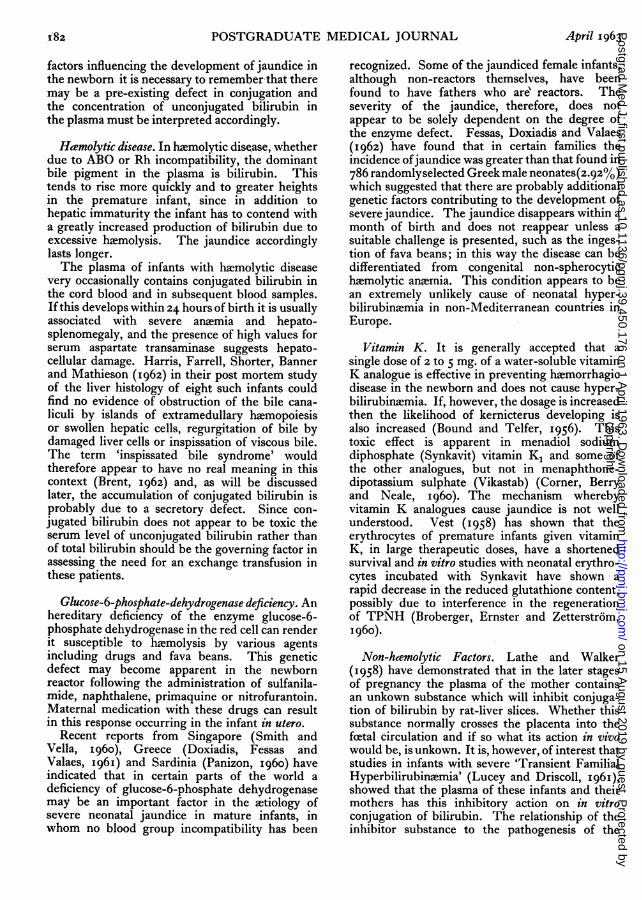
factors influencing the development of jaundice inthe newborn it is necessary to remember that theremay be a pre-existing defect in conjugation andthe concentration of unconjugated bilirubin inthe plasma must be interpreted accordingly.
Haemolytic disease. In hamolytic disease, whetherdue to ABO or Rh incompatibility, the dominantbile pigment in the plasma is bilirubin. Thistends to rise more quickly and to greater heightsin the premature infant, since in addition tohepatic immaturity the infant has to contend witha greatly increased production of bilirubin due toexcessive haemolysis. The jaundice accordinglylasts longer.The plasma of infants with haemolytic disease
very occasionally contains conjugated bilirubin inthe cord blood and in subsequent blood samples.Ifthis develops within 24 hours of birth it is usuallyassociated with severe anaemia and hepato-splenomegaly, and the presence of high values forserum aspartate transaminase suggests hepato-cellular damage. Harris, Farrell, Shorter, Bannerand Mathieson (1962) in their post mortem studyof the liver histology of eight such infants couldfind no evidence of obstruction of the bile cana-liculi by islands of extramedullary haemopoiesisor swollen hepatic cells, regurgitation of bile bydamaged liver cells or inspissation of viscous bile.The term 'inspissated bile syndrome' wouldtherefore appear to have no real meaning in thiscontext (Brent, 1962) and, as will be discussedlater, the accumulation of conjugated bilirubin isprobably due to a secretory defect. Since con-
jugated bilirubin does not appear to be toxic theserum level of unconjugated bilirubin rather thanof total bilirubin should be the governing factor inassessing the need for an exchange transfusion inthese patients.
Glucose-6-phosphate-dehydrogenase deficiency. Anhereditary deficiency of the enzyme glucose-6-phosphate dehydrogenase in the red cell can renderit susceptible to haemolysis by various agentsincluding drugs and fava beans. This geneticdefect may become apparent in the newbornreactor following the administration of sulfanila-mide, naphthalene, primaquine or nitrofurantoin.Maternal medication with these drugs can resultin this response occurring in the infant in utero.
Recent reports from Singapore (Smith andVella, 1960), Greece (Doxiadis, Fessas andValaes, I96I) and Sardinia (Panizon, I960) haveindicated that in certain parts of the world a
deficiency of glucose-6-phosphate dehydrogenasemay be an important factor in the aetiology ofsevere neonatal jaundice in mature infants, inwhom no blood group incompatibility has been
recognized. Some of the jaundiced female infants,although non-reactors themselves, have beenfound to have fathers who are' reactors. Theseverity of the jaundice, therefore, does notappear to be solely dependent on the degree ofthe enzyme defect. Fessas, Doxiadis and Valaes(I962) have found that in certain families theincidence ofjaundice was greater than that found in786 randomlyselected Greek male neonates(2.92%)which suggested that there are probably additionalgenetic factors contributing to the development ofsevere jaundice. The jaundice disappears within amonth of birth and does not reappear unless asuitable challenge is presented, such as the inges-tion of fava beans; in this way the disease can bedifferentiated from congenital non-spherocytichaemolytic anernia. This condition appears to bean extremely unlikely cause of neonatal hyper-bilirubinaemia in non-Mediterranean countries inEurope.
Vitamin K. It is generally accepted that asingle dose of 2 to 5 mg. of a water-soluble vitaminK analogue is effective in preventing haemorrhagicdisease in the newborn and does not cause hyper-bilirubinsemia. If, however, the dosage is increasedthen the likelihood of kernicterus developing isalso increased (Bound and Telfer, 1956). Thistoxic effect is apparent in menadiol sodiumdiphosphate (Synkavit) vitamin K, and some ofthe other analogues, but not in menaphthone-dipotassium sulphate (Vikastab) (Corner, Berryand Neale, I960). The mechanism wherebyvitamin K analogues cause jaundice is not wellunderstood. Vest (1958) has shown that theerythrocytes of premature infants given vitaminK, in large therapeutic doses, have a shortenedsurvival and in vitro studies with neonatal erythro-cytes incubated with Synkavit have shown arapid decrease in the reduced glutathione content,possibly due to interference in the regenerationof TPNH (Broberger, Ernster and Zetterstr6m,1960).
Non-hcemolytic Factors. Lathe and Walker(I958) have demonstrated that in the later stagesof pregnancy the plasma of the mother containsan unkown substance which will inhibit conjuga-tion of bilirubin by rat-liver slices. Whether thissubstance normally crosses the placenta into thefcetal circulation and if so what its action in vivowould be, is unkown. It is, however, of interest thatstudies in infants with severe 'Transient FamilialHyperbilirubinsemia' (Lucey and Driscoll, I96I),showed that the plasma of these infants and theirmothers has this inhibitory action on in vitroconjugation of bilirubin. The relationship of theinhibitor substance to the pathogenesis of the
April 1963q82copyright.
on 15 August 2019 by guest. P
rotected byhttp://pm
j.bmj.com
/P
ostgrad Med J: first published as 10.1136/pgm
j.39.450.176 on 1 April 1963. D
ownloaded from
BILLING: Bilirubin Metabolism
jaundice, which subsided within the first monthof life has, however, not been established.
In a clinical trial of antibiotics in prematureinfants it was observed that the administrationof sulfisoxazole reduced the concentration ofplasma bilirubin and at the same time increasedthe incidence of kernicterus (Harris, Lucey and\Iaclean, 1958). Experiments with in vitrosystems and genetically jaundiced Gunn rats
(Johnson, Garcia, Figueros and Sarmiento, I96I)have shown that organic anions such as sulphona-mides and salicylates compete with bilirubin foralbumin binding sites. An inverse relationshipexists between the blood level of the drug andthe plasma bilirubin. The distribution of bilirubinin the body is therefore altered and there is an
increase in the amount of pigment entering thebrain which may cause kernicterus. Theseinvestigations emphasize the impossibility ofassessing the success of a therapeutic agent inreducing jaundice in the newborn merely bydetermining the serum bilirubin level; evidenceof an increased excretion of bilirubin must beobtained.
Another drug known to cause jaundice isnovobiocin (Cox, Foltz, Raymond and Drewyer,1959); in the newborn it has been reported as
causing a threefold increase in neonatal hyper-bilirubinaemia (Sutherland and Keller, I96I).In vitro studies (Hargreaves and Holton, I962)suggest that novobiocin, which is excreted as a
glucuronide, acts by competing with bilirubin forthe very limited amount of glucuronyl transferasepresent in the newborn liver.
Jaundice in the AdultCrigler-Najjar Syndrome (Congenital non-hcemo-
lyticjaundice). In very rare instances the deficiencyof glucuronyl transferase seen in the newbornmay continue into adult life. This syndrome was
first described by Crigler and Najjar (1952) whostudied the children of three related families withserum levels of unconjugated bilirubin rangingfrom 12-45 mg/ioo ml. Brain damage is oftencaused in the neonatal period so that many ofthese patients, if they survive, are to be found inmental hospitals. However, if neurologicalsymptoms do not develop during early life then theprognosis is probably good (Childs and Najjar,I956; Sugar, 1961) since the toxic effects ofbilirubin are less marked in the adult. Thesepatients have an impairment in their ability toform glucuronides with aglycones such as
salicylates, n-acetyl p-amino phenol, mentholand tetrahydrocortisone (Schmid, I960). Onlytrace amounts of bile pigments are found in thebile and faecal urobilinogen excretion is low. Invitro studies have shown that there is a deficiency
of hepatic glucuronyl transferase but not ofUDPGA, which is responsible for their jaundice(Jervis, 1959; Szabo, Kovacs and Ebrey, I962).In all other respects liver function appears to benormal and there is no evidence of haemolyticdisease.The serum bilirubin levels remain relatively con-
stant in these patients in spite of a normal rate ofhamoglobin breakdown and defective pigmentexcretion. Some alternative pathway for theremoval of bilirubin must therefore be presentwhich, although less efficient than the usualconjugating system since hyperbilirubinaemiaoccurs, is capable of maintaining a 'steady state'with respect to bilirubin. Schmid and Hammaker(I962) have used 14C bilirubin to estimate therate of turnover and the magnitude of the totalmiscible pool of bilirubin in a young boy withcongenital non-haemolytic jaundice. MIost of theisotopic bilirubin appeared in the faeces in theform of metabolites exhibiting properties differentfrom those of the known bile pigments. Furtherwork is needed to establish the site of breakdownof the bilirubir and the nature of the metabolites.
4. Disturbances in hepatic excretion ofbilirubinObstructive jaundice and hepatocellular diseaseSince over 900o of the pigments in hepatic bile
occur as pigment II it would be expected thatthis would be the dominant pigment in the plasmaof patients with extrahepatic biliary obstruction.If however the obstruction continues for someweeks then the proportion of pigment II in theserum will decline while that of pigment I willincrease. A similar pigment pattern is thenobtained to that found in the plasma of patientswith parenchymal liver disease due to hepatitis orcirrhosis which has more pigment I than pigmentII or bilirubin (Billing and Lathe, 1958). Hoffman,Whitcomb, Butt and Bollman (1960) examined thepigment patterns of I50 cases of jaundice andendeavoured to use them to differentiate betweenacute obstructive jaundice and hepatocellulardisease. There was unfortunately a significantoverlap between the two groups which preventsthe procedure from being a useful diagnostictool; it is not helpful in distinguishing betweenextra and intra-hepatic cholestasis.Treatment with ACTH and corticosteroids has
been used for the differential diagnosis of chronicjaundice with some degree of success. In infectivehepatitis there is usually an immediate clinicalimprovement accompanied by a sharp decline inthe plasma bilirubin level, which cannot beexplained in terms of increased biliary excretionor a change in the renal excretion of bile pigments;the rate of red cell breakdown is also unchanged
I83April I963copyright.
on 15 August 2019 by guest. P
rotected byhttp://pm
j.bmj.com
/P
ostgrad Med J: first published as 10.1136/pgm
j.39.450.176 on 1 April 1963. D
ownloaded from

184 POSTGRADUATE MEDICAL JOURNAL April 1963
(Williams and Billing, 196I). In cases of intra-and extra-hepatic obstructive jaundice and drugjaundice a favourable response is rarely seen. Nosatisfactory explanation has so far been putforward for the altered bile pigment metabolismfollowing steroid therapy in hepatitis.Drug JaundiceThe first report of drug jaundice was by Hanger
and Gutman (I940) in which 12 patients receivingintravenous arsphenamine for the treatment ofsyphilis became icteric after the second or thirdinjection. Since then an ever-increasing numberof drug reactions with jaundice due to intra-hepaticcholestasis has been described (Popper andSchaffner, 1959). With many of these drugssuch as chlorpromazine ('Largactil'), thiouracil,para-aminosalicylic acid, chlorpropamide andcetyl urea, the jaundice has been attributed to ahypersensitivity reaction. i% of patients receivingchlorpromazine treatment develop frank jaundicetogether with increases in serum alkaline phos-phatase and transaminases and also bromsulph-thalein retention. Out of eight patients studied byHoffman and others (I960) four had a pigmentpattern similar to that seen with extra-hepaticobstruction, while the other four patients had apattern suggestive of hepatocellular damage.
Anabolic agents, such as methyl testosteroneand norethandrolone ('Nilevar') with a CI7 alkylsubstitution in the steroid molecule, may causejaundice to appear late in the course of therapy,without symptoms of a hypersensitivity reaction.Abnormalities in the microvilli lining the biliarycanaliculi have been reported following theadministration of these drugs and it is thoughtthat these may be responsible for the bile stasis(Schaffner, Popper and Perez, I960). Experimentalwork with rats also supports the view that nor-ethandrolone acts by interfering with the excretoryfunction of the liver cell.
Another group of drugs occasionally causes avery severe type ofjaundice, which is histologicallyand biochemically indistinguishable from viralhepatitis. This type of liver injury may followthe administration of the mono-amine oxidaseinhibitors iproniazid ('Marsilid'), pheniprazine,}-phenyl isopropyl hydrazine, isocarboxazid('Marplan') and phenalzine ('Nardil'). It has alsobeen reported with iso-nicotinic acid, hydrazine,pyrazinamide, zoxazolamine and the oral hypo-glycaemic agents metahexamide and tolbutamide.
Other drugs may cause jaundice as a result ofdirect hepatocellular damage, which affects boththe conjugation and excretion of bilirubin. Drugsin this group include carbon tetrachloride,chloroform, ethyl chloride, dichloro-diphenyl-trichlorethane (D.D.T.) as well as muscarine,
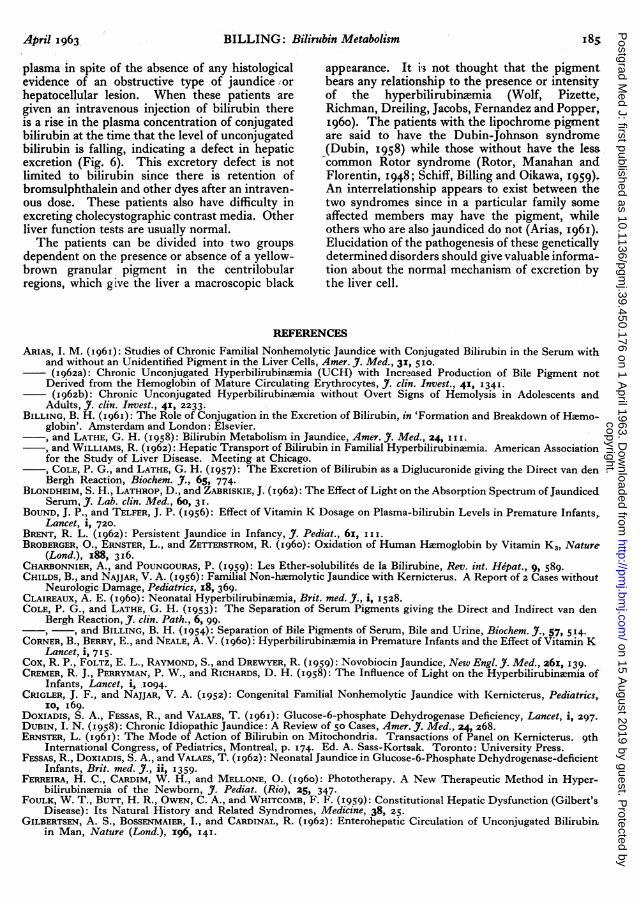
DUBIN-JOHNSON SYNDROME8
iv. Bilirubin (2mgkg)e77
^6 4cz6
J5
|5 U >--^*^-~^~ -Conjugatedi il irub i n
I * 'Bilirubin
0g I i
?0 40 60 120 IO 240Minutes
FIG. 6.-Effect of intravenous bilirubin (2 mg. kg.) onthe serum bile pigment concentration of a patientwith the Dubin-Johnson syndrome.
metallic poisons, naphthalene and benzene deriva-tives.
Functional defects in excretionDefects in the excretory function of the liver
need not necessarily be accompanied by any grosshistological abnormality of the bile ducts. Theaccumulation of conjugated as well as unconjugatedbilirubin suggests that the difficulty lies betweenthe microsomes and the bile canaliculi. It appearsto be developmental in origin but further investi-gations of the mechanism of pigment excretionare necessary to establish whether the defect isa structural or metabolic one.
It has been observed that in some infants withhaemolytic disease that the fall in level of plasmaunconjugated bilirubin is preceded by a rise inthe conjugated pigment concentration. Thisindicates that the conjugating enzyme system hasdeveloped satisfactorily but that the secretoryfunction of the liver cell is still inadequate (Harrisand others, 1962). This impairment in excretionmay occasionally persist for several weeks and insome patients appears to have a familial incidence(Jouvenceaux, Brizard, Michaud and Revol, I959;Billing, 196I).
In some cases of chronic non-haemolyticjaundice in adolescents and adults conjugated aswell as unconjugated bilirubin is present in the
copyright. on 15 A
ugust 2019 by guest. Protected by
http://pmj.bm
j.com/
Postgrad M
ed J: first published as 10.1136/pgmj.39.450.176 on 1 A
pril 1963. Dow
nloaded from
April 1963 BILLING: Bilirubin Metabolism8
plasma in spite of the absence of any histologicalevidence of an obstructive type of jaundice /orhepatocellular lesion. When these patients aregiven an intravenous injection of bilirubin thereis a rise in the plasma concentration of conjugatedbilirubin at the time that the level of unconjugatedbilirubin is falling, indicating a defect in hepaticexcretion (Fig. 6). This excretory defect is notlimited to bilirubin since there is retention ofbromsulphthalein and other dyes after an intraven-ous dose. These patients also have difficulty inexcreting cholecystographic contrast media. Otherliver function tests are usually normal.The patients can be divided into two groups
dependent on the presence or absence of a yellow-brown granular pigment in the centrilobularregions, which give the liver a macroscopic black
appearance. It i's not thought that the pigmentbears any relationship to the presence or intensityof the hyperbilirubinxmia (Wolf, Pizette,Richman, Dreiling, Jacobs, Fernandez and Popper,I960). The patients with the lipochrome pigmentare said to have the Dubin-Johnson syndrome(Dubin, 1958) while those without have the lesscommon Rotor syndrome (Rotor, Manahan andFlorentin, 1948; Schiff, Billing and Oikawa, 1959).An interrelationship appears to exist between thetwo syndromes since in a particular family someaffected members may have the pigment, whileothers who are also jaundiced do not (Arias, I96I).Elucidation of the pathogenesis of these geneticallydetermined disorders should give valuable informa-tion about the normal mechanism of excretion bythe liver cell.
REFERENCES
ARIAS, I. M. (1961): Studies of Chronic Familial Nonhemolytic Jaundice with Conjugated Bilirubin in the Serum withand without an Unidentified Pigment in the Liver Cells, Amer. J. Med., 31, 510.(I962a): Chronic Unconjugated Hyperbilirubinaemia (UCH) with Increased Production of Bile Pigment notDerived from the Hemoglobin of Mature Circulating Erythrocytes, J. clin. Invest., 41, I341.(I962b): Chronic Unconjugated Hyperbilirubinaemia without Overt Signs of Hemolysis in Adolescents and
Adults, J. clin. Invest., 41, 2233.BILLING, B. H. (I96I): The Role of Conjugation in the Excretion of Bilirubin, in 'Formation and Breakdown of Haemo-
globin'. Amsterdam and London: Elsevier., and LATHE, G. H. (1958): Bilirubin Metabolism in Jaundice, Amer. J. Med., 24, I II., and WILLIAMS, R. (1962): Hepatic Transport of Bilirubin in Familial Hyperbilirubinaemia. American Associationfor the Study of Liver Disease. Meeting at Chicago.
--, COLE, P. G., and LATHE, G. H. (I957): The Excretion of Bilirubin as a Diglucuronide giving the Direct van denBergh Reaction, Biochem. .., 65, 774.
BLONDHEIM, S. H., LATHROP, D., and ZABRISKIE, J. (1962): The Effect of Light on the Absorption Spectrum of JaundicedSerum, J. Lab. clin. Med., 60, 31.
BOUND, J. P., and TELFER, J. P. (1956): Effect of Vitamin K Dosage on Plasma-bilirubin Levels in Premature Infants,Lancet, i, 720.
BRENT, R. L. (I962): Persistent Jaundice in Infancy, J. Pediat., 61, III.BROBERGER, O., ERNSTER, L., and ZETTERSTROM, R. (I960): Oxidation of Human Haemoglobin by Vitamin K3, Nature
(Lond.), I88, 316.CHARBONNIER, A., and POUNGOURAS, P. (1959): Les Ether-solubilites de la Bilirubine, Rev. int. Hepat., 9, 589.CHILDS, B., and NAJJAR, V. A. (1956): Familial Non-haemolytic Jaundice with Kernicterus. A Report of 2 Cases without
Neurologic Damage, Pediatrics, r8, 369.CLAIREAUX, A. E. (I960): Neonatal Hyperbilirubinaemia, Brit. med. J., i, 1528.COLE, P. G., and LATHE, G. H. (1953): The Separation of Serum Pigments giving the Direct and Indirect van den
Bergh Reaction, J. clin. Path., 6, 99.--, and BILLING, B. H. (1954): Separation of Bile Pigments of Serum, Bile and Urine, Biochem. J., 57, 514.
CORNER, B., BERRY, E., and NEALE, A. V. (I960)i Hyperbilirubinaemia in Premature Infants and the Effect of Vitamin KLancet, i, 715.
Cox, R. P., FOLTZ, E. L., RAYMOND, S., and DREWYER, R. (I959): Novobiocin Jaundice, New Engl. J. Med., 26I, 139.CREMER, R. J., PERRYMAN, P. W., and RICHARDS, D. H. (1958): The Influence of Light on the Hyperbilirubinaemia of
Infants, Lancet, i, 1094.CRIGLER, J. F., and NAJJAR, V. A. (1952): Congenital Familial Nonhemolytic Jaundice with Kernicterus, Pediatrics,
10, I69.DOXIADIS, S. A., FESSAS, R., and VALAES, T. (I96I): Glucose-6-phosphate Dehydrogenase Deficiency, Lancet, i, 297.DUBIN, I. N. (1958): Chronic Idiopathic Jaundice: A Review of 50 Cases, Amer. J. Med., 24, 268.ERNSTER, L. (I961): The Mode of Action of Bilirubin on Mitochondria. Transactions of Panel on Kernicterus. 9th
International Congress, of Pediatrics, Montreal, p. 174. Ed. A. Sass-Kortsak. Toronto: University Press.FESSAS, R., DOXIADIS, S. A., and VALAES, T. (I962): Neonatal Jaundice in Glucose-6-Phosphate Dehydrogenase-deficient
Infants, Brit. med. J., ii, 1359.FERREIRA, H. C., CARDIM, W. H., and MELLONE, 0. (1960): Phototherapy. A New Therapeutic Method in Hyper-
bilirubinaemia of the Newborn, J. Pediat. (Rio), 25, 347.FOULK, W. T., BUTT, H. R., OWEN, C. A., and WHITCOMB, F. F. (I959): Constitutional Hepatic Dysfunction (Gilbert's
Disease): Its Natural History and Related Syndromes, Medicine, 38, 25.GILBERTSEN, A. S., BOSSENMAIER, I., and CARDINAL, R. (I962): Enterohepatic Circulation of Unconjugated Bilirubin
in Man, Nature (Lond.), 196, I41.
copyright. on 15 A
ugust 2019 by guest. Protected by
http://pmj.bm
j.com/
Postgrad M
ed J: first published as 10.1136/pgmj.39.450.176 on 1 A
pril 1963. Dow
nloaded from

I86 POSTGRADUATE MEDICAL JOURNAL April 1963, and WATSON, C. J. (1962): Studies of the Dipyrrylmethene ('Fuscin') Pigments. III. The Variable Fate of Bili-rubin depending upon Conjugation and other Factors, J. din. Invest., 41, 104I.
GIOVANNETTI, S., MAGGIORE, Q., and VIVALDI, G. (I96I): Separation by Paper Chromatography of Various Fractions ofConjugated Bilirubin occurring in Human Bile, Ital. J. Biochem., o1, 88.
GRAY, C. H., NEUBERGER, A., and SNEATH, P. H. A. (I950): Studies in Congenital Porphyria Incorporation of 15N inStercobilin in the Normal and in the Porphyric, Biochem. J., 47, 87.
GREGORY, C. H. (I962): Studies of Conjugated Bilirubin. III. 'Pigment I'. A Complex of Conjugated and Free Bili-rubin. In the press., and WATSON, C. J. (I962): Studies of Conjugated Bilirubin. II. Problem of Sulfates of Bilirubin 'In Vivo' and'In Vitro', J. Lab. din. Med., 6o, 17.
GRODSKY, G. M., CARBONE, J. V., FANSKA, R., and PENG, C. T. (I962): Tritiated Bilirubin: Preparation and Physio-logical Studies, Amer. J. Physiol., 203, 532.
GROLLMAN, A. P., and ODELL, G. B. (I962): Removal of Bilirubin by Albumin during Intermittent Peritoneal Dialysis,New Engl. J. Med., 267, 279.
HANGER, F. M., and GUTMAN, A. B. (1940): Postarsphenamine Jaundice Apparently Due to Obstruction of the Intra-hepatic Biliary Tract, J. Amer. med. Ass., 15, 263.
HARGREAVES, T., and HOLTON, J. B. (I962): Jaundice of the Newborn due to Novobiocin, Lancet, i, 839.HARRIS, R. C., LUCEY, J. F., and MACLEAN, R. J. (1958): Kernicterus in Premature Infants Associated with Low Con-
centrations of Bilirubin in the Plasma, Pediatrics, 21, 875.HARRIS, L. E., FARRELL, F. J., SHORTER, R. G., BANNER, E. A., and MATHIESON, D. R. (1962): Conjugated Serum
Bilirubin in Erythroblastosis Fetalis: Analysis of 38 Cases, Proc. Mayo Clin., 37, 574.HOFFMAN, H. M., WHITCOMB, F. F., BUTT, H. R., and BOLLMAN, J. L. (1960): Bile Pigments of Jaundice, J. din.
Invest., 39, 132.ISSELBACHER, K. J., and MCCARTHY, E. A. (1959): Studies on Bilirubin Sulfate and Other Non-glucuronide Conjugates
of Bilirubin, J. clin. Invest., 38, 645.ISRAELS, L. G., SUDERMAN, H. J., and RITZMAN, S. E. (1959): Hyperbilirubinaemia due to an Alternate Path of Bilirubin
Production, Amer. J. Med., 27, 693., and ZIPURSKY, A. (1962): Primary Shunt Hyperbilirubinaemia, Nature (Lond.), 193, 73.
JERVIS, G. A. (1959): Constitutional Non-haemolytic Hyperbilirubinaemia with Findings Resembling Kernicterus,Arch. Neurol. Psychiat., 8I, 55.
JOHNSON, L., GARCIA, M. L., FIGUEROS, E., and SARMIENTO, F. (1961): Kernicterus in Rats Lacking GlucuronylTransferase, A.M.A.J. Dis. Child., 101, 322.
JOUVENCEAUX, A., BRIZARD, C. P., MICHAUD, D., and REVOL, L. (1959): Treatment of Haemolytic Disease of the New-born, due to Rh Immunization, Brit. med. J., ii, 336.
KAHAN, I. L., CSERNAY, L., and VARRO, V. (1962): Some Experimental Data on the_IntestinabAbsorption of Urobili-noids, Clin. Chim. Acta., 7, 392.
KLATSKIN, G., and BUNGARDS, L. (1956): Bilirubin-protein Linkages in Serum and their Relationship to the van denBergh reaction, J. din. Invest., 35, 537.
LATHE, G. H., and WALKER, M. (1958): Inhibition of Bilirubin Conjugation in Rat Liver Slices by Human Pregnancyand Neonatal Serum and Steroids, Quart. J. Exp. Physiol., 43, 257.
LESTER, R., and SCHMID, R. (1962): The Mechanism of Intestinal Absorption of Bilirubin, J. clin. Invest., 41, 1379., OSTROW, J. D., and SCHMID, R. (1961): Enterohepatic Circulation of Bilirubin, Nature (Lond.), 192, 372., HAMMAKER, L., and SCHMID, R. (1962): A New Therapeutic Approach to Unconjugated Hyperbilirubinaemia,Lancet, ii, I257., BEHRMAN, H., and LUCEY, J. F. (1962): Placental Transfer of Bilirubin-C4 in the Monkey. American Associationfor study of Liver Disease Meeting at Chicago.
LUCASSEN, J. (I96I): The Diazo Reaction of Bilirubin and Bilirubin-glucuronide. Ph.D. thesis, University of Utrecht.LUCEY, J. F., and DRISCOLL, T. J. (1961): Physiological Jaundice Re-examined. Transactions of panel on Kernicterus.
9th International Congress of Pediatrics, p. 29. Montreal, ed. A. Sass-Kortsak. Toronto: University Press.MCMASTER, P. D., and ELMAN, R. (1927): Urobilin Physiology and Pathology, Ann. intern. Med., x, 68.MERTENS, G. A., and CROAL, A. E. (1960): Ether-extractable Pigment in Icteric Serum: A Simple and Useful Test in
the Investigation of Jaundice, Canad. med. Ass. J., 83, 1148.NOSSLIN, B. (1960): The Direct Diazo Reaction of Bile Pigments in Serum, Scand. .. clin. Lab. Invest., 12, Suppl. 49.ODELL, G. B. (1959): The Dissociation of Bilirubin from Albumin and its Clinical Implications, J. Pediat., 55, 265.OSTROW, J. D., HAMMAKER, L., and SCHMID, R. (1961): The Preparation of Crystalline Bilirubin-C14, J. din. Invest,
40, 1442., and SCHMID, R. (1962): The Binding of Bilirubin-C14 to Human Serum Proteins, Clin. Res., o1, 191.
PANIZON, F. (I960): Erythrocyte Enzyme Deficiency in Unexplained Kernicterus, Lancet, ii, 1093.POPPER, H., and SCHAFFNER, F. (1959): Drug Induced Hepatic Injury, Ann. intern. Med., 5X, 1230.ROBINSON, S., VANIER, T., DESFORGES, J. F., and SCHMID, R. (1962): Jaundice in Thalassemia Minor: A Consequence of
'Ineffective Erythropoiesis', New Engl. J. Med., 267, 523.ROZDILSKY, B. (1961): Toxicity of Bilirubin in Adult Animals, Arch. Path., 72, 8i.ROTOR, A. B., MANAHAN, L., and FLORENTIN, A. (I948): Familial Non-hemolytic Jaundice with Direct van den Bergh
Reaction, Acta med. philipp, 5, 37.SCHAFFNER, F., POPPER, H., and PEREZ, V. (I960): Changes in Bile Canaliculi Produced by Norethandrolone: Electron
Microscopic Study of Human and Rat Liver, J. Lab. clin. Med., 56, 623.SCHENKER, S., DAWBER, N. H., and SCHMID, R. (1962): Disposition of Bilirubin in the Fetus, J. Lab. clin. Med., 60, 1015.SCHIFF, L., BILLING, B. H., and OIKAWA, Y. (1959): Familial Non-hemolytic Jaundice with Conjugated Bilirubin in the
Serum, New Engl. J. Med., 260, 315.SCHMID, R. (1957): The Identification of Direct Reacting Bilirubin as Bilirubin Glucuronide, J. biol. Chem., 229, 88x.
(1960): Hyperbilirubinaemia. 'The Metabolic Basis of Inherited Disease', edited by J. B. Stanbury, J. B. Wyn-gaarden, and D. S. Frederickson. New York: McGraw Hill.
copyright. on 15 A
ugust 2019 by guest. Protected by
http://pmj.bm
j.com/
Postgrad M
ed J: first published as 10.1136/pgmj.39.450.176 on 1 A
pril 1963. Dow
nloaded from

April I963 BILLING: Bilirubin Metabolism i87
, and HAMMAKER, L. (1962): Trans. Ass. Amer. Physcns. In press.SCHOENFIELD, L. J, GRINDLEY, J. H., FOULK, W. T., and BOLLMAN, J. L. (1961): Identification of Extrahepatic Bili-
rubin Monoglucuronide and Its Conversion to Pigment 2 by Isolated Liver, Proc. Soc. exp. Biol. (N. Y.), io6, 438- , BOLLMAN, J. L., and HOFFMAN II, H. N. (I962): Sulfate and Glucuronide Conjugates of Bilirubin in Experimental
Liver Injury, J. clin. Invest., 41, 133.SMITH, G. D., and VELLA, F. (1960): Erythrocyte Enzyme Deficiency in Unexplained Kernicterus, Lancet, i, I133.STEVENSON, T. H., and DUTTON, G. J. (1962): Glucuronide Synthesis in Kidney and Gastrointestinal Tract, Biochem. J.,
82, 330.SUGAR, P. (1961): Familial Nonhemolytic Jaundice, Arch. intern. Med., xo8, I2 .SUTHERLAND, J. M., and KELLER, W. H. (1961): Novobiocin and Neonatal Hyperbilirubinamia, Amer. J. Dis. Child.,
Io1, 447.SZABO, L., KOVACS, Z., and EBREY, P. (1962): Congenital Non-haemolytic Jaundice, Lancet, i, 552.TALAFANT, E. (1956): Properties of the Bile Pigment giving a Direct Diazo Reaction Nature (Lond.) 178, 312.TISDALE W. A., KLATSKIN, G., and KINSELLA, E. D. (1959): The Significance of the Direct Reacting Fraction of Serum
Bilirubin in Hemolytic Jaundice, Amer. J. Med. 26, 214.VEST, M. (1958): Der Einfluss von Naphthohydrochinonderivaten (wassloslichen Vitamin K Ersatzproparoten, Syn-
kavit) auf Erythrocytenabbau und regeneration bei Frutogeburten und auf das Glukuronidbildungstermogen derLeber, Schweiz. Med. Wschr., 88, 1969.
WATERS, W. J. (1961): The Protective Action of Albumin in Bilirubin Toxicity in Newborn Puppies. 'Transactions ofPanel on Kernicterus' p. 219. 9th International Congress of Pediatrics, Montreal, ed. A. Sass-Kortsak. Toronto:University Press.
WATSON, C. J., and WEIMER, M. (1959): Composition of the Urobilin Group in Urine Bile and Faeces andthe Signi-ficance of Variations in Health and Disease, J. Lab. clin. Med. 54, I.
WATSON-JAMES, G., and ABBOT, L. D. (I96I): Abstracts of the Vth International Congress in Biochemistry, p. 529.Oxford: Pergamon.
WEBER, A. R., SCHALM, L., and WITMANS, J. (I953): Bilirubin Monoglucuronide (Pigment I); A Complex, Acta. med.Scand., 173, 19.
WILLIAMS, R. S., and BILLING, B. H. (I96I): Action of Steroid Therapy in Jaundice, Lancet. ii, 392.WOLF, R. L., PIZETTE, M., RICHMAN, A., DREILING, D. A., JACOBS, W., FERNANDEZ, 0., and POPPER, H. (1960): Chronic
Idiopathic Jaundice: A Study of 2 Afflicted Families, Amer. J. Med., 28, 32.
copyright. on 15 A
ugust 2019 by guest. Protected by
http://pmj.bm
j.com/
Postgrad M
ed J: first published as 10.1136/pgmj.39.450.176 on 1 A
pril 1963. Dow
nloaded from