Hypusination paper JNS
13
Development/Plasticity/Repair Cyclic AMP and Polyamines Overcome Inhibition by Myelin- Associated Glycoprotein through eIF5A-Mediated Increases in p35 Expression and Activation of Cdk5 Huifang He, Kangwen Deng, Mustafa M. Siddiq, Aung Pyie, X Wilfredo Mellado, X Sari S. Hannila, and Marie T. Filbin † Department of Biological Sciences, Hunter College, City University of New York, New York, New York 10065 Inhibitory molecules associated with CNS myelin, such as myelin-associated glycoprotein (MAG), represent major obstacles to axonal regeneration following CNS injury. Our laboratory has shown that elevating levels of intracellular cAMP, via application of the nonhy- drolyzable analog dibutyryl cAMP (dbcAMP), can block the inhibitory effects of MAG and myelin. We have also shown that elevation of cAMP results in upregulation of arginase I and increased polyamine synthesis. Treatment with putrescine or spermidine blocks myelin- mediated inhibition of neurite outgrowth, but the mechanism underlying this effect has not yet been elucidated. Here we show that cyclin-dependent kinase 5 (Cdk5) is required for dbcAMP and putrescine to overcome MAG-mediated inhibition. The ability of dbcAMP and putrescine to overcome inhibition by MAG is abolished in the presence of roscovitine, a Cdk inhibitor that has greater selectivity for Cdk5, and expression of dominant negative Cdk5 abolishes the ability of dbcAMP or putrescine to enhance neurite outgrowth in the presence of MAG. Importantly, dbcAMP and putrescine increase expression of p35, the neuron-specific activator of Cdk5, and rat DRG neurons transduced with HSV overexpressing p35 can overcome inhibition by MAG. The upregulation of p35 by putrescine is also reflected in increased localization of p35 to neurites and growth cones. Last, we show that putrescine upregulates p35 expression by serving as a substrate for hypusine modification of eIF5A, and that this hypusination is necessary for putrescine’s ability to overcome inhibi- tion by MAG. Our findings reveal a previously unknown mechanism by which polyamines may encourage regeneration after CNS injury. Key words: Cdk5; eIF5A; MAG; neurite; p35; putrescine Introduction Following injury to the adult CNS, axonal regeneration is inhib- ited by CNS myelin proteins such as myelin-associated glycopro- tein (MAG), Nogo, and oligodendrocyte myelin glycoprotein (Filbin, 2003). Studies from our laboratory and others have es- tablished that elevation of intracellular cAMP can reverse inhibi- tion by CNS myelin and promote regeneration of spinal axons in Received Nov. 6, 2015; revised Jan. 28, 2016; accepted Feb. 5, 2016. Author contributions: M.T.F. designed research; H.H., K.D., M.M.S., A.P., and W.M. performed research; H.H., K.D., and S.S.H. analyzed data; H.H. and S.S.H. wrote the paper. This work was supported by the Specialized Neuroscience Research Program (NINDS Grants 3U54NS041073 and RO1NS037060) and an infrastructure grant from the Research Centers in Minority Institutions Program (NCRR Grant RR003037) at Hunter College. We thank Dr. Solomon H. Snyder of the Johns Hopkins University School of Medicine (Baltimore, MD) for providing the GC-7 compound, and Dr. Rachael Neve, Director of the Viral Gene Transfer core at the Massachusetts Institute of Technology (Boston, MA), for providing the p35 and dominant negative Cdk5 HSV. We also thank Drs. Marco Domeniconi, Nagarathnamma Chaudhry, and Christine R. Cain for their input on this manu- script. This work is dedicated to the memory of Marie T. Filbin. † Deceased January 15, 2014. The authors declare no competing financial interests. Correspondence should be addressed to Dr. Sari S. Hannila, Department of Human Anatomy and Cell Science, University of Manitoba, 745 Bannatyne Avenue, Winnipeg, MB, Canada R3E 0J9. E-mail: [email protected]. M. M. Siddiq’s present address: Pharmacology and Systems Therapeutics, Icahn Medical Institute, Mount Sinai School of Medicine, 1425 Madison Avenue, New York, NY 10029. W. Mellado’s present address: Burke-Cornell Medical Research Institute, 785 Mamaroneck Avenue, White Plains, NY 10605. S. S. Hannila’s present address: Department of Human Anatomy and Cell Science, University of Manitoba, 745 Bannatyne Avenue, Winnipeg, MB, Canada R3E 0J9. DOI:10.1523/JNEUROSCI.4012-15.2016 Copyright © 2016 the authors 0270-6474/16/363079-13$15.00/0 Significance Statement This study describes a novel mechanism for the activation of Cdk5 in neurons and a new role for Cdk5 in promoting axonal growth in the presence of myelin-associated inhibitors. We show that administration of the polyamine putrescine leads to hypusination of the translation initiation factor eIF5A, increased translation of p35, and activation of Cdk5 in cultured cerebellar neurons, and that each of these events is required for putrescine’s ability to overcome inhibition by myelin-associated glycoprotein. These findings have implications for the development of new treatments for spinal cord and traumatic brain injury and provide new insight into the functions of eIF5A and Cdk5 in the nervous system. The Journal of Neuroscience, March 9, 2016 • 36(10):3079 –3091 • 3079
-
Upload
mustafa-siddiq -
Category
Documents
-
view
108 -
download
0
Transcript of Hypusination paper JNS

Development/Plasticity/Repair
Cyclic AMP and Polyamines Overcome Inhibition by Myelin-Associated Glycoprotein through eIF5A-Mediated Increasesin p35 Expression and Activation of Cdk5
Huifang He, Kangwen Deng, Mustafa M. Siddiq, Aung Pyie, X Wilfredo Mellado, X Sari S. Hannila, and Marie T. Filbin†
Department of Biological Sciences, Hunter College, City University of New York, New York, New York 10065
Inhibitory molecules associated with CNS myelin, such as myelin-associated glycoprotein (MAG), represent major obstacles to axonalregeneration following CNS injury. Our laboratory has shown that elevating levels of intracellular cAMP, via application of the nonhy-drolyzable analog dibutyryl cAMP (dbcAMP), can block the inhibitory effects of MAG and myelin. We have also shown that elevation ofcAMP results in upregulation of arginase I and increased polyamine synthesis. Treatment with putrescine or spermidine blocks myelin-mediated inhibition of neurite outgrowth, but the mechanism underlying this effect has not yet been elucidated. Here we show thatcyclin-dependent kinase 5 (Cdk5) is required for dbcAMP and putrescine to overcome MAG-mediated inhibition. The ability of dbcAMPand putrescine to overcome inhibition by MAG is abolished in the presence of roscovitine, a Cdk inhibitor that has greater selectivity forCdk5, and expression of dominant negative Cdk5 abolishes the ability of dbcAMP or putrescine to enhance neurite outgrowth in thepresence of MAG. Importantly, dbcAMP and putrescine increase expression of p35, the neuron-specific activator of Cdk5, and rat DRGneurons transduced with HSV overexpressing p35 can overcome inhibition by MAG. The upregulation of p35 by putrescine is alsoreflected in increased localization of p35 to neurites and growth cones. Last, we show that putrescine upregulates p35 expression byserving as a substrate for hypusine modification of eIF5A, and that this hypusination is necessary for putrescine’s ability to overcome inhibi-tion by MAG. Our findings reveal a previously unknown mechanism by which polyamines may encourage regeneration after CNS injury.
Key words: Cdk5; eIF5A; MAG; neurite; p35; putrescine
IntroductionFollowing injury to the adult CNS, axonal regeneration is inhib-ited by CNS myelin proteins such as myelin-associated glycopro-
tein (MAG), Nogo, and oligodendrocyte myelin glycoprotein(Filbin, 2003). Studies from our laboratory and others have es-tablished that elevation of intracellular cAMP can reverse inhibi-tion by CNS myelin and promote regeneration of spinal axons in
Received Nov. 6, 2015; revised Jan. 28, 2016; accepted Feb. 5, 2016.Author contributions: M.T.F. designed research; H.H., K.D., M.M.S., A.P., and W.M. performed research; H.H., K.D.,
and S.S.H. analyzed data; H.H. and S.S.H. wrote the paper.This work was supported by the Specialized Neuroscience Research Program (NINDS Grants 3U54NS041073 and
RO1NS037060) and an infrastructure grant from the Research Centers in Minority Institutions Program (NCRR GrantRR003037) at Hunter College. We thank Dr. Solomon H. Snyder of the Johns Hopkins University School of Medicine(Baltimore, MD) for providing the GC-7 compound, and Dr. Rachael Neve, Director of the Viral Gene Transfer core atthe Massachusetts Institute of Technology (Boston, MA), for providing the p35 and dominant negative Cdk5 HSV. Wealso thank Drs. Marco Domeniconi, Nagarathnamma Chaudhry, and Christine R. Cain for their input on this manu-script. This work is dedicated to the memory of Marie T. Filbin.
†Deceased January 15, 2014.The authors declare no competing financial interests.
Correspondence should be addressed to Dr. Sari S. Hannila, Department of Human Anatomy and CellScience, University of Manitoba, 745 Bannatyne Avenue, Winnipeg, MB, Canada R3E 0J9. E-mail:[email protected].
M. M. Siddiq’s present address: Pharmacology and Systems Therapeutics, Icahn Medical Institute, Mount SinaiSchool of Medicine, 1425 Madison Avenue, New York, NY 10029.
W. Mellado’s present address: Burke-Cornell Medical Research Institute, 785 Mamaroneck Avenue, White Plains,NY 10605.
S. S. Hannila’s present address: Department of Human Anatomy and Cell Science, University of Manitoba, 745Bannatyne Avenue, Winnipeg, MB, Canada R3E 0J9.
DOI:10.1523/JNEUROSCI.4012-15.2016Copyright © 2016 the authors 0270-6474/16/363079-13$15.00/0
Significance Statement
This study describes a novel mechanism for the activation of Cdk5 in neurons and a new role for Cdk5 in promoting axonal growthin the presence of myelin-associated inhibitors. We show that administration of the polyamine putrescine leads to hypusination ofthe translation initiation factor eIF5A, increased translation of p35, and activation of Cdk5 in cultured cerebellar neurons, and thateach of these events is required for putrescine’s ability to overcome inhibition by myelin-associated glycoprotein. These findingshave implications for the development of new treatments for spinal cord and traumatic brain injury and provide new insight intothe functions of eIF5A and Cdk5 in the nervous system.
The Journal of Neuroscience, March 9, 2016 • 36(10):3079 –3091 • 3079
vivo (Cai et al., 1999; Qiu et al., 2002). We have also demonstratedthat arginase I expression and polyamine synthesis are requiredfor the ability of cAMP to reverse myelin-mediated inhibitionand that treatment with exogenous putrescine overcome inhibi-tion by MAG in vitro (Cai et al. 2002). We subsequently showedthat putrescine must be converted to spermidine to have thiseffect and, most importantly, that spermidine can promote re-generation of retinal ganglion cell axons following optic nerveinjury (Deng et al., 2009). In this study, we report that the abilityof putrescine to overcome inhibition by MAG and myelin arisesfrom spermidine’s function as substrate for the hypusine modi-fication of elongation initiation factor 5A (eIF5A).
eIF5A is highly conserved in eukaryotes and is activated by aunique posttranslational modification known as hypusination, inwhich the 4-aminobutyl group of spermidine is transferred to the�-amino group of Lys50 to generate deoxyhypusine, which isthen hydroxylated to form hypusine (Park, 2006). Studies per-formed in yeast have provided evidence that eIF5A plays an im-portant role in both the initiation and elongation phases oftranslation (Benne and Hershey, 1978, Saini et al., 2009, Gutier-rez et al., 2013), but relatively little is known about its specific rolein the nervous system and the function of the proteins that aretranslated in response to eIF5A activation. Importantly, however,one study reported that eIF5A activity is necessary for nervegrowth factor-induced differentiation of PC12 cells and for neu-rite outgrowth in cultured hippocampal neurons (Huang et al.,2007), which suggests that eIF5A facilitates the translation of pro-teins that are involved in axonogenesis.
Here, we show that eIF5A activation leads to increased trans-lation of p35, which in turn leads to activation of the small serine/threonine kinase cyclin-dependent kinase 5 (Cdk5). p35 is one oftwo regulatory subunits that stimulates Cdk5 kinase activity, theother being p39, and both proteins are expressed exclusively inCNS neurons (Tsai et al., 1994; Tang et al., 1995). Cdk5 has abroader pattern of expression, but it is predominantly observedin the CNS due to the presence of p35 and p39 in neurons (Tsai etal., 1993). Once activated, Cdk5 phosphorylates a wide range ofsubstrates such as microtubule-associated proteins, Tau, synap-sin 1, and DARPP32 (Dhavan and Tsai, 2001), and has thereforebeen implicated in a variety of neuronal processes, including neu-rite outgrowth (Nikolic et al., 1996), synaptogenesis (Johanssonet al., 2005), and neuronal migration in the developing cerebralcortex (Zhao et al., 2009). A variety of extracellular stimuli havebeen shown to activate Cdk5 including neurotrophins, dopa-mine, and integrins (Bibb et al., 2001; Harada et al., 2001), and wenow show that polyamines can have this effect as well.
In this paper, we describe a novel signaling mechanism inwhich hypusination of eIF5A results in increased translation ofp35, induction of Cdk5 activity, and reversal of MAG- andmyelin-mediated inhibition of axonal growth. To the best of ourknowledge, our observation that p35 is translated in response toeIF5A activation is the first description of eIF5A’s downstreameffects in neurons, which provides new insight into eIF5A’s func-tion in the nervous system and the intracellular signaling eventsthat result in activation of Cdk5. Furthermore, we demonstratethat eIF5A, p35, and Cdk5 are all required to overcome inhibitionof neurite outgrowth by myelin, which identifies these proteins aspotential therapeutic targets for enhancing axonal regenerationfollowing spinal cord injury and other forms of CNS trauma.
Materials and MethodsAll animal procedures were approved by the Institutional Animal Careand Use Committee of Hunter College, City University of New York. The
experiments were performed in accordance with all institutional andnational regulations. Animals of both sexes were used in all experiments.
Neuronal preparationsCerebellar granule neurons. Cerebellar cortex was isolated from postnatalday 5 (P5)–P7 Long–Evans rats and treated with 0.025% trypsin for 20min at 37°C. Trypsinization was terminated by the addition of DMEM(Invitrogen) containing 10% fetal bovine serum (FBS; Invitrogen), andthe tissue was mechanically dissociated. Cells were centrifuged at 1000rpm for 5 min at 4°C, resuspended in SATO media (Cai et al., 1999), andcounted using a hemocytometer.
DRG neurons. DRG were isolated from P5–P8 Long–Evans rats andincubated in L15 media containing of 0.025% trypsin and 0.15% colla-genase type I (Worthington) for 90 min at 37°C. The ganglia were thentriturated and trypsinization was stopped by the addition of DMEMcontaining 10% FBS. Cells were centrifuged at 1000 rpm for 5 min andresuspended in SATO before counting.
Cortical neurons. Cerebral cortices from P0 –P2 Long–Evans rats werecollected in plain Neurobasal-A media (Invitrogen) and treated twicewith 0.5 mg/ml papain at 37°C for 20 min (40 min total). Papain wasinhibited by the addition of soybean trypsin inhibitor, and the tissue waswashed twice with plain Neurobasal-A media. The tissue was then tritu-rated in Neurobasal-A media and strained through a 40 �m cell strainer.Cell suspensions were layered on an Optiprep density gradient (Sigma)and centrifuged at 1900 � g for 15 min. Fractions containing enrichedpopulations of neurons were pelleted by centrifugation and the neuronswere resuspended in Neurobasal-A media supplemented with 1X B27(Invitrogen), 0.5 mM L-glutamine (Invitrogen), and 1� antibiotic-antimycotic (Invitrogen) before counting.
Neuronal treatmentsPharmacological agents. Twenty-four-well plates were coated with 20�g/ml poly-L-lysine (PLL; Sigma) for 30 min at room temperatureand air dried. Depending on the nature of the experiment, purifiedneurons received one or more of the following treatments: dibutyrylcAMP (dbcAMP; 1 mM; Calbiochem), putrescine (100 �M; Sigma), ros-covitine (10 �M; Sigma), N1-guanyl-1,7-diaminoheptane (GC-7; 1 �M
or 10 �M; a gift from Dr. Solomon H. Snyder, Johns Hopkins Univer-sity School of Medicine, Baltimore, MD), 5,6-dicholro-1-�-D-ribofuranosylbenzimidazole (DRB; 5 �M; Sigma), cycloheximide (0.5�g/ml; Sigma), BDNF (200 ng/ml; Sigma), or U0126 (1,4-diamino-2,3-dicyano-1,4-bis(o-aminophenylmercapto)butadiene monoethanolate;10 �M; Sigma). Cells were plated at a density of 1 � 10 6 cells/well. Forneurite outgrowth assays, cells were incubated at 37°C for 18 –24 h,washed once with 1� PBS, and then treated with 0.1% trypsin. After a 5min incubation, trypsinization was stopped by the addition of DMEMcontaining 10% FBS. Neurons were triturated, centrifuged at 1000 rpmfor 5 min, resuspended in SATO media, and plated onto either CHO cellmonolayers or CNS myelin substrates. For Western blot analysis, cellswere incubated for 1–24 h as indicated in the experiment and then lysedin 1� RIPA lysis buffer (Millipore) supplemented with phosphatase in-hibitors (1 mM Na3VO4, 1 mM NaF) and protease inhibitors (1 mM
EDTA, 1 mM PMSF, and 1 �g/ml aprotinin, leupeptin, and pepstatin).HSV transduction. DRG neurons were plated on PLL-coated 24-well
plates at a density of 1 � 10 6 cells/well and transduced with HSV express-ing dominant negative Cdk5-GFP (DNCdk5), p35, or LacZ at a finalconcentration of 10 7 PFU/ml. The viruses were obtained from Dr. R.Neve, Director of the Viral Gene Transfer core at the MassachusettsInstitute of Technology. Cells were maintained in virus-containing me-dia overnight at 37°C and treated with dbcAMP or putrescine as de-scribed above. Neurons were then transferred to CHO cell monolayersfor neurite outgrowth assays or lysed in 1� RIPA buffer for Western blotanalysis.
siRNA. Custom siRNAs for the sense strand of rat deoxyhypusinesynthase (DHS) and eIF5A were obtained from Dharmacon. The siRNAduplexes were synthesized with a thiol modification on the sense strandand purified by HPLC. The sequences for the siRNAs and their corre-sponding scrambled siRNAs were as follows: DHS, GCCCAUAAGAACCACAUAC; scrambled, CCAUUAAACCCGGCCAAAA; eIF5A, AAAG
3080 • J. Neurosci., March 9, 2016 • 36(10):3079 –3091 He et al. • Polyamines Promote Growth via eIF5A, p35, and Cdk5

GAAUGAUUUCCAGCUGA; scrambled, CGUUAUGACGAAACAGAAGUU. Annealed siRNA duplexes were resuspended in RNase-free wa-ter. An equimolar ratio of Penetratin I (Q-Biogene) was added, and themixture was incubated at 65°C for 15 min and then further incubated at37°C for 1 h. The penetratin-coupled siRNAs were then added to cere-bellar neurons at a concentration of 300 nM and the neurons were incu-bated for 24 –28 h. Neurons were then treated with putrescine asdescribed above and lysed for Western blot analysis or used for neuriteoutgrowth assays.
siRNA rescue experiment. The following custom siRNA for the 3�UTRof eIF5A and its corresponding scrambled siRNA were obtained fromDharmacon: eIF5A 3�UTR, GCUGGACUCCUAUCCAAUUUA; scram-bled, UUUUUAAAAAAAGGGGGGCCC. siRNAs were coupled to pen-etratin and incubated with P5– 6 DRG rat neurons as described above.
To rescue eIF5A expression, siRNA-transduced neurons were tran-siently transfected with an ORF clone expressing eGFP-tagged humaneIF5A (EX-H4019-M29; Genecopoeia). Transfections were per-formed using the Amaxa Rat Neuron Nucleofector kit (Lonza) at aconcentration of 1–2 �g DNA/10 6 cells. Transfected neurons werethen used for neurite outgrowth assays.
Neurite outgrowth assayCHO cell monolayers. Permanox eight-well chamber slides (Lab-Tek)were coated with 20 �g/ml poly-L-lysine (Sigma) for 30 min at roomtemperature followed by 20 �g/ml fibronectin (Sigma) for 2 h at 37°C.Control or MAG-expressing CHO cells were plated in the wells ata concentration of 75,000 cells/well and grown to confluencyovernight.
CNS myelin substrates. CNS myelin was purified from adult rat me-dulla as described previously (Norton and Poduslo, 1973). Myelin solu-tions containing 0.5–2.0 �g total protein/well were plated in the wells ofeight-well PLL-coated chamber slides and desiccated overnight.
Assay. Neurons were plated at a density of 15,000 cells/well and incu-bated at 37°C for 18 –24 h. Cells were fixed for 30 min with 4% parafor-maldehyde, permeabilized with ice-cold methanol, and blocked for 30min at room temperature with DMEM containing 10% FBS. Cells werethen incubated overnight at 4°C with mouse monoclonal anti-�III tubu-lin antibody (Tuj1, 1:1000; Biolegend, catalog #801201) diluted in 5%BSA in PBS. Cells were washed three times with 1� PBS and then incu-bated for 30 min at room temperature with biotinylated sheep anti-mouse IgG (1:500; GE Healthcare, catalog #RPN1001), followed byincubation with streptavidin-conjugated Texas Red (1:300; GE Health-care, catalog #RPN1233-2ML) for 45 min. The slides were mounted withPermafluor (Thermo Fisher Scientific), and images were taken with afluorescence microscope. A minimum of three independent experimentswere performed for each neurite outgrowth assay, and in each experi-ment the length of the longest neurite for each neuron was measured fora minimum of 200 neurons per treatment using MetaMorph image anal-ysis software (Molecular Devices).
Western blottingProtein concentrations were measured using the Bio-Rad DC proteinassay, and SDS-PAGE was performed using 10% polyacrylamide gels.Proteins were transferred to nitrocellulose and membranes were blockedin 5% nonfat dry milk in PBS containing 0.1% Tween-20 (Sigma). Mem-branes were probed with the following primary antibodies: rabbitanti-p35 (1:1000; Santa Cruz Biotechnology, catalog #sc-820), rabbitanti-Cdk5 (1:1000; Santa Cruz Biotechnology, catalog #sc-173), rab-bit anti-phospho Erk1/2 (Thr202/Tyr204, 1:1000; Cell Signaling Tech-nology, catalog #9101), rabbit anti-Erk1/2 (1:1000; Cell SignalingTechnology, catalog #9102), rabbit-anti DHS (1:1000; Santa Cruz Bio-technology, catalog #sc-67161), mouse anti-eIF5A (1:1000; BD Biosci-ences, catalog #611976), mouse monoclonal anti-�III tubulin antibody(Tuj1, 1:1000; Biolegend, catalog #801201), and rabbit anti-actin (1:1000; Sigma, catalog #A2066) in blocking buffer overnight at 4°C. Mem-branes were washed three times in PBS with 0.1% Tween-20 andincubated with one of the following secondary antibodies for 1 h at roomtemperature: horseradish peroxidase (HRP)-conjugated anti-rabbit IgG(1:2000; Cell Signaling Technology) or HRP-conjugated anti-mouse IgG
(1:2000; Cell Signaling Technology). Proteins were visualized using ECL(GE Healthcare). Membranes were subsequently stripped with RestoreWestern blot stripping buffer (Thermo Fisher Scientific) for 30 min atroom temperature and reprobed as indicated. Densitometric measure-ments were made using ImageJ software (NIH).
Cdk5 kinase assayCell lysates were incubated with rabbit anti-Cdk5 antibody (Santa CruzBiotechnology, catalog #sc-173) overnight at 4°C, and Cdk5 was thenimmunoprecipitated from the lysates using protein G Sepharose beads(Santa Cruz Biotechnology). The beads were washed twice with 1� RIPAlysis buffer and once with kinase reaction buffer (50 mM HEPES, pH 7.55,10 mM MgCl2, and 1 mM dithiothreitol, phosphatase, and protease inhib-itors). The washed beads were mixed with 10 �g of histone H1 in kinasereaction buffer with 2 �Ci �- 32P ATP and incubated for 30 min at 30°C.The reaction was stopped by the addition of sample buffer (62.5 mM
Tris-HCl, pH 6.8 and 2% SDS). SDS-PAGE was performed using 10%polyacrylamide gels, and radiolabeled proteins were visualized usingautoradiography.
ImmunocytochemistryPurified cortical neurons were plated in PLL-coated chamber slides andincubated with 100 �M putrescine for 5, 9, 13, or 17 h. The cells were fixedwith 4% paraformaldehyde, blocked for 30 min at room temperaturewith DMEM containing 10% FBS, and incubated with rabbit anti-p35(1:50; Santa Cruz Biotechnology) and mouse-anti Cdk5 (1:100;Millipore) antibodies overnight at 4°C in 5% BSA in PBS. Cells wererinsed with 1� PBS and then incubated for 1 h at room temperature withTexas Red-conjugated anti-mouse IgG and fluorescein isothiocyante-conjugated anti-rabbit IgG. Slides were coverslipped with Permafluorand viewed under fluorescence optics.
Statistical analysisGraphPad Prism software was used to perform all statistical analyses, anddata are presented as mean � SEM. One-way ANOVAs were performed,followed by Tukey posttests for multiple comparisons.
ResultsActivation of Cdk5 is required for both dbcAMP andpolyamines to overcome inhibition by MAGPrevious studies from our laboratory have demonstrated thattreatment with dbcAMP enhances neurite outgrowth in thepresence of myelin-associated inhibitors (Cai et al., 1999) andthat dbcAMP overcomes inhibition by MAG and CNS myelinby inducing expression of arginase I, which in turn leads toincreased synthesis of polyamines (Cai et al., 2002). We havealso shown that the polyamine putrescine can overcome inhi-bition by MAG when administered in vitro (Cai et al., 2002;Deng et al., 2009), but the molecular events underlying itseffects have not yet been defined. In this study, we now showthat the ability of both dbcAMP and polyamines to overcomeMAG-mediated inhibition is dependent on Cdk5 activity.Neurite outgrowth was strongly inhibited for both cerebellargranule neurons (CGNs; Fig. 1A) and DRG neurons ( B) platedon monolayers of MAG-expressing CHO cells, and treatmentwith 1 mM dbcAMP reversed this inhibition, leading to signif-icant increases in neurite length ( C). However, in the presenceof the Cdk5-selective inhibitor roscovitine (10 �M), the effectsof dbcAMP were completely abolished (Fig. 1A–C). Similarly,CGNs and DRG neurons primed with 100 �M putrescine for18 h were able to overcome inhibition by MAG, and this effectwas lost when roscovitine was administered in conjunctionwith putrescine (Fig. 2A). Roscovitine also negated thegrowth-promoting effects of putrescine when DRG neuronswere plated on CNS myelin substrates (Fig. 2B), which showsthat this phenomenon is not MAG-specific and that Cdk5activity is required to overcome all myelin-associated inhibi-
He et al. • Polyamines Promote Growth via eIF5A, p35, and Cdk5 J. Neurosci., March 9, 2016 • 36(10):3079 –3091 • 3081

tors. In all experiments, neurons treated with roscovitinealone displayed neurite outgrowth that was comparable to thatobserved for untreated controls (Figs. 1C, 2 A, B), which indi-cates that 10 �M roscovitine had no toxic effects and also thatCdk5 activity is not required for basal neurite outgrowth.
Last, to provide further evidence that activation of Cdk5 isnecessary to overcome inhibition by MAG, DRG neurons were
transduced with HSV expressing either LacZ or a dominant neg-ative form of Cdk5 (DNCdk5) that is mutated at N144 (Zuker-berg et al., 2000; Fig. 2C). Neurons were then treated with eitherdbcAMP or putrescine and plated on CHO cell monolayers. ForLacZ-transduced neurons, dbcAMP and putrescine both over-came inhibition by MAG (Fig. 2D,E); however, for neurons ex-pressing DNCdk5, the effects of dbcAMP and putrescine were
Figure 1. Cdk5 activity is required for the ability of dbcAMP to overcome inhibition by MAG. A, Representative images of CGNs treated with 1 mM dbcAMP and 10 �M roscovitine and plated onmonolayers of control or MAG-expressing CHO cells. B, Representative images of DRG neurons treated with 1 mM dbcAMP and 10 �M roscovitine plated on monolayers of control or MAG-expressingCHO cells. Scale bars: A, 20 �m; B, 30 �m. C, Neurite outgrowth was measured from a minimum of 200 neurons for each treatment. Graphs represent the average length of the longest neurite perneuron (depicted as percentage of control) � SEM. ***p � 0.001.
3082 • J. Neurosci., March 9, 2016 • 36(10):3079 –3091 He et al. • Polyamines Promote Growth via eIF5A, p35, and Cdk5

abolished, and neurite outgrowth on MAG-expressing CHO cellswas significantly reduced (Fig. 2D,E). There was no effect onneurite outgrowth for neurons expressing LacZ or DNCdk5alone (Fig. 2D,E), which once again indicates that basal neurite
outgrowth is not affected by the loss of Cdk5 activity. Together,these results demonstrate that the ability of dbcAMP and pu-trescine to overcome inhibition by MAG and myelin is depen-dent on the activation of Cdk5.
Figure 2. Loss of Cdk5 activity blocks the ability of dbcAMP and putrescine to overcome inhibition by myelin-associated inhibitors. A, Quantification of neurite outgrowth for CGNs and DRGneurons primed with 100 �M putrescine and 10 �M roscovitine and plated on monolayers of control or MAG-expressing CHO cells. B, Representative images and quantification of neurite outgrowthfor DRG neurons primed with 100 �M putrescine and 10 �M roscovitine and plated on CNS myelin. C, Western blot of DRG neurons transduced with HSV expressing LacZ, p35, or dominant negativeCdk5 (DNCdk5). D, Quantification of neurite outgrowth for DRG neurons transduced with HSV expressing LacZ, p35, and DNCdk5, treated with 1 mM dbcAMP and plated on monolayers of control orMAG-expressing CHO cells. E, Quantification of neurite outgrowth for DRG neurons transduced with HSV expressing LacZ and DNCdk5, treated with 100 �M putrescine and plated on monolayers ofcontrol or MAG-expressing CHO cells. For all experiments, neurite outgrowth was measured from a minimum of 200 neurons for each treatment. Graphs represent the average length of the longestneurite per neuron (depicted as percentage of control) � SEM. *p � 0.05; ***p � 0.001.
He et al. • Polyamines Promote Growth via eIF5A, p35, and Cdk5 J. Neurosci., March 9, 2016 • 36(10):3079 –3091 • 3083

Polyamines activate Cdk5 and activation of Cdk5 is sufficientto overcome inhibition by MAGTo build on these observations, we performed a Cdk5 kinaseassay to determine whether putrescine could induce Cdk5 activ-ity. CGNs were incubated with 100 �M putrescine for 1, 3, 5, or20 h, and following lysis of the cells, Cdk5 was immunoprecipi-tated. The ability of the immunoprecipitated Cdk5 to phosphor-ylate histone-H1 was then assessed by measuring the amountof 32P incorporated into the protein. After 1 h of putrescine treat-ment, we observed a significant increase in radiolabeled histoneH1 (Fig. 3A), which was indicative of increased phosphorylation
by the immunoprecipitated Cdk5. After 3 h of putrescine treat-ment, levels of phosphorylated histone H1 had increased by�2.5-fold, and we continued to observe significant increases inkinase activity at the 5 and 20 h time points as well (Fig. 3A). Thisled us to conclude that putrescine induces a rapid and sustainedactivation of Cdk5 in cerebellar neurons.
In neurons, activation of Cdk5 is initiated by the binding ofCdk5 monomers to the neuron-specific activator protein, p35(Lew et al. 1994; Tsai et al., 1994). Having observed that theeffects of dbcAMP and putrescine are dependent on Cdk5 activ-ity, our next goal was to determine whether p35-mediated acti-
Figure 3. Putrescine induces Cdk5 activity and p35 expression. A, Representative images of a Cdk5 kinase assay (n � 3). CGNs were treated with 100 �M putrescine for the times indicated, andCdk5 was immunoprecipitated. Cdk5 activity was assessed by combining the immunoprecipitated Cdk5 with 32P and histone H1 and measuring the incorporation of 32P by autoradiography. B,Western blots of CGNs treated with 1 mM dbcAMP for the times indicated (n � 5). C, Western blots of CGNs treated with 100 �M putrescine for the times indicated (n � 3). Graphs representdensitometric measurements that have been normalized to the control (0 h time point) and depicted as average fold changes � SEM. *p � 0.05; **p � 0.01.
3084 • J. Neurosci., March 9, 2016 • 36(10):3079 –3091 He et al. • Polyamines Promote Growth via eIF5A, p35, and Cdk5

vation of Cdk5 could reverse MAG-mediated inhibition. DRGneurons were transduced with HSV expressing p35 (Fig. 2C) andthen plated on CHO cell monolayers. When compared to LacZ-expressing controls, neurite outgrowth on MAG was significantlyincreased for neurons overexpressing p35 (Fig. 2D), and this ef-fect was comparable to that seen in LacZ-transduced neuronsthat were treated with dbcAMP (Fig. 2D). These observationsindicate that direct activation of Cdk5 through overexpression ofp35 is sufficient to overcome inhibition by MAG and suggest thatp35 may play an important role in this process.
Polyamines increase the expression of p35To investigate the effects of polyamines and dbcAMP on p35expression, CGNs were incubated with either 1 mM dbcAMP or100 �M putrescine for 1, 3, 5, or 20 h, and levels of p35 and Cdk5were then analyzed by Western blotting. In neurons treated withdbcAMP, p35 levels were significantly increased after 5 h andremained elevated after 20 h of treatment (Fig. 3B). Similarly, p35levels were significantly increased by approximately twofold aftera 3 h incubation with putrescine, and these increases were sus-tained at 5 and 20 h after treatment (Fig. 3C). Cdk5 levels, how-ever, were unchanged in response to dbcAMP and putrescine inthese experiments (Fig. 3B,C). These results demonstrate thatdbcAMP and its downstream effector putrescine can increase theexpression of p35 protein in cerebellar neurons. It is important tonote that the incubation times that produced these increases areconsistent with those used in the Cdk5 kinase assay and neuriteoutgrowth assays, and so it is likely that these increases in p35
underlie the ability of putrescine to induceactivation of Cdk5 and overcome inhibi-tion by MAG.
It has been reported that in neuronswith actively growing processes, p35 asso-ciates with Cdk5 within the neurites andgrowth cones (Nikolic et al., 1996). Wetherefore examined how putrescine treat-ment affects the subcellular distributionof p35 using P0 –P2 cortical neurons,which have large growth cones that arereadily visualized. The neurons were incu-bated with 100 �M putrescine for 5, 9, 13,or 17 h, and were then fixed and immuno-stained for p35 and Cdk5. In untreatedcontrols, p35 was observed almost exclu-sively in the cell bodies, whereas Cdk5 waspresent in both the cell bodies and neu-rites (Fig. 4). Following treatment withputrescine, the amount of Cdk5 withinthe neurites remained consistent, butbeginning at 9 h after treatment, we ob-served a dramatic and sustained increasein the amount of p35 in the neurites, andaccumulation of p35 in the growth coneswas also readily apparent (Fig. 4, arrows).When the p35 and Cdk5 immunostainedimages were merged, we observed exten-sive colocalization of p35 and Cdk5within the neurites, which indicates thatthese two proteins are in a proper positionto interact with each other. These resultssupport our biochemical data showingthat putrescine increases p35 expression,and they also demonstrate that in re-
sponse to putrescine treatment, p35 localizes to neurites andgrowth cones where it can activate Cdk5 and enhance processoutgrowth.
Putrescine increases the expression of p35 in a translation-dependent mannerIt was reported previously that p35 expression and Cdk5 activa-tion are increased in CGNs in response to BDNF and that BDNFmediates this effect through the Erk signaling pathway (Harada etal., 2001). To determine whether a similar mechanism was re-sponsible for putrescine-induced expression of p35, we testedwhether treatment with putrescine leads to increased phosphor-ylation of Erk1/2. As in our previous experiments (Fig. 3C), weobserved a time-dependent increase in p35 when CGNs weretreated with putrescine, but whereas Erk1/2 phosphorylation wasdramatically increased in neurons treated with 200 ng/ml BDNFfor 30 min, there was no induction of Erk1/2 phosphorylation inresponse to putrescine at any of the time points that were ana-lyzed (Fig. 5A). Furthermore, treatment with the Erk1/2 inhibitorU0126 had no effect on the ability of putrescine to induce p35expression (Fig. 5A). We therefore concluded that putrescinedoes not induce Erk phosphorylation and that Erk signaling islikely not involved in putrescine-induced expression of p35 andactivation of Cdk5.
Having eliminated Erk signaling as a possible mechanism,we next investigated the broader roles of transcription andtranslation in putrescine-mediated expression of p35. CGNswere treated with 100 �M putrescine in the presence of either
Figure 4. p35 is increased in response to putrescine and localizes to the neurites and growth cones of cortical neurons. Corticalneurons were treated with 100 �M putrescine for the times indicated. Cells were immunostained for p35 (green) and Cdk5 (red).Arrows indicate the presence of p35 within neurites and growth cones. Scale bar, 20 �m.
He et al. • Polyamines Promote Growth via eIF5A, p35, and Cdk5 J. Neurosci., March 9, 2016 • 36(10):3079 –3091 • 3085

the transcriptional inhibitor DRB (5 �M) or the translationalinhibitor cycloheximide (0.5 �g/ml), and p35 levels were an-alyzed by Western blotting. After a 20 h incubation with pu-trescine alone, there was a visible increase in p35 protein, andthis increase was not affected by the inclusion of DRB (Fig.5B), which suggests that the effects of putrescine are transcrip-tion independent. By contrast, when the neurons were treatedwith a combination of putrescine and cycloheximide,putrescine-induced expression of p35 was completely blocked(Fig. 5C), thereby demonstrating that the ability of putrescine
to increase p35 expression is translation dependent. Interest-ingly, p35 expression was also abolished in CGNs treated withcycloheximide alone (Fig. 5C), which suggests that there is arapid turnover of p35 within neurons and that new protein isproduced through translation.
We then performed neurite outgrowth assays to determinewhether translation was also required for putrescine-treated neu-rons to overcome inhibition by MAG. Consistent with our pre-vious study showing that the effects of dbcAMP are transcriptiondependent (Cai et al., 2002), we first showed that DRB can block
Figure 5. Putrescine-mediated induction of p35 expression and reversal of MAG-mediated inhibition are translation dependent. A, Western blots of CGNs treated with 200 ng/ml BDNF, 100 �M
putrescine, and 10 �M U0126 for the times indicated. B, Western blots of CGNs treated with 100 �M putrescine and 5 �M DRB for 20 h. C, Western blots of CGNs treated with 100 �M putrescine and0.5 �g/ml cycloheximide for 20 h. D, Quantification of neurite outgrowth for CGNs primed with 1 mM dbcAMP, 100 �M putrescine, 5 �M DRB, and 0.5 �g/ml cycloheximide and plated on monolayersof control or MAG-expressing CHO cells. Neurite outgrowth was measured from a minimum of 200 neurons for each treatment. The graph represents the average length of the longest neurite perneuron (depicted as percentage of control) � SEM. ***p � 0.001. ns, Not significant.
3086 • J. Neurosci., March 9, 2016 • 36(10):3079 –3091 He et al. • Polyamines Promote Growth via eIF5A, p35, and Cdk5

the reversal of MAG-mediated inhibition by dbcAMP (Fig. 5D).Neurite outgrowth on MAG was significantly increased whenneurons were treated with putrescine, and this growth was unaf-fected by treatment with DRB, which confirmed that the effects ofputrescine are not dependent on transcription (Fig. 5D). Con-versely, when neurons were treated with a combination of pu-trescine and cycloheximide, the effects of putrescine wereabolished, and neurite outgrowth in the presence of MAG wassignificantly reduced (Fig. 5D). Based on these findings and thoseof the preceding experiments, we propose that polyamines over-come inhibition by MAG by inducing expression of p35, and thatthis response is regulated at the translational level.
Polyamine-induced expression of p35 is dependent on thehypusination of eIF5AIn mammalian cells, mRNA translation is initiated by eukaryoticinitiation factors, and there is a unique connection between poly-amines and eIF5A, which is the only protein known to undergohypusination. This irreversible posttranslational modification isproduced by deoxyhypusine synthase (DHS)-mediated transfer ofthe 4-aminobutyl group of spermidine to the �-amino group ofLys50 on eIF5A (Park, 2006). This generates deoxyhypusine, whichis then hydroxylated by deoxyhypusine hydroxylase to form hy-pusine (Park, 2006). Hypusination is essential for eIF5A function, asmutation of the hypusine residue abolishes the ability of eIF5A tointeract with the ribosome (Jao and Chen, 2006). Interestingly, ourlaboratory has shown that putrescine must be converted to spermi-dine to overcome inhibition by MAG and promote axonal regener-ation in vivo (Deng et al., 2009), and we have therefore hypothesizedthat hypusination of eIF5A is required for polyamine-induced ex-pression of p35 and the ability of polyamines to overcome inhibitionby MAG.
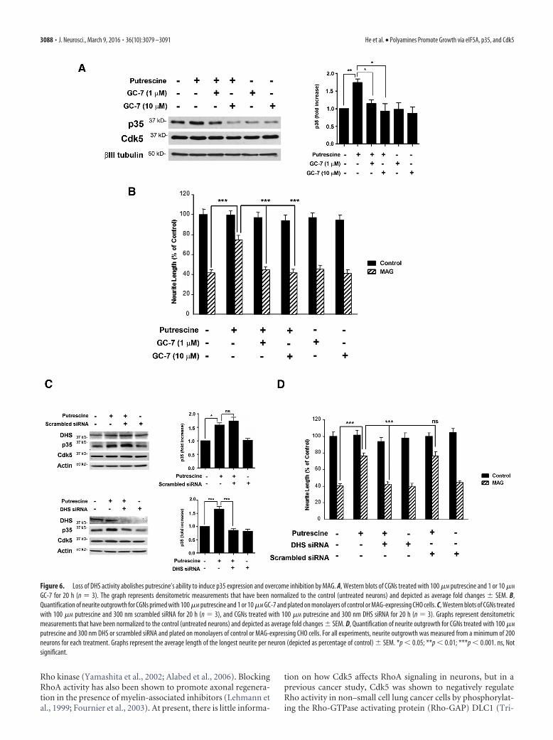
To test this hypothesis, hypusination of eIF5A was blocked byinhibiting DHS activity, and this was accomplished using twodifferent approaches: treatment with GC-7, a pharmacologicalinhibitor of DHS, and siRNA knockdown of DHS. In these ex-periments, p35 levels were significantly increased in CGNstreated with putrescine (Fig. 6A), but in the presence of either 1 or10 �M GC-7, putrescine-induced increases in p35 were blocked(Fig. 6A). Treatment with GC-7 also blocked the ability of pu-trescine to overcome MAG-mediated inhibition of neurite out-growth in vitro (Fig. 6B). Similarly, a scrambled siRNA had noeffect on putrescine-induced expression of p35 (Fig. 6C), butwhen DHS was knocked down, p35 expression was not inducedin response to putrescine (Fig. 6C). In our neurite outgrowthassays, neurons transfected with DHS siRNA and treated withputrescine were unable to overcome inhibition by MAG, andneurite outgrowth was significantly reduced compared to neu-rons treated with putrescine alone, or a combination of pu-trescine and scrambled siRNA (Fig. 6D). It should be noted thatthe effects of GC-7 and DHS siRNA were specific to p35, as thesetreatments had no effect on the expression of Cdk5 (Fig. 6A,C).
Last, we performed additional siRNA experiments to showthat eIF5A is specifically required for polyamine-induced in-creases in p35 expression and the ability to overcome inhibitionby MAG. CGNs were transfected with either eIF5A siRNA orscrambled siRNA and treated with 100 �M putrescine for 24 h.These neurons were then used for Western blotting to analyzeeIF5A and p35 levels, and in neurite outgrowth assays. In neuronstreated with either putrescine alone or putrescine and scrambledsiRNA, we observed a significant increases in p35 (Fig. 7A), andcorresponding significant increases in neurite outgrowth whenthese neurons were plated on MAG substrates (Fig. 7B). When
eIF5A was knocked down, however, putrescine failed to inducep35 expression (Fig. 7A), and its ability to promote neurite out-growth on MAG was abolished (Fig. 7B). To provide furtherevidence of eIF5A’s involvement in this process, we performedgain of function experiments in which the siRNA-knockdown ofeIF5A was rescued by ectopic expression of eIF5A. DRG neuronswere transfected with either scrambled siRNA or siRNA tar-geting the 3�UTR of eIF5A, and this was followed by transfec-tion with an ORF clone expressing eGFP-tagged humaneIF5A. Transfected neurons were then treated with 100 �M
putrescine for 24 h and used for neurite outgrowth assays.Neurons treated with putrescine were able to overcome inhi-bition by MAG, and this effect was sustained in the presence ofthe scrambled siRNA (Fig. 7C). As in the previous experiment,neurons that received eIF5A siRNA were unable to overcomeMAG-mediated inhibition when treated with putrescine (Fig.7C). However, introduction of the eIF5A ORF clone restoredputrescine-mediated neurite outgrowth on MAG in the pres-ence of the eIF5A siRNA (Fig. 7C). This confirms that eIF5A isrequired to mediate the effects of putrescine, and collectivelythese results demonstrate that DHS-mediated hypusination ofeIF5A underlies putrescine’s ability to induce p35 expressionand overcome inhibition by MAG.
DiscussionBased on the results obtained in this study, we propose the fol-lowing mechanism for cAMP- and polyamine-induced neuriteoutgrowth and reversal of inhibition by CNS myelin. Elevation ofintracellular cAMP leads to increased expression of arginase I anda corresponding increase in putrescine levels, which can also beaccomplished through administration of exogenous putrescine.Putrescine is then rapidly converted to spermidine, which servesas a substrate for the hypusination of eIF5A within the neuron.The activated eIF5A will initiate translation of p35, which willthen bind to and activate Cdk5, and Cdk5 will in turn stimulateenhanced neurite outgrowth in the presence of MAG and othermyelin-associated inhibitors.
The elucidation of this mechanism raises interesting questionsabout how Cdk5 promotes axonal growth and the potential sub-strates that may be phosphorylated in response to Cdk5 activa-tion. Proteins that are involved in cytoskeletal regulation arelogical candidates for these roles, and Cdk5 phosphorylates manyproteins that affect actin dynamics, such as WAVE (Wiskott–Aldrich syndrome protein-family verprolin-homologous pro-tein), CaMKII, Cables, amphiphysin, and synapsin I (Matsubaraet al., 1996; Zukerberg et al., 2000; Floyd et al., 2001; Dhavan etal., 2002; Kim et al., 2006). Phosphorylation of these proteins byCdk5 has been shown to have a variety of effects on neuronalmorphology and function including induction of cortical neuri-togenesis (Zukerberg et al., 2000; Floyd et al., 2001), sequestra-tion of synaptic vesicles (Verstegen et al., 2014), and increaseddendritic spine formation in hippocampal neurons (Kim et al.,2006). Cdk5 also regulates actin polymerization by modulatingthe activity of the Rho family of small GTPases. Cdk5 can influ-ence Rac signaling and cytoskeletal remodeling by phosphorylat-ing the Rac effector p21-activated kinase 1 (Pak1; Nikolic et al.,1998; Rashid et al. 2001). This is accomplished through theformation of Rac-p35/Cdk5-Pak1 complexes within neuronalgrowth cones, which leads to the phosphorylation of Pak1 byCdk5/p35 in a Rac-dependent manner (Nikolic et al., 1998). Toovercome inhibition by MAG and CNS myelin, however, themost likely target for Cdk5 would be RhoA, as MAG and Nogohave been shown to activate RhoA and its downstream effector
He et al. • Polyamines Promote Growth via eIF5A, p35, and Cdk5 J. Neurosci., March 9, 2016 • 36(10):3079 –3091 • 3087
Rho kinase (Yamashita et al., 2002; Alabed et al., 2006). BlockingRhoA activity has also been shown to promote axonal regenera-tion in the presence of myelin-associated inhibitors (Lehmann etal., 1999; Fournier et al., 2003). At present, there is little informa-
tion on how Cdk5 affects RhoA signaling in neurons, but in aprevious cancer study, Cdk5 was shown to negatively regulateRho activity in non–small cell lung cancer cells by phosphorylat-ing the Rho-GTPase activating protein (Rho-GAP) DLC1 (Tri-
Figure 6. Loss of DHS activity abolishes putrescine’s ability to induce p35 expression and overcome inhibition by MAG. A, Western blots of CGNs treated with 100 �M putrescine and 1 or 10 �M
GC-7 for 20 h (n � 3). The graph represents densitometric measurements that have been normalized to the control (untreated neurons) and depicted as average fold changes � SEM. B,Quantification of neurite outgrowth for CGNs primed with 100 �M putrescine and 1 or 10 �M GC-7 and plated on monolayers of control or MAG-expressing CHO cells. C, Western blots of CGNs treatedwith 100 �M putrescine and 300 nm scrambled siRNA for 20 h (n � 3), and CGNs treated with 100 �M putrescine and 300 nm DHS siRNA for 20 h (n � 3). Graphs represent densitometricmeasurements that have been normalized to the control (untreated neurons) and depicted as average fold changes � SEM. D, Quantification of neurite outgrowth for CGNs treated with 100 �M
putrescine and 300 nm DHS or scrambled siRNA and plated on monolayers of control or MAG-expressing CHO cells. For all experiments, neurite outgrowth was measured from a minimum of 200neurons for each treatment. Graphs represent the average length of the longest neurite per neuron (depicted as percentage of control) � SEM. *p � 0.05; **p � 0.01; ***p � 0.001. ns, Notsignificant.
3088 • J. Neurosci., March 9, 2016 • 36(10):3079 –3091 He et al. • Polyamines Promote Growth via eIF5A, p35, and Cdk5

pathi et al. 2014). It is therefore possible that Cdk5 may inhibitRhoA activity within neurons by activating Rho-GAPs, whichwould counteract myelin-induced activation of RhoA and facili-tate neurite extension.
Microtubule dynamics are similarly influenced by Cdk5,which is abundant in microtubule preparations isolated frombovine brain (Ishiguro et al., 1992; Sobue et al., 2000). Phosphor-ylation of focal adhesion kinase by Cdk5 plays a critical role inmicrotubule organization and neuronal migration (Xie et al.,2003), and several microtubule-associated proteins (MAPs) in-cluding MAP2, MAP1B, and tau are substrates of Cdk5 (Pagliniand Caceres, 2001). Hyperphosphorylation of tau by Cdk5 pre-vents tau from associating with microtubules (Patrick et al.,1999), and this leads to tau aggregation and the formation of theneurofibrillary tangles that are hallmarks of Alzheimer’s disease
(Noble et al., 2003). However, it should be noted that this path-ological response only occurs when Cdk5 binds to p25, a trun-cated form of p35 produced by calpain-mediated cleavage of thefull-length protein (Patrick et al., 1999). Conversely, phosphory-lation of MAP1B by p35/Cdk5 has been linked to laminin-induced neurite extension in cerebellar neurons (Pigino et al.,1997; Paglini et al., 1998), netrin 1-mediated axonal guidance inthe developing forebrain and hippocampus (Del Río et al., 2004),and the growth and stabilization of embryonic retinal ganglioncell axons (Hahn et al., 2005). Interestingly, in preliminary exper-iments conducted by our laboratory, we observed that treatmentwith putrescine led to increased phosphorylation of MAP1B, butnot tau, in CGNs, and this response could be blocked by rosco-vitine (data not shown). This suggests that Cdk5 can differentiallyphosphorylate its substrates to exert its effects and that Cdk5-
Figure 7. eIF5A is required for putrescine-mediated expression of p35 and reversal of inhibition by MAG. A, Western blots of CGNs treated with 100 �M putrescine and 300 nm eIF5A or scrambledsiRNA for 20 h (n � 3). Graphs represent densitometric measurements that have been normalized to the control (untreated neurons) and depicted as average fold changes � SEM. *p � 0.05. B,Quantification of neurite outgrowth for CGNs treated with 100 �M putrescine and 300 nm eIF5A or scrambled siRNA and plated on monolayers of control or MAG-expressing CHO cells. C,Quantification of neurite outgrowth for DRG neurons treated with 100 �M putrescine, eIF5A or scrambled siRNA, and an eIF5A ORF clone and plated on monolayers of control or MAG-expressing CHOcells. For all experiments, neurite outgrowth was measured from a minimum of 200 neurons for each treatment. Graphs represent the average length of the longest neurite per neuron (depicted aspercentage of control) � SEM. ***p � 0.001. ns, Not significant.
He et al. • Polyamines Promote Growth via eIF5A, p35, and Cdk5 J. Neurosci., March 9, 2016 • 36(10):3079 –3091 • 3089

dependent phosphorylation of MAP1B could underlie the abilityof putrescine to overcome inhibition by CNS myelin.
Whereas the substrates and functions of Cdk5 in the CNS havebeen well characterized, the same cannot be said for eIF5A. Thereis little information available regarding eIF5A’s function in neu-rons, as the vast majority of eIF5A studies have focused on its rolein regulating cell survival and proliferation in both yeast andmammalian cells (Zanelli and Valentini, 2007), but there is someevidence that eIF5A is required for neurite outgrowth from PC12cells and hippocampal neurons (Huang et al., 2007). However,virtually nothing is known about the proteins that are translatedin response to eIF5A activation. In this study we show that hy-pusination of eIF5A leads to increased translation of p35, whichresults in Cdk5 activation and reversal of myelin-mediated inhi-bition. To the best of our knowledge, this is the first time that aneuron-specific protein has been identified as a product ofeIF5A-mediated translation, and we also believe that this is thefirst report to describe functional outcomes that can be directlyattributed to eIF5A activity in neurons. In our immunocyto-chemistry experiments, eIF5A was observed throughout the cellbodies and processes of CGNs (data not shown), and in responseto putrescine treatment, we observed a dramatic upregulation ofp35 that was concentrated in the neurites and growth cones ofCGNs (Fig. 4). This raises the possibility that eIF5A is initiatinglocal translation of p35 within the growth cones, and this is sup-ported by a study reporting that eIF5A is enriched in the dendriticprocesses of Purkinje cells, where it is well established that localprotein synthesis occurs (Luchessi et al., 2008). Although eIF5Ahas classically been defined as a translation initiation factor(Benne and Hershey, 1978; Smit-McBride et al., 1989), it wasshown previously that eIF5A can act as an elongation and termi-nation factor as well (Saini et al., 2009), and the fact that eIF5Acan independently execute all three phases of translation stronglysuggests that the p35 expressed in response to putrescine wasproduced through local translation mediated by eIF5A.
In addition to providing greater insight into the basic mecha-nisms that govern eIF5A and Cdk5 activity, our findings also haveimplications for the development of strategies to overcome inhi-bition by myelin and promote axonal regeneration after CNSinjury. One potential target is p35, as we have shown that over-expression of p35 is sufficient to overcome inhibition by MAG invitro. There are several factors that must be taken into account,however, before a similar approach could be considered for use intranslational studies. The first is that viral overexpression of p35would likely not be a viable approach for clinical applications,from both a technical and safety standpoint, and so alternativemeans of inducing p35 expression would be required. Thesecould include gene-based therapies or, most logically, exogenousadministration of spermidine, which has been shown previouslyto promote axonal regeneration in the optic nerve (Deng et al.,2009). Importantly, spermidine has been shown to cross theblood– brain barrier following cerebral ischemia (Diler et al.,2002), and so it is possible that entry of spermidine into the CNSwould be facilitated after traumatic injury. A second factor toconsider is that elevated expression of p35 could be accompaniedby higher levels of p25, which, as mentioned, has been conclu-sively linked to neurodegeneration (Patrick et al., 1999). It wouldtherefore be prudent to monitor p25 levels in future studies in-volving induction of p35 expression.
In addition to p35, identification of the substrates and signal-ing pathways activated by Cdk5 could also prove to be a valuablesource of new targets for therapeutic intervention, and it wouldbe particularly interesting to test whether these targets could
work synergistically with other proregenerative agents that wehave identified, such as interleukin-6 (Cao et al. 2006), secretoryleukocyte protease inhibitor (Hannila et al., 2013), and metallo-thioinein I/II (Siddiq et al., 2015) to enhance axonal regenera-tion. Our findings also have broader implications for the study ofthe nervous system, as the mechanisms described in this studyadvance our understanding of the functions of eIF5A and Cdk5 inneurons and provide a basis for further studies examining howthese proteins affect fundamental processes such as synaptogen-esis, differentiation, and neuronal survival.
ReferencesAlabed YZ, Grados-Munro E, Ferraro GB, Hsieh SH, Fournier AE (2006)
Neuronal responses to myelin are mediated by rho kinase. J Neurochem96:1616 –1625. CrossRef Medline
Benne R, Hershey JW (1978) The mechanism of action of protein synthesisinitiation factors from rabbit reticulocytes. J Biol Chem 253:3078 –3087.Medline
Bibb JA, Chen J, Taylor JR, Svenningsson P, Nishi A, Snyder GL, Yan Z,Sagawa ZK, Ouimet CC, Nairn AC, Nestler EJ, Greengard P (2001) Ef-fects of chronic exposure to cocaine are regulated by the neuronal proteinCdk5. Nature 410:376 –380. CrossRef Medline
Cai D, Shen Y, De Bellard M, Tang S, Filbin MT (1999) Prior exposure toneurotrophins blocks inhibition of axonal regeneration by MAG andmyelin via a cAMP-dependent mechanism. Neuron 22:89 –101. CrossRefMedline
Cai D, Deng K, Mellado W, Lee J, Ratan RR, Filbin MT (2002) Arginase Iand polyamines act downstream from cyclic AMP in overcoming inhibi-tion of axonal growth MAG and myelin in vitro. Neuron 35:711–719.CrossRef Medline
Cao Z, Gao Y, Bryson JB, Hou J, Chaudhry N, Siddiq M, Martinez J, SpencerT, Carmel J, Hart RB, Filbin MT (2006) The cytokine interleukin-6 issufficient but not necessary to mimic the peripheral conditioning lesioneffect on axonal growth. J Neurosci 26:5565–5573. CrossRef Medline
Del Río JA, Gonzalez-Billault C, Urena JM, Jimenez EM, Barallobre MJ, Pas-cual M, Pujadas L, Simo S, La Torre A, Wandosell F, Avila J, Soriano E(2004) MAP1B is required for Netrin 1 signaling in neuronal migrationand axonal guidance. Curr Biol 2004 May 25;14(10):840 – 850.
Deng K, He H, Qiu J, Lorber B, Bryson JB, Filbin MT (2009) Increasedsynthesis of spermidine as a result of upregulation of arginase I promotesaxonal regeneration in culture and in vivo. J Neurosci 29:9545–9552.CrossRef Medline
Dhavan R, Tsai LH (2001) A decade of CDK5. Nat Rev Mol Cell Biol 2:749 –759. CrossRef Medline
Dhavan R, Greer PL, Morabito MA, Orlando LR, Tsai LH (2002) Thecyclin-dependent kinase 5 activators p35 and p39 interact with the alpha-subunit of Ca2�/calmodulin-dependent protein kinase II and alpha-actinin-1 in a calcium dependent manner. J Neurosci 22:7879 –7891.Medline
Diler AS, Ziylan YZ, Uzum G, Lefauconnier JM, Seylaz J, Pinard E (2002)Passage of spermidine across the blood-brain barrier in short recircula-tion periods following global cerebral ischemia: effects of mild hyperther-mia. Neurosci Res 43:335–342. CrossRef Medline
Filbin MT (2003) Myelin-associated inhibitors of axonal regeneration in theadult mammalian CNS. Nat Rev Neurosci 4:703–713. CrossRef Medline
Floyd SR, Porro EB, Slepnev VI, Ochoa GC, Tsai LH, De Camilli P (2001)Amphiphysin 1 binds the cyclin-dependent kinase (cdk) 5 regulatory sub-unit p35 and is phosphorylated by cdk5 and cdc2. J Biol Chem 276:8104 – 8110. CrossRef Medline
Fournier AE, Takizawa BT, Strittmatter SM (2003) Rho kinase inhibitionenhances axonal regeneration in the injured CNS. J Neurosci 23:1416 –1423. Medline
Gutierrez E, Shin BS, Woolstenhulme CJ, Kim JR, Saini P, Buskirk AR, DeverTE (2013) eIF5A promotes translation of polyproline motifs. Mol Cell51:35– 45. CrossRef Medline
Hahn CM, Kleinholz H, Koester MP, Grieser S, Thelen K, PollerbergGE (2005) Role of cyclin-dependent kinase 5 and its activator P35 inlocal axon and growth cone stabilization. Neuroscience 134:449 – 465.CrossRef Medline
Hannila SS, Siddiq MM, Carmel JB, Hou J, Chaudhry N, Bradley PM, HilaireM, Richman EL, Hart RP, Filbin MT (2013) Secretory leukocyte pro-
3090 • J. Neurosci., March 9, 2016 • 36(10):3079 –3091 He et al. • Polyamines Promote Growth via eIF5A, p35, and Cdk5

tease inhibitor reverses inhibition by CNS myelin, promotes regenerationin the optic nerve, and suppresses expression of the TGF� signaling pro-tein Smad2. J Neurosci 33:5138 –5151. CrossRef Medline
Harada T, Morooka T, Ogawa S, Nishida E (2001) ERK induces p35, aneuron-specific activator of Cdk5, through induction of Egr1. Nat CellBiol 3:453– 459. CrossRef Medline
Huang Y, Higginson DS, Hester L, Park MH, Snyder SH (2007) Neuronalgrowth and survival mediated by eIF5A, a polyamine-modified transla-tion initiation factor. Proc Natl Acad Sci U S A 104:4194 – 4199. CrossRefMedline
Ishiguro K, Takamatsu M, Tomizawa K, Omori A, Takahashi M, Arioka M,Uchida T, Imahori K (1992) Tau protein kinase I converts normal tauprotein into A68-like component of paired helical filaments. J Biol Chem267:10897–10901. Medline
Jao DL, Chen KY (2006) Tandem affinity purification revealed thehypusine-dependent binding of eukaryotic initiation factor 5A to thetranslating 80S ribosomal complex. J Cell Biochem 97:583–598. CrossRefMedline
Johansson JU, Lilja L, Chen XL, Higashida H, Meister B, Noda M, Zhong ZG,Yokoyama S, Berggren PO, Bark C (2005) Cyclin-dependent kinase 5activators p35 and p39 facilitate formation of functional synapses. BrainRes Mol Brain Res 138:215–227. CrossRef Medline
Kim Y, Sung JY, Ceglia I, Lee KW, Ahn JH, Halford JM, Kim AM, Kwak SP,Park JB, Ho Ryu S, Schenck A, Bardoni B, Scott JD, Nairn AC, GreengardP (2006) Phosphorylation of WAVE1 regulates actin polymerizationand dendritic spine morphology. Nature 442:814 – 817. CrossRef Medline
Lehmann M, Fournier A, Selles-Navarro I, Dergham P, Sebok A, Leclerc N,Tigyi G, McKerracher L (1999) Inactivation of Rho signaling pathwaypromotes CNS axon regeneration. J Neurosci 19:7537–7547. Medline
Lew J, Huang QQ, Qi Z, Winkfein RJ, Aebersold R, Hunt T, Wang JH (1994)A brain-specific activator of cyclin-dependent kinase 5. Nature 371:423– 426. CrossRef Medline
Luchessi AD, Cambiaghi TD, Alves AS, Parreiras-E-Silva LT, Britto LR,Costa-Neto CM, Curi R (2008) Insights on eukaryotic translation initi-ation factor 5A (eIF5A) in the brain and aging. Brain Res 1228:6 –13.CrossRef Medline
Matsubara M, Kusubata M, Ishiguro K, Uchida T, Titani K, Taniguchi H(1996) Site specific phosphorylation of synapsin I by mitogen-activatedprotein kinase and Cdk5 and its effects on physiological functions. J BiolChem 271:21108 –21113. CrossRef Medline
Nikolic M, Dudek H, Kwon YT, Ramos YF, Tsai LH (1996) The cdk5/p35kinase is essential for neurite outgrowth during neuronal differentiation.Genes Dev 10:816 – 825. CrossRef Medline
Nikolic M, Chou MM, Lu W, Mayer BJ, Tsai LH (1998) The p35/Cdk5kinase is a neuron-specific Rac effector that inhibits Pak1 activity. Nature395:194 –198. CrossRef Medline
Noble W, Olm V, Takata K, Casey E, Mary O, Meyerson J, Gaynor K, LaFran-cois J, Wang L, Kondo T, Davies P, Burns M, Veeranna, Nixon R, DicksonD, Matsuoka Y, Ahlijanian M, Lau LF, Duff K (2003) Cdk5 is a key factorin tau aggregation and tangle formation in vivo. Neuron 38:555–565.CrossRef Medline
Norton WT, Poduslo SE (1973) Myelination in rat brain: method of myelinisolation. J Neurochem 21:749 –757. CrossRef Medline
Paglini G, Caceres A (2001) The role of the Cdk5–p35 kinase in neuronaldevelopment. Eur J Biochem 268:1528 –1533. Medline
Paglini G, Pigino G, Kunda P, Morfini G, Maccioni R, Quiroga S, Ferreira A,Caceres A (1998) Evidence for the participation of the neuron-specificCDK5 activator P35 during laminin-enhanced axonal growth. J Neurosci18:9858 –9869. Medline
Park MH (2006) The post-translational synthesis of a polyamine-derived
amino acid, hypusine, in the eukaryotic translation initiation factor 5A(eIF5A). J Biochem 139:161–169. CrossRef Medline
Patrick GN, Zukerberg L, Nikolic M, de la Monte S, Dikkes P, Tsai LH (1999)Conversion of p35 to p25 deregulates Cdk5 activity and promotes neuro-degeneration. Nature 402:615– 622. CrossRef Medline
Pigino G, Paglini G, Ulloa L, Avila J, Caceres A (1997) Analysis of the ex-pression, distribution and function of cyclin dependent kinase 5 (cdk5) indeveloping cerebellar macroneurons. J Cell Sci 110:257–270. Medline
Qiu J, Cai D, Dai H, McAtee M, Hoffman PN, Bregman BS, Filbin MT (2002)Spinal axon regeneration induced by elevation of cyclic AMP. Neuron34:895–903. CrossRef Medline
Rashid T, Banerjee M, Nikolic M (2001) Phosphorylation of Pak1 by thep35/Cdk5 kinase affects neuronal morphology. J Biol Chem 276:49043–49052. CrossRef Medline
Saini P, Eyler DE, Green R, Dever TE (2009) Hypusine-containing proteineIF5A promotes translation elongation. Nature 459:118 –121. CrossRefMedline
Siddiq MM, Hannila SS, Carmel JB, Bryson JB, Hou J, Nikulina E, Willis MR,Mellado W, Richman EL, Hilaire M, Hart RP, Filbin MT (2015) Metal-lothionein-I/II promotes axonal regeneration in the central nervous sys-tem. J Biol Chem 290:16343–16356. CrossRef Medline
Smit-McBride Z, Dever TE, Hershey JW, Merrick WC (1989) Sequence de-termination and cDNA cloning of eukaryotic initiation factor 4D, thehypusine-containing protein. J Biol Chem 264:1578 –1583. Medline
Sobue K, Agarwal-Mawal A, Li W, Sun W, Miura Y, Paudel HK (2000)Interaction of neuronal Cdc2-like protein kinase with microtubule-associated protein tau. J Biol Chem 275:16673–16680. CrossRef Medline
Tang D, Yeung J, Lee KY, Matsushita M, Matsui H, Tomizawa K, Hatase O,Wang JH (1995) An isoform of the neuronal cyclin-dependent kinase 5(Cdk5) activator. J Biol Chem 270:26897–26903. CrossRef Medline
Tripathi BK, Qian X, Mertins P, Wang D, Papageorge AG, Carr SA, Lowy DR(2014) CDK5 is a major regulator of the tumor suppressor DLC1. J CellBiol 207:627– 642. CrossRef Medline
Tsai LH, Takahashi T, Caviness VS Jr, Harlow E (1993) Activity and expres-sion pattern of cyclin-dependent kinase 5 in the embryonic mouse ner-vous system. Development 119:1029 –1040. Medline
Tsai LH, Delalle I, Caviness VS Jr, Chae T, Harlow E (1994) p35 is a neural-specific regulatory subunit of cyclin-dependent kinase 5. Nature 371:419 – 423. CrossRef Medline
Verstegen AM, Tagliatti E, Lignani G, Marte A, Stolero T, Atias M, Corradi A,Valtorta F, Gitler D, Onofri F, Fassio A, Benfenati F (2014) Phosphory-lation of synapsin I by cyclin-dependent kinase-5 sets the ratio betweenthe resting and recycling pools of synaptic vesicles at hippocampal syn-apses. J Neurosci 34:7266 –7280. CrossRef Medline
Xie Z, Sanada K, Samuels BA, Shih H, Tsai LH (2003) Serine 732 phosphor-ylation of FAK by Cdk5 is important for microtubule organization, nu-clear movement, and neuronal migration. Cell 114:469 – 482. CrossRefMedline
Yamashita T, Higuchi H, Tohyama M (2002) The p75 receptor transducesthe signal from myelin-associated glycoprotein to Rho. J Cell Biol 157:565–570. CrossRef Medline
Zanelli CF, Valentini SR (2007) Is there a role for eIF5A in translation?Amino Acids 33:351–358. CrossRef Medline
Zhao CT, Li K, Li JT, Zheng W, Liang XJ, Geng AQ, Li N, Yuan XB (2009)PKCdelta regulates cortical radial migration by stabilizing the Cdk5 acti-vator p35. Proc Natl Acad Sci U S A 106:21353–21358. CrossRef Medline
Zukerberg LR, Patrick GN, Nikolic M, Humbert S, Wu CL, Lanier LM,Gertler FB, Vidal M, Van Etten RA, Tsai LH (2000) Cables links Cdk5and c-Abl and facilitates Cdk5 tyrosine phosphorylation, kinase upregu-lation, and neurite outgrowth. Neuron 26:633– 646. CrossRef Medline
He et al. • Polyamines Promote Growth via eIF5A, p35, and Cdk5 J. Neurosci., March 9, 2016 • 36(10):3079 –3091 • 3091

![ERE.] · 2020. 10. 21. · ERE.] 136 NEEDLE ROLLER BEARIN6S . JNS JNS 138 NOSE CO.,LTD. RNA..M NKI..M 30 0 *an: NA69 32) 8mrn V . Created Date: 3/27/2020 9:42:40 AM ...](https://static.fdocuments.us/doc/165x107/610ce98f5446ac55583af75f/ere-2020-10-21-ere-136-needle-roller-bearin6s-jns-jns-138-nose-coltd.jpg)