Hypersensitivity
46
Hypersensitivity Mohammad Mukhit Kazi Lecturer SDCH Pune
-
Upload
mukhit-kazi -
Category
Health & Medicine
-
view
95 -
download
1
Transcript of Hypersensitivity

Hypersensitivity
Mohammad Mukhit Kazi Lecturer SDCH Pune

ObjectivesTo know difference between immunity and
hypersensitivityTo know different types of hypersensitivityTo know mechanisms involved in
hypersensitivity
Mohammad Mukhit Kazi Lecturer SDCH Pune

IntroductionImmunity
A protective processHelps body to overcome infectionsSpecific response to antigens
Immune response – sometime injurious to host
A damage to the host, mediated by preexisting immunity to self or foreign antigen.
Mohammad Mukhit Kazi Lecturer SDCH Pune

Definition of hypersensitivityThe injurious consequences in the sensitized host,
following contact with the specific antigen.
For induction of hypersensitivity reactions the host should have had contact with the antigen (allergen)
Initial contact sensitizes the immune system, leading to the priming of the appropriate B or T lymphocytes this is known as sensitizing or priming dose
Subsequent contact with the allergen causes manifestations of hypersensitivity as shocking dose.
Mohammad Mukhit Kazi Lecturer SDCH Pune

Difference between Immunity and HypersensitivityFocus is on antigen and what happens to it
Antigen are of little concern or often innocuous or bland substances such as serum proteins or pollen
e.g. killing of bacterium or neutralization of toxin
It is concerned with what happens to the host as a result of the immune reaction
Mohammad Mukhit Kazi Lecturer SDCH Pune

Musts for Hypersensitivity
Contact with allergen
Sensitizing/priming dose
Induction of AMI/CMI
Shocking dose
Mohammad Mukhit Kazi Lecturer SDCH Pune

AllergyOriginally used by von Pirquet means
An altered state of reactivity to an antigen and included both types of immune responses protective as well as injurious
Also referred as all immune processes harmful to the host such as hypersensitivity and autoimmunity
Commonly used as hypersensitivity
Mohammad Mukhit Kazi Lecturer SDCH Pune

Classification of Hypersensitivity reactionsI. Immediate hypersensitivity (B cell or Ab
mediated) Anaphylaxis
AtopyAntibody mediated cell damageArthus phenomenonSerum sickenss
II. Delayed hypersensitivity (T cell mediated)
Infection (tuberculin) typecontact dermatitis type
Mohammad Mukhit Kazi Lecturer SDCH Pune

Immediate hypersensitivity
Delayed Hypersensitivity
1. Appears and recedes rapidly
1. Appears slowly, lasts longer
2. Induced by antigen or haptens by any route
2. Antigen or hapten intradermally or with Freund’s adjuvant or by skin contact
3. Circulating antibodies present and responsible for reaction; (Ab mediated reaction)
3. Circulating Abs may be absent and not responsible for reaction; cell mediated reaction
4. Passive transfer possible with serum
4. Cannot be transferred with serum; but possible with T cells or transfer factor
5. Desensitization easy but short lived
5. Difficult, but long lasting
Mohammad Mukhit Kazi Lecturer SDCH Pune

Coombs and Gell (1963) classified hypersensitivity reactions into 4 types based on different mechanisms of pathogenesis
Type I (Anaphylactic, IgE or reagin dependent)
Type II ( cytotoxic or cell stimulating)
Type III ( Immune complexes or toxic complex disease)
Type IV ( Delayed or cell mediated hypersensitivity)
Mohammad Mukhit Kazi Lecturer SDCH Pune

Mohammad Mukhit Kazi Lecturer SDCH Pune

Mohammad Mukhit Kazi Lecturer SDCH Pune

A = Type I - Anaphylaxis Reaction
C = Type II - Cytotoxic Mediated Hypersensitivity Reaction
I = Type III - Immune Complex Mediated Hypersensitivity Reaction
D = Type IV - Delayed Hypersensitivity Or Cell Mediated Hypersensitivity Reaction
Mohammad Mukhit Kazi Lecturer SDCH Pune

Type I Reactions (IgE dependent)Occurs in two forms
The acute : potentially fatal, systemic form called anaphylaxis
The chronic or recurrent : Non fatal, typically localized form called atopy
Mohammad Mukhit Kazi Lecturer SDCH Pune

Type-I hypersensitivityType-I hypersensitivity
The common allergy
Mohammad Mukhit Kazi Lecturer SDCH Pune

AnaphylaxisClassical immediate hypersensitivity
reactionAna = withoutPhylaxis = protection
Coined by Richet (1902)Experiment on Dog
Theobald’s smith (1902)Similar phenomenon in guinea pig
Ehrlich named this ‘Theobald Smith phenomenon’
Mohammad Mukhit Kazi Lecturer SDCH Pune

Type I Reactions
Humans –Itching of scalp & tongue,
flushing of skin, difficulty in breathing, nausea, vomiting, diarrhea, acute hypotension, loss of consciousness, death (rare)
Causes Serum therapy,
antibiotics, insect stingsTreatment
Adrenalin 0.5 ml (1 in 1000 solution) SC/IM repeated up to 2 ml in 15 min
Mohammad Mukhit Kazi Lecturer SDCH Pune

Sensitization is most effective given parenterally
But may occur by any routeMinute doses can sensitize the hostAntigens and haptens can induce anaphylaxisThere should be interval of 2-4 weeks
between sensitizing dose and shocking doseShocking dose more effective when given IVIt should be identical to sensitizing dose
Mohammad Mukhit Kazi Lecturer SDCH Pune
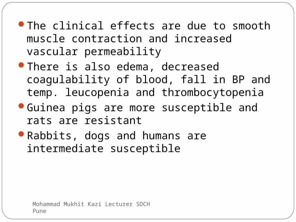
The clinical effects are due to smooth muscle contraction and increased vascular permeability
There is also edema, decreased coagulability of blood, fall in BP and temp. leucopenia and thrombocytopenia
Guinea pigs are more susceptible and rats are resistant
Rabbits, dogs and humans are intermediate susceptible
Mohammad Mukhit Kazi Lecturer SDCH Pune

In human beings fatal anaphylaxis fortunately rare
Signs and symptoms begin with itching of the scalp and tongue, flushing of the skin over whole body and difficult in breathing due to bronchial spasm
There may be nausea, vomiting, abdominal pain and diarrhea, sometime blood in stool
Acute hypotension, loss of consciousness & death follows
Mohammad Mukhit Kazi Lecturer SDCH Pune

Insect stings cause anaphylaxis in human beings
Prompt treatment with adrenalin is life saving0.5 ml of a 1 in 1000 solution SC or IMRepeat dose to total 1 ml over 15 minutes if
necessaryCutaneous anaphylaxis
Local wheal and flare response after Ag is administered intradermally
Wheal – pale central areaFlare – hyperemia and erythema
Mohammad Mukhit Kazi Lecturer SDCH Pune
SDC&H
a congestion of the blood, occurring in any part of the body

Passive cutaneous anaphylaxis (PCA)Developed by Ovary (1952)For detection of Abs e.g. IgG Ab
Anaphylaxis in vitroSchultz – Dale phenomenon : Isolated tissues from
sensitized guinea pig held in Ringers solutions will contract vigorously
Mohammad Mukhit Kazi Lecturer SDCH Pune

Mediators for Anaphylaxis
Primary mediators
Histamine : imp vasoactive amine formed by decarboxylation of histidine found in mast cells, basophils and platelets
Released into skin stimulates nerves producing burning and itching sensations
Causes vasodilatation and hyperemia, edema by increasing capillary permiability
Mohammad Mukhit Kazi Lecturer SDCH Pune

Induces smooth muscle contraction and stimulates
Serotonin
Decarboxylation of tryptophan found in intestinal mucosa, brain tissue and platelts
Causes smooth muscle contraction
Increased capillary permeability and vasoconstriction
Chemotactic factors
Eosinophil chemitactic factors are acidic tetrapeptides released from mast cell granules
Strongly chemotactic
Mohammad Mukhit Kazi Lecturer SDCH Pune

Secondary mediators
Prostaglandins and leukotrienes Formed from disrupted cell membrane of mast cells & other
leucocytes They are powerful bronchoconstrictors Prostaglandins also affect secretion by mucous glands, platelet
adhesion, permeability & dilatation of capillaries and the pain threshold
Platelet activating factor Low molecular weight lipid causes aggregation of platelets and release
vasoactive amines
Other mediators of anaphylaxisAnaphylatoxin released by complement activation and
bradykinin
Mohammad Mukhit Kazi Lecturer SDCH Pune

Anaphylactoid reaction
Intravenous injection of peptone, trypsin provokes a clinical reaction resembling anaphylactic shock termed as anaphylactoid reaction
Resemblance is due to same chemicals participating in the reaction
It has no immunologic basis
Non specific mechanism
Mohammad Mukhit Kazi Lecturer SDCH Pune

Atopy
Introduced by Coca (1923)Naturally occurring : hay fever and asthmaAntigens are inhalant or ingestantsDifficult to induce artificiallyGenetically determined probably linked to MHC
genotypesProduce large quantity of IgE antibodiesBottle fed infants tend to develop atopy in later life IgE differs from other immunoglobulin in many waysClinical expression depends portal of entry
Conjunctivitis, rhinitis, gastrointestinal symptoms, dermatitis
Mohammad Mukhit Kazi Lecturer SDCH Pune

1. Cannot be demonstrated in conventional serological reactions
2. Commonly occur in humans not easy in experimental animals
3. IgE is homocytotropic ie species specific
4. Heat sensitive and is inactivated at 56oc in 2 – 4 hours.
5. Does not pass through placenta
Mohammad Mukhit Kazi Lecturer SDCH Pune
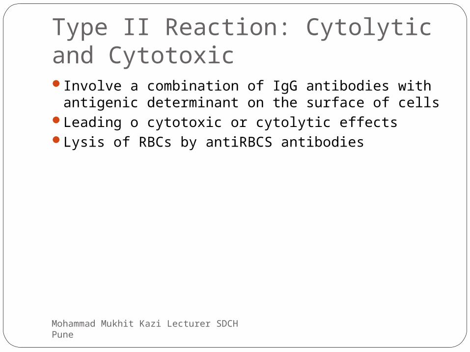
Type II Reaction: Cytolytic and CytotoxicInvolve a combination of IgG antibodies with
antigenic determinant on the surface of cellsLeading o cytotoxic or cytolytic effectsLysis of RBCs by antiRBCS antibodies
Mohammad Mukhit Kazi Lecturer SDCH Pune

Type III Reactions: Immune complexes Diseases
Arthus reactionArthus in 1903 observed after repeated injection
of normal horse serum in rabbits subcutaneously develops edema, indurations and hemorrhagic necrosis
Tissue damage is due to antigen – antibody precipitates causing complement activation and release of inflammatory molecules
This leads to increased vascular permeability and infiltration of the site with neutrophils
Leukocyte platelet thrombi are formed that reduces the blood supply and lead to tissue necrosis
Mohammad Mukhit Kazi Lecturer SDCH Pune

Arthus reactionArthus reaction
Arthus reactionType-III
Wheal & flare reactionType-I
Mohammad Mukhit Kazi Lecturer SDCH Pune

• Serum Sickness systemic form of type III hypersensitivityDescribed by von Pirquet 1905Appeared 7 – 12 days following a single injection of
high concentration of foreign serum such as diphtheria antitoxin
Clinical syndrome consists of Fever Lymphadenopathy Splenomegaly Arthritis Glomerulonephritis Endocarditis Vasculitis Urticarial rashes Abdominal pain Nausea Vomiting
Pathogenesis is the formation immune complex get depositied on inner lining of blood vessels in various parts of body causing inflammatory infiltration
Mohammad Mukhit Kazi Lecturer SDCH Pune

Serum sicknessSerum sickness
Mohammad Mukhit Kazi Lecturer SDCH Pune

Depletion of complement Disease is self limitedSingle injection can serve both as the
sensitizing dose and the shocking doseImmune complexes occurs in many diseases
including bacterial, viral and parasitic infections, disseminated malignancies and autoimmune conditions
Mohammad Mukhit Kazi Lecturer SDCH Pune

Type IV Reactions: Delayed hypersensitivity
Provoked by intra cellular parasites or haptensReaction due to sensitized T cells which on
contact with specific antigen releases cytokines that causes biological effects
Cannot be transferred passivelyBut can be transferred by lymphocytesTwo types
TuberculinContact dermatitis type
Mohammad Mukhit Kazi Lecturer SDCH Pune

Tuberculin typeWhen small dose of tuberculin is injected
intradermally in an individual sensitized to tuberculoprotein by prior infection or immunization an indurated inflammatory reaction develops at the site within 48 – 72 hours
In unsensitized individuals no responseUseful indication of the state of DH to the
bacilli
Mohammad Mukhit Kazi Lecturer SDCH Pune

Contact dermatitis typeDue to variety of chemicals, metals, dyesContact dermatitis is an inflammation of
the skin caused by direct contact with an irritating or allergy-causing substance (irritant or allergen). Reactions may vary in the same person over time. A history of any type of allergies increases the risk for this condition.
Mohammad Mukhit Kazi Lecturer SDCH Pune

Contact dermatitis reactionContact dermatitis reaction
Mohammad Mukhit Kazi Lecturer SDCH Pune

Allergic Contact Dermatitis Response to Poison Ivy Hapten
Mohammad Mukhit Kazi Lecturer SDCH Pune

Type-Type-
IVIV
Type-IIIType-IIIType-IIType-IIType-Type-
II
characteristcharacterist
icic
Comparison of hypersensitivity reactionsComparison of hypersensitivity reactions
TB test, TB test, poison ivy, poison ivy, granulomagranuloma
farmers’ farmers’ lung, lung, SLESLE
pemphiguspemphigus, , GoodpastuGoodpasturere
hay hay fever, fever, asthmaasthma
examplesexamples
antibody IgE IgG, IgM IgG, IgM none
antigen exogenous cell surface intracellularsoluble
response time
15-30 min. Min.-hrs 3-8 hours 48-72 hoursor longer
appearance Wheal & flare
Lysis & necrosis
Erythema & edema
Erythema & induration
baso- and eosinophils
Ab and complement
histology PMN andcomplement
Monocytes & lymphocytes
T-cellsantibodyantibodyantibodytransfer with
Mohammad Mukhit Kazi Lecturer SDCH Pune

AutoimmunitySelf antigens are not immunogenic
Autoimmunity is a condition in which structural or functional damage is produced by the action of immunologically competent cells or antibodies against the normal components of the body.
Autoimmunity literally means – protection
Mohammad Mukhit Kazi Lecturer SDCH Pune

Mechanism of autoimmune diseasesCells undergo antigenic alteration due to
physical, chemical or biological influencesThis results in immune responsePhysical agents – irradiation Chemicals – drugsBiological – infectious agents such as
viruses
Mohammad Mukhit Kazi Lecturer SDCH Pune

Classification of Autoimmune diseasesHemolytic autoimmune disease
- Autoimmune hemolytic anemias- Autoimmune thrombocytopenia- Autoimmune leucopenia
Mohammad Mukhit Kazi Lecturer SDCH Pune

Localized autoimmune diseases- Hashimoto’ disease (enlargement of thyroids)- Grave’s disease (Abs to thyroglobulin)- Addision’s disease (lymphocyte infiltration in
adrenal glands)- Autoimmune orchitis - Myasthenia gravis (malfunction of myoneural
junction)- Autoimmune disease of eye- Pernicius anemia- Autoimmune disease of nervous system- Autoimmune disease of skin
Mohammad Mukhit Kazi Lecturer SDCH Pune

Systemic autoimmune diseaseSystemic lupus erythematosusRheumatoid arthritisPolyarteritis nodosaSjogran’s sundrome
Mohammad Mukhit Kazi Lecturer SDCH Pune
Mohammad Mukhit Kazi Lecturer SDCH Pune