HYGIENE SCIENCES 9TH ISSUE - Tulip Group · This issue of the Journal of Hygiene Sciences is packed...
16
Committed to the advancement of Clinical & Industrial Disinfection & Microbiology n n n n n n n n n n Editorial Mini review Encyclopedia Current Trends In Profile Relax Mood Bug of the Month Did you Know Best Practices In Focus 1 2 6 7 9 10 11 13 14 16 Editorial Contents Contents VOLUME - II ISSUE - III MAY-JUN 2009 For Private Circulation Only 1 This issue of the Journal of Hygiene Sciences is packed with loads of information that would simply keep you reading....... Well, food as we all know has to be hygienic, whether it is a wholesome meal or a dessert, or simply the milk we may consume regularly with breakfast. Milk is available in different processed forms, all being rich in nutrients and therefore the foods containing milk or its products have to prepared in a manner which ensures that the final product is either entirely free from microbes, or has microbes that are desirable in the product, or are intentionally added to the requirement of the taste. Mini Review has an overview of the Dairy products and the underlying microbiology. Bleach, which is a common household disinfectant also has a chemical identity, and in chemical terms is referred to as Sodium hypochlorite, which is very effective as a disinfectant and as a cleaning agent and is employed in various products that are used for cleansing, Current Trends focuses on the role and usefulness of this chemical agent. Antibiotics that are prescribed to individuals that are afflicted with certain infections and diseases, was a term coined for substances that are detrimental to microbes in one or another form, and the term was coined by none other than the person who is the In Profile scientist in this issue; Selman Abraham Waksman, who discovered many antibiotics and pioneered different methods that are used in screening for antibiotics and potential antibiotic producers. Gastritis and peptic ulcers were attributed to different factors like stress, pungent food and acidity, but the actual etiological agent for these conditions is Helicobacter pylori, which is the Bug of the Month this time. Inspissation is a process that is used to dehydrate and coagulate certain media that have certain components that are sensitive to heat. These media are basically those that contain proteins that may denature with excessive heat, a brief description of this process appears in the section 'Did You Know' which would allow you to get an idea of this process that is essential for certain media. The section on Best Practices throws light on an important assay that detects the presence of endotoxins in a system. Endotoxins are substances that are shed by Gram negative bacteria, which aid in the infection of the host and can be life threatening or in some cases fatal. This assay has been described as Limulus Amoebocyte Lysate (LAL) assay. 'Laughter is the music of the soul', it eases the burden on your mind so Relax your mood will obviously let you have a much deserved laugh and also tickle your mind a little....Enjoy!!!! In every issue our focus is the Reader and we look upon feedback from you as a vital ingredient in making the Journal a more effective informer. So go on, turn the page and explore the information.........
Transcript of HYGIENE SCIENCES 9TH ISSUE - Tulip Group · This issue of the Journal of Hygiene Sciences is packed...

Committed to the advancement of Clinical & Industrial Disinfection & Microbiology
n
n
n
n
n
n
n
n
n
n
Editorial
Mini review
Encyclopedia
Current Trends
In Profile
Relax Mood
Bug of the Month
Did you Know
Best Practices
In Focus
1
2
6
7
9
10
11
13
14
16
Editorial
ContentsContents
VOLUME - II ISSUE - III MAY-JUN 2009
For Private Circulation Only 1
This issue of the Journal of Hygiene Sciences is packed with loads of information that would simply keep you reading.......
Well, food as we all know has to be hygienic, whether it is a wholesome meal or a dessert, or simply the milk we may consume regularly with breakfast. Milk is available in different processed forms, all being rich in nutrients and therefore the foods containing milk or its products have to prepared in a manner which ensures that the final product is either entirely free from microbes, or has microbes that are desirable in the product, or are intentionally added to the requirement of the taste. Mini Review has an overview of the Dairy products and the underlying microbiology.
Bleach, which is a common household disinfectant also has a chemical identity, and in chemical terms is referred to as Sodium hypochlorite, which is very effective as a disinfectant and as a cleaning agent and is employed in various products that are used for cleansing, Current Trends focuses on the role and usefulness of this chemical agent.
Antibiotics that are prescribed to individuals that are afflicted with certain infections and diseases, was a term coined for substances that are detrimental to microbes in one or another form, and the term was coined by none other than the person who is the In Profile scientist in this issue; Selman Abraham Waksman, who discovered many antibiotics and pioneered different methods that are used in screening for antibiotics and potential antibiotic producers.
Gastritis and peptic ulcers were attributed to different factors like stress, pungent food and acidity, but the actual etiological agent for these conditions is Helicobacter pylori, which is the Bug of the Month this time.
Inspissation is a process that is used to dehydrate and coagulate certain media that have certain components that are sensitive to heat. These media are basically those that contain proteins that may denature with excessive heat, a brief description of this process appears in the section 'Did You Know' which would allow you to get an idea of this process that is essential for certain media.
The section on Best Practices throws light on an important assay that detects the presence of endotoxins in a system. Endotoxins are substances that are shed by Gram negative bacteria, which aid in the infection of the host and can be life threatening or in some cases fatal. This assay has been described as Limulus Amoebocyte Lysate (LAL) assay.
'Laughter is the music of the soul', it eases the burden on your mind so Relax your mood will obviously let you have a much deserved laugh and also tickle your mind a little....Enjoy!!!!
In every issue our focus is the Reader and we look upon feedback from you as a vital ingredient in making the Journal a more effective informer.
So go on, turn the page and explore the information.........

For Private Circulation Only 2
Dairy is the term given to a facility for the extraction and processing of animal milk, mostly from goats or cows and sometimes also from buffaloes, sheep, horses or camels primarily for human consumption.
Milk is the secretion of the mammary gland of female mammals and is often the sole source of food for the very young mammal. The role of milk is to nourish and provide immunological protection. It is a complex biological fluid containing different components including water, lactose, fat, proteins and minerals. Milk in its natural state is a highly perishable material because it is susceptible to rapid spoilage by the action of naturally occurring enzymes and contaminating microorganisms. However it is desirably converted into a wide variety of milk products using a range of advanced processing technologies. The products include a variety of cheeses, yogurts, butters and spreads, ice cream, and dairy desserts.
In order to assess the quality of the final product; milk or processed milk, it is essential to know:
Initial Microflora of Raw MilkThe numbers and types of microorganisms in milk immediately after production (i.e., the initial microflora) directly reflect microbial contamination during production, collection, and handling. The microflora in the milk when it leaves the farm is influenced significantly by the storage temperature and the
0elapsed time after collection. Where milk is stored at =4 C, this low temperature normally will delay bacterial multiplication for at least 24 hours. The microflora, therefore, is similar to that present initially. However, if unsanitary conditions exist with the milking equipment or storage tank, the low temperature could mask these conditions.
A useful indicator for monitoring the sanitary conditions present during the production, collection, and handling of raw milk is the “total” bacterial count or standard plate count (SPC). The SPC is determined by plating (or using equivalent procedures) on a standardized plate count agar followed by aerobic incubation for
0 02 or 3 days at 32 C or 30 C, respectively. Microorganisms failing to form colonies, of course, will not be counted. The SPC does not indicate the source/s of bacterial contamination or the identity of production deficiencies leading to high counts. Its sole value is to indicate changes in the production, collection, handling, and storage environment.
Follow-up microbial assessments for psychrotrophs or thermoduric bacteria, spore-forming bacteria, streptococci, and coliforms can assist in determining sanitary deficiencies.
Certain groups can be enumerated selectively. For instance, psychrotrophs can be counted either by incubating SPC plates for
010 days at 5 – 7 C or by using a preliminary incubation of the raw
Dairy Microbiology0milk at 13 C for 16 hours followed by performing the SPC
procedure. Thermoduric bacteria can be determined by laboratory pasteurization of milk before plating. Selective or diagnostic media can be used for coliforms, lactic acid bacteria, mastitis pathogens, Gram negative rods, lipolytic, proteolytic and caseinolytic microbial types, and so on. An increased number of automated methods are now being employed for plating and enumerating bacteria. Also, rapid quantifying techniques are being used, such as the direct epifluorescent filtration technique, adenosine triphosphate method, and impedance measurements.
Microbiology of Milk and DairyTypes of microorganisms present in raw milkPathogens for Humans in Raw MilkRaw milk may contain microorganisms that are pathogenic to humans, and their source may lie either within or outside the udder. Historically, the most serious human diseases disseminated by the consumption of contaminated raw milk are tuberculosis and brucellosis. In both the diseases, the causative organisms that may be excreted in milk from infected animals are Mycobacterium bovis or M. tuberculosis and Brucella abortus, B. melitensis, or B. suis. Often with Brucella infections, there is little change in the milk or udder (i.e., mastitis is not present), but, in the case of tuberculosis mastitis, a pronounced and characteristic change in the milk and udder is observed.
Pathogenic bacteria also may be present in raw milk as a direct consequence of udder disease. Among the organisms commonly producing mastitis, Streptococcus agalactiae, Staphylococcus aureus, and Escherichia coli are pathogens known to humans. Streptococcus agalactiae can initiate a variety of clinical conditions, the most serious of which are bacteremia and meningitis in newborns, which are potentially fatal to infected infants.
However, for humans the pathogenicity of bovine strains of Streptococcus agalactiae is uncertain and is carried by a large proportion of the human population. While it seems likely that the consumption of contaminated raw milk may play a part in infections of the population at large, some researchers have reported higher rates of S. agalactiae among consumers of raw milk who do not experience symptoms of a milk-borne illness.
Staphylococcal mastitis of the cow poses a more direct threat to public health because some bovine strains produce enterotoxin. Consumption of food containing enterotoxin leads to a symptomatic illness, usually of approximately 24 hour duration, characterized by nausea, diarrhea, and abdominal pain. The production of enterotoxin usually associated with the multiplication of staphylococci under favorable growth conditions during storage of the milk. Because enterotoxin is relatively heat stable, subsequent pasteurization of the contaminated milk will not make it safe for consumption.
MAY-JUN 2009Mini Review

For Private Circulation Only 3
High numbers of E. coli may be present in milk as a consequence of mastitis, and this bacterium is responsible for several diseases of man and human disease which may have not been reported, a wide range of E. coli serotypes have been isolated from bovine milk, and it is probable that some of these are pathogenic for humans. Interestingly, microorganisms that have a greater pathogenicity for humans seldom produce bovine mastitis and yet may be present in raw milk. They include Leptospira spp., Listeria monocytogenes, Bacillus cereus, Pasteurella multocida, Clostridium perfringens, Nocardia spp., Cryptococcus neoformans and Actinomyces spp.
Further biohazards stem from the adventitious contamination of raw milk by pathogenic bacteria from sources external to the udder. Salmonellae and thermoduric Campylobacter strains fall into this category and have produced many outbreaks of enteritis. Human carriers also may be sources of infection in milk-borne outbreaks. This has been reported for Salmonella infections and for cases of scarlet fever or septic sore throat attributed to Streptococcus pyogenes.
All of these pathogens are destroyed by pasteurization, except Clostridium perfringens and Bacillus cereus, which can survive the pasteurization process because of their ability to sporulate. It is improbable, however, that C. perfringens will germinate and multiply under the modern-day conditions of milk storage.
Contamination of milkContamination of milk can vary widely depending on milk-handling practices ranging from milking a few cows by hand in the out of doors to milking 3000 cows by a complex, automated system in a well equipped parlor. There are however three basic sources of microbial contamination of milk: (1) from within the udder, (2) from the exterior of the teats and udder, and (3) from the milk handling and storage equipment.
Milk is produced at ambient temperatures ranging from subzero centigrade, where it is necessary to protect milk from freezing, to
0above 25 C, where refrigeration is needed. Furthermore, the duration of milk storage time on the farm can vary widely. Therefore, depending on the duration of milk storage on the farm before leaving can differ, often unpredictably, even under apparently similar conditions.
In most dairying areas, milk production methods, equipment, and on-farm storage have improved over time. However, udder disease remains widespread because of the presence of mastitis-associated microorganisms. Refrigeration on the farm all too often masks the effects of unsanitary practices, including the use of inadequately cleaned and sanitized milking equipment. As a result, the microbiological quality of raw milk supplies produced under apparently good sanitary conditions and stored under adequate refrigeration may produce off-flavors, yield poor product, and present a risk of food-borne infections to the consumer.
Environmental sourcesWhile the lactating animal, the production environment, and the milk handling equipment remain the principal sources for microbial contamination of raw milk, other environmental
sources include the following:
AirAir is not considered a significant source for microbial contamination in raw milk. Through its movement, air transfers soil and dust particles from a microbial-laden source into exposed milk surface such as soil and microbes. The main sources of air-borne microbes may include the activity of factory personnel, ventilation and air-conditioning systems, inflow of outdoor air, and packaging materials. The potential contamination from bacterial biofilms is also of major concern because microbial cells may attach, grow and colonize on open exposed wet surfaces.
Outdoor Environment The control of airborne microorganisms in the immediate surroundings of dairy premises is more difficult than in closed, indoor environments where more controlled measures can be taken. Natural agents such as UV light, humidity, temperature, wind direction and speed have a significant influence on the total number of airborne microorganisms in the outdoor atmosphere.
The Milk handlerWhen cows are hand milked, it is possible that the milk handler can contribute to an increased microbial load in the raw milk by dislodging dirt particles from the udder, increasing aerial contamination through accelerated air movement and contacting the milk with infected hands. Risks of contamination from the milk handler are much less with machine milking.
The Water supplyWater used in the milk production process should be of potable quality. This means that the water supply must be from an approved source free from pathogens and fecal contamination.
One must recognize that a potable water supply can become contaminated within the dairy production environment, such as in a farm storage tank that is not properly protected from rodents, birds, insects and dust. Bacteria also may be introduced into the water supply through dirty wash troughs, buckets and hoses. If untreated water gains access into milk or is used for rinsing equipment and containers, the microbes present in the water eventually will contaminate the milk.
The reasons why and how milk gets contaminated are numerous, therefore it is also necessary to know means that are adopted for the treatment of milk, so that dairy products are free from pathogens and other contaminating microbes.
Many processes have been developed over the years to enhance its utilization and safety. These processes can be grouped and analyzed in a variety of ways. l Fractionationl Concentrationl Preservation
Fractionation: the term is used to describe the fractionation or disassembly of the components of milk, utilizing their various properties of the individual components.l Centrifugal separation, utilizing density difference of the
Mini Review MAY-JUN 2009

For Private Circulation Only 4
components. The most common equipment used is the disc bowl separator, which allows the separation of light and heavy phases and also allows removal of any sediment.
l Membrane separation, utilizing size or charge difference. This is normally a pressure – motivated, flow – dependent process, involving the use of a selective membrane, with a wide range of fractionation possible, from simple water removal to separation of different proteins.
l Ion exchange, utilizing charge difference. In this process, tiny resin beads exchange charged ions on their surface with charged ions or larger charged molecules in solution, removing them for subsequent recovery.
l Precipitation and Crystallization, utilizing differences in solubility and suspension stability
l Filtration, utilizing size difference, the principle is similar to that already mentioned with membrane separation but involves the separation of larger components.
l Homogenization is a process of size reduction of the fat globules to prevent fractionation of the cream and skim milk by density difference. A combination of a high pressure pump and special valves provides high shear.
Concentration Processes: this grouping involve removal of one or more components resulting in a concentration of the remaining components. Many of these processes also involve fractionation. The processes include the following:l Evaporation, utilizing phase change of the aqueous
component. An evaporator is a specialized heat exchanger operating under vacuum, facilitating efficient water vapor generation and removal from a liquid with minimal thermal damage to the remaining liquid.
l Freeze concentration, also utilizing phase change. This involves freezing and crystallization of the aqueous component of a liquid by refrigeration followed by crystal removal. It is not widely used in dairy processing.
l Here the permeate, or material passing through the membrane, includes water, enabling concentration of the retentate or material retained.
l Drying, utilizing phase change. This is a very important process, particularly in the production of milk powder, casein and whey products. It involves water removal from a liquid concentrate or solid by heating with hot air.
Preservation Processes: this category are primarily concerned with reducing microbiological and chemical change. They include the following:l Pasteurization, thermalization, and sterilization, utilizing heat
to kill microorganisms. All these processes involve the transfer of heat into the product in order to raise the temperature to achieve a closely controlled time –
0temperature process (e.g., 72 C, 15 s) for pasteurization. l Chilling and freezing, to slow microbial growth and chemical
change and is widely used both during or prior to processing or for final product storage. Heat exchangers of the type can be used for liquid products, with cool stores and freezing chambers for finished goods.
l Reduction of pH, to inhibit microbial growth. This may be achieved by addition of acids or by bacterial fermentation of lactose.
l Dehydration (drying), to inhibit microbial growth and
chemical change. The water content of dairy products influences the microbial changes and hence drying also serves the purpose of storage and to prolong the shelf life of the product.
l Salting, to reduce water activity and inhibit microbial growth. Salt may be added as dry granular salt or by means of a brine solution, with the product being immersed for a period in a tank of concentrated brine.
l Packaging, to contain the product, protect it, and reduce microbiological and chemical changes.
Heat treatment of MilkPasteurizationThis process is done to eliminate all non-spore forming pathogens commonly associated with milk. This process also effectively destroys spoilage organisms and thus contributes to product keeping quality under required refrigeration storage.
0The process of heating every particle of milk to at least 143 F 0(61.7 C) and holding at such temperature for at least 30 minutes,
0 0or to at least 160 F (71.1 C) and holding at such temperature for at least 15 seconds.
Ultra-Pasteurization (Extended Shelf-Life Milk)0In this process milk is 'thermally processed' at or above 138 C for
at least 2 seconds, so as to produce a product that has an extended shelf life under refrigerated conditions.
Ultra-High-Temperature SterilizationUltra-High-Temperature (UHT) sterilization is a process that combines rapid heating of milk to very high temperatures followed by aseptic handling and packaging to produce a shelf-stable, commercially sterile product. Though heat treatments for
0UHT vary indifferent countries, temperatures of 130 – 150 C with holding time of 1 second or more are prescribed, with holding times of 2 – 8 seconds commonly applied.
In-container SterilizationConditions specified for in-container sterilization of milk include
0 0temperatures from 105 C to 120 C for 20 – 40 minutes. With this strategy, milk is prefilled into cans or bottles that are hermetically sealed, and then the milk is heated in an autoclave or a batch or continuous retort. In-container sterilized milk products have expected shelf lives of a year or more with no refrigeration required.
Hazard Analysis Critical Control Points (HACCP)The 'Hazard Analysis Critical Control Points' system offers a structured approach to the control of hazard in food processing and, when properly applied, identifies areas of concern and appropriate control measures before product failure is experienced. It represents a shift from retrospective quality control through end-product testing to a preventative quality assurance approach.
The HACCP procedure is generally targeted at food safety management (pathogenic microorganisms and their toxins), but, as an approach in the context broader quality management, it can be effectively applied to microbiological spoilage, foreign-body contaminations or pesticide contamination. It is preferable to conduct an HACCP program with a narrow scope (a single
Mini Review MAY-JUN 2009

For Private Circulation Only 5
pathogen or possibly pathogens) rather than attempt to cover an extended list of hazard areas then documentation tends to become complex.
Processed Dairy productsHere there is a brief description of the various processed products that are made from milk.
Fluid Milk ProductsThis group of products falls into the 'consumer products' family, competing in the beverage sector of the grocery business. Their manufacture is relatively simple, involving fractionation processes such as centrifugal separation to produce cream, skim milk, or reduced fat milk, concentration processes such as membrane separation (ultrafiltration) to produce high calcium milks, and preservation processes such as pasteurization, ultra-high temperature (UHT), and refrigeration to extend the safety and shelf life of the product range. Homogenization is used to prevent separation of the fat in the liquid product.
Fermented Milk ProductsThere are two groups in this family: (a) cheese products in which part of the original liquid is removed during manufacture as whey and (b) products in which there is no whey drainage, such as yogurts.
Both groups have a very long history of preparation and were probably developed by accident as a means of preserving milk. All or some of the standard food preservation tools of moisture removal, acid development, salt addition, and temperature adjustment may be used. The first letters of the italic words spell the conveniently remembered acronym MAST.
Cheese is an example of a fermented processed milk and its manufacture is a highly complex process. The composition of milk is adjusted or standardized (fractionation) by centrifugal separation and possibly also ultrafiltration. For most cheese types,
0the milk will then be pasteurized (72 C, 15 s) to reduce the risk from pathogenic organisms, adjusted to the desired fermentation temperature, and then pumped into a cheese vat. Starter culture consisting of a carefully selected species of lactic acid bacteria and a coagulate (e.g., calf rennet) are then added and the milk is allowed to coagulate. This is by destabilization of the casein micelle. This permits the beginning of the fractionation and selective concentration processes that form the basis of cheese-making. Once the coagulum is of sufficient strength, it is cut into small particles and, by a process of controlled heating and fermentation, syneresis or expulsion of moisture and minerals (whey) occurs. Separation of the curd from the whey over a screen (filtration) follows. Depending on the cheese type, the curd may be allowed to fuse together (e.g., cheddar) or may be kept in granules (e.g., Colby). Salt may then be incorporated into the curd for preservation, as dry granules or by immersion in brine. The curd is pressed into blocks by either gravity or mechanical compression, and the cheese then goes into controlled storage conditions for final fermentation and maturation.
For fermented milk products, the process of manufacture is somewhat simpler. For example, in yogurt manufacture, the milk to be used is fortified with additional protein (skim milk powder
0or concentrated milk), severely heated (e.g., 95 C, 5 minutes) to reduce the microbial load and to encourage whey protein / casein
0interaction, cooled to fermentation temperature (e.g., 36 C), and transferred to a fermentation vessel. Selected cultures are then added, and fermentation is continued until the desired pH of around 4.5 is reached. This causes the coagulation of the vessel contents, and the plain yogurt is then cooled prior to possible incorporation of fruit and flavoring agents, followed by packaging.
When all the raw materials and the necessary equipment that comes in contact with milk is properly treated and adequate measures are taken for safety, there is a drastic drop in the number of organisms in milk. Some processes however ensure that milk supplied has no microbes at all, which serves as an excellent starting material for any process and also making the milk safe for consumption without prior heating.
Quality control in the Dairy IndustryThe quality of food, such as milk and dairy products, may be defined as that sum of characteristics which enables the food to satisfy definite requirements and which determine its fitness for consumption. In this sense, quality can be judged by means of sensory evaluation, its nutritive value, and according to its chemical, physical, and microbiological characteristics.
Cases of food-borne disease and food poisoning are becoming more and more common throughout the world. Both of these public health problems and the microbiological spoilage of foods can be minimized by the careful choice of raw materials and correct manufacturing and storage procedures. Achievement of such objectives requires, in many cases, monitoring at various stages to assess microbiological load or to look for particular microbial types.
Monitoring procedures commonly used include the air and water supplies in a factory environment, the hygiene of packaging material, and the sampling and testing of raw materials and end products.
The analytical procedures will include standard and rapid methods for assessing microbiological load and for enumerating and detecting specific microbial genera or groups.
Procedures for the direct assessment of the microbial content of milk and milk productsl Cell Counting Procedures
The Breed Microscopic Count: the Breed smear or direct microscopic count (DMC) was developed as a rapid method for counting bacterial cells in milk.
The Direct Epifluorescent Filter Technique (DEFT): is a modern approach to the direct microscope count. With this technique the milk is first pretreated with a proteolytic enzyme and a surfactant which lyses the somatic cells and modifies fat globules sufficiently to filter the sample through a 0.6µm pore size polycarbonate filter. The filtration concentrates bacteria on the surface, after which they are stained with acridine orange. The mounted filter is examined through an epifluorescence microscope. Metabolically active cells fluoresce orange-red,
Mini Review MAY-JUN 2009

Encyclopedia
An Antibiotic is a substance or compound that kills or inhibits the growth of bacteria and other microbes like fungi and protozoa. An antibiotic is produced by one microorganism that is antagonistic to the growth of the other.
Antibiotics of the recent era may not be entirely produced by the microbes and may be semi-synthetically produced, which are effective against a wide range of microorganisms.
Though antibiotics are sometimes the primary and the only line of therapy for certain diseases, there are adverse side effects also that are associated with the antibiotic treatment. The most common side-effects include diarrhea, which can be corrected by consuming probiotics during a course of antibiotics.
Antibiotics have been grouped depending on their synthesis, mode of action and target sites.
Synthesis: Antibiotics can either be obtained directly as they are produced from the microbes itself after purification, or can be modified, by changing a few side chains in the antibiotic molecule, then they are referred to as Semi – synthetic.
Mode of Action: There are also antibiotics that may either be Narrow Spectrum, which are effective only against a few microorganisms, whereas there are others that are effective against a wide variety of microorganisms and these are referred to as Broad Spectrum antibiotics.
Depending on the mode of action, they can either be Bactericidal or Bacteriostatic.
Bactericidal antibiotics kill the target bacterium or fungus and are more effective as compared to bacteriostatic drugs. These types of drugs are used to treat endocarditis, meningitis, osteomyelitis and other invasive bacterial infections. Hosts with low immune system must be treated with bactericidal drugs. These drugs include beta – lactams, aminoglycosides and fluoroquinolones.
Bacteriostatic antibiotics are the class of antibiotics that cease the growth and multiplication of the target bacteria and include antibiotics that stop protein synthesis, this category of drugs include macrolides.
These can be helpful, since they let the normal host defenses destroy the microorganisms. They are used to treat infections such as cystitis. These drugs include tetracycline, sulfonamides, clindamycin and chloramphenicol. Some organisms require two agents for killing such as enterococci.
For Private Circulation Only 6
while inactive bacteria fluoresce green.The clumps of orange-red fluorescing bacteria are counted in the field of view, and a DEFT count per ml of the milk sample is calculated over several fields
Automatic Fluorescent Microscopic Count of Bacteria: in this method too, the samples are pretreated, cells are stained with the fluorochrome acridine orange but are counted electronically as light impulses of a single bacteria.
Flow Cytometry: is the science of measuring components (cells) and the properties of individual cells in liquid suspension. In essence suspended cells are brought to a detector by means of a flow channel. In this technique DNA/RNA of the bacteria is stained with the fluorescent dye ethidium bromide, and 'disturbing' milk constituents are reduced/dispersed by buffers, detergents and enzymes during sample preparation. Fluorescence is excited by laser and light emitted is detected by a fluorescence detector .
l Electronic Counting of Microcolonies
In this method the milk to be tested is mixed with a liquid nutrient gelatin solution to give a milk dilution of 1:500, the mixture is pipetted into tubes and overlayed with 1 ml of the nutrient gelatin. The mixture is allowed to solidify and incubated. The developed colonies in the medium are fixed by overlaying the medium with a 2ml formaldehyde-hydrochloric acid or potassium dichromate. This step is followed by liquefaction of the medium in a waterbath. A volume of 7 ml of electrolyte (sodium chloride and formaldehyde) is added and the mixture is stirred gently. The microcolonies are then counted using a Coulter counter.
There are many more steps that are undertaken in order to provide a good, nutritious and healthy dairy production, which are only dealt with or mentioned here in brief.
ReferencesRobinson R.K. Dairy Microbiology Handbook Third edition 2002
Target sites: Cell Wall inhibitors: These include the glycopeptides; and include vancomycin, bacitracin, cycloserine, fosfomycin and the beta lactams (penicillins, cephalosporins, monobactams, carbapenems). The site of action is peptidoglycan which is the major component of the Gram positive cell wall and if the cross linking of this compound is interferred with, the molecule will weaken and thus may result in cell lysis and death.
Cell Membrane inhibitors: These cause disorganization of the membrane.
Antibacterials – Polymixin, Antifungals – Polyenes (amphotericin), Imidazoles (fluconazole, itraconazole).
Polymixin B and Colistin (polymixin E): these are low molecular weight octapeptides that inhibit Gram negative bacteria. These antibiotics allow nucleic acids to leak out of the cell and hence are responsible for cell death. They can be used only for topical application and not for systemic use as they are toxic to the kidney and nervous system.
Nucleic acid synthesis inhibitors:
Inhibitors of DNA replication: These block the functioning of DNA gyrase and DNA topoisomerase IV that control and modify the topological states of DNA in cells.
Quinolones block topoisomerase by binding to DNA gyrase and DNA.
Inhibitors of RNA polymerase: Rifamycins like Rifampin bind to a beta subunit of RNA polymerases and prevents initiation of DNA transcription.
Inhibitors of nucleotide metabolism: Some antibiotics are effective against certain viruses, for example Acyclovir.
Protein Synthesis Inhibitors:Inhibitors of 30S subunit: Aminoglycosides bind to specific ribosomal proteins and to a major deep groove in the rRNA. Streptomycin binds to the S12 protein and causes the ribosome to misread the genetic code. There are other inhibitors of protein synthesis that bind also the 50S ribosome.
Metabolic inhibitors: Folate inhibitors, bacteria make folic acid while humans take it in from their diet. Sulfonamides and Trimethoprim block the biosynthesis of tetrahydrofolate, which is required for DNA, RNA and cell wall synthesis.
Mini Review MAY-JUN 2009

Sodium Hypochlorite is a chemical compound with the formula NaClO. The solution of this compound is often used as a disinfectant and bleaching agent. It is also very effective for water purification
Sodium hypochlorite is used on a large scale for surface purification and odor removal.
History
Sodium Hypochlorite was initially produced in 1789 in Javelle, France by passing chlorine gas through a solution of sodium carbonate. The resulting liquid, known as 'Eau de Javelle' or 'Javelle water' was a weak solution of sodium hypochlorite. This was not an efficient process and thus alternate measures were sought. One such method involved the extraction of chlorinated lime (known as bleaching powder) with sodium carbonate yield low levels of available chlorine.
The chemical was introduced as bleaching agent in 1785 by the Frenchman Berthollet.
Sodium hypochlorite is a clear, slightly yellowish solution with a characteristic odor. As a bleaching agent for domestic use it usually contains 5% sodium hypochlorite (with a pH of around 11). If the solution is more concentrated it contains a concentration 10 – 15% sodium hypochlorite (with a pH of around 13, it burns and is corrosive).
Sodium hypochlorite is unstable, chlorine evaporates at a rate of 0.75 gram active chlorine per day from the solution. The heated sodium hypochlorite disintegrates. This also happens when sodium hypochlorite comes in contact with acids, sunlight, certain metals and poisonous and corrosive gases, including chlorine gas. Sodium hypochlorite being a strong oxidator reacts with flammable compounds and reductors. Sodium hypochlorite solution is a weak base that is inflammable. These characteristics are essential especially during transportation, storage and use.
How does Sodium Hypochlorite work?
By adding Sodium hypochlorite to water, Hypochlorous acid (HOCl) is formed:
-NaOCl + H O ?HOCl + NaOH2
Hypochlorous acid is divided into hydrochloric acid (HCl) and Oxygen (O). The oxygen atom is a very strong oxidator.
Applications
Sodium hypochlorite has a wide range of applications and is used on a large scale. For example in agriculture, chemical industries and paint – and lime industries, food industries, glass industries, paper industries, pharmaceutical industries, synthetics industries and waste disposal industries. In the textile industries sodium
Sodium Hypochlorite
For Private Circulation Only 7
Current Trends
hypochlorite is used to bleach textile.
It is sometimes added to industrial wastewater. This is done to reduce odors. Hypochlorite neutralizes sulfur hydrogen gas (SH) and ammonia (NH ). It is also used to detoxify cyanide baths in 3
metal industries.
Hypochlorite can be used to prevent algae and shellfish growth in cooling towers in water treatment, hypochlorite is used to disinfect water. In households, hypochlorite is used frequently for the purification and disinfection of the house.
Bleaching
In household bleach form, sodium hypochlorite is used for the removal of stains from laundry. It is particularly effective on cotton fiber, which stains easily but bleaches well. The properties of household bleach that make it effective for removing stains also result in cumulative damage to organic fibers such as cotton, and the useful lifespan of this materials will be shortened with regular bleaching. It is non volatile, and residual amounts of NaOH not rinsed out will continue slowly degrading organic fibers in the presence of humidity. Hot water increases the activity of bleach, owing to the thermal decomposition of hypochlorite which ultimately generates environmentally undesirable chlorate.
Disinfection
A weak solution of 1% household bleach in warm water is used to sanitize smooth surfaces prior to brewing of beer or wine. Surfaces must be rinsed to avoid imparting flavors to the brew; these chlorinated byproducts of sanitizing surfaces are also harmful.
A one in five dilution of household bleach with water (1 part bleach to 4 parts water) is effective against many bacteria and some viruses, and is often the disinfectant of choice in cleaning surfaces in hospitals. The solution is corrosive, and needs to be thoroughly removed afterwards, so the bleach disinfection is sometimes followed by an ethanol disinfection.
Water Treatment
For shock chlorination of wells or water systems, a 2% solution of household bleach is used. The alkalinity of sodium hypochlorite solution also causes the precipitation of minerals such as calcium carbonate, so that the shock chlorination is often accompanied by a clogging effect. The precipitate also preserves bacteria, making this practice somewhat less effective.
Sodium hypochlorite has been used for the disinfection of drinking water. A concentration equivalent to about 1 liter of household bleach per 4000 liters of water is used.
Sodium hypochlorite is applied in swimming pools for waste water disinfection and oxidation. It has the advantage that microorganisms cannot build up any resistance to it. Sodium
MAY-JUN 2009

For Private Circulation Only 8
hypochlorite is effective against Legionella bacteria and biofilm, in which Legionella bacteria can multiply.
Endodontics
Sodium hypochlorite is now used in endodontics during root canal procedures. It is the medicament of choice due to its efficacy against pathogenic organisms and pulp digestion. Historically, Henry Drysdale Dakin's solution (0.5%) had been used. Its concentration for use in endodontics today varies from 0.5% to 5.25%. At low concentrations it will dissolve mainly necrotic tissue whereas at higher concentrations tissue dissolution is better but it also dissolves vital tissue, a generally undesirable effect.
The antibacterial effect of sodium hypochlorite solution may not increase with increasing concentrations of sodium hypochlorite, however the addition of Ethylene Diamine Tetraacetic Acid (EDTA) to this solution exhibits a better antibacterial influence in dental surgeries.
Wastewater Treatment
An alkaline solution (pH 11) of sodium hypochlorite is used to treat dilute (<1g/L) cyanide wastewater. In batch treatment operations, sodium hypochlorite has been used to treat more concentrated cyanide wastes.
Sodium hypochlorite in the form of household bleach is often used to oxidize foul smelling thiol wastes generated in a chemistry laboratory.
When sodium hypochlorite is used, acetic or sulphuric acid are added to water. An overdose can produce poisonous gases. If the dosage is too low, the pH becomes too high and can irritate the eyes. Because sodium hypochlorite is used both to oxidize pollution (urine, sweat and cosmetics) and to remove pathogenic organisms, the required concentrations of sodium hypochlorite depends on the concentration of these pollutants. Especially the amount of organic pollutants determines the required concentration of sodium hypochlorite. If the water is filtered before the sodium hypochlorite is applied, less sodium hypochlorite is needed.
Health effects of Sodium hypochlorite
Exposure
There is no threshold value for sodium hypochlorite exposure. Various health effects occur after exposure. People are usually exposed to sodium hypochlorite by inhalation of aerosols. This causes coughing and a sore throat.
After swallowing sodium hypochlorite the effects are stomach ache, a burning sensation, coughing, diarrhea, sore throat and vomiting. Sodium hypochlorite on skin or eyes causes redness and pain. After prolonged exposure the skin can become sensitive. Sodium hypochlorite is poisonous for water organisms. It is mutagenic and very toxic when it comes in contact with ammonium salts.
Sodium hypochlorite in swimming pools
The concentration of sodium hypochlorite that is found in swimming pools is generally not harmful to people. When there is
Current Trends
too much chlorine in the water, this burns the body tissues, which causes damage to the air tracts; stomach; intestines; eyes and the skin.
When sodium hypochlorite is used in swimming pools, it sometimes causes red eyes and it gives off a typical chlorine odor. However the eye irritation disappears after a while. When there is a lot of ureum (a mixture of urine and sweat) present, hypochlorous acid and ureum react to form chloramines. These chloramines irritate mucous membranes and cause the so called 'chlorine smell'. In most swimming pools these problems are prevented by water purification and ventilation.
Sodium Hypochlorite as a disinfectant has the following advantages:
It can easily be stored and transported when it is produced on – site.
Dosage is simple.
Transport and storage of sodium hypochlorite are safe.
Sodium hypochlorite is as effective as chlorine gas for disinfection.
It also produces residual disinfection.
However the disadvantages of sodium hypochlorite include the following:
Sodium hypochlorite is a dangerous and corrosive substance and thus safety measures have to be taken, for the protection of workers and environmental safety.
It should not come in contact with air, because it will disintegrate and may this may decrease activity.
Both chlorine and sodium hypochlorite do not deactivate Giardia lamblia and Cryptosporidium species which are the etiological agents for giardiasis and crytosporidiosis respectively.
Safety
Sodium hypochlorite being a strong oxidizing agent produces byproducts of the oxidation reaction which are highly corrosive. Solutions burn the skin and cause eye damage, particularly when used in concentrated form.
Household bleach and pool chlorinator solutions are typically stabilized by a significant concentration of lye (caustic soda, NaOH) as part of the manufacturing reaction. Skin contact will produce irritation or burns due to defatting and saponification of skin oils and destruction of tissue. The slippery feel of bleach on skin is due to this process.
Chlorination of drinking water can oxidize organic contaminants, producing haloforms, which are carcinogenic. The extent of the hazard thus created is debatable.
Mixing bleach with some household cleaners can be hazardous. For instance, mixing an acid cleaner with sodium hypochlorite bleach generates chlorine gas.
Bleach can also react violently with hydrogen peroxide producing oxygen gas.
l
l
l
l
l
l
l
l
l
l
l
l
l
MAY-JUN 2009
Selman Abraham Waksman
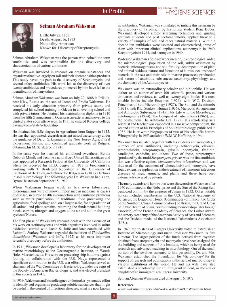
Birth: July 22, 1888Death: August 16, 1973Nationality: AmericanKnown for: Discovery of Streptomycin
Selman Abraham Waksman was the person who coined the term 'antibiotic' and was responsible for the discovery and characterization of various antibiotics.
Waksman was involved in research on organic substances and organisms that live largely on soil and their decomposition products. This study paved his path to the discovery of Streptomycin, and several other antibiotics. His work led to the discovery of over twenty antibiotics and procedures pioneered by him have led to the identification of many others.
Selman Abraham Waksman was born on July 22, 1888 in Prikula, near Kiev, Russia as, the son of Jacob and Fradia Waksman. He received his early education primarily from private tutors, and completed his school training in Odessa in an evening school and with private tutors. He obtained his matriculation diploma in 1910 from the fifth Gymnasium in Odessa as an extern, and moved to the United States soon afterwards. In 1911 he entered Rutgers college having won a State Scholarship.
He obtained his B.Sc. degree in Agriculture from Rutgers in 1915. He was then appointed research assistant in soil bacteriology under the guidance of Dr. J. G. Lipman at the New Jersey Agricultural Experiment Station, and continued graduate work at Rutgers, obtaining his M. Sc. degree in 1916.
In the same year he married his childhood sweetheart Bertha Deborah Mitnik and became a naturalized United States citizen and was appointed a Research Fellow at the University of California where he received his Ph.D. degree in 1918 in biochemistry, working with T. Brailsford Robertson at the University of California at Berkeley, and returned to Rutgers in 1918 as a lecturer in soil microbiology. The following year Dr. Waksman had a son, Byron Helsted on September 15, 1919.
When Waksman began work in his own laboratory, microorganisms were of known importance in medicine as causes of disease, in public health in connection with sanitation problems such as water purification, in traditional food processing and agriculture, food spoilage and, on a larger scale, for degradation of all animal and plant remains, returning the fundamental building blocks carbon, nitrogen and oxygen to the air and soil in the great cycles of Nature.
The first phase of Waksman's research dealt with the extension of his work on Actinomycetes and with organisms involved in sulfur oxidation, carried with Jacob S. Joffe and later continued with Robert L. Starkey. Waksman regarded the isolation of Thiobacillus thiooxidans (Waksman and Joffe, 1922) as his most important scientific discovery before the antibiotics.
In 1931, Waksman developed a laboratory for the development of marine microbiology at the Oceanographic Institute, in Woods Hole, Massachusetts. His work on protecting ship bottoms against fouling, in collaboration with the U.S. Navy, represented a significant contribution to the U.S. war effort. Waksman served as chairman of the War Committee on Bacteriology, under the aegis of the Society of American Bacteriologists, and was elected president of this society in 1941.
In 1939, Waksman and his colleagues undertook a systematic effort to identify soil organisms producing soluble substances that might be useful in the control of infectious diseases, what are now known
For Private Circulation Only 9
In Profile
as antibiotics. Waksman was stimulated to initiate this program by the discovery of Tyrothricin by his former student Rene Dubos. Waksman developed simple screening techniques and, guiding graduate students and post doctoral fellows, applied these to a variety of samples of soil and other natural materials. Within a decade ten antibiotics were isolated and characterized, three of them with important clinical applications: actinomycin in 1940, Streptomycin in 1944, and neomycin in 1949.
Professor Waksman's fields of work include, in chronological order, the microbiological population of the soil, sulfur oxidation by bacteria, microorganisms and soil fertility; decomposition of plant and animal residues, nature and formation of humus; occurrence of bacteria in the sea and their role in marine processes; production and nature of antibiotic substances; taxonomy, physiology, and biochemistry of the Actinomycetes.
Waksman was an extraordinary scholar and bibliophile. He was author or co author of over 400 scientific papers and various obituaries and reviews, as well as twenty eight books. His most notable books include Enzymes (1924), with W.C. Davison; Principles of Soil Microbiology (1927); The Soil and the microbe (1931); with R.L. Starkey; Humus (1936); Microbial Antagonisms and Antibiotic Substances (1945); My Life with the Microbes, an autobiography (1954); The Conquest of Tuberculosis (1965); and the posthumous The Antibiotic Era (1975). His scholarship as a scientist and teacher was embodied in a massive compendium, the second edition of his Principles of Soil Microbiology, published in 1932. He later wrote biographies of two of his scientific heroes: Winogradsky, in 1953 and about W.M.W. Haffkine, in 1964.
Waksman has isolated, together with his students and associates, a number of new antibiotics, including actinomycin, clavacin, streptothricin, streptomycin, grisein, neomycin, fradicin, candicidin, candidin, and others. Two of these, streptomycin (produced by the mold Streptomyces griseus was the first antibiotic that was effective against Mycobacterium tuberculosis, and was thus used for the treatment of tuberculosis) and neomycin, have found extensive application in the treatment of numerous infectious diseases of men, animals, and plants and these have been extensively covered by patents.
The many awards and honors that were showered on Waksman after 1940 culminated in the Nobel prize and the Star of the Rising Sun, bestowed on him by the emperor of Japan in 1952. Other notable honors included membership in the U.S. National Academy of Sciences, the Legion of Honor (Commander) of France, the Order of the Southern Cross (Commendatore) of Brazil, the Grand Cross of Public Health of Spain, corresponding membership (later foreign associate) of the French Academy of Sciences, the Lasker Award, the Amory Academy of the American Activity of Arts and Sciences, and the Trudeau medal of the National Tuberculosis Association (U.S.).
In 1949, the trustees of Rutgers University voted to establish an Institute of Microbiology and made Professor Waksman its first Director. The larger portion of the funds derived from royalties obtained from streptomycin and neomycin have been assigned for the building and support of this Institute, which is being used for research and advanced teaching in microbiology. Out of the small portion of the royalties assigned to him personally, Dr. and Mrs. Waksman established the 'Foundation for Microbiology' for the support of research and publications in the field of microbiology at various institutions of the world. He and his wife have also established a scholarship for an immigrant student, or the son or daughter of an immigrant, at Rutgers University.
Selman Abraham Waksman died on August 16, 1973
Referencewww.waksman.rutgers.edu/Waks/Waksman/Dr.Waksman.html
MAY-JUN 2009

Thoughts to live by
§
§
§
§
§
Cheerfulness is the best promoter of health and is as
friendly to the mind as to the body. (Joseph Addison)
An eye for an eye only ends up making the whole world
blind. (Mohandas Gandhi)
Laziness may appear attractive, but work gives
satisfaction. (Anne Frank)
Forgiveness is a virtue of the brave. (Indira Gandhi)
Nobody can bring you peace but yourself. (Ralph Waldo
Emerson)
nE joy the humour§
§
§
A blind man walks into a store with his seeing eye dog. All of a sudden he picks up the leash and begins swinging the dog over his head. The manager runs to the man and asks, “What are you doing?!!!”. The blind man replies, “Just looking around.”
A door knocks twice.
Insider asks: Who's there?
Outsider: Its 'OPPORTUNITY'.
Insider: Wrong....
Outsider: How did you know?
Insider: OPPORTUNITY never knocks twice.
You are a genius, your mind is a master piece,
It is divided into left and right.
In the left part; nothing is right,
and in the right part; nothing is left.
For Private Circulation Only 10
Relax Mood
Track your brain
Check your Answers on Page 16Check your Answers on Page 16
MAY-JUN 2009
ACROSS2. Inspissation is a process used for media containing a high
_______ content.3. H. pylori, was previously called _______ pylori.5. LAL assay detects the presence of ________ in a system.6. _______ is a process done in order to eliminate non-spore
forming pathogens from certain fluids.
7. _______ is a substance produced by one microbe that is detrimental to another microbe.
DOWN1. _______ An antibiotic that was used to treat tuberculosis,
which was patented.4. A fermented milk product _______, which is also a probiotic.8. _______ is the common name for Sodium hypochlorite.
1
2
3
4
5
6
7 8

Helicobacter pylori
For Private Circulation Only 11
Bug of the Month
Helicobacter pylori is a Gram negative, microaerophilic (requires oxygen at a lower concentration than is found in the environment) bacterium. This bacterium inhabits various areas of the stomach and the duodenum. It causes a chronic low level inflammation of the stomach lining and is strongly linked to the development of duodenal and gastric ulcers and stomach cancer.
Earlier gastric ulcers were attributed to stress, pungent food and also a buildup of acid. But John Warren and Barry Marshal re-discovered Helicobacter pylori and were successful in establishing, the bacterium as the etiological agent of gastric ulcers. This discovery not only asserted the role of the bacterium in gastric ulcers but also led the duo to receive the Nobel Prize for Medicine and Physiology in the year 2005, after Dr. Marshall intentionally consumed a beaker-full of Helicobacter pylori, fell ill, developed nausea and vomiting and was diagnosed with gastric ulcers. Subsequently the bacterium was isolated from his body (this was done in accordance to Robert Koch's postulates of confirming a particular microbe as the etiological agent for a particular disease).
It is found commonly in the lining of the stomach and the duodenum because it adapts well to the acidic, low pH environment. Urease is the central mechanism of H. pylori. In order to survive, the organism uses urea to produce ammonia and bicarbonate to neutralize the acid in the stomach. The metabolic products of H. pylori can alter the host, and change the acidity of the environment and increase the supplements of nutrients to colonize the stomach.
Pathophysiology
To colonize the stomach H. pylori must survive the acidic pH of the lumen and burrow into the mucus to reach its niche, close to the stomach's epithelial cell layer. The bacterium has flagella and moves through the stomach lumen and drills into the mucus lining of the stomach. The organism weakens the mucus lining of the stomach and allows acid to enter to the sensitive coating of the stomach. The acid and the bacteria irritate the lining and cause an ulcer. Since H. pylori is a slow growing organism, some infected individuals would be asymptomatic and show no signs of apparent disease, that is they don't get ulcers or gastritis. Colonization of the stomach by H. pylori results in chronic gastritis, an inflammation of the stomach lining. The severity of the inflammation is likely to underlie H. pylori related diseases.
Various virulence factors in Helicobacter pylori include
Urease enzyme
Vacuolating toxin (VacA)
Cytotoxin associated antigen (CagA)
Blood group antigen binding adhesin (BabA)
Adherence factors.
Epidemiology
At least half the world's population are infected by the bacterium,
l
l
l
l
l
making it the most widespread infection in the world. The infection rates however vary from nation to nation and the developing countries are more affected than the developed countries. Infections are usually acquired in early childhood.
H. pylori infection is contagious although the exact route of transmission is not adequately understood. Person to person transmission by either oral – oral or fecal - oral route is most likely through the ingestion of waste - tainted water. Consistent with these transmission routes, the bacteria have been isolated from feces, saliva and dental plaque of infected individuals.
Transmission occurs within families in developed nations yet can also be acquired from the community in developing countries.
Therefore a hygienic environment and a good sanitation facility could aid in decreasing the rate of H. pylori infections.
Signs and Symptoms
A Helicobacter pylori infection usually starts as a silent infection and may not exhibit any visible signs and symptoms. But when these bacteria produce a disease; the symptoms may be either those of peptic ulcer disease or of gastritis.
In kids, symptoms may include nausea, vomiting and frequent complaints about pain in the abdomen. However these may not be characteristic symptoms, and are seen in many childhood illnesses.
Kids who have peptic ulcer disease can have ulcers that bleed, causing hematemesis (bloody vomit or vomit that looks like coffee grounds) or melena (stool that's black, bloody, or looks like tar). Younger children with peptic ulcers disease may not have symptoms as clear cut, so their illness may be harder to diagnose.
H. pylori, which used to be called Campylobacter pylori, can also cause peptic ulcers. In older kids and adults, the most common of peptic ulcer disease causes a gnawing or burning pain in the abdomen, usually in the area below the ribs and above the navel. This pain often gets worse on an empty stomach and improves as soon as the person eats food, drinks milk, or takes antacid medicine.
Contagiousness
Scientists suspect that H. pylori infection may be contagious because the infection seems to run in families and is more common where people live in crowded or unsanitary conditions.
Diagnosis
Symptoms of H. pylori infection are not characteristic and may be confused with other general unwellness and abdomen problems and hence there are a few tests that are performed in order to precisely determine the cause of the infection.
A look at the stomach lining directly: This is performed under sedation and involves inserting an endoscope (a small flexible
MAY-JUN 2009

For Private Circulation Only 12
tube with a tiny camera on the end) – down the throat and into the stomach and duodenum. The doctor may then take samples of the lining to be checked in the laboratory for microscopic signs of infection and for H. pylori bacteria.
A Steiner's stain for the microscopic examination: of gastric antral biopsy specimens is considered the gold standard for detecting the presence of H. pylori. Cultures of biopsy specimens obtained during endoscopy can be tested for antimicrobial resistance in cases of treatment failure.
If necessary a specimen for culture can also be obtained using a string test, a less invasive but also a reliable technique in which a highly absorbent polymer string is partially swallowed and then removed manually to recover gastric material.
Biochemical media used for the cultivation of H. pylori are as follows:
Mueller Hinton Broth
Brain Heart Infusion Broth
Columbia Agar
Modified Glupczynski's Brussels Camplylobacter Charcoal Agar
Johnson – Murano Agar
Helicobacter Pylori Special Peptone Agar (HPSPA)
Serology / ELISA: Enzyme linked Immunosorbent Assay detects IgG antibodies to H. pylori, indicating current or past infection. Because H. pylori is not known to spontaneous resolve, there can be positive serological tests in patients who have not undergone eradication therapy.
Blood tests: which can detect the presence of H. pylori antibodies. Blood tests are common, although they typically aren't as accurate for children as they are for adults.
Urease test: is a rapid test for the diagnosis of Helicobacter pylori. The basis of the test is the ability of H. pylori to secrete the urease enzyme, which catalyzes the conversion of urea to ammonia and bicarbonate. The test is performed at the time of gastroscopy. A biopsy of mucosa is taken from the antrum of the stomach, and is placed into a medium containing an indicator such as phenol red. The urease produced by H. pylori hydrolyzes urea to ammonia, which raises the pH of the medium, and changes the color of the specimen.
Breath test for H. pylori: during the test the individual is made to exhale in a balloon-like bag. The air the person breathes into this bag is tested to provide a basis for comparison (called a baseline sample). The individual will then be asked to drink a small amount of a pleasant lemon-flavored solution (which is a urea containing liquid that is either carbon 13 or carbon 14 labeled). Fifteen minutes after drinking the solution, a second breath sample will be taken. The air that is breathed into this bag is tested for an increase in carbon dioxide (indicated by labeled carbon).
Principle of the test: is that the bacterium rapidly utilizes urea and the carbon is immediately absorbed into the system which after 15 to 20 minutes will be visibly tested in the breath test.
Precaution: that therapy consisting of proton pump inhibitors (PPI) have to be discontinued for a minimum of two weeks with the physicians recommendation.
Treatment
The physicians usually treat an H. pylori infection by prescribing an antibiotic therapy, because a single antibiotic may not kill the bacteria.
Antimicrobial resistance and incomplete treatment are major reasons for treatment failure. Because of the lengthy period of therapy, convenience and tolerability become important considerations in choosing a treatment plan. While success with shorter durations of treatment has been reported, continued therapy for 14 days is the most reliable and effective regimen.
Triple therapies with a combination of a PPI and two antibiotics have largely replaced the use of the classical Bismuth therapy due to better tolerability and higher efficacy. PPI have a synergistic effect with several antibiotics by raising the pH and making it optimal for the activity of the other antibiotics. These therapy regimen are usually prescribed for seven to fourteen days with omeprazoel, lansoprazole or pantoprazole in combination with clarithromycin, amoxicillin and metronidazole (or tinidazole).
A quadruple therapy combination by addition of anti – secretory agents like PPI to the classic triple therapy has been done. The major advantage of this regimen over the classical triple therapy are high and consistent eradication rates, a one week treatment offering a cure rate similar to a two week therapy and a significant reduction in complications seen with the two week bismuth based triple therapy. The total duration of the therapy is reduced.
Prevention
At the moment, there are no vaccines against H. pylori and because the mode of transmission isn't clearly understood, prevention guidelines aren't available. However it is always important to make sure you and your family:
Wash your hands thoroughly.
Eat food that has been properly prepared.
Drink water from a safe source.
Make sure that prolonged abdominal disorders are not neglected.
References
www.healthscout.com
l
l
l
l
Bug of the Month MAY-JUN 2009

For Private Circulation Only 13
Did You Know
The study of microbial life is dependent on the ability of the microorganism to utilize and grow on a particular media. Microbiological media which is essential for the microorganism’s growth and multiplication is used only after sterilization. Sterilization is a process of making a certain material entirely free from microbial life. This process is extremely essential and indispensable in the field of medicine, pharmaceuticals, research and microbiology. There are however some methods that are used during or prior to the process which aid in the overall sterilization, and 'Inspissation' is one such method.
Inspissation may be defined as the process of thickening by dehydration, it is this process which is used when heating high protein containing media for instance media containing egg and / or serum.
Lowenstein Jensen (L - J) media for Mycobacterium tuberculosis is prepared by the process which employs inspissation since the medium contains egg and glycerol which when moist heat is applied begin to precipitate and the medium looses its homogeneity in concentration. Whereas the coagulation of egg albumin during the inspissation process provides a solid medium for inoculation purposes.
Phases of Serum Inspissation or Fractional Sterilization
Day Temperature Time Purpose
1 85°C 60 Drying of the mediumminutes and killing the organisms
in their vegetative form
Time in Overnight Growth of vegetativebetween incubation forms from spores
2 75 to 80°C 20 minutes Killing the organisms in their vegetative form
Time in overnight Growth of vegetativebetween incubation forms from any spores
remaining
3 75 to 85°C 20 minutes Killing the organisms in their vegetative form
Inspissation and sterilization of Serum Medium platesSingle chambered bacteriological autoclave(1) Preheat the autoclave by running in steam for 5 minutes at 15 pounds pressure and allow the pressure to return to zero. (2) Line the floor of the autoclave or the shelves of a rack with several layers of dry newspaper and place plates in the autoclave in a single layer. If hardware cloth is used instead of solid metal shelves, paper lining is unnecessary. (3) Plates should never be placed directly onto a good heat conductor nor piled on one another. (4) Close all water, steam and air outlets. (5) Allow steam to enter slowly so that 15 pounds pressure is reached in 3 minutes. Keep original air entrapped. (6) The plates are allowed to remain for an additional 7 minutes in the closed system. The valve containing the water exit is then opened slightly to replace entrapped air with steam and to allow the condensed water and a small amount of steam to escape so that the pressure is not appreciably changed.
The serum containing medium is sterilized 10 minutes more: a total of 20 minutes, whereas casamino acids containing serum
medium is sterilized 15 minutes more: a total of 25 minutes.At the end of sterilization, close the steam inlets and open ALL the outlets quickly, the largest first, opening the safety valve if necessary to reduce pressure to zero in 5 to 10 seconds. Remove the plates as soon as possible from the autoclave.
Pressure cooker type field autoclave(1) Pour a small amount of water into the bottom of the cooker and allow it to come to a boil. (2) Place the plates of medium in a single layer on the bottom or in single layers in a rack in the autoclave. Do not pile plates on one another. (3) The lid is clamped on quickly, the petcocks closed and the pressure brought slowly to 15 pounds. (4) The plates are allowed to remain 10 minutes from the beginning of pressure rise in the closed system. The air vent is then opened slightly so that there is no appreciable change in pressure. The serum containing medium is sterilized 10 minutes more: a total of 20 minutes. The serum casamino acids medium is sterilized 15 minutes more: a total of 25 minutes. (5) At the end of sterilization, open the outlets in the lid and start opening the cover clamps as quickly as possible. Remove the plates immediately from the autoclave.
Double chambered dressing type autoclaveThis type of autoclave is not recommended unless the water trap line is equipped with a manually operated valve in lieu of or in addition to the thermostatic valve. It is then operated similarly to the single chambered bacteriological autoclave.
Discrepancies in the finished productBubbles in the finished media may be due to:
The plate resting on the heat conductor or a wet paper.Failure to trap air during inspissation period, for instance; when outlet valve is not securely closed.An acidity greater than that recommended.Too rapid rise in the steam pressure so that inspissation begins before the chloroform in the serum (chloroform brings about the activation of plasminogen in serum) has boiled off.
Fragmentation after inspissation which may occur if the pressure at the end of sterilization is reduced too slowly.
Coagulation of mediumoBefore loading heat the inspissator to 80 C to quicken the build-
up of temperature. Place the bottles in a slanted position in the oinspissator and coagulate the medium for 45 minutes at 80 – 85 C
(since the medium has been prepared with sterile precautions this heating is to solidify the media, not to sterilize it). Heating for a second or third time has a detrimental effect on the quality of the medium.The quality of the egg medium deteriorates when coagulation is done at too high a temperature or for too long. Discoloration of the coagulated medium may be due to excessive temperature. The appearance of little holes or bubbles on the surface of the medium also serves as an indicator of faulty coagulation procedures.
Sterility checkAfter inspissation the whole media batch or a representative
osample of culture bottles should be incubated at 35 – 37 C for 24 hours as a check for sterility
l
l
l
l
Inspissation
MAY-JUN 2009

The Limulus Amoebocyte Lysate (LAL) assay is the most specific and sensitive test to detect the presence of endotoxins in systems, including sterile products like injectables.
Bacterial endotoxin, lipopolysaccharide (LPS), is a fever producing byproduct of Gram negative bacteria commonly known as pyrogen. LPS consists of a polysaccharide, a core oligosaccharide and lipid A, which is responsible for the toxic effects. In human, lipopolysaccharide binding protein (LBP) plays a central role in response to LPS in the activation pathway and in the neutralization of LPS. Removal of LPS from the system is one of the most difficult downstream processes in protein purification.
HistoryBacteria are ubiquitous in nature and are found everywhere, from hydrothermal vents to Polar regions and are also present in the intestines of healthy individuals. However though bacteria are present in the intestines, they are not allowed to enter the blood stream but are capable of entering the blood stream only in cases of trauma, wherein they cause high, potentially fatal fevers.
Bacteria are classified into two broad categories based on their cell walls as Gram negative and Gram positive. This differentiation is visible to the bacteriologist only on Gram staining the bacteria and this staining technique was put forth by Dr. Hans Christian Gram. Scientists discovered that the fevers were caused by endotoxins which are found in the cell walls of Gram negative bacteria. The term 'pyrogens' , meaning 'burning bodies', was given to these endotoxins.
The discovery of the horseshoe crab's most important role in human medicine was made by Frederick Bang in the early 1950s. Bang discovered that the blood cells (called amoebocytes) contain a clotting agent that attaches to dangerous endotoxins produced by Gram negative bacteria. Thus the present method of endotoxin detection is based on his studies.
Bang observed that bacteria caused intra vascular coagulation in the American horseshoe crab, Limulus polyphemus. In collaboration Levin and Bang found that the agent responsible for the clotting phenomena resided in the crab's amoebocytes, or circulating blood cells, and that the pyrogen triggers the turbidity and gel forming reaction enzymatically. Thus endotoxicity causes an opacity and gelation in limulus amoebocyte lysate, which is based on an enzymatic reaction.
Bang's studies on horseshoe crab revealed that the amoebocyte cells in the horseshoe crab blood act like a primitive immune system. When a crab is wounded, the amoebocytes swarm to the area and coagulate, forming a viscous gel surrounding the invading bacteria. Unable to escape the bacteria are soon devoured by defense molecules such as antimicrobial proteins and polypeptides. This blood clotting mechanism prevents infection from occurring in the horseshoe crab. Bang realized this
For Private Circulation Only 14
Best Practices
clotting could be used as a fast and accurate way to test pharmaceutical drugs for the presence of Gram negative bacteria.
Up until then, drugs were tested by injecting rabbits with the drug and then waiting for 48 hours to see if they developed a fever. Within a few years of his initial discovery, Dr. Bang and Dr. Levin had created Limulus amoebocyte lysate, or LAL, and a new method to test for the presence of Gram negative bacteria. This assay was so effective that the U.S. Food and Drug Administration (FDA) accepted it as a standard test for endotoxins in 1983. Since then LAL has gained widespread use, replacing rabbit tests for clinical and biomedical applications.
LAL Milestones
1880 W. H. Lowell first studies coagulation of L. polyphemus blood at Johns Hopkins University.
1956 Bang discovers that horseshoe crab blood forms clots when bacteria (Vibrio sp.) are present.
1964 Levin and Bang discover that the blood clotting agent in horseshoe crabs is an amoebocyte.
1968 Scientists discover that an endotoxin causes blood-clot reaction in horseshoe crabs. The method for preparing LAL from horseshoe crab blood is developed.
1971 The standard LAL assay is developed.
1977 The FDA replaces the standard rabbit test for endotoxins with the LAL test.
1983 The FDA accepts LAL as a standard test for bacterial endotoxin contamination.
1987 The FDA establishes guidelines for LAL testing of pharmaceuticals and medical devices.
LAL Production In order to produce limulus amoebocyte lysate, large horseshoe crabs are caught, examined for health, and bled using a stainless steel needle that is inserted into their circulatory system. Blood is removed from the horseshoe crab's pericardium; the conditions for extracting blood must be sterile and pyrogen – free else the amoebocytes would immediately do their job and form a clot. The crabs are returned to the water and recover fully. It is noteworthy to mention that the horseshoe crabs are not seriously harmed during this process and studies have shown that the bled crabs have a relatively low mortality rate (approximately 10 %).
The horseshoe crab blood has a milky – blue color due to the copper pigmented hemocyanin molecule which turns blue upon contact with oxygen the same way our blood turns red. It is a mixture of liquid serum and suspended amoebocytes. If all the required conditions for the blood extraction are met, the blood can be centrifuged and the result is a separation of blood cells (amoebocytes) from the serum. A small whitish pellet forms
LAL Assay
MAY-JUN 2009

For Private Circulation Only 15
at the bottom of the tube. Technicians pour off the serum and rinse the pellet with saline. It is then resuspended and added to the collection of amoebocytes. Eventually pyrogen – free distilled water is mixed with the suspension of blood cells. This causes the cells to absorb fresh water and balloon until they eventually burst. This releases the chemicals from inside of the cell (the “lysate”), which is then purified and freeze – dried to form a white powder of the lysate and processed for use in the pharmaceutical industry. It is this lysate that is packaged and sold; which has to be reconstituted with pyrogen-free distilled water to use in the LAL assay.
Types of LAL AssaysA variety of LAL assay options are available; gel clot; turbidimetric; and chromogenic measurement. The latter 2 can be measured either as an End-point or Kinetic reaction. The primary requirement for all methods is that the test reagents, containers and pipettes have to be pyrogen-free.
The first, referred to as Gel clot method is based on the fact that LAL clots in the presence of endotoxin and forms an opaque gel. Depending on the amount of endotoxin in the gel, it may even become firm and hard.This is the simplest and most widely used LAL assay. The method can be qualitative or semi-quantitative when comparing a sample against a dilution series of an endotoxin standard. This assay can be used to provide a pass / fail for a certain limit and is best used with low sample numbers. The method may be performed manually with little or no requirement for instrumentation.
The Kinetic Turbidimetric method is a quantitative LAL method that uses the appearance of turbidity to determine endotoxin content. Turbidimetric systems depend upon an increasing concentration of insoluble coagulin released as the test progresses, this changes the turbidity of the sample solution. The change in turbidity can be measured either as transmission or absorbance requiring at least a spectrophotometer.
The third is the Kinetic Chromogenic LAL, chromogenic assays depend upon a chromogen which changes color if endotoxin is present in the sample. The higher the concentration of the endotoxin the more chromogen is released. Optical reading device operating at the chromogen wavelength is required.
Both the turbidimetric and chromogenic assays can be performed either as End-point or Kinetic assays.
End-point assays involve the measurement of the turbidity or color after incubation at a fixed temperature and a time period. Instrumentation requirements can be more basic than those required for kinetic assays.
Kinetic assays measure the rate of change in turbidity or color during the assay. Instrumentation is used to incubate at a fixed temperature, take measurements, compare those measurements and generate the results. Kinetic assays can provide a greater sensitivity over a wider range than endpoint assays but more sophisticated instrumentation is usually required.
Because the potency of an endotoxin to cause pyrogenic reaction will vary according to the nature of the toxin, the FDA developed endotoxin Units (EU) for result comparisons.
LAL AssayThe LAL assay is an in vitro assay for the detection and quantitation of bacterial endotoxin.
To test a sample for endotoxins, the packaged freeze-dried lysate is carefully removed from the pouch and under sterile conditions the required lyophilized powder is reconsitituted with pyrogen free distilled water, which in most cases is provided with a commercially available kit.
A LAL Assay takes as little as 45 minutes, in comparison with a rabbit assay, which can take as long as 48 hours. A suspect sample is mixed with reconstituted LAL and allowed to sit in a small tube. After 45 minutes the tube is inverted and if a clot has formed it will stick to the top of the inverted tube.
Precautions to be taken while conducting the testCareful technique must be used to avoid microbiological or endotoxin contamination. All the materials coming in contact with the specimen or test reagents must be endotoxin - free.Clean glassware and materials may be rendered endotoxin –
ofree by heating at 250 C for 30 minutes.Appropriate precautions must be taken to protect depyrogenated materials from subsequent environmental contamination.Samples to be tested must be stored in such a way that all bacteriological activity is stopped or the endotoxin level may
oincrease with time. For instance, store samples at 2 – 8 C for less than 24 hours; samples stored longer than 24 hours should be frozen.LAL assay is pH dependent and hence in case the LAL mixture is a reconstituted one, always must make sure that the pH is ranging between 6.5 to 8.0.In cases where a buffer is required the use of endotoxin-free TRIS buffer is recommended.
Choice of LAL technique may be prescribed by the pharmacopoeias but may also be affected by other factors such as the inherent turbidity or color of the sample as well as required sensitivities. Electronics technology has also advanced to the stage that portable endotoxin systems with results available in 15 minutes are now in the market, which are more suited for lower sample numbers but are to be processed with more sophisticated instrumentation in the form of dedicated systems or incubated microplate readers with appropriate software programs. Automatic robotic systems are also available.
Recent research in this area, is focused on decreasing the reliance on horseshoe crabs and resorting to alternate measures like:Genetic engineering: recombinant factor C, which is the initial element of the LAL clotting cascade is linked to fluorogenic substrates.Cell lines sensitive to LPS linked to color changes in the test medium.
l
l
l
l
l
l
Best Practices MAY-JUN 2009

Microxpress is Proud to be Introducing the Veg Media Bases For The Preparation Of Different Types Of Culture Media.
• Accu Veg Extract BE (Beef Extract)
An ingredient for preparation of a variety of culture media.
• Accu Veg Extract ME (Meat Extract)
An ingredient for preparation of culture media for fastidious microorganisms.
• Accu Veg Infusion Powder BH (Brain heart infusion powder)
An ingredient for preparation of a variety of culture media for fastidious microorganisms.
• Accu Veg Peptone MP (Meat Peptone)
An ingredient for use in bacteriological culture media for routine cultivation of a variety of microorganisms.
• Accu Veg Peptone PB (Peptone Bacteriological)
An ingredient for preparation of culture media for a variety of bacteria and fungi.
• Accu Veg Peptone PR (Proteose Peptone)
An ingredient for preparation of culture media for a variety of microorganisms and in producing bacterial toxins.
In Focus
TM Novacide - Aldehyde Free Cold Sterilant For Medical Devices and Surfaces
NOVACIDE is a 40X concentrated, highly effective aldehyde free high level disinfectant and cold sterilant for all types of medical devices and surfaces. Low level / intermediate level disinfection can be achieved with NOVACIDE in just 5 minutes and high level disinfection / complete sterilization in 30 min. NOVACIDE is suitable for use in automated endoscope reprocessors and in automated dental & medical machines. Once diluted to working strength of 2.5% v/v solution NOVACIDE remains active up to 14 days. The concentrated solution is stable till the expiry date mentioned on the container label.
COMPOSITION 3% w/v PHMB [poly(hexamethylenebiguanide) hydrochloride]10% w/v DDAC (Didecyl dimethyl ammonium chloride)
BENEFITSHigh level disinfection/Complete sterilization in 30 minutes. Low
level/intermediate level disinfection in 5 minutes. Excellent synergistic action. Bioburden tolerant. Compatible with all materials and surfaces. Resistance free. Tough against biofilms.
USAGE DIRECTIONS PROCEDURE TO PREPARE WORKING SOLUTION
Working Solution Novacide Potable Water Dilution Achieved
1000 ml 25 ml 975 ml 1:40
5000 ml 125 ml 4875 ml 1:40
10000 ml 250 ml 9750 ml 1:40
MEDICAL EQUIPMENTS: Pre-clean the surgical instruments/ endoscopes with mild detergent solution/enzymatic cleaner (CLENZYME). Rinse with water and dry. Immerse the clean instrument/endoscope in 2.5% v/v (1:40 dilution) solution of NOVACIDE for 5 minutes for low /intermediate level disinfection and for 30 minutes to achieve high level disinfection/complete sterilization. Rinse thoroughly with water and dry. Store in sterile packaging for reuse.
SURFACES: Use 2.5% v/v (1:40 dilution) solution of NOVACIDE and mop the surface using a clean mop. Allow a contact time of 10 minutes.
l l l
l l l l
ie
M ni Revi w
N al o l a hen bod
orm Micr f or of t humay
ands
i .
opportuni tic infect ons
ndCurrent Tre s
t i t g.S eril ty estin
n P eI rofil
mi dol n B
E l A f Vo ehring.
th
Bug of the Mon
r l ertu s s.
Bo dete la p s i
D d ou oi Y Kn w
ydroph G d-m brane t
Hobic ri em
fil er
iqTechn ue.
ac sBest Pr tice
s nfe on e
Di i cti of Lin n.
ost fhgt
ih
le
h co
g
m
i
in
H
g issue
VO ME LU - II
JUL-AUG 2009
ISSUE - IV
MAY-JUN 2009
Track your brain - Answer
1
2
3
4
5
6
7 8
S
T
R
E
P
T
O
M
Y
C
I
N
Y
O
G
U
R
B
L
E
C
H
P R O E I N
A M P Y L O B A C T E R
A N T I I O T I C
P A S T E R I Z A T I O N
E N D O T X I N
Printed and published by D.G. Tripathi, Edited by Gracinda Mesquita for and on behalf of Tulip Diagnostics (P) Ltd., Tulip House, Dr. Antonio Do Rego Bagh, Alto Santacruz, Bambolim Complex, Post Office Goa, 403 202, India. Fax- 0832 2458544, E-mail: [email protected], Website- www.tulipgroup.com.
Bimonthly Journal Published Exclusively For Tulip Group Customers
For Private Circulation Only 16