Drink H2O2 Drinking Hydrogen Peroxide Hydrogen Peroxide Therapy
Upload
truongkhuongCategory
view
228download
0
The Plant Cell, Vol. 13, 179–191, January 2001, www.plantcell.org © 2001 American Society of Plant Physiologists
Hydrogen Peroxide Acts as a Second Messenger for the Induction of Defense Genes in Tomato Plants in Responseto Wounding, Systemin, and Methyl Jasmonate
Martha L. Orozco-Cárdenas, Javier Narváez-Vásquez, and Clarence A. Ryan
1
Institute of Biological Chemistry, Washington State University, Pullman, Washington 99164-6340
The systemic accumulation of both hydrogen peroxide (H
2
O
2
) and proteinase inhibitor proteins in tomato leaves in re-sponse to wounding was inhibited by the NADPH oxidase inhibitors diphenylene iodonium (DPI), imidazole, and pyri-dine. The expression of several defense genes in response to wounding, systemin, oligosaccharides, and methyljasmonate also was inhibited by DPI. These genes, including those of four proteinase inhibitors and polyphenol oxi-dase, are expressed within 4 to 12 hr after wounding. However, DPI did not inhibit the wound-inducible expression ofgenes encoding prosystemin, lipoxygenase, and allene oxide synthase, which are associated with the octadecanoidsignaling pathway and are expressed 0.5 to 2 hr after wounding. Accordingly, treatment of plants with the H
2
O
2
-gener-ating enzyme glucose oxidase plus glucose resulted in the induction of only the later-expressed defensive genes andnot the early-expressed signaling-related genes. H
2
O
2
was cytochemically detected in the cell walls of vascular paren-chyma cells and spongy mesophyll cells within 4 hr after wounding of wild-type tomato leaves, but not earlier. The cu-mulative results suggest that active oxygen species are generated near cell walls of vascular bundle cells byoligogalacturonide fragments produced by wound-inducible polygalacturonase and that the resulting H
2
O
2
acts as asecond messenger for the activation of defense genes in mesophyll cells. These data provide a rationale for the se-quential, coordinated, and functional roles of systemin, jasmonic acid, oligogalacturonides, and H
2
O
2
signals for sys-temic signaling in tomato plants in response to wounding.
INTRODUCTION
Reactive oxygen species (ROS) are common components ofthe defense responses of plants against pathogen and her-bivore attacks. Inoculation of plant tissues with pathogensor treatment of cell cultures with microbial elicitors causesan oxidative burst characterized by the rapid generation ofhydrogen peroxide (H
2
O
2
; reviewed by Low and Merida,1996; Lamb and Dixon, 1997; Bolwell, 1999). Similarly, ROSare generated in plant tissues in response to wounding(Angelini et al., 1990; Bradley et al., 1992; Olson and Varner,1993; Felton et al., 1994; Bi and Felton, 1995; Orozco-Cárdenas and Ryan, 1999). Mechanical stimulation of iso-lated cells (Yahraus et al., 1995; Gus-Mayer et al., 1998) andthe treatment of cell suspension cultures with plant cell wall–derived oligogalacturonic acid (OGA; Legendre et al., 1993;Stennis et al., 1998) also generate H
2
O
2
accumulation. Wound-induced H
2
O
2
accumulation is observed both locally andsystemically in leaves of several plant species, apparentlycaused by OGA that was released by a systemically wound-
induced polygalacturonase (PG; Bergey et al., 1999; Orozco-Cárdenas and Ryan, 1999).
H
2
O
2
can act as a local signal for hypersensitive cell deathand also as a diffusible signal for the induction of defensivegenes in adjacent cells (Alvarez et al., 1998). For example,transgenic potato plants that overexpress a fungal glucoseoxidase gene and protein contain a constitutive increase insublethal levels of H
2
O
2
and exhibit enhanced disease resis-tance (Wu et al., 1995, 1997). Similarly, transgenic tobaccoplants engineered to have low levels of antioxidant defenses(enzymes) show higher constitutive and inducible levels ofH
2
O
2
and pathogenesis-related (PR) proteins, together withincreased sensitivity and general resistance to pathogenchallenge (Chamnongpol et al., 1998; Mittler et al., 1999).
In several model systems investigated in plants, the oxi-dative burst and the accumulation of H
2
O
2
appear to be me-diated by the activation of a membrane-bound NADPHoxidase complex (Doke et al., 1996; Lamb and Dixon, 1997;Ogawa et al., 1997; Del Río et al., 1998; Potikha et al., 1999;Pei et al., 2000). In animal cells, this enzymatic complexconsists of two membrane-associated polypeptides (gp91-phox and gp22-phox) that become active when at least threeproteins from the cytosol (p47-phox, p67-fox, and rac) bindto the membrane components (Jones, 1994; Henderson and
1
To whom correspondence should be addressed. E-mail [email protected]; fax 509-335-7643.
180 The Plant Cell
Chappel, 1996). Plant homologs of the animal NADPH oxi-dase protein subunits have been identified (Desikan et al.,1996; Xing et al., 1997; Potikha et al., 1999), and some oftheir genes have been sequenced (Groom et al., 1996; Kelleret al., 1998; Torres et al., 1998). How the plant NADPH oxi-dase is regulated is still unknown. Some chemical inhibitorsof the NADPH oxidase enzyme complex found in mamma-lian neutrophils inhibit the pathogen-, elicitor-, and wound-induced accumulation of H
2
O
2
derived from the oxidativeburst in plants (Levine et al., 1994; Auh and Murphy, 1995;Alvarez et al., 1998; Piedras et al., 1998; Orozco-Cárdenasand Ryan, 1999). In mammalian cells, the induction of de-fense genes by H
2
O
2
involves the activation of the NF-
k
Btranscription factor by mediating its release from the inhibi-tory I
k
B proteins (Li and Karin, 1999). In plants, the mecha-nisms by which H
2
O
2
activates genes are not understood.A model has been presented (Farmer and Ryan, 1992) for
the expression of defense-related genes in tomato leaves inresponse to wounding and systemin. In this model, sys-temin initiates a cascade of intracellular events resulting inthe activation of a cytoplasmic phospholipase that releaseslinolenic acid from membranes. Linolenic acid is convertedto jasmonic acid, which is a powerful activator of genescoding for both signal pathway enzymes and defensive pro-teinase inhibitors and polyphenol oxidase. The model wasmodified recently to note that signal pathway genes are ex-pressed within 0.5 hr after wounding, whereas defensivegenes are expressed within 4 hr (Ryan, 2000). Recently, PGwas shown to be among the early-expressed genes (Bergeyet al., 1999), which raised questions concerning its role inthe signal transduction pathway, because it was known toproduce oligogalacturonide fragments from plant cell wallsthat are activators of both the defensive genes (Ryan andFarmer, 1991) and of the production of H
2
O
2
(Stennis et al.,1998) in tomato leaves.
In this study, H
2
O
2
was generated in vascular bundles oftomato leaves in response to wounding and behaved as if itwas a diffusible signal for the expression of defense genesin mesophyll cells but not for signaling pathway genes invascular bundle cells. Based on these results, a modifiedsignal transduction pathway model is proposed (Figure 1)for the wound- and elicitor-induced defense response inwhich H
2
O
2
is generated by wound-inducible PG and actsas a second messenger for the induction of defensive genesin mesophyll cells.
RESULTS
The localized and systemic activation of a leaf PG gene andits enzyme activity occur in response to wounding of theleaves of several plant families (Bergey et al., 1999). Thetime course of induction of PG activity in leaves correlateswell with the accumulation of ROS, namely H
2
O
2
(Orozco-Cárdenas and Ryan, 1999). Because oligogalacturonide
fragments produced by PG are known to induce a substantialoxidative burst and the activation of defense gene expres-sion (Legendre et al., 1993; Stennis et al., 1998), a relation-ship between the wound-inducible H
2
O
2
and the wound-inducible defense gene activation after PG induction was in-vestigated (Orozco-Cárdenas and Ryan, 1999).
A membrane-bound NADPH oxidase has been implicatedin the production of ROS during the defense response ofplants against pathogen and herbivore attacks (Doke et al.,1996; Low and Merida, 1996; Lamb and Dixon, 1997;Orozco-Cárdenas and Ryan, 1999). Accordingly, someknown chemical inhibitors of the mammalian neutrophilNADPH oxidase inhibit the generation of ROS after patho-gen infection in plants (Levine et al., 1994; Auh and Murphy,1995; Piedras et al., 1998) and the accumulation of H
2
O
2
in wounded or systemin-treated tomato leaves (Orozco-Cárdenas and Ryan, 1999). The production of H
2
O
2
fromROS in tomato leaves likely results from the action of the en-zyme SOD (Auh and Murphy, 1995). Therefore, the effect ofthree different NADPH oxidase inhibitors on the accumula-tion of proteinase inhibitor I and II proteins in leaves ofyoung tomato plants in response to wounding were investi-
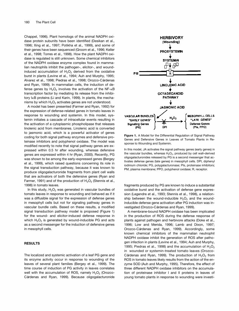
Figure 1. A Model for the Differential Regulation of Signal PathwayGenes and Defensive Genes in Leaves of Tomato Plants in Re-sponse to Wounding and Systemin.
In this model, JA activates the signal pathway genes (early genes) inthe vascular bundles, whereas H2O2, produced by cell wall–derivedoligogalacturonides released by PG is a second messenger that ac-tivates defense genes (late genes) in mesophyll cells. DPI, diphenyliodinium chloride; PG, polygalacturonase; PIs, proteinase inhibitors;PM, plasma membrane; PPO, polyphenol oxidase; R, receptor.

Hydrogen Peroxide and Wound Signaling 181
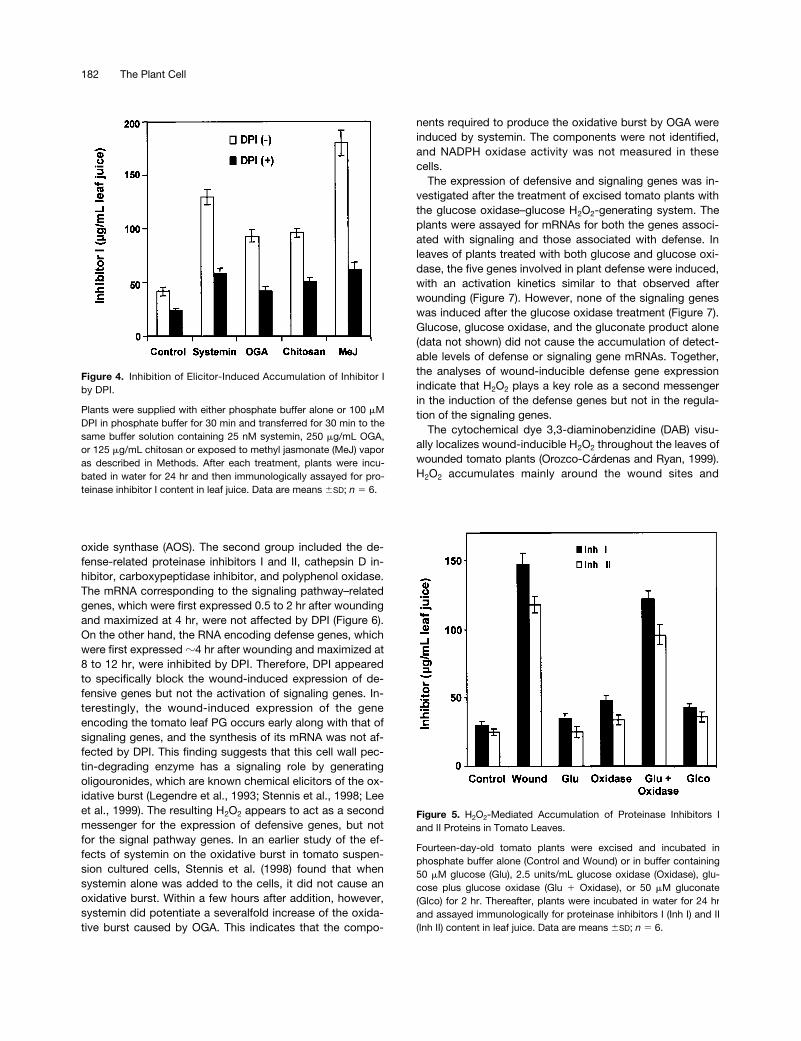
gated. As shown in Figure 2, when young, excised tomatoplants were supplied through their cut stems for 30 min withsolutions of the inhibitors diphenylene iodonium (DPI), pyri-dine, or imidazole, the accumulation of proteinase inhibitorsI and II in response to wounding was severely diminishedcompared with untreated controls. Among the three inhibi-tors, DPI had the greatest inhibitory effect. Figure 3 showsthat increasing concentrations of DPI (10 to 250
m
M) pro-gressively inhibited the accumulation of wound-inducibleproteinase inhibitors in a concentration-dependent manner.The concentration of DPI required for half-maximal inhibitionwas
z
50
m
M (Figure 3).DPI also inhibited the accumulation of proteinase inhibi-
tors induced in tomato plants after treatment with differentchemical elicitors of the wound response (Figure 4). DPI-pretreated plants accumulated significantly lower levels ofproteinase inhibitor I in their leaves than control plants in re-sponse to systemin, OGA, chitosan, and the plant hormonemethyl jasmonate (Figure 4). Together, these results suggestthat an active NADPH oxidase enzyme is necessary for de-fense gene activation in response to wounding and chemi-cal elicitors and the generation of H
2
O
2
and may be involvedas a signaling pathway component.
Glucose oxidase has been used to generate H
2
O
2
inplants (Wu et al., 1995, 1997; Alvarez et al., 1998). Supply-
ing plants with glucose oxidase together with glucosecauses a continuous production of H
2
O
2
within plant tissues(Levine et al., 1994; Alvarez et al., 1998). Excised tomatoplants were incubated in a buffer solution containing glu-cose, gluconate, glucose oxidase, or a mixture of bothglucose and glucose oxidase, and the accumulation of pro-teinase inhibitors I and II was assayed. Treatment with eithercompound or with the enzyme alone caused a slight tomoderate increase of both proteinase inhibitor proteins Iand II over levels observed in untreated controls (Figure 5).However, when the plants were supplied with glucose plusglucose oxidase, the levels of inhibitors I and II were in-duced to accumulate to
z
80% of the levels found inwounded plants (Figure 5). This result suggests that theH
2
O
2
generated by glucose oxidase within the apoplast wasable to trigger signaling events leading to the induction ofproteinase inhibitor protein accumulation.
To further investigate the role of H
2
O
2
in defense gene ac-tivation, the effect of DPI on the wound induction of defensegenes at the mRNA level was investigated. Gel blot analysesof total RNA isolated from wounded leaves of plants thathad been preincubated in buffer alone (untreated controls)or pretreated with DPI were performed using cDNA clonesof different wound-inducible genes as probes. These cDNAsencoded for two groups of functionally related proteins (Ryan,2000). The first group included the signaling pathway-associated proteins prosystemin, lipoxygenase, and allene
Figure 2. Inhibition of Wound-Inducible Accumulation of ProteinaseInhibitors I and II Proteins by Different Chemical Inhibitors of NADPHOxidase.
Fourteen-day-old tomato plants were excised at the base of thestems and supplied with solutions of phosphate buffer alone (Con-trol and Wound), 40 mM pyridine, 20 mM imidazole, or 100 mM DPIin phosphate buffer for 30 min. Plants, except controls, then werewounded and incubated in water under light as described in Meth-ods. Proteinase inhibitors I (Inh I) and II (Inh II) were assayed immu-nologically in leaf juice 24 hr later. Data are means 6SD; n 5 6.
Figure 3. Inhibition of Wound-Inducible Accumulation of ProteinaseInhibitors by the NADPH Oxidase Inhibitor DPI at Different Concen-trations.
Plants were treated and assayed as described for Figure 1. Inh I,proteinase inhibitor I; Inh II, proteinase inhibitor II. Data are means6SD; n 5 6.
182 The Plant Cell
oxide synthase (AOS). The second group included the de-fense-related proteinase inhibitors I and II, cathepsin D in-hibitor, carboxypeptidase inhibitor, and polyphenol oxidase.The mRNA corresponding to the signaling pathway–relatedgenes, which were first expressed 0.5 to 2 hr after woundingand maximized at 4 hr, were not affected by DPI (Figure 6).On the other hand, the RNA encoding defense genes, whichwere first expressed
z
4 hr after wounding and maximized at8 to 12 hr, were inhibited by DPI. Therefore, DPI appearedto specifically block the wound-induced expression of de-fensive genes but not the activation of signaling genes. In-terestingly, the wound-induced expression of the geneencoding the tomato leaf PG occurs early along with that ofsignaling genes, and the synthesis of its mRNA was not af-fected by DPI. This finding suggests that this cell wall pec-tin-degrading enzyme has a signaling role by generatingoligouronides, which are known chemical elicitors of the ox-idative burst (Legendre et al., 1993; Stennis et al., 1998; Leeet al., 1999). The resulting H
2
O
2
appears to act as a secondmessenger for the expression of defensive genes, but notfor the signal pathway genes. In an earlier study of the ef-fects of systemin on the oxidative burst in tomato suspen-sion cultured cells, Stennis et al. (1998) found that whensystemin alone was added to the cells, it did not cause anoxidative burst. Within a few hours after addition, however,systemin did potentiate a severalfold increase of the oxida-tive burst caused by OGA. This indicates that the compo-
nents required to produce the oxidative burst by OGA wereinduced by systemin. The components were not identified,and NADPH oxidase activity was not measured in thesecells.
The expression of defensive and signaling genes was in-vestigated after the treatment of excised tomato plants withthe glucose oxidase–glucose H
2
O
2
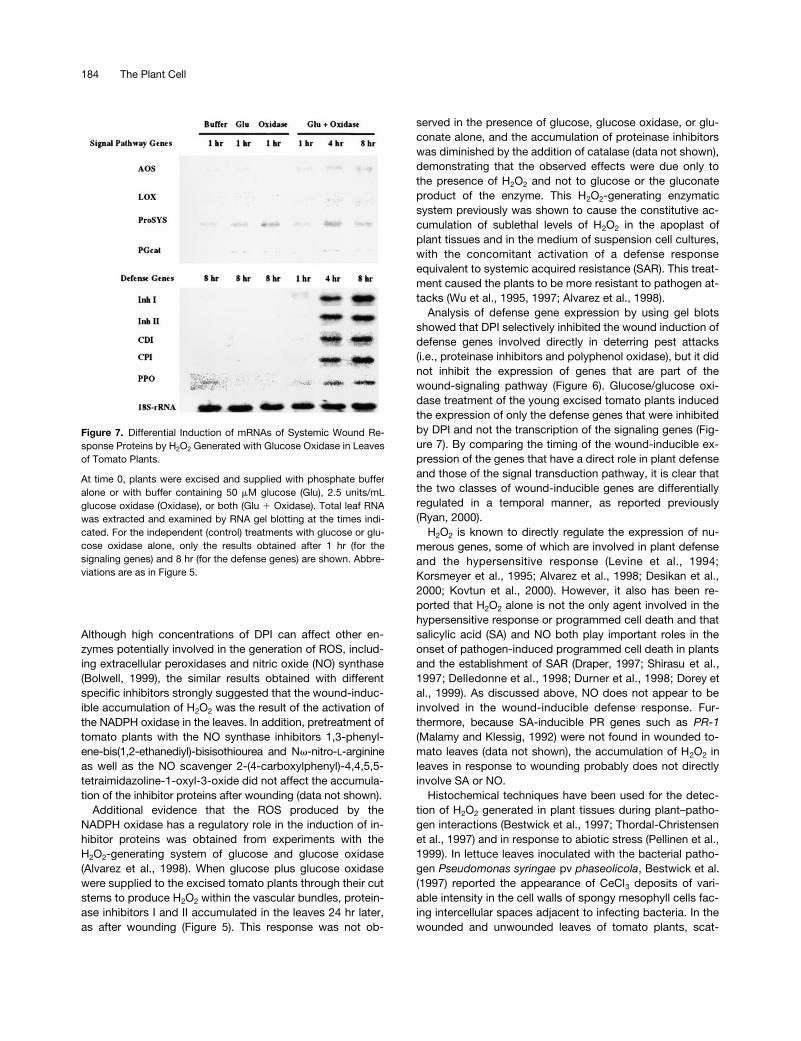
-generating system. Theplants were assayed for mRNAs for both the genes associ-ated with signaling and those associated with defense. Inleaves of plants treated with both glucose and glucose oxi-dase, the five genes involved in plant defense were induced,with an activation kinetics similar to that observed afterwounding (Figure 7). However, none of the signaling geneswas induced after the glucose oxidase treatment (Figure 7).Glucose, glucose oxidase, and the gluconate product alone(data not shown) did not cause the accumulation of detect-able levels of defense or signaling gene mRNAs. Together,the analyses of wound-inducible defense gene expressionindicate that H
2
O
2
plays a key role as a second messengerin the induction of the defense genes but not in the regula-tion of the signaling genes.
The cytochemical dye 3,3-diaminobenzidine (DAB) visu-ally localizes wound-inducible H
2
O
2
throughout the leaves ofwounded tomato plants (Orozco-Cárdenas and Ryan, 1999).H
2
O
2
accumulates mainly around the wound sites and
Figure 4. Inhibition of Elicitor-Induced Accumulation of Inhibitor Iby DPI.
Plants were supplied with either phosphate buffer alone or 100 mMDPI in phosphate buffer for 30 min and transferred for 30 min to thesame buffer solution containing 25 nM systemin, 250 mg/mL OGA,or 125 mg/mL chitosan or exposed to methyl jasmonate (MeJ) vaporas described in Methods. After each treatment, plants were incu-bated in water for 24 hr and then immunologically assayed for pro-teinase inhibitor I content in leaf juice. Data are means 6SD; n 5 6.
Figure 5. H2O2-Mediated Accumulation of Proteinase Inhibitors Iand II Proteins in Tomato Leaves.
Fourteen-day-old tomato plants were excised and incubated inphosphate buffer alone (Control and Wound) or in buffer containing50 mM glucose (Glu), 2.5 units/mL glucose oxidase (Oxidase), glu-cose plus glucose oxidase (Glu 1 Oxidase), or 50 mM gluconate(Glco) for 2 hr. Thereafter, plants were incubated in water for 24 hrand assayed immunologically for proteinase inhibitors I (Inh I) and II(Inh II) content in leaf juice. Data are means 6SD; n 5 6.

Hydrogen Peroxide and Wound Signaling 183
within the major and minor veins of the leaves, reaching lev-els of
z
1 to 10
m
M. DAB staining is blocked by pretreatmentof the plants with different NADPH oxidase inhibitors and bycatalase treatment after wounding of the leaves (M.L.Orozco-Cárdenas and C.A. Ryan, unpublished results). Tofurther investigate the subcellular location of H
2
O
2
genera-tion and/or accumulation in the wounded leaves, we used acerium perhydroxide (CeCl
3
)–based cytochemical technique(Bestwick et al., 1997). In leaves of unwounded young to-mato plants, CeCl
3
deposits, indicative of the presence ofH
2
O
2
, were found in developing lignified secondary cellwalls of xylem vessels (Figure 8A) but were not observed inthe cell walls of vascular parenchyma (Figure 8B) or meso-phyll cells (Figure 9A). On the other hand, in leaves ofwounded plants, electron-dense CeCl
3
deposits were de-tected in the cell walls of vascular parenchyma (Figures 8Cand 8D) and in nearby spongy mesophyll cells in bothwounded and unwounded (systemic) leaves (Figures 9B to9D). Similar deposits were found in cell walls of leaves oftransgenic tomato plants overexpressing prosystemin (Fig-ure 9E). Heavy CeCl
3
staining often was observed in the cellwalls of spongy mesophyll cells facing intercellular spaces,a few cells away from the vascular traces (Figures 9C and9D). The cytosol and internal cell organelles, including thechloroplasts, mitochondria, peroxisomes, nuclei, and tono-plasts of these cells, were almost completely free of electron-dense material. However, CeCl
3
deposits were observedreadily within the chloroplasts of palisade and spongy meso-phyll cells of plant leaves pretreated with paraquat, a ROS-inducing herbicide (data not shown), indicating that CeCl
3
could penetrate throughout the leaves and permeate cellmembranes, as has been reported (Pellinen et al., 1999). Inaddition, supplying excised tomato plants with the DPI in-hibitor before wounding did not result in an accumulation ofCeCl
3
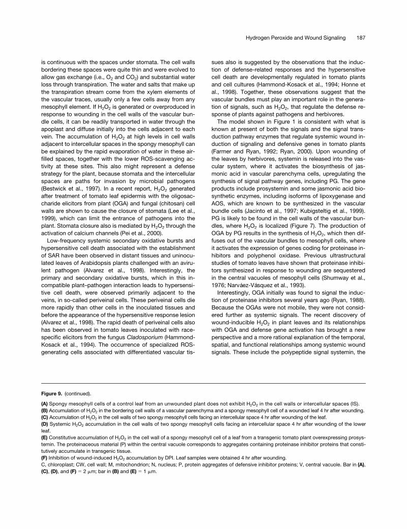
deposits in the cell walls of the vascular parenchymaand the mesophyll cells of the leaves (Figure 9F).
DISCUSSION
ROS are generated in plant tissues and organs during plantgrowth and development and also in response to environ-mental and biological stress (reviewed by Dangl et al., 1996;Greenberg, 1996; Pennell and Lamb, 1997). A previousstudy using several known chemical inhibitors of NADPHoxidase implicated this enzyme complex in the generationof ROS and the accumulation of H
2
O
2
in wounded and un-wounded leaves of several plant species in response to me-chanical wounding (Orozco-Cárdenas and Ryan, 1999). Theaccumulation of H
2
O
2
occurs near wound sites and also indistal unwounded leaves, indicating that the process is reg-ulated by a systemic signaling system. In the present study,NADPH oxidase inhibitors were used to investigate their ef-fects on the wound induction of defense proteinase inhibitorproteins in leaves of tomato plants. All of the NADPH oxi-
dase inhibitors tested had an inhibitory effect not only onthe generation of H
2
O
2
but on the accumulation of protein-ase inhibitors I and II as well (Figures 2 and 3). This findingsupported the hypothesis that the H
2
O
2
produced by theNADPH oxidase in response to wounding may have a regu-latory role in the wound inducibility of defensive proteins.
DPI, a suicide inhibitor that binds irreversibly to the fla-vonoid group of the membrane-associated gp91-phox sub-unit of the NADPH oxidase complex (O’Donnell et al., 1993),was the most effective of all of the inhibitors tested. As withwounding, DPI inhibited the accumulation of proteinase in-hibitor I that was induced after treatment of the plants withchemical elicitors of the wound response, namely systemin,OGA, and chitosan, as well as methyl jasmonate (Figure 4).
Figure 6. Differential Inhibition of Systemic Wound ResponseGenes by DPI.
Young excised tomato plants with two expanding leaves and ayoung growing leaf were supplied with phosphate buffer alone orwith 100 mM DPI for 0.5 hr, wounded on the lower leaf at time 0,transferred to water, and assayed by RNA gel blotting at the timesindicated (see Methods). 18S-rRNA, rRNA (loading control); AOS, al-lene oxide oxidase; CDI, aspartic proteinase inhibitor; CPI, metal-locarboxypeptidase inhibitor; Inh I, proteinase inhibitor I; Inh II,proteinase inhibitor II; LOX, lipoxygenase; PGcat, leaf polygalact-uronase catalytic subunit; PPO, polyphenol oxidase; ProSYS, pro-systemin.
184 The Plant Cell
Although high concentrations of DPI can affect other en-zymes potentially involved in the generation of ROS, includ-ing extracellular peroxidases and nitric oxide (NO) synthase(Bolwell, 1999), the similar results obtained with differentspecific inhibitors strongly suggested that the wound-induc-ible accumulation of H
2
O
2
was the result of the activation ofthe NADPH oxidase in the leaves. In addition, pretreatment oftomato plants with the NO synthase inhibitors 1,3-phenyl-ene-bis(1,2-ethanediyl)-bisisothiourea and N
v
-nitro-
L
-arginineas well as the NO scavenger 2-(4-carboxylphenyl)-4,4,5,5-tetraimidazoline-1-oxyl-3-oxide did not affect the accumula-tion of the inhibitor proteins after wounding (data not shown).
Additional evidence that the ROS produced by theNADPH oxidase has a regulatory role in the induction of in-hibitor proteins was obtained from experiments with theH
2O2-generating system of glucose and glucose oxidase(Alvarez et al., 1998). When glucose plus glucose oxidasewere supplied to the excised tomato plants through their cutstems to produce H2O2 within the vascular bundles, protein-ase inhibitors I and II accumulated in the leaves 24 hr later,as after wounding (Figure 5). This response was not ob-
served in the presence of glucose, glucose oxidase, or glu-conate alone, and the accumulation of proteinase inhibitorswas diminished by the addition of catalase (data not shown),demonstrating that the observed effects were due only tothe presence of H2O2 and not to glucose or the gluconateproduct of the enzyme. This H2O2-generating enzymaticsystem previously was shown to cause the constitutive ac-cumulation of sublethal levels of H2O2 in the apoplast ofplant tissues and in the medium of suspension cell cultures,with the concomitant activation of a defense responseequivalent to systemic acquired resistance (SAR). This treat-ment caused the plants to be more resistant to pathogen at-tacks (Wu et al., 1995, 1997; Alvarez et al., 1998).
Analysis of defense gene expression by using gel blotsshowed that DPI selectively inhibited the wound induction ofdefense genes involved directly in deterring pest attacks(i.e., proteinase inhibitors and polyphenol oxidase), but it didnot inhibit the expression of genes that are part of thewound-signaling pathway (Figure 6). Glucose/glucose oxi-dase treatment of the young excised tomato plants inducedthe expression of only the defense genes that were inhibitedby DPI and not the transcription of the signaling genes (Fig-ure 7). By comparing the timing of the wound-inducible ex-pression of the genes that have a direct role in plant defenseand those of the signal transduction pathway, it is clear thatthe two classes of wound-inducible genes are differentiallyregulated in a temporal manner, as reported previously(Ryan, 2000).
H2O2 is known to directly regulate the expression of nu-merous genes, some of which are involved in plant defenseand the hypersensitive response (Levine et al., 1994;Korsmeyer et al., 1995; Alvarez et al., 1998; Desikan et al.,2000; Kovtun et al., 2000). However, it also has been re-ported that H2O2 alone is not the only agent involved in thehypersensitive response or programmed cell death and thatsalicylic acid (SA) and NO both play important roles in theonset of pathogen-induced programmed cell death in plantsand the establishment of SAR (Draper, 1997; Shirasu et al.,1997; Delledonne et al., 1998; Durner et al., 1998; Dorey etal., 1999). As discussed above, NO does not appear to beinvolved in the wound-inducible defense response. Fur-thermore, because SA-inducible PR genes such as PR-1(Malamy and Klessig, 1992) were not found in wounded to-mato leaves (data not shown), the accumulation of H2O2 inleaves in response to wounding probably does not directlyinvolve SA or NO.
Histochemical techniques have been used for the detec-tion of H2O2 generated in plant tissues during plant–patho-gen interactions (Bestwick et al., 1997; Thordal-Christensenet al., 1997) and in response to abiotic stress (Pellinen et al.,1999). In lettuce leaves inoculated with the bacterial patho-gen Pseudomonas syringae pv phaseolicola, Bestwick et al.(1997) reported the appearance of CeCl3 deposits of vari-able intensity in the cell walls of spongy mesophyll cells fac-ing intercellular spaces adjacent to infecting bacteria. In thewounded and unwounded leaves of tomato plants, scat-
Figure 7. Differential Induction of mRNAs of Systemic Wound Re-sponse Proteins by H2O2 Generated with Glucose Oxidase in Leavesof Tomato Plants.
At time 0, plants were excised and supplied with phosphate bufferalone or with buffer containing 50 mM glucose (Glu), 2.5 units/mLglucose oxidase (Oxidase), or both (Glu 1 Oxidase). Total leaf RNAwas extracted and examined by RNA gel blotting at the times indi-cated. For the independent (control) treatments with glucose or glu-cose oxidase alone, only the results obtained after 1 hr (for thesignaling genes) and 8 hr (for the defense genes) are shown. Abbre-viations are as in Figure 5.

Hydrogen Peroxide and Wound Signaling 185
tered and localized dense deposits of CeCl3 were observedin the walls of vascular parenchyma cells (Figures 8C and8D) and nearby spongy mesophyll cells (Figures 9B to 9E).The intensity of the CeCl3 staining would correspond to sub-lethal levels of H2O2, not high enough to cause cell collapseand death, as observed by Bestwick et al. (1997). As notedabove, using the DAB histochemical dye to detect H2O2 inwounded tomato leaves, we calculated the levels of wound-inducible H2O2 to be z1 to 10 mM (Orozco-Cárdenas andRyan, 1999), which again might not be high enough to
cause hypersensitive cell death. In fact, no signs of necrotictissue were observed in the transgenic tomato plants over-expressing prosystemin (McGurl et al., 1994), which consti-tutively accumulate H2O2 in their leaves (Figure 9E; Orozco-Cárdenas and Ryan, 1999).
The greatest accumulation of H2O2 was observed in thewalls of spongy mesophyll cells facing large intercellularspaces (Figures 9C and 9D). These gas-filled cavities, whichin some leaves accounted for as much as 70% of the leafvolume, constituted a labyrinth that surrounds the cells and
Figure 8. Cytochemical Localization of Wound-Inducible H2O2 in Vascular Bundles of Tomato Leaves.
(A) Electron-dense deposits of CeCl3 indicative of the presence of H2O2 in developing secondary cell walls of xylem vessels (XV) of control un-wounded leaves (arrows show typical deposits). Note that the cell walls of an adjacent vascular parenchyma cell (VP) show little CeCl3 staining. (B) Absence of CeCl3 staining in the cell walls of vascular parenchyma (VP) and neighboring spongy mesophyll (SM) cells associated with thephloem in control unwounded leaves.(C) H2O2 generation in the vascular bundle of a wounded tomato leaf 4 hr after wounding. H2O2 accumulates strongly in the cell walls of vascularparenchyma cells bordering spongy mesophyll cells and at the intercellular spaces (IS).(D) Systemic accumulation of H2O2 in vascular bundles of upper unwounded leaves of young tomato plants 4 hr after wounding of the lower leaf. CC, companion cell; CW, cell wall; SE, sieve element. Bar 5 5 mm.

186 The Plant Cell
Figure 9. Cytochemical Localization of Wound-Inducible H2O2 in Mesophyll Cells of Tomato Leaves.
Hydrogen Peroxide and Wound Signaling 187
is continuous with the spaces under stomata. The cell wallsbordering these spaces were quite thin and were evolved toallow gas exchange (i.e., O2 and CO2) and substantial waterloss through transpiration. The water and salts that make upthe transpiration stream come from the xylem elements ofthe vascular traces, usually only a few cells away from anymesophyll element. If H2O2 is generated or overproduced inresponse to wounding in the cell walls of the vascular bun-dle cells, it can be readily transported in water through theapoplast and diffuse initially into the cells adjacent to eachvein. The accumulation of H2O2 at high levels in cell wallsadjacent to intercellular spaces in the spongy mesophyll canbe explained by the rapid evaporation of water in these air-filled spaces, together with the lower ROS-scavenging ac-tivity at these sites. This also might represent a defensestrategy for the plant, because stomata and the intercellularspaces are paths for invasion by microbial pathogens(Bestwick et al., 1997). In a recent report, H2O2 generatedafter treatment of tomato leaf epidermis with the oligosac-charide elicitors from plant (OGA) and fungal (chitosan) cellwalls are shown to cause the closure of stomata (Lee et al.,1999), which can limit the entrance of pathogens into theplant. Stomata closure also is mediated by H2O2 through theactivation of calcium channels (Pei et al., 2000).
Low-frequency systemic secondary oxidative bursts andhypersensitive cell death associated with the establishmentof SAR have been observed in distant tissues and uninocu-lated leaves of Arabidopsis plants challenged with an aviru-lent pathogen (Alvarez et al., 1998). Interestingly, theprimary and secondary oxidative bursts, which in this in-compatible plant–pathogen interaction leads to hypersensi-tive cell death, were observed primarily adjacent to theveins, in so-called periveinal cells. These periveinal cells diemore rapidly than other cells in the inoculated tissues andbefore the appearance of the hypersensitive response lesion(Alvarez et al., 1998). The rapid death of periveinal cells alsohas been observed in tomato leaves inoculated with race-specific elicitors from the fungus Cladosporium (Hammond-Kosack et al., 1994). The occurrence of specialized ROS-generating cells associated with differentiated vascular tis-
sues also is suggested by the observations that the induc-tion of defense-related responses and the hypersensitivecell death are developmentally regulated in tomato plantsand cell cultures (Hammond-Kosack et al., 1994; Honne etal., 1998). Together, these observations suggest that thevascular bundles must play an important role in the genera-tion of signals, such as H2O2, that regulate the defense re-sponse of plants against pathogens and herbivores.
The model shown in Figure 1 is consistent with what isknown at present of both the signals and the signal trans-duction pathway enzymes that regulate systemic wound in-duction of signaling and defensive genes in tomato plants(Farmer and Ryan, 1992; Ryan, 2000). Upon wounding ofthe leaves by herbivores, systemin is released into the vas-cular system, where it activates the biosynthesis of jas-monic acid in vascular parenchyma cells, upregulating thesynthesis of signal pathway genes, including PG. The geneproducts include prosystemin and some jasmonic acid bio-synthetic enzymes, including isoforms of lipoxygenase andAOS, which are known to be synthesized in the vascularbundle cells (Jacinto et al., 1997; Kubigsteltig et al., 1999).PG is likely to be found in the cell walls of the vascular bun-dles, where H2O2 is localized (Figure 7). The production ofOGA by PG results in the synthesis of H2O2, which then dif-fuses out of the vascular bundles to mesophyll cells, whereit activates the expression of genes coding for proteinase in-hibitors and polyphenol oxidase. Previous ultrastructuralstudies of tomato leaves have shown that proteinase inhibi-tors synthesized in response to wounding are sequesteredin the central vacuoles of mesophyll cells (Shumway et al.,1976; Narváez-Vásquez et al., 1993).
Interestingly, OGA initially was found to signal the induc-tion of proteinase inhibitors several years ago (Ryan, 1988).Because the OGAs were not mobile, they were not consid-ered further as systemic signals. The recent discovery ofwound-inducible H2O2 in plant leaves and its relationshipswith OGA and defense gene activation has brought a newperspective and a more rational explanation of the temporal,spatial, and functional relationships among systemic woundsignals. These include the polypeptide signal systemin, the
Figure 9. (continued).
(A) Spongy mesophyll cells of a control leaf from an unwounded plant does not exhibit H2O2 in the cell walls or intercellular spaces (IS).(B) Accumulation of H2O2 in the bordering cell walls of a vascular parenchyma and a spongy mesophyll cell of a wounded leaf 4 hr after wounding. (C) Accumulation of H2O2 in the cell walls of two spongy mesophyll cells facing an intercellular space 4 hr after wounding of the leaf. (D) Systemic H2O2 accumulation in the cell walls of two spongy mesophyll cells facing an intercellular space 4 hr after wounding of the lowerleaf. (E) Constitutive accumulation of H2O2 in the cell wall of a spongy mesophyll cell of a leaf from a transgenic tomato plant overexpressing prosys-temin. The proteinaceous material (P) within the central vacuole corresponds to aggregates containing proteinase inhibitor proteins that consti-tutively accumulate in transgenic tissue.(F) Inhibition of wound-induced H2O2 accumulation by DPI. Leaf samples were obtained 4 hr after wounding.C, chloroplast; CW, cell wall; M, mitochondrion; N, nucleus; P, protein aggregates of defensive inhibitor proteins; V, central vacuole. Bar in (A),(C), (D), and (F) 5 2 mm; bar in (B) and (E) 5 1 mm.

188 The Plant Cell
lipid signal jasmonic acid, the oligosaccharide signal OGA,and the inorganic chemical signal H2O2.
METHODS
Plant Growth
Tomato plants (Lycopersicon esculentum cv Castlemart) were grownfrom seed for 2 weeks in a growth chamber with 18-hr days (light at300 mE·m22·sec21) at 288C and 6-hr nights at 188C. Transgenic to-mato plants expressing a prosystemin cDNA gene under the controlof the cauliflower mosaic virus 35S promoter (McGurl et al., 1994)were grown under the same conditions.
Assays
To assay chemical elicitors of the wound response, plants with twoexpanding leaves and a small terminal leaf were used. The plantswere excised at the base of the stem with a razor blade, and the cutstem was placed in 1023 M potassium phosphate buffer, pH 6.0, or ina solution of systemin (25 nM), oligogalacturonic acid (OGA; 0.5 mg/mL), or chitosan (125 mg/mL) in phosphate buffer for 30 min. Aftertreatment, the plants were incubated in water under constant light at300 mE·m22·sec21 at 28°C within closed Plexiglas boxes as de-scribed previously (Orozco-Cárdenas and Ryan, 1999). For experi-ments in which plants were induced by glucose oxidase plusglucose, excised plants were incubated in buffer alone or in buffercontaining glucose (50 mM), gluconate (50 mM), glucose oxidase (2.5units/mL; [all from Sigma, St. Louis, MO], or glucose (50 mM) plusglucose oxidase (2.5 units/mL) for 2 hr.
Excised plants also were exposed to methyl jasmonate vapors byapplying 1 mL of absolute methyl jasmonate (Bedoukian Research,Danbury, CT) to a cotton wick inside a Plexiglas box, as describedpreviously (Farmer and Ryan, 1992). The box was sealed, placed in agrowth chamber, and incubated under the light and temperatureconditions described above. Wounding was performed by crushingthe leaflets of the lower leaf with a hemostat once near their tips andonce across the main veins. Inhibitors of NADPH oxidase, includingdiphenylene iodonium (DPI), pyridine, and imidazole (all from Sigma,St. Louis, MO), also were supplied through the excised stem in thesame phosphate buffer for 0.5 to 4 hr before wounding or elicitortreatments.
Proteinase inhibitor concentrations in leaf juice were assayed after24 hr by immunoradial diffusion as reported previously (Ryan, 1967;Trautman et al., 1971).
RNA Extraction and RNA Gel Blot Analyses
At different time intervals after treatment of detached tomato plants,leaves were excised, immersed in liquid nitrogen, ground to a finepowder, and stored at 2808C to isolate total RNA. Total leaf RNAwas extracted using the Trizol reagent (Gibco BRL) according to themanufacturer’s recommendations. Total RNA pellets were dissolvedin 25 mL of RNase-free water and quantified spectrophotometrically.RNA quality was determined by gel fractionation in agarose/formal-dehyde followed by ethidium bromide staining and UV light visualiza-tion before analyzing for specific mRNAs (Maniatis et al., 1982).
Total leaf RNA (10 to 20 mg) was fractionated by electrophoresison 1.4% agarose gels with formaldehyde, blotted onto nitrocellulosemembranes, and incubated at 658C for 1 hr in hybridization buffer (5 3SSPE [1 3 SSPE is 0.115 M NaCl, 10 mM sodium phosphate, and 1mM EDTA, pH 7.4], 5 3 Denhardt’s solution [1 3 Denhardt’s solutionis 0.02% Ficoll, 0.02% polyvinylpyrrolidone, and 0.02% BSA], 1%SDS, and 10% dextran sulfate). Radioactive 32P-dCTP–labeledprobes were generated by random priming according to the manu-facturer’s recommendations (DECA prime II kit; Ambion, Austin, TX)by using tomato cDNAs encoding allene oxide synthase (AOS)(Howe et al., 2000), lipoxygenase (Heitz et al., 1997), prosystemin(McGurl et al., 1992), leaf polygalacturonase catalytic subunit(Bergey et al., 1999), serine proteinase inhibitor I (Graham et al.,1985a), serine proteinase inhibitor II (Graham et al., 1985b), cathep-sin D inhibitor (Hildemann et al., 1992), carboxypeptidase inhibitor(Moura and Ryan, 2000), and polyphenol oxidase (Constabel et al.,1995). An 18S rRNA gene probe was used as a loading control. Syn-thetic oligonucleotide probes were purified using Bio-spin P6 chro-matography columns (Bio-Rad). The probes were heat denatured,added to the hybridization buffer, and incubated with the blockedmembranes overnight at 658C. Membranes were washed once with2 3 SSPE for 20 min at room temperature, twice with 2 3 SSPE and1% SDS for 15 to 30 min at 658C, and then exposed for 15 to 32 hr tox-ray film or to a PhosphorImager (Bio-Rad).
Cytochemical Detection of H2O2
H2O2 was visualized at the subcellular level using CeCl3 for localiza-tion (Bestwick et al., 1997; Pellinen et al., 1999). Electron-denseCeCl3 deposits are formed in the presence of H2O2 and are visible bytransmission electron microscopy. Briefly, tissue pieces (2 3 5 mm2)were excised from leaves of wounded and unwounded wild-type andtransgenic tomato plants and then vacuum infiltrated with freshlyprepared 5 mM CeCl3 in 50 mM 3-(N-morpholino)-propanesulfonicacid at pH 7.2 for 30 min. Tissues then were fixed in 1.25% (v/v) glu-taraldehyde and 1.25% (v/v) paraformaldehyde in 50 mM sodium ca-codylate buffer (CAB), pH 7.2, for 1 hr at room temperature and keptovernight at 48C. After fixation, tissues were washed twice for 10 minin CAB and postfixed for 45 min in 1% (v/v) osmium tetroxide in CAB.Tissues were then washed twice for 10 min in CAB and dehydrated ina graded acetone series (30, 50, 70, 80, 90, and 100% [v/v]), progres-sively embedded in rising concentrations of acetone-resin mixtures,and finally incubated in two 24-hr changes of pure epoxy resin(Eponate 12 resin; Ted Pella Inc., Redding, CA) before polymerizationat 608C for 48 hr. Ultrathin sections (50 to 100 nm) were obtained ona Reichert Ultracut E Microtome (Leica AG, Wein, Austria) using a di-amond knife (Delaware Diamond Knives, Wilmington, DE), mountedon nickel grids (200 mesh), and examined without staining with atransmission electron microscope (model JEM-1200Ex; JEOL Ltd.,Tokyo, Japan) at an accelerating voltage of 80 kV.
ACKNOWLEDGMENTS
Transmission electron microscopy was performed at the ElectronMicroscopy Center (EMC) of Washington State University. We thankSue Vogtman for growing the plants for this study, and the EMC stafffor their technical advice and collaboration. This research was sup-ported by Washington State University College of Agriculture and
Hydrogen Peroxide and Wound Signaling 189
Home Economics (Project No. 1791) and by grants from the UnitedStates Department of Agriculture Competitive Grants Program (GrantNo. 9801502) and the National Science Foundation (Grant No.9601099).
Received July 31, 2000; accepted October 26, 2000.
REFERENCES
Alvarez, M.E., Penell, R.I., Meijer, P.-J., Ishikawa, A., Dixon, R.A.,and Lamb, C. (1998). Reactive oxygen intermediates mediate asystemic signal network in the establishment of plant immunity.Cell 92, 773–784.
Angelini, R., Manes, F., and Federico, R. (1990). Spatial and func-tional correlation between diamine-oxidase and peroxidase activi-ties and their dependence upon de-etiolation and wounding inchick-pea stems. Planta 182, 89–96.
Auh, C.-K., and Murphy, T.M. (1995). Plasma membrane redoxenzyme is involved in the synthesis of O2
and H2O2 by Phyto-phthora elicitor-stimulated rose cells. Plant Physiol. 107, 1241–1247.
Bergey, D.R., Orozco-Cárdenas, M.L., de Moura, D.S., and Ryan,C.A. (1999). A wound- and systemic-inducible polygalacturonasein tomato leaves. Proc. Natl. Acad. Sci. USA 96, 1756–1760.
Bestwick, C.S., Brown, I.R., Bennett, M.H.R., and Mansfield, J.W.(1997). Localization of hydrogen peroxide accumulation duringthe hypersensitive reaction of lettuce cells to Pseudomonas syrin-gae pv phaseolicola. Plant Cell 9, 209–221.
Bi, J.L., and Felton, G.W. (1995). Foliar oxidative stress and insectherbivory: Primary compounds, secondary metabolites, and reac-tive oxygen species as components of induced resistance. J.Chem. Ecol. 21, 1511–1530.
Bolwell, G.P. (1999). Role of active oxygen species and NO in plantdefense responses. Curr. Opin. Plant Biol. 2, 287–294.
Bradley, D.J., Kjellbom, P., and Lamb, C.J. (1992). Elicitor- andwound-induced oxidative cross-linking of a proline-rich plant cellwall protein: A novel, rapid defense response. Cell 70, 21–30.
Chamnongpol, S., Willekens, H., Moeder, W., Langebartels, C.,Sandermann, H., Van Montagu, M., Inze, D., and Van Camp,W. (1998). Defense activation and enhanced pathogen toleranceinduced by H2O2 in transgenic tobacco. Proc. Natl. Acad. Sci.USA 95, 5818–5823.
Constabel, C.P., Bergey, D.R., and Ryan, C.A. (1995). Systeminactivates synthesis of wound-inducible tomato leaf polyphenoloxidase via the octadecanoid defense signaling pathway. Proc.Natl. Acad. Sci. USA 92, 407–411.
Dangl, J.L., Dietrich, R.A., and Richberg, M.H. (1996). Death don’thave no mercy: Cell death programs in plant–microbe interac-tions. Plant Cell 8, 1793–1807.
Delledonne, M., Xia, Y., Dixon, R.A., and Lamb, C. (1998). Nitricoxide functions as a signal in plant disease resistance. Nature394, 585–588.
Del Río, L.A., Pastori, G.M., Palma, J.M., Sandalio, L.M., Sevilla,F., Corpas, F.J., Jiménez, A., López-Huertas, E., and Hernández,J.A. (1998). The activated oxygen role of peroxisomes in senes-cence. Plant Physiol. 116, 1195–1200.
Desikan, R., Hancock, J.T., Coffey, M.J., and Neill, S.J. (1996).Generation of active oxygen species in elicited cells of Arabidop-sis thaliana is mediated by NADPH oxidase-like enzyme. FEBSLett. 382, 213–217.
Desikan, R., Neill, S.J., and Hancock, J.T. (2000). Hydrogen per-oxide–induced gene expression in Arabidopsis thaliana. FreeRadic. Biol. Med. 28, 773–778.
Doke, N., Miura, Y., Sanchez, L.M., Park, H.J., Noritake, T.,Yoshioka, H., and Kawakita, K. (1996). The oxidative burst pro-tects plants against pathogen attack: Mechanism and role as anemergency signal for plant bio-defence. A review. Gene 179, 45–51.
Dorey, S., Kopp, M., Geoffroy, P., Fritig, B., and Kauffmann, S.(1999). Hydrogen peroxide from the oxidative burst is neither nec-essary nor sufficient for hypersensitive cell death induction, phe-nylalanine ammonia lyase stimulation, salicylic acid accumulation,or scopoletin consumption in cultured tobacco cells treated withelicitin. Plant Physiol. 121, 163–171.
Draper, D. (1997). Salicylate, superoxide synthesis and cell suicidein plant defence. Trends Plant Sci. 2, 162–165.
Durner, J., Wendehenne, D., and Klessig, D. (1998). Defense geneinduction in tobacco by nitric oxide, cyclic GMP, and cyclic ADP-ribose. Proc. Natl. Acad. Sci. USA 95, 10328–10333.
Farmer, E.E., and Ryan, C.A. (1992). Octadecanoid precursors ofjasmonic acid activate the synthesis of wound-inducible protein-ase inhibitors. Plant Cell 4, 129–134.
Felton, G.W., Summers, C.B., and Mueller, A.J. (1994). Oxidativeresponses in soybean foliage to herbivory by bean leaf beetle andthree-cornered alfalfa hopper. J. Chem. Ecol. 20, 639–649.
Graham, J.S., Pearce, G., Merryweather, J., Titani, K., Ericson,L., and Ryan, C.A. (1985a). Wound-induced proteinase inhibitorsfrom tomato leaves. I. The cDNA deduced primary structure ofpre-inhibitor I and its post-translational processing. J. Biol. Chem.260, 6555–6560.
Graham, J.S., Pearce, G., Merryweather, J., Titani, K., Ericson,L., and Ryan, C.A. (1985b). Wound-induced proteinase inhibitorsfrom tomato leaves. II. The cDNA deduced primary structure ofpre-inhibitor II and its post-translational processing. J. Biol.Chem. 260, 6561–6564.
Greenberg, J.T. (1996). Programmed cell death: A way of life forplants. Proc. Natl. Acad. Sci. USA 93, 12094–12097.
Groom, O.J., Torres, M.A., Fordham-Skeleton, P., Hammond-Kosak, K.E., Robinson, N.J., and Jones, J.D.G. (1996). RbohA,a rice homologue of the mammalian pg91phox respiratory burstoxidase gene. Plant J. 10, 515–522.
Gus-Mayer, S., Naton, B., Hahlbrock, K., and Schmelzer, E.(1998). Local mechanical stimulation induces components of thepathogen defense response in parsley. Proc. Natl. Acad. Sci. USA95, 8398–8403.
Hammond-Kosack, K.E., Harrison, K., and Jones, J.D.G. (1994).Developmentally regulated cell death on expression of the fungalavirulence gene Avr9 in tomato seedlings carrying the disease-resistance gene Cf-9. Proc. Natl. Acad. Sci. USA 91, 10445–10449.
Heitz, T., Berger, D.R., and Ryan, C.A. (1997). A gene encoding achloroplast-targeted lipoxygenase in tomato leaves is transientlyinduced by wounding, systemin, and methyl jasmonate. PlantPhysiol. 114, 1085–1093.
190 The Plant Cell
Henderson, L.M., and Chappel, J.B. (1996). NADPH oxidase ofneutrophils. Biochim. Biophys. Acta 1273, 87–107.
Hildemann, T., Ebneth, M., Pena-Cortes, H., Sanchez-Serrano,J.J., Wilmitzer, L., and Prat, S. (1992). General roles of abscisicand jasmonic acids in gene activation as a result of mechanicalwounding. Plant Cell 4, 1157–1170.
Honne, G., Buitink, J., Jabs, T., De Kloe, J., Sijbolts, F.,Apotheker, M., Weide, R., Sije, T., Stuiver, M., and De Wit,P.J.G.M. (1998). Induction of defense-related responses in Cf9tomato cells by the AVR9 elicitor peptide of Cladosporium fulvumis developmentally regulated. Plant Physiol. 117, 809–820.
Howe, G., Lee, G.I., Itoh, A., Li, L., and DeRocher, A.E. (2000).Cytochrome P450-dependent metabolism of oxylipins in tomato:Cloning and expression of allene oxide synthase and fatty acidhydroperoxide lyase. Plant Physiol. 123, 711–724.
Jacinto, T., McGurl, B., Franceschi, V., Delano-Freier, J., andRyan, C.A. (1997). Tomato prosystemin confers wound-inducible,vascular bundle–specific expression of the b-glucoronidase genein transgenic tomato plants. Planta 203, 406–412.
Jones, O.T.G. (1994). The regulation of superoxide production bythe NADPH oxidase of neutrophils and other mammalian cells.Bioessays 16, 919–923.
Keller, T., Damude, H.G., Werner, D., Doaner, P., Dixon, R.A., andLamb, C. (1998). A plant homolog of the neutrophil NADPH oxi-dase gp91phox subunit gene encodes a plasma membrane proteinwith Ca21 binding motifs. Plant Cell 10, 255–266.
Korsmeyer, S.J., Yin, X.M., Oltuai, Z.N., Veis-Novack, D.J., andLinette, G.P. (1995). Reactive oxygen species and the regulationof cell death by the Bcl-2 gene family. Biochim. Biophys. Acta1271, 63–66.
Kovtun, Y., Chiu, W.L., Tena, G., and Sheen, J. (2000). Functionalanalysis of oxidative stress–activated mitogen-activated proteinkinase cascade in plants. Proc. Natl. Acad. Sci. USA 97, 2940–2945.
Kubigsteltig, I., Laudert, D., and Weiler, E.W. (1999). Structureand regulation of the Arabidosis thaliana allene oxide synthasegene. Planta 208, 463–471.
Lamb, C., and Dixon, R.A. (1997). The oxidative burst in plant dis-ease resistance. Annu. Rev. Plant Physiol. Plant Mol. Biol. 48,251–275.
Lee, S., Choi, H., Suh, S., Doo, I.-S., Oh, K.-Y., Choi, E.J., Taylor,A.T.S., Low, P.S., and Lee, Y. (1999). Oligogalacturonic acid andchitosan reduce stomatal aperture by inducing the evolution ofreactive oxygen species from guard cells of tomato and Com-melina communis. Plant Physiol. 121, 147–152.
Legendre, L., Rueter, S., Heinstein, P.F., and Low, P.S. (1993).Characterization of the oligogalacturonide-induced oxidativeburst in cultured soybean (Glycine max) cells. Plant Physiol. 102,233–240.
Levine, A., Tenhaken, R., Dixon, R., and Lamb, C. (1994). H2O2
from the oxidative burst orchestrates the plant hypersensitive dis-ease resistance response. Cell 79, 583–593.
Li, N., and Karin, M. (1999). Is NF-kB the sensor of oxidative stress?FASEB J. 13, 1137–1143.
Low, P.S., and Merida, J.R. (1996). The oxidative burst in plantdefense: Function and signal transduction. Physiol. Plant. 96,533–542.
Malamy, J., and Klessig, D.F. (1992). Salicylic acid and plant dis-ease resistance. Plant J. 2, 643–654.
Maniatis, T., Fritsch, E.F., and Sambrook, J. (1982). MolecularCloning: A Laboratory Manual. (Cold Spring Harbor, NY: ColdSpring Harbor Laboratory Press).
McGurl, B., Pearce, G., Orozco-Cárdenas, M.L., and Ryan, C.A.(1992). Structure, expression, and antisense inhibition of the sys-temin precursor gene. Science 255, 1570–1573.
McGurl, B., Orozco-Cárdenas, M.L., Pearce, G., and Ryan, C.A.(1994). Overexpression of the prosystemin gene in transgenictomato plants generates a systemic signal that constitutivelyinduces proteinase inhibitor synthesis. Proc. Natl. Acad. Sci. USA91, 9799–9802.
Mittler, R., Herr, E.H., Orvar, B.L., van Camp, W., Willekens, H.,and Ellis, B.E. (1999). Transgenic tobacco plants with reducedcapability to detoxify reactive oxygen intermediates are hyperre-sponsive to pathogen infection. Proc. Natl. Acad. Sci. USA 96,14165–14170.
Moura, D., Bergey, D.R., and Ryan, C.A. (2000). Characterizationand localization of a wound inducible type I serine-carboxypepti-dase from tomato (Lycopersicon esculentum Mill.) leaves. Planta,in press.
Narváez-Vásquez, J., Franceschi, V.R., and Ryan, C.A. (1993).Proteinase-inhibitor synthesis in tomato plants: Evidence forextracellular deposition in roots through the secretory pathway.Planta 189, 257–266.
O’Donnell, V.B., Tew, D.G., Jones, O.T.G., and England, P.J.(1993). Studies on the inhibitory mechanism of iodonium com-pounds with special reference to neutrophil NADPH oxidase. Bio-chem. J. 290, 41–49.
Ogawa, K., Kanematsu, S., and Asada, K. (1997). Generation ofsuperoxide anion and localization of CuZn-superoxide dismutasein the vascular tissue of spinach hypocotyls: Their associationwith lignification. Plant Cell Physiol. 38, 1118–1126.
Olson, P.D., and Varner, J.E. (1993). Hydrogen peroxide and lignifi-cation. Plant J. 4, 887–892.
Orozco-Cárdenas, M.L., and Ryan, C. (1999). Hydrogen peroxideis generated systemically in plant leaves by wounding and sys-temin via the octadecanoid pathway. Proc. Natl. Acad. Sci. USA96, 6553–6557.
Pei, Z.-M., Murata, Y., Benning, G., Thomine, S., Klusener, B.,Allen, G.J., Grill, E., and Schroeder, J.I. (2000). Calcium chan-nels activated by hydrogen peroxide mediate abscisic acid signal-ling in guard cells. Nature 406, 731–734.
Pellinen, R., Palva, T., and Kangasjarvi, J. (1999). Subcellularlocalization of ozone-induced hydrogen peroxide production inbirch (Betula pendula) leaf cells. Plant J. 20, 349–356.
Pennell, R.I., and Lamb, C. (1997). Programmed cell death inplants. Plant Cell 9, 1157–1168.
Piedras, P., Hammond-Kosack, K.E., Harrison, K., and Jones,J.D.G. (1998). A rapid Cf-9 and Avr9-dependent production ofactive oxygen species in tobacco suspension cultures. Mol. Plant-Microbe Interact. 11, 1155–1166.
Potikha, T.S., Collins, C.C., Johnson, D.I., Delmer, D.P., andLevine, A. (1999). The involvement of hydrogen peroxide in thedifferentiation of secondary walls in cotton fibers. Plant Physiol.119, 849–858.

Hydrogen Peroxide and Wound Signaling 191
Ryan, C.A. (1967). Quantitative determination of soluble cellular pro-teins by radial diffusion in agar gels containing antibodies. Anal.Biochem. 19, 434–440.
Ryan, C.A. (1988). Oligosaccharides as recognition signals for theexpression of defensive genes in plants. Biochemistry 27, 8879–8883.
Ryan, C.A. (2000). The systemin signaling pathway: Differential acti-vation of plant defensive genes. Biochem. Biophys. Acta 1477,112–121.
Ryan, C.A., and Farmer, E.E. (1991). Oligosaccharide signals inplants: A current assessment. Annu. Rev. Plant Physiol. Plant Mol.Biol. 42, 651–674.
Shirasu, K., Nakajima, H., Rajasekhar, V.K., Dixon, R.A., andLamb, C. (1997). Salicylic acid potentiates an agonist-dependentgain control that amplifies pathogen signals in the activation ofdefense mechanisms. Plant Cell 9, 261–270.
Shumway, L.K., Yang, V.V., and Ryan, C.A. (1976). Evidence forthe presence of proteinase inhibitor I in vacuolar protein bodies ofplant cells. Planta 129, 161–165.
Stennis, M.J., Chandra, S., Ryan, C.A., and Low, P.S. (1998). Sys-temin potentiates the oxidative burst in cultured tomato cells.Plant Physiol. 117, 1031–1036.
Thordal-Christensen, H., Zhang, Z., Wei, Y., and Collinge, D.B.(1997). Subcellular localization of H2O2 in plants: H2O2 accumula-
tion in papillae and hypersensitive response during the barley-powdery mildew interaction. Plant J. 11, 1187–1194.
Torres, M.A., Onouchi, H., Hamada, S., Machida, C., Hammond-Kosack, K.E., and Jones, J.D.G. (1998). Six Arabidopsis thalianahomologues of the human respiratory burst oxidase (gp91phox).Plant J. 14, 365–370.
Trautman, R., Cowan, K.M., and Wagner, G.G. (1971). Data pro-cessing for radial immunodiffusion. Immunochemistry 8, 901–916.
Wu, G., Shortt, B.J., Lawrence, E.B., Fitzsimmons, K.C., Levine,E.B., and Shah, D.P. (1995). Disease resistance conferred byexpression of a gene encoding H2O2-generating glucose oxidasein transgenic potato plants. Plant Cell 7, 1357–1368.
Wu, G., Shortt, B.J., Lawrence, E.B., Leon, J., Fitzsimmons, K.C.,Levine, E.B., Raskin, I., and Shah, D.P. (1997). Activation of hostdefense mechanisms by elevated production of H2O2 in trans-genic plants. Plant Physiol. 115, 427–435.
Xing, T., Higgins, V.J., and Blumwald, E. (1997). Race-specificelicitors of Cladosporium fulvum promote translocation of cyto-solic components of NADPH oxidase to the plasma membrane oftomato cells. Plant Cell 9, 249–259.
Yahraus, T., Chandra, S., Legendre, L., and Low, P.S. (1995). Evi-dence for mechanically induced oxidative burst. Plant Physiol.109, 1259–1266.
DOI 10.1105/tpc.13.1.179 2001;13;179-191Plant Cell
Martha L. Orozco-Cárdenas, Javier Narváez-Vásquez and Clarence A. RyanPlants in Response to Wounding, Systemin, and Methyl Jasmonate
Hydrogen Peroxide Acts as a Second Messenger for the Induction of Defense Genes in Tomato
This information is current as of July 19, 2019
References /content/13/1/179.full.html#ref-list-1
This article cites 71 articles, 35 of which can be accessed free at:
Permissions https://www.copyright.com/ccc/openurl.do?sid=pd_hw1532298X&issn=1532298X&WT.mc_id=pd_hw1532298X
eTOCs http://www.plantcell.org/cgi/alerts/ctmain
Sign up for eTOCs at:
CiteTrack Alerts http://www.plantcell.org/cgi/alerts/ctmain
Sign up for CiteTrack Alerts at:
Subscription Information http://www.aspb.org/publications/subscriptions.cfm
is available at:Plant Physiology and The Plant CellSubscription Information for
ADVANCING THE SCIENCE OF PLANT BIOLOGY © American Society of Plant Biologists