Macro-micron-nano-featured surface topography of Ti-6Al-4V ...
materials
Review
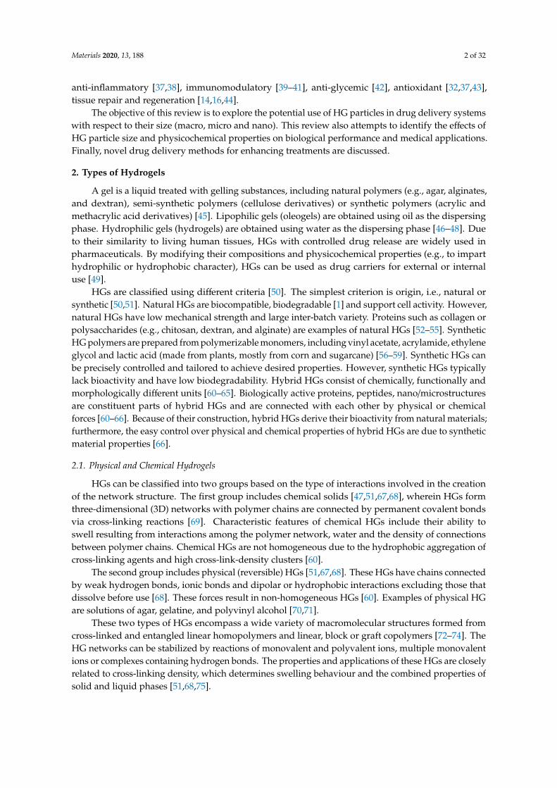
Hydrogels as Potential Nano-, Micro- andMacro-Scale Systems for Controlled Drug Delivery
Adam Chyzy † , Monika Tomczykowa † and Marta E. Plonska-Brzezinska *
Department of Organic Chemistry, Faculty of Pharmacy with the Division of Laboratory Medicine, MedicalUniversity of Bialystok, Mickiewicza 2A, 15-222 Bialystok, Poland; [email protected] (A.C.);[email protected] (M.T.)* Correspondence: [email protected]; Tel.: +4885-748-5683† These authors contributed equally to this work.
Received: 16 December 2019; Accepted: 27 December 2019; Published: 2 January 2020�����������������
Abstract: This review is an extensive evaluation and essential analysis of the design and formation ofhydrogels (HGs) for drug delivery. We review the fundamental principles of HGs (their chemicalstructures, physicochemical properties, synthesis routes, different types, etc.) that influence theirbiological properties and medical and pharmaceutical applications. Strategies for fabricating HGswith different diameters (macro, micro, and nano) are also presented. The size of biocompatible HGmaterials determines their potential uses in medicine as drug carriers. Additionally, novel drugdelivery methods for enhancing treatment are discussed. A critical review is performed based on thelatest literature reports.
Keywords: hydrogel; drug delivery; polymer; biocompatibility; immobilization of drug
1. Introduction
Drug delivery systems grounded on hydrogels (HGs) are interesting because of their highbiocompatibility and biodegradability. These properties are especially relevant for materials usedfor biomedical engineering applications, an example of which may be drug delivery or tissueengineering [1–3]. HGs are water-swollen polymeric networks containing chemical or physicalcross-links able to absorb large quantities of water or biological fluids [4]. HGs have a variety ofstructures, architectures, sizes (from centimetres to sub-nanometres), and functions, and together withother properties, these variables determine HG use for drug delivery [1–3,5].
HGs can be prepared from one polymer (homo-polymeric HG), two or more polymers(multi-polymeric HG); they may also contain other nanostructures/nanoparticles in a polymericnetwork [6–9]. These polymeric networks can be chemically and physically modified imparting newand unusual properties [9–11]. Chemical structures, compositions, biodegradability, biological functionsand different physicochemical properties (e.g., mechanical, rheological, spectral, thermosensitive, pHstability) can be modified [2,6,9,12]. These variations influence the performances of HGs and affectloading and releasing properties for drugs [7,9,11–15].
HGs can be used to form microparticles, nanoparticles, micelles and films [15,16]. For HGparticles, the particle size (macro, micro and nano) determines the route by which HGs can bedelivered into the human body [17–21]. For micro- or nano-sized HGs, the effects of variousphysical and chemical factors on drug release should be considered [15,21]. Therefore, drugimmobilization in a polymer matrix should be considered in the context of controlled release attarget sites. Various in vivo and in vitro drug application techniques have been developed with varioustherapeutic properties [12,22–24], including antifungal [25–27], antibacterial [28–32], antitumor [33–36],
Materials 2020, 13, 188; doi:10.3390/ma13010188 www.mdpi.com/journal/materials
Materials 2020, 13, 188 2 of 32
anti-inflammatory [37,38], immunomodulatory [39–41], anti-glycemic [42], antioxidant [32,37,43],tissue repair and regeneration [14,16,44].
The objective of this review is to explore the potential use of HG particles in drug delivery systemswith respect to their size (macro, micro and nano). This review also attempts to identify the effects ofHG particle size and physicochemical properties on biological performance and medical applications.Finally, novel drug delivery methods for enhancing treatments are discussed.
2. Types of Hydrogels
A gel is a liquid treated with gelling substances, including natural polymers (e.g., agar, alginates,and dextran), semi-synthetic polymers (cellulose derivatives) or synthetic polymers (acrylic andmethacrylic acid derivatives) [45]. Lipophilic gels (oleogels) are obtained using oil as the dispersingphase. Hydrophilic gels (hydrogels) are obtained using water as the dispersing phase [46–48]. Dueto their similarity to living human tissues, HGs with controlled drug release are widely used inpharmaceuticals. By modifying their compositions and physicochemical properties (e.g., to imparthydrophilic or hydrophobic character), HGs can be used as drug carriers for external or internaluse [49].
HGs are classified using different criteria [50]. The simplest criterion is origin, i.e., natural orsynthetic [50,51]. Natural HGs are biocompatible, biodegradable [1] and support cell activity. However,natural HGs have low mechanical strength and large inter-batch variety. Proteins such as collagen orpolysaccharides (e.g., chitosan, dextran, and alginate) are examples of natural HGs [52–55]. SyntheticHG polymers are prepared from polymerizable monomers, including vinyl acetate, acrylamide, ethyleneglycol and lactic acid (made from plants, mostly from corn and sugarcane) [56–59]. Synthetic HGs canbe precisely controlled and tailored to achieve desired properties. However, synthetic HGs typicallylack bioactivity and have low biodegradability. Hybrid HGs consist of chemically, functionally andmorphologically different units [60–65]. Biologically active proteins, peptides, nano/microstructuresare constituent parts of hybrid HGs and are connected with each other by physical or chemicalforces [60–66]. Because of their construction, hybrid HGs derive their bioactivity from natural materials;furthermore, the easy control over physical and chemical properties of hybrid HGs are due to syntheticmaterial properties [66].
2.1. Physical and Chemical Hydrogels
HGs can be classified into two groups based on the type of interactions involved in the creationof the network structure. The first group includes chemical solids [47,51,67,68], wherein HGs formthree-dimensional (3D) networks with polymer chains are connected by permanent covalent bondsvia cross-linking reactions [69]. Characteristic features of chemical HGs include their ability toswell resulting from interactions among the polymer network, water and the density of connectionsbetween polymer chains. Chemical HGs are not homogeneous due to the hydrophobic aggregation ofcross-linking agents and high cross-link-density clusters [60].
The second group includes physical (reversible) HGs [51,67,68]. These HGs have chains connectedby weak hydrogen bonds, ionic bonds and dipolar or hydrophobic interactions excluding those thatdissolve before use [68]. These forces result in non-homogeneous HGs [60]. Examples of physical HGare solutions of agar, gelatine, and polyvinyl alcohol [70,71].
These two types of HGs encompass a wide variety of macromolecular structures formed fromcross-linked and entangled linear homopolymers and linear, block or graft copolymers [72–74]. TheHG networks can be stabilized by reactions of monovalent and polyvalent ions, multiple monovalentions or complexes containing hydrogen bonds. The properties and applications of these HGs are closelyrelated to cross-linking density, which determines swelling behaviour and the combined properties ofsolid and liquid phases [51,68,75].
Materials 2020, 13, 188 3 of 32
2.2. Conventional and Stimuli-Responsive Hydrogels
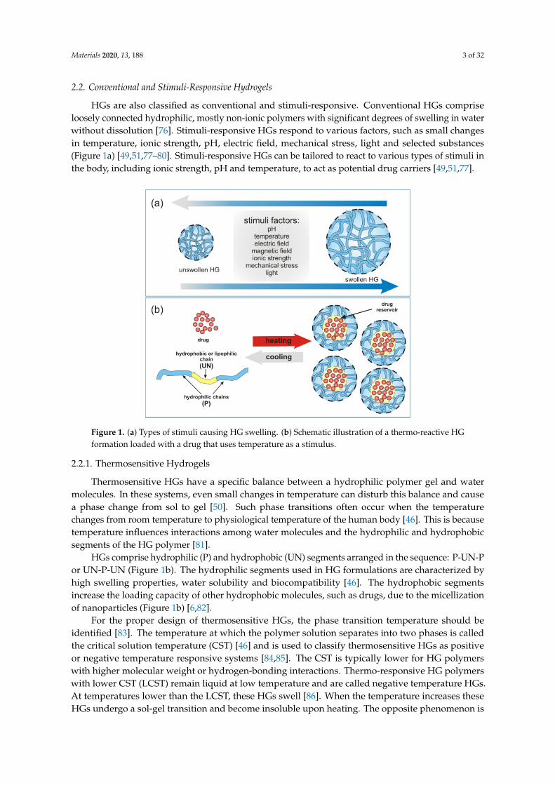
HGs are also classified as conventional and stimuli-responsive. Conventional HGs compriseloosely connected hydrophilic, mostly non-ionic polymers with significant degrees of swelling in waterwithout dissolution [76]. Stimuli-responsive HGs respond to various factors, such as small changesin temperature, ionic strength, pH, electric field, mechanical stress, light and selected substances(Figure 1a) [49,51,77–80]. Stimuli-responsive HGs can be tailored to react to various types of stimuli inthe body, including ionic strength, pH and temperature, to act as potential drug carriers [49,51,77].
Materials 2019, 12, x FOR PEER REVIEW 3 of 32
changes in temperature, ionic strength, pH, electric field, mechanical stress, light and selected substances (Figure 1a) [49,51,77–80]. Stimuli-responsive HGs can be tailored to react to various types of stimuli in the body, including ionic strength, pH and temperature, to act as potential drug carriers [49,51,77].
Figure 1. (a) Types of stimuli causing HG swelling. (b) Schematic illustration of a thermo-reactive HG formation loaded with a drug that uses temperature as a stimulus.
2.2.1. Thermosensitive Hydrogels
Thermosensitive HGs have a specific balance between a hydrophilic polymer gel and water molecules. In these systems, even small changes in temperature can disturb this balance and cause a phase change from sol to gel [50]. Such phase transitions often occur when the temperature changes from room temperature to physiological temperature of the human body [46]. This is because temperature influences interactions among water molecules and the hydrophilic and hydrophobic segments of the HG polymer [81].
HGs comprise hydrophilic (P) and hydrophobic (UN) segments arranged in the sequence: P-UN-P or UN-P-UN (Figure 1b). The hydrophilic segments used in HG formulations are characterized by high swelling properties, water solubility and biocompatibility [46]. The hydrophobic segments increase the loading capacity of other hydrophobic molecules, such as drugs, due to the micellization of nanoparticles (Figure 1b) [6,82].
For the proper design of thermosensitive HGs, the phase transition temperature should be identified [83]. The temperature at which the polymer solution separates into two phases is called the critical solution temperature (CST) [46] and is used to classify thermosensitive HGs as positive or negative temperature responsive systems [84,85]. The CST is typically lower for HG polymers with higher molecular weight or hydrogen-bonding interactions. Thermo-responsive HG polymers with lower CST (LCST) remain liquid at low temperature and are called negative temperature HGs. At temperatures lower than the LCST, these HGs swell [86]. When the temperature increases these HGs undergo a sol-gel transition and become insoluble upon heating. The opposite phenomenon is observed for HG polymers with upper CST (UCST, positive temperature HGs), which become soluble upon heating [46]. At temperatures lower than the UCST, positive temperature HGs dehydrate [87]. LCST HGs are used in the medical field because they form gels in situ at physiological temperatures, i.e., 30–37 °C (Table 1) [88]. Examples of synthetic polymers used to develop LCST HGs include copolymer blocks of poly(ethylene oxide) and poly(pentapeptide),
Figure 1. (a) Types of stimuli causing HG swelling. (b) Schematic illustration of a thermo-reactive HGformation loaded with a drug that uses temperature as a stimulus.
2.2.1. Thermosensitive Hydrogels
Thermosensitive HGs have a specific balance between a hydrophilic polymer gel and watermolecules. In these systems, even small changes in temperature can disturb this balance and causea phase change from sol to gel [50]. Such phase transitions often occur when the temperaturechanges from room temperature to physiological temperature of the human body [46]. This is becausetemperature influences interactions among water molecules and the hydrophilic and hydrophobicsegments of the HG polymer [81].
HGs comprise hydrophilic (P) and hydrophobic (UN) segments arranged in the sequence: P-UN-Por UN-P-UN (Figure 1b). The hydrophilic segments used in HG formulations are characterized byhigh swelling properties, water solubility and biocompatibility [46]. The hydrophobic segmentsincrease the loading capacity of other hydrophobic molecules, such as drugs, due to the micellizationof nanoparticles (Figure 1b) [6,82].
For the proper design of thermosensitive HGs, the phase transition temperature should beidentified [83]. The temperature at which the polymer solution separates into two phases is calledthe critical solution temperature (CST) [46] and is used to classify thermosensitive HGs as positiveor negative temperature responsive systems [84,85]. The CST is typically lower for HG polymerswith higher molecular weight or hydrogen-bonding interactions. Thermo-responsive HG polymerswith lower CST (LCST) remain liquid at low temperature and are called negative temperature HGs.At temperatures lower than the LCST, these HGs swell [86]. When the temperature increases theseHGs undergo a sol-gel transition and become insoluble upon heating. The opposite phenomenon is

Materials 2020, 13, 188 4 of 32
observed for HG polymers with upper CST (UCST, positive temperature HGs), which become solubleupon heating [46]. At temperatures lower than the UCST, positive temperature HGs dehydrate [87].LCST HGs are used in the medical field because they form gels in situ at physiological temperatures, i.e.,30–37 ◦C (Table 1) [88]. Examples of synthetic polymers used to develop LCST HGs include copolymerblocks of poly(ethylene oxide) and poly(pentapeptide), poly(N-isopropylacrylamide) (PNIPAM), andpoly(N,N-diethyl acrylamide) (PDEAM) [88]. Natural LCST polymers include cellulose derivatives,chitosan and gellan gum (Table 1) [88–90].
Table 1. Phase transition temperatures for selected polymers with LCST and UCST.
Polymer Name Abbreviation Transition Temperature (in Water) Refs.
Poly(N-isopropylacrylamide) PNIPAM 30–34 ◦C [88]Poly(N,N-diethylacrylamide) PDEAM 32–34 ◦C [88]
Poly(methyl vinyl ether) PMVE 37 ◦C [91]Polyvinyl chloride PVC 30–50 ◦C [92,93]
Gellan gum - 50–60 ◦C [88]Methylcellulose - 40 ◦C [94,95]
Acrylamide and acrylic acid AAm and AAc 15–25 ◦C [96]
2.2.2. Photo-Responsive Hydrogels
Light-responsive HGs are subjected to sol-gel changes under the influence of light at differentwavelengths: infrared (IR), ultraviolet (UV), or visible (VIS) [50]. The initiation of sol-gel phasetransitions in photo-responsive HGs using light is straightforward and non-invasive making these HGsa potential source of functional material in biomedicine, e.g., for drug delivery [50,97,98]. To preparea photo-responsive HG, a light-responsive chromophore group is attached to HG polymers [99,100].Incident light initiates swelling/deswelling processes or sol-gel phase transition to release drugs atdesired locations. UV-light-responsive HGs undergo photopolymerization or photocleavage dependingon the exposure length [101,102]. Light wavelength is also extremely important because it affects thedrug release control process [103].
2.2.3. pH-, Electric- and Magnetic-Responsive Hydrogels
pH-responsive HGs have specific pH-dependent physicochemical properties [104]. These HGshave acidic and alkaline groups associated with HG polymer chains [50,105]. The human body isa dynamic environment with different tissues having different pH ranges, which provides deliverymeans for in vivo pH-responsive HGs in pharmaceutical and biomedical applications [106–108].
Ionizable groups embedded in the polymer network of pH-responsive HGs accept or donateprotons in response to pH change, resulting in changes to the structure and solubility of the HGs andconsequently, swelling or deswelling (Figure 1a). The most frequently used synthetic monomer withacidic characteristics are acrylic acid, methacrylic acid, maleic anhydride, N,N-dimethylaminoethylmethacrylate and sulphonamide-containing polymers [2,18,20,91,109]. Weak synthetic polybases, suchas aromatics 4-vinylpyridine, 2-vinylpyridine, poly(vinyl imidazole), poly(N,N-dimethyl aminoethylmethacrylate), and poly(N,N-diethyl aminoethyl methacrylate), accept protons at low pH [50,110,111].There is also a large group of natural polymers used in pH-responsive HGs that has advantagesover synthetic polymers due to their degradability within the human body, making them excellentbiocompatible components in drug delivery [9,33,88,112].
There are also HGs that respond to external physical stimuli, like electric and magneticfields [52,105]. Magnetic or electrically responsive HG networks have been developed for drug-deliveryapplications. The influence of an electric field causes electro-responsive HGs to swell or de-swell,which can be adapted for drug delivery systems [113]. Similar applications have been determined formagnetic field-based stimuli, especially high frequency fields, which are much safer for humans thanelectric fields or UV light [114]. Magnetic and electrical stimuli-responsive HGs allow for the study

Materials 2020, 13, 188 5 of 32
of cell behaviour by altering hydrogel properties in situ. These stimuli-responsive HGs can serve inpotential drug delivery systems for targeting and localizing to delivery sites for controlled drug releaseupon application of electric or magnetic fields [115–117].
2.3. Other Classifications for Hydrogels
HGs could also be classified by the substrates used for their production. Polymer networksmay be synthesized from linking monomers to form high-molecular weight polymers. Hydrophilicmonomers, such as polyphenylene oxide (PPO) or polyethylene glycol (PEG), are copolymerised withcrosslinkers to form networks for drug delivery [118,119]. When prepolymers (oligomers) are usedas synthetic substrates, different types of HGs are obtained. A series of model networks consistingof PEG, tetra-PEG gels, have been prepared by Sakai et al. [119,120]. Small-angle neutron scatteringmeasurements confirmed, that network structure is extremely uniform, even in the presence of differentfunctional groups at the ends, i.e., amine group and succinimidyl ester group [121,122]. Additionally,the modified with ionic liquid form of tetra-PEG gels possesses high ion conductivity and highmechanical properties [123,124]. For example, polyurethane-based HGs are used in the fabrication offlexible medical devices, prosthetics for example [125]. Other substrate-specific HGs are those withnetwork structures made by crosslinking hydrophilic polymer chains, e.g., chitosan crosslinked withglutaraldehyde [75].
Due to large differences in their chemical compositions, HGs are sometimes broadlyclassified [51,126]. For example, in terms of the types of monomers used to prepare HGs (polymericcomposition), three groups of HGs are recognized: (i) homopolymeric HGs (consisting of one type ofhydrophilic monomer); (ii) copolymeric HGs (two or more monomer types of which at least one ishydrophilic), and (iii) multipolymer HGs (obtained from more than one type of polymer).
Multipolymer interpenetrating polymeric (IPN) HGs represent a class of HGs made of twoindependent cross-linked synthetic and/or natural polymers [51,75,127–129]. HGs there are twopolymer networks, one of which is polymerized around and within the on the second polymer network,and what is very important, there are no covalent linkages between those two polymeric networks.
HGs may take significantly different morphological forms, which can be a criterion for their divisiondepending on the state of aggregation (physical form) [17,105,129]: (i) solid (amorphous, semicrystallineor crystalline); (ii) semisolid (for example shear-thinning HGs), and (iii) liquid [17,51,75,130]. Solid HGscan be useful in formation of functional tissues [75,131]. At room temperature they are solid in naturewith strong cross-linked network structure [17]. Solid HGs swell in the presence of water or otherhydrophilic solutions like buffers or biological fluids. Semisolid HG consists of two types componentswhere at least one possess biological nature, e.g., plant resins or gums [17]. The shear-thinning HGsalso belong to this group [17,132,133]. These HGs can be pre-gelled outside of the body and injected byapplying shear stress and flow like low-viscosity fluids. The shear-thinning behaviour is a result ofreversible physical cross-links. That group of HGs is often called bio- or muco-adhesive HGs accordingto their specific adhesive properties [16,75,134]. Liquid HGs are injected in liquid form and a sol-geltransition is performed inside the human body [19,109,135–137]. The resulting HGs takes the shapeof space available at the injection site which allows to achieve the sol–gel transition with variety ofstrategies. Slow-gelling systems based on gelation mechanisms (charge interaction, stereocomplexation,click chemistry, etc.), termo- or pH-responsive systems are good representatives of this group.
It is also possible to classify HG based on ionic charges into the following four groups: (i) neutral(no charge, e.g., dextran); (ii) anionic (negative charge) like carrageenan; (iii) cationic (with positivecharge) such as chitosan, and (iv) amphiphilic (having the ability to strongly interact with both polarand nonpolar solvent molecules—such as collagen) [17,138–143].
3. Types of Physical Appearance of Hydrogels
HGs have a number of properties, such as oxygen and nutrient permeability and the ability to bind,transport and control drug delivery and release, attractive for use in biological applications. Different
Materials 2020, 13, 188 6 of 32
tissues have different requirements and would be better served using different types of HGs [144]. HGscan be formed in almost any shape and size [145], e.g., as a matrix, a film, microparticles, nanoparticles,macro-beads, membranes, or coatings, depending on the polymerization technique [46,51,82,146]. HGsare altered to improve their compatibility with hydrophobic or hydrophilic compounds or drugs withdifferent properties [147,148].
Polymeric HGs are classified as macro-, micro- and nanogels with respect to particle size [17,149].Microgels, cross-linked structure is large, usually from millimetres to centimetres [150]. A group ofgels called microgels, whose particles are smaller, separate and cross-linked, having a size between1 µm and 100 nm [151]. Nanogels are those gels whose particles have a size smaller than 100nm [49,106,152]. It should be noted that gels with particle sizes only slightly larger than 100 nm werecalled quasi-nanogels [153].
Microgel is a submicron- or micron-sized network polymer which is highly swellable but insolublein water [154]. Cross-linked microgel particles possess special kind of control over particle shape andsize, which is probably due to specific extensive swelling pattern [155]. Microgels are obtained in twoways. One of them is molecular assembly of existing polymer molecules in aqueous solutions. Theother one, particle-forming polymerization, includes two types of polymerization: precipitation andinverse emulsion polymerization [154,156].
Nanogels are prepared in water by self-aggregation of polymers, mostly of natural origin; naturalpolysaccharides (dextran, pullulan) or cholesterol-containing polysaccharide [14,157]. The dimensionsof these HGs are usually of 20–30 nm. Such a small size causes the use of nanogels for cell targeting,because swelling caused by pH changes in the surrounding environment is the reason to release theentrapped drug [158]. Nanogels react to the external stimuli changes much quicker than macrogels.The reason for this is probably their small size and short relaxation time [159,160]. Swelling anddeswelling properties and small size of biocompatible and biodegradable nanogel particles are thecause of easy crossing the blood brain barrier [21,149,153].
Obtaining a specific shape of HG at a nanoscale is difficult. HG nanofibers, for example, can beobtained by the by electrostatic spinning method [161]. HGs are also given the form of microcapsulesand microspheres, whose size is 1–1000 µm. This allows more protection of substances enclosed in theinterior of microcapsules and microspheres. HGs can form micelles in an aqueous environment, as aresult of aggregation of amphiphilically connected blocks or end-modified polymers. It is often difficultto differentiate micelles from reversible HGs, because HGs can form micelles above a appropriateconcentration called the micelle gel concentration [162]. Similarly, biodegradable nanoparticles of HGsare classified as nanocapsules or nanospheres having drug molecules adsorbed on the surface or closedinside [163,164].
There also exist the group of in situ-gelling HGs, which undergo a sol-gel transition inside thehuman body [165]. That group of HG always adapts to the free space with its shape. An example ofsuch HGs are shear-thinning HGs. Because of reversible physical cross-links shear-thinning hydrogelspre-gelled outside of the human body, after shear stress injection can regain its initial stiffness.
Despite of many nano- and microgels advantages, it should remember about the possibilityof using macroporous HGs and the benefits resulting from it. Macroporous HGs can mechanicallycollapse at up to 90% and recover reversibly almost immediately after injection in the human body.Such macroporous HGs allow for the preparation of highly defined shapes for drug delivery [151].
4. Immobilization of Drugs in Hydrogel
4.1. HG Structure and Physical Properties
A physical and chemical properties, including structural parameters, swelling behaviour, diffusivecharacteristics, and surface properties, determine the biomedical and pharmaceutical applications ofHGs [5]. After the creation of porous HG structures, different drug molecules may be incorporatedinto the scaffolding (post-loading) or in the network during formation (in situ loading); these drugs are
Materials 2020, 13, 188 7 of 32
subsequently released from the hydrophilic matrix to biological media. Thus, network mesh size (orpore size) is a major parameter which determines the sizes of the drug system that can be immobilizedin the porous matrix of the HG. The network mesh size (ξ) indicates the distance between adjacentjunctions, cross-links and tie points. Cross-links may be chemical (mainly covalent bonds) or physical(e.g., electrostatic, hydrophobic and dipole-dipole) [5].
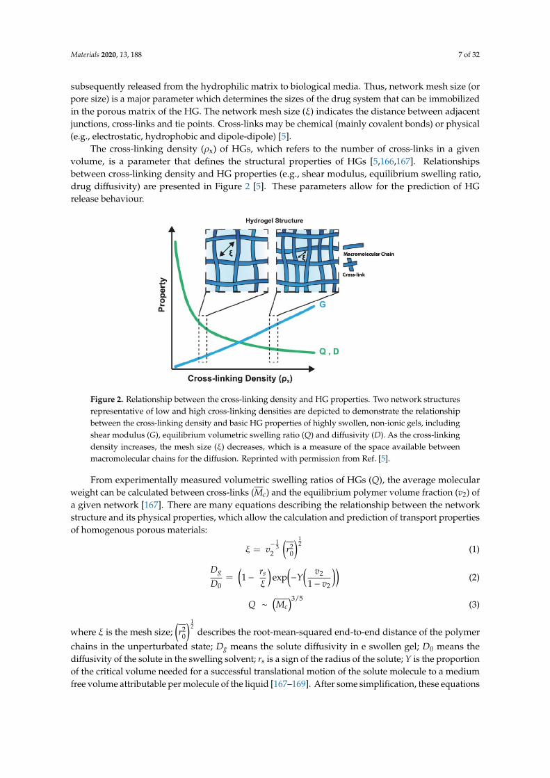
The cross-linking density (ρx) of HGs, which refers to the number of cross-links in a givenvolume, is a parameter that defines the structural properties of HGs [5,166,167]. Relationshipsbetween cross-linking density and HG properties (e.g., shear modulus, equilibrium swelling ratio,drug diffusivity) are presented in Figure 2 [5]. These parameters allow for the prediction of HGrelease behaviour.
Materials 2019, 12, x FOR PEER REVIEW 7 of 32
distance between adjacent junctions, cross-links and tie points. Cross-links may be chemical (mainly covalent bonds) or physical (e.g., electrostatic, hydrophobic and dipole-dipole) [5].
The cross-linking density (ρx) of HGs, which refers to the number of cross-links in a given volume, is a parameter that defines the structural properties of HGs [5,166,167]. Relationships between cross-linking density and HG properties (e.g., shear modulus, equilibrium swelling ratio, drug diffusivity) are presented in Figure 2 [5]. These parameters allow for the prediction of HG release behaviour.
Figure 2. Relationship between the cross-linking density and HG properties. Two network structures representative of low and high cross-linking densities are depicted to demonstrate the relationship between the cross-linking density and basic HG properties of highly swollen, non-ionic gels, including shear modulus (G), equilibrium volumetric swelling ratio (Q) and diffusivity (D). As the cross-linking density increases, the mesh size (ξ) decreases, which is a measure of the space available between macromolecular chains for the diffusion. Reprinted with permission from Ref. [5].
From experimentally measured volumetric swelling ratios of HGs (Q), the average molecular weight can be calculated between cross-links (𝑀 ) and the equilibrium polymer volume fraction (v2) of a given network [167]. There are many equations describing the relationship between the network structure and its physical properties, which allow the calculation and prediction of transport properties of homogenous porous materials: 𝜉 = 𝑣 𝑟 (1)
𝐷𝐷 = 1 − 𝑟𝜉 exp −𝑌 𝑣1 − 𝑣 (2) 𝑄 ~ 𝑀 / (3)
where ξ is the mesh size; 𝑟 describes the root-mean-squared end-to-end distance of the polymer chains in the unperturbated state; Dg means the solute diffusivity in e swollen gel; D0 means the diffusivity of the solute in the swelling solvent; rs is a sign of the radius of the solute; Y is the proportion of the critical volume needed for a successful translational motion of the solute molecule to a medium free volume attributable per molecule of the liquid [167–169]. After some simplification, these equations can be used to find the correlation between all mentioned parameters (Equation (4)) for highly swollen, non-ionic HGs, and scale with 𝑀 : 1 − 𝐷𝐷 = 𝑟𝜉 ~ 𝑀 (4)
The HG network structure is also important in determining the mechanical properties of porous material. The network structure controls swelling behaviour and drug release in biological
Figure 2. Relationship between the cross-linking density and HG properties. Two network structuresrepresentative of low and high cross-linking densities are depicted to demonstrate the relationshipbetween the cross-linking density and basic HG properties of highly swollen, non-ionic gels, includingshear modulus (G), equilibrium volumetric swelling ratio (Q) and diffusivity (D). As the cross-linkingdensity increases, the mesh size (ξ) decreases, which is a measure of the space available betweenmacromolecular chains for the diffusion. Reprinted with permission from Ref. [5].
From experimentally measured volumetric swelling ratios of HGs (Q), the average molecularweight can be calculated between cross-links (Mc) and the equilibrium polymer volume fraction (v2) ofa given network [167]. There are many equations describing the relationship between the networkstructure and its physical properties, which allow the calculation and prediction of transport propertiesof homogenous porous materials:
ξ = v−
13
2
(r2
0
) 12
(1)
Dg
D0=(1−
rs
ξ
)exp(−Y( v2
1− v2
))(2)
Q ∼
(Mc)3/5
(3)
where ξ is the mesh size;(r2
0
) 12
describes the root-mean-squared end-to-end distance of the polymer
chains in the unperturbated state; Dg means the solute diffusivity in e swollen gel; D0 means thediffusivity of the solute in the swelling solvent; rs is a sign of the radius of the solute; Y is the proportionof the critical volume needed for a successful translational motion of the solute molecule to a mediumfree volume attributable per molecule of the liquid [167–169]. After some simplification, these equations
Materials 2020, 13, 188 8 of 32
can be used to find the correlation between all mentioned parameters (Equation (4)) for highly swollen,non-ionic HGs, and scale with Mc:
1−Dg
D0=
rs
ξ∼
(Mc)− 7
10 (4)
The HG network structure is also important in determining the mechanical properties of porousmaterial. The network structure controls swelling behaviour and drug release in biological environment.Swelling behaviour is defined as the ratio of the volume of the water-swollen gel to the volume ofdry polymer and is indicative of the water content of the swollen HG. However, higher water contentresulting from high network porosity is more beneficial for immobilizing active substances and cansimultaneously lead to polymer degradation and prevent the controlled delivery of drugs. In thiscontext, the cross-linking density must be high enough to allow for the immobilization of the activesubstance in the polymer matrix. Beyond the structural properties, the chemical composition of HGsregulates the final biochemical properties, e.g., charge, hydrophilicity and bioactivity.
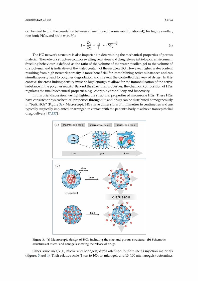
In this brief discussion, we highlighted the structural properties of macroscale HGs. These HGshave consistent physicochemical properties throughout, and drugs can be distributed homogeneouslyin “bulk HGs” (Figure 3a). Macroscopic HGs have dimensions of millimetres to centimetres and aretypically surgically implanted or arranged in contact with the patient’s body to achieve transepithelialdrug delivery [17,137].
Materials 2019, 12, x FOR PEER REVIEW 8 of 32
environment. Swelling behaviour is defined as the ratio of the volume of the water-swollen gel to the volume of dry polymer and is indicative of the water content of the swollen HG. However, higher water content resulting from high network porosity is more beneficial for immobilizing active substances and can simultaneously lead to polymer degradation and prevent the controlled delivery of drugs. In this context, the cross-linking density must be high enough to allow for the immobilization of the active substance in the polymer matrix. Beyond the structural properties, the chemical composition of HGs regulates the final biochemical properties, e.g., charge, hydrophilicity and bioactivity.
In this brief discussion, we highlighted the structural properties of macroscale HGs. These HGs have consistent physicochemical properties throughout, and drugs can be distributed homogeneously in “bulk HGs” (Figure 3a). Macroscopic HGs have dimensions of millimetres to centimetres and are typically surgically implanted or arranged in contact with the patient’s body to achieve transepithelial drug delivery [17,137].
Figure 3. (a) Macroscopic design of HGs including the size and porous structure. (b) Schematic structures of micro- and nanogels showing the release of drugs.
Other structures, e.g., micro- and nanogels, draw attention to their use as injection materials (Figures 3 and 4). Their relative scale (1 µm to 100 nm microgels and 10–100 nm nanogels) determines the course by which HGs can be delivered to the human body [17–20]. Small HG particles are needle-injectable, provide large surfaces for bioconjugation and enhance penetration through tissue barriers [135,136,170,171].
Figure 3. (a) Macroscopic design of HGs including the size and porous structure. (b) Schematicstructures of micro- and nanogels showing the release of drugs.
Other structures, e.g., micro- and nanogels, draw attention to their use as injection materials(Figures 3 and 4). Their relative scale (1 µm to 100 nm microgels and 10–100 nm nanogels) determines
Materials 2020, 13, 188 9 of 32
the course by which HGs can be delivered to the human body [17–20]. Small HG particles areneedle-injectable, provide large surfaces for bioconjugation and enhance penetration through tissuebarriers [135,136,170,171].Materials 2019, 12, x FOR PEER REVIEW 9 of 32
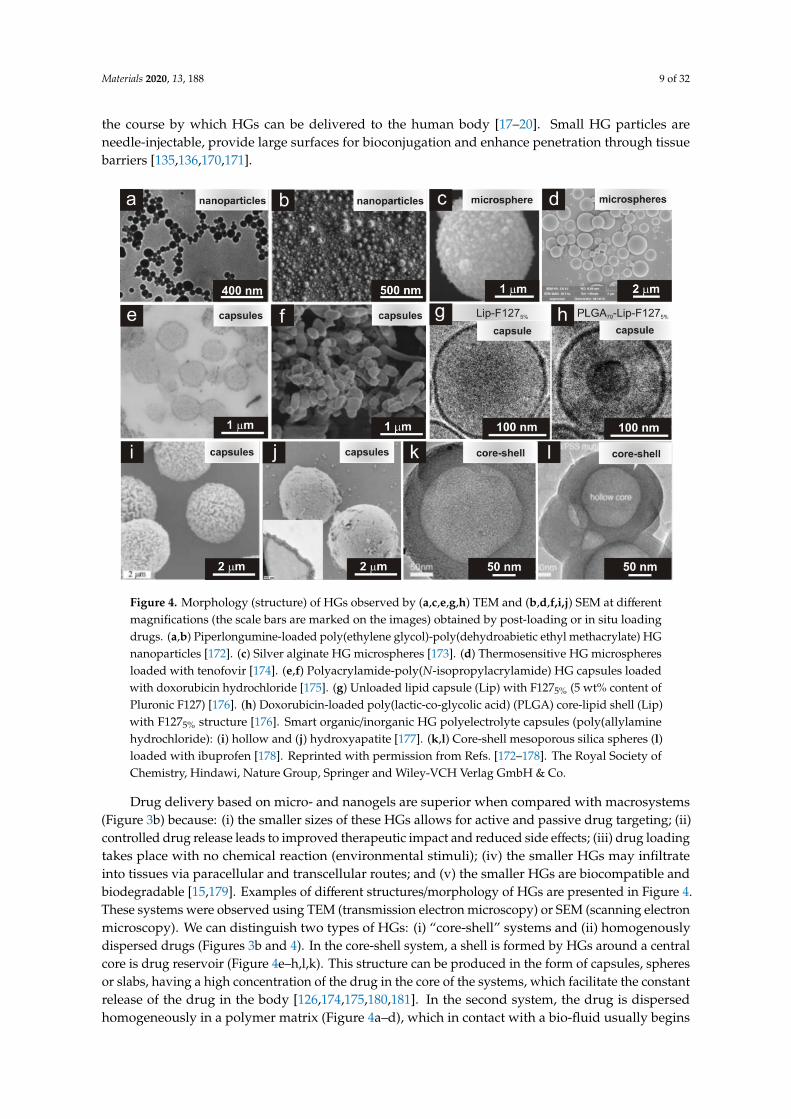
Figure 4. Morphology (structure) of HGs observed by (a,c,e,g,h) TEM and (b,d,f,i,j) SEM at different magnifications (the scale bars are marked on the images) obtained by post-loading or in situ loading drugs. (a,b) Piperlongumine-loaded poly(ethylene glycol)-poly(dehydroabietic ethyl methacrylate) HG nanoparticles [172]. (c) Silver alginate HG microspheres [173]. (d) Thermosensitive HG microspheres loaded with tenofovir [174]. (e,f) Polyacrylamide-poly(N-isopropylacrylamide) HG capsules loaded with doxorubicin hydrochloride [175]. (g) Unloaded lipid capsule (Lip) with F1275% (5 wt% content of Pluronic F127) [176]. (h) Doxorubicin-loaded poly(lactic-co-glycolic acid) (PLGA) core-lipid shell (Lip) with F1275% structure [176]. Smart organic/inorganic HG polyelectrolyte capsules (poly(allylamine hydrochloride): (i) hollow and (j) hydroxyapatite [177]. (k,l) Core-shell mesoporous silica spheres (l) loaded with ibuprofen [178]. Reprinted with permission from Refs. [172–178]. The Royal Society of Chemistry, Hindawi, Nature Group, Springer and Wiley-VCH Verlag GmbH & Co.
Drug delivery based on micro- and nanogels are superior when compared with macrosystems (Figure 3b) because: (i) the smaller sizes of these HGs allows for active and passive drug targeting; (ii) controlled drug release leads to improved therapeutic impact and reduced side effects; (iii) drug loading takes place with no chemical reaction (environmental stimuli); (iv) the smaller HGs may infiltrate into tissues via paracellular and transcellular routes; and (v) the smaller HGs are biocompatible and biodegradable [15,179]. Examples of different structures/morphology of HGs are presented in Figure 4. These systems were observed using TEM (transmission electron microscopy) or SEM (scanning electron microscopy). We can distinguish two types of HGs: (i) “core-shell” systems and (ii) homogenously dispersed drugs (Figures 3b and 4). In the core-shell system, a shell is formed by HGs around a central core is drug reservoir (Figure 4e–h,l,k). This structure can be produced in the form of capsules, spheres or slabs, having a high concentration of the drug in the core of the systems, which facilitate the constant release of the drug in the body [126,174,175,180,181]. In the second system, the drug is dispersed homogeneously in a polymer matrix (Figure 4a–d), which in contact with a bio-fluid usually begins swelling, and takes forms of nanoparticles, polymeric micelles, microspheres, etc. [68,172,180,181]. For both types of HGs, releasing of drugs is time-depended process and it is mainly diffusion controlled. Drug releasing from the HG network will be discussed in detail in the next paragraph.
Figure 4. Morphology (structure) of HGs observed by (a,c,e,g,h) TEM and (b,d,f,i,j) SEM at differentmagnifications (the scale bars are marked on the images) obtained by post-loading or in situ loadingdrugs. (a,b) Piperlongumine-loaded poly(ethylene glycol)-poly(dehydroabietic ethyl methacrylate) HGnanoparticles [172]. (c) Silver alginate HG microspheres [173]. (d) Thermosensitive HG microspheresloaded with tenofovir [174]. (e,f) Polyacrylamide-poly(N-isopropylacrylamide) HG capsules loadedwith doxorubicin hydrochloride [175]. (g) Unloaded lipid capsule (Lip) with F1275% (5 wt% content ofPluronic F127) [176]. (h) Doxorubicin-loaded poly(lactic-co-glycolic acid) (PLGA) core-lipid shell (Lip)with F1275% structure [176]. Smart organic/inorganic HG polyelectrolyte capsules (poly(allylaminehydrochloride): (i) hollow and (j) hydroxyapatite [177]. (k,l) Core-shell mesoporous silica spheres (l)loaded with ibuprofen [178]. Reprinted with permission from Refs. [172–178]. The Royal Society ofChemistry, Hindawi, Nature Group, Springer and Wiley-VCH Verlag GmbH & Co.
Drug delivery based on micro- and nanogels are superior when compared with macrosystems(Figure 3b) because: (i) the smaller sizes of these HGs allows for active and passive drug targeting; (ii)controlled drug release leads to improved therapeutic impact and reduced side effects; (iii) drug loadingtakes place with no chemical reaction (environmental stimuli); (iv) the smaller HGs may infiltrateinto tissues via paracellular and transcellular routes; and (v) the smaller HGs are biocompatible andbiodegradable [15,179]. Examples of different structures/morphology of HGs are presented in Figure 4.These systems were observed using TEM (transmission electron microscopy) or SEM (scanning electronmicroscopy). We can distinguish two types of HGs: (i) “core-shell” systems and (ii) homogenouslydispersed drugs (Figures 3b and 4). In the core-shell system, a shell is formed by HGs around a centralcore is drug reservoir (Figure 4e–h,l,k). This structure can be produced in the form of capsules, spheresor slabs, having a high concentration of the drug in the core of the systems, which facilitate the constantrelease of the drug in the body [126,174,175,180,181]. In the second system, the drug is dispersedhomogeneously in a polymer matrix (Figure 4a–d), which in contact with a bio-fluid usually begins
Materials 2020, 13, 188 10 of 32
swelling, and takes forms of nanoparticles, polymeric micelles, microspheres, etc. [68,172,180,181]. Forboth types of HGs, releasing of drugs is time-depended process and it is mainly diffusion controlled.Drug releasing from the HG network will be discussed in detail in the next paragraph.
4.2. Immobilization of Drugs
HGs due to their 3D structures may be applied as carriers for drugs, proteins, lipids orcells [15,17,68,182,183]. Additionally, a large amount of water within their structures makes a convenientenvironment for the immobilization of drugs. However, 3D structures of HG allow the immobilizationof drugs in their matrix, in this regard, when immobilization process of active substances is performed,two aspects must be taken into account. The large pore size of HG (large mesh size), as well as the highwater content causes that water-soluble drugs with small particles quickly escape from the network,thanks to which they have a short release time [182]. Drug release is much slower when the drugparticle size is comparable to the mesh size [17]. When the drug particle size exceeds the mesh size,then the drug is physically trapped in the network [17]. To minimize these problems, the pore size inHG (mesh size) should be “matched” to the size of the immobilized drug.
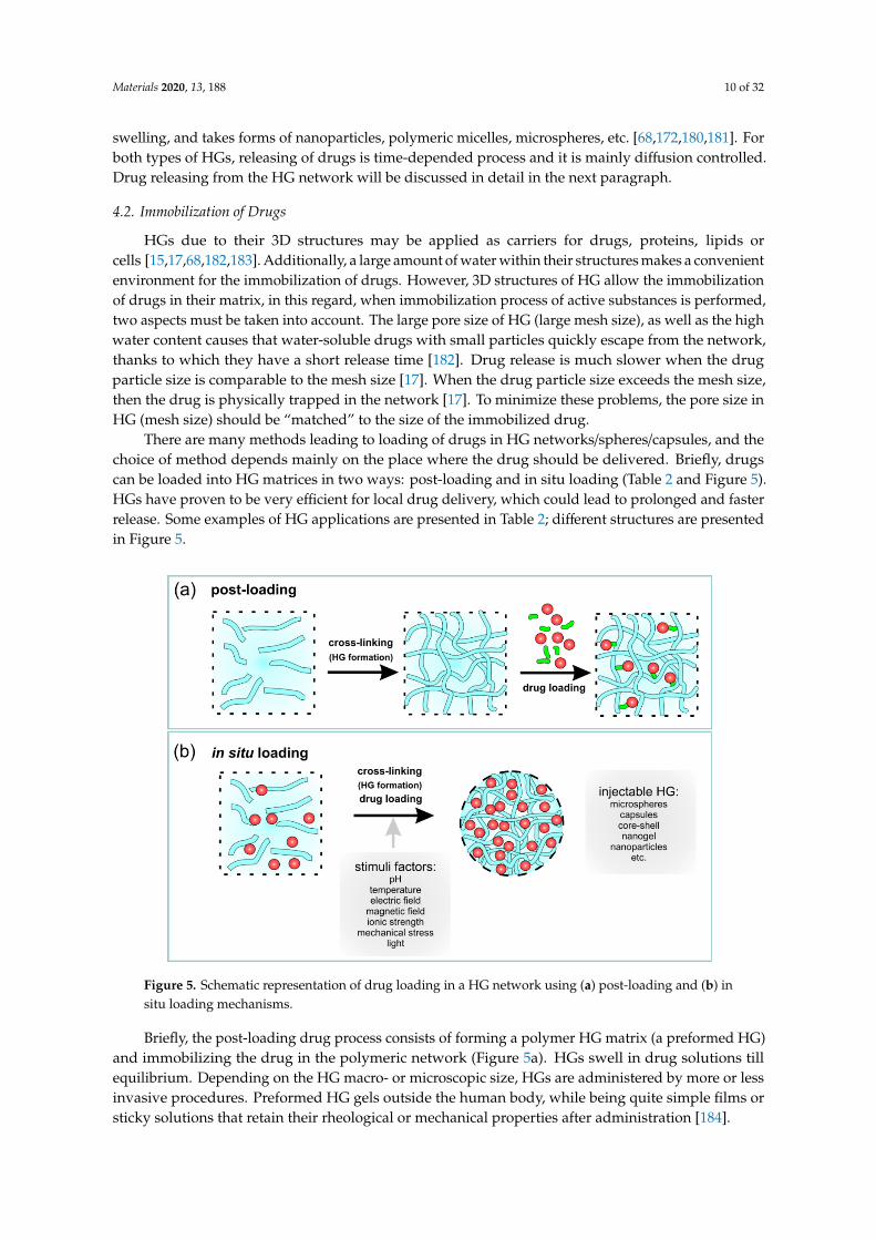
There are many methods leading to loading of drugs in HG networks/spheres/capsules, and thechoice of method depends mainly on the place where the drug should be delivered. Briefly, drugscan be loaded into HG matrices in two ways: post-loading and in situ loading (Table 2 and Figure 5).HGs have proven to be very efficient for local drug delivery, which could lead to prolonged and fasterrelease. Some examples of HG applications are presented in Table 2; different structures are presentedin Figure 5.
Materials 2019, 12, x FOR PEER REVIEW 12 of 32
Figure 5. Schematic representation of drug loading in a HG network using (a) post-loading and (b) in situ loading mechanisms.
Immobilization of Drugs in Hydrogel Composites
Some systems containing drugs in matrix/capsules are poorly water-soluble or insoluble. In this sense, soluble therapeutic agents are poorly retained in HGs due to their hydrophilic nature. In this case, there are many problems with loading process of drugs due to their tendency to aggregate resulting in high local concentrations causing toxicity [195]. To overcome these problems, HG composites have been created to exploit the hydrophilic-hydrophobic nature of various components [192,196]. HG composites contain polymeric networks (hydrophilic) swollen with water and nanostructures/microstructures with different physico-chemical properties [192,197–199]. These composites represent a new class of materials with new properties.
The inclusion of nanomaterials in the HG polymer network is an interesting way to adjust the mechanical properties of HG and/or to provide the composite with sensitivity to external stimuli [192,196,197,199]. Different nanomaterials have been immobilized in polymeric networks, including inorganic nanoparticles [200,201], carbon nanomaterials [202], and lipids [99]. Nanoparticle systems have gained considerable attention by being one of the most interesting and promising biomedical materials with the exceptional physicochemical properties, controlled shapes, nano-sized characteristics, comprehensive modification options and well-defined multi-functionality [83]. The preparation of HG composites may be performed using physical and chemical forces.
To overcome the incompatibility of hydrophobic drugs and hydrophilic HG networks, lipid nanoparticles (LPNs) are frequently used to promote good solubility. LPNs have been used for dermal, mucosal, transdermal and intramuscular applications [176,182]. Three types of LPNs have been used in drug delivery systems: (i) lipid nano emulsions (LNEs, where the core is composed of liquid lipids), (ii) solid lipid nanoparticles (SLNs, where the core has lipids in a solid state at room and body temperatures), and (iii) nanostructured lipid carriers (NLCs, where the lipid core is a heterogeneous mixture of solid and liquid lipids) [182]. The lipophilic core of LNPs entraps active ingredients, whereas the surfactant membrane (consisting of phospholipids) ensures the stability of LNPs in hydrophilic environments. In pure lipophilic form, these systems have unsuitable rheological properties and therefore, require structural modifications. These modifications result in the formation of LNP-HG composites [187] as shown in Figure 6.
Figure 5. Schematic representation of drug loading in a HG network using (a) post-loading and (b) insitu loading mechanisms.
Briefly, the post-loading drug process consists of forming a polymer HG matrix (a preformed HG)and immobilizing the drug in the polymeric network (Figure 5a). HGs swell in drug solutions tillequilibrium. Depending on the HG macro- or microscopic size, HGs are administered by more or lessinvasive procedures. Preformed HG gels outside the human body, while being quite simple films orsticky solutions that retain their rheological or mechanical properties after administration [184].
Materials 2020, 13, 188 11 of 32
Table 2. Examples of HGs with different routes drug immobilization of and their structuresand applications.
Type ofLoading Drugs Hydrogel Precursors Drug Structure Applications Refs.
Post-loading
AA17 –BA18 –DEAP19 17-DMAPG 15 Core-shell 16 Antitumor activity [152]Carbopol 940 20 Vor 11 Core-shell LPN Dermal applications [185]
Carbopol/stearic acid Vor Core-shell OphthalmicApplication [186]
PNIPAAm-b-PLA-b-PEG-b-PLA7-b-PNIPAAm Riluzole - Neuroprotective drug [183]
PAA-PNIPAAm DOX Core-shellcapsule Antitumor activity [175]
PEG-b-PDAEMA 8 PLGM 9 Nanoparticle Antitumor activity [172]Chitosan/Polysaccharide Tenofovir Microsphere Vaginal drug [174]
Chitosan/β-GB DOX-loaded in LTSL 29 PP33 HG Antitumor activity [187]
PLGA 30 MicroCPEG derivatives (HG)
DOX-loaded in MicroC31
Fu 32-loaded in HGPP HG Antitumor activity [188]
In situ loading
Polysaccharide Ibuprofen Core-shellcapsule Oral drug [126]
Carbohydrate-NIPAM 22 Bupivacaine HG-microgel composite Anaesthetic drug [189]PNIPAAm-co-AIA 21 Lopinavir Microspheres Antiretroviral drug [190]CmetCel 25 -Dextran AmB 23 MacroHG Antifungal therapy [191]
NiPAAm 26-NtBAAm27 Fluvastatin PP HG HMG-CoA 25 [192]
PPZ 34 Silibinin Microspheres Anticancer andantiangiogenic activity [193]
PEG–PCL 35–PEG PTX micelles 36 PP HG Antitumor activity [194]
Abbreviations: 1 PNIPAAm: poly(N-isopropylacrylamide); 2 PAA: polyacrylamide; 3 PHEMA: poly(hydroxylethylmethacrylate); 4 PVP: poly(vinylpyrrolidone); 5 PEG: poly(ethylene glycol); 6 LPN: lipid nanoparticles; 7 PLA:poly(lactide); 8 PDAEMA: poly(dehydroabietic ethyl methacrylate); 9 PLGM: piperlongumine; 10 DOX: Doxorubicinhydrochloride; 11 Vor: Voriconazole; 12 EGDMA: 2-(2-methyl-acryloyloxy)ethyl 2-methyl-acrylate; 13 HEMA:2-hydroxethyl 2-methylprop-2-enoate; 14 Indo: Indomethacin; 15 17-DMAPG: geldanamycin derivative (aminatedform, which readily protonates at low pH; 16 core-shell structure of HG-in-LPN; 17 AA: acrylic acid; 18 BA:N,N’-methylenebis(acrylamide); 19 DEAP: 2,2-diethoxyacetophenone; 20 Carbopol 940: HG composed of PrecirolATO 5, Labrafil 1944 CS, and Tween 80; 21 AIA: allylamine; 22 NIPAM: poly(N-isopropylacrylamide); 23 AmB:Amphotericin B; 24 CmetCel: carboxymethylcellulose-hydrazide; 25 HMG-CoA: reductase inhibitor (statin);26 NiPAAm: N-isopropylacrylamide; 27 NtBAAm: N-tert-butylacrylamide; 28 β-GB: β-glycerophosphate; 29
LTSL: lysolipid thermally sensitive liposomes; 30 PLGA: poly(lactide-co-glycolide); 31 MicroC: microcapsule; 32
Fu: 5-fluorouracil; 33 PP: “plum padding”; 34 PPZ: poly(organophosphazene); 35 PCL: poly(ε-caprolactone); 36
PTX: Paclitaxel.
Drugs can be physically or chemically immobilized in HGs and can also be loaded into otherspecies (secondary delivery vehicles) to provide the appropriate environment for targeting. PreformedHGs are solid even when injected [184].
In situ loading of drugs is associated with the simultaneous formation of injectable HGs in thebody and the encapsulation of drugs (Figure 5b). Briefly, the whole process shows a sol-to-gel transitionin situ that form after injection in vivo.
The resulting HG take the shape of the available space. In situ gel formation usually includesthe subsequent steps: (i) gelation as a response to the changes of temperature or pH changes(thermoresponsive or chemical responsive polymers; i.e., “smart” polymers), (ii) ionic or covalentcross-linking, (iii) solvent exchange or crystallization, and/or (iv) thickening upon removal of theinjection shear. These external stimuli are shortly summarized in Figure 1a (stimuli factors).
Taking into account the lifetime of HGs, their formation mechanism can be categorized as: (i)pre-gelation (polymer precursor in solution), (ii) therapeutic window (after injection and gelationcontaining drug), and (iii) degradation (HG degradation products) [18]. These systems may behomopolymeric or multipolymeric or may also be composed of different components, including othersystems (e.g., capsule, microgels, and nanoparticles) serving as drug carriers. This latter mentioned gelsystems are characterized in the next section.
Immobilization of Drugs in Hydrogel Composites
Some systems containing drugs in matrix/capsules are poorly water-soluble or insoluble. In thissense, soluble therapeutic agents are poorly retained in HGs due to their hydrophilic nature. In this case,there are many problems with loading process of drugs due to their tendency to aggregate resulting inhigh local concentrations causing toxicity [195]. To overcome these problems, HG composites have been
Materials 2020, 13, 188 12 of 32
created to exploit the hydrophilic-hydrophobic nature of various components [192,196]. HG compositescontain polymeric networks (hydrophilic) swollen with water and nanostructures/microstructureswith different physico-chemical properties [192,197–199]. These composites represent a new class ofmaterials with new properties.
The inclusion of nanomaterials in the HG polymer network is an interesting way to adjustthe mechanical properties of HG and/or to provide the composite with sensitivity to externalstimuli [192,196,197,199]. Different nanomaterials have been immobilized in polymeric networks,including inorganic nanoparticles [200,201], carbon nanomaterials [202], and lipids [99]. Nanoparticlesystems have gained considerable attention by being one of the most interesting and promisingbiomedical materials with the exceptional physicochemical properties, controlled shapes, nano-sizedcharacteristics, comprehensive modification options and well-defined multi-functionality [83]. Thepreparation of HG composites may be performed using physical and chemical forces.
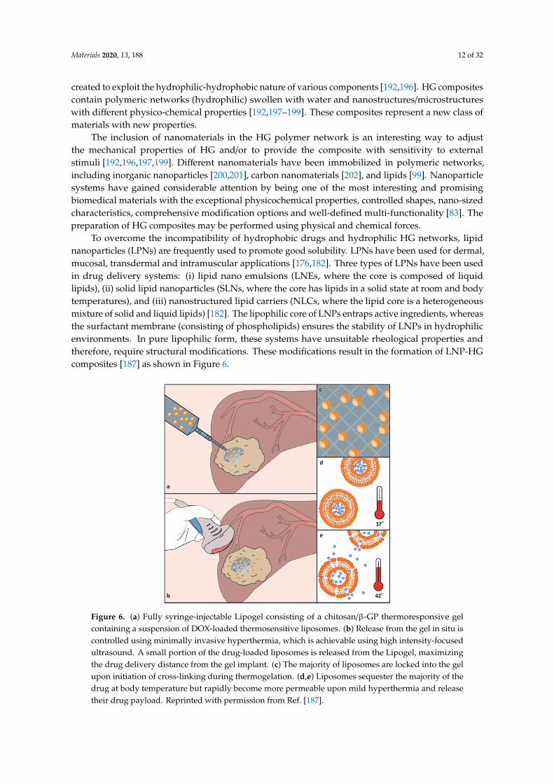
To overcome the incompatibility of hydrophobic drugs and hydrophilic HG networks, lipidnanoparticles (LPNs) are frequently used to promote good solubility. LPNs have been used for dermal,mucosal, transdermal and intramuscular applications [176,182]. Three types of LPNs have been usedin drug delivery systems: (i) lipid nano emulsions (LNEs, where the core is composed of liquidlipids), (ii) solid lipid nanoparticles (SLNs, where the core has lipids in a solid state at room and bodytemperatures), and (iii) nanostructured lipid carriers (NLCs, where the lipid core is a heterogeneousmixture of solid and liquid lipids) [182]. The lipophilic core of LNPs entraps active ingredients, whereasthe surfactant membrane (consisting of phospholipids) ensures the stability of LNPs in hydrophilicenvironments. In pure lipophilic form, these systems have unsuitable rheological properties andtherefore, require structural modifications. These modifications result in the formation of LNP-HGcomposites [187] as shown in Figure 6.
Materials 2019, 12, x FOR PEER REVIEW 13 of 32
An in situ gelation of chitosan/β-glycerophosphate (GP) and thermoresponsive liposomes was performed [187]. This biocompatible and biodegradable HG was used as a matrix for lysolipid thermally sensitive liposomes (LTSL) loaded with doxorubicin (DOX). LTSLs are bi-layered spherical vesicles that rapidly change structure upon mild hyperthermia (41–43 °C), creating openings in the liposome. DOX was loaded into LTSL by changing solution pH. DOX delivery was also based on the pH-sensitivity of liposomes to acidic pH. A schematic of the controlled-delivery of chitosan/β-GP/DOX-loaded LTSLs and DOX release is presented in Figure 6.
Figure 6. (a) Fully syringe-injectable Lipogel consisting of a chitosan/β-GP thermoresponsive gel containing a suspension of DOX-loaded thermosensitive liposomes. (b) Release from the gel in situ is controlled using minimally invasive hyperthermia, which is achievable using high intensity-focused ultrasound. A small portion of the drug-loaded liposomes is released from the Lipogel, maximizing the drug delivery distance from the gel implant. (c) The majority of liposomes are locked into the gel upon initiation of cross-linking during thermogelation. (d,e) Liposomes sequester the majority of the drug at body temperature but rapidly become more permeable upon mild hyperthermia and release their drug payload. Reprinted with permission from Ref. [187].
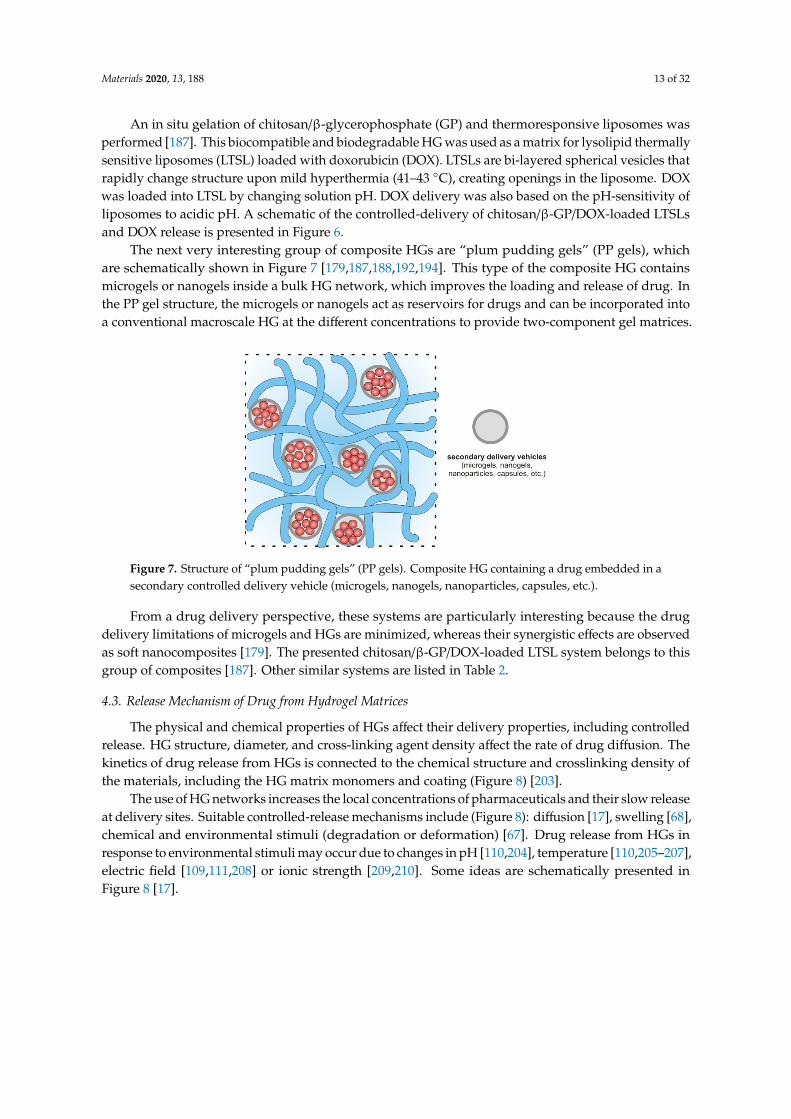
The next very interesting group of composite HGs are “plum pudding gels” (PP gels), which are schematically shown in Figure 7 [179,187,188,192,194]. This type of the composite HG contains microgels or nanogels inside a bulk HG network, which improves the loading and release of drug. In the PP gel structure, the microgels or nanogels act as reservoirs for drugs and can be incorporated into a conventional macroscale HG at the different concentrations to provide two-component gel matrices.
Figure 6. (a) Fully syringe-injectable Lipogel consisting of a chitosan/β-GP thermoresponsive gelcontaining a suspension of DOX-loaded thermosensitive liposomes. (b) Release from the gel in situ iscontrolled using minimally invasive hyperthermia, which is achievable using high intensity-focusedultrasound. A small portion of the drug-loaded liposomes is released from the Lipogel, maximizingthe drug delivery distance from the gel implant. (c) The majority of liposomes are locked into the gelupon initiation of cross-linking during thermogelation. (d,e) Liposomes sequester the majority of thedrug at body temperature but rapidly become more permeable upon mild hyperthermia and releasetheir drug payload. Reprinted with permission from Ref. [187].
Materials 2020, 13, 188 13 of 32
An in situ gelation of chitosan/β-glycerophosphate (GP) and thermoresponsive liposomes wasperformed [187]. This biocompatible and biodegradable HG was used as a matrix for lysolipid thermallysensitive liposomes (LTSL) loaded with doxorubicin (DOX). LTSLs are bi-layered spherical vesicles thatrapidly change structure upon mild hyperthermia (41–43 ◦C), creating openings in the liposome. DOXwas loaded into LTSL by changing solution pH. DOX delivery was also based on the pH-sensitivity ofliposomes to acidic pH. A schematic of the controlled-delivery of chitosan/β-GP/DOX-loaded LTSLsand DOX release is presented in Figure 6.
The next very interesting group of composite HGs are “plum pudding gels” (PP gels), whichare schematically shown in Figure 7 [179,187,188,192,194]. This type of the composite HG containsmicrogels or nanogels inside a bulk HG network, which improves the loading and release of drug. Inthe PP gel structure, the microgels or nanogels act as reservoirs for drugs and can be incorporated intoa conventional macroscale HG at the different concentrations to provide two-component gel matrices.
Materials 2019, 12, x FOR PEER REVIEW 13 of 32
An in situ gelation of chitosan/β-glycerophosphate (GP) and thermoresponsive liposomes was performed [187]. This biocompatible and biodegradable HG was used as a matrix for lysolipid thermally sensitive liposomes (LTSL) loaded with doxorubicin (DOX). LTSLs are bi-layered spherical vesicles that rapidly change structure upon mild hyperthermia (41–43 °C), creating openings in the liposome. DOX was loaded into LTSL by changing solution pH. DOX delivery was also based on the pH-sensitivity of liposomes to acidic pH. A schematic of the controlled-delivery of chitosan/β-GP/DOX-loaded LTSLs and DOX release is presented in Figure 6.
Figure 6. (a) Fully syringe-injectable Lipogel consisting of a chitosan/β-GP thermoresponsive gel containing a suspension of DOX-loaded thermosensitive liposomes. (b) Release from the gel in situ is controlled using minimally invasive hyperthermia, which is achievable using high intensity-focused ultrasound. A small portion of the drug-loaded liposomes is released from the Lipogel, maximizing the drug delivery distance from the gel implant. (c) The majority of liposomes are locked into the gel upon initiation of cross-linking during thermogelation. (d,e) Liposomes sequester the majority of the drug at body temperature but rapidly become more permeable upon mild hyperthermia and release their drug payload. Reprinted with permission from Ref. [187].
The next very interesting group of composite HGs are “plum pudding gels” (PP gels), which are schematically shown in Figure 7 [179,187,188,192,194]. This type of the composite HG contains microgels or nanogels inside a bulk HG network, which improves the loading and release of drug. In the PP gel structure, the microgels or nanogels act as reservoirs for drugs and can be incorporated into a conventional macroscale HG at the different concentrations to provide two-component gel matrices.
Figure 7. Structure of “plum pudding gels” (PP gels). Composite HG containing a drug embedded in asecondary controlled delivery vehicle (microgels, nanogels, nanoparticles, capsules, etc.).
From a drug delivery perspective, these systems are particularly interesting because the drugdelivery limitations of microgels and HGs are minimized, whereas their synergistic effects are observedas soft nanocomposites [179]. The presented chitosan/β-GP/DOX-loaded LTSL system belongs to thisgroup of composites [187]. Other similar systems are listed in Table 2.
4.3. Release Mechanism of Drug from Hydrogel Matrices
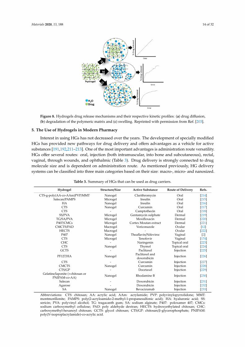
The physical and chemical properties of HGs affect their delivery properties, including controlledrelease. HG structure, diameter, and cross-linking agent density affect the rate of drug diffusion. Thekinetics of drug release from HGs is connected to the chemical structure and crosslinking density ofthe materials, including the HG matrix monomers and coating (Figure 8) [203].
The use of HG networks increases the local concentrations of pharmaceuticals and their slow releaseat delivery sites. Suitable controlled-release mechanisms include (Figure 8): diffusion [17], swelling [68],chemical and environmental stimuli (degradation or deformation) [67]. Drug release from HGs inresponse to environmental stimuli may occur due to changes in pH [110,204], temperature [110,205–207],electric field [109,111,208] or ionic strength [209,210]. Some ideas are schematically presented inFigure 8 [17].
Materials 2020, 13, 188 14 of 32
Materials 2019, 12, x FOR PEER REVIEW 14 of 32
Figure 7. Structure of “plum pudding gels” (PP gels). Composite HG containing a drug embedded in a secondary controlled delivery vehicle (microgels, nanogels, nanoparticles, capsules, etc.).
From a drug delivery perspective, these systems are particularly interesting because the drug delivery limitations of microgels and HGs are minimized, whereas their synergistic effects are observed as soft nanocomposites [179]. The presented chitosan/β-GP/DOX-loaded LTSL system belongs to this group of composites [187]. Other similar systems are listed in Table 2.
4.3. Release Mechanism of Drug from Hydrogel Matrices
The physical and chemical properties of HGs affect their delivery properties, including controlled release. HG structure, diameter, and cross-linking agent density affect the rate of drug diffusion. The kinetics of drug release from HGs is connected to the chemical structure and crosslinking density of the materials, including the HG matrix monomers and coating (Figure 8) [203].
The use of HG networks increases the local concentrations of pharmaceuticals and their slow release at delivery sites. Suitable controlled-release mechanisms include (Figure 8): diffusion [17], swelling [68], chemical and environmental stimuli (degradation or deformation) [67]. Drug release from HGs in response to environmental stimuli may occur due to changes in pH [110,204], temperature [110,205–207], electric field [109,111,208] or ionic strength [209,210]. Some ideas are schematically presented in Figure 8 [17].
Figure 8. Hydrogels drug release mechanisms and their respective kinetic profiles: (a) drug diffusion, (b) degradation of the polymeric matrix and (c) swelling. Reprinted with permission from Ref. [203].
5. The Use of Hydrogels in Modern Pharmacy
Interest in using HGs has not decreased over the years. The development of specially modified HGs has provided new pathways for drug delivery and offers advantages as a vehicle for active substances [191,192,211–213]. One of the most important advantages is administration route versatility. HGs offer several routes: oral, injection (both intramuscular, into bone and subcutaneous), rectal, vaginal, through wounds, and ophthalmic (Table 3). Drug delivery is strongly connected to drug molecule size and is dependent on administration route. As mentioned previously, HG delivery systems can be classified into three main categories based on their size: macro-, micro- and nanosized.
We present some examples of HGs applications in drug delivery. Most informative reports from the last couple of years regarding HGs in health care have been well summarized; however, the
Figure 8. Hydrogels drug release mechanisms and their respective kinetic profiles: (a) drug diffusion,(b) degradation of the polymeric matrix and (c) swelling. Reprinted with permission from Ref. [203].
5. The Use of Hydrogels in Modern Pharmacy
Interest in using HGs has not decreased over the years. The development of specially modifiedHGs has provided new pathways for drug delivery and offers advantages as a vehicle for activesubstances [191,192,211–213]. One of the most important advantages is administration route versatility.HGs offer several routes: oral, injection (both intramuscular, into bone and subcutaneous), rectal,vaginal, through wounds, and ophthalmic (Table 3). Drug delivery is strongly connected to drugmolecule size and is dependent on administration route. As mentioned previously, HG deliverysystems can be classified into three main categories based on their size: macro-, micro- and nanosized.
Table 3. Summary of HGs that can be used as drug carriers.
Hydrogel Structure/Size Active Substance Route of Delivery Refs.
CTS-g-poly(AA-co-AAm)PVP/MMT Nanogel Clarithromycin Oral [214]Salecan/PAMPS Microgel Insulin Oral [215]
HA Nanogel Insulin Oral [216]CTS Nanogel Curcumin Oral [217]CTS - Camptothecin Oral [218]
SS/PVA Microgel Gentamycin sulphate Dermal [219]TG/SA/PVA Microgel Moxifloxacin Dermal [220]P407/CMCs Microgel Cortex Moutan extract Dermal [221]
CMCTS/PAD Macrogel Voriconazole Ocular [12]HECTS Macrogel - Ocular [222]
P407 Nanogel Theaflavin/Nifeviroc Vaginal [2]CTS Microgel Tenofovir Vaginal [174]CHC - Naringenin Topical oral [223]CTS Nanogel Thymol Topical oral [224]
GCTS - Paclitaxel Injection [225]
PF127/HA Nanogel Paclitaxel anddoxorubicin Injection [226]
CTS - Curcumin Injection [227]CMCTS Nanogel Curcumin Injection [228]CTS/GP - Docetaxel Injection [229]
Gelatine/laponite (+chitosan orPNIPAM-co-AA) Nanogel Rhodamine B Injection [230]
Salecan - Doxorubicin Injection [231]Agarose - Doxorubicin Injection [232]
SA Nanogel Bevacizumab Injection [233]
Abbreviations: CTS: chitosan; AA: acrylic acid; AAm: acrylamide; PVP: polyvinylopyrrolidone; MMT:montmorillonite; PAMPS: poly(2-acryloamido-2-methyl-1-propanesulfonic acid); HA: hyaluronic acid; SS:sericin; PVA: polyvinyl alcohol; TG: tragacanth gum; SA: sodium alginate; P407: poloxamer 407; CMCs:sodium carboxymethyl cellulose; PAD: poly aldehyde dextran; HECTS: hydroxyethylated chitosan; CHC:carboxymethyl-hexanoyl chitosan; GCTS: glycol chitosan; CTS/GP: chitosan/β-glycerophosphate; PNIPAM:poly(N-isoproplacrylamide)-co-acrylic acid.
Materials 2020, 13, 188 15 of 32
We present some examples of HGs applications in drug delivery. Most informative reports fromthe last couple of years regarding HGs in health care have been well summarized; however, theliterature on HGs is constantly increasing, as well as interest in HG materials. Herein, we highlightonly a few studies that only begins to hint at the wide applications of HGs (Table 3).
5.1. Application of Hydrogels for Oral Administration
Oral administrative routes are classic and accepted means for delivering drugs. The vast majorityof medicines available on the market are taken through oral routes. Several pills are often taken daily atappropriate intervals to achieve effective therapy. This reduces associated risks of skipping or missingdoses and thereby lowering the effectiveness of the therapy. The development of delayed releasecapsules that prolong the delivery of active substances has been a breakthrough and has allowed forincreased compliance. With these delayed release capsules, the amount of tablets consumed is reduced,usually to only one per day. Acid-sensitive drugs require protection against harmful effects of gastricjuice typically encountered when delivering drugs vial oral routes. Protection can be offered by usingspecial tablets coated with polymers soluble only at basic pH, such as in the intestines. As a result,drugs survive transit through the stomach and are released only in the intestines thereby promotinghigher absorption into the bloodstream.
HGs have also been explored for oral delivery applications. The appropriate selection of gel orthe addition of pH-dependent coatings enable the controlled release of active substances from HGs.
The first modification was presented by Panahi et al. [214] (Table 3). Chitosan (CTS)-basedgels with acrylic acid (AA), acrylamide (AAm) and polyvinylpyrrolidone (PVP) were prepared [214].During HG formation, a mineral (montmorillonite, MMT), which has ability to absorb water, was used.Clarithromycin (CAM), a macrolide antibiotic, was used to eradicate H. Pylori from the gastrointestinaltract was immobilized in the HG network. It was important to achieve a prolonged release of theantibiotic to increase the chances of an effective therapy. MMT increased the pore diameter in the gelstructure, which increased the immobilization of the active substance. However, this also preventedsolvent from readily reaching CAM through more intricate pathways in the network, which complicateddrug release.
The dependence of the release of the drug on the pH level was explored by Qi et al. [215](Table 3). An HG system was prepared based on salecan (beta glucan) with pH-sensitivepoly(2-acrylamido-2-methyl-1-propanesulfonic acid) (PAMPS). The system had the ability to take ordonate protons depending on the pH of the environment, while reducing or increasing the networkvolume. The authors showed that salecan in combination with PAMPS had the ability to release theactive substance depending on the pH of the environment. Using insulin as an exemplary drug, atacidic pH insulin was released at a lower level than at neutral or slightly alkaline pH.
Insulin release from HGs was more extensively studied [216]. Special nanocarriers based onmethacrylic acid were synthesized. In a neutral environment, the methacrylic polymer chains startedto repel each other, thereby loosening the nanocarrier network and allowing the release of insulin. Thisproperty was exploited in preparing HA-doped HG. In the intestines, where the pH is above 7, insulinrelease increased dramatically compared with release in the stomach. By using HGs as a drug carrier,the release of insulin was extended over time.
In another study, a unique biodegradable, super porous, swellable and pH-sensitive nanocellulosereinforced CTS HG was prepared for the oral administration of curcumin [217]. The in vitro degradationof HG was dependent on the swelling ratio and the number of cellulose nanocrystals (CNCs) in the HG.All HGs showed maximum swelling ratios greater than 300%. The drug release occurred in simulatedgastric media; the drug maintained its chemical activity after in vitro release. According to this study,CNC-reinforced CTS HGs can be used to improve the bioavailability of curcumin for absorption fromthe stomach and upper intestinal tract.
Finally, tetrakis(hydroxymethyl)phosphonium chloride was used as a crosslinking agent in aMannich reaction to obtain chitosan-based HGs [218]. These pH-sensitive HGs showed low toxicity, high
Materials 2020, 13, 188 16 of 32
biocompatibility, and allowed for the modified release of encapsulated drugs, namely camptothecin,for 48 h. According to the obtained results, the oral administration of camptothecin through HGsprovided low concentrations of the drug at the absorption site, avoiding carrier saturation and reducingintestinal toxicity [218].
5.2. Hydrogels for Dermal Applications
Due to their fairly compact consistency, HGs can also be applied to wounds or other skin issues.Commercially available HG-based dressings used for exudative wounds, pressure sores or burns,exploit the adhesive properties and ability of HGs to absorb liquids. The addition of antibacterialsubstances may further improve the applications of HG dressings.
Researchers from Southwest University in China developed a sericin (SS) and polyvinylalcohol (PVA)-based HG (Table 3) [219]. This gel showed good biocompatibility, humidity andself-healing properties, i.e., ideal for dressings. Gentamycin sulphate, a known antibiotic substance andaminoglycoside, was added to this HG. To determine the properties of the obtained antibacterial HG, anumber of in vitro tests were carried out, which measured wettability, swelling, microbiological activity,drug substance release, cytotoxicity, and immunotoxicity. The studies showed that the antibacterialHG ensured a prolonged release. For a deeper analysis of the HG properties, a model infected tissuewas prepared. Tests confirmed that the HG was cytocompatible with mammalian cells and did notaffect the growth of healthy cells.
The material used in wound dressing should fulfil many requirements [220], including isolationfrom harmful external factors, such as secondary wound infection, but should also provide sufficientwater vapour permeability and oxygen availability. HGs based on tragacanth gum (TG), sodiumalginate (SA) and PVA meet these properties and have potential applications [220]. In addition to goodpermeability for water vapour and oxygen, these HGs also provide barriers against secondary woundinfection. These properties were confirmed by in vitro studies. Additionally, the HGs had haemolyticand mucoadhesive properties. Dressings based on these HGs were able to release antimicrobial activesubstances (moxifloxacin, an antibiotic from the fluoroquinolone group used to treat a wide spectrumof microorganisms). The results showed prolonged release of up to 24 h without an initial burst release.
Patients struggling with atopic skin changes very often use strong steroid drugs to alleviateemerging inflammation. Unfortunately, topical steroid therapy also leads to skin dryness and irritation,which patients with atopic dermatitis should avoid. Therefore, adequate skin hydration in atopicdermatitis therapy is maintained through the systematic and frequent use of appropriate emollients.Wang et al. combined the administration of an anti-inflammatory substance and maintenance of properskin hydration (Figure 9) [221].
A HG prepared from a mixture of poloxamer 407 (P407) and sodium carboxymethyl cellulose(CMC) was used to achieve this goal (Table 3) [221]. A special nonwoven fabric was covered with the HGmixture. The addition of CMCs resulted in an increase in hydrophilicity of the resulting gel structureand also significantly reduced the sol-gel transition temperature, which advantageously promotedthe fabrication of the coated nonwoven fabric. The HG exhibited moisture retaining properties, andthe nonwoven fabric material prevented excessive water transpiration. The anti-inflammatory effectwas provided by the addition of a Cortex Moutan extract, a well-known plant popular in Chinesemedicine, during HG preparation. To determine the drug release profile, in vitro and ex vivo studieswere performed. The in vitro studies showed that the release of the anti-inflammatory substanceoccurred in a prolonged manner up to several dozen hours, ensuring prolonged and elevated levelsof the anti-inflammatory substance. Ex vivo tests on pork ear confirmed the results of the in vitrotests. The nonwoven fabric coated with a mixture of HGs composed of P407 and CMCs and CortexMoutan extract showed potential for treating and caring for atopic lesions in patients with activeskin inflammation.
Materials 2020, 13, 188 17 of 32
Materials 2019, 12, x FOR PEER REVIEW 17 of 32
properties, a model infected tissue was prepared. Tests confirmed that the HG was cytocompatible with mammalian cells and did not affect the growth of healthy cells.
The material used in wound dressing should fulfil many requirements [220], including isolation from harmful external factors, such as secondary wound infection, but should also provide sufficient water vapour permeability and oxygen availability. HGs based on tragacanth gum (TG), sodium alginate (SA) and PVA meet these properties and have potential applications [220]. In addition to good permeability for water vapour and oxygen, these HGs also provide barriers against secondary wound infection. These properties were confirmed by in vitro studies. Additionally, the HGs had haemolytic and mucoadhesive properties. Dressings based on these HGs were able to release antimicrobial active substances (moxifloxacin, an antibiotic from the fluoroquinolone group used to treat a wide spectrum of microorganisms). The results showed prolonged release of up to 24 h without an initial burst release.
Patients struggling with atopic skin changes very often use strong steroid drugs to alleviate emerging inflammation. Unfortunately, topical steroid therapy also leads to skin dryness and irritation, which patients with atopic dermatitis should avoid. Therefore, adequate skin hydration in atopic dermatitis therapy is maintained through the systematic and frequent use of appropriate emollients. Wang et al. combined the administration of an anti-inflammatory substance and maintenance of proper skin hydration (Figure 9) [221].
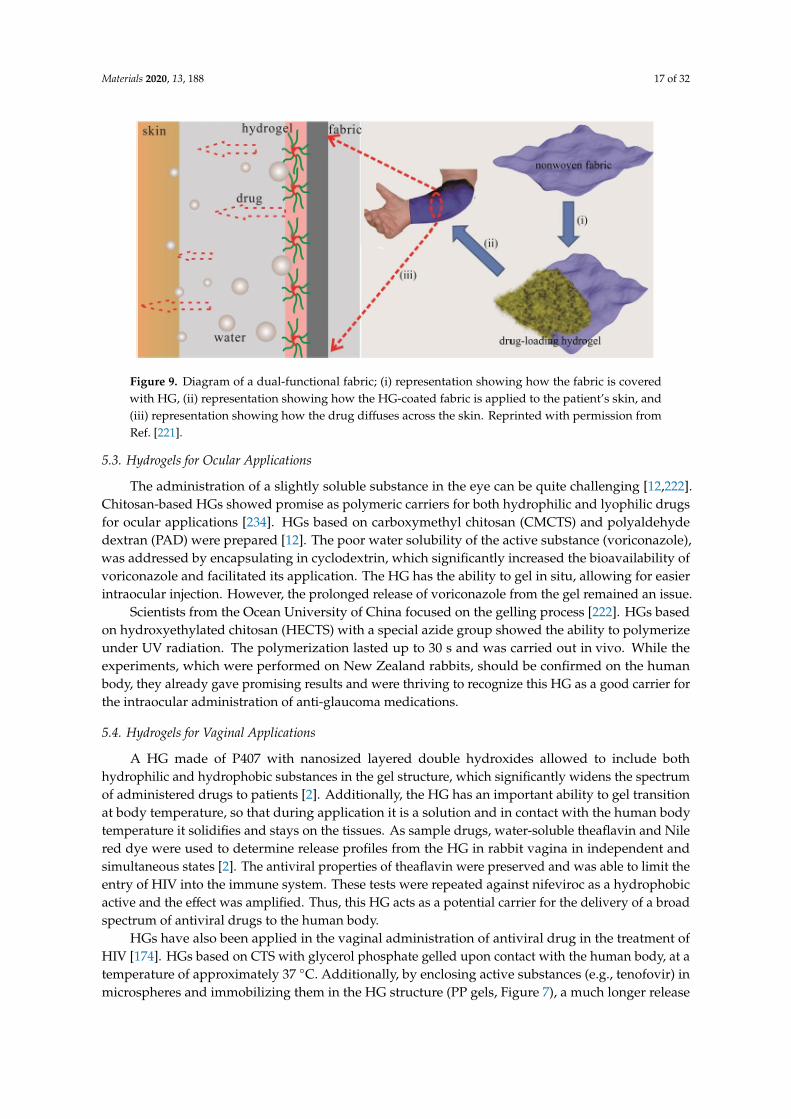
Figure 9. Diagram of a dual-functional fabric; (i) representation showing how the fabric is covered with HG, (ii) representation showing how the HG-coated fabric is applied to the patient’s skin, and (iii) representation showing how the drug diffuses across the skin. Reprinted with permission from Ref. [221].
A HG prepared from a mixture of poloxamer 407 (P407) and sodium carboxymethyl cellulose (CMC) was used to achieve this goal (Table 3) [221]. A special nonwoven fabric was covered with the HG mixture. The addition of CMCs resulted in an increase in hydrophilicity of the resulting gel structure and also significantly reduced the sol-gel transition temperature, which advantageously promoted the fabrication of the coated nonwoven fabric. The HG exhibited moisture retaining properties, and the nonwoven fabric material prevented excessive water transpiration. The anti-inflammatory effect was provided by the addition of a Cortex Moutan extract, a well-known plant popular in Chinese medicine, during HG preparation. To determine the drug release profile, in vitro and ex vivo studies were performed. The in vitro studies showed that the release of the anti-inflammatory substance occurred in a prolonged manner up to several dozen hours, ensuring prolonged and elevated levels of the anti-inflammatory substance. Ex vivo tests on pork ear confirmed the results of the in vitro tests. The nonwoven fabric coated with a mixture of HGs composed of P407 and CMCs and Cortex Moutan extract showed potential for treating and caring for atopic lesions in patients with active skin inflammation.
Figure 9. Diagram of a dual-functional fabric; (i) representation showing how the fabric is coveredwith HG, (ii) representation showing how the HG-coated fabric is applied to the patient’s skin, and(iii) representation showing how the drug diffuses across the skin. Reprinted with permission fromRef. [221].
5.3. Hydrogels for Ocular Applications
The administration of a slightly soluble substance in the eye can be quite challenging [12,222].Chitosan-based HGs showed promise as polymeric carriers for both hydrophilic and lyophilic drugsfor ocular applications [234]. HGs based on carboxymethyl chitosan (CMCTS) and polyaldehydedextran (PAD) were prepared [12]. The poor water solubility of the active substance (voriconazole),was addressed by encapsulating in cyclodextrin, which significantly increased the bioavailability ofvoriconazole and facilitated its application. The HG has the ability to gel in situ, allowing for easierintraocular injection. However, the prolonged release of voriconazole from the gel remained an issue.
Scientists from the Ocean University of China focused on the gelling process [222]. HGs basedon hydroxyethylated chitosan (HECTS) with a special azide group showed the ability to polymerizeunder UV radiation. The polymerization lasted up to 30 s and was carried out in vivo. While theexperiments, which were performed on New Zealand rabbits, should be confirmed on the humanbody, they already gave promising results and were thriving to recognize this HG as a good carrier forthe intraocular administration of anti-glaucoma medications.
5.4. Hydrogels for Vaginal Applications
A HG made of P407 with nanosized layered double hydroxides allowed to include bothhydrophilic and hydrophobic substances in the gel structure, which significantly widens the spectrumof administered drugs to patients [2]. Additionally, the HG has an important ability to gel transitionat body temperature, so that during application it is a solution and in contact with the human bodytemperature it solidifies and stays on the tissues. As sample drugs, water-soluble theaflavin and Nilered dye were used to determine release profiles from the HG in rabbit vagina in independent andsimultaneous states [2]. The antiviral properties of theaflavin were preserved and was able to limit theentry of HIV into the immune system. These tests were repeated against nifeviroc as a hydrophobicactive and the effect was amplified. Thus, this HG acts as a potential carrier for the delivery of a broadspectrum of antiviral drugs to the human body.
HGs have also been applied in the vaginal administration of antiviral drug in the treatment ofHIV [174]. HGs based on CTS with glycerol phosphate gelled upon contact with the human body, at atemperature of approximately 37 ◦C. Additionally, by enclosing active substances (e.g., tenofovir) inmicrospheres and immobilizing them in the HG structure (PP gels, Figure 7), a much longer release
Materials 2020, 13, 188 18 of 32
profile was obtained when compared with the release profile from the microspheres or HG alone. Thisprovides an opportunity to improve on the effectiveness of immunotherapy.
5.5. Hydrogels for Topical Oral Applications
In the use of HG structures in the treatment of inflammatory periodontal bent, Chang et al.prepared a gel based on carboxymethyl-hexanoyl chitosan (CHC) [223]. Through the addition ofglycerol phosphate into the HG network, the HG was modified to solidify at human body temperatures,which provided significant advantages over the use of other substances in oral inflammation therapy.Anti-inflammatory properties were carried out using naringenin. Experiments on rats showed that theanti-inflammatory substance was more rapidly released from the HG when the pH of the environmentwas acidic (~5.5), thereby rapidly treating the periodontal inflammation.
CTS HG can also be used to treat oral inflammation when used with thymol [224]. In vitro studiesshowed that the resulting structure allowed for the prolonged release and high antimicrobial activityof thymol against microorganisms up to 72 h. Furthermore, in the first two days, the active substancewas increasingly released. This dual action HG demonstrated anti-inflammatory properties and thetreatment of bacterial biofilms in the oral cavity.
5.6. Injection of Hydrogels
Therapeutic injections are usually least favoured by the patient and requires specialized HGs. Thekey challenge is to prepare a structure that will be fluid enough to squeeze through the needle butalso sufficiently rigid that when applied does not spread and remains at the injection site [17,135,136].Therefore, an important property of these structures is shear thinning, i.e., the ability to decreaseviscosity of fluid under shear strain.
CTS-based in situ HGs are frequently adopted in the treatment for age-related macular degeneration(AMD), glaucoma and mucosal allergic diseases [234]. The HGs offer a convenient matrix for in situgelling systems containing other nanoparticles, such as micelles. Injectable CTS-based in situ gelshave been applied as in situ forming implants and as drug carriers in nasal and ocular delivery dueto good biocompatibility, simple manufacture and sustained-release properties. These systems havebeen utilized to deliver several chemotherapeutic agents, including camptothecin [218,235], paclitaxel(PTX) [225,226], DOX [226], curcumin [217,227,228], and docetaxel (DTX) [229,236].
An injectable drug delivery system based on a visible light-cured glycol chitosan (GCS) HGcontaining PTX-complexed beta-cyclodextrin (β-CD) (GCS/CD/PTX) was tested for ovarian cancertherapy using a tumour-bearing mouse model [225]. The swollen GCS HG affected the release of PTXand CD/PTX. These systems exhibited a controlled release of PTX for 7 days. Additionally, GC/CD/PTXresulted in a superior antitumor effect against ovarian cancer.
An interesting application of HGs as dual carriers for PTX and DOX was proposed by Rezazadehet al. [226]. An injectable thermosensitive HG for simultaneous intra-tumoral administration ofPTX and DOX was prepared using mixed micelles composed of Pluronic F127 and α-tocopherylpolyethylene glycol 1000 succinate (TPGS) and a thermosensitive Pluronic F127/hyaluronic acid(PF127/HA) HG containing fixed amount of DOX [226]. PP gel formation temperature, rheologicalproperties, injectability, degradation rates of the HG, and the release rate of DOX and PTX from theHG were examined. The HG containing PTX-loaded micelles and DOX converted to a semisolid withincreasing temperature to 35 ◦C. DOX and PTX were released from the HG at different rates within 12h and 3 days, respectively. This novel thermosensitive HG could be used for the co-delivery of PTXand DOX in solid tumours.
A series of injectable in situ-forming chitosan-based HGs were prepared by the chemicalcross-linking of CTS and genipin in the presence of different inorganic salts [227]. In situ HGformation, with curcumin as an active substance, was detected after subcutaneous injections in rats.In vitro curcumin release profiles exhibited sustained release properties with an initial burst releasewith approximately three to six times higher cumulative release than other gel controls.
Materials 2020, 13, 188 19 of 32
CTS-based HGs may also show magnetic properties, when magnetic Fe3O4 and MnFe2O4
nanoparticles are incorporated into their networks [228]. The Fe3O4 and MnFe2O4 on CMCTS HGsindicated a pH-sensitive behaviour; their release performances were investigated by curcumin as amodel drug. The effects of applied magnetic fields on drug release for composites containing Fe3O4
were higher compared with MnFe2O4. The results showed that HGs containing Fe3O4 and MnFe2O4
can be applied for novel drug delivery systems.DTX is an anticancer drug used for treatment of various solid tumours [229]. But it’s low water
solubility and a lack of specification has limited its clinical use. Therefore, new solutions havebeen explored to use this drug in targeted therapy. A thermosensitive chitosan/β-glycerophosphate(CTS/GP) DTX-loaded HG for intratumoral delivery was studied. The results of an in vitro releasestudy demonstrated that DTX-C/GP was released over a period of 3 weeks. The tumour volume wasminimized by the intratumoral injection of DTX-CTS/GP (at 20 mg/kg in H22 tumour-bearing mice).Further, the in vivo pharmacokinetic characteristics of DTX-CTS/GP correlated well with the in vitrorelease. DTX-CTS/GP significantly prolonged DTX retention, supported a high DTX concentration intumours, and toxicity was effectively reduced.
Carbon nanostructures were also successfully applied in hydrophilic, porous materials as acomponent of HGs for DTX carriers [236]. A functionalized graphene oxide (GO)-based thermosensitiveHG loaded with DTX for intratumoral delivery was designed. First, GO was functionalized by usingchitosan to achieve high stability in physiological solutions. Next, HGs containing few componentswere formed: GO-chitosan, Poloxamer 407 and Poloxamer 188 and DTX. GO/chitosan HG was stablein physiological solution; DTX released much more slowly from this gel compared with free DTX witha pH-responsive feature. The DTX–GO/chitosan gel released higher concentrations and longer residenttimes in the tumour tissues of mice in vivo giving nontoxic effects to normal organs. Additionally,the combination of near-infrared laser irradiation at 808 nm significantly enhanced tumour inhibitionin vitro and in vivo.
Other composite-based HGs were also evaluated. Gharaie et al. used gelatin and laponite invarious proportions to obtain appropriate shear thinning properties (Figure 10) [230]. Additionally, byloading with CTS or poly(N-isopropylacrylamide)-co-acrylic acid (PNIPAM-co-AA) to the gel structure,pH-responsive abilities were gained, resulting in a different release profiles. As a model substance,rhodamine B was used to test the release. The results of these experiments showed that the gel couldbe easily and accessibly applied, whereas the release of the active substance was dependent on thepH level (in acidic and neutral environments, the release was negligible, whereas release increased atbasic pH.
The key point of interest of many scientists is the fight against cancer. Also in this case HG canalso be used to treat cancer [36]. Hu et al. combined salecan, a beta-glucan polysaccharide, with Fe3O4
nanoparticles and agarose [231]. The resulting HG gained magnetic properties and was used as acarrier for DOX (an anthracycline cytostatic used in anti-cancer therapy). The salecan-based HG withiron oxide nanoparticles had a pH-dependent drug release profile. In an acidic environment, unlikealkaline environments, a larger amount of DOX was released. Due to the acidic pH of cancer cells,this result indicated significant promise. Additionally, due to the presence of Fe3O4 nanoparticles,the amount of active drug released was also manipulated. With an external electromagnetic field,inorganic particles can be vibrated, thereby loosening the gel structure enabling easier release of DOX.
The technical aspects of DOX release from HGs were examined by a group from DrexelUniversity [232]. In this study, an agarose-based HG was used wherein DOX-dextran sulphatecomplexes were provided. The structure was doped with divalent metallic ions, Ca2+ from CaCl2 andMg2+ from MgCl2, at different concentrations. The divalent ions interfered with the binding betweenDOX and dextran sulphate, promoting prolonged release of the active substance from the HG andthereby providing a sustained chemotherapeutic effect for destroying cancer cells. Alginate (sodiumalginate, SA)-based HGs with immobilized bevacizumab were also examined [233]. Anti-angiogenic
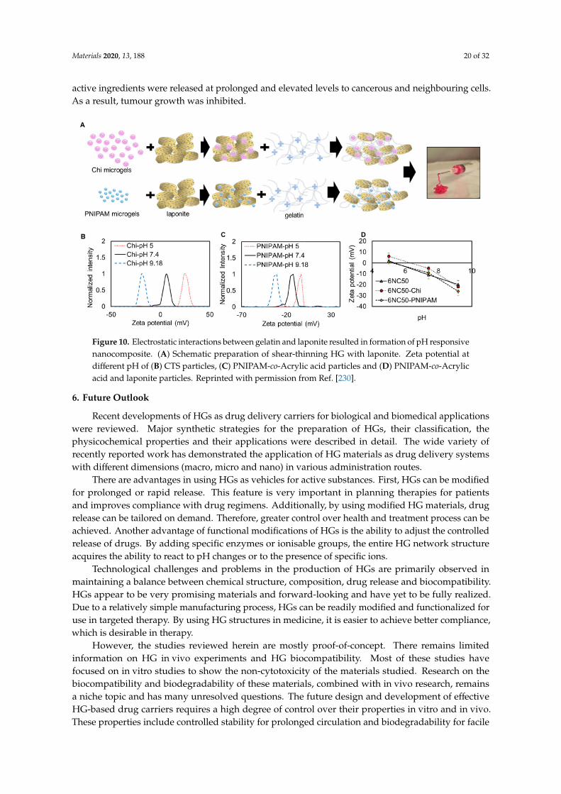
Materials 2020, 13, 188 20 of 32
active ingredients were released at prolonged and elevated levels to cancerous and neighbouring cells.As a result, tumour growth was inhibited.
Materials 2019, 12, x FOR PEER REVIEW 20 of 32
functionalized by using chitosan to achieve high stability in physiological solutions. Next, HGs containing few components were formed: GO-chitosan, Poloxamer 407 and Poloxamer 188 and DTX. GO/chitosan HG was stable in physiological solution; DTX released much more slowly from this gel compared with free DTX with a pH-responsive feature. The DTX–GO/chitosan gel released higher concentrations and longer resident times in the tumour tissues of mice in vivo giving nontoxic effects to normal organs. Additionally, the combination of near-infrared laser irradiation at 808 nm significantly enhanced tumour inhibition in vitro and in vivo.
Other composite-based HGs were also evaluated. Gharaie et al. used gelatin and laponite in various proportions to obtain appropriate shear thinning properties (Figure 10) [230]. Additionally, by loading with CTS or poly(N-isopropylacrylamide)-co-acrylic acid (PNIPAM-co-AA) to the gel structure, pH-responsive abilities were gained, resulting in a different release profiles. As a model substance, rhodamine B was used to test the release. The results of these experiments showed that the gel could be easily and accessibly applied, whereas the release of the active substance was dependent on the pH level (in acidic and neutral environments, the release was negligible, whereas release increased at basic pH.
Figure 10. Electrostatic interactions between gelatin and laponite resulted in formation of pH responsive nanocomposite. (A) Schematic preparation of shear-thinning HG with laponite. Zeta potential at different pH of (B) CTS particles, (C) PNIPAM-co-Acrylic acid particles and (D) PNIPAM-co-Acrylic acid and laponite particles. Reprinted with permission from Ref. [230].
The key point of interest of many scientists is the fight against cancer. Also in this case HG can also be used to treat cancer [36]. Hu et al. combined salecan, a beta-glucan polysaccharide, with Fe3O4 nanoparticles and agarose [231]. The resulting HG gained magnetic properties and was used as a carrier for DOX (an anthracycline cytostatic used in anti-cancer therapy). The salecan-based HG with iron oxide nanoparticles had a pH-dependent drug release profile. In an acidic environment, unlike alkaline environments, a larger amount of DOX was released. Due to the acidic pH of cancer cells, this result indicated significant promise. Additionally, due to the presence of Fe3O4 nanoparticles, the amount of active drug released was also manipulated. With an external electromagnetic field, inorganic particles can be vibrated, thereby loosening the gel structure enabling easier release of DOX.
The technical aspects of DOX release from HGs were examined by a group from Drexel University [232]. In this study, an agarose-based HG was used wherein DOX-dextran sulphate complexes were provided. The structure was doped with divalent metallic ions, Ca2+ from CaCl2 and Mg2+ from MgCl2, at different concentrations. The divalent ions interfered with the binding between DOX and dextran sulphate, promoting prolonged release of the active substance from the HG and
Figure 10. Electrostatic interactions between gelatin and laponite resulted in formation of pH responsivenanocomposite. (A) Schematic preparation of shear-thinning HG with laponite. Zeta potential atdifferent pH of (B) CTS particles, (C) PNIPAM-co-Acrylic acid particles and (D) PNIPAM-co-Acrylicacid and laponite particles. Reprinted with permission from Ref. [230].
6. Future Outlook
Recent developments of HGs as drug delivery carriers for biological and biomedical applicationswere reviewed. Major synthetic strategies for the preparation of HGs, their classification, thephysicochemical properties and their applications were described in detail. The wide variety ofrecently reported work has demonstrated the application of HG materials as drug delivery systemswith different dimensions (macro, micro and nano) in various administration routes.
There are advantages in using HGs as vehicles for active substances. First, HGs can be modifiedfor prolonged or rapid release. This feature is very important in planning therapies for patientsand improves compliance with drug regimens. Additionally, by using modified HG materials, drugrelease can be tailored on demand. Therefore, greater control over health and treatment process can beachieved. Another advantage of functional modifications of HGs is the ability to adjust the controlledrelease of drugs. By adding specific enzymes or ionisable groups, the entire HG network structureacquires the ability to react to pH changes or to the presence of specific ions.
Technological challenges and problems in the production of HGs are primarily observed inmaintaining a balance between chemical structure, composition, drug release and biocompatibility.HGs appear to be very promising materials and forward-looking and have yet to be fully realized.Due to a relatively simple manufacturing process, HGs can be readily modified and functionalized foruse in targeted therapy. By using HG structures in medicine, it is easier to achieve better compliance,which is desirable in therapy.
However, the studies reviewed herein are mostly proof-of-concept. There remains limitedinformation on HG in vivo experiments and HG biocompatibility. Most of these studies havefocused on in vitro studies to show the non-cytotoxicity of the materials studied. Research on thebiocompatibility and biodegradability of these materials, combined with in vivo research, remainsa niche topic and has many unresolved questions. The future design and development of effectiveHG-based drug carriers requires a high degree of control over their properties in vitro and in vivo.These properties include controlled stability for prolonged circulation and biodegradability for facile
Materials 2020, 13, 188 21 of 32
removal after drug release. One future goal should be the improved design of HG with specific targetingresidues to enable highly selective uptake in particular cells or organs. In this way, careful control oversize, biodegradability, stability, functionality, and bioconjugation will guide the development of nextgeneration HGs.
Author Contributions: Conceptualization, M.E.P.-B.; writing—original draft preparation, M.E.P.-B.; M.T.; A.C.;writing—review and editing, M.E.P.-B., M.T.; visualization, supervision and project administration, M.E.P.-B.;funding acquisition, M.E.P.-B. All authors have read and agreed to the published version of the manuscript.
Funding: This research was funded by the National Science Centre, Poland, grant number 2017/25/B/ST5/01414 toM.E.P-B.
Conflicts of Interest: The authors declare no conflict of interest.
References
1. Korde, J.M.; Kandasubramanian, B. Naturally biomimicked smart shape memory hydrogels for biomedicalfunctions. Chem. Eng. J. 2020, 379, 122430. [CrossRef]
2. Tian, W.; Han, S.; Huang, X.; Han, M.; Cao, J.; Liang, Y.; Sun, Y. LDH hybrid thermosensitive hydrogel forintravaginal delivery of anti-HIV drugs. Artif. Cells Nanomed. Biotechnol. 2019, 47, 1234–1240. [CrossRef][PubMed]
3. Tibbitt, M.W.; Dahlman, J.E.; Langer, R. Emerging frontiers in drug delivery. J. Am. Chem. Soc. 2016, 138,704–717. [CrossRef] [PubMed]
4. Fujiyabu, T.; Yoshikawa, Y.; Chung, U.; Sakai, T. Structure-property relationship of a model networkcontaining solvent. Sci. Technol. Adv. Mater. 2019, 20, 608–621. [CrossRef] [PubMed]
5. Kirschner, C.M.; Anseth, K.S. Hydrogels in healthcare: From static to dynamic material microenvironments.Acta Mater. 2013, 61, 931–944. [CrossRef] [PubMed]
6. Chatterjee, S.; Chi-leung Hui, P. Stimuli-responsive hydrogels: An interdisciplinary overview. InHydrogels—Smart Materials for Biomedical Applications; Popa, L., Violeta Ghica, M., Dinu-Pîrvu, C.-E., Eds.;IntechOpen: London, UK, 2019; ISBN 978-1-78985-875-4.
7. Shafiee, S.; Ahangar, H.A.; Saffar, A. Taguchi method optimization for synthesis of Fe3O4
@chitosan/Tragacanth Gum nanocomposite as a drug delivery system. Carbohydr. Polym. 2019, 222,114982. [CrossRef] [PubMed]
8. Ullah, K.; Sohail, M.; Buabeid, M.A.; Murtaza, G.; Ullah, A.; Rashid, H.; Khan, M.A.; Khan, S.A. Pectin-based(LA-co-MAA) semi-IPNS as a potential biomaterial for colonic delivery of oxaliplatin. Int. J. Pharm. 2019,569, 118557. [CrossRef]
9. Nazlı, A.B.; Açıkel, Y.S. Loading of cancer drug resveratrol to pH-Sensitive, smart, alginate-chitosan hydrogelsand investigation of controlled release kinetics. J. Drug Deliv. Sci. Technol. 2019, 53, 101199. [CrossRef]
10. Kim, C.; Jeong, D.; Kim, S.; Kim, Y.; Jung, S. Cyclodextrin functionalized agarose gel with low gellingtemperature for controlled drug delivery systems. Carbohydr. Polym. 2019, 222, 115011. [CrossRef]
11. Cocarta, A.-I.; Hobzova, R.; Sirc, J.; Cerna, T.; Hrabeta, J.; Svojgr, K.; Pochop, P.; Kodetova, M.; Jedelska, J.;Bakowsky, U.; et al. Hydrogel implants for transscleral drug delivery for retinoblastoma treatment. Mater.Sci. Eng. C 2019, 103, 109799. [CrossRef]
12. Yang, C.; Gao, L.; Liu, X.; Yang, T.; Yin, G.; Chen, J.; Guo, H.; Yu, B.; Cong, H. Injectable Schiff basepolysaccharide hydrogels for intraocular drug loading and release. J. Biomed. Mater. Res. 2019, 107.[CrossRef] [PubMed]
13. Alvarez-Rivera, F.; Serro, A.P.; Silva, D.; Concheiro, A.; Alvarez-Lorenzo, C. Hydrogels for diabetic eyes:Naltrexone loading, release profiles and cornea penetration. Mater. Sci. Eng. C 2019, 105, 110092. [CrossRef][PubMed]
14. Dragan, E.S.; Dinu, M.V. Polysaccharides constructed hydrogels as vehicles for proteins and peptides. Areview. Carbohydr. Polym. 2019, 225, 115210. [CrossRef] [PubMed]
15. Duwa, R.; Emami, F.; Lee, S.; Jeong, J.-H.; Yook, S. Polymeric and lipid-based drug delivery systems fortreatment of glioblastoma multiforme. J. Ind. Eng. Chem. 2019, 79, 261–273. [CrossRef]
16. Riley, L.; Schirmer, L.; Segura, T. Granular hydrogels: Emergent properties of jammed hydrogel microparticlesand their applications in tissue repair and regeneration. Curr. Opin. Biotechnol. 2019, 60, 1–8. [CrossRef]
Materials 2020, 13, 188 22 of 32
17. Li, J.; Mooney, D.J. Designing hydrogels for controlled drug delivery. Nat. Rev. Mater. 2016, 1, 10671.[CrossRef]
18. Bakaic, E.; Smeets, N.M.B.; Hoare, T. Injectable hydrogels based on poly (ethylene glycol) and derivatives asfunctional biomaterials. RSC Adv. 2015, 5, 35469–35486. [CrossRef]
19. Norouzi, M.; Nazari, B.; Miller, D.W. Injectable hydrogel-based drug delivery systems for local cancer therapy.Drug Dis. Today 2016, 21, 1835–1849. [CrossRef]
20. Nguyen, Q.V.; Huynh, D.P.; Park, J.H.; Lee, D.S. Injectable polymeric hydrogels for the delivery of therapeuticagents: A review. Eur. Polym. J. 2015, 72, 602–619. [CrossRef]
21. Soni, K.S.; Desale, S.S.; Bronich, T.K. Nanogels: An overview of properties, biomedical applications andobstacles to clinical translation. J. Control. Release 2016, 240, 109–126. [CrossRef]
22. Nita, L.E.; Chiriac, A.P.; Rusu, A.G.; Bercea, M.; Ghilan, A.; Dumitriu, R.P.; Mititelu-Tartau, L. New self-healinghydrogels based on reversible physical interactions and their potential applications. Eur. Polym. J. 2019, 118,176–185. [CrossRef]
23. Wang, J.; Liu, L.; Chen, J.; Deng, M.; Feng, X.; Chen, L. Supramolecular nanoplatforms via cyclodextrinhost-guest recognition for synergistic gene-photodynamic therapy. Eur. Polym. J. 2019, 118, 222–230.[CrossRef]
24. Marafon, P.; Fachel, F.N.S.; Dal Prá, M.; Bassani, V.L.; Koester, L.S.; Henriques, A.T.; Braganhol, E.; Teixeira, H.F.Development, physico-chemical characterization and in-vitro studies of hydrogels containing rosmarinicacid-loaded nanoemulsion for topical application. J. Pharm. Pharmacol. 2019, 71, 1199–1208. [CrossRef][PubMed]
25. Miranda, M.; Cruz, M.T.; Vitorino, C.; Cabral, C. Nanostructuring lipid carriers using Ridolfia segetum (L.)Moris essential oil. Mater. Sci. Eng. C 2019, 103, 109804. [CrossRef] [PubMed]
26. Sharaf, S.; El-Naggar, M.E. Wound dressing properties of cationized cotton fabric treated withcarrageenan/cyclodextrin hydrogel loaded with honey bee propolis extract. Int. J. Biol. Macromol. 2019, 133,583–591. [CrossRef]
27. Liu, Z.; Zheng, Y.; Dang, J.; Zhang, J.; Dong, F.; Wang, K.; Zhang, J. A novel antifungal plasma-activatedhydrogel. ACS Appl. Mater. Interfaces 2019, 11, 22941–22949. [CrossRef]
28. Wang, K.; Wang, J.; Li, L.; Xu, L.; Feng, N.; Wang, Y.; Fei, X.; Tian, J.; Li, Y. Synthesis of a novel anti-freezing,non-drying antibacterial hydrogel dressing by one-pot method. Chem. Eng. J. 2019, 372, 216–225. [CrossRef]
29. Chen, W.; Zhu, Y.; Zhang, Z.; Gao, Y.; Liu, W.; Borjihan, Q.; Qu, H.; Zhang, Y.; Zhang, Y.; Wang, Y.-J.; et al.Engineering a multifunctional N-halamine-based antibacterial hydrogel using a super-convenient strategyfor infected skin defect therapy. Chem. Eng. J. 2020, 379, 122238. [CrossRef]
30. Xue, H.; Hu, L.; Xiong, Y.; Zhu, X.; Wei, C.; Cao, F.; Zhou, W.; Sun, Y.; Endo, Y.; Liu, M.; et al. Quaternizedchitosan-Matrigel-polyacrylamide hydrogels as wound dressing for wound repair and regeneration.Carbohydr. Polym. 2019, 226, 115302. [CrossRef]
31. Huang, L.; Zhu, Z.; Wu, D.; Gan, W.; Zhu, S.; Li, W.; Tian, J.; Li, L.; Zhou, C.; Lu, L. Antibacterial poly (ethyleneglycol) diacrylate/chitosan hydrogels enhance mechanical adhesiveness and promote skin regeneration.Carbohydr. Polym. 2019, 225, 115110. [CrossRef]
32. Liang, Y.; Zhao, X.; Hu, T.; Han, Y.; Guo, B. Mussel-inspired, antibacterial, conductive, antioxidant, injectablecomposite hydrogel wound dressing to promote the regeneration of infected skin. J. Coll. Interface Sci. 2019,556, 514–528. [CrossRef] [PubMed]
33. Solomevich, S.O.; Bychkovsky, P.M.; Yurkshtovich, T.L.; Golub, N.V.; Mirchuk, P.Y.; Revtovich, M.Y.;Shmak, A.I. Biodegradable pH-sensitive prospidine-loaded dextran phosphate based hydrogels for localtumor therapy. Carbohydr. Polym. 2019, 226, 115308. [CrossRef] [PubMed]
34. Chen, H.; Wei, X.; Chen, H.; Wei, H.; Wang, Y.; Nan, W.; Zhang, Q.; Wen, X. The study of establishment ofan in vivo tumor model by three-dimensional cells culture systems methods and evaluation of antitumoreffect of biotin-conjugated pullulan acetate nanoparticles. Artif. Cells Nanomed. Biotechnol. 2019, 47, 123–131.[CrossRef] [PubMed]
35. Cao, D.; Zhang, X.; Akabar, M.D.; Luo, Y.; Wu, H.; Ke, X.; Ci, T. Liposomal doxorubicin loadedPLGA-PEG-PLGA based thermogel for sustained local drug delivery for the treatment of breast cancer. Artif.Cells Nanomed. Biotechnol. 2019, 47, 181–191. [CrossRef]
Materials 2020, 13, 188 23 of 32
36. Wu, Q.; He, Z.; Wang, X.; Zhang, Q.; Wei, Q.; Ma, S.; Ma, C.; Li, J.; Wang, Q. Cascade enzymes withinself-assembled hybrid nanogel mimicked neutrophil lysosomes for singlet oxygen elevated cancer therapy.Nat. Commun. 2019, 10, 240. [CrossRef]
37. Puertas-Bartolomé, M.; Benito-Garzón, L.; Fung, S.; Kohn, J.; Vázquez-Lasa, B.; San Román, J. Bioadhesivefunctional hydrogels: Controlled release of catechol species with antioxidant and antiinflammatory behavior.Mater. Sci. Eng. C 2019, 105, 110040. [CrossRef]
38. Zheng, J.; Fan, R.; Wu, H.; Yao, H.; Yan, Y.; Liu, J.; Ran, L.; Sun, Z.; Yi, L.; Dang, L.; et al. Directed self-assemblyof herbal small molecules into sustained release hydrogels for treating neural inflammation. Nat. Commun.2019, 10, 1604. [CrossRef]
39. Uehara, M.; Li, X.; Sheikhi, A.; Zandi, N.; Walker, B.; Saleh, B.; Banouni, N.; Jiang, L.; Ordikhani, F.; Dai, L.;et al. Anti-IL-6 eluting immunomodulatory biomaterials prolong skin allograft survival. Sci. Rep. 2019, 9,6535. [CrossRef]
40. García, J.R.; Quirós, M.; Han, W.M.; O’Leary, M.N.; Cox, G.N.; Nusrat, A.; García, A.J. IFN-γ-tetheredhydrogels enhance mesenchymal stem cell-based immunomodulation and promote tissue repair. Biomaterials2019, 220, 119403. [CrossRef]
41. Hertegård, S.; Nagubothu, S.R.; Malmström, E.; Ström, C.E.; Tolf, A.; Davies, L.C.; Le Blanc, K. Hyaluronanhydrogels for the local delivery of mesenchymal stromal cells to the injured vocal fold. Stem Cells Dev. 2019,28, 1177–1190. [CrossRef]
42. Grigoras, A.G. Drug delivery systems using pullulan, a biocompatible polysaccharide produced by fungalfermentation of starch. Environ. Chem. Lett. 2019, 17, 1209–1223. [CrossRef]
43. Ge, W.; Cao, S.; Shen, F.; Wang, Y.; Ren, J.; Wang, X. Rapid self-healing, stretchable, moldable, antioxidantand antibacterial tannic acid-cellulose nanofibril composite hydrogels. Carbohydr. Polym. 2019, 224, 115147.[CrossRef] [PubMed]
44. Clegg, J.R.; Wagner, A.M.; Shin, S.R.; Hassan, S.; Khademhosseini, A.; Peppas, N.A. Modular fabrication ofintelligent material-tissue interfaces for bioinspired and biomimetic devices. Prog. Mater. Sci. 2019, 106,100589. [CrossRef]
45. Bezerra, U.T.; Ferreira, H.S.; Barbosa, N.P. Hydrogels: Types, structure, properties, and applications. InFrontiers in Biomaterials; Razavi, M., Ed.; Bentham Science Publishers Ltd.: Sharjah, UAE, 2017; Volume 4,pp. 143–169. ISBN 978-1-68108-536-4.
46. Chatterjee, S.; Hui, P.; Kan, C. Thermoresponsive hydrogels and their biomedical applications: Special insightinto their applications in textile based transdermal therapy. Polymers 2018, 10, 480. [CrossRef]
47. Akhtar, M.F.; Hanif, M.; Ranjha, N.M. Methods of synthesis of hydrogels . . . A review. Saudi Pharm. J. 2016,24, 554–559. [CrossRef]
48. Buwalda, S.J.; Boere, K.W.M.; Dijkstra, P.J.; Feijen, J.; Vermonden, T.; Hennink, W.E. Hydrogels in a historicalperspective: From simple networks to smart materials. J. Control. Release 2014, 190, 254–273. [CrossRef]
49. Zhang, Y.S.; Khademhosseini, A. Advances in engineering hydrogels. Science 2017, 356, eaaf3627. [CrossRef]50. Chai, Q.; Jiao, Y.; Yu, X. Hydrogels for biomedical applications: Their characteristics and the mechanisms
behind them. Gels 2017, 3, 6. [CrossRef]51. Ahmed, E.M. Hydrogel: Preparation, characterization, and applications: A review. J. Adv. Res. 2015, 6,
105–121. [CrossRef]52. Straccia, M.; d’Ayala, G.; Romano, I.; Oliva, A.; Laurienzo, P. Alginate hydrogels coated with chitosan for
wound dressing. Mar. Drugs 2015, 13, 2890–2908. [CrossRef]53. Yuan, L.; Wu, Y.; Fang, J.; Wei, X.; Gu, Q.; El-Hamshary, H.; Al-Deyab, S.S.; Morsi, Y.; Mo, X. Modified
alginate and gelatin cross-linked hydrogels for soft tissue adhesive. Artif. Cells Nanomed. Biotechnol. 2017, 45,76–83. [CrossRef] [PubMed]
54. Kaczmarek-Pawelska, A. Alginate-based hydrogels in regenerative medicine. In Alginates; IntechOpen:London, UK, 2019.
55. Hu, Y.; Zheng, M.; Dong, X.; Zhao, D.; Cheng, H.; Xiao, X. Preparation and characterization ofalginate-hyaluronic acid-chitosan based composite gel beads. J. Wuhan Univ. Technol.-Mater. Sci. Ed.2015, 30, 1297–1303. [CrossRef]
56. Parlato, M.; Reichert, S.; Barney, N.; Murphy, W.L. Poly(ethylene glycol) hydrogels with adaptable mechanicaland degradation properties for use in biomedical applications. Macromol. Biosci. 2014, 14, 687–698. [CrossRef][PubMed]
Materials 2020, 13, 188 24 of 32
57. Zustiak, S.P.; Leach, J.B. Hydrolytically degradable poly(ethylene glycol) hydrogel scaffolds with tunabledegradation and mechanical properties. Biomacromolecules 2010, 11, 1348–1357. [CrossRef]
58. Basu, A.; Kunduru, K.R.; Doppalapudi, S.; Domb, A.J.; Khan, W. Poly(lactic acid) based hydrogels. Adv.Drug Deliv. Rev. 2016, 107, 192–205. [CrossRef]
59. Korzhikov-Vlakh, V.; Krylova, M.; Sinitsyna, E.; Ivankova, E.; Averianov, I.; Tennikova, T. Hydrogel layerson the surface of polyester-based materials for improvement of their biointeractions and controlled release ofproteins. Polymers 2016, 8, 418. [CrossRef]
60. Palmese, L.L.; Thapa, R.K.; Sullivan, M.O.; Kiick, K.L. Hybrid hydrogels for biomedical applications. Curr.Opin. Chem. Eng. 2019, 24, 143–157. [CrossRef]
61. Stagnaro, P.; Schizzi, I.; Utzeri, R.; Marsano, E.; Castellano, M. Alginate-polymethacrylate hybrid hydrogelsfor potential osteochondral tissue regeneration. Carbohydr. Polym. 2018, 185, 56–62. [CrossRef]
62. Jia, X.; Kiick, K.L. Hybrid multicomponent hydrogels for tissue engineering. Macromol. Biosci. 2009, 9,140–156. [CrossRef]
63. Vieira, V.M.P.; Hay, L.L.; Smith, D.K. Multi-component hybrid hydrogels—Understanding the extent oforthogonal assembly and its impact on controlled release. Chem. Sci. 2017, 8, 6981–6990. [CrossRef]
64. Hu, S.; Zhou, L.; Tu, L.; Dai, C.; Fan, L.; Zhang, K.; Yao, T.; Chen, J.; Wang, Z.; Xing, J.; et al. Elastomericconductive hybrid hydrogels with continuous conductive networks. J. Mater. Chem. B 2019, 7, 2389–2397.[CrossRef]
65. Radvar, E.; Azevedo, H.S. Supramolecular peptide/polymer hybrid hydrogels for biomedical applications.Macromol. Biosci. 2019, 19, 1800221. [CrossRef] [PubMed]
66. Li, J.; Illeperuma, W.R.K.; Suo, Z.; Vlassak, J.J. Hybrid hydrogels with extremely high stiffness and toughness.ACS Macro Lett. 2014, 3, 520–523. [CrossRef]
67. Narayanaswamy, R.; Torchilin, V.P. Hydrogels and their applications in targeted drug delivery. Molecules2019, 24, 603. [CrossRef] [PubMed]
68. Caccavo, D.; Cascone, S.; Lamberti, G.; Barba, A.A.; Larsson, A. Swellable hydrogel-based systems forcontrolled drug delivery. In Smart Drug Delivery System; Sezer, A.D., Ed.; InTech: London, UK, 2016; ISBN978-953-51-2247-0.
69. Parhi, R. Cross-linked hydrogel for pharmaceutical applications: A review. Adv. Pharm. Bull. 2017, 7,515–530. [CrossRef] [PubMed]
70. Tonda-Turo, C.; Gnavi, S.; Ruini, F.; Gambarotta, G.; Gioffredi, E.; Chiono, V.; Perroteau, I.; Ciardelli, G.Development and characterization of novel agar and gelatin injectable hydrogel as filler for peripheral nerveguidance channels: Novel agar and gelatin injectable hydrogel for peripheral nerve guidance channels. J.Tissue Eng. Regen. Med. 2017, 11, 197–208. [CrossRef]
71. Wakhet, S.; Singh, V.K.; Sahoo, S.; Sagiri, S.S.; Kulanthaivel, S.; Bhattacharya, M.K.; Kumar, N.; Banerjee, I.;Pal, K. Characterization of gelatin–agar based phase separated hydrogel, emulgel and bigel: A comparativestudy. J. Mater. Sci. Mater. Med. 2015, 26, 118. [CrossRef]
72. Sadeghi, M.; Heidari, B. Crosslinked graft copolymer of methacrylic acid and gelatin as a novel hydrogelwith pH-responsiveness properties. Materials 2011, 4, 543–552. [CrossRef]
73. Lanzalaco, S.; Turon, P.; Weis, C.; Alemán, C.; Armelin, E. The mechanism of adhesion and graft polymerizationof a PNIPAAm thermoresponsive hydrogel to polypropylene meshes. Soft Matter 2019, 15, 3432–3442.[CrossRef]
74. Singh, R.; Mahto, V. Synthesis, characterization and evaluation of polyacrylamide graft starch/claynanocomposite hydrogel system for enhanced oil recovery. Pet. Sci. 2017, 14, 765–779. [CrossRef]
75. Varaprasad, K.; Raghavendra, G.M.; Jayaramudu, T.; Yallapu, M.M.; Sadiku, R. A mini review on hydrogelsclassification and recent developments in miscellaneous applications. Mater. Sci. Eng. C 2017, 79, 958–971.[CrossRef] [PubMed]
76. Mohammadi, S.; Vafaie Sefti, M.; Baghban Salehi, M.; Mousavi Moghadam, A.; Rajaee, S.; Naderi, H.Hydrogel swelling properties: Comparison between conventional and nanocomposite hydrogels for watershutoff treatment: Comparing Conventional hydrogels with Nanocomposite. Asia Pac. J. Chem. Eng. 2015,10, 743–753. [CrossRef]
77. Monir, T.S.B.; Afroz, S.; Khan, R.A.; Miah, M.Y.; Takafuji, M.; Alam, M.A. pH-sensitive hydrogel frompolyethylene oxide and acrylic acid by gamma radiation. J. Compos. Sci. 2019, 3, 58. [CrossRef]
Materials 2020, 13, 188 25 of 32
78. Hennink, W.E.; van Nostrum, C.F. Novel crosslinking methods to design hydrogels. Adv. Drug Deliv. Rev.2012, 64, 223–236. [CrossRef]
79. Bauri, K.; Nandi, M.; De, P. Amino acid-derived stimuli-responsive polymers and their applications. Polym.Chem. 2018, 9, 1257–1287. [CrossRef]
80. Kahn, J.S.; Hu, Y.; Willner, I. Stimuli-responsive dna-based hydrogels: From basic principles to applications.Acc. Chem. Res. 2017, 50, 680–690. [CrossRef] [PubMed]
81. Alexander, A.; Ajazuddin; Khan, J.; Saraf, S.; Saraf, S. Poly(ethylene glycol)–poly(lactic-co-glycolic acid)based thermosensitive injectable hydrogels for biomedical applications. J. Control. Release 2013, 172, 715–729.[CrossRef]
82. Larrañeta, E.; Stewart, S.; Ervine, M.; Al-Kasasbeh, R.; Donnelly, R. Hydrogels for hydrophobic drug delivery.Classification, synthesis and applications. J. Funct. Biomater. 2018, 9, 13. [CrossRef]
83. Zhao, F.; Yao, D.; Guo, R.; Deng, L.; Dong, A.; Zhang, J. Composites of polymer hydrogels and nanoparticulatesystems for biomedical and pharmaceutical applications. Nanomaterials 2015, 5, 2054–2130. [CrossRef]
84. Laftah, W.A.; Hashim, S.; Ibrahim, A.N. Polymer hydrogels: A review. Polym. Plast. Technol. Eng. 2011, 50,1475–1486. [CrossRef]
85. Purushotham, S.; Ramanujan, R.V. Thermoresponsive magnetic composite nanomaterials for multimodalcancer therapy. Acta Biomater. 2010, 6, 502–510. [CrossRef] [PubMed]
86. Qiu, Y.; Park, K. Environment-sensitive hydrogels for drug delivery. Adv. Drug Deliv. Rev. 2012, 64, 49–60.[CrossRef]
87. Gong, C.; Qi, T.; Wei, X.; Qu, Y.; Wu, Q.; Luo, F.; Qian, Z. Thermosensitive polymeric hydrogels as drugdelivery systems. Curr. Med. Chem. 2012, 20, 79–94. [CrossRef]
88. Ferreira, N.N.; Ferreira, L.M.B.; Cardoso, V.M.O.; Boni, F.I.; Souza, A.L.R.; Gremião, M.P.D. Recent advancesin smart hydrogels for biomedical applications: From self-assembly to functional approaches. Eur. Polym. J.2018, 99, 117–133. [CrossRef]
89. Cochis, A.; Bonetti, L.; Sorrentino, R.; Contessi Negrini, N.; Grassi, F.; Leigheb, M.; Rimondini, L.; Farè, S. 3Dprinting of thermo-responsive methylcellulose hydrogels for cell-sheet engineering. Materials 2018, 11, 579.[CrossRef]
90. Contessi Negrini, N.; Bonetti, L.; Contili, L.; Farè, S. 3D printing of methylcellulose-based hydrogels.Bioprinting 2018, 10, e00024. [CrossRef]
91. Larrañeta, E.; Barturen, L.; Ervine, M.; Donnelly, R.F. Hydrogels based on poly(methyl vinyl ether-co-maleicacid) and Tween 85 for sustained delivery of hydrophobic drugs. Int. J. Pharm. 2018, 538, 147–158. [CrossRef]
92. Hwang, T.; Frank, Z.; Neubauer, J.; Kim, K.J. High-performance polyvinyl chloride gel artificial muscleactuator with graphene oxide and plasticizer. Sci. Rep. 2019, 9, 1–9. [CrossRef]
93. Bae, J.W.; Shin, E.-J.; Jeong, J.; Choi, D.-S.; Lee, J.E.; Nam, B.U.; Lin, L.; Kim, S.-Y. High-performance PVC gelfor adaptive micro-lenses with variable focal length. Sci. Rep. 2017, 7, 2068. [CrossRef]
94. Kim, M.H.; Kim, B.S.; Park, H.; Lee, J.; Park, W.H. Injectable methylcellulose hydrogel containing calciumphosphate nanoparticles for bone regeneration. Int. J. Biol. Macromol. 2018, 109, 57–64. [CrossRef]
95. Contessi, N.; Altomare, L.; Filipponi, A.; Farè, S. Thermo-responsive properties of methylcellulose hydrogelsfor cell sheet engineering. Mater. Lett. 2017, 207, 157–160. [CrossRef]
96. Safronov, A.P.; Terziyan, T.V. Formation of chemical networks of acrylamide and acrylic acid hydrogelsinitiated by ammonium persulfate. Polym. Sci. Ser. B 2015, 57, 481–487. [CrossRef]
97. Jalani, G.; Naccache, R.; Rosenzweig, D.H.; Haglund, L.; Vetrone, F.; Cerruti, M. Photocleavablehydrogel-coated upconverting nanoparticles: A multifunctional theranostic platform for nir imagingand on-demand macromolecular delivery. J. Am. Chem. Soc. 2016, 138, 1078–1083. [CrossRef]
98. Li, L.; Scheiger, J.M.; Levkin, P.A. Design and applications of photoresponsive hydrogels. Adv. Mater. 2019,31, 1807333. [CrossRef]
99. Makhlouf, A.S.H.; Abu-Thabit, N.Y. (Eds.) Stimuli Responsive Polymeric Nanocarriers for Drug DeliveryApplications; Elsevierz: Amsterdam, The Netherlands, 2018; Volume 1, ISBN 978-0-08-101997-9.
100. Tomatsu, I.; Peng, K.; Kros, A. Photoresponsive hydrogels for biomedical applications. Adv. Drug Deliv. Rev.2011, 63, 1257–1266. [CrossRef]
101. Truong, V.X.; Li, F.; Ercole, F.; Forsythe, J.S. Wavelength-selective coupling and decoupling of polymer chainsvia reversible [2 + 2] photocycloaddition of styrylpyrene for construction of cytocompatible photodynamichydrogels. ACS Macro Lett. 2018, 7, 464–469. [CrossRef]
Materials 2020, 13, 188 26 of 32
102. Rosales, A.M.; Vega, S.L.; DelRio, F.W.; Burdick, J.A.; Anseth, K.S. Hydrogels with reversible mechanics toprobe dynamic cell microenvironments. Angew. Chem. Int. Ed. 2017, 56, 12132–12136. [CrossRef]
103. Yan, B.; Boyer, J.-C.; Habault, D.; Branda, N.R.; Zhao, Y. near infrared light triggered release ofbiomacromolecules from hydrogels loaded with upconversion nanoparticles. J. Am. Chem. Soc. 2012, 134,16558–16561. [CrossRef]
104. Ariffin, A.; Musa, M.S.; Othman, M.B.H.; Razali, M.A.A.; Yunus, F. Effects of various fillers on anionicpolyacrylamide systems for treating kaolin suspensions. Coll. Surf. A Physicochem. Eng. Asp. 2014, 441,306–311. [CrossRef]
105. Ullah, F.; Othman, M.B.H.; Javed, F.; Ahmad, Z.; Akil, H.M. Classification, processing and application ofhydrogels: A review. Mater. Sci. Eng. C 2015, 57, 414–433. [CrossRef]
106. Steichen, S.D.; Caldorera-Moore, M.; Peppas, N.A. A review of current nanoparticle and targeting moietiesfor the delivery of cancer therapeutics. Eur. J. Pharm. Sci. 2013, 48, 416–427. [CrossRef] [PubMed]
107. Sharpe, L.A.; Daily, A.M.; Horava, S.D.; Peppas, N.A. Therapeutic applications of hydrogels in oral drugdelivery. Exp. Opin. Drug Deliv. 2014, 11, 901–915. [CrossRef] [PubMed]
108. Liu, L.; Yao, W.; Rao, Y.; Lu, X.; Gao, J. pH-Responsive carriers for oral drug delivery: Challenges andopportunities of current platforms. Drug Deliv. 2017, 24, 569–581. [CrossRef] [PubMed]
109. Qu, J.; Zhao, X.; Ma, P.X.; Guo, B. Injectable antibacterial conductive hydrogels with dual response to anelectric field and pH for localized “smart” drug release. Acta Biomater. 2018, 72, 55–69. [CrossRef] [PubMed]
110. Chen, Y.; Cheng, G.; Hu, R.; Chen, S.; Lu, W.; Gao, S.; Xia, H.; Wang, B.; Sun, C.; Nie, X.; et al. A nasaltemperature and pH dual-responsive in situ gel delivery system based on microemulsion of huperzine A:Formulation, evaluation, and in vivo pharmacokinetic study. AAPS PharmSciTech 2019, 20, 301. [CrossRef][PubMed]
111. Kiaee, G.; Mostafalu, P.; Samandari, M.; Sonkusale, S. A pH-mediated electronic wound dressing forcontrolled drug delivery. Adv. Healthcare Mater. 2018, 7, 1800396. [CrossRef]
112. Anirudhan, T.S.; Mohan, A.M. Novel pH switchable gelatin based hydrogel for the controlled delivery of theanti cancer drug 5-fluorouracil. RSC Adv. 2014, 4, 12109. [CrossRef]
113. Indermun, S.; Choonara, Y.E.; Kumar, P.; du Toit, L.C.; Modi, G.; Luttge, R.; Pillay, V. An interfaciallyplasticized electro-responsive hydrogel for transdermal electro-activated and modulated (TEAM) drugdelivery. Int. J. Pharm. 2014, 462, 52–65. [CrossRef]
114. Reddy, N.N.; Mohan, Y.M.; Varaprasad, K.; Ravindra, S.; Joy, P.A.; Raju, K.M. Magnetic and electric responsivehydrogel-magnetic nanocomposites for drug-delivery application. J. Appl. Polym. Sci. 2011, 122, 1364–1375.[CrossRef]
115. Siangsanoh, C.; Ummartyotin, S.; Sathirakul, K.; Rojanapanthu, P.; Treesuppharat, W. Fabrication andcharacterization of triple-responsive composite hydrogel for targeted and controlled drug delivery system. J.Mol. Liq. 2018, 256, 90–99. [CrossRef]
116. Uva, M.; Pasqui, D.; Mencuccini, L.; Fedi, S.; Barbucci, R. Influence of alternating and static magnetic fieldson drug release from hybrid hydrogels containing magnetic nanoparticles. J. Biomater. Nanobiotechnol. 2014,5, 116–127. [CrossRef]
117. Crippa, F.; Moore, T.L.; Mortato, M.; Geers, C.; Haeni, L.; Hirt, A.M.; Rothen-Rutishauser, B.; Petri-Fink, A.Dynamic and biocompatible thermo-responsive magnetic hydrogels that respond to an alternating magneticfield. J. Magn. Magn. Mater. 2017, 427, 212–219. [CrossRef]
118. Jain, E.; Hill, L.; Canning, E.; Sell, S.A.; Zustiak, S.P. Control of gelation, degradation and physical propertiesof polyethylene glycol hydrogels through the chemical and physical identity of the crosslinker. J. Mater.Chem. B 2017, 5, 2679–2691. [CrossRef]
119. Deng, Y.; Yang, L. Preparation and characterization of polyethylene glycol (PEG) hydrogel as shape-stabilizedphase change material. Appl. Therm. Eng. 2017, 114, 1014–1017. [CrossRef]
120. Fukasawa, M.; Sakai, T.; Chung, U.; Haraguchi, K. Synthesis and mechanical properties of a nanocompositegel consisting of a tetra-PEG/clay network. Macromolecules 2010, 43, 4370–4378. [CrossRef]
121. Matsunaga, T.; Sakai, T.; Akagi, Y.; Chung, U.; Shibayama, M. SANS and SLS studies on tetra-arm PEG gelsin as-prepared and swollen states. Macromolecules 2009, 42, 6245–6252. [CrossRef]
122. Matsunaga, T.; Sakai, T.; Akagi, Y.; Chung, U.; Shibayama, M. Structure characterization of tetra-PEG gel bysmall-angle neutron scattering. Macromolecules 2009, 42, 1344–1351. [CrossRef]
Materials 2020, 13, 188 27 of 32
123. Fujii, K.; Asai, H.; Ueki, T.; Sakai, T.; Imaizumi, S.; Chung, U.; Watanabe, M.; Shibayama, M. High-performanceion gel with tetra-PEG network. Soft Matter 2012, 8, 1756–1759. [CrossRef]
124. Asai, H.; Fujii, K.; Ueki, T.; Sakai, T.; Chung, U.; Watanabe, M.; Han, Y.-S.; Kim, T.-H.; Shibayama, M.Structural analysis of high performance ion-gel comprising tetra-PEG network. Macromolecules 2012, 45,3902–3909. [CrossRef]
125. Oveissi, F.; Naficy, S.; Le, T.Y.L.; Fletcher, D.F.; Dehghani, F. Tough hydrophilic polyurethane-based hydrogelswith mechanical properties similar to human soft tissues. J. Mater. Chem. B 2019, 7, 3512–3519. [CrossRef]
126. Chen, Z.; Wang, T.; Yan, Q. Building a polysaccharide hydrogel capsule delivery system for control release ofibuprofen. J. Biomater. Sci. Polym. Ed. 2018, 29, 309–324. [CrossRef] [PubMed]
127. Jana, S.; Saha, A.; Nayak, A.K.; Sen, K.K.; Basu, S.K. Aceclofenac-loaded chitosan-tamarind seedpolysaccharide interpenetrating polymeric network microparticles. Coll. Surf. B Biointerfaces 2013, 105,303–309. [CrossRef] [PubMed]
128. Liou, G.-S.; Lin, P.-H.; Yen, H.-J.; Yu, Y.-Y.; Tsai, T.-W.; Chen, W.-C. Highly flexible and optical transparent6F-PI/TiO2 optical hybrid films with tunable refractive index and excellent thermal stability. J. Mater. Chem.2010, 20, 531–536. [CrossRef]
129. Koetting, M.C.; Peters, J.T.; Steichen, S.D.; Peppas, N.A. Stimulus-responsive hydrogels: Theory, modernadvances, and applications. Mater. Sci. Eng. R Rep. 2015, 93, 1–49. [CrossRef]
130. Jayaramudu, T.; Raghavendra, G.M.; Varaprasad, K.; Sadiku, R.; Raju, K.M. Development of novelbiodegradable Au nanocomposite hydrogels based on wheat: For inactivation of bacteria. Carbohydr.Polym. 2013, 92, 2193–2200. [CrossRef]
131. Gaharwar, A.K.; Peppas, N.A.; Khademhosseini, A. Nanocomposite hydrogels for biomedical applications:Nanocomposite hydrogels. Biotechnol. Bioeng. 2014, 111, 441–453. [CrossRef]
132. Chen, M.H.; Wang, L.L.; Chung, J.J.; Kim, Y.-H.; Atluri, P.; Burdick, J.A. Methods to assess shear-thinninghydrogels for application as injectable biomaterials. ACS Biomater. Sci. Eng. 2017, 3, 3146–3160. [CrossRef]
133. Rodell, C.B.; Lee, M.E.; Wang, H.; Takebayashi, S.; Takayama, T.; Kawamura, T.; Arkles, J.S.; Dusaj, N.N.;Dorsey, S.M.; Witschey, W.R.T.; et al. Injectable shear-thinning hydrogels for minimally invasive deliveryto infarcted myocardium to limit left ventricular remodeling. Circ. Cardiovasc. Interv. 2016, 9, e004058.[CrossRef]
134. Nep, E.I.; Conway, B. Grewia gum 2: Mucoadhesive properties of compacts and gels. Trop. J. Pharm. Res.2011, 10, 393–401. [CrossRef]
135. Tsintou, M.; Dalamagkas, K.; Seifalian, A. Injectable hydrogel versus plastically compressed collagen scaffoldfor central nervous system applications. Int. J. Biomater. 2018, 2018, 1–10. [CrossRef]
136. Bae, K.H.; Wang, L.-S.; Kurisawa, M. Injectable biodegradable hydrogels: Progress and challenges. J. Mater.Chem. B 2013, 1, 5371. [CrossRef]
137. Mathew, A.P.; Uthaman, S.; Cho, K.-H.; Cho, C.-S.; Park, I.-K. Injectable hydrogels for delivering biotherapeuticmolecules. Int. J. Biol. Macromol. 2018, 110, 17–29. [CrossRef] [PubMed]
138. Tong, R.; Chen, G.; Pan, D.; Qi, H.; Li, R.; Tian, J.; Lu, F.; He, M. Highly stretchable and compressible celluloseionic hydrogels for flexible strain sensors. Biomacromolecules 2019, 20, 2096–2104. [CrossRef]
139. Zhou, Y.; Wan, C.; Yang, Y.; Yang, H.; Wang, S.; Dai, Z.; Ji, K.; Jiang, H.; Chen, X.; Long, Y. Highly Stretchable,Elastic, and Ionic Conductive Hydrogel for Artificial Soft Electronics. Adv. Funct. Mater. 2019, 29, 1806220.[CrossRef]
140. Zhang, X.; Sheng, N.; Wang, L.; Tan, Y.; Liu, C.; Xia, Y.; Nie, Z.; Sui, K. Supramolecular nanofibrillar hydrogelsas highly stretchable, elastic and sensitive ionic sensors. Mater. Horiz. 2019, 6, 326–333. [CrossRef]
141. Liu, S.; Oderinde, O.; Hussain, I.; Yao, F.; Fu, G. Dual ionic cross-linked double network hydrogel withself-healing, conductive, and force sensitive properties. Polymer 2018, 144, 111–120. [CrossRef]
142. Franzén, H.; Draget, K.; Langebäck, J.; Nilsen-Nygaard, J. Characterization and properties of hydrogelsmade from neutral soluble chitosans. Polymers 2015, 7, 373–389. [CrossRef]
143. Shams Es-haghi, S.; Weiss, R.A. Fabrication of tough hydrogels from chemically cross-linked multiple neutralnetworks. Macromolecules 2016, 49, 8980–8987. [CrossRef]
144. Weller, C.; Weller, C.; Team, V. Interactive dressings and their role in moist wound management. In AdvancedTextiles for Wound Care; Elsevier: Amsterdam, The Netherlands, 2019; pp. 105–134. ISBN 978-0-08-102192-7.
145. Seliktar, D. Designing cell-compatible hydrogels for biomedical applications. Science 2012, 336, 1124–1128.[CrossRef]
Materials 2020, 13, 188 28 of 32
146. Kumar, A.; Kumar, A. Antimicrobial polymeric gels. In Polymeric Gels; Elsevier: Amsterdam, The Netherlands,2018; pp. 357–371. ISBN 978-0-08-102179-8.
147. McKenzie, M.; Betts, D.; Suh, A.; Bui, K.; Kim, L.; Cho, H. Hydrogel-based drug delivery systems for poorlywater-soluble drugs. Molecules 2015, 20, 20397–20408. [CrossRef]
148. Gu, D.; O’Connor, A.J.; Qiao, G.G.H.; Ladewig, K. Hydrogels with smart systems for delivery of hydrophobicdrugs. Exp. Opin. Drug Deliv. 2017, 14, 879–895. [CrossRef] [PubMed]
149. Jiang, Y.; Chen, J.; Deng, C.; Suuronen, E.J.; Zhong, Z. Click hydrogels, microgels and nanogels: Emergingplatforms for drug delivery and tissue engineering. Biomaterials 2014, 35, 4969–4985. [CrossRef] [PubMed]
150. Barthelat, F.; Yin, Z.; Buehler, M.J. Structure and mechanics of interfaces in biological materials. Nat. Rev.Mater. 2016, 1, 16007. [CrossRef]
151. Thorne, J.B.; Vine, G.J.; Snowden, M.J. Microgel applications and commercial considerations. Coll. Polym. Sci.2011, 289, 625–646. [CrossRef]
152. Wang, Y.; Tu, S.; Pinchuk, A.N.; Xiong, M.P. Active drug encapsulation and release kinetics fromhydrogel-in-liposome nanoparticles. J. Coll. Interface Sci. 2013, 406, 247–255. [CrossRef]
153. Nayak, A.K.; Das, B. Introduction to polymeric gels. In Polymeric Gels; Elsevier: Amsterdam, The Netherlands,2018; pp. 3–27. ISBN 978-0-08-102179-8.
154. Kawaguchi, H. Micro hydrogels: Preparation, properties, and applications. J. Oleo Sci. 2013, 62, 865–871.[CrossRef]
155. Vinogradov, S. Colloidal microgels in drug delivery applications. Curr. Pharm. Des. 2006, 12, 4703–4712.[CrossRef]
156. Panda, J.J.; Mishra, A.; Basu, A.; Chauhan, V.S. Stimuli responsive self-assembled hydrogel of a low molecularweight free dipeptide with potential for tunable drug delivery. Biomacromolecules 2008, 9, 2244–2250.[CrossRef]
157. Kim, B.; Peppas, N.A. In vitro release behavior and stability of insulin in complexation hydrogels as oraldrug delivery carriers. Int. J. Pharm. 2003, 266, 29–37. [CrossRef]
158. Na, K.; Lee, K.H.; Bae, Y.H. pH-sensitivity and pH-dependent interior structural change of self-assembledhydrogel nanoparticles of pullulan acetate/oligo-sulfonamide conjugate. J. Control. Release 2004, 97, 513–525.[CrossRef]
159. Fleige, E.; Quadir, M.A.; Haag, R. Stimuli-responsive polymeric nanocarriers for the controlled transport ofactive compounds: Concepts and applications. Adv. Drug Deliv. Rev. 2012, 64, 866–884. [CrossRef] [PubMed]
160. Setia, A.; Ahuja, P. Nanohydrogels. In Organic Materials as Smart Nanocarriers for Drug Delivery; Elsevier:Amsterdam, The Netherlands, 2018; pp. 293–368. ISBN 978-0-12-813663-8.
161. Kim, S.H.; Kim, S.-H.; Nair, S.; Moore, E. Reactive electrospinning of cross-linked poly(2-hydroxyethylmethacrylate) nanofibers and elastic properties of individual hydrogel nanofibers in aqueous solutions.Macromolecules 2005, 38, 3719–3723. [CrossRef]
162. Gil, E.; Hudson, S. Stimuli-reponsive polymers and their bioconjugates. Prog. Polym. Sci. 2004, 29, 1173–1222.[CrossRef]
163. Konwar, R.; Ahmed, A.B. Nanoparticle: An overview of preparation, characterization and application. Int.Res. J. Pharm. 2016, 4, 47–57. [CrossRef]
164. Kumari, A.; Yadav, S.K.; Yadav, S.C. Biodegradable polymeric nanoparticles based drug delivery systems.Coll. Surf. B Biointerfaces 2010, 75, 1–18. [CrossRef]
165. Guvendiren, M.; Lu, H.D.; Burdick, J.A. Shear-thinning hydrogels for biomedical applications. Soft Matter2012, 8, 260–272. [CrossRef]
166. Metters, A.T.; Bowman, C.N.; Anseth, K.S. Verification of scaling laws for degrading PLA-b-PEG-b-PLAhydrogels. AIChE J. 2001, 47, 1432–1437. [CrossRef]
167. Mason, M.N.; Metters, A.T.; Bowman, C.N.; Anseth, K.S. Predicting controlled-release behavior of degradablePLA-b-PEG-b-PLA hydrogels. Macromolecules 2001, 34, 4630–4635. [CrossRef]
168. Lustig, S.R.; Peppas, N.A. Solute diffusion in swollen membranes. IX. Scaling laws for solute diffusion ingels. J. Appl. Polym. Sci. 1988, 36, 735–747. [CrossRef]
169. Metters, A.T.; Anseth, K.S.; Bowman, C.N. A statistical kinetic model for the bulk degradation ofPLA-b-PEG-b-PLA hydrogel networks: Incorporating network non-idealities. J. Phys. Chem. B 2001,105, 8069–8076. [CrossRef]
Materials 2020, 13, 188 29 of 32
170. Tan, H.; Marra, K.G. Injectable, biodegradable hydrogels for tissue engineering applications. Materials 2010,3, 1746–1767. [CrossRef]
171. Lee, J.H. Injectable hydrogels delivering therapeutic agents for disease treatment and tissue engineering.Biomater. Res. 2018, 22, 1–14. [CrossRef] [PubMed]
172. Wang, J.; Yao, K.; Wang, C.; Tang, C.; Jiang, X. Synthesis and drug delivery of novel amphiphilic blockcopolymers containing hydrophobic dehydroabietic moiety. J. Mater. Chem. B 2013, 1, 2324. [CrossRef]
173. Lengert, E.; Yashchenok, A.M.; Atkin, V.; Lapanje, A.; Gorin, D.A.; Sukhorukov, G.B.; Parakhonskiy, B.V.Hollow silver alginate microspheres for drug delivery and surface enhanced Raman scattering detection.RSC Adv. 2016, 6, 20447–20452. [CrossRef]
174. Yang, T.-T.; Cheng, Y.-Z.; Qin, M.; Wang, Y.-H.; Yu, H.-L.; Wang, A.-L.; Zhang, W.-F. Thermosensitive chitosanhydrogels containing polymeric microspheres for vaginal drug delivery. BioMed Res. Int. 2017, 2017, 1–12.[CrossRef]
175. Nan, J.; Chen, Y.; Li, R.; Wang, J.; Liu, M.; Wang, C.; Chu, F. Polymeric hydrogel nanocapsules: A thermo andpH dual-responsive carrier for sustained drug release. Nano-Micro Lett. 2014, 6, 200–208. [CrossRef]
176. Yu, M.; Xu, L.; Tian, F.; Su, Q.; Zheng, N.; Yang, Y.; Wang, J.; Wang, A.; Zhu, C.; Guo, S.; et al. Rapid transportof deformation-tuned nanoparticles across biological hydrogels and cellular barriers. Nat. Commun. 2018, 9,2607. [CrossRef]
177. Shchukin, D.G.; Sukhorukov, G.B.; Möhwald, H. Smart inorganic/organic nanocomposite hollowmicrocapsules. Angew. Chem. Int. Ed. 2003, 42, 4472–4475. [CrossRef]
178. Zhu, Y.; Shi, J.; Shen, W.; Dong, X.; Feng, J.; Ruan, M.; Li, Y. Stimuli-responsive controlled drug release from ahollow mesoporous silica sphere/polyelectrolyte multilayer core-shell structure. Angew. Chem. Int. Ed. 2005,44, 5083–5087. [CrossRef]
179. Bayat, M.; Nasri, S. Injectable microgel–hydrogel composites “plum pudding gels”: New system for prolongeddrug delivery. In Nanomaterials for Drug Delivery and Therapy; Elsevier: Amsterdam, The Netherlands, 2019;pp. 343–372. ISBN 978-0-12-816505-8.
180. Basso, J.; Miranda, A.; Nunes, S.; Cova, T.; Sousa, J.; Vitorino, C.; Pais, A. Hydrogel-based drug deliverynanosystems for the treatment of brain tumors. Gels 2018, 4, 62. [CrossRef]
181. Motornov, M.; Roiter, Y.; Tokarev, I.; Minko, S. Stimuli-responsive nanoparticles, nanogels and capsules forintegrated multifunctional intelligent systems. Prog. Polym. Sci. 2010, 35, 174–211. [CrossRef]
182. Desfrançois, C.; Auzély, R.; Texier, I. Lipid nanoparticles and their hydrogel composites for drug delivery: Areview. Pharmaceuticals 2018, 11, 118. [CrossRef] [PubMed]
183. Pertici, V.; Pin-Barre, C.; Rivera, C.; Pellegrino, C.; Laurin, J.; Gigmes, D.; Trimaille, T. Degradable andinjectable hydrogel for drug delivery in soft tissues. Biomacromolecules 2019, 20, 149–163. [CrossRef] [PubMed]
184. Sosnik, A.; Seremeta, K. Polymeric hydrogels as technology platform for drug delivery applications. Gels2017, 3, 25. [CrossRef]
185. Song, S.H.; Lee, K.M.; Kang, J.B.; Lee, S.G.; Kang, M.J.; Choi, Y.W. Improved skin delivery of voriconazolewith a nanostructured lipid carrier-based hydrogel formulation. Chem. Pharm. Bull. 2014, 62, 793–798.[CrossRef]
186. Khare, A.; Singh, I.; Pawar, P.; Grover, K. Design and evaluation of voriconazole loaded solid lipidnanoparticles for ophthalmic application. J. Drug Deliv. 2016, 2016, 1–11. [CrossRef]
187. López-Noriega, A.; Hastings, C.L.; Ozbakir, B.; O’Donnell, K.E.; O’Brien, F.J.; Storm, G.; Hennink, W.E.;Duffy, G.P.; Ruiz-Hernández, E. Hyperthermia-induced drug delivery from thermosensitive liposomesencapsulated in an injectable hydrogel for local chemotherapy. Adv. Healthcare Mater. 2014, 3, 854–859.[CrossRef]
188. Kim, D.Y.; Kwon, D.Y.; Kwon, J.S.; Park, J.H.; Park, S.H.; Oh, H.J.; Kim, J.H.; Min, B.H.; Park, K.; Kim, M.S.Synergistic anti-tumor activity through combinational intratumoral injection of an in-situ injectable drugdepot. Biomaterials 2016, 85, 232–245. [CrossRef]
189. Sivakumaran, D.; Maitland, D.; Hoare, T. Injectable microgel-hydrogel composites for prolongedsmall-molecule drug delivery. Biomacromolecules 2011, 12, 4112–4120. [CrossRef]
190. Town, A.R.; Giardiello, M.; Gurjar, R.; Siccardi, M.; Briggs, M.E.; Akhtar, R.; McDonald, T.O. Dual-stimuliresponsive injectable microgel/solid drug nanoparticle nanocomposites for release of poorly soluble drugs.Nanoscale 2017, 9, 6302–6314. [CrossRef]
Materials 2020, 13, 188 30 of 32
191. Hudson, S.P.; Langer, R.; Fink, G.R.; Kohane, D.S. Injectable in situ cross-linking hydrogels for local antifungaltherapy. Biomaterials 2010, 31, 1444–1452. [CrossRef] [PubMed]
192. McGillicuddy, F.C.; Lynch, I.; Rochev, Y.A.; Burke, M.; Dawson, K.A.; Gallagher, W.M.; Keenan, A.K. Novel“plum pudding” gels as potential drug-eluting stent coatings: Controlled release of fluvastatin. J. Biomed.Mater. Res. Part A 2006, 79, 923–933. [CrossRef] [PubMed]
193. Cho, J.-K.; Park, J.W.; Song, S.-C. Injectable and biodegradable poly(organophosphazene) gel containingsilibinin: Its physicochemical properties and anticancer activity. J. Pharm. Sci. 2012, 101, 2382–2391.[CrossRef]
194. Lei, N.; Gong, C.; Qian, Z.; Luo, F.; Wang, C.; Wang, H.; Wei, Y. Therapeutic application of injectablethermosensitive hydrogel in preventing local breast cancer recurrence and improving incision wound healingin a mouse model. Nanoscale 2012, 4, 5686. [CrossRef]
195. Chen, M.-C.; Tsai, H.-W.; Liu, C.-T.; Peng, S.-F.; Lai, W.-Y.; Chen, S.-J.; Chang, Y.; Sung, H.-W. A nanoscaledrug-entrapment strategy for hydrogel-based systems for the delivery of poorly soluble drugs. Biomaterials2009, 30, 2102–2111. [CrossRef]
196. Biondi, M.; Borzacchiello, A.; Mayol, L.; Ambrosio, L. Nanoparticle-integrated hydrogels as multifunctionalcomposite materials for biomedical applications. Gels 2015, 1, 162–178. [CrossRef]
197. Li, Y.; Huang, G.; Zhang, X.; Li, B.; Chen, Y.; Lu, T.; Lu, T.J.; Xu, F. Magnetic hydrogels and their potentialbiomedical applications. Adv. Funct. Mater. 2013, 23, 660–672. [CrossRef]
198. Meid, J.; Friedrich, T.; Tieke, B.; Lindner, P.; Richtering, W. Composite hydrogels with temperaturesensitive heterogeneities: Influence of gel matrix on the volume phase transition of embeddedpoly-(N-isopropylacrylamide) microgels. Phys. Chem. Chem. Phys. 2011, 13, 3039–3047. [CrossRef]
199. Salvati, A.; Söderman, O.; Lynch, I. Plum-pudding gels as a platform for drug delivery: Understanding theeffects of the different components on the diffusion behavior of solutes. J. Phys. Chem. B 2007, 111, 7367–7376.[CrossRef]
200. Li, X.; Rombouts, W.; van der Gucht, J.; de Vries, R.; Dijksman, J.A. Mechanics of composite hydrogelsapproaching phase separation. PLoS ONE 2019, 14, e0211059. [CrossRef]
201. Tan, H.-L.; Teow, S.-Y.; Pushpamalar, J. Application of metal nanoparticle–hydrogel composites in tissueregeneration. Bioengineering 2019, 6, 17. [CrossRef] [PubMed]
202. Alam, A.; Zhang, Y.; Kuan, H.-C.; Lee, S.-H.; Ma, J. Polymer composite hydrogels containing carbonnanomaterials—Morphology and mechanical and functional performance. Prog. Polym. Sci. 2018, 77, 1–18.[CrossRef]
203. Rocha-García, D.; Guerra-Contreras, A.; Rosales-Mendoza, S.; Palestino, G. Role of porous silicon/hydrogelcomposites on drug delivery. Open Mater. Sci. 2016, 3, 93–101. [CrossRef]
204. Ayub, A.D.; Chiu, H.I.; Mat Yusuf, S.N.A.; Abd Kadir, E.; Ngalim, S.H.; Lim, V. Biocompatible disulphidecross-linked sodium alginate derivative nanoparticles for oral colon-targeted drug delivery. Artif. CellsNanomed. Biotechnol. 2019, 47, 353–369. [CrossRef] [PubMed]
205. Havanur, S.; Batish, I.; Cheruku, S.P.; Gourishetti, K.; JagadeeshBabu, P.E.; Kumar, N. Poly(N,N-diethylacrylamide)/functionalized graphene quantum dots hydrogels loaded with doxorubicin as a nano-drugcarrier for metastatic lung cancer in mice. Mater. Sci. Eng. C 2019, 105, 110094. [CrossRef]
206. Hou, F.; Xi, B.; Wang, X.; Yang, Y.; Zhao, H.; Li, W.; Qin, J.; He, Y. Self-healing hydrogel with cross-linkinginduced thermo-response regulated light emission property. Coll. Surf. B Biointerfaces 2019, 183, 110441.[CrossRef]
207. Zarrintaj, P.; Jouyandeh, M.; Ganjali, M.R.; Hadavand, B.S.; Mozafari, M.; Sheiko, S.S.;Vatankhah-Varnoosfaderani, M.; Gutiérrez, T.J.; Saeb, M.R. Thermo-sensitive polymers in medicine: A review.Eur. Polym. J. 2019, 117, 402–423. [CrossRef]
208. Zhang, Y.; An, Q.; Tong, W.; Li, H.; Ma, Z.; Zhou, Y.; Huang, T.; Zhang, Y. A new way to promotemolecular drug release during medical treatment: A polyelectrolyte matrix on a piezoelectric-dielectricenergy conversion substrate. Small 2018, 14, 1802136. [CrossRef]
209. Dadou, S.M.; El-Barghouthi, M.I.; Antonijevic, M.D.; Chowdhry, B.Z.; Badwan, A.A. Elucidation of thecontrolled-release behavior of metoprolol succinate from directly compressed xanthan gum/chitosan polymers:Computational and experimental studies. ACS Biomater. Sci. Eng. 2019. [CrossRef]
Materials 2020, 13, 188 31 of 32
210. Gu, S.; Yang, L.; Li, S.; Yang, J.; Zhang, B.; Yang, J. Thermo- and glucose-sensitive microgels with improvedsalt tolerance for controlled insulin release in a physiological environment: Thermo- and glucose-sensitivemicrogels with improved salt tolerance. Polym. Int. 2018, 67, 1256–1265. [CrossRef]
211. Wang, X.; Wang, C.; Zhang, Q.; Cheng, Y. Near infrared light-responsive and injectable supramolecularhydrogels for on-demand drug delivery. Chem. Commun. 2016, 52, 978–981. [CrossRef] [PubMed]
212. Sattari, S.; Dadkhah Tehrani, A.; Adeli, M. pH-responsive hybrid hydrogels as antibacterial and drug deliverysystems. Polymers 2018, 10, 660. [CrossRef] [PubMed]
213. Li, X.; Fu, M.; Wu, J.; Zhang, C.; Deng, X.; Dhinakar, A.; Huang, W.; Qian, H.; Ge, L. pH-sensitive peptidehydrogel for glucose-responsive insulin delivery. Acta Biomater. 2017, 51, 294–303. [CrossRef]
214. Panahi, Y.; Gharekhani, A.; Hamishehkar, H.; Zakeri-Milani, P.; Gharekhani, H. Stomach-specific drugdelivery of clarithromycin using asemi interpenetrating polymeric network hydrogel made ofmontmorilloniteand chitosan: Synthesis, characterization and in vitro drug release study. Adv. Pharm. Bull. 2019, 9, 159–173.[CrossRef]
215. Qi, X.; Wei, W.; Li, J.; Zuo, G.; Pan, X.; Su, T.; Zhang, J.; Dong, W. Salecan-based pH-sensitive hydrogels forinsulin delivery. Mol. Pharm. 2017, 14, 431–440. [CrossRef]
216. Li, L.; Jiang, G.; Yu, W.; Liu, D.; Chen, H.; Liu, Y.; Huang, Q.; Tong, Z.; Yao, J.; Kong, X. A composite hydrogelsystem containing glucose-responsive nanocarriers for oral delivery of insulin. Mater. Sci. Eng. C 2016, 69,37–45. [CrossRef]
217. Udeni Gunathilake, T.; Ching, Y.; Chuah, C. Enhancement of curcumin bioavailability using nanocellulosereinforced chitosan hydrogel. Polymers 2017, 9, 64. [CrossRef]
218. Martínez-Martínez, M.; Rodríguez-Berna, G.; Bermejo, M.; Gonzalez-Alvarez, I.; Gonzalez-Alvarez, M.;Merino, V. Covalently crosslinked organophosphorous derivatives-chitosan hydrogel as a drug deliverysystem for oral administration of camptothecin. Eur. J. Pharm. Biopharm. 2019, 136, 174–183. [CrossRef]
219. Tao, G.; Wang, Y.; Cai, R.; Chang, H.; Song, K.; Zuo, H.; Zhao, P.; Xia, Q.; He, H. Design and performance ofsericin/poly(vinyl alcohol) hydrogel as a drug delivery carrier for potential wound dressing application.Mater. Sci. Eng. C 2019, 101, 341–351. [CrossRef]
220. Singh, B.; Varshney, L.; Francis, S.; Rajneesh. Designing tragacanth gum based sterile hydrogel by radiationmethod for use in drug delivery and wound dressing applications. Int. J. Biol. Macromol. 2016, 88, 586–602.[CrossRef]
221. Wang, W.; Wat, E.; Hui, P.C.L.; Chan, B.; Ng, F.S.F.; Kan, C.-W.; Wang, X.; Hu, H.; Wong, E.C.W.; Lau, C.B.S.; et al.Dual-functional transdermal drug delivery system with controllable drug loading based on thermosensitivepoloxamer hydrogel for atopic dermatitis treatment. Sci. Rep. 2016, 6, 24112. [CrossRef] [PubMed]
222. Qiao, X.; Peng, X.; Qiao, J.; Jiang, Z.; Han, B.; Yang, C.; Liu, W. Evaluation of a photocrosslinkable hydroxyethylchitosan hydrogel as a potential drug release system for glaucoma surgery. J. Mater. Sci. Mater. Med. 2017,28, 149. [CrossRef] [PubMed]
223. Chang, P.-C.; Chao, Y.-C.; Hsiao, M.-H.; Chou, H.-S.; Jheng, Y.-H.; Yu, X.-H.; Lee, N.; Yang, C.; Liu, D.-M.Inhibition of periodontitis induction using a stimuli-responsive hydrogel carrying naringin. J. Periodontol.2017, 88, 190–196. [CrossRef] [PubMed]
224. Alvarez Echazú, M.I.; Olivetti, C.E.; Anesini, C.; Perez, C.J.; Alvarez, G.S.; Desimone, M.F. Developmentand evaluation of thymol-chitosan hydrogels with antimicrobial-antioxidant activity for oral local delivery.Mater. Sci. Eng. C 2017, 81, 588–596. [CrossRef] [PubMed]
225. Hyun, H.; Park, M.; Jo, G.; Kim, S.; Chun, H.; Yang, D. Photo-cured glycol chitosan hydrogel for ovariancancer drug delivery. Mar. Drugs 2019, 17, 41. [CrossRef]
226. Rezazadeh, M.; Akbari, V.; Amuaghae, E.; Emami, J. Preparation and characterization of an injectablethermosensitive hydrogel for simultaneous delivery of paclitaxel and doxorubicin. Res. Pharm. Sci. 2018, 13,181.
227. Songkroh, T.; Xie, H.; Yu, W.; Liu, X.; Sun, G.; Xu, X.; Ma, X. Injectable in situ forming chitosan-basedhydrogels for curcumin delivery. Macromol. Res. 2015, 23, 53–59. [CrossRef]
228. Naderi, Z.; Azizian, J. Synthesis and characterization of carboxymethyl chitosan/Fe3O4 and MnFe2O4nanocomposites hydrogels for loading and release of curcumin. J. Photochem. Photobiol. B Biol. 2018, 185,206–214. [CrossRef]
Materials 2020, 13, 188 32 of 32
229. Li, C.; Ren, S.; Dai, Y.; Tian, F.; Wang, X.; Zhou, S.; Deng, S.; Liu, Q.; Zhao, J.; Chen, X. Efficacy, pharmacokinetics,and biodistribution of thermosensitive chitosan/β-glycerophosphate hydrogel loaded with docetaxel. AAPSPharmSciTech 2014, 15, 417–424. [CrossRef]
230. Samimi Gharaie, S.; Dabiri, S.; Akbari, M. Smart shear-thinning hydrogels as injectable drug delivery systems.Polymers 2018, 10, 1317. [CrossRef]
231. Hu, X.; Wang, Y.; Zhang, L.; Xu, M.; Zhang, J.; Dong, W. Magnetic field-driven drug release from modifiediron oxide-integrated polysaccharide hydrogel. Int. J. Biol. Macromol. 2018, 108, 558–567. [CrossRef][PubMed]
232. Niu, X.; Zhang, Z.; Zhong, Y. Hydrogel loaded with self-assembled dextran sulfate-doxorubicin complexesas a delivery system for chemotherapy. Mater. Sci. Eng. C 2017, 77, 888–894. [CrossRef] [PubMed]
233. Ferreira, N.N.; Ferreira, L.M.; Miranda-Gonçalves, V.; Reis, R.M.; Seraphim, T.V.; Borges, J.C.; Baltazar, F.;Gremião, M.P.D. Alginate hydrogel improves anti-angiogenic bevacizumab activity in cancer therapy. Eur. J.Pharm. Biopharm. 2017, 119, 271–282. [CrossRef]
234. Liu, L.; Gao, Q.; Lu, X.; Zhou, H. In situ forming hydrogels based on chitosan for drug delivery and tissueregeneration. Asian J. Pharm. Sci. 2016, 11, 673–683. [CrossRef]
235. Li, X.; Kong, X.; Zhang, J.; Wang, Y.; Wang, Y.; Shi, S.; Guo, G.; Luo, F.; Zhao, X.; Wei, Y.; et al. Pharmaceuticalnanotechnology: A novel composite hydrogel based on chitosan and inorganic phosphate for local drugdelivery of camptothecin nanocolloids. J. Pharm. Sci. 2011, 100, 232–241. [CrossRef]
236. Zhu, X.; Zhang, Y.; Huang, H.; Zhang, H.; Hou, L.; Zhang, Z. Functionalized graphene oxide-basedthermosensitive hydrogel for near-infrared chemo-photothermal therapy on tumor. J. Biomater. Appl. 2016,30, 1230–1241. [CrossRef]
© 2020 by the authors. Licensee MDPI, Basel, Switzerland. This article is an open accessarticle distributed under the terms and conditions of the Creative Commons Attribution(CC BY) license (http://creativecommons.org/licenses/by/4.0/).