Hydration of acids: Towards molecular level understanding
182
Hydration of acids: Towards molecular level understanding By Parvathi K. CHEM01201204022 Bhabha Atomic Research Centre, Mumbai A thesis submitted to the Board of Studies in Chemical Sciences In partial fulfillment of requirements for the Degree of DOCTOR OF PHILOSOPHY of HOMI BHABHA NATIONAL INSTITUTE August, 2017
Transcript of Hydration of acids: Towards molecular level understanding

Hydration of acids: Towards molecular level
understanding
By
Parvathi K.
CHEM01201204022
Bhabha Atomic Research Centre, Mumbai
A thesis submitted to the
Board of Studies in Chemical Sciences
In partial fulfillment of requirements
for the Degree of
DOCTOR OF PHILOSOPHY
of
HOMI BHABHA NATIONAL INSTITUTE
August, 2017

Homi Bhabha National lnstituteRecommendations of the Viva Voce Committee
As members of the Viva Voce Committee, we certifr that we have read the
dissertation prepared by Ms. Parvathi K. entitled "Hydration of acids: Towards
understanding at molecular level" and recommend that it may be accepted as fulfillingthe thesis requirement for the award of Degree of Doctor of Philosophy.
Chairman - Prof. A. Arya Date: Orf rl 1t-olT
Guide / Convener - Maity
a.Examiner - Prof. R. B. Sunoj,IIT Bombay
Date:
Date: tlttltTqlrrlz-:t7
af nlutT
ff/A-4*Member 1- Prof. T. K. Ghanty Date:
Member 2- Prof. H. P. Upadhyaya
t)
Date:
Member 3- Prof. C. Majumder Date:
Final approval and acceptance ofthis thesis is contingent upon the candidate'ssubmission of the final copies of the thesis to HBNI.
I/We hereby certifu that Vwe have read this thesis prepared under my/ourdirection and recommend that it may be accepted as fulfilling the thesis requirement.
l^t", O l/ ,rl17
rrnha/Guide:
Place: rt

STATEMENT BY AUTHOR
This dissertation has been submitted in partial fulfillment of requirements for an
advanced degree at Homi Bhabha National Institute (HBNI) and is deposited in the
Library to be made available to borrowers under rules ofthe HBNI.
Brief quotations from this dissertation are allowable without special permission,
provided that accurate acknowledgement of source is made. Requests for permission for
extended quotation ffom or reproduction of this manuscript in whole or in part may be
granted by the Competent Authority ofHBNI when in his or herjudgment the proposed
use of the material is in the interests of scholarship. In all other instances, however,
permission must be obtained from the author.
V
\"@Parvathi K.

DECLARATION
I, hereby declare that the investigation presented in the thesis has been carried out by me.
The work is original and has not been submitted earlier as a whole or in part for a degree
/ diploma at this or any other Institution / University.
Parvathi K.t*P1'

List of Publications arising from the thesis
Journal
l. "Microhydration of Oxalic Acid Leading to Dissociation", Parvathi Krishnakumar,
Dilip Kumar Maity, Molecular Physics,2017,DOl: 10.1080/00268976.2017.1359346.
2. "Microhydration of a benzoic acid molecule and its dissociation", Parvathi
Krishnakumar, Dilip Kumar Maity, New Journal of Chemistry,2017 , 4 I , 7195-7202.
3. "Microhydration of Neutral and Charged Acetic Acid", Parvathi Krishnakumar,
Dilip Kumar Maity, Journal of Physical Chemistry A,2017, 121,493-504.
4. "Theoretical studies on dimerization vs. microhydration of carboxylic acids",
Parvathi Krishnakumar, Dilip Kumar Maity, Computational and Theoretical Chemistry,
2017, 1099,185-194.
5. "Effect of Microhydration on Dissociation of Trifluoroacetic Acid", Parvathi
Krishnakumar, Dilip Kumar Maity, Journal of Physical Chemistry A,2014, ll8,
5443-s4s3.
Conferences
1. Parvathi Krishnakumar, Dilip Kumar Maity, "Microhydration of Acids", /3"
Discussion Meeting on the Structure and Dynamics of Molecular Clusters
(SDMC) -2 01 6, February I 8-21, 20 1 6, Mahabaleswar, India.

2. Parvathi Krishnakumar, Dilip Kumar Maity, 'Dimerization v/s Microhydration of
Carboxylic acids", 13th Trombay Symposium on Radiation and Photochemistry
(ISRP) -2 0 1 6, l aruary 5-9, 201 6, Mumbai, India.
3. Parvathi Krishnakumar, Dilip Kumar Maity, "Microhydration of Oxalic Acid,,,
13'h National Symposium on Radiation and Photochemistry (NSW)-201 S,March
9-11,2015, Kanpur, India. Received best poster award.
4. Parvathi Krishnakumar, Dilip Kumar Maity, "Effect of microhydration on
dissociation oftrifluoroacetic acid", l lth Dtscnss ion Meeting on the Structure and
Dynamics of Molecular Clusters (SDMC)-20l4,February 20-23,2014, P:ui,,
India.
5. Parvathi Krishnakumar, Dilip Kumar Maity, "How much water is needed to
ionize trifluroacetic acid?", International symposium on Current Trends in
Theoretical Chemistry (CTTC)-2013, September 26-28, 2013, Mumbai, lndia.
Other Journal Publications
l. "Enhanced fluorescence of aqueous BODIPY by interaction with cavitand
cucurbit[7]uril", Monika Gupta, K. Parvathi, Soumyaditya Mula, Dilip K.
Maity, Alok K. Ray, Photochemical & Photobiological Sciences,2OlT, 16,
499-506.
2. "Supramolecularly Assisted Modulation of Optical Properties of BODIPY-
Benzimidazole Conjugates" Shrikant S. Thakare, Goutam Chakraborty,
Parvathi Krishnakumar, Alok K. Ray, Dilip K. Maity, Haridas Pal, Nagayan
Sekar, Journal of Physical Chemistry B,2016, I20, 11266-tt278.

3. "Steric effects in complexes ofdiphenyl (2-pyridyl) phosphine oxide with the
uranyl ion. Synthetic, structural and theoretical studies" Bal Govind Vats, S.
Kannan, K. Pawathi, D.K. Maity, M.G.B. Drew, Polyhedron,20lS, 89, 116-
t2't .
4. "An insight into the electrocatalysis of uranyl sulphate on gold nanoparticles
modified glassy carbon electrode" Saurav K. Guin, Parvathi K., Arvind S.
Ambolikar, Jisha S. Pillai, Dilip K. Maity, S. Kannan, Suresh K. Aggarwal,
Electrochimica Acta, 2015, I 54, 413420.
lL
Parvathi K.
\fl\rt%1

i
ACKNOWLEDGEMENT
I would like to thank all the people, who in some way, contributed to the completion of
this thesis. Foremost, I would like to express my sincere gratitude to my advisor Prof.
Dilip K. Maity, for his patience, motivation, enthusiasm and immense knowledge. He
contributed to a rewarding research experience by giving me intellectual freedom in my
work, supporting my attendance at various conferences, engaging me in new ideas, and
demanding a high quality of work in all my endeavors. Additionally, I would like to
thank my doctoral committee members Professor A. K. Arya, Professor T. K. Ghanty,
Professor C. Majumdar and Professor H. P. Upadhyaya, for their interest in my work and
for giving me helpful suggestions. The availability of ANUPAM parallel computing
facility in BARC is also highly acknowledged. Special thanks to Shri Vaibhav Kumar for
his spontaneous software related help. I would also like to thank HBNI and DAE, for my
fellowship.
I thank Uma, my fellow colleagues Rahul Kumar, Vinod Balwanshi and Ashish Shelke,
for the stimulating discussions and technical help. Also I thank my dear friends for
making these five years memorable.
I thank my parents, Apputan, chechi and Neenus for being my pillars of strength.

ii
CONTENTS
Page No.
SYNOPSIS i
LIST OF FIGURES viii
LIST OF TABLES xvi
CHAPTER 1 How Many Water Molecules are Needed to Ionize an Acid
Molecule? ..................................................................................................... 1
1.1 Clusters ............................................................................................................ 2
1.2 Experimental techniques to study clusters ....................................................... 3
1.3 Theoretical investigation of clusters ................................................................ 5
1.4 Microhydrated Clusters ................................................................................... 6
1.5 Hydrated Acids ................................................................................................ 6
1.6 Microhydration of Carboxylic Acids (RCOOH) ........................................... 12
1.6.1 Trifluoroacetic acid (tfa) ........................................................................ 13
1.6.2 Acetic acid (acoh) ................................................................................... 13
1.6.3 Benzoic acid (bza) .................................................................................. 14
1.6.4 Oxalic acid (oxa) .................................................................................... 15
1.7 Motivation of the thesis ................................................................................. 15
1.8 Brief overview of the thesis ........................................................................... 16
CHAPTER 2 Theoretical Background .......................................................... 20
2.1 Born-Oppenheimer Approximation ............................................................... 22
2.2 Theoretical model chemistry ......................................................................... 22
2.3 Hartree Fock (HF) model .............................................................................. 23
2.4 Basis Sets ....................................................................................................... 24
2.5 Electron correlation methods ......................................................................... 27
2.5.1 Configuration Interaction (CI) ............................................................... 27

iii
2.5.2 Perturbation Theory ................................................................................ 28
2.6 Density Functional Theory ............................................................................ 28
2.7 Microhydration-Theoretical background ....................................................... 30
CHAPTER 3 Structures of Hydrated Carboxylic acid Clusters ................ 34
3.1 Theoretical method ........................................................................................ 36
3.2 Results and Discussion .................................................................................. 38
3.2.1 Benchmark studies ................................................................................. 38
3.2.2 Macroscopic Hydration of Carboxylic Acids ......................................... 42
3.2.3 Dimerization versus Microhydration of Carboxylic acid ....................... 42
3.2.4 Structure ................................................................................................. 43
3.3 Conclusion ..................................................................................................... 78
CHAPTER 4 Energetics of Hydrated Carboxylic acid Clusters ................ 80
4.1 Theoretical Methods ...................................................................................... 81
4.2 Results and Discussion .................................................................................. 83
4.2.1 Free energy of formation ........................................................................ 83
4.2.2 Solvent Stabilization Energy .................................................................. 85
4.2.3 Interaction Energy .................................................................................. 87
4.3 Conclusion ..................................................................................................... 91
CHAPTER 5 Selected Properties of Hydrated Carboxylic acid Clusters .. 92
5.1 Theoretical Methods ...................................................................................... 94
5.1.1 Isotropic Polarizability ........................................................................... 94
5.1.2 O-H Bond Dipole Moment ..................................................................... 95
5.1.3 IR Spectra ............................................................................................... 95
5.1.4 O-H Bond Dissociation Curve ............................................................... 96
5.1.5 Hydrogen Bond Energy .......................................................................... 96
5.2 Results and Discussion .................................................................................. 96
5.2.1 Isotropic Polarizability ........................................................................... 96

iv
5.2.2 Dipole moment ....................................................................................... 98
5.2.3 IR spectra .............................................................................................. 101
5.2.4 Bond dissociation curve ....................................................................... 110
5.2.5 Hydrogen Bond Energy ........................................................................ 115
5.3 Conclusion ................................................................................................. 1177
CHAPTER 6 Microhydration of 2-Naphthol at Ground, First
Excited Triplet and First Excited Singlet states:A Case Study on Photo
Acids ................................................................................... 120
6.1 Theoretical Methods .................................................................................... 122
6.2 Results and Discussion ................................................................................ 123
6.2.1 Microhydration at ground state ............................................................ 123
6.2.2 Microhydration at first excited triplet state .......................................... 126
6.2.3 Microhydration at first excited singlet state ......................................... 129
6.2.4 O-H Potential Energy Profile of nap .................................................... 132
6.3 Conclusion ................................................................................................... 134
CHAPTER 7 Prediction of pKa: From Molecule to Bulk ........... 135
Summary and Future Perspectives 141
REFERENCES 144

i
SYNOPSIS
Water plays a crucial role in many biological, chemical and environmental
phenomena by facilitating proton transfer between molecules. Acidic solutions are
ubiquitous in nature and hence microscopic description of hydrated acids would help in
understanding a broad spectrum of topics ranging from proton transport to acid rain.
Hydration of an acid molecule involves formation of stabilizing interactions between the
solute acid molecule and the solvent water molecules. Macroscopic hydration models
describe the effect of bulk hydration on a solute molecule. In these models, the solute
molecule is placed in a cavity within a continuous dielectric medium imitating the
solvent. Studies on the effect of macroscopic hydration on various properties of different
molecules are numerous. Several macroscopic solvation models have been developed as
well.[1,2]
These implicit solvation models consider solvent effect as a perturbation to the
electronic cloud of the solute. Though this leads to a reduction in the number of degrees
of freedom of the system studied, it only gives an average effect of the solvent on the
solute molecule. Explicit solute-solvent interactions are not included in the calculations.
Such an approximation cannot accurately describe the process of hydration, especially
when non-covalent interactions are involved.
When a solute acid molecule is placed in solvent water pool, the water molecules
reorient to accommodate the solute into their hydrogen bonded network. This leads to
weakening of the acidic bond and a contact ion pair is formed. Further, the cation and the
anion parts of the acid molecule are hydrated separately and we get solvent separated ion
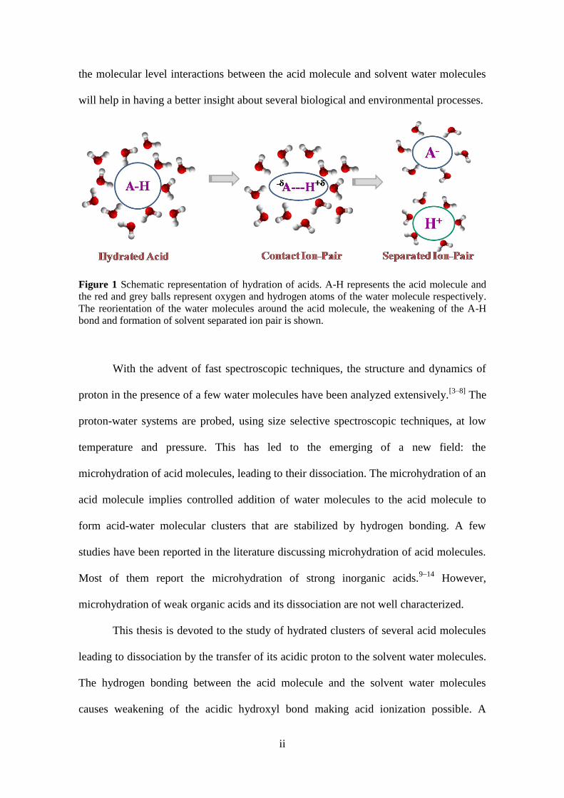
pair. This can be depicted by the schematic diagram shown in Figure 1. Understanding
ii
the molecular level interactions between the acid molecule and solvent water molecules
will help in having a better insight about several biological and environmental processes.
Figure 1 Schematic representation of hydration of acids. A-H represents the acid molecule and
the red and grey balls represent oxygen and hydrogen atoms of the water molecule respectively.
The reorientation of the water molecules around the acid molecule, the weakening of the A-H
bond and formation of solvent separated ion pair is shown.
With the advent of fast spectroscopic techniques, the structure and dynamics of
proton in the presence of a few water molecules have been analyzed extensively.[3–8]
The
proton-water systems are probed, using size selective spectroscopic techniques, at low
temperature and pressure. This has led to the emerging of a new field: the
microhydration of acid molecules, leading to their dissociation. The microhydration of an
acid molecule implies controlled addition of water molecules to the acid molecule to
form acid-water molecular clusters that are stabilized by hydrogen bonding. A few
studies have been reported in the literature discussing microhydration of acid molecules.
Most of them report the microhydration of strong inorganic acids.9–14
However,
microhydration of weak organic acids and its dissociation are not well characterized.
This thesis is devoted to the study of hydrated clusters of several acid molecules
leading to dissociation by the transfer of its acidic proton to the solvent water molecules.
The hydrogen bonding between the acid molecule and the solvent water molecules
causes weakening of the acidic hydroxyl bond making acid ionization possible. A

iii
stronger acid molecule needs a lesser number of solvent water molecules to form
dissociated products. This implies that at molecular level, the strength of an acid is
related to the number of solvent water molecules needed to ionize the acid molecule. In
the present study, the molecular interactions between weak organic acid molecules and
solvent water molecules are studied to get a better understanding of the hydration of acid
molecules. Equilibrium geometries of different size hydrated clusters of selected acid
molecules are calculated. The minimum number of water molecules needed for the
dissociation of an acid molecule is determined. Microhydration of a photo acid is also
considered. Photo acids have different pKa values at ground and excited states. Often
excited state pKa values of photo acids are much lower compared to the ground state. It
is reported that in case of 2-naphthol, the excited singlet state has a low pKa value
compared to its ground state and excited triplet state.15
Analysis of the microhydrated
clusters of both ground state and first excited singlet and triplet states of 2-naphthol is
carried out. At macroscopic level, strength of an acid is described by its pKa value, while
at molecular level the acidic strength can be expressed in terms of the number of water
molecules needed for its dissociation. A linear relation is observed between the two
quantities. Thus, this study also presents a non-thermodynamic route to calculate the pKa
value of an acid molecule, if the number of water molecules needed for its dissociation is
known and vice-versa. The thesis is organized in 7 chapters. A brief description of each
chapter is given below:
Chapter 1: It is the introductory chapter of the thesis. It gives a general description about
microhydration of acid molecules and its relevance. The experimental and theoretical
studies on microhydrated clusters reported in the literature are discussed. A summary of
the contents of each chapter is also given.

iv
Chapter 2: This chapter gives an overview of the important computational methods
including ab initio, electron correlation and DFT methods, along with a brief account on
basis sets and geometry optimization. Applying these methods, the present study aims to
understand at molecular level, the hydration of an acid.
Chapter 3: This chapter deals with the determination of minimum energy conformers of
different size hydrated clusters of selected acid molecules, namely: trifluoroacetic acid,
acetic acid, benzoic acid and oxalic acid. The selection is made so as to understand the
electronic effects on the ionization of an acid molecule. These acids also play a
significant role in many atmospheric processes. The bench-marking done to decide upon
a suitable level of theory is provided in this chapter. The effect of bulk solvation models
and their inability to accurately describe the hydrogen bonded acid-water clusters is
mentioned. The evolution from acid dimer to microhydrated acid cluster is also
discussed. The minimum number of water molecules needed for the dissociation of the
selected acid molecules is determined. Geometrical parameters and microwave spectra
generated is compared with available experimental data.
Chapter 4: The interaction between the acid molecule and solvent water molecules are
analyzed in this chapter. The free energy of formation of the hydrated acid clusters is
calculated at room temperature and atmospheric pressure as well as at low temperature
and pressure, to determine the temperature-pressure dependence on their thermodynamic
stability. The solvent stabilization energy and interaction energy are also determined to
understand the molecular interactions present in the acid-water clusters. The solvent
stabilization energy considers all types of interactions, both solute-solvent and solvent-

v
solvent. Hence, the interaction energy is defined to exclude all solvent-solvent
interactions and exclusively gives solute-solvent interactions. The variation of solvent
stabilization energy and interaction energy with increase in size of the hydrated cluster is
studied.
Chapter 5: The formation of contact ion-pair should be reflected in some of the
properties of the hydrated acid clusters. This chapter describes the variation in various
properties of the hydrated acid clusters, with increase in cluster size. The variations in net
dipole moment, acidic O-H bond dipole moment, isotropic polarizability and hydrogen
bond energy of the selected acid-water clusters have been studied. Intermolecular
interactions in a solvated system can be understood by the system‟s response to an
external electric field, which is given by its electronic polarizability. The variation of
static polarizability of the hydrated acid clusters, with increase in the size of the cluster,
is studied. The addition of water molecules to the acid molecule causes a weakening of
the acidic O-H bond. The elongation of the hydroxyl bond of the acid molecule due to
hydrogen bonding with solvent water molecules causes a change in the dipole moment of
the hydrated acid clusters. Hence, dipole moment can be a property which can be
monitored to determine the proton transfer in carboxylic acid-water cluster. Two bonds
crucial in the study of dissociation of the acid molecule are: the acidic O-H bond and the
hydrogen bond between the acidic H atom and the O atom of the neighboring water
molecule. The breaking of the former and the formation of the latter bonds can be studied
from their hydrogen bond energy. The dissociation energy curve of the hydrated acid
clusters is useful in obtaining information about the proton transfer barrier of the cluster.
The dissociation energy curve is obtained by carrying out a rigid potential energy scan of
the acidic hydroxyl bond of the hydrated acid cluster. Marked differences in some of

vi
these properties are observed upon ionization of the acid molecules. The simulated
infrared spectra of the hydrated acid clusters are also discussed here and are compared to
available experimental data. The IR spectrum is generated by calculating the hessian
matrix of the system. As this calculation is done considering a harmonic approximation,
a scaling factor is included to account for the anharmonicity. The weakening of the
acidic O-H bond, with increase in size of the hydrated cluster, is manifested as a red shift
in its IR stretching frequency. Large red shifts in acidic O-H stretching frequency, with
respect to free acid, are observed in case of ionized acid-water clusters. Formation of
new peaks corresponding to vibrational frequencies of hydrated proton is also noticed in
case of hydrated clusters of ionized acid molecule.
Chapter 6: This chapter is dedicated to the microhydration of photo acid. Photo acids
have different pKa values at ground and excited state. 2-naphthol is taken up as a case
study. The minimum energy geometries of 2-naphthol and its hydrated clusters, at both
ground state and first excited singlet and triplet states, are reported. It is noted that the
minimum number of water molecules needed to dissociate 2-naphthol at first excited
singlet state is different from that of ground and excited triplet state.
Chapter 7: It is the concluding chapter. It gives a summary of the study on
microhydration of weak organic acids. Based on the present study, an interesting
relationship is observed between the number of water molecules needed to dissociate an
acid molecule and its pKa. A non-thermodynamic route for determining the pKa of an
acid is reported.
The future direction of the current work would be to study the microhydration of
other systems such as alcohols, amines and photo acids. This would help in expanding

vii
the database to verify the correlation observed between the pKa and number of water
molecules needed by the system for proton transfer. Moreover, a microscopic theory
based approach may be considered to predict number of water molecules needed to
dissociate an acid in the ground state.

viii
LIST OF FIGURES AND PAGE No.
Figure 1.1 Schematic representation of hydration of acids. A-H represents the acid
molecule and the red and grey balls represent oxygen and hydrogen atoms of the water
molecule respectively. The reorientation of the water molecules around the acid
molecule, the weakening of the A-H bond and formation of solvent separated ion pair is
shown. ............................................................................................................................ 8
Figure 3.1 Minimum energy equilibrium structures of (a) CF3COOH; (b)
CF3COOH.1H2O; (c) CF3COOH.2H2O; (d) CF3COOH.3H2O; (e) CF3COOH.4H2O; (f)
CF3COOH.5H2O; (g) CF3COOH.6H2O and (h) CF3COOH.7H2O calculated at B97X-
D/aug-cc-pVDZ level of theory. O1and O2 are hydroxyl and carbonyl oxygen atoms of
acid molecule. O3 is the oxygen atom on the water molecule closest to hydroxyl
hydrogen atom of acid molecule. Yellow balls represent fluorine, red balls represent
oxygen, grey balls represent carbon and blue balls represent hydrogen atoms. .......... 44
Figure 3.2 Higher energy equilibrium structures of (a)CF3COOH.6H2O and (b)-(e)
CF3COOH.7H2O showing contact ion pair formation. Zero point energy corrected
relative energy (in kcal/mol) of the conformers with respect to minimum energy
conformer of that size cluster is also given. Yellow balls represent fluorine, red balls
represent oxygen, grey balls represent carbon and blue balls represent hydrogen atoms.
...................................................................................................................................... 45
Figure 3.3 Higher energy equilibrium structures of (i)CF3COOH.2H2O (ii-
v)CF3COOH.3H2O, (vi-ix) CF3COOH.4H2O, (x-xiii) CF3COOH.5H2O, (xiv-xvi)
CF3COOH.6H2O and (xvii) CF3COOH.7H2O. Zero point energy corrected relative
energy (in kcal/mol) of the conformers with respect to minimum energy conformer of
that size cluster is also given. Yellow balls represent fluorine, red balls represent oxygen,
grey balls represent carbon and blue balls represent hydrogen atoms. ...................47-48
Figure 3.4 Minimum energy equilibrium structures of (a) CH3COOH; (b)
CH3COOH.1H2O; (c) CH3COOH.2H2O; (d) CH3COOH.3H2O; (e) CH3COOH.4H2O; (f)
CH3COOH.5H2O; (g) CH3COOH.6H2O (h) CH3COOH.7H2O; and (i) CH3COOH.8H2O.
O1and O2 are hydroxyl and carbonyl oxygen atoms of acid molecule. O3 is the oxygen
atom on the water molecule closest to hydroxyl hydrogen atom of acid molecule. Red
balls represent oxygen, grey balls represent carbon and blue balls represent hydrogen
atoms. ........................................................................................................................... 49
Figure 3.5 Higher energy equilibrium structures of (a)-(b)CH3COOH.6H2O, (c)-(e)
CH3COOH.7H2O and (f)-(h) CH3COOH.8H2O showing contact ion pair formation. Zero
point energy corrected relative energy (in kcal/mol) of the conformers with respect to
minimum energy conformer of that size cluster is also given. Red balls represent oxygen,
grey balls represent carbon and blue balls represent hydrogen atoms. ........................ 50

ix
Figure 3.6 Higher energy equilibrium structures of (i-ii) CH3COOH.1H2O, (iii-v)
CH3COOH.2H2O, (vi-xi )CH3COOH.3H2O and(xii-xx) CH3COOH.4H2O. Zero point
energy corrected relative energy (in kcal/mol) of the conformers with respect to
minimum energy conformer of that size cluster is also given. Red balls represent oxygen,
grey balls represent carbon and blue balls represent hydrogen atoms. ........................ 53
Figure 3.7(i-xv) Higher energy equilibrium structures of CH3COOH.5H2O. Zero point
energy corrected relative energy (in kcal/mol) of the conformers with respect to
minimum energy conformer of that size cluster is also given. Red balls represent oxygen,
grey balls represent carbon and blue balls represent hydrogen atoms. ........................ 54
Figure 3.8(i-xix) Higher energy equilibrium structures of CH3COOH.6H2O. Zero point
energy corrected relative energy (in kcal/mol) of the conformers with respect to
minimum energy conformer of that size cluster is also given. Red balls represent oxygen,
grey balls represent carbon and blue balls represent hydrogen atoms. ........................ 55
Figure 3.9 (i-xvi) Higher energy equilibrium structures of CH3COOH.7H2O. Zero point
energy corrected relative energy (in kcal/mol) of the conformers with respect to
minimum energy conformer of that size cluster is also given. Red balls represent oxygen,
grey balls represent carbon and blue balls represent hydrogen atoms. ........................ 56
Figure 3.10 (i-xx) Higher energy equilibrium structures of CH3COOH.8H2O. Zero point
energy corrected relative energy (in kcal/mol) of the conformers with respect to
minimum energy conformer of that size cluster is also given. Red balls represent oxygen,
grey balls represent carbon and blue balls represent hydrogen atoms. ........................ 57
Figure 3.11 Minimum energy equilibrium structures of (a) C6H5COOH; (b)
C6H5COOH.1H2O; (c) C6H5COOH.2H2O; (d) C6H5COOH.3H2O; (e) C6H5COOH.4H2O;
(f) C6H5COOH.5H2O; (g) C6H5COOH.6H2O (h) C6H5COOH.7H2O; and (i)
C6H5COOH.8H2O. O1and O2 are hydroxyl and carbonyl oxygen atoms of acid
molecule. O3 is the oxygen atom on the water molecule closest to hydroxyl hydrogen
atom of acid molecule. Red balls represent oxygen, grey balls represent carbon and blue
balls represent hydrogen atoms. ................................................................................... 58
Figure 3.12 Higher energy equilibrium structures of (a)C6H5COOH.7H2O and (b)-(d)
C6H5COOH.8H2O showing contact ion pair formation. Zero point energy corrected
relative energy (in kcal/mol) of the conformers with respect to minimum energy
conformer of that size cluster is also given. Red balls represent oxygen, grey balls
represent carbon and blue balls represent hydrogen atoms. ......................................... 60
Figure 3.13 Higher energy equilibrium structures of (i)C6H5COOH, (ii-iii)
C6H5COOH.1H2O, (iv) C6H5COOH.2H2O, (v-viii) C6H5COOH.3H2O and (ix-xx)
C6H5COOH.4H2O. Zero point energy corrected relative energy (in kcal/mol) of the
conformers with respect to minimum energy conformer of that size cluster is also given.
Red balls represent oxygen, grey balls represent carbon and blue balls represent
hydrogen atoms. ........................................................................................................... 62
Figure 3.14 (i-xi) Higher energy equilibrium structures of C6H5COOH.5H2O. Zero point
energy corrected relative energy (in kcal/mol) of the conformers with respect to

x
minimum energy conformer of that size cluster is also given. Red balls represent oxygen,
grey balls represent carbon and blue balls represent hydrogen atoms. ........................ 63
Figure 3.15 (i-xx) Higher energy equilibrium structures of C6H5COOH.6H2O. Zero point
energy corrected relative energy (in kcal/mol) of the conformers with respect to
minimum energy conformer of that size cluster is also given. Red balls represent oxygen,
grey balls represent carbon and blue balls represent hydrogen atoms. ........................ 64
Figure 3.16 (i-ix) Higher energy equilibrium structures of C6H5COOH.7H2O. Zero point
energy corrected relative energy (in kcal/mol) of the conformers with respect to
minimum energy conformer of that size cluster is also given. Red balls represent oxygen,
grey balls represent carbon and blue balls represent hydrogen atoms. ........................ 65
Figure 3.17 (i-xx) Higher energy equilibrium structures of C6H5COOH.8H2O. Zero point
energy corrected relative energy (in kcal/mol) of the conformers with respect to
minimum energy conformer of that size cluster is also given. Red balls represent oxygen,
grey balls represent carbon and blue balls represent hydrogen atoms. ........................ 66
Figure 3.18 Minimum energy equilibrium structures of (a) (COOH)2; (b)
(COOH)2.1H2O; (c) (COOH)2.2H2O; (d) (COOH)2.3H2O; (e) (COOH)2.4H2O; (f)
(COOH)2.5H2O; (g) (COOH)2.6H2O (h) (COOH)2.7H2O; and (i) (COOH)2.8H2O. O1and
O2 are hydroxyl oxygen atoms of acid molecule. Oa and Ob are the oxygen atoms on the
water molecules closest to hydroxyl hydrogen atoms of acid molecule. Red balls
represent oxygen, grey balls represent carbon and blue balls represent hydrogen atoms.
...................................................................................................................................... 68
Figure 3.19 Higher energy equilibrium structures of (a)-(f) (COOH)2.5H2O and (g)-(q)
(COOH)2.6H2O showing contact ion pair formation. Zero point energy corrected relative
energy (in kcal/mol) of the conformers with respect to minimum energy conformer of
that size cluster is also given. Red balls represent oxygen, grey balls represent carbon
and blue balls represent hydrogen atoms. .................................................................... 70
Figure 3.20 Higher energy equilibrium structures of (COOH)2.7H2O showing contact ion
pair formation. Zero point energy corrected relative energy (in kcal/mol) of the
conformers with respect to minimum energy conformer of that size cluster is also given.
Red balls represent oxygen, grey balls represent carbon and blue balls represent
hydrogen atoms. ........................................................................................................... 71
Figure 3.21 Higher energy equilibrium structures of (i-v) (COOH)2, (vi-viii)
(COOH)2.1H2O, (ix-xvi) (COOH)2.2H2O. Zero point energy corrected relative energy (in
kcal/mol) of the conformers with respect to minimum energy conformer of that size
cluster is also given. Red balls represent oxygen, grey balls represent carbon and blue
balls represent hydrogen atoms. ................................................................................... 73
Figure 3.22 (i-xii) Higher energy equilibrium structures of (COOH)2.3H2O. Zero point
energy corrected relative energy (in kcal/mol) of the conformers with respect to
minimum energy conformer of that size cluster is also given. Red balls represent oxygen,
grey balls represent carbon and blue balls represent hydrogen atoms. ........................ 74

xi
Figure 3.23 (i-xv) Higher energy equilibrium structures of (COOH)2.4H2O. Zero point
energy corrected relative energy (in kcal/mol) of the conformers with respect to
minimum energy conformer of that size cluster is also given. Red balls represent oxygen,
grey balls represent carbon and blue balls represent hydrogen atoms. ........................ 75
Figure 3.24 (i-xx) Higher energy equilibrium structures of (COOH)2.5H2O. Zero point
energy corrected relative energy (in kcal/mol) of the conformers with respect to
minimum energy conformer of that size cluster is also given. Red balls represent oxygen,
grey balls represent carbon and blue balls represent hydrogen atoms. ........................ 76
Figure 3.25 Higher energy equilibrium structures of (i-xix) (COOH)2.6H2O and (xx)
(COOH)2.7H2O. Zero point energy corrected relative energy (in kcal/mol) of the
conformers with respect to minimum energy conformer of that size cluster is also given.
Red balls represent oxygen, grey balls represent carbon and blue balls represent
hydrogen atoms. ........................................................................................................... 77
Figure 4.1 Schematic diagram depicting the solvent stabilization energy, Estab, for a
hydrated carboxylic acid system, RCOOH.nH2O. This energy parameter includes solute-
solvent as well as solvent-solvent interactions. The red and white balls represent oxygen
and hydrogen atoms respectively of water molecule. .................................................. 82
Figure 4.2 Schematic diagram depicting the interaction energy, Eint, for a hydrated
carboxylic acid system, RCOOH.nH2O. This energy parameter contains only solute-
solvent interactions. The red and white balls represent oxygen and hydrogen atoms
respectively of water molecule. .................................................................................... 83
Figure 4.3 Variation of solvent stabilization energy (Estab) of hydrated clusters of
carboxylic acids with increase in size of the hydrated cluster (n). The brown, green, blue
and orange line shows solvent stabilization energies of CF3COOH.nH2O,
CH3COOH.nH2O, C6H5COOH.nH2O and (COOH)2.nH2O, n=1-8, clusters respectively,
calculated at ωB97X-D/aug-cc-pVDZ level of theory. ................................................ 86
Figure 4.4 Variation of interaction energy (Eint) for hydrated carboxylic acid clusters
with increase in size of the hydrated cluster (n). The brown, orange, blue and green lines
show interaction energy profiles for CF3COOH.nH2O, (COOH)2.nH2O,
C6H5COOH.nH2O and CH3COOH.nH2O, n=1-8, clusters respectively, calculated at
ωB97X-D/aug-cc-pVDZ level of theory. ..................................................................... 87
Figure 4.5 Variation of solvent stabilization energy (Estab) and interaction energy (Eint) of
hydrated oxalic acid clusters, (COOH)2.nH2O, n=1-7, with increase in size of the cluster
(n). The blue and black lines show Estab profiles calculated at CCSD(T)/6-311++G(d,p)
and ωB97X-D/aug-cc-pVDZ levels of theory while green and brown lines show Eint
profiles calculated at CCSD(T)/6-311++G(d,p) and ωB97X-D/aug-cc-pVDZ levels of
theory, respectively. ..................................................................................................... 90
Figure 5.1 Variation of acidic O-H bond dipole moment (μO-H) of hydrated clusters of
carboxylic acids with increase in size of the hydrated cluster (n). The violet, green, black
and orange line shows the bond dipole moments of the most acidic O-H bonds of

xii
C6H5COOH.nH2O, CH3COOH.nH2O, CF3COOH.nH2O and (COOH)2.nH2O, n=1-8,
clusters respectively, calculated at ωB97X-D/aug-cc-pVDZ level of theory. ........... 100
Figure 5.2 Simulated scaled IR spectra of (a) H2O and (b-i) CF3COOH.nH2O, n=0-7,
clusters calculated at ωB97X-D/aug-cc-pVDZ level of theory. Calculated scaled O-H
(νO-H) and C=O (νC=O) stretching frequencies of the acid molecule (in cm-1
) are also
given. .......................................................................................................................... 103
Figure 5.3 (a-i) Simulated scaled IR spectra of CH3COOH.nH2O, n=0-8, clusters
calculated at ωB97X-D/aug-cc-pVDZ level of theory. Calculated scaled O-H (νO-H) and
C=O (νC=O) stretching frequencies of the acid molecule (in cm-1
) are also given. ..... 106
Figure 5.4 (a-i) Simulated scaled IR spectra of C6H5COOH.nH2O, n=0-8, clusters
calculated at ωB97X-D/aug-cc-pVDZ level of theory. Calculated scaled O-H (νO-H) and
C=O (νC=O) stretching frequencies of the acid molecule (in cm-1
) are also given. ..... 107
Figure 5.5 (a-h) Simulated scaled IR spectra of (COOH)2.nH2O, n=0-7, clusters
calculated at ωB97X-D/aug-cc-pVDZ level of theory. Calculated scaled O-H (νO-H) and
C=O (νC=O) stretching frequencies of the acid molecule (in cm-1
) are also given. ..... 109
Figure 5.6 Rigid potential energy scan of O-H bond of CF3COOH.nH2O, n=1-6,
calculated at ωB97X-D/aug-cc-pVDZ level of theory. Curves marked as tn (n=1-6)
represents potential energy surface of hydrated cluster having n solvent water molecules
upon increasing bond length of dissociating O-H bond of the acid. .......................... 111
Figure 5.7 Rigid potential energy scan for O-H bond of CH3COOH.nH2O, n=1-8,
calculated at ωB97X-D/aug-cc-pVDZ level of theory. Curves marked as an (n=1-8)
represents potential energy surface of hydrated cluster having n solvent water molecules
upon increasing bond length of dissociating O-H bond of the acid. .......................... 112
Figure 5.8 Rigid potential energy scan for O-H bond of C6H5COOH.nH2O, n=1-8,
calculated at ωB97X-D/aug-cc-pVDZ level of theory. Curves marked as bn (n=1-8)
represents potential energy surface of hydrated cluster having n solvent water molecules
upon increasing bond length of dissociating O-H bond of the acid. .......................... 113
Figure 5.9 Rigid potential energy scan for O-H bond of (COOH)2.nH2O, n=1-7,
calculated at ωB97X-D/aug-cc-pVDZ level of theory. Curves marked as On (n=1-7)
represents potential energy surface of hydrated cluster having n solvent water molecules
upon increasing bond length of dissociating O-H bond of the acid. .......................... 114
Figure 5.10 Variation of hydrogen bond energy of most acidic O-H bond (EO-H) and
H..O(H2) bond (EH..O(H2)) of hydrated clusters of carboxylic acids with increase in size of
the hydrated cluster (n). The violet, green, black and orange line shows the hydrogen
bond energy of CH3COOH.nH2O, (COOH)2.nH2O, CF3COOH.nH2O and
C6H5COOH.nH2O, n=1-8, clusters respectively, calculated at ωB97X-D/aug-cc-pVDZ
level of theory. ............................................................................................................ 116
Figure 6.1 Equilibrium structures of 2-naphthol, at ground state, calculated at ωB97X-
D/aug-cc-pVDZ level of theory. Zero point energy corrected relative energy (in
kcal/mol) of the higher energy conformer with respect to minimum energy conformer is

xiii
also given. Red balls represent oxygen, grey balls represent carbon and blue balls
represent hydrogen atoms. .......................................................................................... 123
Figure 6.2 Equilibrium structures of mono-hydrate of 2-naphthol, at ground state,
calculated at ωB97X-D/aug-cc-pVDZ level of theory. Zero point energy corrected
relative energy (in kcal/mol) of the higher energy conformer with respect to minimum
energy conformer is also given. Red balls represent oxygen, grey balls represent carbon
and blue balls represent hydrogen atoms. .................................................................. 124
Figure 6.3 Equilibrium structures of di-hydrate of 2-naphthol, at ground state, calculated
at ωB97X-D/aug-cc-pVDZ level of theory. Zero point energy corrected relative energy
(in kcal/mol) of the higher energy conformer with respect to minimum energy conformer
is also given. Red balls represent oxygen, grey balls represent carbon and blue balls
represent hydrogen atoms. .......................................................................................... 124
Figure 6.4 Equilibrium structures of tri-hydrate of 2-naphthol, at ground state, calculated
at ωB97X-D/aug-cc-pVDZ level of theory. Zero point energy corrected relative energy
(in kcal/mol) of the higher energy conformer with respect to minimum energy conformer
is also given. Red balls represent oxygen, grey balls represent carbon and blue balls
represent hydrogen atoms. .......................................................................................... 125
Figure 6.5 Equilibrium structures of tetra-hydrate of 2-naphthol, at ground state,
calculated at ωB97X-D/aug-cc-pVDZ level of theory. Zero point energy corrected
relative energy (in kcal/mol) of the higher energy conformer with respect to minimum
energy conformer is also given. Red balls represent oxygen, grey balls represent carbon
and blue balls represent hydrogen atoms. .................................................................. 126
Figure 6.6 Equilibrium structures of 2-naphthol, at first excited triplet state, calculated at
CAM-B3LYP/aug-cc-pVDZ level of theory. Zero point energy corrected relative energy
(in kcal/mol) of the higher energy conformer with respect to minimum energy conformer
is also given. Red balls represent oxygen, grey balls represent carbon and blue balls
represent hydrogen atoms. .......................................................................................... 127
Figure 6.7 Equilibrium structures of 2-naphthol.1H2O, at first excited triplet state,
calculated at CAM-B3LYP/aug-cc-pVDZ level of theory. Zero point energy corrected
relative energy (in kcal/mol) of the higher energy conformer with respect to minimum
energy conformer is also given. Red balls represent oxygen, grey balls represent carbon
and blue balls represent hydrogen atoms. .................................................................. 127
Figure 6.8 Equilibrium structures of 2-naphthol.2H2O, at first excited triplet state,
calculated at CAM-B3LYP/aug-cc-pVDZ level of theory. Zero point energy corrected
relative energy (in kcal/mol) of the higher energy conformer with respect to minimum
energy conformer is also given. Red balls represent oxygen, grey balls represent carbon
and blue balls represent hydrogen atoms. .................................................................. 128
Figure 6.9 Equilibrium structures of 2-naphthol.3H2O, at first excited triplet state,
calculated at CAM-B3LYP/aug-cc-pVDZ level of theory. Zero point energy corrected
relative energy (in kcal/mol) of the higher energy conformer with respect to minimum

xiv
energy conformer is also given. Red balls represent oxygen, grey balls represent carbon
and blue balls represent hydrogen atoms. .................................................................. 128
Figure 6.10 Equilibrium structures of 2-naphthol.4H2O, at first excited triplet state,
calculated at CAM-B3LYP/aug-cc-pVDZ level of theory. Zero point energy corrected
relative energy (in kcal/mol) of the higher energy conformer with respect to minimum
energy conformer is also given. Red balls represent oxygen, grey balls represent carbon
and blue balls represent hydrogen atoms. .................................................................. 129
Figure 6.11 Equilibrium structures of 2-naphthol, at first excited singlet state, calculated
at CAM-B3LYP/aug-cc-pVDZ level of theory. Zero point energy corrected relative
energy (in kcal/mol) of the higher energy conformer with respect to minimum energy
conformer is also given. Red balls represent oxygen, grey balls represent carbon and blue
balls represent hydrogen atoms. ................................................................................. 129
Figure 6.12 Equilibrium structures of 2-naphthol.1H2O, at first excited singlet state,
calculated at CAM-B3LYP/aug-cc-pVDZ level of theory. Zero point energy corrected
relative energy (in kcal/mol) of the higher energy conformer with respect to minimum
energy conformer is also given. Red balls represent oxygen, grey balls represent carbon
and blue balls represent hydrogen atoms. .................................................................. 130
Figure 6.13 Equilibrium structures of 2-naphthol.2H2O, at first excited singlet state,
calculated at CAM-B3LYP/aug-cc-pVDZ level of theory. Zero point energy corrected
relative energy (in kcal/mol) of the higher energy conformer with respect to minimum
energy conformer is also given. Red balls represent oxygen, grey balls represent carbon
and blue balls represent hydrogen atoms. .................................................................. 130
Figure 6.14 Equilibrium structures of 2-naphthol.3H2O, at first excited singlet state,
calculated at CAM-B3LYP/aug-cc-pVDZ level of theory. Zero point energy corrected
relative energy (in kcal/mol) of the higher energy conformer with respect to minimum
energy conformer is also given. Red balls represent oxygen, grey balls represent carbon
and blue balls represent hydrogen atoms. .................................................................. 131
Figure 6.15 Equilibrium structures of 2-naphthol.4H2O, at first excited singlet state,
calculated at CAM-B3LYP/aug-cc-pVDZ level of theory. Zero point energy corrected
relative energy (in kcal/mol) of the higher energy conformer with respect to minimum
energy conformer is also given. Red balls represent oxygen, grey balls represent carbon
and blue balls represent hydrogen atoms. .................................................................. 132
Figure 6.16 Rigid potential energy scan of O-H bond of 2-naphthol.4H2O, at ground, first
excited triplet and singlet state. Curves marked as S0, T1 and S1 represent potential
energy surfaces of ground, first excited triplet and singlet states respectively of 2-
naphthol.4H2O, upon increasing the bond distance of dissociating O-H bond of the acid.
.................................................................................................................................... 133
Figure 7.1 Thermodynamic cycle for determining pKa indirectly from free energy. AH
refers to the acid. Subscript (g) and (aq) denote the species in gas and aqueous phase
respectively. ................................................................................................................ 136

xv
Figure 7.2 Plot of pKa v/s number of water molecules needed for dissociation (n), for
HCl, trifluoroacetic acid (tfa), formic acid (fa) and benzoic acid (bz), calculated at
ωB97X-D/aug-cc-pVDZ level of theory. ................................................................... 138
Figure 7.3 Plot of pKa v/s number of water molecules needed for dissociation (n), for HI,
HBr, HCl, HNO3, trifluoroacetic acid, formic acid and benzoic acid. ...................... 139

xvi
LIST OF TABLES
Table 3.1 Comparison of geometrical parameters and hydroxyl stretching frequency of
isolated water and trifluoroacetic acid molecule, calculated at different levels of theory
using aug-cc-pVDZ basis set, with available experimental data.................................. 40
Table 3.2 Comparison of rotational constants (A, B, and C in MHz) for hydrated clusters
of trifluoroacetic acid, CF3COOH.nH2O (n = 1−3) calculated at different levels of theory
...................................................................................................................................... 41
Table 3.3 Selected bond lengths of tfa.nH2O, n=0-7, calculated at B97X-D/aug-cc-
pVDZ level of theory. rO1-H, rC=O2 and rH-O3 represent hydroxyl O-H and carbonyl C=O
bond of the acid molecule and hydrogen bond between the hydroxyl hydrogen of acid
molecule and oxygen of the nearest water molecule, respectively. ............................. 46
Table 3.4. Selected bond lengths of CH3COOH.nH2O, n=0-8, calculated at B97X-
D/aug-cc-pVDZ level of theory. rO1-H, rC=O2 and rH-O3 represent hydroxyl O-H and
carbonyl C=O bond of the acid molecule and hydrogen bond between the hydroxyl
hydrogen of acid molecule and oxygen of the nearest water molecule, respectively. . 52
Table 3.5 Selected bond lengths of C6H5COOH.nH2O, n=0-8, calculated at B97X-
D/aug-cc-pVDZ level of theory. rO1-H, rC=O2 and rH-O3 represent hydroxyl O-H and
carbonyl C=O bond of the acid molecule and hydrogen bond between the hydroxyl
hydrogen of acid molecule and oxygen of the nearest water molecule, respectively. . 61
Table 3.6. Selected bond lengths of (COOH)2.nH2O, n=0-7, calculated at B97X-D/aug-
cc-pVDZ level of theory. rO1-H, rO2-H, rH-Oaand rH-Ob represent hydroxyl O-H and
hydrogen bond between the hydroxyl hydrogen of acid molecule and oxygen of the
nearest water molecule, respectively.δO1-C-C-O2 gives the dihedral angle between the C-O
bonds ............................................................................................................................ 72
Table 4.1 Free energy of formation, at room temperature/atmospheric pressure
(1atmΔG298K
) and 100K/1μTorr (μTorrΔG100K
), of CF3COOH.nH2O, CH3COOH.nH2O,
C6H5COOH.nH2O and (COOH)2.nH2O, n=1-8, calculated at ωB97X-D/aug-cc-pVDZ
level of theory. .............................................................................................................. 84
Table 4.2 Solvent stabilization energy and Interaction energy of CF3COOH.nH2O,
CH3COOH.nH2O, C6H5COOH.nH2O and (COOH)2.nH2O, n=1-8, calculated at ωB97X-
D/aug-cc-pVDZ level of theory. .................................................................................. 86
Table 4.3 Solvent stabilization energy (Estab) and Interaction energy (Eint) of
CF3COOH.nH2O, CH3COOH.nH2O, C6H5COOH.nH2O and (COOH)2.nH2O, n=1-8,
clusters calculated at CCSD(T)/6-311++G(d,p) level of theory. ................................. 89
Table 5.1 Comparison of isotropic polarizability of RCOOH.nH2O (n = 0−8) clusters
calculated at ωB97X-D/aug-cc-pVDZ level of Theory ............................................... 97
Table 5.2 Net dipole moment and acidic O-H bond dipole moments of the most stable
conformers of hydrated carboxylic acid, RCOOH.nH2O, n=0-8 systems. .................. 99

xvii
Table 5.3 Scaled IR stretching frequency of acidic O-H bond (O-H) of hydrated
carboxylic acid, RCOOH.nH2O, n=0-8 systems. The red-shift of the O-H stretching
frequency (O-H) of the carboxylic acid of the hydrated clusters, with respect to free
acid molecule is also given. ........................................................................................ 102
Table 5.4 Hydrogen bond energy of acidic O-H bond (EO-H) and H..O(H2) bond
(EH..O(H2)) in the most stable conformers of hydrated carboxylic acid, RCOOH.nH2O,
n=0-8 systems. ............................................................................................................ 115

Chapter 1
1
How Many Water Molecules are Needed to
Ionize an Acid Molecule?
If we were to name the most powerful assumption of all, which leads one
on and on in an attempt to understand life, it is that all things are made
of atoms, and that everything that living things do can be understood in
terms of the jigglings and wigglings of atoms.
-Richard Feynman, The Feynman Lectures on Physics, 1970

2
Carboxylic acids are known to ionize in water to form carboxylate anion and
proton. The extent of ionization of an acid is defined in terms of the pKa value of the
acid. Observing from a microscopic point of view, the degree of ionization depends on
the strength of the O-H bond of the acid molecule, which is reflected in the number of
water molecules needed for its dissociation. Understanding the microscopic description
of hydration is essential for the fundamental understanding of acid dissociation as well as
for understanding and modeling several atmospheric as well as biological phenomena.
This thesis deals with the dissociation of acid molecule in the presence of water
molecules. Molecular level acid dissociation can be studied using acid-water clusters.
1.1 Clusters
Clusters are defined as finite aggregates of particles (atoms or molecules). They
bridge the gap between molecules and bulk materials. Although cluster science is an
emerging area of research, clusters have been in use for ages, for example, the beautiful
colors imparted to the glass artifacts by the Romans, and AgBr clusters used in
photography. Most of the elements of the periodic table form clusters. The size of a
cluster varies from a few to several thousand atoms or molecules. The properties of
clusters are very different from that of its individual constituents and that of the bulk.
The electronic, optic, magnetic and chemical properties of a cluster depend on its size.
The evolution of properties with the variation in size of the cluster is of great
fundamental interest. The physical and chemical properties can be tuned by varying the
size and constituent particles of the cluster. Cluster science tries to find answers to
fundamental questions such as „how large should the cluster be for its properties to
resemble that of the bulk material‟.[16]
Depending on the composition and nature of
bonding in the clusters, they can be broadly classified as metallic, ionic and weakly

3
bound clusters. In metallic clusters, like Al77, Pd145 etc., the atoms are held together by
long range forces originating from valence electron sharing over many adjacent atoms.[17]
The binding force in ionic clusters is the electrostatic interaction between oppositely
charged species.[18]
In weakly bound molecular clusters inter-molecular interactions are
non-covalent in nature.[19–23]
The size-dependent evolution of properties in a cluster is a popular field of
theoretical and experimental research. The development of molecular beam techniques
has helped in better understanding of cluster geometries and properties in an interaction-
free environment. Cluster generation begins with vaporization, where the
atoms/molecules constituting the cluster are produced in gas phase. The atoms/molecules
then condense to form the cluster nucleus. The cluster grows with the addition of more
atoms/molecules to the nucleus. The merging of small clusters to form larger clusters is
known as coalescence. The structure and properties of the cluster are studied using
molecular beam techniques, inert matrices, supporting surfaces or in solid state.
Theoretical chemistry has also been playing a crucial role to understand the structure and
properties of atomic and molecular clusters.
1.2 Experimental techniques to study clusters
A few of the experimental techniques used to study clusters are described here
briefly. Gas phase clusters can be generated by pulsed supersonic jet expansion
techniques, where a dilute mixture, of the sample to be studied and an inert gas, is
allowed to expand through a slit-jet source.[24,25]
The expansion can be coupled with
experimental techniques such as IR spectroscopy, microwave spectroscopy, photo
electron spectroscopy etc to study the properties of the clusters generated. Mass
spectrometry is used along with other experimental techniques to determine the size of
the cluster. Direct absorption spectroscopy employs FTIR to obtain accurate information

4
over a broad spectral coverage.[26,27]
In IR cavity ring down spectroscopy, a laser is used
to illuminate a high finesse optical cavity formed by two highly reflective mirrors.[28–30]
Intensity builds up in the cavity when the laser is in resonance with a cavity mode. The
ring down pulse intersects a pulsed slit jet expansion in a vacuum chamber. The sample
is seeded in to the expansion by bubbling an inert carrier gas through the room
temperature liquid. The ability to provide quantitative absorption intensity information
and to probe gaseous samples with high sensitivity makes this technique very popular.
Matrix isolation IR spectroscopy is also used to study cluster properties.[31]
In this
method, the sample is trapped in an inert gas matrix, like Ar or N2 at low temperature.
The sample concentration is kept very low to ensure molecular isolation; the sample is
surrounded only by the inert gas molecules. This method can be applied to study van der
Waals complexes, polycyclic aromatic hydrocarbons and hydrogen bonded clusters due
to its small line-width and spectral sharpening features. In supersonic jet expansion, the
vibrational-rotational energy levels of the sample are separated by the expansion of the
gas at high pressure.[25,32]
Microwave spectrum of such a system can then be studied
thoroughly.[33]
The rotational spectra of weakly bound clusters have been widely
investigated using Fourier Transform microwave spectroscopy. Other important tools for
studying clusters are electron attachment and photo electron spectroscopies.[34,35]
They
are valuable in determining the electron affinity and ionization energy of the clusters as
well as for obtaining information about the production and identification of ions in mass
spectrometry. Photo electron spectroscopy has been used to determine the size of CO2
clusters.[36]
The free neutral CO2 clusters are generated in a free jet condensation source
and are expanded from a stagnation chamber. The cluster-rich part of the beam
consisting of molecules and clusters is extracted by a skimmer. The recorded C1s

5
photoelectron spectrum shows two distinct peaks corresponding to the clusters of various
size and uncondensed monomers.
1.3 Theoretical investigation of clusters
Theoretical methods play an important role in the development and application of
cluster science. Many cluster properties, like geometry, binding energy etc. cannot be
easily measured from experiments directly. Computational models also help in
interpreting the experimental results. Early ab initio level calculations of small water
clusters were reported by Clementi and co-workers in 1974.[37]
Due to limited
computational resources, HF calculations were the only options available until the end of
1980s. During the 1990s, ab initio calculations at MP2 levels became feasible for dimers
and trimers of small molecules. With advancement in computational resources, larger
clusters of larger molecules can be studied using ab initio as well as DFT calculations.[38–
43] The inherent O(N
5) (N is the number of electrons) scaling of MP2 level calculations
limits its use in geometry optimization to less than 100 first row atoms. DFT based
methods have become increasingly popular for the computational study of molecular
clusters. It is observed that the geometry and energy of water clusters up to size four,
obtained from DFT functionals like BLYP and BP86 coupled with correlation consistent
basis sets, are comparable to those obtained from MP2 calculations.[44]
However, these
functionals are not suitable for larger water clusters, where hydrogen bonding plays a
major role in shaping geometry. Introduction of long range dispersion corrected density
functionals have helped in the modeling of large weakly bound clusters.[45–50]
HF, DFT
and MP2 are the most popular levels of theory for geometry optimization for large
clusters. The energy of the clusters so obtained can then further be improved using
higher levels of theory like MP4, coupled cluster method etc. It may be noted that

6
coupled cluster method with single and double excitations including triplet correction
(CCSD(T)) is regarded as the contemporary gold standard of quantum chemistry.
1.4 Microhydrated Clusters
Water clusters as well as microhydrated clusters are among the most extensively
investigated clusters, because of their role in a wide range of phenomena from solution
chemistry to large number of biochemical processes. Microhydrated clusters are formed
by the step-wise addition of water molecules to a chemical species. These clusters are
stabilized by hydrogen bonding. Hydrogen bonds play a key role in determining the three
dimensional structure of the cluster. Microhydration of chemical species has been a
subject of intense research, to understand the structural, energetic, spectroscopic, and
dynamic aspects of hydration at molecular level.[51–55]
The size-dependent variation in
the properties of hydrated systems can be studied from molecular level to the bulk
solution. Explicit hydration studies are also important in understanding the correlation
between solubility and acidity of the solute. The importance of including specific
interactions between solute and solvent molecules in the condensed phase has been
reported previously.[56]
A large part of the experimental and theoretical microhydration
studies reported, are on acid molecules.[9–14,57–68]
1.5 Hydrated Acids
Acids (A-H) are ubiquitous in nature. Acidic solutions play a major role in a wide
range of fields. A plethora of chemical, physical and biological phenomena involves
acid–water interactions. Be it many enzyme catalyzed reactions in biological systems or
the formation of cloud condensation nuclei in the atmosphere, it all depends on the
proton transfer process between the acid and water molecules, that is, acidity of the acid.
With the advent of ultra-fast spectroscopic techniques and high level computational

7
facilities, the nature and characterization of the excess proton in water, shifting between
Eigen and Zundel forms, has been a field of extensive study, which is summarized in the
review by Hassanali et al.[3]
The understanding of spectral signatures of hydrated proton
has led to the progress of the study of microhydration of acids.
An accurate description of hydration of acids, at molecular level, can deepen our
understanding of a broad spectrum of phenomena, from proton transfer to acid rain.
Further, acid-water molecular clusters provide an ideal system to study the relation
between solvation and reactivity, as ionization of the acid molecule occurs during the
progress from molecular clusters to aqueous solution. Studying the structure and
properties of acid-water clusters provides basic understanding of the fundamental
interactions responsible for the phenomenon of hydration, which is important not just to
chemists, but also to physicists, biologists and material scientists. The intermolecular
interactions determine the stability and geometry of the molecular clusters.
In macroscopic description, a strong acid signifies an acid that ionizes completely
in an aqueous solution whereas a weak acid does not ionize fully in such an environment.
Thus, the ability to transfer a proton to a water molecule is the answer to differentiate an
acid based on its strength. In the context of the microscopic or molecular level
description of the strength of an acid, one may ask a fundamental question: how much
water is required to ionize an acid? The answer to this question involves a microscopic
study of the hydration of acids. In acid-water clusters (A-H.nH2O), solute acid molecule
forms hydrogen bonds with the solvent water molecules present in the immediate
neighborhood. As a result of intermolecular interactions between the acid molecule and
solvent water molecules, the A-H bond of the acid molecule weakens and proton transfer
from the acid molecule to solvent water molecules occurs, leading to dissociation of the
acid molecule, as depicted in Figure 1.1.

8
Figure 1.1 Schematic representation of hydration of acids. A-H represents the acid molecule and
the red and grey balls represent oxygen and hydrogen atoms of the water molecule respectively.
The reorientation of the water molecules around the acid molecule, the weakening of the A-H
bond and formation of solvent separated ion pair is shown.
The process of hydration of an acid molecule involves formation of contact ion
pair followed by solvent separated ion pair, and yields hydrated protons in either Eigen
(H3O+·3H2O) or Zundel (H3O
+·H2O) forms.
[7,69–71] Thus, determining the number of
water molecules needed to stabilize the hydrated ion pair will tell us the number of water
molecules that are needed to ionize an acid.
A few recent theoretical studies report the number of water molecules needed to
ionize H2SO4, HCl, HNO3 and HClO4.[61],[9],[67],[14]
When a solute is added to a solvent
water pool, the water molecules in the immediate neighborhood of the solute get
rearranged to form a hydrogen-bonded cluster of solute and solvent water molecules. The
electron distribution pattern of the added solute molecule plays a key role in forming
stable hydrogen-bonded water network around the solute. Spectroscopy and first
principle based quantum chemical studies have been successfully applied to determine
the detailed structure of hydration shell around the solute molecule.
In experimental front, Matrix isolation studies, Helium nano-droplet spectroscopy
and thin film IR studies have been employed to analyze acid-water molecular
clusters.[11,59,61,64]
From experimental and theoretical studies, a general trend noted in the
geometry of mono-hydrates of acid molecules is that a linear or near linear hydrogen

9
bond is formed between the acidic proton of the acid and the oxygen atom of the water
molecule. The covalent O-H bond of the acid molecule increases only slightly from that
of the free acid molecule and the O..H hydrogen bond distance range between 1.6 to 2.2
Å. A red-shift in the O-H stretching frequency of the acidic proton is also noted, due to
the formation of H bond between the acid molecule and the water molecule. Number of
possible geometries increases with increase in size of the acid-water cluster as the
number of H bond donors and acceptors increases. However the relative stability of the
isomers becomes very small. As the size of the cluster increases, the potential energy
surface of the hydrated acid clusters become complex due to the availability of multiple
H bond acceptors. Characterization of such a potential energy surface which has several
low lying minima is essential for studying the hydrated acid clusters. The possibility for
proton transfer from the acid molecule to the water molecules also increases with
increase in size of the acid-water cluster. Low concentrations, absence of size selectivity
etc makes it difficult to obtain experimental insights about higher hydrated acid clusters
and majority of the reported studies on higher hydrates are based on theoretical
studies.[9,10,12–14,52,54,57,59,61,63,66–68]
However a few IR and microwave studies for higher
hydrates have also been reported. [3,14,46,50,54,57,64,68]
A dominant structural motif
consistent in higher hydrates of acid-water clusters is the hydrogen bonded closed rings.
The water molecule added either forms part of a new ring or expands an existing ring.
This introduces conformational variety. The hydrated acid clusters prefer closed ring
conformers, even when the size of the cluster is very small. This observation is very
different from that of hydrated proton systems, where closed ring conformers are first
observed only for the hepta-hydrated cluster.[8]
Unlike smaller clusters of hydrated
proton clusters, conformers with dangling O-H bonds have higher energy in case of
hydrated acid clusters.

10
Experimental reports of proton transfer in hydrated acid molecules are mainly
based on matrix isolation and thin-film IR studies. Based on the study of amorphous thin
films of HNO3 and water, Ritzhaupt and Devlin reported that three waters of hydration
are required to stabilize ionized HNO3.[76]
Gas-phase ultra-fast pump-probe experiments
on HBr-water clusters by Castleman et al led to the conclusion that five water molecules
are needed to induce ionization of HBr.[77]
High resolution mass-selective infrared laser
spectroscopy was employed by Gutberlet et al to observe the formation of hydronium ion
in the tetra-hydrated cluster of HCl, within superfluid He cluster, at a temperature of 0.37
K.[62]
Ab initio molecular dynamics was used to explore the free energy surface of the
HCl-water system and explain how the proton transfer barrier was surmounted under the
cryogenic conditions of the experiment. The addition of a fourth water molecule to the
unionized tri-hydrate was predicted to yield a “partially aggregated” complex which then
transforms into the structure containing Cl- and H3O
+(H2O)3. HF undergoes significant
ionization in aqueous solution and results combining matrix IR spectroscopy and
molecular dynamics simulations conclude that ionization of HF depend on the local
hydration environment.[65,78]
As of yet majority of studies on hydrated acid clusters come from computational
calculations. In a few of the acid-water systems studied, local minima on the potential
energy surface show contact ion pair formation. With increase in number of water
molecules, the global minimum of these clusters also show acid dissociation. Though
variation in level of theory used may cause a slight difference in the results, in general it
can be concluded that inorganic acid molecules dissociate in the presence of 3-5 water
molecules, which is consistent with available experimental results. The formation of ion
pairs in smaller hydrated clusters is observed to cause a structural transformation from
the closed ring conformers to conformers with dangling O-H bonds. The ionized

11
conformers of hydrated acid clusters resemble Eigen cation interacting with the
corresponding counter anions.[11,52,60,62,63]
Zundel-like structures are not so commonly
observed.
The A-H bond length is an indicator for ionization of acid molecules in hydrated
acid clusters. Upon hydration, due to hydrogen bonding between the proton of the acid
molecule and O atom of the nearest water molecule, weakening of the A-H bond occurs.
This leads to elongation of A-H bond. With increase in size of the hydrated acid cluster,
the A-H bond also increases and finally after dissociation of the acid, the A-H bond
distance would be in the range of H-Bond lengths. There is a corresponding decrease in
the H..O H-bond distance, which finally reduces to O-H covalent bond length of the
hydronium ion, upon acid dissociation. These distances are usually determined
computationally, as their experimental measurement is difficult because the increase is
usually a few hundredths of an angstrom. Another manifestation of acid dissociation lies
in the vibrational stretching frequency of the A-H bond of the hydrated acid cluster,
which can indicate the weakening of the A-H bond. A red-shift of the IR stretching
frequency of the A-H bond is expected for the hydrated acid cluster, relative to free A-H,
due to weakening of the A-H covalent bond upon hydration. The red-shift in vibrational
stretching frequency of hydrated acids and H-bonded systems in general has been widely
studied computationally incorporating anharmonic effects.[79–81]
Atmospheric impacts of the hydrated acids have certainly been one of the
motivations to study these systems. Nitric acid, sulfuric acid and a number of carboxylic
acids and their hydrates play a major role in many environmental phenomena and have
been thus studied.[82,83]
They are involved in the formation of cloud condensation nuclei,
aerosol formation, acid rain etc. Smaller clusters are intermediate in the formation of
micron-sized particles, from gas phase species, and they can affect particle formation
12
rate. Molecular interactions of acid molecules and water are also important in
understanding many surface and interfacial phenomena.
The importance of microhydration studies of acids is cited in a review by
Leopold.[84]
It gives an overview of the acids that have been studied and goes on to
mention the lack of systematic theoretical studies on weak carboxylic acids, which are
important in the modeling of many environmental as well as biological systems. The
scarcity in microhydration studies of weak acids could probably be due to their higher
apK values and the associated complexity of calculations.
1.6 Microhydration of Carboxylic Acids (RCOOH)
Carboxylic acids are one of the dominant classes of organic species found in the
troposphere up to the lower stratosphere.[85]
Low molecular weight carboxylic acids, in
particular, formic acid, acetic acid, and oxalic acid, play a significant role in cloud
formation due to their polarity and characteristic hygroscopicity.[86]
They are the
prevalent carboxylic acids found in rain, fog, and snow samples from both urban and
rural sites.[87]
These pollutants significantly contribute to important environmental
concerns, such as rain acidification and aerosol formation. In this context, the study of
microhydration of carboxylic acids is environmentally relevant as the acid molecules are
present along with water vapor in the upper atmosphere. Thus, understanding the
interactions of these weak organic acid molecules with water molecules can shed light on
various atmospheric phenomena. The present study deals with the microhydration of
selected acids, namely, trifluoroacetic acid, acetic acid, benzoic acid and oxalic acid. The
acids are chosen to understand the electronic effect on the hydrated acid cluster as well
as for their atmospheric impact. A comparison between these acids and previously
reported formic acid is made. The structures of different size hydrated clusters of these

13
acids are determined and their properties are studied. A brief summary of the studies
done so far on the selected acid-water clusters is mentioned below.
1.6.1 Trifluoroacetic acid (tfa)
Trifluoroacetic acid is the most abundant halogenated acid in the atmosphere,
produced mainly from the degradation of hydrochlorofluorocarbons (HCFCs). Because
of the presence of water vapor in the atmosphere, the formation of its water cluster is
inevitable. Its removal in the gas phase proceeds mainly in the wet deposition because it
is inert toward reaction with hydroxyl radical (•OH), the major chlorofluorocarbon
scavenger from atmosphere. Infrared spectra and first principle-based theoretical reports
are available in the literature on stable structures of tfa·nH2O (n ≤ 5) clusters.[72,88,89]
Experimental studies on clusters of tfa and water, tfa·nH2O (n ≤ 3) have been reported
based on pulsed nozzle Fourier transform microwave spectroscopy.[73]
Assignments of
the observed microwave emission lines have also been made to H-bonded ring structures
of tfa and water molecules with the support from ab initio quantum-chemical results.
However, no report is available in the literature on the hydrated cluster of trifluoroacetic
acid (pKa =0.52), where contact ion pair is the most stable structure, leading to the
dissociation of acidic proton.
1.6.2 Acetic acid (acoh)
Acetic acid is highly soluble in water and this is due to strong interaction of
acetic acid molecules with solvent water molecules through hydrogen bonding. IR and
Raman spectroscopic studies in the condensed phase have suggested that in dilute
aqueous solution of acetic acid, major components are hydrates of acetic acids.[90,91]
Under atmospheric conditions, formation of hydrates of acetic acid is expected due to
strong H-bonding interaction between the acid and water. Microwave spectroscopic
14
studies have been carried out on acetic acid and its small size hydrates, CH3COOH.nH2O
(n =1, 2) to extract rotational constants and geometrical information.[74]
Recently, a
combined experimental and theoretical study has been reported on two isomers of acetic
acid and water complex (1:1) trapped in an argon matrix at 11 K.[75]
The most stable
isomer has a six-member ring structure involving two hydrogen bonds: one between the
hydroxyl hydrogen atom of the acid molecule and the oxygen atom of the water
molecule and the other between the hydrogen atom of the water molecule and carbonyl
oxygen atom of the acid molecule. Non-covalent interaction of only 1:1 acetic acid and
water cluster (CH3COOH.1H2O) has been studied experimentally by IR spectroscopy in
a nitrogen matrix and theoretically at MP2 and CCSD(T) level.[92]
Only a few theoretical
studies have been reported in the literature on small size (n =1, 2) hydrated clusters of
acetic acid, elucidating mainly hydrogen bonding interactions.[93,94]
Gao and Leung
reported DFT results on equilibrium structures and binding energies of mono and di-
hydrated clusters of acetic acid applying B3LYP functional.[93]
Structure, energy
parameters, and infrared vibrational frequencies of CH3COOH·nH2O (n = 1, 2) clusters
have also been calculated at the MP2 level of theory. This thesis reports the sequential
addition of water molecules to acetic acid molecule to study the dissociation of acetic
acid molecule.
1.6.3 Benzoic acid (bza)
Benzoic acid is a pollutant present in the atmosphere, from engine exhaust[95]
and
from the photo-oxidation of toluene etc forming secondary organic aerosols.[96]
Apart
from its environmental impact, it is the smallest aromatic acid, making it the standard on
which Hammett rules are based upon. Its derivatives are used as templates for many drug
syntheses[97]
as well as precursors in many organic syntheses. Its interaction with water
would help shed light on various atmospheric and biological processes. An attempt has

15
been made earlier to calculate the pKa of benzoic acid using MD simulations, based on
free energy of ionized and unionized conformers of benzoic acid-water clusters.[98]
Microwave spectrum of mono-hydrate of benzoic acid has been reported.[99]
No reports
of higher hydrates of benzoic acid are reported. Microhydration leading to dissociation of
benzoic acid molecule is reported here.
1.6.4 Oxalic acid (oxa)
Though a few studies have been reported on oxalic acid-water systems,[100–102]
a
systematic comprehensive study of sequential addition of water molecules, leading to
dissociation of the oxalic acid molecule, is missing. Theoretical studies have shown that
oxalic acid can enhance the stability of ionic clusters.[103]
Hence determining the number
of water molecules needed to ionize an oxalic acid molecule could help in environmental
modeling. The dissociation of one of the protons of oxalic acid molecule, following
sequential addition of water molecules is reported in this thesis.
1.7 Motivation of the thesis
Macroscopic or bulk strength of an acid (A-H) is determined by the pKa value of
the acid. At molecular level, the acidic strength may be correlated with the ease of proton
transfer from the acid to the neighboring solvent water molecules. Proton transfer in an
acid-water hydrated cluster depends on the bond strength of the A-H bond. The proton-
transfer process involves polarization of A-H bond induced by solvent water molecules,
followed by contact ion pair (Aδ−
···Hδ+
···δ−
OH2) formation. Depending on the strength
of an acid, several molecules of water are needed to form stable contact ion pair in an
acid molecule and make proton transfer possible. A weak acid molecule needs more
number of solvent water molecules whereas a strong acid needs less number of water
molecules. It is interesting to investigate if any relation exists between these two

16
parameters, the bulk parameter: pKa and the molecular parameter: number (n) of solvent
water molecules needed to form contact ion-pair. The aim of this thesis is to determine
the correlation that exists between the acid strength at molecular and bulk level. This
could help in determining the pKa of an acid through a non-thermodynamic route.
1.8 Brief overview of the thesis
This thesis aims to understand hydration of carboxylic acids at molecular level.
Hydrated acid clusters represent embryonic form of their aqueous solutions, illuminating
the transition between molecular to bulk behavior. The study of microhydration of acids
is relevant not only from a fundamental point of view but also for modeling reliable
atmospheric processes. The strength of an acid in microscopic context is defined by the
ease of transfer of proton from the acid molecule to the neighboring solvent water
molecules. This in turn is reflected in the minimum number of water molecules needed to
break the hydroxyl bond of the acid molecule or in other words, the minimum number of
water molecules needed to stabilize the charge separated ion pair. Structure of
microhydrated clusters of weak organic acids are studied in the present work and their
properties are determined. The thesis is divided into seven chapters. The second chapter
gives an overview of the theoretical methods used in the thesis. The third chapter is
„Structures of Hydrated Carboxylic acid Clusters‟. The determination of geometry of
acid-water cluster is the crucial step in discerning the number of water molecules needed
for dissociation of an acid molecule. The delicate balance between proton transfer energy,
coulombic attraction and stabilization due to H bond energy determines the structure of a
hydrated cluster. A systematic study on step-wise microhydration of selected the acid
molecules following first-principle-based electronic structure calculations are discussed
in this chapter. The acid-water molecular clusters are stabilized by H bonds. These
hydrogen bonds help in shaping the molecular cluster. Understanding the geometry of

17
the clusters is also important in determining the properties of the clusters. In this chapter,
geometry of each size cluster of tfa, acoh, bza and oxa are determined. Several initial
input structures of each size cluster are optimized to obtain equilibrium geometries. The
geometry optimization follows Newton-Raphson algorithm which is infamous for its
initial value problem, i.e., the geometry converges to the local minimum nearest to the
initial geometry. Hence the initial guess structures must be chosen with care and is
explained in this chapter. To obtain initial input geometries, ab initio Molecular Dynamic
simulations have been carried out in a few cases. All the structures within a cut-off
energy range are then re-optimized at present level of theory. The minimum energy
conformer is then selected. The most stable hydrated cluster showing contact ion pair
formation is selected for each acid. The electronic effect on the minimum number of
water molecules needed for contact ion pair formation is also mentioned in this chapter.
Geometrical parameters like bond length and microwave rotational constants are
compared with available experimental data.
The fourth chapter reports variation in energy parameters of the hydrated acid clusters.
The stability of hydrated clusters is determined by their free energy of formation. In this
chapter, the free energy of formation of the minimum energy structures of each size
cluster of the selected acids is determined at different temperatures and pressure. The
solvent stabilization energy is defined to understand the molecular interactions between
the acid and water molecules. The variation of the energy parameter of each acid system
with increase in size of the cluster is discussed. As the solvent stabilization energy fails
to show any characteristic feature representing the ionization of the acid molecule, a new
energy parameter, interaction energy, is defined. Its variation with respect to increase in
size of the cluster is also monitored.

18
The Fifth chapter attempts to decipher the correlation between the structure and
properties of the different hydrated acid clusters. The addition of water molecules to the
acid molecules weakens the hydroxyl bond of the acid molecule. This is reflected in
some of the properties of the hydrated acid. The polarizability, dipole moment, hydrogen
bond energy, O-H bond dissociation curve and IR spectra of the most stable structure of
different size hydrated clusters are presented. The isotropic polarizability, which shows
the response of the hydrated cluster to an external electric field, is calculated. The
variation of net dipole moments and selected bond dipole moments with increase in size
of the hydrated cluster, for the selected acid molecules, are reported. The IR spectra of
the hydrated clusters of the selected acid molecules are simulated to monitor the
weakening of the hydroxyl bond, which manifests as a red-shift in the O-H stretching
frequency. The potential energy scan for the acidic hydroxyl bond shows the potential
energy barrier for proton transfer in the hydrated acid cluster. The hydrogen bond energy
of the dissociating hydroxyl bond of the acid molecule and the bond forming between the
hydroxyl H atom of the acid molecule and the O atom of the nearest water molecule are
also calculated.
Chemists have been fascinated by the stark difference in dissociation
characteristics of photo acids in the ground and excited state. Photo acids have different
pKa values at ground and excited state. It has been reported that the pKa of ground state
and excited singlet and triplet state of 2-naphthol are different. The sixth chapter deals
with systematic studies carried out to elucidate the structure of the most stable
conformers of hydrated 2-naphthol in the ground and excited states. The number of water
molecules needed to ionize the ground state and first excited singlet and triplet states of
2-naphthol is examined in this chapter.

19
The final chapter of the thesis is „Prediction of pKa: From Molecule to Bulk‟. Depending
on the extent of dissociation in aqueous medium, the strength of an acid (A-H) is defined
in terms of apK value, at macroscopic level.
aa KpK log 1.1
where, aK is the acid dissociation constant. At molecular level, the acid strength
depends on the strength of the A-H bond, which in turn is reflected in the number of
water molecules need to dissociate the acid molecule. An empirical linear relation is
observed between the number of water molecules needed to dissociate a number of acid
molecules and their pKa. In this chapter, a novel non-thermodynamic route is presented
to predict pKa of an acid based on the studies of its hydrated clusters. This can be used to
determine the pKa of an acid if number of water molecules needed for its dissociation is
known and vice-versa.
The summary and future prospects of the thesis are also provided.

Chapter 2
2
Theoretical Background
An atom consists of a nucleus and electrons. The motion of the
electrons is described by the laws of quantum mechanics. When these laws
were formulated more than 70 years ago, researchers realized immediately
that in them was contained the explanation of the chemical bond. It was
realized that if the quantum mechanical equations could be solved, one
would be able to explain how atoms are bound together to form molecules.
It would be possible to explain why molecules look as they do, what their
properties are, and how they react with each other to form new molecules. A
theoretical description of all of chemistry was within reach.
-Presentation Speech for the 1998 Nobel Prize in Chemistry for John Pople,
by Professor Björn Roos of the Swedish Academy of Sciences, December
10, 1998.
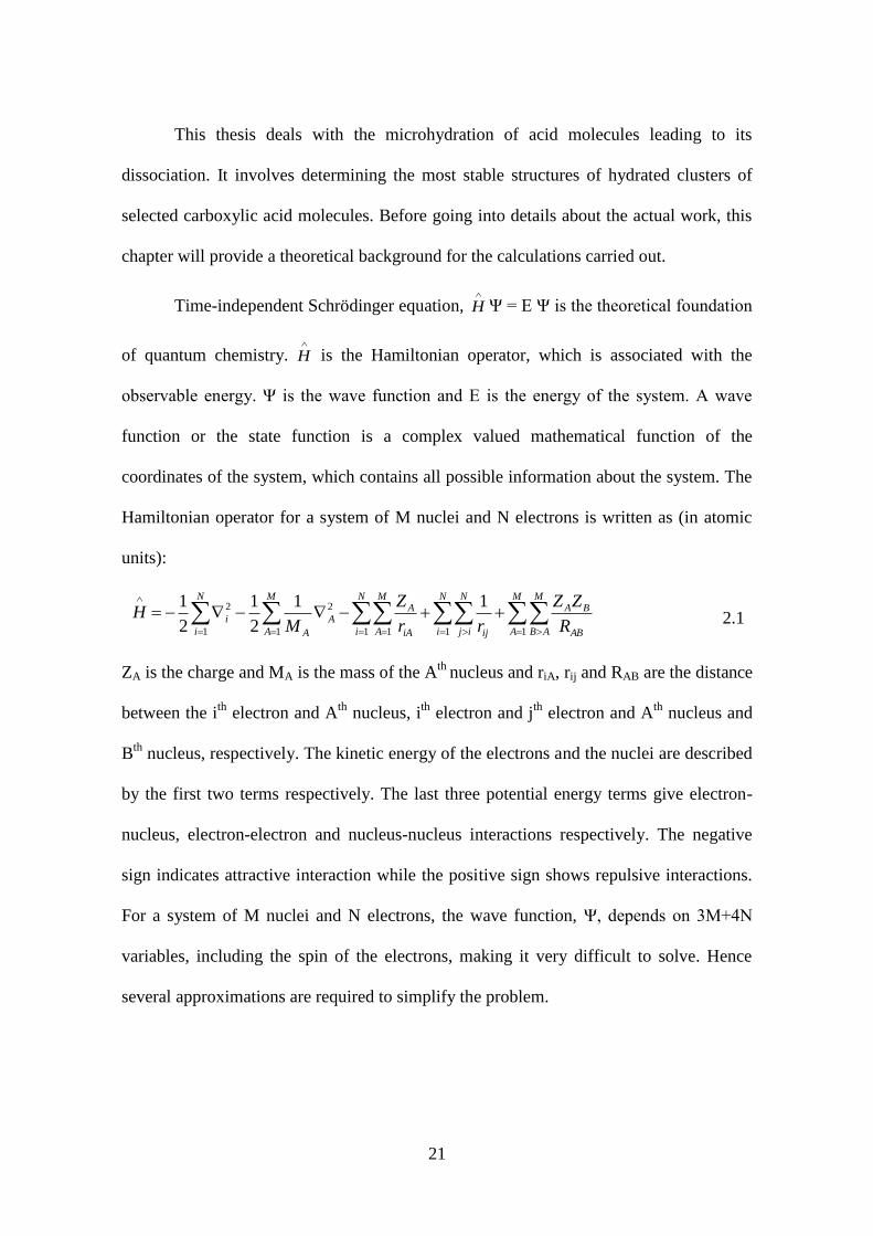
21
This thesis deals with the microhydration of acid molecules leading to its
dissociation. It involves determining the most stable structures of hydrated clusters of
selected carboxylic acid molecules. Before going into details about the actual work, this
chapter will provide a theoretical background for the calculations carried out.
Time-independent Schrödinger equation,
H Ψ = E Ψ is the theoretical foundation
of quantum chemistry.
H is the Hamiltonian operator, which is associated with the
observable energy. Ψ is the wave function and E is the energy of the system. A wave
function or the state function is a complex valued mathematical function of the
coordinates of the system, which contains all possible information about the system. The
Hamiltonian operator for a system of M nuclei and N electrons is written as (in atomic
units):
M
A
M
AB AB
BAN
i
N
ij ij
M
A
N
i
M
A iA
AA
A
N
i
iR
ZZ
rr
Z
MH
111 1 1
2
1
2 11
2
1
2
1
2.1
ZA is the charge and MA is the mass of the Ath
nucleus and riA, rij and RAB are the distance
between the ith
electron and Ath
nucleus, ith
electron and jth
electron and Ath
nucleus and
Bth
nucleus, respectively. The kinetic energy of the electrons and the nuclei are described
by the first two terms respectively. The last three potential energy terms give electron-
nucleus, electron-electron and nucleus-nucleus interactions respectively. The negative
sign indicates attractive interaction while the positive sign shows repulsive interactions.
For a system of M nuclei and N electrons, the wave function, Ψ, depends on 3M+4N
variables, including the spin of the electrons, making it very difficult to solve. Hence
several approximations are required to simplify the problem.

22
2.1 Born-Oppenheimer Approximation
The huge mass difference between the nuclei and the electrons enables the
separation of motion of electrons from that of the nuclei. The Born-Oppenheimer
approximation assumes that the nuclei are stationary during electronic motion. Thus,
kinetic energy term of the nuclei becomes zero and nucleus-nucleus repulsion term
becomes constant in the Hamiltonian. The Hamiltonian is now reduced to electronic
Hamiltonian:
N
i
N
ij ij
N
i
M
A iA
AN
i
ielecrr
ZH
11 11
2 1
2
1
2.2
Total energy can then be defined as the sum of electronic and nuclear energy: Etot= Eelec+
Enuc. However, Schrödinger equation cannot be solved exactly for many-electron systems.
As most of the practical problems involve systems with more than one electron, attempts
were made to develop other theoretical models that could describe such systems and help
in understanding as well as predicting chemical phenomena.
2.2 Theoretical model chemistry
A theoretical model is a well-defined mathematical procedure of simulation that
can approximately describe a complex process. John Pople, in his Nobel lecture on
quantum chemical models, described the features of theoretical models.[104]
He
mentioned five stages in the development and use of a model. These are (a) determining
the level of accuracy of the systems predicted;(b) precisely formulating an approximate
mathematical procedure to describe a system, while making the formulation as general
and continuous as possible;(c) implementing the formulated method as efficient and
easily used computer programs;(d) verifying the model against experimental data; and (e)

23
using the model to predict unknown chemical problems. Models can be broadly
classified as semi-empirical and ab initio, on the basis of the parameters used in the
models.
Models which include empirical parameters, obtained from experiments, are
classed as semi-empirical methods. The ab initio methods do not rely on any empirical
values. Instead they use fundamental constants of physics to define a system. When
applied to chemistry, a model describes the molecular behavior of a system. Electronic
structure theory deals with the description of electrons in atoms or molecules. Hartree-
Fock (HF) theory is the basic and fundamental method on which most of electronic
structure theory is based on.
2.3 Hartree Fock (HF) model
The concept of orbital theory of many electron systems was popularized by Slater,
Hartree and Fock. The many electron wave function for a molecule having n electrons
can be written as a Slater determinant:
)...]))()(det[(!
12211
n 2.3
where i is the orthonormal molecular orbital and and are spin functions.
The Hamiltonian operator applying on the Slater determinant gives the energy as[105]
:
Nelec
ij
nnjijjiji
Nelec
i
ii VKJhE )||||(2
1||
1
2.4
where,
Nnuclei
a ia
aii
rR
Zh
||2
1 2
2.5
)2(|)1(|||
1|)1()2(|
21
jiijirr
J
2.6

24
)2(|)1(|||
1|)1()2(|
21
jjijirr
K
2.7
J is the coulomb integral while K is the exchange integral. 1 and 2 represent electron
coordinates while i and j are molecular orbitals. Vnn is the nuclear repulsion term,
which is a constant.
As the actual wave functions are not known, trial wave functions are used to
solve the Hartree Fock equation iteratively. This is done by Self Consistent Field (SCF)
method. It involves solving Schrödinger equation iteratively, by choosing an
approximate Hamiltonian to operate on the trial wave function to obtain more accurate
wave functions, applying variation principle, until the results converge. The resulting
energy is then consistent with the orbitals that generate it and hence the procedure is
called Self Consistent Field. Variational principle states that the ground state energy is
always less than or equal to the expectation value of
H :
|| H E0 2.8
E0 is the ground state energy.
In case of HF model, motion of each electron is described by a single particle function
and it does not explicitly depend on the motion of other electrons.
2.4 Basis Sets
Molecular orbitals are approximated as the linear combination of basis functions,
which are one particle functions. A complete basis set is not an approximation. However,
for a complete basis set, infinite number of basis functions must be used, which is not
practical. A finite basis set defines the components of molecular orbitals in the direction
of the selected basis functions. As the computational cost for ab initio methods scales as
M4, where M is the number of basis functions, there is a trade-off between expense and

25
accuracy. There are mainly two types of basis functions: the Slater Type Orbital and the
Gaussian Type Orbital.[105]
The STO is of the type:
rn
mlmln erNYr 1
,,,, ),(),,( 2.9
where, N is the normalization constant, mlY , is the spherical harmonic function, r is the
radial function and ζ controls the width of the orbital. STOs do not have radial nodes.
Radial nodes are introduced by taking linear combination of STOs. But, analytical
calculation of three and four-centre two-electron integrals cannot be done in case of
STOs. Hence the much more popular GTOs are used which are easier to calculate. GTOs
are represented as:
222
,,,, ),(),,( rln
mlmln erNYr 2.10
The r2 in the exponential causes the GTO to poorly represent proper behavior near the
nucleus and also at the tail of the wave function. Hence, a combination of GTOs, i.e.,
Contracted type GTOs (CGTO) are mostly used, which mimics the STOs but are easy to
calculate.
Depending on the combination of the GTOs there are several classifications of
basis sets like the minimal basis sets, which contain just one basis function for each
atomic orbital in the atom; the split valence basis sets which treat the core and valence
orbitals differently; Pople basis sets, Dunning‟s correlation-consistent basis sets, which
are optimized using correlated wave functions, etc. There are also the plane wave basis
sets, which are used to describe periodic systems.
The molecular orbitals, i , are expanded in terms of linear combination of basis
functions, χ.
basisM
ii C
2.11
Applying the Fock operator the energy is obtained as:

26
basisbasis M
ii
M
ii CCF
2.12
The Roothan-Hall equations for closed shell systems are obtained by multiplying the
LHS by a specific basis function followed by integration.
|||basisbasis MM
CFC 2.13
This can be written in the matrix notation as:
FC=SCε 2.14
F =
|| F and S = | 2.15
F matrix contains the Fock matrix elements and S matrix contains the overlap elements
between basis function. Roothan-Hall equation gives the Eigen values of the Fock matrix.
The unknown molecular orbital coefficients are obtained by diagonalzing the Fock
matrix. However, to determine the Fock matrix, the molecular orbital coefficients must
be known. So a self consistent procedure is followed wherein, some guess values are
taken as the MO coefficients to form a Fock matrix which is diagonalized to obtain new
coefficients. This process is repeated till a self consistent field solution is obtained.
However, in case of HF theory, the trial wave function consists only of a single
Slater determinant. Thus HF scheme does not take into account electron correlation
completely, and is referred to as mean-field approximation, as the electron-electron
repulsion is accounted for only in an average manner. HF accounts for correlation of
electrons with same spin. However, electrons of opposite spins are uncorrelated. With
sufficiently large basis set, HF method has an accuracy of 99%. Unfortunately, most of
chemical phenomena belong to the 1% that is not accounted for by the HF method.
Dispersion forces are a consequence of electron correlation and thus HF fails for systems
with non-covalent interactions. Hence electron correlation methods are important.

27
2.5 Electron correlation methods
The exact representation of a wave function requires infinite number of
determinants, while HF theory uses just one, which is a crude approximation. The
difference between the HF and lowest possible energy for a given basis set is called the
correlation energy. Addition of determinants, for a given set of molecular orbitals
implies considering different electron configurations, which are essential for better
description of the system. There are several methods to introduce electron correlation.
The Configuration Interaction and Perturbation theory consider electron correlation, by
including more Slater determinants to describe the system. As corrections are added to
the HF method, these correlation techniques are called post HF methods.
2.5.1 Configuration Interaction (CI)
In CI, the wave function is expressed as a linear combination of Slater
determinants. The coefficients are determined variationally, by minimizing the total
energy. A complete CI, though accurate, is impractical as it involves arrangement of all
electrons in all possible ways in a complete basis set. A full CI also considers all possible
arrangements of all the electrons, but in a finite basis set. This becomes computationally
very costly and impractical except for very small systems. CI singles and doubles (CISD)
and CI singles, doubles and triples (CISDT) are much more popular. They truncate the
expansion according to the excitation level. This is possible because the contribution of
the determinants to the system drops off with higher excited states. In the CISD, single
and double excitations of electrons are considered and in CISDT, single, double and
triple excitations are considered.

28
2.5.2 Perturbation Theory
In Perturbation Theory, the correction to the HF energy is added as a Taylor
expansion in powers of a perturbation parameter. Depending on the perturbations added,
there is Møller-Plesset (MPn) theory, Coupled Cluster (CC) theory etc. As the
computational cost as well as accuracy increases with increase in order of perturbation,
there is a trade off and expansion is truncated after achieving suitable level of accuracy.
Post-HF methods are costly and in most cases they cannot be applied for
geometry determination as the memory requirements will be very high. Therefore the use
of Density Functional Theory (DFT) is very popular. The DFT is based on the electron
density of the system. The electron correlation is incorporated into DFT as a function of
electron density. For an N electron system, while the wave function depends on 4N
variables (3N spatial and N spin variables), the electron density depends only on three
spatial coordinates, irrespective of the number of electrons. DFT is computationally
comparable to HF and provides better results. However, there is no systematic way of
improving the results obtained through DFT, to get exact solution.
2.6 Density Functional Theory
The Hohenberg and Kohn theorems (HK theorems) form the basis of DFT. The first HK
theorem states that the external potential applied on a system, in ground state, is defined
as a unique functional of electron density. The external potential determines the
Hamiltonian,
H , axiomatically making it a unique functional of density. The second HK
theorem states that the approximate electron density associated with an external potential
is always greater than or equal to the exact ground state energy.
Thus, the ground state energy of the system can be expressed as:
][][][ 0000 Neee EETE 2.16

29
T is the kinetic energy, Eee is the electron-electron interaction energy and ENe is the
nucleus-electron interaction energy. The first two terms are independent of number of
electrons, electron-nucleus distance and nuclear charge and are universally valid while
the ENe depends on those variables and is therefore system dependent. The independent
parts of the above equation is the Hohenberg-Kohn functional, FHK[ρ]. The exact HK
functional is not known. The Eee term can be written as the sum of coulomb term and a
term containing all non-classical contributions to the electron-electron interaction. The
energy is then determined using variational principle. To improve the results obtained,
Kohn-Sham orbitals have been developed. Here, the hypothetical system consists of non-
interacting electrons, having ρ equivalent to the real system, which experiences average
repulsion field from other electrons. The total energy is then written as:
][)()]()(V[][T][ exts XCEdrrrJrE 2.17
where, Ts[ρ] is the electron kinetic energy of the hypothetical system, )(rJ
gives the
classical coulomb interaction between electrons and )(V rext
is the potential due to the
nuclei.
Kinetic energy is computed in terms of one electron function:
N
i
iiST1
2 ||2
1][ 2.18
A A
Aext
rR
ZV
|| 2.19
'|'|
)'()( dr
rr
rrJ
2.20
r and are 'r the coordinates of the two electrons and ZA is the nuclear charge. EXC
contains all the other contributions to energy such as electron exchange, correlation
energy, correction for self-interaction and the difference in kinetic energy between

30
interacting and non-interacting system. The Hamiltonian for the non-interacting system
is:
N
irS
N
i
isi
VH)(
2
2
1 2.21
The non-interaction fictitious system is represented by Kohn-Sham orbitals, i . They are
determined using one-electron Kohn-Sham operator, KSf .
iii
KSf
2.22
)(
2
2
1rS
KS Vf
2.23
Vs is the effective potential of non-interacting system.
The major part of kinetic energy can thus be computed and the residual part is merged
with non-classical EXC term. EXC is the unknown term and different approximations for
its determination have led to the development of several density functionals.
2.7 Microhydration-Theoretical background
The correlation between electronic structure of a system and its properties forms
the basis of chemistry. First principle calculations and experiments act complementary to
each other in determining the structure and properties of chemical systems.
Computational assistance is particularly helpful in cases were experimental
determination of structure and properties are difficult. Microhydration refers to step-wise
addition of water molecules to a solute molecule. The crucial step of in-silico studies of
microhydration is the geometry determination of the microhydrated cluster. Each
conformation of the molecular cluster is associated with a definite energy. The variation
of the energy with respect to change in geometry of the molecule, within Born-

31
Oppenheimer approximation, is given by its potential energy surface. The minimum in
the potential energy surface of a given system is obtained through geometry optimization.
Optimization in general refers to determining stationary points of a function,
where the first derivative of energy with respect to the geometric coordinates of the
system, is zero. Geometry optimization essentially predicts the equilibrium energy
structure of a system. The minimum energy configuration of a system is determined
iteratively, by minimizing the force on the comprising atoms in each step. Several
procedures are available to locate minimum energy structures, like Steepest Descent (SD)
method, Conjugate Gradient (CG) method, Newton-Raphson (NR) method etc.
In SD method, the search direction is defined along the negative gradient.
di= -gi 2.24
Though there is a guarantee to approach a minimum, there is an oscillation around the
minimum path. Also the rate of convergence drops as the minima is reached.
The CG method improves on this by performing the search along a line that is
conjugate to the direction of the previous search.
di= -gi+idi-1 2.25
values can be chosen in several ways, depending on the method. CG method is not
suitable for non-linear surfaces.
In the NR method, the function is approximated as a quadratic surface and
minimum is determined. The true function is expanded about the current position, 0x up
to second order and its gradient is made zero.
2
000 )(2
1)()f()f( xxHxxgxx 2.26
gHxx 1
0)( 2.27
32
i
i
i
i
fxx
'' 2.28
where Hessian, H, is the diagonal in the coordinate system ( 'x ). fi is projection of
gradient along Hessian Eigen vector with Eigen value εi. Since the real function may
have terms beyond second order, the NR method iteratively approaches towards
stationary point. NR method always converges to the nearest local minimum. Hence care
should be taken in preparing the initial geometries to get the correct configuration. Many
optimization algorithms in quantum chemical software packages use some variant of the
NR method. One such is the “Berny” algorithm developed by Bernhard Schlegel[106]
,
which is used in the present study.
In Berny optimization, the local minimum on the potential energy surface is
located using the forces on the atoms as well as the Hessian matrix. Since explicit
calculation of the Hessian is costly, an approximated Hessian is constructed at the
beginning, applying valence force field and is updated during each iteration, using the
energies and gradients calculated in each step.
The optimized geometry can be a local/global minimum or a saddle point on the
potential energy surface. The first (gradient) and second (Hessian) derivatives of energy
of the system with respect to geometrical parameters, provides information about the
nature of the minima. The negative of the gradient gives the vector of forces on the
atoms. The eigenvectors of the mass-weighted Hessian matrix gives the normal modes of
vibration. A minimum in the potential energy surface has zero gradient and all positive
vibrational frequencies. A first order saddle point (Transition State) has maximum in one
direction and minimum in all other directions in the potential energy surface. Thus, it has
zero gradient and one negative vibrational frequency. Similarly, nth order saddle point
will have zero gradient and n imaginary frequencies.
33
All these methods can be implemented computationally. Manual calculations of
even the simplest systems would be very time consuming and complicated. With the
evolution of computers into easily accessible machines with large memory and
computing powers, the field of computational chemistry has expanded and relied upon
by many industries. Many modern chemistry softwares have graphical interfaces and
most of them can be used without much training in theoretical chemistry.

Chapter 3
3
Structures of Hydrated Carboxylic acid Clusters
The underlying physical laws necessary for the mathematical theory of a
large part of physics and the whole of chemistry are thus completely
known, and the difficulty is only that exact application of these laws
leads to equations much too complicated to be soluble.
-Paul A. M. Dirac (1929)
35
This chapter deals with structures of the different size hydrated clusters of
trifluoroacetic acid (tfa), acetic acid (acoh), benzoic acid (bza) and oxalic acid (oxa)
molecules. The addition of the solute acid molecule to solvent water pool causes a
reorganization of the immediate neighboring water molecules to accommodate the solute
molecule in their hydrogen bonded network. The electron distribution of the added acid
molecule plays the key role in the formation of hydrogen bonded network of water
molecules around the solute. The hydrogen bonding interactions between the acid
molecule and the solvent water molecules determine the shape of the hydrated clusters,
and hence their properties. Thus determining the geometry of the hydrated acid clusters
is important in understanding their properties. Study on microhydration of formic acid
has been previously reported.[107]
In the present thesis, the electronic effect of the acid
molecule on the geometry of the hydrated acid clusters is examined. The effect of
electron donating and withdrawing groups is studied by considering trifluoroacetic acid
and acetic acid hydrated clusters respectively. Benzoic acid-water system is investigated
to determine the effect of an aromatic ring. Microhydrated clusters of the simplest
dicarboxylic acid, namely, oxalic acid, are also studied.
The effect of bulk solvation on the acid molecule is examined. Carboxylic acids
are known to form dimers in the vapor phase as well as in non-polar solvents. The
transition of dimer to microhydrated clusters is also discussed. This chapter describes
how the initial guess structures of the hydrated clusters are chosen, the different local
minima obtained, the minimum energy structures obtained for each size acid cluster and
the geometric parameters of the minimum energy structures. Comparison is made with
available experimental data. The minimum number of water molecules needed to ionize
each acid molecule studied is predicted and reported.

36
The task of obtaining the exact potential energy surface for system with weak
interactions, like hydrogen-bonded systems, is a herculean, as there would be numerous
local minima, and the potential energy surface is shallow, especially for large size
clusters. Thus, choice of initial guess structures is very important. As the size of the
hydrated acid clusters increase, number of possible initial guess structures becomes very
large and the risk of missing out important structures becomes high. Hence, the studies
are limited to hydrated clusters with maximum eight water molecules.
3.1 Theoretical method
As the geometry, and hence properties of hydrated clusters depend very much on
hydrogen bonding, it is important to choose a level of theory that describes these
interactions accurately. Hence, all optimization calculations are done using long range
corrected hybrid density functional, ωB97X-D, including empirical atom-atom
dispersion corrections.[108]
Dunning‟s correlation consistent, augmented double zeta set
of basis functions is used. The present level of theory is decided based on the bench-
marking studies carried out on a few selected systems (described in detail in section 3.2.1)
as well as from reported study on carboxylic acid.[107]
The equilibrium structures for each
size hydrated carboxylic acid clusters, RCOOH.nH2O, are obtained by geometry
optimization of several possible initial guess structures, following Newton Raphson
algorithm. The choice of guess structures is important as the geometry optimization
following this algorithm does not ensure that the geometry corresponding to the global
minimum is achieved. The input geometry converges to the nearest local minimum
energy conformer. The objective is to determine the minimum number of water
molecules needed for breaking the hydroxyl bond of the acid molecule. The initial
geometries are prepared so as to facilitate the dissociation of the O-H bond of the acid
molecule and maximize solute-solvent as well as solvent-solvent interactions. The water

37
molecules are placed in such a way that they form hydrogen bonding with the carbonyl
oxygen, hydroxyl oxygen as well as the hydroxyl hydrogen of the acid molecule.
Optimization of the input geometries, without any symmetry restrictions, results in
various equilibrium conformers of which, the zero-point energy corrected minimum
energy conformer is chosen for each size hydrated cluster. To obtain initial guess
structures, ab initio Molecular Dynamic simulations have been carried out in a few cases.
All the MD calculations are performed using Quantum ESPRESSO software, a plane
wave based implementation of density functional theory.[109]
The electronic exchange
and correlation potential are described by the generalized gradient approximation (GGA)
as parameterized by Perdew-Burke-Ernzerhof (PBE).[110]
Projector-Augmented Wave
(PAW) methods are used for the elemental constituents.[111]
The simulations are allowed
to run for 2000 ionic steps with 1fs time step. The cut-off energy for the plane wave
basis set was fixed at 600 eV. The molecule is placed in a unit cell of fixed volume,
ensuring that the periodically repeating clusters do not interact with each other. The
minimum energy structure and all the structures with relative energy within 200meV (~5
kcal/mol) of the minimum energy structure are selected and optimized at ω-B97XD/aug-
cc-pVDZ level of theory. Hessian calculations are done to identify the nature of the
optimized geometries, obtain thermodynamic parameters and to simulate the IR spectra
of the microhydrated clusters. In cases where the optimized geometry has normal modes
with imaginary frequency, they are optimized further to obtain only positive frequencies.
The molecular electronic calculations are done on a Linux cluster platform, with general
ab initio quantum package, GAMESS.[112]
Structure and IR spectra of the microhydrated
systems are visualized using MOLDEN.[113]

38
3.2 Results and Discussion
For easy identification, the hydroxyl oxygen of the acid molecule studied is
referred as O1, the carbonyl oxygen of the acid molecule is referred as O2 and the
oxygen of the water molecule, closest to the hydroxyl hydrogen of acid molecule, is
referred as O3 in case of trifluoroacetic acid (tfa), acetic acid (acoh) and benzoic acid
(bza).In case of oxalic acid (oxa), O1 and O2 are the two hydroxyl oxygen atoms of the
acid molecule and Oa and Ob are the oxygen atoms on the water molecule closest to O1
and O2 respectively. In the following discussions, these conventions will be used.
3.2.1 Benchmark studies
To decide upon a suitable level of theory, geometrical parameters of isolated
water molecule and trifluoroacetic acid molecule, calculated using different DFT
functionals and MP2 method are compared with available experimental data.
Trifluoroacetic acid is chosen for benchmark studies as experimental data of hydrated
clusters (up to three water molecules) of trifluoroacetic acid are available from literature.
For benchmark studies, popular hybrid density functional, B3LYP; a functional
including dispersion correction, ωB97X-D and a modern meta-hybrid GGA DFT
functional that accounts for non-covalent interactions, M06-2X, are tried. Selected
Geometrical parameters for minimum energy equilibrium structures of CF3COOH and
H2O molecules calculated applying different DFT functional and MP2 method are
supplied in Table 3.1 and are compared with reported experimental values. As the size of
the hydrated clusters can be very large, Dunning‟s correlation consistent double zeta set
of atomic basis functions, augmented with diffuse functions (aug-cc-pVDZ) is used as
trade-off between accuracy and computational expense and is seen to serve well to
describe these hydrated systems. From Table 3.1, it is seen that B3LYP performs well for
trifluoroacetic acid as well as water molecule. But this functional does not involve
39
dispersion corrections which is very important for systems with non-covalent
interactions, and is hence known to fail for such systems. As the systems studied are
hydrogen bonded systems, B3LYP is not chosen, because this functional cannot treat
such systems accurately. The gross performance of the selected DFT functionals and
MP2 method are similar in case of these two molecules. Calculated rotational constants,
applying rigid rotor model, are compared to the available experimental data. The
comparison of rotational constants is given in Table 3.2. For mono and di-hydrates of
trifluoroacetic acid (tfa), rotational constants calculated using B97X-D are within 1%
error with respect to experimental values, while for tri-hydrate the percentage-error is
higher.

40
Table 3.1 Comparison of geometrical parameters and hydroxyl stretching frequency of isolated water and trifluoroacetic acid molecule,
calculated at different levels of theory using aug-cc-pVDZ basis set, with available experimental data
Method
CF3COOH H2O
rC-F
(Å)a
rC-C
(Å)
rC=O
(Å)
rC-O
(Å)
rO-H
(Å)
C-F
(cm-1
)
C=O
(cm-1
)
O-H
(cm-1
)
rO-H
(Å)
HOH
(degree)
symO-H
(cm-1
)
asymO-H
(cm-1
)
MP2 1.349 1.544 1.212 1.349 0.974 1182 1819 3736 0.966 103.8 3804 3937
B97X-D 1.337 1.547 1.197 1.333 0.967 1216 1898 3821 0.961 104.7 3881 3992
LC-wPBE 1.338 1.540 1.196 1.331 0.969 1208 1922 3813 0.962 105.1 3871 3982
M062X 1.334 1.542 1.194 1.333 0.969 1248 1928 3798 0.962 104.8 3867 3977
B3LYP 1.345 1.550 1.200 1.341 0.972 1162 1850 3735 0.965 104.8 3795 3905
Exptb 1.325 1.546 1.192 1.342 0.960 1240 1792 3587 0.957 104.5 3657 3756
aaverage of the three C-F bonds;
bReferences
[114],
[115]
However, when compared to other DFT functionals and MP2 method, overall performance of B97X-D is better. Also, as
mentioned earlier, previous study has shown that B97X-D works well for microhydrated formic acid clusters.[107]
Thus, B97X-D/aug-cc-pVDZ is chosen as the level of theory for geometry optimization of all systems presented in this thesis.
The bond length and bond angle of free H2O molecule at B97X-D/aug-cc-pVDZ level of theory is 0.961Å and 104.76o, which is
very close to the experimentally determined values of 0.9575 Å and 104.51o, respectively.

41
Table 3.2 Comparison of rotational constants (A, B, and C in MHz) for hydrated clusters of trifluoroacetic acid, CF3COOH.nH2O (n = 1−3)
calculated at different levels of theory
Rotational
constant Method CF3COOH CF3COOH.H2O CF3COOH.2H2O CF3COOH.3H2O
A/MHz
MP2 3771 3737 2506 1711
wB97XD 3848 3811 2549 1905
LC-wPBE 3849 3815 2559 1769
M062X 3855 3821 2562 1910
B3LYP 3805 3737 2530 1752
Expta 3865.13 3835.11 2533.71 1733.35
B/MHz
MP2 2471 1085 716 499
wB97XD 2484 1090 724 497
LC-wPBE 2495 1100 727 494
M062X 2501 1103 730 504
B3LYP 2463 1085 719 488
Expta 2498.79 1082.58 718.46 493.41
C/MHz
MP2 2044 993 621 428
wB97XD 2063 1000 628 465
LC-wPBE 2071 1009 631 416
M062X 2073 1010 633 477
B3LYP 2046 993 624 411
Expta 2075.2 993.78 622.5 414.75
aReference
[73]

42
3.2.2 Macroscopic Hydration of Carboxylic Acids
To determine the effect of macroscopic hydration of acid molecule, optimization
of trifluoroacetic acid, acetic acid, benzoic acid and oxalic acid molecules are done using
macroscopic solvation models. Macroscopic solvation models like Polarized Continuum
Model (PCM) and Solute Density Model (SMD) are used applying B97X-D/aug-cc-
pVDZ level of theory. In PCM model, a cavity is formed by the union of spheres which
are centered on each atom and the electrostatic interaction, between the wave function of
the solute and a dielectric model of the bulk solvent, is determined.[1]
The solute electron
density interacts with the solvent, taken into account as a dielectric continuum, in case of
the SMD model.[2]
Trifluoroacetic acid, acetic acid, benzoic acid and oxalic acid
molecules are known to dissociate in water to form corresponding carboxylate anion.
However, the O-H bond length of acetic acid molecule, calculated using PCM and SMD
models are 0.968 and 0.970 Å respectively, indicating the acid is still undissociated.
Similar results are observed in case of trifluoroacetic acid, benzoic acid and oxalic acid.
The O-H bond length of trifluoroacetic acid, benzoic acid and oxalic acid calculated
using SMD model is 0.972, 0.969 and 0.974 Å respectively, which is not correct. Hence
for an accurate description of the hydration of carboxylic acids, explicit solvation models
must be adopted at least for the primary shell of solvation. This is especially needed, as
the systems studied are hydrogen bonded clusters and the implicit solvation models fail
to take into account such kind of solute-solvent interactions.
3.2.3 Dimerization versus Microhydration of Carboxylic acid
Before going into details about the possible structures of acid-water molecular
clusters, a brief note on the relative stability of carboxylic dimers and microhydrated acid

43
clusters is interesting to know. It is well known that carboxylic acids form dimeric
complexes in gas phase and non-polar solvents. Hence to fully understand the
fundamental nature of acid dissociation, the breaking of the acid dimers, to form
microhydrated acid clusters, needs to be studied. It has applications in biological as well
as environmental modeling. The number of water molecules needed for the dimer to
break and form micro-hydrated acid clusters is studied for the two cases: formic and
acetic acid.
The free energy of formation calculated for formic and acetic acid systems shows
that at room temperature and pressure, the microhydrated systems are not stable.
However, at lower temperatures and pressures, all the systems studied are predicted to be
stable. It may be noted that these systems will be stable in the upper atmosphere. It is
observed that with four or more water molecules, the microhydrated clusters are formed
predominantly over their dimer counterparts.[116]
The calculated interaction energy also
supports the above statement. Another engaging point noticed is that when a single water
molecule is added to the dimers of formic or acetic acid, the most stable conformer is the
one with the water molecule incorporated in between the two acid molecules of the dimer.
In other words, the presence of a single water molecule can break the carboxylic acid
dimer.
3.2.4 Structure
3.2.4.1 Trifluoroacetic Acid (tfa)
The electron withdrawing -CF3 group makes the removal of proton of tfa easier,
compared to formic acid. The minimum energy equilibrium geometry of tfa is given in
Figure 3.1(a), with O-H bond length of 0.968Å. Computed geometrical parameters of
free tfa in the gas phase are noted to be fairly close to the experimental data. The largest

44
deviation in bond length is predicted for C−F bond, and it is 0.017 Å longer than the
reported experimental data when the calculation is carried out without any symmetry
restriction (see Table 3.1). It is to be noted that the present calculated O−H bond distance
in free tfa at ωB97X-D/aug-cc-pVDZ level is the same as the earlier value calculated
applying MP2/6-311++G(2df,2pd)level.[73]
Minimum energy equilibrium structures of
tfa and hydrated clusters of tfa (tfa.nH2O, n=1-7) are given in Figure 3.1. Only one
equilibrium structure is obtained for the mono-hydrated cluster, and is shown as Figure
3.1(b). Addition of the first solvent water molecule to tfa increases its C=O bond
distance by 0.01 Å and O−H bond distance by 0.024 Å. The present calculated value of
O-H bond length compares well with the previous reported value.[73]
The calculated
distance between the transferring H atom and O atom of the neighboring solvent H2O
molecule is 1.713 Å, which is less than that observed in case of formic acid.[107]
The
present calculated values are very close to previously reported values at B971 and MP2
levels of theory.[72]
The C−O bond of tfa is also shortened significantly on addition of a
water molecule to tfa. The most favored structure for di-hydrated cluster of tfa is
displayed as Figure 3.1(c). The present calculated values of O1-H and H-O3 bond
lengths are lesser than the earlier reported MP2 values.[72,73]
a b c
g he f
1
2
1
1 1
1
1 1
2 2
2 2
2 2
33
33
3 3
1.592
1
2
3
1.481 1.419
d
Figure 3.1 Minimum energy equilibrium structures of (a) CF3COOH; (b) CF3COOH.1H2O; (c)
CF3COOH.2H2O; (d) CF3COOH.3H2O; (e) CF3COOH.4H2O; (f) CF3COOH.5H2O; (g)

45
CF3COOH.6H2O and (h) CF3COOH.7H2O calculated at B97X-D/aug-cc-pVDZ level of theory.
O1and O2 are hydroxyl and carbonyl oxygen atoms of acid molecule. O3 is the oxygen atom on
the water molecule closest to hydroxyl hydrogen atom of acid molecule. Yellow balls represent
fluorine, red balls represent oxygen, grey balls represent carbon and blue balls represent
hydrogen atoms.
The minimum energy structure of the five equilibrium conformers obtained for
tfa.3H2O is shown in Figure 3.1(d). The three water molecules form a hydrogen bonded
closed-ring motif, which is hydrogen bonded to the hydroxyl hydrogen and carbonyl
oxygen of the acid molecule. The O-H bond length of the acid molecule increases to
1.016 Å and the H bond distance between the H atom of the acid molecule and O atom of
the nearest water molecule is 1.561 Å. For the tetra-hydrate of tfa, tfa.4H2O, the most
stable conformer obtained has hydrogen bonded close ring structure, with hydrogen
bonds between the acid molecule and the solvent water molecules as well as between the
water molecules (see Figure 3.1(e)).
a ΔE = +1.7 b ΔE = +2.8 c ΔE = +3.5
d ΔE = +4.0 e ΔE = +4.6
Figure 3.2 Higher energy equilibrium structures of (a)CF3COOH.6H2O and (b)-(e)
CF3COOH.7H2O showing contact ion pair formation. Zero point energy corrected relative energy
(in kcal/mol) of the conformers with respect to minimum energy conformer of that size cluster is
also given. Yellow balls represent fluorine, red balls represent oxygen, grey balls represent
carbon and blue balls represent hydrogen atoms.

46
There is 0.02 Å increase in O-H bond length and 0.08 Å decrease in H-O3 H-bond length,
relative to the tfa.3H2O. Of the eight equilibrium conformers obtained after optimization,
the minimum energy structure of penta hydrate of tfa, tfa.5H2O, is given in Figure 3.1(f),
where the O-H and H-O3 H-bond distances are1.064 and 1.419 Å, respectively.
Table 3.3 Selected bond lengths of tfa.nH2O, n=0-7, calculated at B97X-D/aug-cc-pVDZ level
of theory. rO1-H, rC=O2 and rH-O3 represent hydroxyl O-H and carbonyl C=O bond of the acid
molecule and hydrogen bond between the hydroxyl hydrogen of acid molecule and oxygen of the
nearest water molecule, respectively.
System
Bond Length
(Ǻ)
rO1-H rC=O rH-O3
CF3COOH.1H2O 0.992 1.207 1.713
CF3COOH.2 H2O 1.009 1.211 1.592
CF3COOH.3 H2O 1.016 1.213 1.561
CF3COOH.4 H2O 1.036 1.217 1.481
CF3COOH.5 H2O 1.064 1.222 1.419
CF3COOH.6 H2O 1.415 1.226 1.057
CF3COOH.7 H2O 1.564 1.228 1.012
As is seen from Figure 3.1(g), the minimum energy equilibrium conformer of tfa.6H2O
shows dissociation of trifluoroacetic acid and formation of contact ion pair. The O-H
bond length of the acid molecule increases significantly to 1.415 Å and there is a drastic
reduction in the bond distance between the transferring H atom of the acid molecule and
the O atom of the nearest water molecule, to 1.057 Å, indicating proton transfer from
acid molecule to solvent water molecules. A higher energy equilibrium conformer of
tfa.6H2O containing contact ion pair is given in Figure 3.2(a). In case of tfa.7H2O also,
the most stable conformer shows the dissociation of the acid molecule, the O1-H and H-
O3 bond distances being 1.564 and1.012 Å respectively (see Figure 3.1(h)). Higher

47
energy ionized conformers (relative energy < 5 kcal/mol with respect to minimum
energy structure) of tfa.7H2O is given in Figure 3.2(b)-(e).
It is observed that at least six water molecules are needed for ionizing
trifluoroacetic acid molecule while seven are needed in case of formic acid molecule.
The hydrated clusters of tfa prefer hydrogen bonded close ring geometry. Selected bond
distance parameters of tfa.nH2O, n=0-7 are given in Table 3.3.[117]
The calculated
rotational constants are comparable to available experimental data (see Table 3.2).
Many higher energy conformers of each size hydrated clusters are obtained for
trifluoroacetic acid molecule. The higher energy conformers of each size cluster of tfa
and their relative energy with respect to the minimum energy conformer of that size is
given in Figure 3.3.
(i) ΔE = +1.0 (ii) ΔE = +1.7 (iii) ΔE =+3.4 (iv) ΔE = +5.0
(v) ΔE = +7.4 (vi) ΔE =+1.2 (vii) ΔE =+1.8 (viii) ΔE =+6.5
(ix) ΔE = +7.3 (x) ΔE =+0.6 (xi) ΔE =+1.9 (xii) ΔE =+2.4
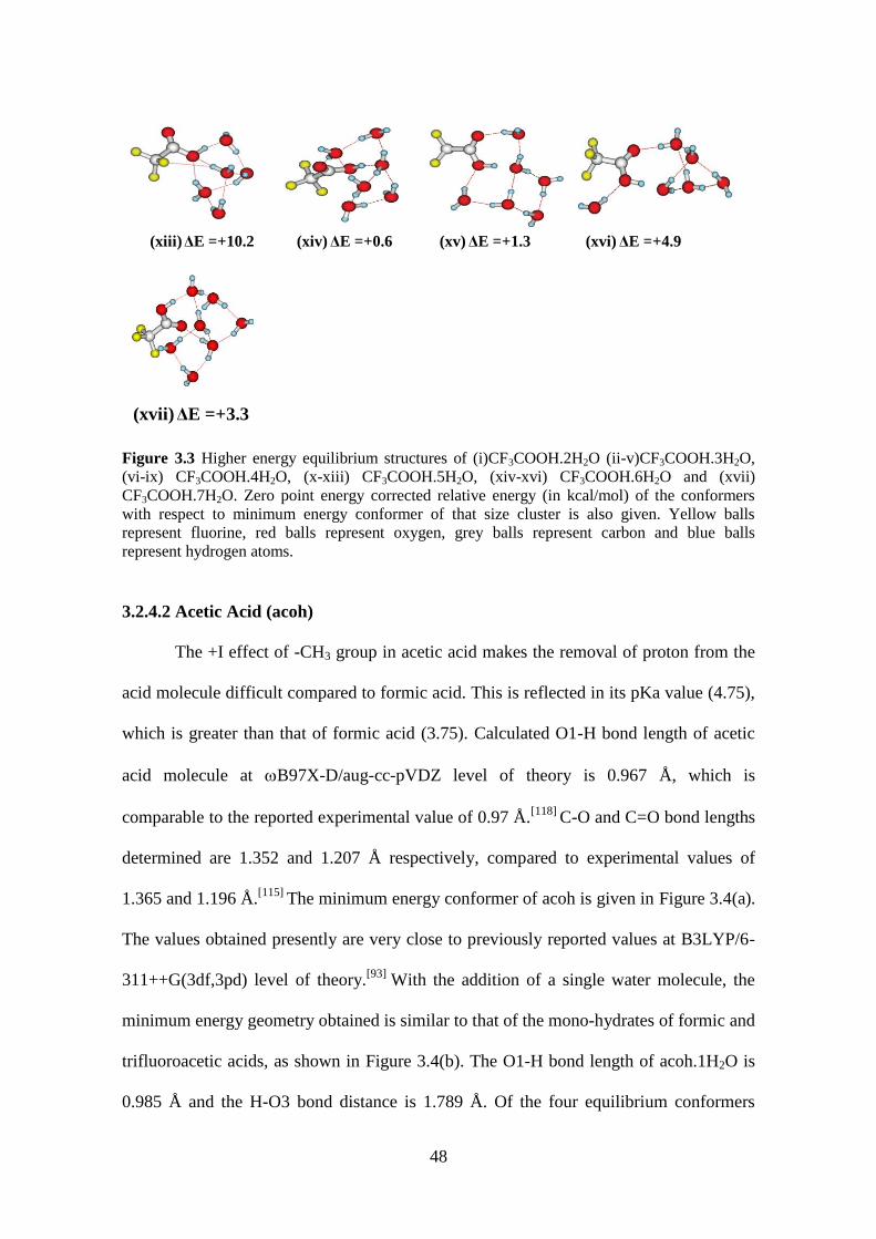
48
(xiii) ΔE =+10.2 (xiv) ΔE =+0.6 (xv) ΔE =+1.3 (xvi) ΔE =+4.9
(xvii) ΔE =+3.3
Figure 3.3 Higher energy equilibrium structures of (i)CF3COOH.2H2O (ii-v)CF3COOH.3H2O,
(vi-ix) CF3COOH.4H2O, (x-xiii) CF3COOH.5H2O, (xiv-xvi) CF3COOH.6H2O and (xvii)
CF3COOH.7H2O. Zero point energy corrected relative energy (in kcal/mol) of the conformers
with respect to minimum energy conformer of that size cluster is also given. Yellow balls
represent fluorine, red balls represent oxygen, grey balls represent carbon and blue balls
represent hydrogen atoms.
3.2.4.2 Acetic Acid (acoh)
The +I effect of -CH3 group in acetic acid makes the removal of proton from the
acid molecule difficult compared to formic acid. This is reflected in its pKa value (4.75),
which is greater than that of formic acid (3.75). Calculated O1-H bond length of acetic
acid molecule at B97X-D/aug-cc-pVDZ level of theory is 0.967 Å, which is
comparable to the reported experimental value of 0.97 Å.[118]
C-O and C=O bond lengths
determined are 1.352 and 1.207 Å respectively, compared to experimental values of
1.365 and 1.196 Å.[115]
The minimum energy conformer of acoh is given in Figure 3.4(a).
The values obtained presently are very close to previously reported values at B3LYP/6-
311++G(3df,3pd) level of theory.[93]
With the addition of a single water molecule, the
minimum energy geometry obtained is similar to that of the mono-hydrates of formic and
trifluoroacetic acids, as shown in Figure 3.4(b). The O1-H bond length of acoh.1H2O is
0.985 Å and the H-O3 bond distance is 1.789 Å. Of the four equilibrium conformers

49
determined for acoh.2H2O, the minimum energy structure has hydrogen bonded closed
ring structure as shown in Figure 3.4(c). The O1-H bond length increases to 0.996 Å and
H-O3 bond distance decreases to 1.675 Å. It must be noted that initial geometries with
hydrogen bonded interactions between water molecules and -CH3 group of the acid
molecule are not considered for the present study as they are not directly relevant to acid
dissociation.
1
2
1
2
3 1.675
0.9961
2
30.9971
2
3
a b c d
1
2
3
1
2
3
0.999
1.638
1
2
3
1
2
3
e f g h
1
2
3
i
Figure 3.4 Minimum energy equilibrium structures of (a) CH3COOH; (b) CH3COOH.1H2O; (c)
CH3COOH.2H2O; (d) CH3COOH.3H2O; (e) CH3COOH.4H2O; (f) CH3COOH.5H2O; (g)
CH3COOH.6H2O (h) CH3COOH.7H2O; and (i) CH3COOH.8H2O. O1and O2 are hydroxyl and
carbonyl oxygen atoms of acid molecule. O3 is the oxygen atom on the water molecule closest to
hydroxyl hydrogen atom of acid molecule. Red balls represent oxygen, grey balls represent
carbon and blue balls represent hydrogen atoms.
Also, optimization of a few such structures of lower hydrates showed that they are higher
in energy compared to the other structures. The minimum energy structure, of the seven
equilibrium conformers of acoh.3H2O, has the three water molecules forming hydrogen
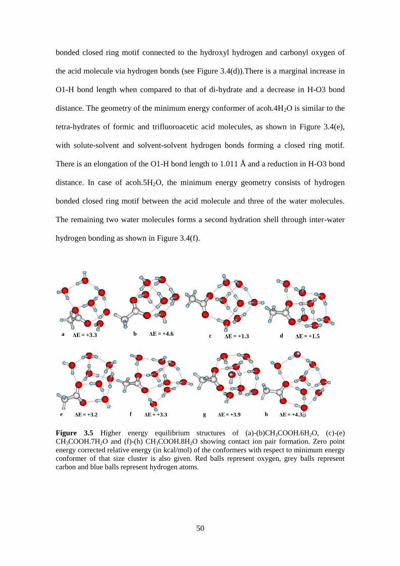
50
bonded closed ring motif connected to the hydroxyl hydrogen and carbonyl oxygen of
the acid molecule via hydrogen bonds (see Figure 3.4(d)).There is a marginal increase in
O1-H bond length when compared to that of di-hydrate and a decrease in H-O3 bond
distance. The geometry of the minimum energy conformer of acoh.4H2O is similar to the
tetra-hydrates of formic and trifluoroacetic acid molecules, as shown in Figure 3.4(e),
with solute-solvent and solvent-solvent hydrogen bonds forming a closed ring motif.
There is an elongation of the O1-H bond length to 1.011 Å and a reduction in H-O3 bond
distance. In case of acoh.5H2O, the minimum energy geometry consists of hydrogen
bonded closed ring motif between the acid molecule and three of the water molecules.
The remaining two water molecules forms a second hydration shell through inter-water
hydrogen bonding as shown in Figure 3.4(f).
ΔE = +3.3 ΔE = +4.6a b c ΔE = +1.3 d ΔE = +1.5
e ΔE = +3.2 f ΔE = +3.3 g ΔE = +3.9 h ΔE = +4.3
Figure 3.5 Higher energy equilibrium structures of (a)-(b)CH3COOH.6H2O, (c)-(e)
CH3COOH.7H2O and (f)-(h) CH3COOH.8H2O showing contact ion pair formation. Zero point
energy corrected relative energy (in kcal/mol) of the conformers with respect to minimum energy
conformer of that size cluster is also given. Red balls represent oxygen, grey balls represent
carbon and blue balls represent hydrogen atoms.

51
For the hexa-hydrate of acetic acid, acoh.6H2O, though the minimum energy structure
has the undissociated acid molecule in it, a few higher energy conformers are obtained
which show contact ion pair formation. The minimum energy structure has O1-H and H-
O3 bond distances of 0.999 and 1.638 Å respectively, as shown in Figure 3.4(g). Higher
energy conformers of acoh.6H2O (relative energy <5kcal/mol with respect to minimum
energy structure) are given in Figure 3.5(a) and (b). The un-ionized minimum energy
conformer of the hexa-hydrate is more stable than the most stable conformer, containing
charge separated ion-pair formation, by 3.3 kcal/mol. Twenty minimum energy
structures are predicted in the case of the hepta-hydrated cluster, acoh·7H2O; out of
which eight structures show formation of a charge-separated ion pair and hydronium ion,
H3O+. The most stable conformer has a cage-like structure (Figure 3.4(h)), and it is more
stable than the most stable ionized structure, shown in Figure 3.5(c), by 1.3 kcal/mol. The
O1-H and H-O3 bond distances are 1.048 and 1.459 Å respectively. With eight water
molecules, the minimum energy structure of acoh.8H2O has an un-ionized cage like
structure, consisting of four and five hydrogen bonded rings of water molecules. O1-H
bond length of 1.019 Å and H-O3 bond distance of 1.559 Å indicate that the acid
molecule still remains undissociated. However, higher energy conformers with charge
separated ion-pairs are observed for the octa-hydrate (see Figure 3.5(f)-(h)).
A general trend observed is that hydrogen bonded closed ring structures are
favored in case of smaller hydrates while cage-like motifs are prominent in higher
hydrates of the acid molecule. The shaping of the structures of the hydrated clusters
involves a delicate balance in the formation of solute-solvent and solvent-solvent
hydrogen bonding. With seven water molecules, twenty equilibrium geometries were
obtained for acoh.7H2O, after optimization, while for octa-hydrate, it is twenty four.
With nine water molecules, number of possible guess geometries becomes very large.

52
Applying the present strategy for determining the global minima is not suitable for larger
size clusters. As the risk of missing out important structures increases with increase in
size of the hydrated cluster, interaction of acetic acid molecule up to eight water
molecules are only considered at present. The formation of ion pair does not occur in the
most stable structure, even in case of acoh.8H2O, though elongation of the O1-H bond is
observed for the hydrates of acetic acid molecule. Selected bond distance parameters of
acoh.nH2O, n=0-8 are given in Table 3.4.[119]
Table 3.4. Selected bond lengths of CH3COOH.nH2O, n=0-8, calculated at B97X-D/aug-cc-
pVDZ level of theory. rO1-H, rC=O2 and rH-O3 represent hydroxyl O-H and carbonyl C=O bond of
the acid molecule and hydrogen bond between the hydroxyl hydrogen of acid molecule and
oxygen of the nearest water molecule, respectively.
System
Bond distance
(Å)
rO1-H rC=O2 rH-O3
CH3COOH 0.967 1.207 -
CH3COOH.H2O 0.985 1.220 1.789
CH3COOH.2H2O 0.996 1.222 1.675
CH3COOH.3H2O 0.997 1.221 1.654
CH3COOH.4H2O 1.011 1.227 1.580
CH3COOH.5H2O 1.007 1.225 1.596
CH3COOH.6H2O 0.999 1.225 1.638
CH3COOH.7H2O 1.048 1.225 1.459
CH3COOH.8H2O 1.019 1.227 1.559
Experimental microwave rotational constants A, B and C of free acetic acid and
its mono and di-hydrates have been reported previously.[74,120]
The calculated rotational
constants, applying rigid rotor approximation, are comparable with the available
experimental data. For free acetic acid molecule the calculated rotational constants, A, B
and C are 11380, 9462 and 5339 MHz respectively and they differ from the experimental
values by +45,-17 and +14 MHz respectively. In case of acoh.1H2O (with calculated A,

53
B and C values of 11064, 2639 and 2169 MHz) the A, B and C rotational constants differ
by +4, +55 and +42 MHz respectively. For acoh.2H2O, A, B and C values observed
experimentally are 4445, 1618 and 1200 MHz respectively. The variations in the
calculated rotational constants, A, B and C, from the experimental values are +84, +28
and +23 MHz respectively.
(i)ΔE = +3.5 (ii)ΔE = +5.7 (iii)ΔE = +5.2 (iv)ΔE = +6.3 (v)ΔE = +7.0
(vi)ΔE = +0.5 (vii)ΔE = +2.3 (viii)ΔE = +2.8 (ix)ΔE = +3.5 (x)ΔE = +3.7
(xi)ΔE = +8.8 (xii)ΔE = +1.0 (xiii)ΔE = +1.3 (xiv)ΔE = +2.2 (xv)ΔE = +3.1
(xvi)ΔE = +4.0 (xvii)ΔE = +4.3 (xviii)ΔE = +4.8 (xix)ΔE = +7.5 (xx)ΔE = +7.6
Figure 3.6 Higher energy equilibrium structures of (i-ii) CH3COOH.1H2O, (iii-v)
CH3COOH.2H2O, (vi-xi )CH3COOH.3H2O and(xii-xx) CH3COOH.4H2O. Zero point energy
corrected relative energy (in kcal/mol) of the conformers with respect to minimum energy
conformer of that size cluster is also given. Red balls represent oxygen, grey balls represent
carbon and blue balls represent hydrogen atoms.
Similar to hydrated clusters of trifluoroacetic acid, several higher energy
conformers for each size cluster are also obtained for acetic acid-water systems. The
higher energy conformers of mono, di, tri and tetra-hydrated clusters and the relative

54
energy of these conformers with respect to the minimum energy conformer of that size
are given in Figure 3.6.
Fifteen higher energy conformers, along with their relative energy with respect to
the minimum energy conformer, obtained for penta-hydrate of acetic acid are shown in
Figure 3.7. Three of these conformers are less than 0.5 kcal/mol higher in energy than
the most stable conformer, implying that the penta-hydrate can exist in those
conformations as well.
(i)ΔE = +0.3 (ii)ΔE = +0.4 (iii)ΔE = +0.5 (iv)ΔE = +0.7 (v)ΔE = +1.6
(vi)ΔE = +1.8 (vii)ΔE = +2.0 (viii)ΔE = +2.3 (ix)ΔE = +2.6 (x)ΔE = +2.8
(xi)ΔE = +3.7 (xii)ΔE = +6.0 (xiii)ΔE = +6.8 (xiv)ΔE = +8.5 (xv)ΔE = +8.5
Figure 3.7(i-xv) Higher energy equilibrium structures of CH3COOH.5H2O. Zero point energy
corrected relative energy (in kcal/mol) of the conformers with respect to minimum energy
conformer of that size cluster is also given. Red balls represent oxygen, grey balls represent
carbon and blue balls represent hydrogen atoms.
Seventeen of the higher energy equilibrium conformers of hexa-hydrate of acetic acid,
not shown previously, are given in Figure 3.8. Structures shown in Figure 3.8 (i)-(iii) are
very close in energy to the minimum energy conformer and hence are possible structures
for the hexa-hydrated cluster of acetic acid. Three ionized conformers of the hexa-

55
hydrate (see Figure 3.8 (xii),(xv),(xvii)-(xix)) are observed to be very high in energy
compared to the minimum energy conformer.
(i)ΔE = +0.1 (ii)ΔE = +0.2 (iii)ΔE = +0.4 (iv)ΔE = +0.7 (v)ΔE = +0.8
(vi)ΔE = +1.2 (vii)ΔE = +1.4 (viii)ΔE = +2.0 (ix)ΔE = +2.4 (x)ΔE = +2.8
(xi)ΔE = +3.0 (xii)ΔE = +3.3 (xiii)ΔE = +3.8 (xiv)ΔE = +4.6 (xv)ΔE = +4.6
(xvi)ΔE = +5.4 (xvii)ΔE = +5.8 (xviii)ΔE = +6.5 (xix)ΔE = +9.5
Figure 3.8(i-xix) Higher energy equilibrium structures of CH3COOH.6H2O. Zero point energy
corrected relative energy (in kcal/mol) of the conformers with respect to minimum energy
conformer of that size cluster is also given. Red balls represent oxygen, grey balls represent
carbon and blue balls represent hydrogen atoms.
Sixteen higher energy conformers of hepta-hydrate of acetic acid, other than
those mentioned previously, are also provided in Figure 3.9. The structure shown in
Figure 3.9(i) is also a possible conformation of the hepta-hydrate, its energy being higher
than the minimum energy conformer by just 0.4 kcal/mol. Five conformers of the hepta-

56
hydrate (see Figure 3.9 (x),(xii)-(xv)) show formation of ion pair. However their relative
energy with respect to minimum energy conformer is > 5.0 kcal/mol and hence the
probability of the hepta-hydrate to be in those conformations is very low.
(i)ΔE = +0.4 (ii)ΔE = +1.6 (iii)ΔE = +1.7 (iv)ΔE = +2.2 (v)ΔE = +3.4
(vi)ΔE = +3.6 (vii)ΔE = +4.0 (viii)ΔE = +5.0 (ix)ΔE = +5.0 (x)ΔE = +5.8
(xi)ΔE = +6.6 (xii)ΔE = +7.2 (xiii)ΔE = +7.4 (xiv)ΔE = +7.9 (xv)ΔE = +8.1
(xvi)ΔE = +8.9
Figure 3.9 (i-xvi) Higher energy equilibrium structures of CH3COOH.7H2O. Zero point energy
corrected relative energy (in kcal/mol) of the conformers with respect to minimum energy
conformer of that size cluster is also given. Red balls represent oxygen, grey balls represent
carbon and blue balls represent hydrogen atoms.
Other than the structures given previously, twenty conformers obtained for the octa-
hydrate of acetic acid is given in Figure 3.10. All of them have relative energy >2.0
kcal/mol than the minimum energy conformer. It is observed that open chain
conformations are not preferred. As can be seen from Figure 3.10, conformers showing
formation of contact ion pair have relative energy 5.0 kcal/mol or higher compared to the

57
most stable conformer. Hence it can be concluded that even in the presence of eight
water molecules, acetic acid does not dissociate to form acetate anion and hydrated
proton.
(i)ΔE = +2.6 (ii)ΔE = +2.8 (iii)ΔE = +2.9 (iv)ΔE = +4.3 (v)ΔE = +5.0
(vi)ΔE = +5.4 (vii)ΔE = +5.9 (viii)ΔE = +6.0 (ix)ΔE = +6.1 (x)ΔE = +7.0
(xi)ΔE = +7.3 (xii)ΔE = +7.5 (xiii)ΔE = +7.6 (xiv)ΔE = +7.9 (xv)ΔE = +8.3
(xvi)ΔE = +9.3 (xvii)ΔE = +9.3 (xviii)ΔE = +9.4 (xix)ΔE = +9.5 (xx)ΔE = +12.8
Figure 3.10 (i-xx) Higher energy equilibrium structures of CH3COOH.8H2O. Zero point energy
corrected relative energy (in kcal/mol) of the conformers with respect to minimum energy
conformer of that size cluster is also given. Red balls represent oxygen, grey balls represent
carbon and blue balls represent hydrogen atoms.
3.2.4.3 Benzoic Acid (bza)
The pKa value of benzoic acid is 4.20, higher than that of formic acid. The +M
effect of the phenyl ring makes the removal of proton from benzoic acid molecule
difficult. At ωB97X-D/aug-cc-pVDZ level of theory, three equilibrium geometries are

58
obtained for bza: a syn-conformer and two anti-conformers. The syn conformer has the
COOH group and the phenyl ring in the same plane, with the O-H bond eclipsing the
C=O bond, as shown in Figure 3.11 (a). It is more stable than the anti-conformers by 5.8
kcal/mol. The two anti-conformers are degenerate, with one having the COOH and
phenyl ring in the same plane while the other having 22° angle between the two.
However, neither gas phase electron diffraction[121]
nor microwave studies[122]
could
detect the anti-conformer. The relative energy of the anti-conformer, calculated at
MP2/6-311+G(d,p)[121]
and B3LYP/cc-pVTZ[122]
levels of theory, is more than 6
kcal/mol higher than the syn conformer. The calculated O1-H bond length of the
minimum energy conformer of bza is 0.966 Å and C=O bond length is 1.210 Å, at
present level of theory, which is comparable to the O1-H and C=O bond lengths at
MP2/6-311+G(d,p)(0.968 and 1.213 Å respectively).
1
2
δ = 0.4
3 1
2
δ = 0.6
3
δ = 0.0
1
3
2
1
2
δ = 0.0
a b c d
1
2
δ = 0.0
1.0153 1
2
δ = 2.7
1.6333 1
2
δ = 4.0
1.008
31
2
δ = 2.6
3
e f g h
1
2δ = 14.0
1.4483
i
Figure 3.11 Minimum energy equilibrium structures of (a) C6H5COOH; (b) C6H5COOH.1H2O;
(c) C6H5COOH.2H2O; (d) C6H5COOH.3H2O; (e) C6H5COOH.4H2O; (f) C6H5COOH.5H2O; (g)
C6H5COOH.6H2O (h) C6H5COOH.7H2O; and (i) C6H5COOH.8H2O. O1and O2 are hydroxyl and

59
carbonyl oxygen atoms of acid molecule. O3 is the oxygen atom on the water molecule closest to
hydroxyl hydrogen atom of acid molecule. Red balls represent oxygen, grey balls represent
carbon and blue balls represent hydrogen atoms.
Of the three equilibrium conformer obtained for the mono-hydrate of benzoic acid,
bza.1H2O, the minimum energy conformer is similar to the mono-hydrates of previously
studied acid molecules, with the oxygen of the water molecule hydrogen bonded to the
hydroxyl hydrogen of the acid molecule (see Figure 3.11 (b)). The O1-H bond length
increases to 0.985 Å. The C=O2 and H-O3 bond distances are 1.222 and 1.778 Å
respectively. The rotational constants determined using B3LYP/6-311++G(d,p) level of
theory is previously reported to be closer to the experimentally determined values, when
compared to those obtained from M06-2X or MP2 calculations.[99]
The O1-H and H-O3
bond distances calculated at B3LYP, M06-2X & MP2 levels of theory are 0.990, 0.980
& 0.990 and 1.800, 1.780 & 1.760 Å respectively, which are close to the values obtained
at present level of theory. A hydrogen bonded closed ring structure is the most stable
conformer obtained for bza.2H2O. As shown in Figure 3.11(c), the di-hydrate has O1-H
bond length of 0.997 Å and H-O3 bond length of 1.665 Å. Of the five equilibrium
conformers of the tri-hydrate, the minimum energy structure has the water molecules
forming a hydrogen bonded closed ring connecting the carbonyl oxygen and hydroxyl
hydrogen atom of the acid molecule, as seen in Figure 3.11 (d). The O1-H and H-O3
bond lengths of bza.3H2O are 0.998 and 1.223 Å respectively. The structural motif of
bza.4H2O is similar to that predicted in the previous cases. It has a pyramid-like structure,
with the four water molecules forming the base and the planar acid molecule forming the
apex of the pyramid, as shown in Figure 3.11 (e). The O1-H bond length increases to
1.015 Å while there is a reduction in H-O3 bond distance to 1.559 Å. The minimum
energy conformer, of the twelve equilibrium geometries of bza.5H2O, consists of the
water molecules forming two hydrogen bonded closed rings, one of which is H bonded

60
to the acid molecule as shown in Figure 3.11 (f). The O1-H and H-O3 bond distances of
the minimum energy conformer of bza.5H2O are 1.003 and 1.633 Å respectively. With
six water molecules, though the most stable conformer remains un-ionized, a few higher
energy structures are obtained, containing contact ion pairs. However, the most stable
ionized conformer is higher in energy than the minimum energy conformer by 5.4
kcal/mol. The minimum energy hexa-hydrated cluster of benzoic acid molecule has the
water molecules forming many hydrogen bonded closed rings, connecting the acid
molecule as well as each other (see Figure 3.11 (g)). The O1-H and H-O3 bond lengths
are 1.006 and 1.614 Å respectively. The hepta-hydrate of benzoic acid molecule has a
structural motif resembling that of bza.4H2O (see Figure 3.11 (h)), with O1-H and H-O3
bond distances of 1.008 and 1.600 Å respectively. Out of the 11 equilibrium geometries
determined, a higher energy hepta-hydrate shows the formation of charge-separated ion
pair. This ionized structure (see Figure 3.12 (a)) is higher in energy than the minimum
energy un-ionized conformer by 1.5 kcal/mol.
ΔE= +1.5 ΔE= +0.6 ΔE= +3.8a ΔE= +4.0b c d
Figure 3.12 Higher energy equilibrium structures of (a)C6H5COOH.7H2O and (b)-(d)
C6H5COOH.8H2O showing contact ion pair formation. Zero point energy corrected relative
energy (in kcal/mol) of the conformers with respect to minimum energy conformer of that size
cluster is also given. Red balls represent oxygen, grey balls represent carbon and blue balls
represent hydrogen atoms.
The proton transfer from the acid molecule to the solvent water molecules occur with the
addition of eight water molecules, as shown in Figure 3.11(i). The minimum energy
conformer of bza.8H2O has O1-H and H-O3 bond distances of 1.448 and 1.047 Å
respectively, indicating that the acid molecule has dissociated and the proton now resides

61
with the solvent water molecules. A few higher energy conformers obtained, also
containing the dissociated acid molecule, are given in Figure 3.12 (b)-(d). Thus, a
minimum of eight water molecules are needed to dissociate benzoic acid molecule,
which is one water molecule greater than that required for formic acid molecule. The
geometrical parameters of bza.nH2O, n=0-8 are given in Table 3.5.[123]
Table 3.5 Selected bond lengths of C6H5COOH.nH2O, n=0-8, calculated at B97X-D/aug-cc-
pVDZ level of theory. rO1-H, rC=O2 and rH-O3 represent hydroxyl O-H and carbonyl C=O bond of
the acid molecule and hydrogen bond between the hydroxyl hydrogen of acid molecule and
oxygen of the nearest water molecule, respectively.
System
Bond distance
(Å)
rO1-H rC=O2 rH-O3
C6H5COOH 0.966 1.210 -
C6H5COOH.1H2O 0.985 1.222 1.778
C6H5COOH.2H2O 0.997 1.224 1.665
C6H5COOH.3H2O 0.998 1.223 1.643
C6H5COOH.4H2O 1.015 1.230 1.559
C6H5COOH.5H2O 1.003 1.222 1.633
C6H5COOH.6H2O 1.006 1.222 1.614
C6H5COOH.7H2O 1.008 1.223 1.600
C6H5COOH.8H2O 1.448 1.252 1.047
The microwave rotational constants of benzoic acid and its mono-hydrate have been
reported.[99,122]
The reported rotational constants A, B and C for free benzoic acid
molecule are 3872, 1227 and 933 MHz and that for bza.1H2O are 3838, 652 and 558
MHZ, respectively. The calculated rotational constants, A, B and C of free benzoic acid
molecule differ from the experimental values by +12, -2 and -2 MHZ while that of
bza.1H2O differ by +8, +5 and +4 MHZ respectively. Calculated microwave data are
comparable to the experimental data.

62
Based on the many initial guess structures chosen, several other higher energy
equilibrium structures are obtained for benzoic acid and its hydrated clusters. The higher
energy conformers of benzoic acid molecule and mono, di, tri and tetra-hydrated benzoic
acid molecules, along with their relative energy with respect to minimum energy
conformer of that size cluster, is given in Figure 3.13.
(i)ΔE = +5.8 (ii)ΔE = +3.9 (iii)ΔE = +6.4 (iv)ΔE = +6.9 (v)ΔE = +0 .3
(vi)ΔE = +3.0 (vii)ΔE = +3.6 (viii)ΔE = +3.8 (ix)ΔE = +0.9 (x)ΔE = +1.7
(xi)ΔE = +2.3 (xii)ΔE = +3.5 (xiii)ΔE = +4.1 (xiv)ΔE = +4.6 (xv)ΔE = +5.0
(xvi)ΔE = +5.3 (xvii)ΔE = +5.8 (xviii)ΔE = +6.2 (xix)ΔE = +6.5 (xx)ΔE = +7.0
Figure 3.13 Higher energy equilibrium structures of (i)C6H5COOH, (ii-iii) C6H5COOH.1H2O,
(iv) C6H5COOH.2H2O, (v-viii) C6H5COOH.3H2O and (ix-xx) C6H5COOH.4H2O. Zero point
energy corrected relative energy (in kcal/mol) of the conformers with respect to minimum energy
conformer of that size cluster is also given. Red balls represent oxygen, grey balls represent
carbon and blue balls represent hydrogen atoms.
It can be seen that open chain conformers are not preferred and the water molecules in
the hydrated clusters tend to remain near the carboxyl group of the acid molecule. The

63
conformer shown in Figure 3.13(v) is very similar to the minimum energy conformer of
the tri-hydrated benzoic acid and is just 0.3 kcal/mol higher in energy. The difference
between the two structures is in the orientation of the closed ring motif formed by the
three water molecules: while in the minimum energy conformer the ring is planar to the
benzene ring, in the higher energy conformer it is perpendicular to the benzene ring. All
other structures are found to be much higher in energy than their minimum energy
counterparts. Eleven higher energy equilibrium structures are obtained for the penta-
hydrate of benzoic acid (see Figure 3.14). Most of the conformers are observed to
contain hydrogen bonded closed ring motifs formed by two or three water molecules.
(i)ΔE = +0.5 (ii)ΔE = +0.8 (iii)ΔE = +2.5 (iv)ΔE = +2.6 (v)ΔE = +3.2
(vi)ΔE = +4.6 (vii)ΔE = +4.9 (viii)ΔE = +5.2 (ix)ΔE = +5.3 (x)ΔE = +6.2
(xi)ΔE = +6.5
Figure 3.14 (i-xi) Higher energy equilibrium structures of C6H5COOH.5H2O. Zero point energy
corrected relative energy (in kcal/mol) of the conformers with respect to minimum energy
conformer of that size cluster is also given. Red balls represent oxygen, grey balls represent
carbon and blue balls represent hydrogen atoms.
Of the twenty higher energy equilibrium structures obtained for the hexa-hydrated cluster,
none showed the formation of contact ion-pair. As in previous cases, all the structures
are observed to contain H-bonded closed ring motifs (see Figure 3.15). The structure

64
shown in Figure 3.15(i) is a possible conformation of the hexa-hydrated cluster, being
only 0.2 kcal/mol higher in energy than the minimum energy conformer.
(i)ΔE = +0.2 (ii)ΔE = +0.5 (iii)ΔE = +0.9 (iv)ΔE = +1.2 (v)ΔE = +1.3
(vi)ΔE = +1.7 (vii)ΔE = +1.9 (viii)ΔE = +1.9 (ix)ΔE = +2.0 (x)ΔE = +2 .1
(xi)ΔE = +2.1 (xii)ΔE = +2.7 (xiii)ΔE = +2.9 (xiv)ΔE = +3.1 (xv)ΔE = +3.1
(xvi)ΔE = +3.9 (xvii)ΔE = +4.2 (xviii)ΔE = +4.6 (xix)ΔE = +4.8 (xx)ΔE = +5.0
Figure 3.15 (i-xx) Higher energy equilibrium structures of C6H5COOH.6H2O. Zero point energy
corrected relative energy (in kcal/mol) of the conformers with respect to minimum energy
conformer of that size cluster is also given. Red balls represent oxygen, grey balls represent
carbon and blue balls represent hydrogen atoms.
Nine other higher energy conformers of hepta-hydrated cluster of benzoic acid,
which do not show formation of contact ion-pair, are shown in Figure 3.16. Interestingly
none of these structures show the cage-like motif of hydrogen bonded network of water
molecules, observed in case of acetic acid and trifluoroacetic acid.

65
(i)ΔE = +1.6 (ii)ΔE = +2.6 (iii)ΔE = +3.1 (iv)ΔE = +3.4 (v)ΔE = +3.5
(vi)ΔE = +4.1 (vii)ΔE = +4.3 (viii)ΔE = +5.4 (ix)ΔE = +6.3
Figure 3.16 (i-ix) Higher energy equilibrium structures of C6H5COOH.7H2O. Zero point energy
corrected relative energy (in kcal/mol) of the conformers with respect to minimum energy
conformer of that size cluster is also given. Red balls represent oxygen, grey balls represent
carbon and blue balls represent hydrogen atoms.
The higher energy conformations of the octa-hydrated cluster are shown in Figure
3.17. Another possible conformation of the octa-hydrate of benzoic acid is shown in
Figure 3.17(i), which is just 0.3 kcal/mol less stable than the most stable conformer. It is
observed that few of the higher energy conformers also show contact ion-pair formation.
The proton in all the cases remain within the first shell of hydration, i.e., there is direct
contact between the benzoate group and the hydrated proton.
(i)ΔE = +0.3 (ii)ΔE = +2.3 (iii)ΔE = +3.0 (iv)ΔE = +3.2 (v)ΔE = +3.8
(vi)ΔE = +4.0 (vii)ΔE = +4.4 (viii)ΔE = +4.5 (ix)ΔE = +5.2 (x)ΔE = +5.5

66
(xi)ΔE = +5.7 (xii)ΔE = +5.7 (xiii)ΔE = +6.4 (xiv)ΔE = +6.4 (xv)ΔE = +6.5
(xvi)ΔE = +6.7 (xvii)ΔE = +8.0 (xviii)ΔE = +8.4 (xix)ΔE = +8.5 (xx)ΔE = +9.5
Figure 3.17 (i-xx) Higher energy equilibrium structures of C6H5COOH.8H2O. Zero point energy
corrected relative energy (in kcal/mol) of the conformers with respect to minimum energy
conformer of that size cluster is also given. Red balls represent oxygen, grey balls represent
carbon and blue balls represent hydrogen atoms.
3.2.4.4 Oxalic Acid (oxa)
The simplest dicarboxylic acid, oxalic acid, has first pKa value of 1.25 and second pKa
value of 3.81. Six equilibrium conformers are obtained for free oxalic acid (oxa)
molecule. Three of the conformers have both the hydroxyl bonds on the same side of the
C-C bond (cis) while other three have them on the opposite side (trans). The most stable
structure is a planar trans conformer with both the hydroxyl bonds facing inward, as
shown in Figure 3.18(a) and having O(H)-C-C-O(H) dihedral angle (δ ) of 180°. This is
consistent with the gas phase electron diffraction and IR study[124,125]
. The gas phase
electron diffraction study gives the C-C, C=O, C-O and O-H bond lengths to be 1.548,
1.208, 1.339 and 1.056 Å respectively[124]
, while the calculated bond lengths, at ωB97X-
D/aug-cc-pVDZ level of theory are: 1.542, 1.205, 1.320 and 0.974 Å respectively. There
is an excellent agreement for all the bond lengths except the O-H bond. It is under
estimated by 0.082 Å at the present level of theory. A previous study at B3LYP/6-
311++G(d,p) also shows this geometry to be the minimum energy conformer of free
oxalic acid, with the O-H bond lengths being 0.975 Å.[100]
The C-C, C=O, C-O and O-H

67
bond lengths at MP2/6-311++G(3df,3pd) level of theory are 1.536, 1.207, 1.322 and
0.973 Å respectively while those at MP2/aug-cc-pVTZ are 1.527, 1.206, 1.319 and 0.974
Å respectively.[103]
The minimum energy conformer is 2.7 kcal/mol lower in energy than
the next stable conformer, which has a trans geometry, with one hydroxyl group facing
outward and the other facing inward. The stability of the minimum energy conformer is
justifiable as there are two intra-molecular hydrogen bonding interactions possible in this
conformer, while the other conformers have one or none. The minimum energy
conformer of mono-hydrate of oxalic acid, oxa.1H2O, as shown in Figure 3.18(b), has
the water molecule hydrogen bonded to one of the hydroxyl hydrogen atoms. The acid
molecule is planar and there is an increase in O1-H bond length to 0.996 Å. The H-Oa
bond length is 1.662 Å while there is only a marginal increase in the O2-H bond length,
to 0.978 Å. These are comparable to the previously reported O1-H, O2-H and H-Oa bond
lengths of 0.998, 0.979 and 1.669 Å respectively at B3LYP/6-311++G(d,p) level of
theory.[100]
The minimum energy conformer of di-hydrated oxalic acid molecule,
oxa.2H2O, has a hydrogen bonded closed ring planar conformation, as shown in Figure
3.18(c). It is observed that the O1-H bond length further increases to 1.004 Å but the O2-
H bond length remains unchanged on addition of the second solvent water molecule.
With three water molecules, the most stable geometry of oxa.3H2O also forms a
hydrogen bonded closed ring, planar structure (see Figure 3.18(d)), having O1-H and
O2-H bond lengths as 1.009 and 0.979 Å respectively. It is seen that up to three water
molecules, the minimum energy conformers have water molecules forming a closed
hydrogen bonded network.

68
δ = 180
1
2
1
2
δ = 179.6
aa
1
δ = 179.2
2
1
2
δ = 179.2
a
a b c d
a
b
1
2
δ = 126.3
1.625
1.625
a
b
1
2
δ = 129.3
1.466δ = 179.0
e f g
1
2
a
δ = 7.4
1
2
δ = 5.2
a
h i
Figure 3.18 Minimum energy equilibrium structures of (a) (COOH)2; (b) (COOH)2.1H2O; (c)
(COOH)2.2H2O; (d) (COOH)2.3H2O; (e) (COOH)2.4H2O; (f) (COOH)2.5H2O; (g)
(COOH)2.6H2O (h) (COOH)2.7H2O; and (i) (COOH)2.8H2O. O1and O2 are hydroxyl oxygen
atoms of acid molecule. Oa and Ob are the oxygen atoms on the water molecules closest to
hydroxyl hydrogen atoms of acid molecule. Red balls represent oxygen, grey balls represent
carbon and blue balls represent hydrogen atoms.
However, with four water molecules, the most stable conformer of oxa.4H2O is
the one in which two water molecules form hydrogen bonded closed ring motif on
one side of the acid molecule, while the other two water molecules form similar
motif on the other side, giving rise to a symmetrical structure, as given in Figure
3.18(e). The planarity of the acid molecule is seen to be lost and the calculated
O1-C-C-O2 dihedral angle is 126.3°. The bond length of O1-H and O2-H bonds is
1.004 Å while the distance of hydrogen bond between the dissociating protons and
solvent water molecules (H-Oa and H-Ob) is 1.625 Å. This conformer is the most

69
stable one and is more stable than the next stable conformer by 1.7 kcal/mol,
where all the four water molecules form hydrogen bonded closed ring geometry
on one side of the molecule. Out of the many equilibrium structures obtained for
oxa.5H2O, the minimum energy structure (see Figure 3.18(f)) has an un-ionized
acid molecule, with three water molecules forming hydrogen bonded closed ring
on one side and the two remaining water molecules forming a closed ring motif on
the other side of the acid molecule. Calculated O1-H and O2-H bonds are 1.007
and 1.004 Å respectively, indicating that the proton transfer from acid molecule to
water molecules has not occurred even after addition of five solvent water
molecules. Calculated O1-C-C-O2 dihedral angle is 129.3°. An equilibrium
geometry is obtained for the penta-hydrate, that is higher in energy than the
structure shown in Figure 3.18(f) by only 0.4 kcal/mol. This structure has all the
five water molecules forming closed hydrogen bonded network in one side of the
acid molecule as shown in Figure 3.19(a). This conformer depicts the dissociation
of one of the O-H bonds of the acid molecule and the formation of contact ion
pair. The O1-H and H-Oa bond lengths are calculated as 1.418 and 1.051 Å
respectively and the acid molecule remains planar. Since this is lower than the
thermal energy, at room temperature, there is a possibility of dissociation of oxalic
acid in the presence of five water molecules. It must be noted, however that
hydrated oxalic acid systems are not stable at room temperature and are stable
only at low temperature (~ 150 K or lower). Hence, the minimum number of water
molecules needed for dissociating oxalic acid molecule cannot be five.

70
a ΔE=0.4 b ΔE=1.1 c ΔE=2.0 d ΔE=2.8 e ΔE=3.2
f ΔE=3.5 g ΔE=0.5 h ΔE=1.0 i ΔE=2.0 j ΔE=2.2
k ΔE=2.3 l ΔE=2.4 m ΔE=2.8 n ΔE=3.1 o ΔE=3.5
p ΔE=4.0 q ΔE=4.4
Figure 3.19 Higher energy equilibrium structures of (a)-(f) (COOH)2.5H2O and (g)-(q)
(COOH)2.6H2O showing contact ion pair formation. Zero point energy corrected relative energy
(in kcal/mol) of the conformers with respect to minimum energy conformer of that size cluster is
also given. Red balls represent oxygen, grey balls represent carbon and blue balls represent
hydrogen atoms.
The hexa-hydrate of oxalic acid molecule, oxa.6H2O, gives an un-ionized
conformer as the most stable geometry; very different from that reported by Tao et
al.[100]
The calculated most stable structure, as shown in Figure 3.18(g), has a
hydrogen bonded network of the six water molecules connected to one of the
hydroxyl hydrogen atoms of the acid molecule. The O1-H and O2-H bond lengths
are 1.044 and 0.974 Å respectively, with the planarity of the acid molecule intact.
However, another conformer having relative energy 0.5 kcal/mol higher than the
minimum energy conformer is also predicted for the hexa-hydrate, which shows

71
the formation of charge separated ion pair, as shown in Figure 3.189(g). The bond
distances O1-H and H-Oa in this higher energy conformer are 1.443 and 1.047 Å
respectively. In this structure, solvent water molecules form hydrogen bonded
cluster which is connected to one side of the acid molecule through hydrogen
bonds with hydroxyl hydrogen and carbonyl oxygen atoms. Contact ion-pair
formation is also noted in some other higher energy conformers, which are given
in Figure 3.19(h)-(q).
a ΔE=0.2 b ΔE=2.1 c ΔE=2.2 d ΔE=3.8 e ΔE=3.8
f ΔE=4.0 g ΔE=4.3 h ΔE=4.3 i ΔE=4.5 j ΔE=4.6
k ΔE=4.8
Figure 3.20 Higher energy equilibrium structures of (COOH)2.7H2O showing contact ion pair
formation. Zero point energy corrected relative energy (in kcal/mol) of the conformers with
respect to minimum energy conformer of that size cluster is also given. Red balls represent
oxygen, grey balls represent carbon and blue balls represent hydrogen atoms.
In case of hepta-hydrated oxalic acid cluster, the minimum energy structure shows
dissociation of one of the hydroxyl hydrogen atoms of the oxalic acid molecule, as
shown in Figure 3.18(h). It has hydrogen bonded cage-like structure, with O1-H
and H-Oa bond distances equal to 1.517 and 1.022 Å respectively, and the acid

72
molecule is planar. Higher energy conformers of hepta-hydrate, showing contact
ion-pair formation is given in Figure 3.20(a)-(k). The minimum energy
equilibrium geometry for octa-hydrated oxalic acid molecule, oxa.8H2O, is a
solvent separated ion-pair, as shown in Figure 3.18(i). Solvent water molecules are
present between the acid molecule and hydronium ion in the hydrated cluster.
Table 3.6. Selected bond lengths of (COOH)2.nH2O, n=0-7, calculated at B97X-D/aug-cc-
pVDZ level of theory. rO1-H, rO2-H, rH-Oa and rH-Ob represent hydroxyl O-H and hydrogen bond
between the hydroxyl hydrogen of acid molecule and oxygen of the nearest water molecule,
respectively.δO1-C-C-O2 gives the dihedral angle between the C-O bonds
System rO1-H rO2-H rH-Oa rH-Ob δO1-C-C-O2
(Å) (degree)
(COOH)2 0.974 0.974 - - 180.0
(COOH)2.1H2O 0.996 0.978 1.661 - 179.6
(COOH)2.2H2O 1.004 0.978 1.625 - 179.2
(COOH)2.3H2O 1.009 0.979 1.588 - 179.2
(COOH)2.4H2O 1.004 1.004 1.625 1.625 126.3
(COOH)2.5H2O 1.007 1.004 1.599 1.628 129.3
(COOH)2.6H2O 1.044 0.974 1.466 - 179.0
(COOH)2.7H2O 1.517 0.975 1.022 - 174.1
An interesting observation is that in the smaller hydrated clusters, the water
molecules are distributed across the oxalic acid molecule while in larger size clusters, the
water molecules prefer a particular region of the acid molecule. A possible explanation
for this observation is that, in case of smaller clusters, there is not much weakening of
bonds in the acid molecule. The water molecules arrange themselves uniformly across
the acid molecule to form stabilizing hydrogen bonds. In larger clusters, the hydroxyl
bonds weaken due to hydrogen bonding with solvent water molecules and their polar
character increases, attracting the water molecules to those regions. Another point noted
is that the distribution of water molecules on either side of the acid molecule leads to loss
in planarity of the acid molecule, implying that the hydrogen bonding, between the acid

73
molecule and water molecules as well as between the water molecules, are strong enough
to break the planarity of the oxalic acid molecule.
As the number of possible input geometries increases largely with increase in size
of the hydrated oxalic acid cluster, only the first dissociation of oxalic acid is presently
considered. Based on a rough estimate, second ionization was not observed in the
minimum energy conformer, even up to twelve water molecules. However, as the
possibility of missing out important conformers is large in large size clusters, a
conclusive statement cannot be given. Selected geometrical parameters of oxa.nH2O,
n=1-7 are given in Table 3.6.[126]
(i)ΔE = +2.7 (ii)ΔE = +4.3 (iii)ΔE = +4.9 (iv)ΔE = +6.0 (v)ΔE = +18.7
(vi)ΔE = +2.4 (vii)ΔE = +4.2 (viii)ΔE = +4.5 (ix)ΔE = +0.2 (x)ΔE = +0.5
(xi)ΔE = +2.0 (xii)ΔE = +2.1 (xiii)ΔE = +2.1 (xiv)ΔE = +2.2 (xv)ΔE = +2.8
(xvi)ΔE = +4.8 Figure 3.21 Higher energy equilibrium structures of (i-v) (COOH)2, (vi-viii) (COOH)2.1H2O,
(ix-xvi) (COOH)2.2H2O. Zero point energy corrected relative energy (in kcal/mol) of the
conformers with respect to minimum energy conformer of that size cluster is also given. Red
balls represent oxygen, grey balls represent carbon and blue balls represent hydrogen atoms.

74
Many input geometries for each size hydrated oxalic acid cluster have been tried,
to ensure that the most stable conformer is obtained. This has resulted in many higher
energy equilibrium conformers of oxalic acid and its hydrated clusters. Higher energy
conformers of free, mono and di-hydrates of oxalic acid and their relative energy with
respect to the minimum energy conformer of that size cluster are given in Figure 3.21.
All the conformers of free oxalic acid molecule given in Figure 3.21(i)-(v) are more than
2.0 kcal/mol higher in energy than the global minimum. The planarity of the acid
molecule is lost in one of the three conformers of the mono-hydrate of oxalic acid (see
Figure 3.21(viii)), which is 4.5 kcal/mol higher in energy than its minimum energy
conformer.
(i)ΔE = +0.1 (ii)ΔE = +0.3 (iii)ΔE = +0.7 (iv)ΔE = +1.0 (v)ΔE = +1.5
(vi)ΔE = +2.1 (vii)ΔE = +2.6 (viii)ΔE = +2.7 (ix)ΔE = +2.8 (x)ΔE = +2.8
(xi)ΔE = +4.5 (xii)ΔE = +4.6
Figure 3.22 (i-xii) Higher energy equilibrium structures of (COOH)2.3H2O. Zero point energy
corrected relative energy (in kcal/mol) of the conformers with respect to minimum energy
conformer of that size cluster is also given. Red balls represent oxygen, grey balls represent
carbon and blue balls represent hydrogen atoms.

75
12 higher energy equilibrium conformers are obtained for tri-hydrate of oxalic acid, as
shown in Figure 3.22(i)-(xii). Four of them are less than 1.0 kcal/mol higher in energy
than the most stable conformer. It is seen that the planarity of the acid molecule is lost in
few of the higher energy conformers. Another interesting observation is that clusters with
similar hydrogen bonded water ring motifs differ very much in energy depending on the
conformation of the acid molecule. Open chain motifs of water molecules are not
observed.
(i)ΔE = +1.7 (ii)ΔE = +1.7 (iii)ΔE = +2.2 (iv)ΔE = +2.4 (v)ΔE = +2.5
(vi)ΔE = +2.5 (vii)ΔE = +2.7 (viii)ΔE = +3.1 (ix)ΔE = +3.3 (x)ΔE = +3.3
(xi)ΔE = +3.7 (xii)ΔE = +3.8 (xiii)ΔE = +4.7 (xiv)ΔE = +5.0 (xv)ΔE = +5 .0
Figure 3.23 (i-xv) Higher energy equilibrium structures of (COOH)2.4H2O. Zero point energy
corrected relative energy (in kcal/mol) of the conformers with respect to minimum energy
conformer of that size cluster is also given. Red balls represent oxygen, grey balls represent
carbon and blue balls represent hydrogen atoms.
Fifteen higher energy equilibrium geometries obtained for the tetra-hydrate of
oxalic acid is shown in Figure 3.23. It is observed that hydrogen bonded closed ring
motifs are preferred. All the structures obtained are more than 1.0 kcal/mol higher in

76
energy than the minimum energy conformer. Conformer with structural motifs similar to
those observed in minimum energy equilibrium conformers of tetra-hydrates of
trifluoroacetic acid, acetic acid and benzoic acid molecules are found to be much higher
in energy in case of oxalic acid.
(i)ΔE = +0.4 (ii)ΔE = +1.1 (iii)ΔE = +1.5 (iv)ΔE = +1.6 (v)ΔE = +1.7
(vi)ΔE = +1.7 (vii)ΔE = +1.8 (viii)ΔE = +1.9 (ix)ΔE = +2.0 (x)ΔE = +2 .1
(xi)ΔE = +2.3 (xii)ΔE = +2.9 (xiii)ΔE = +3.0 (xiv)ΔE = +3.0 (xv)ΔE = +3.1
(xvi)ΔE = +3.1 (xvii)ΔE = +3.6 (xviii)ΔE = +3.9 (xix)ΔE = +4.6 (xx)ΔE = +4.6
Figure 3.24 (i-xx) Higher energy equilibrium structures of (COOH)2.5H2O. Zero point energy
corrected relative energy (in kcal/mol) of the conformers with respect to minimum energy
conformer of that size cluster is also given. Red balls represent oxygen, grey balls represent
carbon and blue balls represent hydrogen atoms.
Twenty higher energy equilibrium conformers of penta-hydrate of oxalic acid,
other than the ionized conformers given previously, are given in Figure 3.24. One of

77
them (Figure 3.24(i)) is just 0.4 kcal/mol higher in energy than the minimum energy
conformer. The water molecules are observed to form hydrogen bonded closed ring
motifs. Structures where water molecules have dangling hydrogen bonds are less
preferred. The relative energy of the penta-hydrates with similar hydrogen bonded motifs
of water molecules vary greatly, depending on the conformation of the acid molecule.
(i)ΔE = +0.7 (ii)ΔE = +1.0 (iii)ΔE = +1.0 (iv)ΔE = +1.4 (v)ΔE = +1.5
(vi)ΔE = +1.8 (vii)ΔE = +2.2 (viii)ΔE = +2.5 (ix)ΔE = +2.6 (x)ΔE = +2 .7
(xi)ΔE = +3.1 (xii)ΔE = +3.2 (xiii)ΔE = +3.6 (xiv)ΔE = +3.6 (xv)ΔE = +3.7
(xvi)ΔE = +3.9 (xvii)ΔE = +4.4 (xviii)ΔE = +4.5 (xix)ΔE = +4.9 (xx)ΔE = +2.2
Figure 3.25 Higher energy equilibrium structures of (i-xix) (COOH)2.6H2O and (xx)
(COOH)2.7H2O. Zero point energy corrected relative energy (in kcal/mol) of the conformers with
respect to minimum energy conformer of that size cluster is also given. Red balls represent
oxygen, grey balls represent carbon and blue balls represent hydrogen atoms.

78
Higher energy equilibrium conformers of hexa and hepta-hydrates of oxalic acid,
other than those discussed previously, along with the relative stability of each conformer
with respect to the minimum energy conformer of that size cluster, are given in Figure
3.25. As can be seen from the figure, open-chain conformers are not observed. It is seen
that the planarity of the acid molecule is lost in a few of the higher energy conformers.
Most of the higher energy conformers of the hepta-hydrate also show contact ion pair
formation, and are given in Figure 3.20. As in previous cases, hydrogen bonded closed
ring motifs of water molecules are preferred.
3.3 Conclusion
Range-separated, dispersion-corrected, correlated hybrid density functional,
namely, ωB97X-D, is applied along with aug-cc-pVDZ basis sets for determining the
minimum number of water molecules needed to dissociate trifluoroacetic acid, acetic
acid, benzoic acid and oxalic acid molecules. Available macroscopic solvation models
cannot fully describe acid−water systems, which are important in many phenomena
ranging from biological to atmospheric. From the present study it is concluded that
implicit macroscopic solvation model fails to accurately describe the effect of hydration
on the structure of carboxylic acid molecules. Explicit hydration (microhydration),
leading to dissociation, of trifluoroacetic acid, acetic acid, benzoic acid and oxalic acid
molecules are studied. Various initial guess structures are considered for each size
hydrated acid clusters for geometry optimization to determine the most stable conformers.
The structure of a hydrated acid cluster depends on the delicate balance between
the hydrogen bond stabilization, coulombic attraction and proton transfer energy. An
interesting point noted is the extra stability of hydrogen bonded closed ring conformers
of the hydrated acid clusters, relative to open chain conformers. This is not surprising as
the number of stabilizing hydrogen bonds are more in case of the closed ring conformers.

79
An elongation is seen in the acidic O-H bonds of the hydrated acid clusters, in all cases,
with increase in number of water molecules. This is expected as the hydrogen bonds
between the acid molecule and the water molecules causes weakening of the acidic
hydroxyl bonds. It is observed that a minimum of six water molecules are needed to
dissociate trifluoroacetic acid molecule while eight water molecules are needed for the
dissociation of benzoic acid molecule. In case of oxalic acid molecule, the dissociation of
the first acidic proton requires the presence of at least seven water molecules. The
structure of the ionized acid-water cluster resembles that of the Eigen cation interacting
with the carboxylate anion surrounded by the remaining water molecules. The proton
transfer in CH3COOH.nH2O and second proton transfer in (COOH)2.nH2O are not
observed even when n=8. The proton transfer from an acid molecule to the solvent water
molecules in a hydrated acid cluster can be explained by the stronger stabilization of the
contact ion pairs upon hydration, compared to the neutral acid molecule. As the
possibility of missing out important conformers increases with increase in size of the
hydrated clusters, the present study is limited to octa-hydrates. Geometrical parameters
like bond length and microwave rotational constants, of the minimum energy conformers
of the hydrated acid clusters, are found to be comparable to the available experimental
data.

Chapter 4
4
Energetics of Hydrated Carboxylic acid Clusters
The whole is more than the sum of the parts
- Aristotle

81
The stability of the hydrated acid clusters at a finite temperature can be
understood by studying their thermodynamic parameter. The free energy of formation,
ΔG, determines the feasibility of formation and thermodynamic stability of the
microhydrated acid clusters. The interactions of the solvent water molecules with solute
acid molecule can be qualitatively analyzed by solvent stabilization energy and
interaction energy.
In the present chapter, the free energy of formation of the hydrated clusters of
carboxylic acids is calculated at different conditions to determine the temperature and
pressure dependence on their stability. The solvent stabilization energy and interaction
energy of the hydrated carboxylic acid clusters are also defined and their variation is
monitored with increase in size of the hydrated acid cluster.
4.1 Theoretical Methods
The geometries are optimized and the most stable conformers of each size
hydrated clusters of carboxylic acids are selected, as mentioned in Chapter 3. The free
energy calculations are carried out for the most stable conformers of each size hydrated
cluster of carboxylic acid molecule at B97X-D/aug-cc-pVDZ level of theory. The free
energy of formation of a hydrated cluster is calculated as the difference in free energy of
the product and the individual reactant molecules. The free energy of individual reactants
and the product is calculated applying principles of statistical thermodynamics, assuming
a non-interacting NVT ensemble, which is obtained during Hessian calculations.
Based on the geometry of the most stable conformers of free and hydrated
carboxylic acids optimized at B97X-D/aug-cc-pVDZ level of theory, energy
parameters are defined to understand the molecular interactions existing within the
hydrated acid clusters. Solvent-induced stabilization energy of hydrated clusters of the
acid molecule, RCOOH.nH2O, is defined as:

82
Estab = ERCOOH.nH2O− (nEH2O + ERCOOH) 4.1
where, ERCOOH.nH2O refers to the energy of the hydrated acid cluster, RCOOH.nH2O, EH2O
and ERCOOH correspond to the energy of a single water and acid molecule, respectively.
Figure 4.1 Schematic diagram depicting the solvent stabilization energy, Estab, for a hydrated
carboxylic acid system, RCOOH.nH2O. This energy parameter includes solute-solvent as well as
solvent-solvent interactions. The red and white balls represent oxygen and hydrogen atoms
respectively of water molecule.
By definition, Estab represents stabilization of the acid molecule due to its
interactions with the solvent water molecules. Estab gives the stabilization of the acid
molecule on account of addition of solvent water molecules, which includes both
solute−solvent as well as solvent−solvent interactions, as depicted in Figure 4.1. To
isolate the solute−solvent interactions from the solvent stabilization energy, excluding
any solvent−solvent stabilization, interaction energy, Eint is defined as:
Eint = ERCOOH·nH2O− (E(H2O)n + ERCOOH*) 4.2
E(H2O)n and ERCOOH* correspond to the energy of the water cluster and the acid molecule,
respectively, in the same geometry as is present in the hydrated acid cluster.

83
Figure 4.2 Schematic diagram depicting the interaction energy, Eint, for a hydrated carboxylic
acid system, RCOOH.nH2O. This energy parameter contains only solute-solvent interactions.
The red and white balls represent oxygen and hydrogen atoms respectively of water molecule.
For evaluation of E(H2O)n, solute part RCOOH is deleted from the optimized geometry of
the cluster, followed by a single-point energy calculation. Following a similar procedure
for the evaluation of ERCOOH, the (H2O)n part is removed from the optimized geometry of
the hydrated cluster, followed by a single-point energy calculation. The interaction
energy includes only solute-solvent interactions, removing all solvent-solvent
interactions as depicted in Figure 4.2. The calculations are done at B97X-D/aug-cc-
pVDZ level of theory. The energy parameters are improved applying CCSD(T) level of
theory/6-311++G(d,p).
4.2 Results and Discussion
4.2.1 Free energy of formation
The calculated free energy of formation, at room temperature and atmospheric
pressure (1atmΔG298K
), of hydrated clusters of tfa, acoh, bza and oxa are given in Table
4.1. In case of trifluoroacetic acid, the calculated free energy of formation at room
temperature and atmospheric pressure are negative only for mono, di, tri and tetra-
hydrates while for penta, hexa and hepta-hydrates, it is +1.1, +2.1 and +3.3 kcal/mol
respectively.

84
Table 4.1 Free energy of formation, at room temperature/atmospheric pressure (1atmΔG298K
) and
100K/1μTorr (μTorrΔG100K
), of CF3COOH.nH2O, CH3COOH.nH2O, C6H5COOH.nH2O and
(COOH)2.nH2O, n=1-8, calculated at ωB97X-D/aug-cc-pVDZ level of theory.a
aReferences
[117,119,126,127]
Similarly, it is seen that the hydrated clusters of acetic acid have positive free energy of
formation at room temperature and atmospheric pressure. The positive value of the
calculated free energy of formation of the hydrated clusters of benzoic acid and oxalic
acid also indicate that not all sized hydrated clusters of carboxylic acids are stable at
room temperature and atmospheric pressure.
Free energy of formation of the hydrated clusters of the carboxylic acids is also
calculated at a lower temperature (100 K) and pressure (1 μTorr), mimicking the
conditions of upper atmosphere. Calculated data is provided in Table 4.1 as μTorrΔG100K
.
It is observed that all the hydrated acid clusters are thermodynamically stable under such
conditions. The calculated free energy of formation of mono, di, tri, tetra, penta, hexa
and hepta-hydrates of trifluoroacetic acid at 100K and 1μTorr are -4.3, -9.2, -10.8, -14.2,
-16.4, -18.2 and -23.4. Similarly, at 100 K and 1 μTorr pressure, it is seen that there is a
near linear increase in the ΔG value of the acoh·nH2O clusters, with an increase in the
n
CF3COOH.nH2O CH3COOH.nH2O C6H5COOH.nH2O (COOH)2.nH2O
μTorrΔG100K
1atmΔG298K
μTorrΔG100K
1atmΔG298K
μTorrΔG100K
1atmΔG298K
μTorrΔG100K
1atmΔG298K
(kcal/mol)
1 -4.3 -1.1 -2.7 +1.8 -3.3 +0.03 -3.4 +0.0
2 -9.2 -2.5 -6.9 +0.3 -7.7 -1.1 -5.9 +0.2
3 -10.8 -1.2 -8.6 +0.9 -9.3 -0.3 -8.2 +0.8
4 -14.2 -0.4 -10.9 +2.9 -11.6 +1.7 -11.6 +2.1
5 -16.4 +1.1 -12.7 +3.0 -12.4 +3.9 -13.6 +2.6
6 -18.2 +2.1 -14.8 +5.7 -14.7 +5.6 -15.7 +5.1
7 -23.4 +3.3 -18.9 +6.3 -18.3 +5.5 -22.0 +4.1
8 - - -22.8 +7.2 -21.8 +7.5 - -

85
number of water molecules. The free energy change for acoh·1H2O is −2.7 kcal/mol at
100 K and 1 μTorr pressure. With the addition of another water molecule, the free energy
change for di-hydrate is −6.9 kcal/mol. At 100 K and 1 μTorr pressure, ΔG for
acoh·3H2O, acoh·4H2O, acoh.5H2O, acoh·6H2O, acoh·7H2O, and acoh·8H2O are,
respectively, −8.6, −10.9, −12.7, −14.8, −18.9, and −22.8 kcal/mol. At 100K and 1μTorr
pressure, the calculated free energy of formation for all the hydrated clusters of benzoic
acid is found to be negative. The calculated free energy of formation at 100 K and 1
μTorr, for mono, di, tri, tetra, penta, hexa and hepta-hydrate of oxalic acid are -3.4, -5.9,
-8.2, -11.6, -13.6, -15.7 and -22.0 kcal/mol respectively, indicating that they too are
stable at low temperature and pressure. The negative value of the free energy of
formation of the hydrated carboxylic acid clusters at low temperature and pressure
implies that there is a possibility for their existence in the upper atmosphere, where these
acid molecules are known to be present, along with water vapor. These hydrated acid
clusters determine many atmospheric phenomena such as cloud nucleation, aerosol
formation, etc. The microhydration studies of carboxylic acid molecules are
environmentally relevant and can help in developing theoretical models that accurately
describe the atmosphere. For that, understanding the intermolecular interactions of these
hydrated acid clusters is important. These interactions can be qualitatively analyzed by
solvent stabilization energy and interaction energy
4.2.2 Solvent Stabilization Energy
Solvent stabilization energy (Estab) of tfa·nH2O clusters, calculated applying
ωB97X-D/aug-cc-pVDZ level of theory, is supplied in Table 4.2. It is noted that Estab
changes linearly with the addition of solvent water molecules. This is understandable
because the Estab value corresponds to the internal energy that increases with the increase
in size of the hydrated cluster.

86
Table 4.2 Solvent stabilization energy and Interaction energy of CF3COOH.nH2O,
CH3COOH.nH2O, C6H5COOH.nH2O and (COOH)2.nH2O, n=1-8, calculated at ωB97X-D/aug-
cc-pVDZ level of theory.a
aReferences
[117,119,126,127]
Similarly, for microhydrated clusters of acetic acid molecule, Estab varies linearly with
the addition of water molecules. Estab of hydrated clusters of benzoic acid and oxalic acid
molecules also increase linearly with addition of water molecules, as expected, and does
not provide any information regarding the ionization of the acid molecule.
1 2 3 4 5 6 7 80
20
40
60
80
100
(COOH)2
C6H
5COOH CH
3COOH
CF3COOH
Estab
- E
ne
rgy (
kc
al/
mo
l)
n
Figure 4.3 Variation of solvent stabilization energy (Estab) of hydrated clusters of carboxylic
acids with increase in size of the hydrated cluster (n). The brown, green, blue and orange line
n
CF3COOH.nH2O CH3COOH.nH2O C6H5COOH.nH2O (COOH)2.nH2O
Estab Eint Estab Eint Estab Eint Estab Eint
(kcal/mol)
1 -11.6 -12.3 -10.8 -11.5 -11.0 -11.8 -10.9 -13.7
2 -24.1 -22.1 -22.7 -20.3 -23.0 -20.8 -20. 6 -19.3
3 -32.8 -25.2 -31.5 -21.7 -31.8 -22.1 -30.3 -22.5
4 -44.4 -32.2 -42.4 -28.4 -42.5 -29.8 -42.0 -42.1
5 -54 -39.6 -51.2 -25.7 -51.2 -22.8 -51.2 -44.0
6 -62.3 -96.2 -62.4 -23.3 -61.3 -33.8 -62.4 -31.8
7 -78.2 -150.3 -74.9 -35.7 -73.7 -27.6 -78.5 -124.7
8 - - -88.3 -33.4 -85.3 -109.8 - -

87
shows solvent stabilization energies of CF3COOH.nH2O, CH3COOH.nH2O, C6H5COOH.nH2O
and (COOH)2.nH2O, n=1-8, clusters respectively, calculated at ωB97X-D/aug-cc-pVDZ level of
theory.
This cumulative solvent stabilization masks the solute-solvent interactions which is the
focus of our study. The plotted energy profile of Estab does not display any characteristic
feature to indicate the formation of an ion pair for any of the hydrated acid clusters
studied at present, as shown in Figure 4.3.
4.2.3 Interaction Energy
Interaction energy (Eint) essentially represents the net interaction of the acid
molecule with (H2O)n systems in these hydrated acid clusters. Interaction energy of the
RCOOH.nH2O (n = 1−8) clusters, calculated applying ωB97X-D/aug-cc-pVDZ method
is tabulated in Table 4.2. One may easily notice that stabilization and interaction energy
are very close in mono and di-hydrated clusters of tfa. But when intermolecular
hydrogen bonds among solvent water molecules start building up, the Estab exceeds
interaction energy. Calculated interaction energy is significantly smaller than Estab for the
hydrated cluster of size n = 3−5 of tfa.
1 2 3 4 5 6 7 8
20
40
60
80
100
120
140
160
(COOH)2
C6H
5COOH
CF3COOH
CH3COOH
Eint
- E
ne
rgy (
kc
al/
mo
l)
n
Figure 4.4 Variation of interaction energy (Eint) for hydrated carboxylic acid clusters with
increase in size of the hydrated cluster (n). The brown, orange, blue and green lines show
interaction energy profiles for CF3COOH.nH2O, (COOH)2.nH2O, C6H5COOH.nH2O and

88
CH3COOH.nH2O, n=1-8, clusters respectively, calculated at ωB97X-D/aug-cc-pVDZ level of
theory.
The interaction energy profile shows a sharp increase for hexa- and hepta- hydrated
clusters of tfa. This increase in Eint values over Estab may be explained from the definition
of these two energy parameters. In the case of hexa and hepta-hydrated clusters of tfa,
the O−H bond of tfa is close to dissociation limit. However, by definition of Estab, the
energy of tfa with equilibrium O−H bond length is considered. This makes Estab lower
than Eint, where energy of the system with actual geometrical parameters is calculated.
The difference between the two energy parameters, Estab and Eint, for hydrated clusters up
to n = 5 should be the measure of inter-water hydrogen bonding energy. The interaction
energy profiles clearly point out that six solvent water molecules are enough to dissociate
tfa to form a contact ion pair. The absence of ion pair formation is reflected in the
interaction energy profile of hydrated clusters of acetic acid. Eint varies irregularly with
the addition of water molecules, depending on the geometry of the hydrated acetic acid
cluster. The interaction energy profile of the hydrated clusters of carboxylic acid
molecules is shown in Figure 4.4. It should be noted that except for the mono-hydrate,
the solvent stabilization energies of acetic acid clusters are higher than solvent
interaction energies. This is justified as the solvent stabilization energy includes
solvent−solvent dispersion interactions whereas the interaction energy accounts only for
the net dispersion interaction between solute and solvent clusters. Observations similar to
the case of tfa are noted for benzoic acid and oxalic acid hydrated clusters. While the
solvent stabilization energy of acid-water systems increases linearly with the addition of
water molecules, a sharp increase of interaction energy is observed when moving from
unionized to ionized system. For benzoic acid system, from hepta to octa hydrate, the
interaction energy increases steeply from -27.6 kcal/mol to -109.8 kcal/mol. A sharp

89
increase is seen in the interaction energy of the hydrated oxalic acid cluster on moving
from unionized hexa-hydrate to ionized hepta-hydrate, as depicted in Figure 4.4.
Table 4.3 Solvent stabilization energy (Estab) and Interaction energy (Eint) of CF3COOH.nH2O,
CH3COOH.nH2O, C6H5COOH.nH2O and (COOH)2.nH2O, n=1-8, clusters calculated at
CCSD(T)/6-311++G(d,p) level of theory.a
n CF3COOH.nH2O CH3COOH.nH2O C6H5COOH.nH2O (COOH)2.nH2O
Estab Eint Estab Eint Estab Eint Estab Eint
1 -11.4 -12.2 -10.2 -11.1 -10.6 -11.5 -10.2 -13.4
2 -23.8 -21.6 -22.1 -19.7 -22.8 -20.4 -21.3 -20.2
3 -32.9 -24.4 -32.0 -21.5 -32.7 -22.2 -31.8 -23.6
4 -44.7 -31.4 -42.2 -27.4 -42.6 -28.9 -43.9 -41.3
5 -54.8 -39.3 -52.3 -25.4 -52.1 -22.3 -54.2 -43.7
6 -63.9 -94.9 -63.4 -23.1 - - -66.3 -34.0
7 -78.5 -128.7 -76.3 -35.5 - - -80.7 -123.1
8 - - -89.3 -32.8 - - - -
aReferences
[117,119,126,127]
It is observed that the stabilization energy dominates the interaction energy in the
unionized hydrated acid clusters while the trend reverses for ionized clusters. This
becomes obvious when we understand that the stabilization energy incorporates the weak
van der Waals interactions which are present in the hydrogen bonded systems while the
interaction energy takes into account the electrostatic interactions involved, which rises
sharply in the ionized cluster.
To confirm that the above data is not a computational artifact, the interaction energy and
stabilization energy values are further improved applying CCSD(T)/6-311++G(d,p) level
of theory (see Table 4.3). Due to resource limitations, coupled cluster calculations on
systems beyond penta-hydrated clusters of benzoic acid could not be performed at
present. However, one can clearly see that the energy variation follow similar trend at
both levels of theory. To correlate the solvent stabilization energy and interaction energy,

90
a plot depicting the two energy profiles, calculated at both DFT and Coupled Cluster
levels of theory, for oxalic acid-water clusters, is shown in
Figure 4.5, as an example. The two intersecting energy curves do indicate the formation
of charge-separated ion pair of oxalic acid in the case of hepta-hydrated cluster. As can
be seen from the figure, the Estab and Eint profiles, obtained from DFT and coupled cluster
calculations, remain similar verifying the ωB97X-D/aug-cc-pVDZ level data as reliable.
Similar plots are obtained for of all the carboxylic acids studied.
1 2 3 4 5 6 7
20
40
60
80
100
120
Eint
Estab
Estab
Eint
- Ene
rgy
(kca
l/mol
)
n
Figure 4.5 Variation of solvent stabilization energy (Estab) and interaction energy (Eint) of
hydrated oxalic acid clusters, (COOH)2.nH2O, n=1-7, with increase in size of the cluster (n). The
blue and black lines show Estab profiles calculated at CCSD(T)/6-311++G(d,p) and ωB97X-
D/aug-cc-pVDZ levels of theory while green and brown lines show Eint profiles calculated at
CCSD(T)/6-311++G(d,p) and ωB97X-D/aug-cc-pVDZ levels of theory, respectively.
The molecular cluster calculations suffer from Basis Set Super position Error
(BSSE) which leads to artificial inter-molecular distances and energies. These errors
become very pronounced when small basis sets are used. The BSSE calculations based
on counterpoise procedure, reveal that the errors are negligible.The BSSE energy is
calculated as 0.4, 0.6, and 0.8 kcal/mol for mono, di, and tri-hydrated clusters of tfa,
respectively. For mono, di, tri and tetra-hydrate of acetic acid, the calculated BSSE
energy is 0.4, 0.6, 0.7 and 0.9 kcal/mol respectively. BSSE for C6H5COOH.nH2O, n=1-8
are 0.5, 0.7, 0.8, 1.0, 1.1, 1.1, 1.5 and 2.1 kcal/mol respectively, which are negligible

91
compared to the stabilization and interaction energies obtained. The BSSE energy for the
hydrated oxalic acid clusters are also determined to be very low compared to the
interaction energy. The BSSE up to tetra-hydrate oxalic acid clusters are found to be less
than 2 kcal/mol.
4.3 Conclusion
Free energy calculations show that the microhydrated clusters of carboxylic
acid molecules are not favoured at room temperature and atmospheric pressure.
They are stable only at very low temperature and pressure (100 K and 1 μTorr).
The thermodynamic stability of the RCOOH.nH2O systems at low temperature
and pressure implies the possibility of their existence in the upper atmosphere. The
interaction and stabilization energy are calculated for RCOOH·nH2O clusters (n =
1−8). The calculated stabilization energy increases linearly on successive addition
of solvent water molecules whereas the interaction energy increases largely for
ionized clusters. Similar results are obtained upon improving these energy
parameters applying CCSD(T)/6-311++G(d,p) level of theory. No characteristic
feature is observed in the plot of interaction energy v/s size of the cluster, for
acetic acid hydrated clusters, indicating that more than eight water molecules are
needed for its dissociation. As these acids are known to assist cloud nuclei
formation and aerosol production, further studies, both theoretical and
experimental, in this respect would be helpful for environmental modelling.

Chapter 5
5
Selected Properties of Hydrated Carboxylic acid
Clusters
Who has seen the wind? Neither you nor I; But when the trees bow down
their heads, the wind is passing by.
- Christina G. Rossetti 1830-1894

93
The addition of solvent water molecules to carboxylic acid molecule, leading to
the formation of ion pair, should be reflected in some of the properties of the hydrated
acid clusters. This chapter analyses a few properties of the hydrated acid clusters and
their variation with the size of the cluster. It must be noted that as the rate of evolution of
different properties of the hydrated acid systems may be different along the proton-
transfer coordinate, measurement of these properties may suggest difference in the
position of the proton along the proton transfer coordinate. Static polarizability, dipole
moment, bond dipole moment, IR spectrum, bond dissociation curve and hydrogen bond
energy of carboxylic acid molecules and their hydrated clusters have been examined.
Intermolecular interactions in a solvated system can be understood by the
system‟s response to an external electric field, which is given by its electronic
polarizability. Electronic polarizability is an important property for understanding the
process of solvation in the hydrated clusters. The variation of static polarizability of the
hydrated acid clusters, with increase in the size of the cluster, is studied.
The addition of water molecules to the acid molecule causes a weakening of the
acidic O-H bond. The elongation of the hydroxyl bond of the acid molecule due to
hydrogen bonding with solvent water molecules causes a change in the dipole moment of
the hydrated acid clusters. Hence, dipole moment can be a property which can be
monitored to determine the proton transfer in carboxylic acid-water cluster.
Water clusters encapsulating carboxylic acid molecule are stable due to the
formation of hydrogen bonds between the acid molecule and neighboring water
molecules as well as between the water molecules. Peak positions in IR spectra due to
O−H stretching modes of the acid and water molecules are expected to shift compared
with that of free acid and water, due to H-bonding interactions. Thus, simulating the IR

94
spectra of the hydrated acid clusters can serve to keep track of the weakening of the O−H
bond of the acid molecule. It can also help in determining the proton transfer from the
solute acid molecule to solvent water molecules, as the presence of hydrated proton
introduces new peaks in the IR spectrum. Comparing the simulated IR spectrum of the
hydrated acid cluster with that of experimental gas phase IR spectrum can help in
verifying the structures predicted. Hence Hessian calculation for the most stable structure
of each size hydrated acid cluster is carried out.
The dissociation energy curve of the hydrated acid clusters is useful in obtaining
information about the proton transfer barrier of the cluster. The dissociation energy curve
is obtained by carrying out a rigid potential energy scan of the acidic hydroxyl bond of
the hydrated acid cluster.
Two bonds crucial in the study of dissociation of the acid molecule are: the acidic
O-H bond and the hydrogen bond between the acidic H atom and the O atom of the
neighboring water molecule. The breaking of the former and the formation of the latter
bonds can be studied from their hydrogen bond energy.
5.1 Theoretical Methods
5.1.1 Isotropic Polarizability
The charge density of a molecule can rearrange in the presence of an external
electric field, E. This leads to a change in the electric dipole moment of the molecule
which can be described by the tensor equation:
...:2
1:0 EEE
5.1

95
where 0 is the dipole moment of the molecule in the absence of electric field, is a
second rank tensor called the polarizability tensor and is the first in an infinite series
of dipole hyperpolarizabilities. The isotropic polarizability, α, of hydrated acid clusters
are calculated from the diagonal elements of the polarizability tensor.
α = 1/3(αxx + αyy + αzz) 5.29
Isotropic polarizabilities of the most stable structures of each size hydrated acid cluster
are calculated applying B97X-D/aug-cc-pVDZ level of theory.
5.1.2 O-H Bond Dipole Moment
The weakening of the acidic O-H bond will be reflected in its bond dipole
moment. The bond dipole moment is calculated as the sum of products of density
matrix and dipole moment integrals between the basis functions involved in the
selected bond. It gives the electric dipole moment around the geometric centre of
the two atoms involved. The bond dipole moments are calculated using Multiwfn -
A Multifunctional Wavefunction Analyzer.[128]
5.1.3 IR Spectra
Vibrational frequency calculations are carried out by determining the second
derivative of energy with respect to Cartesian nuclear coordinates of the molecular
cluster and transforming to mass-weighted coordinates. Hessian calculations for the most
stable structure of RCOOH.nH2O cluster (n = 1−8), bare H2O and RCOOH are carried
out at B97X-D/aug-cc-pVDZ level of theory. The Lorentzian line shape has been
applied with a peak half-width of 10 cm−1
for all the vibrational spectra plots. Hessian
calculations are based on harmonic approximation. As full length anharmonicity
calculations are expensive, a scaling factor is used to account for the anharmonicity in

96
vibration. Since the main focus of this study is the hydroxyl stretching frequency of the
acid molecule, the scaling factor is taken as the ratio of experimental to calculated O-H
stretching frequency of the acid molecule.
5.1.4 O-H Bond Dissociation Curve
A dissociation energy curve is obtained from a rigid potential energy scan of the
hydrated acid cluster, keeping all but the length of dissociating O-H bond constant. This
energy profile can explain the process of proton transfer, which occurs only in a few
hydrated acid clusters. A single point energy calculation varying only the acidic O-H
bond is carried out for the most stable conformers of each size hydrated cluster. The
relative energy of each variation with respect to the equilibrium energy of that size
conformer is plotted against the O-H bond length, to obtain the dissociation energy curve.
The calculations are done at B97X-D/aug-cc-pVDZ level of theory.
5.1.5 Hydrogen Bond Energy
The proton transfer from the acid molecule to the water molecule will be
indicated by the hydrogen bond energy of the forming and dissociating bonds. Hydrogen
bond energy is obtained as EHB= 0.5V(rcp), where V(rcp) is potential energy density at
bond critical point.[129]
The hydrogen bond energies are calculated for the most stable
conformers of each size hydrated clusters of the acid molecule.
5.2 Results and Discussion
5.2.1 Isotropic Polarizability
The variation of isotropic polarizability () with the size (n) of hydrated clusters
of carboxylic acids is observed to be linear. The isotropic polarizabilities of the hydrated
clusters of the carboxylic acids are given in Table 5.1.

97
Table 5.1 Comparison of isotropic polarizability of RCOOH.nH2O (n = 0−8) clusters calculated
at ωB97X-D/aug-cc-pVDZ level of Theory
n CF3COOH.nH2O CH3COOH.nH2O C6H5COOH.nH2O (COOH)2.nH2O
in Bohr3
0 35.4 33.8 89.2 38.6
1 45.0 43.6 99.9 49.0
2 55.2 53.9 110.6 59.2
3 64.7 63.9 120.8 69.5
4 74.5 73.2 130.3 80.0
5 83.9 83.3 140.1 90.1
6 95.8 92.3 150.0 98.2
7 103.4 100.7 159.1 106.7
8 - 109.6 168.5 -
The calculated isotropic polarizability of CF3COOH is 35.4. The addition of a
single water molecule increases the value to 45.0. The calculated isotropic polarizability
for CF3COOH.nH2O, n=2-7 are 55.2, 64.7, 74.5, 83.9, 95.8 and 103.4 respectively. The
variation in with the size of the hydrated clusters of trifluoroacetic acid is linear and
best fitted as:
n8.93.35 5.30
with a regression value > 0.99.
Isotropic polarizability of acetic acid molecule is calculated as 33.8 Bohr3 at the
present level of theory. A linear increase in the isotropic polarizability, with increase in
size of the acetic acid hydrated clusters, is observed, similar to that seen in trifluoroacetic
acid-water clusters. For the most stable conformer of mono-hydrate of acetic acid cluster,
isotropic polarizability is 43.6 Bohr3 and that for di-hydrate, tri-hydrate, and tetra-
hydrate are 53.9, 63.9, and 73.2 Bohr3, respectively. With five, six, seven, and eight
water molecules, the isotropic polarizability of the hydrated acetic acid clusters increases

98
to 83.3, 92.3, 100.7, and 109.6 Bohr3, respectively. This linear relation is best fitted, with
a regression value of 0.99, as:
n5.96.34 5.4
where n is the number water molecules present in the cluster.
The calculated isotropic values of benzoic acid-water clusters are also seen to
increase linearly with size of the cluster. for C6H5COOH.nH2O, n=0-8 are given in
Table 5.1. With a regression value > 0.99, the linear variation in calculated isotropic
polarizability with increase in size of the cluster is best fitted as:
n9.93.90 5.5
Similarly, for oxalic acid molecule the linear variation in calculated isotropic
polarizability is observed to best fit in the equation:
n8.95.39 5.6
These linear fittings have a correlation coefficient > 0.99, showing the reliable
predictability of the polarizability of larger size clusters. However, it is noted that
calculated isotropic polarizability does not show any characteristic feature indicating the
formation of ion pair. Isotropic polarizability cannot be used in monitoring the breaking
of the acidic hydroxyl bond in carboxylic acid clusters.
5.2.2 Dipole moment
The elongation of the hydroxyl bond of the hydrated acid clusters,
compared to free acid molecule, makes dipole moment a property which can be
studied to determine the proton transfer in acid-water clusters. It would be
interesting to understand the variation of dipole moment (μ) of the hydrated acid
cluster with increase in the size of the hydrated cluster. It has been previously
reported that the cumulative effect of aligning dipoles due to the water molecules

99
create an electric field which induces acid dissociation in hydrated acid
clusters.[130,131]
The net dipole moment of the hydrated clusters of the carboxylic
acid molecules are given in Table 5.2.
Table 5.2 Net dipole moment and acidic O-H bond dipole moments of the most stable
conformers of hydrated carboxylic acid, RCOOH.nH2O, n=0-8 systems.
n
CF3COOH.nH2O CH3COOH.nH2O C6H5COOH.nH2O (COOH)2.nH2O
μ μO-H μ μO-H μ μO-H μ μO-H
in Debye
0 2.2 1.0 1.8 1.1 2.1 1.3 0.0 0.9
1 3.4 1.1 1.4 1.2 1.7 1.5 2.2 1.1
2 2.9 1.3 0.5 1.4 1.0 1. 8 2.6 1.1
3 4.4 1.3 1.0 1.4 1.3 1. 8 2.6 1.1
4 4.8 1.4 3.3 1.5 4.5 1. 8 0.1 1.3
5 4.9 1.3 2.3 1.4 3.1 2.0 0.9 1.3
6 4.2 1.0 0.5 1.5 2.7 1. 8 2.5 1.1
7 3.6 1.0 5.0 1.4 2.8 2.5 4.4 0.8
8 - - 3.7 1.3 5.0 1.5 - -
However no characteristic feature corresponding to dissociation of the acid
molecules or formation of ion-pair is noted in the net dipole moment of the
hydrated acid clusters. This is not surprising as the net dipole moment depends on
geometry of the cluster. The direction of the net dipole moment vector need not be
in the direction of the dissociating hydroxyl-bond. Hence the bond dipole moment
of acidic O-H bond (μO-H), which is in the direction of the dissociating hydroxyl-
bond, is calculated for the hydrated acid clusters.
Interestingly, calculated acidic O-H bond dipole moments of the
microhydrated clusters shows a characteristic feature indicating formation of ion
pair. The O-H bond dipole moment (μO-H) of CF3COOH is 1.0 D. With the
addition of a single water molecule, it increases to 1.1 D. The variation in O-H

100
bond dipole moment of hydrated acid clusters is given in Table 5.2. It is seen that
there is a sharp decrease in μO-H on going from penta to hexa-hydrate of
trifluoroacetic acid. This is understandable as the bond dipole moment becomes
insignificant when the distance between the atoms connected by the bond becomes
large, as in the case of the ionized acid cluster. A similar feature is absent in the
case of hydrated acetic acid clusters, as the formation of an ion pair does not take
place. A sharp decrease in μO-H is observed for octa-hydrate of benzoic acid, from
2.5 to 1.5 D, and it is seen that from hexa to hepta-hydrate of oxalic acid, there is a
clear decrease of the most acidic O-H bond dipole moment, from 1.1 to 0.8 D.
Figure 5.1 shows the variation of O−H bond dipole moment of trifluoroacetic acid,
acetic acid, benzoic acid and oxalic acid, with increase in size of the hydrated
cluster.
0 1 2 3 4 5 6 7 80.5
1.0
1.5
2.0
2.5
(COOH)2.nH
2O
C6H
5COOH.nH
2O
CH3COOH.nH
2O
CF3COOH.nH
2O
O
-H (
D)
n
Figure 5.1 Variation of acidic O-H bond dipole moment (μO-H) of hydrated clusters of carboxylic
acids with increase in size of the hydrated cluster (n). The violet, green, black and orange line
shows the bond dipole moments of the most acidic O-H bonds of C6H5COOH.nH2O,
CH3COOH.nH2O, CF3COOH.nH2O and (COOH)2.nH2O, n=1-8, clusters respectively, calculated
at ωB97X-D/aug-cc-pVDZ level of theory.
It is observed that the variation of bond dipole moment with number of water molecules
is not regular. This is because the bond dipole moment depends on the geometry of the

101
hydrated clusters. However, a sharp decrease in μO-H on moving from a lower to higher
hydrate can be taken as an indication for acid ionization. In the case of trifluoroacetic
acid, benzoic acid and oxalic acid, proton transfer from the acid molecule to solvent
water molecules occurs in the presence of six, eight and seven water molecules,
respectively. The plot shows a decrease in the O−H bond dipole moment corresponding
to n = 6 for trifluoroacetic acid, n = 8 for benzoic acid and n = 7 for oxalic acid hydrated
clusters, whereas there is no such significant variation in case of acetic acid hydrated
clusters, where even with eight water molecules, proton transfer is not observed.
5.2.3 IR spectra
On the basis of the previously reported experimental values[114,115]
of stretching
frequency of CF3COOH (νO-H = 3587 cm−1
) and calculated harmonic data (ωO-H = 3822
cm−1
) at ωB97X-D/aug-cc-pVDZ level, 0.94 is considered to be the anharmonic scaling
factor for tfa and its hydrated clusters. The ratio of experimental stretching frequency of
H2O (νsym = 3657 cm−1
, νasym = 3756 cm−1
) to calculated harmonic data (ωsym = 3881
cm−1
, ωasym = 3992 cm−1
), at ωB97X-D/aug-cc-pVDZ level, is also 0.94. Scaled
frequencies for free water are thus calculated as 3645 and 3753 cm−1
, respectively, for
symmetrical stretching (ωsym) and asymmetrical stretching (ωasym). Present calculated
scaled stretching frequencies of C-F, C=O and O−H bonds in free tfa are 1143, 1783, and
3592 cm−1
, respectively. Frequencies of hydrated clusters of tfa are also calculated with
the same scaling factor, and the acidic O-H stretching frequencies and their red-shift,
with respect to free acid, are supplied in Table 5.3. Simulated IR spectrum of isolated
free water molecule is presented in Figure 5.2 (a). IR peaks due to asymmetric stretching,
symmetric stretching and bending modes in H2O are computed at 3753, 3645 and 1530
cm−1
, respectively. The IR spectrum of free tfa molecule in the gas phase is displayed in
Figure 5.2 (b), showing a peak at 3592 cm−1
corresponding to O−H stretching mode and

102
at 1783 cm−1
corresponding to C=O stretching. The peak at 1357 cm−1
in the IR spectrum
is for C−C stretching mode, and large intensity peaks around 1100 cm−1
correspond to
different stretching modes of C−F bonds.
Table 5.3 Scaled IR stretching frequency of acidic O-H bond (O-H) of hydrated carboxylic acid,
RCOOH.nH2O, n=0-8 systems. The red-shift of the O-H stretching frequency (O-H) of the
carboxylic acid of the hydrated clusters, with respect to free acid molecule is also given.a
n
CF3COOH.nH2O CH3COOH.nH2O C6H5COOH.nH2O (COOH)2.nH2O
O-H O-H O-H O-H O-H O-H O-H O-H
(cm-1
)
0 3592 - 3549 - 3567 - 3475 -
1 3142 -450 3222 327 3208 359 3035 -440
2 2808 -784 3014 535 2999 568 2909 -566
3 2699 -893 3002 547 2982 585 2807 -668
4 2354 -1238 2745 804 2676 891 2869 -606
5 1969 -1623 2815 734 2864 703 2837 -638
6 454 -3138 2962 587 2823 744 2249 -1226
7 214 -3378 2168 1381 2763 804 257 -3218
8 - - 2619 930 192 3375 - -
aReferences
[117,119,126,127]
Figure 5.2 (c) reports simulated IR spectrum of monohydrated cluster of tfa. The
strongest IR band at 3142 cm−1
corresponds to O−H stretching of tfa, and IR peak at
1746 cm−1
is due to C=O stretching. Thus, on hydration of tfa by a single H2O molecule,
a large red shift of 450 cm−1
for O−H stretching mode is noted. A very small red shift of
37 cm−1
is also seen for C=O stretching mode. It is interesting to observe that while
symmetric stretching of H2O gets red-shifted by 90 cm−1
, a blue shift of 38 cm−1
is seen
for the asymmetric stretching mode. A weak band is also observed at 171 cm−1
for the H
bond between O atom of H2O and transferring H atom of tfa. In di-hydrated cluster, a red
shift of 784 cm−1
is obtained compared with the isolated tfa molecule for stretching mode
of O−H bond. IR bands higher than 3000 cm−1
in Figure 5.2 (d) are due to H-bonded and

103
free O−H bond stretching frequencies in solvent H2O molecules. The peak at 3449 cm−1
is due to the stretching mode of H bond between carbonyl O atom and H atom of solvent
H2O, while the same at 3270 cm−1
refers to inter water H bonding in the hydrated cluster.
A weak band at 3706 cm−1
is assigned to stretching of free O−H bond of water molecules.
Band due to the formation of H bond between O atom of solvent H2O and transferring H
atom of tfa is predicted at 188cm−1
that is blue-shifted by 17 cm−1
compared with mono-
hydrated cluster. IR band due to C=O stretching mode of tfa in this di-hydrated cluster is
observed at 1732cm−1
, which is red-shifted by 51 cm−1
.
0 1000 2000 3000 40000
20
40
60
80
bend
=1530
asym=3753
sym
=3645
H2O
IR In
ten
sity
wavenumber (cm-1) 0 1000 2000 3000 4000
0
100
200
300
400
C=O
=1783
OH
=3592
CF3COOH
IR In
ten
sity
wavenumber (cm-1)
0 1000 2000 3000 40000
200
400
600
800
1000
C=O
=1746
OH
=3142CF
3COOH.1H
2O
IR In
ten
sity
wavenumber (cm-1)
0 1000 2000 3000 40000
500
1000
1500
2000
C=O
=1732
OH
=2808
CF3COOH.2H
2O
IR In
ten
sity
wavenumber (cm-1)
0 1000 2000 3000 40000
500
1000
1500
2000
2500
C=O
=1718
OH
=2699
CF3COOH.3H
2O
IR In
ten
sity
wavenumber (cm-1) 0 1000 2000 3000 4000
0
700
1400
2100
2800
C=O
=1700
OH
=2354CF
3COOH.4H
2O
IR In
ten
sity
wavenumber (cm-1)
a b c
d e f
0 1000 2000 3000 40000
700
1400
2100
2800
C=O
=1648
OH
=1969CF
3COOH.5H
2O
IR In
ten
sity
wavenumber (cm-1)
0 1000 2000 3000 40000
1000
2000
3000
4000
C=O
=1669
OH
=454
CF3COOH.6H
2O
IR In
ten
sity
wavenumber (cm-1)
0 1000 2000 3000 40000
500
1000
1500
2000
2500
C=O
=1666
OH
=214
CF3COOH.7H
2O
IR In
ten
sity
wavenumber (cm-1)
g h i
Figure 5.2 Simulated scaled IR spectra of (a) H2O and (b-i) CF3COOH.nH2O, n=0-7, clusters
calculated at ωB97X-D/aug-cc-pVDZ level of theory. Calculated scaled O-H (νO-H) and C=O
(νC=O) stretching frequencies of the acid molecule (in cm-1
) are also given.

104
Calculated IR spectrum of tri-hydrated cluster is depicted in Figure 5.2 (e) showing IR
band at 2699 cm−1
, which corresponds to O−H stretching of tfa. IR band for this
rupturing O−H bond is red-shifted by 893 cm−1
, and the IR peak due to formation of
hydrogen bond between O atom of solvent H2O and transferring H atom of tfa is blue-
shifted by 130 cm−1
. Band for C=O stretching mode of tfa is calculated at 1718 cm−1
,
suggesting a red shift of 65 cm−1
. A strong band predicted at 3395 cm−1
refers to
stretching mode of H bond between carbonyl O atom and H atom of H2O. IR peaks
observed beyond 3500 cm−1
are due to stretching of free O−H bond of H2O units, and
bands in the 3300−3500 cm−1
range are assigned to O−H bonds of water that are H-
bonded either to tfa or to another H2O. O−H stretching of tfa (2354 cm−1
) is further red-
shifted on addition of another solvent watermolecule to tri-hydrated cluster of tfa. IR
spectrum of the most stable structure of tfa·4H2O cluster is displayed in Figure 5.2 (f). A
very weak band appearing at 317 cm−1
is due to the newly formed hydrogen bond
between O atom of solvent H2O and transferring H atom of tfa. IR peak observed at 1700
cm−1
is due to C=O stretching mode of tfa, and this is red-shifted by 83 cm−1
compared
with free tfa. The band predicted at 3301 cm−1
is assigned to stretching mode of the H
bond between carbonyl O atom and H atom of water and peak positions beyond this band
represents stretching vibrations of inter water hydrogen-bonded O−H bond and free O−H
bond of solvent H2O molecules. In the case of penta-hydrated cluster of tfa, the
calculated red shift of the peak position due to O−H stretching of tfa in IR spectrum is
1623 cm−1
, as shown in Figure 5.2 (g). Because of shortening of H-bond length between
O atom of H2O and transferring H atom of tfa, the corresponding IR band is blue-shifted
by 148 cm−1
. The peak due to C=O stretching is also predicted to be red-shifted. Peaks in
the region of 3200−3700 cm−1
are due to O−H bonds; those are H-bonded either to
another solvent molecule or to tfa in this penta-hydrated cluster. One can easily notice

105
that the calculated red shift for O−H stretching mode in tfa is systematically larger from
monohydrated to penta-hydrated cluster. However, the calculated red shift for C=O
stretching mode in tfa, because of the formation of hydrogen bond between carbonyl O
atom and H atom of nearest solvent H2O molecule, is not as significant. At the same time,
because of the formation of new hydrogen bond between transferring H atom and O
atom of nearest solvent H2O, a blue shift in the IR peak position is observed in these
hydrated clusters, and the shift is systematically larger from di-hydrated to penta-
hydrated cluster. Calculated IR spectrum for the minimum energy structure of hexa-
hydrated cluster is shown in Figure 5.2 (h). Note that the proton of tfa is transferred to
the solvent water molecule in this structure. IR peaks at 2033 and 2730cm−1
are assigned
to O−H asymmetric stretching frequencies of H3O+. The peak at 2827 cm
−1 refers to the
symmetric stretching of O−H bonds in H3O+, indicating a large blue shift in the newly
formed O−H bond in this system. Bands past 3600 cm−1
refer to water O−H bonds that
are not H-bonded. Peaks in the range of 3000−3500 cm−1
are because of H-bonded O−H
bonds of water. Stretching of C=O and C−O bonds of tfa in this cluster are coupled to
bending motions of H3O+ unit. A weak band at 454 cm
−1 assigned to the stretching of the
rupturing O−H bond in tfa undergoes a huge red shift of 3138 cm−1
.In the case of hepta-
hydrated cluster, symmetric stretching of O−H bonds in H3O+ is at 2800 cm
−1, and bands
at 2283 and2528 cm−1
in Figure 5.2 (i) are assigned to asymmetric modes of O−H bonds
in H3O+. Bands beyond 3700 cm
−1 refer to water O−H bonds that are not H-bonded, and
bands due to H-bonded O−H bonds of water are predicted in 3000−3700 cm−1
range. A
weak band at 214 cm−1
is observed that refers to stretching of the rupturing O−H bond in
tfa. Similar salient features are noted in the IR spectra of hydrated clusters of acetic acid,
benzoic acid and oxalic acid molecules. The prominent aspects of their IR spectra are
alone discussed, which has relevance to the dissociation of the acid molecule.

106
In the case of free acoh, the experimentally reported[132]
O−H stretching
frequency (νOH) is 3566 cm−1
whereas the calculated value is 3829 cm−1
. The average
scaling factor for the O−H stretching frequency, considering acoh, acoh.1H2O, and
acoh.2H2O is 0.93. The scaled O−H stretching (ωOH) in free acetic acid is thus 3549
cm−1
.With the addition of a water molecule to the acetic acid molecule, the O-H
stretching frequency of the −COOH group, ωOH, in acoh.1H2O, is observed to be red-
shifted to 3222 cm−1
. Simulated scaled IR spectra of acetic acid (acoh) and its hydrated
clusters, acoh.nH2O (n = 1−8), are provided in Figure 5.3 (a)-(i).
0 1000 2000 3000 40000
100
200
300
400
500
600
C=O
= 1679
OH
= 3222CH3COOH.1H
2O
IR In
ten
sity
Wavenumber (cm-1)0 1000 2000 3000 4000
0
50
100
150
200
250
300
350 C=O
= 1718
OH
= 3549
CH3COOH
IR In
ten
sity
Wavenumber (cm-1)
0 1000 2000 3000 40000
200
400
600
800
1000
1200
C=O
= 1661
OH
= 3014CH3COOH.2H
2O
IR In
ten
sity
Wavenumber (cm-1)
0 1000 2000 3000 40000
500
1000
1500
2000
C=O
= 1638
OH
= 2745CH3COOH.4H
2O
IR In
ten
sity
Wavenumber (cm-1)
0 1000 2000 3000 40000
300
600
900
1200
1500
C=O
= 1665
OH
= 3002CH
3COOH.3H
2O
IR In
ten
sity
Wavenumber (cm-1) 0 1000 2000 3000 4000
0
300
600
900
1200
1500
1800
C=O
= 1648
OH
= 2815CH
3COOH.5H
2O
IR In
ten
sity
Wavenumber (cm-1)
0 1000 2000 3000 40000
500
1000
1500
2000
2500
C=O
= 1650
OH
= 2962
CH3COOH.6H
2O
IR In
ten
sity
Wavenumber (cm-1)
0 1000 2000 3000 40000
200
400
600
800
1000
1200
1400
C=O
= 1641
OH
= 2619CH3COOH.8H
2O
IR In
ten
sity
Wavenumber (cm-1)
a b c
d e f
g h i0 1000 2000 3000 4000
0
500
1000
1500
2000
2500
C=O
= 1631
OH
= 2168CH3COOH.7H
2O
IR In
ten
sity
Wavenumber (cm-1)
Figure 5.3 (a-i) Simulated scaled IR spectra of CH3COOH.nH2O, n=0-8, clusters calculated at
ωB97X-D/aug-cc-pVDZ level of theory. Calculated scaled O-H (νO-H) and C=O (νC=O) stretching
frequencies of the acid molecule (in cm-1
) are also given.
Similarly, for the di-hydrated cluster, the O-H stretching frequency of acetic acid is
calculated to be red-shifted to 3014 cm−1
compared to the available experimental value

107
of 2992 cm−1
. In the case of acoh.3H2O and acoh.4H2O clusters, the calculated ωOH is
3002 and 2745 cm−1
, respectively. The O−H stretching frequency of acetic acid in the
penta-hydrated cluster, though red-shifted compared to that of the free acid by 734 cm−1
,
is slightly blue-shifted relative to that for acoh.4H2O (ΔωOH = 804cm−1
). The acoh.6H2O
also has a slight blue shift compared to acoh.5H2O but is red-shifted by 587 cm−1
compared to free acoh. The cage-like hepta-hydrate has a largely red-shifted ωOH of 2168
cm−1
. However, the calculated ωOH of acoh.8H2O is 2619 cm−1
, showing that the O-H
bond is not as weakened as in the case of the hepta-hydrate. The calculated scaled IR
stretching frequencies of the O−H bond of the −COOH group of acetic acid hydrated
clusters are given in Table 5.3. There is no characteristic change observed in the scaled
C=O stretching frequencies (ωC=O) of hydrated acetic acid clusters.
0 1000 2000 3000 40000
50
100
150
200
250
300
350
400
C=O
= 1700
OH
= 3567
C6H
5COOH
IR In
ten
sity
wavenumber (cm-1) 0 1000 2000 3000 4000
0
200
400
600
800
C=O
=1661
OH
=3208C
6H
5COOH.1H
2O
IR In
ten
sity
wavenumber (cm-1)
0 1000 2000 3000 40000
300
600
900
1200
1500
1800
C=O
=1645
OH
=2999
C6H
5COOH.2H
2O
IR In
ten
sity
wavenumber (cm-1)
0 1000 2000 3000 40000
500
1000
1500
2000
C=O
=1649
OH
=2982C
6H
5COOH.3H
2O
IR In
ten
sity
wavenumber (cm-1)
0 1000 2000 3000 40000
500
1000
1500
2000
2500
3000
C=O
=1621
OH
=2676C
6H
5COOH.4H
2O
IR In
ten
sity
wavenumber (cm-1)
0 1000 2000 3000 40000
500
1000
1500
2000
C=O
=1652
OH
=2864C
6H
5COOH.5H
2O
IR In
ten
sity
wavenumber (cm-1)
0 1000 2000 3000 40000
400
800
1200
1600
2000
C=O
=1649
OH
=2823C6H
5COOH.6H
2O
IR In
ten
sity
wavenumber (cm-1)
0 1000 2000 3000 40000
300
600
900
1200
1500
1800
2100
C=O
=1646
OH
=2763C6H
5COOH.7H
2O
IR In
ten
sity
wavenumber (cm-1)
0 1000 2000 3000 40000
700
1400
2100
2800
3500
C=O
=1494
OH
=192
C6H
5COOH.8H
2O
IR In
ten
sity
wavenumber (cm-1)
a b c
d e f
g h i
Figure 5.4 (a-i) Simulated scaled IR spectra of C6H5COOH.nH2O, n=0-8, clusters calculated at
ωB97X-D/aug-cc-pVDZ level of theory. Calculated scaled O-H (νO-H) and C=O (νC=O) stretching
frequencies of the acid molecule (in cm-1
) are also given.

108
Simulated scaled IR spectra of hydrated clusters of benzoic acid molecule are
given in Figure 5.4 (a)-(i). The harmonic OH stretching frequency for free benzoic acid
molecule, calculated at the current level of theory, is 3838 cm-1
while that which is
experimentally observed is 3567 cm-1
.[133]
Due to lack of experimental data, for higher
hydrates of benzoic acid, O-H stretching frequency of benzoic acid is the taken as the
basis for calculating scaling factor. Scaling factor, determined by taking the ratio of
experimental to calculated O-H stretching frequency, is 0.93. The scaled O-H stretching
frequencies of hydrated clusters of benzoic acid are given in Table 5.3. The red-shift of
carboxylic O-H stretching frequency in the hydrated clusters with respect to free benzoic
acid molecule is also given. It is seen that moving from the unionized hepta-hydrate to
the ionized octa-hydrate, there is a large red-shift of 2571 cm-1
. Though red-shift in C=O
stretching frequency due to the weakening of carbonyl bonds is also expected, no
significant red-shift is observed in the C=O peaks of hydrated benzoic acid clusters. The
newly formed peaks corresponding to the hydronium ions are clearly observed in case of
bz.8H2O.
The calculated O-H stretching frequency of free oxalic acid molecule,
based on harmonic approximation is 3720 cm-1
while the experimentally reported
value is 3475 cm-1
.[124]
The scaling factor, taking the ratio of experimental to
calculated O-H stretching frequency of oxalic acid molecule, is determined to be
0.93. The scaled O-H stretching frequencies of the most acidic O-H bond of the
hydrated clusters of oxalic acid, as well their shift with respect to isolated oxalic
acid molecule, are given in Table 5.3. The symmetric free oxalic acid molecule
has a calculated scaled O-H stretching frequency of 3475 cm-1
. For hydrated
clusters of oxalic acid, the nomenclature given in Chapter 3 is followed. With the

109
addition of a water molecule, the O1-H bond elongates while there is no
significant change in O2-H bond length. There is a red shift of 440 cm-1
in the
calculated O1-H stretching frequency while for O2-H stretching frequency, it is
only 83 cm-1
. Similarly, for di and tri-hydrate of oxalic acid molecule, where the
water molecules form hydrogen bonds with only O1-H bond and not O2-H bond,
the red shift in the calculated O1-H stretching frequency are 566 and 668 cm-1
respectively. The red shifts in O2-H stretching frequency in both cases are
negligible.
0 1000 2000 3000 40000
200
400
600
800
C=O
=1763
OH
=3475
(COOH)2
IR In
ten
sity
wavenumber (cm-1)
0 1000 2000 3000 40000
300
600
900
1200
C=O
=1718
OH
=3035
(COOH)2.1H
2O
IR In
ten
sity
wavenumber (cm-1)
0 1000 2000 3000 40000
400
800
1200
1600
C=O
=1713
OH
=2909
(COOH)2.2H
2O
IR In
ten
sity
wavenumber (cm-1)
0 1000 2000 3000 40000
400
800
1200
1600
2000
O-H
=2807
C=O
=1713
(COOH)2.3H
2O
IR In
ten
sity
wavenumber (cm-1)
0 1000 2000 3000 40000
700
1400
2100
2800
3500
C=O
=1679
OH
=2869
(COOH)2.4H
2O
IR In
ten
sity
wavenumber (cm-1) 0 1000 2000 3000 4000
0
500
1000
1500
2000
2500
C=O
=1700
OH
=2837
(COOH)2.5H
2O
IR In
ten
sity
wavenumber (cm-1)
0 1000 2000 3000 40000
500
1000
1500
2000
C=O
=1691
OH
=2249(COOH)
2.6H
2O
IR In
ten
sity
wavenumber (cm-1)
0 1000 2000 3000 40000
500
1000
1500
2000
2500
C=O
=1605
OH
=257
(COOH)2.7H
2O
IR In
ten
sity
wavenumber (cm-1)
a b c
d e f
g h
Figure 5.5 (a-h) Simulated scaled IR spectra of (COOH)2.nH2O, n=0-7, clusters calculated at
ωB97X-D/aug-cc-pVDZ level of theory. Calculated scaled O-H (νO-H) and C=O (νC=O) stretching
frequencies of the acid molecule (in cm-1
) are also given.
The symmetric oxa.4H2O has calculated O-H stretching frequency of 2869 cm-1
.
The penta-hydrate of oxalic acid molecule has three water molecules hydrogen

110
bonded to one hydroxyl bond and two water molecules hydrogen bonded to the
other hydroxyl bond of the acid molecule. The calculated red shift in O1-H and
O2-H stretching frequencies are 638 and 576 cm-1
, respectively. In the hexa-
hydrate, the solvent water molecules are concentrated more on one of the hydroxyl
bonds. This is reflected in the red shifts of O1-H and O2-H stretching frequencies.
The red shift for O1-H is 1226 cm-1
while that for O2-H is only 26 cm-1
. It is
interesting to note that for oxa.7H2O, upon the dissociation of the oxalic acid
molecule, the calculated O1-H stretching frequency has a very large red shift of
3218 cm-1
. There is no significant change in O2-H stretching frequency. The very
large red shift of the O1-H stretching frequency is an indication of the hydroxyl
bond dissociation of the oxalic acid molecule. The IR spectrum of the hepta-
hydrate also shows formation of new peaks corresponding to the stretching
frequencies of hydrated proton. The simulated, scaled IR spectra of oxalic acid
and its hydrated clusters are given in Figure 5.5 (a)-(h). Not much change is
observed in the carbonyl stretching frequencies of the hydrated oxalic acid
clusters.
5.2.4 Bond dissociation curve
A rigid potential energy scan of the acidic O-H bond can help in understanding
the energy barrier for the proton transfer from the acid molecule to the neighboring water
molecule in hydrated clusters of carboxylic acid molecules. The relative energy of the
system, at each variation of the acidic O-H bond, with respect to the equilibrium
geometry of that size cluster, is plotted against the varying O-H bond length to get the O-
H bond dissociation curve. O-H bond length of ~1.0 Å implies that the proton is still with
the acid molecule while O-H bond distance of 1.4 Å and beyond is a clear indication that
the proton has transferred from the acid to water molecule. Depending on the position of

111
the energy minima with respect to the O-H bond length, the process of proton transfer
from the acid to water molecule can be explained. In case of CF3COOH.nH2O, the O-H
bond dissociation curve is given in Figure 5.6.
0.8 1.0 1.2 1.4 1.6 1.8 2.0
0
20
40
60
80
100
120
140
t6
t5
t4
t3
t2
t1
-
E (
kcal/m
ol)
RO-H
(Å)
Figure 5.6 Rigid potential energy scan of O-H bond of CF3COOH.nH2O, n=1-6, calculated at
ωB97X-D/aug-cc-pVDZ level of theory. ΔE gives the relative energy of the system at each point
of the scan of the dissociating O-H bond, with respect to the energy of the equilibrium geometry.
Curves marked as tn (n=1-6) represents potential energy surface of hydrated cluster having n
solvent water molecules upon increasing bond length of dissociating O-H bond of the acid.
Up to penta-hydrated cluster, the energy minima of the dissociation curve is
found to be around 1.0 Å. Increasing the O-H bond length further causes an
increase in the energy of the system. However, in case of CF3COOH.6H2O, the
minimum in energy for the acidic O-H bond dissociation curve is when the O-H
bond length is ~1.4 Å, indicating the transfer of proton from the acid to water
molecule. The destabilization of the system due to increase in O-H bond length
does not seem to follow any particular order. The degree of destabilization
depends on the ability of the geometry of the hydrated cluster to accommodate the

112
increase in O-H bond length. Maximum destabilization is observed for
CF3COOH.2H2O.
0.8 1.0 1.2 1.4 1.6 1.8 2.00
20
40
60
80
100
120
140 a8
a3
a7
a6
a5
a4
a2
a1-
E (
kc
al/
mo
l)
RO-H
(Å)
Figure 5.7 Rigid potential energy scan for O-H bond of CH3COOH.nH2O, n=1-8, calculated at
ωB97X-D/aug-cc-pVDZ level of theory. ΔE gives the relative energy of the system at each point
of the scan of the dissociating O-H bond, with respect to the energy of the equilibrium geometry.
Curves marked as an (n=1-8) represents potential energy surface of hydrated cluster having n
solvent water molecules upon increasing bond length of dissociating O-H bond of the acid.
For acetic acid hydrated clusters, the dissociation curve for acidic O-H bond has a
minimum when O-H bond length is ~1.0 Å. With further elongation of the O-H
bond, an increase in energy is observed, indicating that there is a barrier for proton
transfer from the acid molecule to the water molecule. Though the energy barrier
for proton transfer is observed to decrease with the addition of water molecules, it
is seen that even with eight water molecules, a barrier-less transfer of proton is not
achieved. The dissociation curve of the O-H bond for hydrated clusters of acetic
acid molecule is given in Figure 5.7.
In case of benzoic acid-water clusters, the bond dissociation curve for acidic O-H
bond is given in Figure 5.8. As can be seen from the figure, the energy minimum
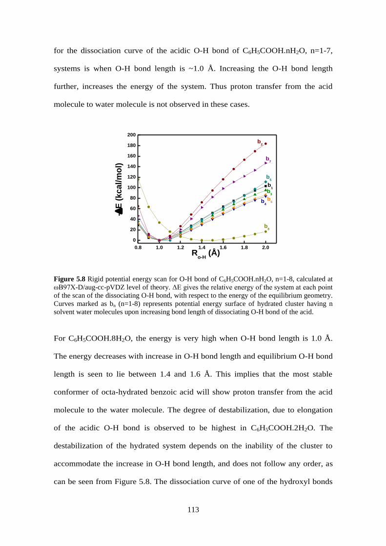
113
for the dissociation curve of the acidic O-H bond of C6H5COOH.nH2O, n=1-7,
systems is when O-H bond length is ~1.0 Å. Increasing the O-H bond length
further, increases the energy of the system. Thus proton transfer from the acid
molecule to water molecule is not observed in these cases.
0.8 1.0 1.2 1.4 1.6 1.8 2.0
0
20
40
60
80
100
120
140
160
180
200
b8
b7
b6
b5
b4
b3
b2
b1
-E
(kcal/m
ol)
Ro-H
(Å)
Figure 5.8 Rigid potential energy scan for O-H bond of C6H5COOH.nH2O, n=1-8, calculated at
ωB97X-D/aug-cc-pVDZ level of theory. ΔE gives the relative energy of the system at each point
of the scan of the dissociating O-H bond, with respect to the energy of the equilibrium geometry.
Curves marked as bn (n=1-8) represents potential energy surface of hydrated cluster having n
solvent water molecules upon increasing bond length of dissociating O-H bond of the acid.
For C6H5COOH.8H2O, the energy is very high when O-H bond length is 1.0 Å.
The energy decreases with increase in O-H bond length and equilibrium O-H bond
length is seen to lie between 1.4 and 1.6 Å. This implies that the most stable
conformer of octa-hydrated benzoic acid will show proton transfer from the acid
molecule to the water molecule. The degree of destabilization, due to elongation
of the acidic O-H bond is observed to be highest in C6H5COOH.2H2O. The
destabilization of the hydrated system depends on the inability of the cluster to
accommodate the increase in O-H bond length, and does not follow any order, as
can be seen from Figure 5.8. The dissociation curve of one of the hydroxyl bonds

114
of oxalic acid molecule shows that except for hepta-hydrated oxalic acid cluster,
there is a barrier for proton transfer from the acid molecule to solvent water
molecules, as shown in Figure 5.9.
0.8 1.0 1.2 1.4 1.6 1.8 2.0
0
100
200
300
400
500
o7
o6
o5
o4
o3
o2
o1
-E
(kcal/m
ol)
ROH
(Å)
Figure 5.9 Rigid potential energy scan for O-H bond of (COOH)2.nH2O, n=1-7, calculated at
ωB97X-D/aug-cc-pVDZ level of theory. ΔE gives the relative energy of the system at each point
of the scan of the dissociating O-H bond, with respect to the energy of the equilibrium geometry.
Curves marked as On (n=1-7) represents potential energy surface of hydrated cluster having n
solvent water molecules upon increasing bond length of dissociating O-H bond of the acid.
A rigid potential energy scan, keeping all but the length of dissociating O-H bond
of the acid-water cluster constant shows an energy minimum when the acidic O-H
bond length is around 1.0 Å for oxa.nH2O, n=1-6. There is an increase in energy
with further elongation of the O-H bond length implying that there is barrier for
the process of proton transfer from the acid molecule to solvent water molecule. In
the hepta-hydrated oxalic acid cluster, however, the energy is high when O-H
bond length is 1.0 to 1.2 Å. It is observed to decrease beyond O-H bond length of
1.4 Å. This implies that the proton is already transferred to the solvent water
molecule in this size of hydrated cluster. The bond dissociation curve provides
further confirmation that first dissociation of oxalic acid occurs in presence seven

115
solvent water molecules. It also signifies that in presence of seven water
molecules, barrier for the transfer of proton from oxalic acid to solvent water
molecule becomes zero. The degree of destabilization of the hydrated clusters of
oxalic acid (excluding hepta-hydrate) with increase in O-H bond distance does not
follow any particular order as this depends on the geometry of the hydrated acid
cluster and its ability to accommodate the elongated hydroxyl bond. It is noted that
the destabilization of oxa.1H2O and oxa.5H2O are relatively low when compared
to other hydrated acid clusters while the highest destabilization is observed in case
of oxa.3H2O.
5.2.5 Hydrogen Bond Energy
The dissociation of the acidic O-H bond as well as the strengthening of the
H bond between the acidic H atom and the O atom of the nearest water molecule,
Table 5.4 Hydrogen bond energy of acidic O-H bond (EO-H) and H..O(H2) bond (EH..O(H2)) in the
most stable conformers of hydrated carboxylic acid, RCOOH.nH2O, n=0-8 systems.
n
CF3COOH.nH2O CH3COOH.nH2O C6H5COOH.nH2O (COOH)2.nH2O
EO-H EH..O(H2) EO-H EH..O(H2) EO-H EH..O(H2) EO-H EH..O(H2)
in kcal/mol
0 211.4 - 210.9 - 211.5 - 210.5 --
1 196.4 10.9 201.7 8.6 201.1 8.9 193.2 13.0
2 179.5 17.1 192.2 12.4 191.1 12.9 186.4 14.9
3 172.6 19.7 191.4 13.2 190.3 13.8 181.1 17.1
4 153.7 27.4 178.1 18.1 173.9 19.8 184.2 15.0
5 130.7 35.8 182.1 16.8 184.4 15.0 182.1 16.5
6 35.9 136.4 189.6 14.1 182.2 16.1 146.9 29.8
7 19.1 177.9 143.8 30.5 179.8 17.0 23.2 169.7
8 - - 169.9 20.2 31.7 144.8 - -

116
H..O(H2), can be tracked to study the proton transfer in the hydrated carboxylic
acid clusters. Table 5.4 gives the hydrogen bond energy of the acidic O-H bond
and the forming H..O(H2) bond. Hydrogen bond energy for acidic O-H and
H..O(H2) bonds shows that the former decreases and the latter increases, as the
size of the cluster increases. It is seen that the hydrogen bond energy value for
acidic O-H bond is higher than that of H..O(H2), when the proton resides with acid
molecule while the reverse is observed when proton transfer occurs. This is
expected as before ionization, the acidic O-H bond is stronger while after
ionization, the H..O(H2) bond is stronger.
0 1 2 3 4 5 6 7 80
50
100
150
200
EH..O(H2)
EO-H
CH3COOH.nH
2O
C6H
5COOH.nH
2O
(COOH)2.nH
2O
CF3COOH.nH
2O
En
erg
y (
kc
al/
mo
l)
n
Figure 5.10 Variation of hydrogen bond energy of most acidic O-H bond (EO-H) and H..O(H2)
bond (EH..O(H2)) of hydrated clusters of carboxylic acids with increase in size of the hydrated
cluster (n). The violet, green, black and orange line shows the hydrogen bond energy of
CH3COOH.nH2O, (COOH)2.nH2O, CF3COOH.nH2O and C6H5COOH.nH2O, n=1-8, clusters
respectively, calculated at ωB97X-D/aug-cc-pVDZ level of theory.
The H..O(H2) bond increases in strength with addition of more water molecules,
and finally when the proton transfer occurs from the acid molecule to solvent
water molecules, the H..O(H2) bond becomes stronger than the O-H bond of the
acid. The variation of hydrogen bond energy of acidic O-H and H..O(H2) bonds of

117
hydrated clusters of the carboxylic acids are given in Figure 5.10. The cross over
in the hydrogen bond energy profiles of the two bonds can be taken as an
indication for bond dissociation of the acid molecule. It is seen that cross-over
occurs at n=6, n=7 and n=8 for CF3COOH.nH2O, (COOH)2.nH2O and
C6H5COOH.nH2O respectively. No such cross-over is observed in case of
CH3COOH.nH2O, implying that even eight water molecules are not sufficient to
break the acidic O-H bond in acetic acid molecule.
5.3 Conclusion
The dissociation of hydrated carboxylic acid molecules, to form contact ion pairs, will be
reflected in some of its properties. Analyzing some of the properties reveal that while
static polarizability and net dipole moment show no characteristic features corresponding
to proton transfer, the acidic O-H bond dipole, IR spectra, O-H bond dissociation curve
and hydrogen bond energy give indication about the formation of contact ion pair.
Static polarizability of the acid-water clusters is observed to vary linearly with
the size of the clusters (n). Hence, though prediction of static polarizability of higher
hydrates of the acid clusters can be done very reliably (regression value > 0.99), it does
not give an indication about the formation of contact ion pair.
The net dipole moment of hydrated acid clusters depend on the geometry of the
system. The direction of the dipole moment vector is not always aligned with the
breaking O-H bond of the acid molecule and thus cannot indicate the formation of
contact ion pair. The bond dipole moment of the dissociating O−H bond does show a
characteristic feature of acid dissociation in case of hydrated clusters of trifluoroacetic
acid, benzoic acid and oxalic acid. A sharp dip is seen in the acidic O-H bond dipole
moment, in case of the ionized acid clusters when compared to unionized clusters. Such

118
a dip is absent in case of acetic acid hydrated clusters, where even with eight water
molecules, proton transfer does not occur.
To compare with experimental data, IR spectral properties of the acid hydrates
are simulated. A strong correlation is found between the reported values and the present
calculated data. The formation of a charge-separated ion pair is manifested in simulated
IR spectra. IR spectra for the most stable structures of each size hydrated acid clusters
are simulated, and the variation in acidic O-H peak positions is in accordance with the
extent of elongation of the O−H bond in the acid molecule. In the case of tfa, a minimum
of six water molecules are needed for ionization, which is characterized by formation of
new peaks in the hexa-hydrated cluster corresponding to stretching frequencies of
hydrated proton. Similarly huge red shift; with respect to isolated acid molecule, is
observed in the hydroxyl bond stretching frequency of C6H5COOH.8H2O and
(COOH)2.7H2O, along with formation of new IR peaks. This indicates the dissociation of
the O-H bond of the acid molecule and proton transfer from the acid molecule to solvent
water molecules. CH3COOH.nH2O systems provide no such observations.
A rigid potential energy scan, altering the dissociating hydroxyl bond, shows that
there is a barrier in proton transfer from the acid to water molecules in case of mono to
penta-hydrate of trifluoroacetic acid, mono to hepta-hydrate of benzoic acid, mono to
octa-hydrate of acetic acid and mono to hexa-hydrate of oxalic acid. In the case of hexa,
octa and hepta-hydrates of trifluoroacetic acid, benzoic acid and oxalic acid, the transfer
is barrier-less.
The hydrogen bond energy profiles of the dissociating hydroxyl bond of the acid
molecule and the hydrogen bond between the acidic H atom of the acid molecule and the
O atom of the nearest water molecule shows characteristic features upon dissociation of
the acid molecule. For trifluoroacetic acid-water clusters, hydrogen bond energy of the

119
dissociating O-H bond is greater than the hydrogen bond energy of the forming H..O
bond between the acidic H atom and the O atom of the nearest water molecule, for all
except hexa and hepta-hydrates. In case of hexa and hepta-hydrates clusters the reverse is
true. For benzoic acid-water clusters, the breaking O-H hydrogen bond energy is greater
than that of forming H..O bond till hepta hydrated cluster but in case of octa hydrate, the
same for forming H..O bond is stronger. Similarly for oxalic acid-water clusters, the
hydrogen bond energy of the most acidic O-H bond of the acid molecule is greater than
the forming H..O bond for (COOH)2.nH2O, n=1-6, while reverse is true for hepta-hydrate.
No such cross-over in the energy profiles of the two bonds is observed in case of acetic
acid molecule. These properties can be used as indicators to study microhydration in acid
molecules to track proton transfer and formation of contact ion pairs.

Chapter 6
6
Microhydration of 2-Naphthol at Ground, First
Excited Triplet and First Excited Singlet states:
A Case Study on Photo Acids
The only existing things are atoms and empty space;
all else is mere opinion.
- Democritus of Abdera d. 370B.C.

121
Photo acids undergo significant enhancement in their acidity upon electronic
excitation. This leads to excited state proton transfer to the solvent. The change in
electron density distribution of a photo acid can be determined from its excited state
acidity constant. This information is of much interest to both photo chemists, who can
study the differences in reactivity of molecules in ground and excited state, and
theoretical chemists, who can calculate the excited state electronic structure and the
nature of the transitions involved. The significant change in acidity of these molecules
makes them an ideal system to probe solute-solvent interactions and solvent
polarities.[134]
Photo acids can be used as molecular probes for determining the structural
transitions of proteins under various conditions.[135]
Water accessibility in biological
surfaces can also be probed using photo acids.[136,137]
The excited state pKa of a photo
acid is generally determined from the Fӧrster cycle, based on absorption and emission
data.[138]
It can also be determined using photo-potentiometry.[139]
In aqueous solution, aromatic compounds like phenol and 2-naphthol have
ground state pKa values of 10.0 and 9.2 respectively, while their excited state pKa values
are just 3.6 and 2.8 respectively.[140]
Theoretical calculations to explain excited state
proton transfer in phenols has been reported.[141]
Two different explanations have been
proposed for the increase in acidity in the first excited singlet state. One proposition
attributes the increase in acidity to a charge transfer in the photo acid.[142,143]
According
to the other proposition, the enhanced excited state acidity is largely determined by the
deprotonated photo acid.[141]
Jackson and Porter reported that the acidity constants of ground and first excited
triplet states of 2-naphthol are comparable while that of the first excited singlet state
differ by a factor of ~106.[15]
The studies were based on flash photolysis, fluorescence

122
and phosphorescence measurements. They provide a qualitative explanation for this
observation based on electron density. The difference in the pKa value of ground state
and excited states of 2-naphthol could be reflected in the number (n) of water molecules
needed for their ionization. This chapter explores the microhydration of 2-naphthol (nap)
at ground (napS0), first excited triplet (napT1) and first excited singlet (napS1) states, to
understand the correlation between n and pKa at different electronic states.
6.1 Theoretical Methods
The ground state geometries are optimized at B97X-D/aug-cc-pVDZ level of
theory. The first excited singlet and first excited triplet states are optimized at CAM-
B3LYP/ aug-cc-pVDZ level of theory. CAM-B3LYP is a long-range corrected hybrid
exchange-correlation functional designed to better describe electronic excitations and
charge-transfer processes.[144–147]
The excited state calculations are done using Time-
Dependent DFT (TD-DFT). The input geometries are chosen such that minimum number
of water molecules is needed to ionize the O-H bond of nap. The water molecules are
placed in such a way so as to increase the number of hydrogen bonds formed between
the solvent water molecules and nap, facilitating the easy dissociation of the O-H bond of
nap. The minimum energy equilibrium structure of each size cluster at ground, first
excited triplet and first excited singlet state is selected to determine the minimum number
of water molecules needed to dissociate nap. A rigid potential energy scan, at CAM-
B3LYP/aug-cc-pVDZ level of theory, of the most stable conformer of each size cluster,
at S0, T1 and S1 states, is carried out by keeping all but the dissociating O-H bond of nap
constant. The potential energy profile of the hydrated nap system is obtained by plotting
the relative energy, with respect to most stable structure, at each O-H bond distance
against the O-H bond distance. All calculations are carried out using the general ab initio
quantum package, Gaussian.[148]

123
6.2 Results and Discussion
6.2.1 Microhydration at ground state
At ground state, two conformers are possible for napS0. Structures of both the
conformers, along with their relative energy, are given in Figure 6.1. The two structures
differ in the relative position of the O-H bond with respect to the naphthalene ring. In the
most stable conformer (see Figure 6.1 (i)), the O-H bond faces the ring while in the higher
energy conformer, the O-H bond faces away from the ring. This is consistent with the
results obtained from MP2/aug-cc-pVDZ level of theory reported recently.[149]
The O-H
bond length is 0.961 Å and the C-O bond length is 1.364 Å in the most stable conformer
at the present level of theory.
(i) (ii)ΔE= +0.4
Figure 6.1 Equilibrium structures of 2-naphthol, at ground state, calculated at ωB97X-D/aug-cc-
pVDZ level of theory. Zero point energy corrected relative energy (in kcal/mol) of the higher
energy conformer with respect to minimum energy conformer is also given. Red balls represent
oxygen, grey balls represent carbon and blue balls represent hydrogen atoms.
With the addition of a single water molecule, the most stable conformer has the H atom
of the nap forming hydrogen bond with the O atom of the water molecule. The O-H bond
length of the naphthol molecule increases to 0.972 Å and the C-O bond length decreases
to 1.356 Å. the hydrogen bond distance between the acidic hydrogen atom of nap and the
O atom of the water molecule is calculated to be 1.853 Å. Another structure is also
observed for the mono-hydrate of nap (napS0.1H2O), which is higher in energy than the
most stable conformer by just 0.3 kcal/mol, as shown in Figure 6.2 (ii). The difference
between this conformer and the most stable conformer is in the relative position of the O-

124
H bond of the nap with respect to the naphthalene ring. All the equilibrium conformers
obtained for napS0.1H2O along with their zero point corrected relative energy are given in
Figure 6.2.
(i) (ii)ΔE= +0.3 (iii) ΔE= +2.5
Figure 6.2 Equilibrium structures of mono-hydrate of 2-naphthol, at ground state, calculated at
ωB97X-D/aug-cc-pVDZ level of theory. Zero point energy corrected relative energy (in
kcal/mol) of the higher energy conformer with respect to minimum energy conformer is also
given. Red balls represent oxygen, grey balls represent carbon and blue balls represent hydrogen
atoms.
With two water molecules, the most stable conformer has the O-H bond of nap hydrogen
bonded to the oxygen atom of one of the water molecules. The other water molecule is
hydrogen bonded to the first water molecule, without direct bonding with the nap. The
four equilibrium geometries obtained for the di-hydrate of nap (napS0.2H2O) along with
their zero point corrected relative energy is given in Figure 6.3. Unlike as observed in
carboxylic acids, a closed ring motif is absent in case of the di-hydrates, possibly due to
the steric strain involved in such a ring formation. The O-H and C-O bond lengths of nap
in the most stable conformer are 0.994 and 1.326 Å respectively.
(i) (ii)ΔE= +0.5 (iii) ΔE= +0.9 (iv) ΔE= +1.0
Figure 6.3 Equilibrium structures of di-hydrate of 2-naphthol, at ground state, calculated at
ωB97X-D/aug-cc-pVDZ level of theory. Zero point energy corrected relative energy (in
kcal/mol) of the higher energy conformer with respect to minimum energy conformer is also

125
given. Red balls represent oxygen, grey balls represent carbon and blue balls represent hydrogen
atoms.
Six equilibrium structures are obtained for the tri-hydrate of nap (napS0.3H2O), as shown
in Figure 6.4. The most stable conformer has the three water molecules and the O-H
bond of the nap forming hydrogen bonded close-ring motif. Open chain structures are
found to be higher in energy than the closed ring conformers. The O-H and C-O bond
length of nap are 0.985 and 1.362 Å respectively, for the most stable conformer of
napS0.3H2O, while the hydrogen bond distance between the acidic O-H of nap and the O
atom of the nearest water molecule is 1.627 Å.
(i) (ii)ΔE= +0.7 (iii) ΔE= +4.4
(iv) ΔE= +4.7 (v)ΔE= +4.7 (vi) ΔE= +5.3
Figure 6.4 Equilibrium structures of tri-hydrate of 2-naphthol, at ground state, calculated at
ωB97X-D/aug-cc-pVDZ level of theory. Zero point energy corrected relative energy (in
kcal/mol) of the higher energy conformer with respect to minimum energy conformer is also
given. Red balls represent oxygen, grey balls represent carbon and blue balls represent hydrogen
atoms.
A closed ring conformer is the most stable geometry obtained for the tetra-hydrate of nap
(napS0.4H2O). All the six equilibrium conformers obtained for nap.4H2O are given in
Figure 6.5 along with the relative energy of each conformer with respect to the most

126
stable conformer. The O-H and C-O bond lengths of nap in the most stable conformer
are 0.987 and 1.360 Å respectively.
(i) (ii)ΔE= +0.7 (iii) ΔE= +2.6
(iv)ΔE= +3.3 (v) ΔE= +8.8 (vi) ΔE= +9.0
Figure 6.5 Equilibrium structures of tetra-hydrate of 2-naphthol, at ground state, calculated at
ωB97X-D/aug-cc-pVDZ level of theory. Zero point energy corrected relative energy (in
kcal/mol) of the higher energy conformer with respect to minimum energy conformer is also
given. Red balls represent oxygen, grey balls represent carbon and blue balls represent hydrogen
atoms.
It can be seen that none of the conformers show proton transfer from nap to the solvent
water molecules at the ground state.
6.2.2 Microhydration at first excited triplet state
Of the two equilibrium conformers obtained for nap at the first excited triplet
state (T1), the most stable conformer has the O-H bond facing away from the
naphthalene ring. The other conformer, with O-H bond facing towards the ring, is 16.8
kcal/mol higher in energy than the most stable conformer. The O-H and C-O bond
lengths of the most stable conformer are 0.961 and 1.364 Å respectively. Both the
conformers are shown in Figure 6.6.

127
(i) (ii) ΔE= +16.8
Figure 6.6 Equilibrium structures of 2-naphthol, at first excited triplet state, calculated at CAM-
B3LYP/aug-cc-pVDZ level of theory. Zero point energy corrected relative energy (in kcal/mol)
of the higher energy conformer with respect to minimum energy conformer is also given. Red
balls represent oxygen, grey balls represent carbon and blue balls represent hydrogen atoms.
With the addition of a single water molecule, the most stable conformation at first
excited triplet state has the O-H bond facing away from the ring and the O atom of the
water molecule forming hydrogen bond with H atom of nap. All conformations obtained
for the first excited triplet state of mono-hydrate of nap (napT1.H2O) are given in Figure
6.7. The O-H and C-O bond length of nap are respectively, 0.975 and 1.358 Å. The
hydrogen bond distance between the hydroxyl H atom of nap and O atom of water
molecule is 1.840 Å.
(i) (ii) ΔE= + 16.3 (ii) ΔE= +18.5
Figure 6.7 Equilibrium structures of 2-naphthol.1H2O, at first excited triplet state, calculated at
CAM-B3LYP/aug-cc-pVDZ level of theory. Zero point energy corrected relative energy (in
kcal/mol) of the higher energy conformer with respect to minimum energy conformer is also
given. Red balls represent oxygen, grey balls represent carbon and blue balls represent hydrogen
atoms.
The most stable conformation of the first excited triplet state of di-hydrate of nap
(napT1.2H2O) has an open chain conformation with the O-H bond facing way from the
naphthalene ring as shown in Figure 6.8 (i). The O-H and C-O bond lengths are 0.979
and 1.353 Å respectively. Higher energy conformations of napT1.2H2O are also given in
Figure 6.8.
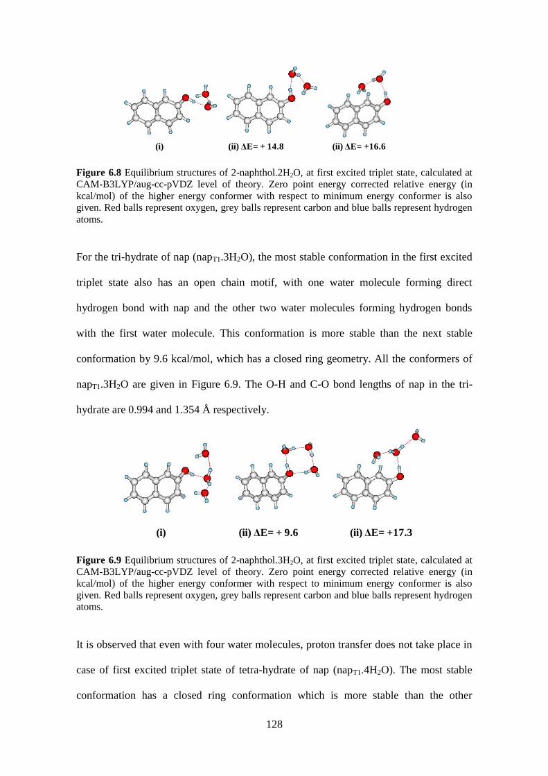
128
(i) (ii) ΔE= + 14.8 (ii) ΔE= +16.6
Figure 6.8 Equilibrium structures of 2-naphthol.2H2O, at first excited triplet state, calculated at
CAM-B3LYP/aug-cc-pVDZ level of theory. Zero point energy corrected relative energy (in
kcal/mol) of the higher energy conformer with respect to minimum energy conformer is also
given. Red balls represent oxygen, grey balls represent carbon and blue balls represent hydrogen
atoms.
For the tri-hydrate of nap (napT1.3H2O), the most stable conformation in the first excited
triplet state also has an open chain motif, with one water molecule forming direct
hydrogen bond with nap and the other two water molecules forming hydrogen bonds
with the first water molecule. This conformation is more stable than the next stable
conformation by 9.6 kcal/mol, which has a closed ring geometry. All the conformers of
napT1.3H2O are given in Figure 6.9. The O-H and C-O bond lengths of nap in the tri-
hydrate are 0.994 and 1.354 Å respectively.
(i) (ii) ΔE= + 9.6 (ii) ΔE= +17.3
Figure 6.9 Equilibrium structures of 2-naphthol.3H2O, at first excited triplet state, calculated at
CAM-B3LYP/aug-cc-pVDZ level of theory. Zero point energy corrected relative energy (in
kcal/mol) of the higher energy conformer with respect to minimum energy conformer is also
given. Red balls represent oxygen, grey balls represent carbon and blue balls represent hydrogen
atoms.
It is observed that even with four water molecules, proton transfer does not take place in
case of first excited triplet state of tetra-hydrate of nap (napT1.4H2O). The most stable
conformation has a closed ring conformation which is more stable than the other

129
conformations obtained, by 19.2 kcal/mol (see Figure 6.10). The O-H and C-O bond
length of nap are 0.993 and 1.370 Å respectively.
(i) (ii) ΔE= + 19.2 (ii) ΔE= +26.6
Figure 6.10 Equilibrium structures of 2-naphthol.4H2O, at first excited triplet state, calculated at
CAM-B3LYP/aug-cc-pVDZ level of theory. Zero point energy corrected relative energy (in
kcal/mol) of the higher energy conformer with respect to minimum energy conformer is also
given. Red balls represent oxygen, grey balls represent carbon and blue balls represent hydrogen
atoms.
6.2.3 Microhydration at first excited singlet state
The most stable conformer of nap, at the first excited singlet state (S1), has the O-
H bond facing away from the naphthalene ring. The O-H and C-O bond lengths of nap
are 0.966 and 1.346 Å respectively. This structure is more stable than the structure in
which the O-H bond is facing towards the naphthalene ring, by 0.7 kcal/mol. Both the
structures are given in Figure 6.11.
(i) (ii)ΔE= +0.7
Figure 6.11 Equilibrium structures of 2-naphthol, at first excited singlet state, calculated at
CAM-B3LYP/aug-cc-pVDZ level of theory. Zero point energy corrected relative energy (in
kcal/mol) of the higher energy conformer with respect to minimum energy conformer is also
given. Red balls represent oxygen, grey balls represent carbon and blue balls represent hydrogen
atoms.
Three equilibrium conformers are obtained for the first excited singlet state of mono-
hydrate of 2-naphthol (napS1.1H2O), as shown in Figure 6.12. The most stable

130
conformation of napS1.1H2O, at first excited singlet state has the O-H bond of the nap
facing towards the naphthalene ring. The H atom of the water molecule forms hydrogen
bonding with the oxygen atom of the nap. The O-H and C-O bond lengths of nap are
0.965 and 1.356 Å respectively.
(i) (ii)ΔE= +31.6 (iii) ΔE= +32.2
Figure 6.12 Equilibrium structures of 2-naphthol.1H2O, at first excited singlet state, calculated at
CAM-B3LYP/aug-cc-pVDZ level of theory. Zero point energy corrected relative energy (in
kcal/mol) of the higher energy conformer with respect to minimum energy conformer is also
given. Red balls represent oxygen, grey balls represent carbon and blue balls represent hydrogen
atoms.
With two water molecules, five conformers are observed for the first excited singlet state
of di-hydrate of nap (napS1.2H2O) as shown in Figure 6.13. The most stable conformer
has the O-H bond of nap facing away from the ring and forming open chain hydrogen
bonds with the water molecules. The O-H and C-O bond lengths of nap are 0.994 and
1.326 Å respectively.
(i) (ii)ΔE= +1.3 (iii) ΔE= +1.3
Figure 6.13 Equilibrium structures of 2-naphthol.2H2O, at first excited singlet state, calculated at
CAM-B3LYP/aug-cc-pVDZ level of theory. Zero point energy corrected relative energy (in
kcal/mol) of the higher energy conformer with respect to minimum energy conformer is also
given. Red balls represent oxygen, grey balls represent carbon and blue balls represent hydrogen
atoms.

131
The most stable conformation at the first excited singlet state of tri-hydrate of nap (napS1.3H2O)
has a hydrogen bonded closed ring conformation (see Figure 6.14 (i)). The O-H and C-O
bond length of nap are 1.001 and 1.341 Å respectively. A slightly higher energy structure
is also obtained, differing only in the relative position of the O-H bond of nap (see Figure
6.14 (ii)). Higher energy equilibrium conformers of napS1.3H2O at the first excited
singlet state are also given in Figure 6.14. It must be noted that closed ring conformation
are more stable than the open chain ones.
(i) (ii)ΔE= +0.2 (iii) ΔE= +0.4 (iv) ΔE= +3.4
(v) ΔE= +3.6 (vi) ΔE= +4.7 (vii) ΔE= +4.9
Figure 6.14 Equilibrium structures of 2-naphthol.3H2O, at first excited singlet state, calculated at
CAM-B3LYP/aug-cc-pVDZ level of theory. Zero point energy corrected relative energy (in
kcal/mol) of the higher energy conformer with respect to minimum energy conformer is also
given. Red balls represent oxygen, grey balls represent carbon and blue balls represent hydrogen
atoms.
Interestingly, the most stable conformer of tetra-hydrate of nap (napS1.4H2O), at the first
excited singlet state, shows proton transfer from nap to the neighboring water molecule.
This conformer is more stable than the next stable conformer by 1.0 kcal/mol. The most
stable conformer has an open chain conformation with two water molecules stabilizing
the first water molecule through hydrogen bonding and the H atom of the fourth water
molecule forming hydrogen bond with the oxygen atom of nap (see Figure 6.15 (i)). The

132
O..H hydrogen bond distance between O atom of nap and nearest H atom of the hydrated
proton is 1.598 Å. Four other higher energy equilibrium conformers of napS1.4H2O are
also obtained, as shown in Figure 6.15.
(i) (ii)ΔE= +1.0 (iii) ΔE= +1.7
(iv)ΔE= +7.4 (v) ΔE= +8.7
Figure 6.15 Equilibrium structures of 2-naphthol.4H2O, at first excited singlet state, calculated at
CAM-B3LYP/aug-cc-pVDZ level of theory. Zero point energy corrected relative energy (in
kcal/mol) of the higher energy conformer with respect to minimum energy conformer is also
given. Red balls represent oxygen, grey balls represent carbon and blue balls represent hydrogen
atoms.
6.2.4 O-H Potential Energy Profile of nap
The dissociating O-H bond potential energy profile of the hydrated clusters of
nap gives the barrier for proton transfer from nap to the neighboring solvent water
molecule. The potential energy profiles for the dissociating O-H bond of tetra-hydrate of
nap, at the ground, first excited triplet and first excited singlet state are given in Figure
6.16. The equilibrium bond length of O-H in nap is ~1 Å for mono, di, tri and tertra-
hydrates of nap in the ground and first excited triplet state. In these cases, it is seen that
when the O-H bond distance is increased, the energy increases and the system becomes
less stable. The potential energy profile of the dissociating O-H bond of nap at the first

133
excited singlet state of mono, di and tri-hydrates also show a minimum at O-H bond
distance ~1 Å. Further increase in O-H bond length leads to destabilization of the system.
0.8 1.0 1.2 1.4 1.6 1.8 2.0 2.2-30
0
30
60
90
120
2-naphthol.4H2O
S0
T1
S1
-E
(kcal/m
ol)
ROH
(Å)
Figure 6.16 Rigid potential energy scan of O-H bond of 2-naphthol.4H2O, at ground, first
excited triplet and singlet state. ΔE gives the relative energy of the system at each point of the
scan of the dissociating O-H bond, with respect to the energy of the equilibrium geometry.
Curves marked as S0, T1 and S1 represent potential energy surfaces of ground, first excited triplet
and singlet states respectively of 2-naphthol.4H2O, upon increasing the bond distance of
dissociating O-H bond of the acid.
However, a rigid potential energy scan of the acidic O-H bond of nap.4H2O at the first
excited singlet state reveals that in the presence of four water molecules, the acidic
proton transfer from nap to the neighboring water molecule becomes a barrier-less
process, as shown in Figure 6.16. In fact, in the first excited singlet state, the tetra-
hydrate becomes unstable at O-H bond distances less than 1.4 Å. The potential energy
profiles show that a barrier exists for proton transfer from nap to solvent water molecules
in all cases except the tetra-hydrate of nap at the first excited singlet state. In case of
napS1.4H2O, spontaneous dissociation of the O-H bond is observed. This is in line with
the experimentally observed fact that first excited singlet state is more acidic than the
ground and first excited triplet state.

134
6.3 Conclusion
Microhydration studies of 2-naphthol at ground, first excited triplet and singlet
states are carried out using TD-DFT. It is seen that even in the presence of four water
molecules, proton transfer from naphthol to water molecules is not observed for either
ground or first excited triplet state. However, proton transfer becomes a barrier-less
process for first excited singlet state of tetra-hydrate of naphthol.
Single determinant methods are not enough to accurately describe the excited
states of a system. To completely define the excited state chemistry, multi-reference
calculations are required. Hence excited state calculations are complicated and time-
consuming. Although TD-DFT is a powerful and widely used procedure, higher multi-
reference methods have to be employed to get a better picture of the photo-chemistry of
hydrated-naphthol systems. The present chapter deals with the structures and the
potential energy profiles of hydrated clusters of 2-naphthol, at different energy states. To
get a comprehensive understanding of the microhydration of photo acids, the following
actions are planned to be undertaken in the future. A multi-reference method, like
CASSCF, can be used to verify the present results. Further studies to understand the
reason behind the difference in pKa of S0, T1 and S1 states of 2-naphthol could be carried
out. A wider range of systems will be studied so that a general expression can be derived
to predict the pKa of photo acids, if n is known.

Chapter 7
7
Prediction of pKa:
From Molecule to Bulk
Truth, in science, can be defined as the working hypothesis best fitted
to open the way to the next better one.
- Konrad Lorenz

136
Proton transfer reactions are widely studied because of their fundamental
importance in biological systems, atmospheric phenomena and synthetic applications.
Understanding of the effect of chemical environment on the acidity of a system and vice-
versa are important in comprehending the mechanism of several processes like enzyme-
catalysis. The acidity of a system is defined by its pKa. Spectrophotometry, pH titrations
etc are some of the methods for determining pKa of a system experimentally. But
accurate experimental pKa determination becomes complicated in many cases like very
strong acids and very weak acids, due to leveling effect of the solvent etc. The
experimental pKa value for H2SO4 ranges from -3 to -9 while that for CF3SO3H range
from -6 to -14.[150,151]
Accurate pKa values for some very weak acids have been reported
from kinetic measurements employing sensitive techniques such as NMR.[152–157]
However, accurate pKa determination from experiment remains difficult for many
systems.
Quantum chemical methods have been used to obtain pKa values. The pKa can
be determined indirectly from free energies calculations in solution. An excellent review
by Ho and Coote and the references given therein, as well as a few recent reports are a
few among the numerous publications that discuss the strategies applied to determine
pKa values using quantum chemical calculations in a thermodynamic route.[158–162]
Figure 7.1 Thermodynamic cycle for determining pKa indirectly from free energy. AH refers to
the acid. Subscript (g) and (aq) denote the species in gas and aqueous phase respectively.

137
The free energy of acid (AH) dissociation calculated following the simplest
thermodynamic cycle is as shown in Figure 7.1. Here, first the acid is desolvated, then it
is deprotonated in the gas phase and finally the conjugate base and the separated proton
are solvated. This is the most commonly used thermodynamic cycle due to its simplicity.
The free energy of acid dissociation can then be defined as:
AH
sol
H
sol
A
solga GGGGG 7.1
where ΔGa and ΔGg refers to the free energy change of the system in the aqueous
solution and gas phase respectively. A
solG , H
solG and AH
solG represent free energy of
solvation of A-, H
+ and the acid, AH respectively. The free energy change of the system
in gas phase is given by:
AH
g
H
g
A
ggGGGG
7.2
where A
gG , H
gG and AH
gG are the gas phase free energies of the anion, proton and acid,
respectively. All these free energy values can be obtained from computational
calculations except H
gG and H
solG . Since properties of systems without electron cannot
be studied using quantum chemical methods, these values have to be determined
experimentally or from thermodynamic theory. The H
solG value has been under debate
for some time, but generally accepted value is -265.9 kcal/mol.[163–165]
pKa is then
calculated as:
RT
GpK a
a303.2
7.3
Though accurate gas phase reaction energies can now be calculated using high level
computational methods available, free energy calculations in solution remains difficult.
For neutral solutes, the dielectric continuum models like PCM and SMD give solvation

138
free energies with an accuracy of 1 kcal/mol.[166,167]
However, ionic species are involved
in pKa calculations which have much lower accuracy. Accurate prediction of pKa of an
acid is very difficult as a small error in free energy calculations will cause a large
deviation of the pKa from the true value. The main source of error in thermodynamic
determination of pKa arises from the uncertainty in free energy of hydration of proton
and neglect of the solute-solvent molecular interactions, which are not considered in
continuum solvation models, as discussed in Chapter 3 of this thesis.
1 2 3 4 5 6 7 8
-16
-12
-8
-4
0
4
8
bzfa
tfa
HClpK
a
n
Figure 7.2 Plot of pKa v/s number of water molecules needed for dissociation (n), for HCl,
trifluoroacetic acid (tfa), formic acid (fa) and benzoic acid (bz), calculated at ωB97X-D/aug-cc-
pVDZ level of theory.
A novel non-thermodynamic route for calculating pKa value is presented here. A
linear correlation is observed between the pKa of acid and the number of water
molecules (n) needed for its dissociation, in case of acids studied at present applying the
same level of theory, as depicted in Figure 7.2. Number of solvent water molecules
needed to dissociate strong acid molecule, HCl has been a point of debate. Recent
computational studies explained the difference in results on the basis of temperature

139
dependence of relative stability of the structures.[9]
Based on our own independent study,
at the present level of theory, it is predicted that dissociation of HCl molecule and
formation of ion-pair occurs with four water molecules. Linear fitting produces the
relation: pKa = 2.9n-18.0, with R2 = 0.94. This relation can be exploited to qualitatively
predict unknown pKa values based on electron structure theory, where experimental
measurement is rather difficult.[127]
A more generalized relation, including acids previously reported in literature,
also shows linear dependence of pKa and n, as shown in Figure 7.3. It may be noted that
the calculations reported for other acids are not at the same level of theory.
1 2 3 4 5 6 7 8-16
-12
-8
-4
0
4
8
CF3COOH (COOH)
2
HCOOH
C6H
5COOH
HNO3
HCl
HBrHI
pK
a
n
Figure 7.3 Plot of pKa v/s number of water molecules needed for dissociation (n), for HI, HBr,
HCl, HNO3, trifluoroacetic acid, formic acid, oxalic acid and benzoic acid.
It has been previously reported that HI, HBr and HNO3, having pKa values -9.5, -9 and -
1.4 respectively, require 3, 4 and 5 water molecules for acid dissociation.[13,67]
Including
these data points to our study, the correlation between pKa and n can be best fitted as:
pKa=3.1n-19.2, with an R2 value of 0.93.
[126] Thus, the pKa of an acid can be predicted if

140
n is known. This provides a non-thermodynamic route for determining pKa of an acid,
connecting a molecular level property of the acid molecule, to macroscopic property, the
pKa of the acid, in the ground state. To obtain a more reliable relation, a larger set of
data is required, with all the calculations carried out at the same level of theory.
In the case of excited state, microhydration leading to dissociation is studied considering
2-naphthol as a case study (see chapter 6). The pKa of the first excited singlet state of 2-
naphthol is very small compared to its ground and first excited triplet state. This is
reflected in its „n‟. In the first excited singlet state, the presence of just four water
molecules leads to proton transfer from 2-naphthol to the solvent water molecules. This
is not observed in case of ground or first excited triplet state. However, the correlation
between pKa and „n‟ of a system that exists for the ground state acids, may not be
applicable in the excited state, as is suggested by the case study on 2-naphthol. Further
excited state studies need to be carried out to obtain a better correlation between pKa and
„n‟.

141
Summary and Future Perspectives
Salient features of theoretical results presented in the previous chapters may be
summarized as follows. The structure of the hydrated acid cluster depends on the H-
bonded network between the acid molecule and water molecules. On successive addition
of each water molecule to isolated carboxylic acid molecule, O−H bond of −COOH
group becomes longer and weaker. The distance between H atom of the acid and O atom
of the neighboring water molecule decreases. As is expected, it is seen that electronic
effect affects the strength of the acidic O-H bond. Trifluoroacetic acid, oxalic acid and
benzoic acid molecules require 6, 7 and 8 water molecules respectively, for their
dissociation, while even in the presence of eight water molecules, acetic acid molecule
remains undissociated. The geometry of the ionized hydrated acid cluster resembles that
of Eigen cation interacting with the corresponding carboxylate anion surrounded by the
remaining water molecules. The weakening of the O-H bond of the acid molecule, with
the addition of water molecules, and subsequent formation of contact ion pair are
reflected in the bond dipole moment, H bond energy, IR spectra and bond dissociation
energy profiles of the O-H bond of the acid molecule in the hydrated acid clusters. On
successive addition of solvent water molecules, the barrier for proton transfer from the
acid molecule to the neighboring solvent water molecule reduces. Microhydration of
photo acid is also considered by taking 2-naphthol as a case study. It is seen that fewer
number of water molecules are required to dissociate 2-naphthol in the first excited
singlet state when compared to its ground and first excited triplet state (pKa of S1 state is
much lower compared to S0 and T1 state).
One of the major challenges of studying microhydration of acids is the increase in
structural complexity with increase in size of the hydrated acid clusters. A large

142
conformational variety within a small relative energy range is obtained. This indicates a
shallow potential energy surface. The number of H bond donors and acceptors increases
with increase in number of water molecules in the hydrated acid clusters. Converging to
a local minimum is an inherent defect of Newton-Raphson based algorithm which is used
for geometry optimization. Thus selection of input geometries is crucial in determining
the geometry of the cluster. As the increase in size of the hydrated clusters increases the
number of possible input geometries, only clusters up to octa-hydrates are studied here to
avoid missing out an important conformation.
The main motive of the thesis was to understand microhydration of acids at the
molecular level and obtain a correlation between molecular and bulk level acid strength.
As was discussed in the previous chapters, number of water molecules needed to
dissociate an acid molecule depends on the strength of the acid. A weak acid molecule
needs more number of solvent water molecules whereas a strong acid needs less number
of water molecules. Bulk property like pKa value of an acid also indicates the strength of
an acid. It is interesting to investigate if any correlation exists between these two
parameters, bulk parameter: pKa and molecular parameter: number (n) of solvent water
molecules needed to form contact ion-pair. A linear relation is observed between the
number of water molecules needed to dissociate an acid molecule and its pKa.
For obtaining a valid equation connecting the pKa of the acid and the number of
water molecules needed to dissociate the acid molecule, a larger set of data is required,
with all the calculations done at a same level of theory. Microhydration studies of a
larger group of chemical systems, like amines, alcohols etc. can help in establishing a
reliable equation to calculate the pKa of an acid from n. Also from the case study on 2-
naphthol it is inferred that the linear correlation between pKa and n, that exists in the
ground state, may not be applicable in the excited states. Further, observed linear

143
correlation between n and pKa is to be explained. The author aspires to pursue these
studies to obtain logical conclusions.

144
References
[1] J. Tomasi, B. Mennucci, R. Cammi, Chem. Rev. 2005, 105, 2999–3093.
[2] A. V. Marenich, C. J. Cramer, D. G. Truhlar, J. Phys. Chem. B 2009, 113, 6378–
6396.
[3] N. Agmon, H. J. Bakker, R. K. Campen, R. H. Henchman, P. Pohl, S. Roke, M.
Thämer, A. Hassanali, Chem. Rev. 2016, 116, 7642–7672.
[4] S. Pylaeva, C. Allolio, B. Koeppe, G. S. Denisov, H. Limbach, D. Sebastiani, P.
M. Tolstoy, Phys. Chem. Chem. Phys. 2015, 17, 4634–4644.
[5] J. A. Fournier, C. J. Johnson, C. T. Wolke, G. H. Weddle, A. B. Wolk, M. A.
Johnson, Science (80-. ). 2014, 344, 1009–1012.
[6] C. Lao-ngam, P. Asawakun, S. Wannarat, K. Sagarik, Phys. Chem. Chem. Phys.
2011, 13, 4562–4575.
[7] S. Woutersen, H. J. Bakker, Phys. Rev. Lett. 2006, 96, 138305-1–4.
[8] J. M. Headrick, E. G. Diken, R. S. Walters, N. I. Hammer, R. A. Christie, J. Cui, E.
M. Myshakin, M. A. Duncan, M. A. Johnson, K. D. Jordan, Science (80-. ). 2005,
308, 1765–1769.
[9] A. Vargas-Caamal, J. L. Cabellos, F. Ortiz-Chi, H. S. Rzepa, A. Restrepo, G.
Merino, Chem. - A Eur. J. 2016, 22, 2812–2818.
[10] S. Sugawara, T. Yoshikawa, T. Takayanagi, M. Tachikawa, Chem. Phys. Lett.
2011, 501, 238–244.
[11] H. Forbert, M. Masia, A. Kaczmarek-kedziera, N. N. Nair, D. Marx, J. Am. Chem.
Soc. 2011, 133, 4062–4072.
[12] S. Li, F. Tao, R. Gu, Chem. Phys. Lett. 2006, 426, 1–7.
[13] S. Odde, B. J. Mhin, S. Lee, H. M. Lee, K. S. Kim, J. Chem. Phys 2004, 120,

145
9524–9535.
[14] K. H. Weber, F.-M. Tao, J. Phys. Chem. A 2001, 105, 1208–1213.
[15] G. Jackson, G. Porter, Proc. R. Soc. A Math. Phys. Eng. Sci. 1961, 260, 13–30.
[16] R. L. Johnston, Atomic and Molecular Clusters, Taylor And Francis, 2002.
[17] M. Moskovits, Annu. Rev. Phys. Chem. 1999, 42, 465–499.
[18] A. W. Castleman, R. G. Keesee, Chem. Rev. 1986, 86, 589–618.
[19] A. Kumar, S. R. Gadre, X. Chenxia, X. Tianlv, S. R. Kirk, S. Jenkins, Phys. Chem.
Chem. Phys. 2015, 17, 15258–15273.
[20] M. Becucci, S. Melandri, Chem. Rev. 2016, 116, 5014–5037.
[21] S. R. Gadre, S. D. Yeole, N. Sahu, Chem. Rev. 2014, 114, 12132–12173.
[22] T. C. Cheng, B. Bandyopadhyay, J. D. Mosley, M. A. Duncan, J. Am. Chem. Soc.
2012, 134, 13046–13055.
[23] C. Desfrancois, S. Carles, J. P. Schermann, Chem. Rev. 2000, 100, 3943–3962.
[24] T. A. Miller, Science (80-. ). 1984, 223, 545–553.
[25] F. Sylla, M. Veltcheva, S. Kahaly, A. Flacco, V. Malka, Rev. Sci. Instrum. 2012,
83, 33507-1–7.
[26] L. M. Goss, S. W. Sharpe, T. A. Blake, V. Vaida, J. W. Brault, J. Phys. Chem. A
1999, 103, 8620–8624.
[27] V. Chandrasekaran, L. Biennier, E. Arunan, D. Talbi, R. Georges, J. Phys. Chem.
A 2011, 115, 11263–11268.
[28] F. N. Keutsch, R. J. Saykally, Proc. Natl. Acad. Sci. U. S. A. 2001, 98, 10533–
10540.
[29] K. Liu, R. S. Fellers, M. R. Viant, R. P. McLaughlin, M. G. Brown, R. J. Saykally,
Rev. Sci. Instrum. 1996, 67, 410–416.
[30] J. B. Paul, C. P. Collier, R. J. Saykally, J. J. Scherer, A. O‟Keefe, J. Phys. Chem.

146
A 1997, 101, 5211–5214.
[31] V. E. Bondybey, A. M. Smith, J. Agreiter, Chem. Rev. 1996, 96, 2113–2134.
[32] R. E. Smalley, L. Wharton, D. H. Levy, Acc. Chem. Res. 1977, 10, 139–145.
[33] T. J. Balle, W. H. Flygare, Rev. Sci. Instrum. 1981, 52, 33–45.
[34] J. M. Weber, E. Leber, M. W. Ruf, H. Hotop, Phys. Rev. Lett. 1999, 82, 516–519.
[35] A. Grubisic, Y. J. Ko, H. Wang, K. H. Bowen, J. Am. Chem. Soc. 2009, 131,
10783–10790.
[36] J. Harnes, M. Winkler, A. Lindblad, L. J. Sæthre, K. J. Børve, J. Phys. Chem. A
2011, 115, 10408–10415.
[37] H. Kistenmacher, G. C. Lie, H. Popkie, E. Clementi, J. Chem. Phys. 1974, 61,
546–561.
[38] P. Hobza, H. L. Selzle, E. W. Schlag, J. Phys. Chem. 1996, 100, 18790–18794.
[39] S. Tsuzuki, T. Uchimaru, K. Matsumura, M. Mikami, K. Tanabe, Chem. Phys.
Lett. 2000, 319, 547–554.
[40] S. S. Xantheas, J. Chem. Phys. 1995, 102, 4505–4517.
[41] S. Yoo, E. Aprà, X. C. Zeng, S. S. Xantheas, J. Phys. Chem. Lett. 2010, 1, 3122–
3127.
[42] L.-J. Guo, X. Liu, G.-F. Zhao, Y.-H. Luo, J. Chem. Phys. 2007, 126, 234704-1–7.
[43] D. R. Kauffman, D. Alfonso, C. Matranga, H. Qian, R. Jin, J. Am. Chem. Soc.
2012, 134, 10237–10243.
[44] S. S. Xantheas, T. H. Dunning, J. Chem. Phys. 1993, 99, 8774–8792.
[45] S. Grimme, J. Comput. Chem. 2006, 27, 1787–1799.
[46] T. Schwabe, S. Grimme, Phys. Chem. Chem. Phys. 2007, 9, 3397–3406.
[47] S. Grimme, J. Antony, S. Ehrlich, H. Krieg, J. Chem. Phys. 2010, 132, 154104-1–
19.

147
[48] S. Gohr, S. Grimme, T. Söhnel, B. Paulus, P. Schwerdtfeger, J. Chem. Phys. 2013,
139, 174501-1–8.
[49] S. Grimme, Wiley Interdiscip. Rev. Comput. Mol. Sci. 2011, 1, 211–228.
[50] L. Goerigk, S. Grimme, J. Chem. Theory Comput. 2011, 7, 291–309.
[51] A. Abo-Riziq, B. Crews, L. Grace, M. S. De Vries, J. Am. Chem. Soc. 2005, 127,
2374–2375.
[52] A. Luchow, D. Spangenberg, C. Janzen, A. Jansen, M. Gerhards, K. Kleinermanns,
Phys. Chem. Chem. Phys. 2001, 3, 2771–2780.
[53] U. Buck, F. Huisken, Chem. Rev. 2000, 100, 3863–3890.
[54] V. B. Pacheco, P. Chaudhuri, J. Phys. Chem. A 2013, 117, 5675–5684.
[55] H. Ohtaki, T. Radnai, Chem. Rev. 1993, 93, 1157–1204.
[56] R. B. Sunoj, M. Anand, Phys. Chem. Chem. Phys. 2012, 14, 12715–12736.
[57] J. S. Mancini, J. M. Bowman, J. Phys. Chem. Lett. 2014, 5, 2247–2253.
[58] M. B. Craddock, C. S. Brauer, K. R. Leopold, J. Phys. Chem. A 2008, 112, 488–
96.
[59] A. A. Hassanali, J. Cuny, M. Ceriotti, C. J. Pickard, M. Parrinello, J. Am. Chem.
Soc. 2012, 134, 8557–8569.
[60] M. Rozenberg, A. Loewenschuss, J. Phys. Chem. A 2009, 113, 4963–4971.
[61] S. Re, Y. Osamura, K. Morokuma, J. Phys. Chem. A 1999, 103, 3535–3547.
[62] A. Gutberlet, G. Schwaab, Ö. Birer, M. Masia, A. Kaczmarek, H. Forbert, M.
Havenith, D. Marx, Science (80-. ). 2009, 324, 1545–1548.
[63] J. S. Mancini, J. M. Bowman, Phys. Chem. Chem. Phys. 2015, 17, 6222–6226.
[64] P. Ayotte, M. Hébert, P. Marchand, J. Chem. Phys 2005, 123, 184501-1–8.
[65] R. Iftimie, V. Thomas, S. Plessis, P. Marchand, P. Ayotte, J. Am. Chem. Soc. 2008,
130, 5901–5907.

148
[66] S. Odde, B. J. Mhin, K. H. Lee, H. M. Lee, P. Tarakeshwar, K. S. Kim, J. Phys.
Chem. A 2006, 110, 7918–7924.
[67] J. R. Scott, J. B. Wright, J. Phys. Chem. A 2004, 108, 10578–10585.
[68] A. Smith, M. A. Vincent, I. H. Hillier, J. phys. chem A 1999, 103, 1132–1139.
[69] R. Biswas, W. Carpenter, J. A. Fournier, G. A. Voth, A. Tokmakoff, J. Chem.
Phys. 2017, 146, 154507-1–11.
[70] F. Dahms, R. Costard, E. Pines, B. P. Fingerhut, E. T. J. Nibbering, T. Elsaesser,
Angew. Chemie - Int. Ed. 2016, 55, 10600–10605.
[71] D. Marx, M. E. Tuckerman, J. Hutter, M. Parrinello, Nature 1999, 397, 601–604.
[72] F. Ito, Chem. Phys. 2011, 382, 52–57.
[73] B. Ouyang, T. G. Starkey, B. J. Howard, J. Phys. Chem. A 2007, 111, 6165–6175.
[74] B. Ouyang, B. J. Howard, Phys. Chem. Chem. Phys. 2009, 11, 366–373.
[75] K. Haupa, A. Bil, A. Barnes, Z. Mielke, J. phys. chem A 2015, 119, 2522–2531.
[76] G. Ritzhaupt, J. P. Devlin, J. Phys. Chem. 1991, 95, 90–95.
[77] S. M. Hurley, T. E. Dermota, D. P. Hydutsky, A. W. Castleman, J. Chem. Phys.
2003, 118, 9272–9277.
[78] P. A. Giguère, S. Turrell, J. Am. Chem. Soc. 1980, 102, 5473–5477.
[79] G. M. Chaban, R. B. Gerber, K. C. Janda, J. Phys. Chem. A 2001, 105, 8323–8332.
[80] Y. Miller, G. M. Chaban, R. B. Gerber, Chem. Phys. 2005, 313, 213–224.
[81] S. S. Xantheas, Int. Rev. Phys. Chem. 2006, 25, 719–733.
[82] V. Vaida, H. G. Kjaergaard, K. J. Feierabend, Int. Rev. Phys. Chem. 2003, 22,
203–219.
[83] V. Vaida, J. Phys. Chem. A 2009, 113, 5–18.
[84] K. R. Leopold, Annu. Rev. Phys. Chem. 2011, 62, 327–349.
[85] G. G. Abad, P. F. Bernath, C. D. Boone, S. D. McLeod, G. L. Manney, G. C.

149
Toon, Atmos. Chem. Phys. 2009, 9, 8039–8047.
[86] S. Yu, Atmos. Res. 2000, 53, 185–217.
[87] Y. I. Tsai, S. Kuo, Atmos. Environ. 2013, 81, 270–279.
[88] F. Ito, Vib. Spectrosc. 2014, 71, 57–61.
[89] F. Ito, Comput. Theor. Chem. 2013, 1016, 48–53.
[90] F. D‟Amico, F. Bencivenga, A. Gessini, E. Principi, R. Cucini, C. Masciovecchio,
J. Phys. Chem. B 2012, 116, 13219–13227.
[91] L. Pu, Y. Sun, Z. Zhang, J. Phys. Chem. A 2010, 114, 10842–10849.
[92] S. Lopes, R. Fausto, L. Khriachtchev, J. Chem. Phys. 2016, 144, 84308-1–11.
[93] Q. Gao, K. T. Leung, J. Chem. Phys. 2005, 123, 74325-1–11.
[94] M. V Fedotova, S. E. Kruchinin, J. Mol. Liq. 2011, 164, 201–206.
[95] K. Kawamura, N. Lai-Ling, I. R. Kaplan, Environ. Sci. Technol. 1985, 19, 1082–
1086.
[96] M. Jang, R. M. Kamens, Environ. Sci. Technol. 2001, 35, 3626–3639.
[97] S. Berne, L. Kovacic, M. Sova, N. Krasevec, S. Gobec, I. Krizaj, R. Komel,
Bioorg. Med. Chem. 2015, 23, 4264–4276.
[98] A. K. Tummanapelli, S. Vasudevan, Phys. Chem. Chem. Phys. 2015, 17, 6383–
6388.
[99] E. G. Schnitzler, W. Jäger, Phys. Chem. Chem. Phys. 2014, 16, 2305–2314.
[100] K. H. Weber, F. J. Morales, F.-M. Tao, J. Phys. Chem. A 2012, 116, 11601–11617.
[101] M. Darvas, S. Picaud, P. Jedlovszky, Phys. Chem. Chem. Phys. 2011, 13, 19830–
19839.
[102] J. M. Hermida-Ramón, E. M. Cabaleiro-Lago, J. Rodríguez-Otero, Chem. Phys.
2004, 302, 53–60.
[103] Y. Xu, A. B. Nadykto, F. Yu, L. Jiang, W. Wang, J. Mol. Struct. THEOCHEM

150
2010, 951, 28–33.
[104] J. A. Pople, Rev. Mod. Phys. 1999, 71, 1267–1274.
[105] F. Jensen, Introduction to Computational Chemistry, 2007.
[106] H. B. Schlegel, J. Comput. Chem. 1982, 3, 214–218.
[107] D. K. Maity, J. Phys. Chem. A 2013, 117, 8660–8670.
[108] J. Da Chai, M. Head-Gordon, Phys. Chem. Chem. Phys. 2008, 10, 6615–6620.
[109] P. Giannozzi, S. Baroni, N. Bonini, M. Calandra, R. Car, C. Cavazzoni, D.
Ceresoli, G. L. Chiarotti, M. Cococcioni, I. Dabo, et al., J. Phys. Cond. Mat. 2009,
21, 395502-1–19.
[110] J. P. Perdew, K. Burke, M. Ernzerhof, Phys. Rev. Lett. 1996, 77, 3865–3868.
[111] P. E. Blöchl, Phys. Rev. B 1994, 50, 17953–17979.
[112] M. W. Schmidt, K. K. Baldridge, J. A. Boatz, S. T. Elbert, M. S. Gordon, J. H.
Jensen, S. Koseki, N. Matsunaga, K. A. Nguyen, S. Su, et al., J. Comput. Chem.
1993, 14, 1347–1363.
[113] G. Schaftenaar, J. H. Noordik, J. Comput. Aided. Mol. Des. 2000, 14, 123–134.
[114] R. E. Kagarise, J. Chem. Phys. 1957, 27, 519–522.
[115] D. R. Lide, CRC Handbook of Chemistry and Physics, CRC Press Taylor And
Francis: Boca Raton, FL, 2005.
[116] P. Krishnakumar, D. K. Maity, Comput. Theor. Chem. 2017, 1099, 185–194.
[117] P. Krishnakumar, D. K. Maity, J. Phys. Chem. A 2014, 118, 5443–5453.
[118] J. L. Derissen, J. Mol. Struct. 1971, 7, 67–80.
[119] P. Krishnakumar, D. K. Maity, J Phys. Chem. A 2017, 121, 493–504.
[120] B. P. van Eijck, Van Opheusden J., van Schaik M. M. M, van Zoeren E., J. Mol.
Spectrosc. 1981, 86, 465–479.
[121] K. Aarset, E. M. Page, D. A. Rice, J. Phys. Chem. A 2006, 110, 9014–9019.

151
[122] P. D. Godfrey, D. Mcnaughton, J. Chem. Phys. 2013, 138, 24303-1–8.
[123] P. Krishnakumar, D. K. Maity, New J. Chem. 2017, 41, 7195–7202.
[124] Z. Nahlovska, B. Nahlovsky, T. G. Strand, acta Chem. Scand. 1970, 24, 2617–
2628.
[125] R. L. Redington, T. E. Redington, J. Mol. Struct. 1978, 48, 165–176.
[126] P. Krishnakumar, D. K. Maity, Mol. Phys. 2017, DOI
10.1080/00268976.2017.1359346.
[127] P. Krishnakumar, D. K. Maity, New J. Chem. 2017, 41, 7195–7202.
[128] T. Lu, F. Chen, J. Comput. Chem. 2012, 33, 580–592.
[129] E. Espinosa, E. Molins, C. Lecomte, Chem. Phys. Lett. 1998, 285, 170–173.
[130] C. Conley, F. Tao, Chem. Phys. Lett. 1999, 301, 29–36.
[131] E. M. Cabaleiro-Lago, J. M. Hermida-Ramon, J. Rodriguez-Otero, J. Chem. Phys.
2002, 117, 3160–3168.
[132] C. V Berney, R. L. Redington, K. C. Lin, J. Chem. Phys. 1970, 53, 1713–1721.
[133] S. G. Stepanian, I. D. Reva, E. D. Radchenko, G. G. Sheina, Vib. Spectrosc. 1996,
11, 123–133.
[134] C. Reichardt, Solvents and Solvent Effects in Organic Chemistry, 2003.
[135] N. Amdursky, Phys. Chem. Chem. Phys. 2015, 17, 32023–32032.
[136] R. Simkovitch, D. Huppert, J. Phys. Chem. A 2015, 119, 1973–1982.
[137] R. Simkovitch, D. Huppert, J. Phys. Chem. A 2015, 119, 641–651.
[138] T. Förster, Pure Appl. Chem 1973, 34, 225–234.
[139] D. D. Rosebrook, W. W. Brandt, J. Phys. Chem. 1966, 70, 3857–3862.
[140] J. Gao, N. Li, M. Freindorf, J. Am. Chem. Soc. 1996, 118, 4912–4913.
[141] G. Granucci, J. T. Hynes, P. Millié, T. H. Tran-Thi, J. Am. Chem. Soc. 2000, 122,
12243–12253.
152
[142] E. Vander Donckt, Progress in Reaction Kinetics, Pergamon Press: London, 1970.
[143] L. Salem, Electrons in Chemical Reactions: First Principles, Wiley: Newyork,
1982.
[144] E. Rudberg, P. Sałek, H. Ågren, J. Chem. Phys. 2005, 123, 184108-1–7.
[145] M. J. G. Peach, T. Helgaker, P. Salek, T. W. Keal, O. B. Lutnaes, D. J. Tozer, N.
C. Handy, Phys. Chem. Chem. Phys. 2006, 8, 558–562.
[146] T. Yanai, D. P. Tew, N. C. Handy, Chem. Phys. Lett. 2004, 393, 51–57.
[147] R. Kobayashi, R. D. Amos, Chem. Phys. Lett. 2006, 420, 106–109.
[148] M. J. Frisch, G. W. Trucks, H. B. Schlegel, G. E. Scuseria, M. A. Robb, J. R.
Cheeseman, G. Scalmani, V. Barone, 2016, Revision A.
[149] J. Novak, A. Prlj, N. Basaric, C. Corminboeuf, N. Doslic, Chem. - A Eur. J. 2017,
23, 8244–8251.
[150] K. E. Gutowski, D. A. Dixon, J. Phys. Chem. A 2006, 110, 12044–12054.
[151] J. P. Guthrie, Can. J. Chem. 1978, 56, 2342–2354.
[152] F. M. Wong, C. C. Capule, D. X. Chen, S. Gronert, W. Wu, Org. Lett. 2008, 10,
2757–2760.
[153] T. Amyes, J. Richard, J. Am. Chem. Soc. 1996, 118, 3129–3141.
[154] J. P. Richard, G. Williams, J. Gao, J. Am. Chem. Soc. 1999, 121, 715–726.
[155] A. Rios, J. P. Richard, T. L. Amyest, J. Am. Chem. Soc. 2002, 124, 8251–8259.
[156] A. Sievers, R. Wolfenden, J. Am. Chem. Soc. 2002, 124, 13986–13987.
[157] F. M. Wong, C. C. Capule, W. Wu, Org. Lett. 2006, 8, 6019–6022.
[158] P. G. Seybold, G. C. Shields, Wiley Interdiscip. Rev. Comput. Mol. Sci. 2015, 5,
290–297.
[159] A. D. Bochevarov, M. A. Watson, J. R. Greenwood, D. M. Philipp, J. Chem.
Theory Comput. 2016, 12, 6001–6019.

153
[160] J. Ho, M. L. Coote, J. Chem. Theory Comput. 2009, 5, 295–306.
[161] J. Ho, M. L. Coote, Theor. Chem. Acc. 2010, 125, 3–21.
[162] J. T. Muckerman, J. H. Skone, M. Ning, Y. Wasada-Tsutsui, Biochim. Biophys.
Acta - Bioenerg. 2013, 1827, 882–891.
[163] M. D. Tissandier, K. A. Cowen, W. Y. Feng, E. Gundlach, M. H. Cohen, A. D.
Earhart, J. V. Coe, T. R. Tuttle, J. Phys. Chem. A 1998, 102, 7787–7794.
[164] C. Lim, D. Bashford, M. Karplus, J. Phys. Chem. 1991, 95, 5610–5620.
[165] C. P. Kelly, C. J. Cramer, D. G. Truhlar, J. Phys. Chem. B 2006, 110, 16066–
16081.
[166] Y. Takano, K. N. Houk, J. Chem. Theory Comput. 2005, 1, 70–77.
[167] C. J. Cramer, D. G. Truhlar, Acc. Chem. Res. 2008, 41, 760–768.