HYBRID NEUROMUSCULOSKELETAL MODELING … NEUROMUSCULOSKELETAL MODELING OF THE HUMAN LOWER EXTREMITY...
2
HYBRID NEUROMUSCULOSKELETAL MODELING OF THE HUMAN LOWER EXTREMITY 1 Massimo Sartori, 2 David G. Lloyd and 1 Dario Farina 1 Dept of Neurorehab. Eng., University Medical Center Goettingen, Germany, [email protected] 2 Musculoskeletal Research Center, Griffith University, QLD, Australia INTRODUCTION Human movement is the result of the neural drive sent to muscles that propel the skeletal system. The human neuromusculoskeletal system is highly non-linear. Therefore, computational modeling is an important, often used, tool to understand how neural inputs are translated into mechanical outputs by the multiple musculotendon units (MTUs) that span the joints. Recordings of surface electromyography (EMG) signals indirectly reflect the neural drive sent to MTUs and can be easily recorded during human movement. For this reason EMG signals have been used to perform forward dynamic simulations of the human musculoskeletal system during a variety of motor tasks [1, 2]. However, surface EMG amplitude only provides a crude indicator of the neural excitation sent to MTUs, i.e. the ensemble of discharges of the active motor neuron pools. Therefore, when surface EMG linear envelopes are used as input drive in open-loop forward dynamic musculoskeletal models of human extremities, it is not always possible to exactly match the joint moments experimentally recorded from the respective degrees of freedom (DOFs). The limitations of surface EMG include the inability of recording EMG data from deep muscles, and the procedure of extraction of the linear envelope that requires low-pass filtering with a pre-defined cut-off frequency, which may not correspond to the actual bandwidth of the neural excitation. This is in fact continuously modulated as a function of multiple factors including the human effort and the motor task demand. On the other hand, simply providing a large bandwidth for EMG envelope extraction would not filter out the contribution of the shapes of the motor unit action potentials, biasing the estimation of the neural drive. In this study we combined together a calibrated, open-loop forward dynamic EMG-driven model [2] with an inverse dynamic optimization-based approach [3] into a novel closed-loop hybrid model. The proposed hybrid model predicts the behavior of MTUs for which EMGs are not available, and can adjust the EMG recordings to account for joint moment tracking errors and limitations in EMG processing. METHODS Movement data were collected from four healthy male subjects (age: 25.4±1.5years, weight: 72±5.4Kg, height: 1.7±0.1m) in a motion analysis laboratory. Each subject performed 15 trials of walking (1.3±0.25m/s), running (2.5±0.5m/s), sidestepping (1.9±0.35m/s), and crossover (1.8±0.15m/s) cutting maneuvers. A subject-specific musculoskeletal model of the human lower extremity was created in OpenSim [4] to individually match each subject’s anthropometry and MTU force generating properties. The lower limb joint motion and moments about six DOFs were determined for each trial using inverse kinematics and dynamics respectively. The subject-specific model included 34 MTUs, which were gathered into three groups: 1) MTUs driven by experimental EMGs (EMG exp ), 2) MTUs without Figure 1: Relationship between the adjustment of the experimental EMG linear envelopes and the resulting joint moment tracking error. Figure 2: Ensemble average curves (filled lines) and standard deviations (dotted lines) for the experimental joint moments about six DOFs: hip flexion-extension (HipFE), hip adduction-abduction (HipAA), hip internal-external rotation (HipROT), knee flexion-extension (KneeFE), ankle plantar-dorsi flexion (AnkleFE), and ankle subtalar-flexion (AnkleSF). Ensemble average curves are also shown the same joint moments predicted using EMG exp (EMG-driven) and EMG adj (adjusted) as input to the musculoskeletal model. EMG exp available for which excitations were predicted using static optimization, and 3) MTUs for which EMG exp were further adjusted to better track experimental joint moments. The proposed model was first calibrated to each person using selected trials to adjust EMG-to-activation parameters and MTU properties as previously presented [1,2]. The calibrated model was then operated on novel trials that were not included in the calibration. In this, the EMG exp were then adjusted (EMG adj ) by increasing amounts to obtain closer and closer fits to the experimental lower limb joint moments (Figure 1). RESULTS AND DISCUSSION Results showed that minimally adjusting all MTUs’ EMG and simulating the missing EMG linear envelopes for the iliacus and psoas MTUs allowed significantly reducing the
Transcript of HYBRID NEUROMUSCULOSKELETAL MODELING … NEUROMUSCULOSKELETAL MODELING OF THE HUMAN LOWER EXTREMITY...
HYBRID NEUROMUSCULOSKELETAL MODELING OF THE HUMAN LOWER EXTREMITY
1 Massimo Sartori, 2 David G. Lloyd and 1 Dario Farina 1Dept of Neurorehab. Eng., University Medical Center Goettingen, Germany, [email protected]
2Musculoskeletal Research Center, Griffith University, QLD, Australia
INTRODUCTION Human movement is the result of the neural drive sent to muscles that propel the skeletal system. The human neuromusculoskeletal system is highly non-linear. Therefore, computational modeling is an important, often used, tool to understand how neural inputs are translated into mechanical outputs by the multiple musculotendon units (MTUs) that span the joints. Recordings of surface electromyography (EMG) signals indirectly reflect the neural drive sent to MTUs and can be easily recorded during human movement. For this reason EMG signals have been used to perform forward dynamic simulations of the human musculoskeletal system during a variety of motor tasks [1, 2].
However, surface EMG amplitude only provides a crude indicator of the neural excitation sent to MTUs, i.e. the ensemble of discharges of the active motor neuron pools. Therefore, when surface EMG linear envelopes are used as input drive in open-loop forward dynamic musculoskeletal models of human extremities, it is not always possible to exactly match the joint moments experimentally recorded from the respective degrees of freedom (DOFs). The limitations of surface EMG include the inability of recording EMG data from deep muscles, and the procedure of extraction of the linear envelope that requires low-pass filtering with a pre-defined cut-off frequency, which may not correspond to the actual bandwidth of the neural excitation. This is in fact continuously modulated as a function of multiple factors including the human effort and the motor task demand. On the other hand, simply providing a large bandwidth for EMG envelope extraction would not filter out the contribution of the shapes of the motor unit action potentials, biasing the estimation of the neural drive. In this study we combined together a calibrated, open-loop forward dynamic EMG-driven model [2] with an inverse dynamic optimization-based approach [3] into a novel closed-loop hybrid model. The proposed hybrid model predicts the behavior of MTUs for which EMGs are not available, and can adjust the EMG recordings to account for joint moment tracking errors and limitations in EMG processing. METHODS Movement data were collected from four healthy male subjects (age: 25.4±1.5years, weight: 72±5.4Kg, height: 1.7±0.1m) in a motion analysis laboratory. Each subject performed 15 trials of walking (1.3±0.25m/s), running (2.5±0.5m/s), sidestepping (1.9±0.35m/s), and crossover (1.8±0.15m/s) cutting maneuvers. A subject-specific musculoskeletal model of the human lower extremity was created in OpenSim [4] to individually match each subject’s anthropometry and MTU force generating properties. The lower limb joint motion and moments about six DOFs were determined for each trial using inverse kinematics and dynamics respectively. The subject-specific model included 34 MTUs, which were gathered into three groups: 1) MTUs driven by experimental EMGs (EMGexp), 2) MTUs without
Figure 1: Relationship between the adjustment of the experimental EMG linear envelopes and the resulting joint moment tracking error.
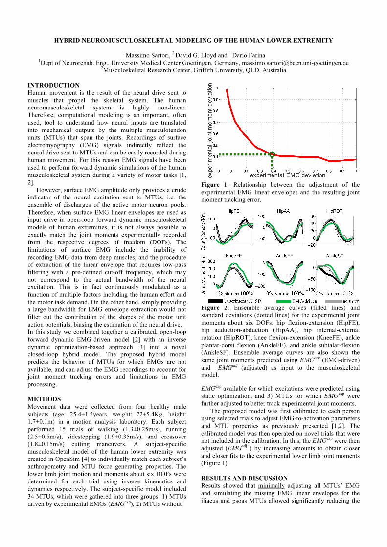
Figure 2: Ensemble average curves (filled lines) and standard deviations (dotted lines) for the experimental joint moments about six DOFs: hip flexion-extension (HipFE), hip adduction-abduction (HipAA), hip internal-external rotation (HipROT), knee flexion-extension (KneeFE), ankle plantar-dorsi flexion (AnkleFE), and ankle subtalar-flexion (AnkleSF). Ensemble average curves are also shown the same joint moments predicted using EMGexp (EMG-driven) and EMGadj (adjusted) as input to the musculoskeletal model. EMGexp available for which excitations were predicted using static optimization, and 3) MTUs for which EMGexp were further adjusted to better track experimental joint moments.
The proposed model was first calibrated to each person using selected trials to adjust EMG-to-activation parameters and MTU properties as previously presented [1,2]. The calibrated model was then operated on novel trials that were not included in the calibration. In this, the EMGexp were then adjusted (EMGadj ) by increasing amounts to obtain closer and closer fits to the experimental lower limb joint moments (Figure 1). RESULTS AND DISCUSSION Results showed that minimally adjusting all MTUs’ EMG and simulating the missing EMG linear envelopes for the iliacus and psoas MTUs allowed significantly reducing the
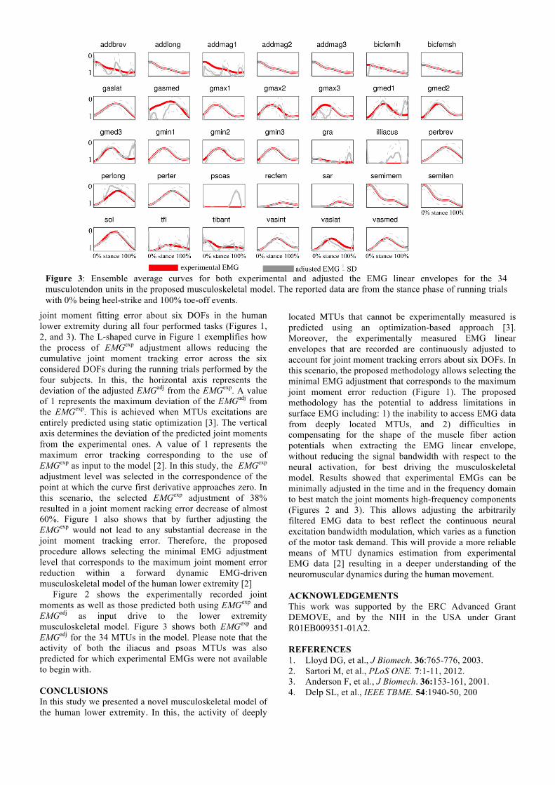
Figure 3: Ensemble average curves for both experimental and adjusted the EMG linear envelopes for the 34 musculotendon units in the proposed musculoskeletal model. The reported data are from the stance phase of running trials with 0% being heel-strike and 100% toe-off events.
joint moment fitting error about six DOFs in the human lower extremity during all four performed tasks (Figures 1, 2, and 3). The L-shaped curve in Figure 1 exemplifies how the process of EMGexp adjustment allows reducing the cumulative joint moment tracking error across the six considered DOFs during the running trials performed by the four subjects. In this, the horizontal axis represents the deviation of the adjusted EMGadj from the EMGexp. A value of 1 represents the maximum deviation of the EMGadj from the EMGexp. This is achieved when MTUs excitations are entirely predicted using static optimization [3]. The vertical axis determines the deviation of the predicted joint moments from the experimental ones. A value of 1 represents the maximum error tracking corresponding to the use of EMGexp as input to the model [2]. In this study, the EMGexp adjustment level was selected in the correspondence of the point at which the curve first derivative approaches zero. In this scenario, the selected EMGexp adjustment of 38% resulted in a joint moment racking error decrease of almost 60%. Figure 1 also shows that by further adjusting the EMGexp would not lead to any substantial decrease in the joint moment tracking error. Therefore, the proposed procedure allows selecting the minimal EMG adjustment level that corresponds to the maximum joint moment error reduction within a forward dynamic EMG-driven musculoskeletal model of the human lower extremity [2]
Figure 2 shows the experimentally recorded joint moments as well as those predicted both using EMGexp and EMGadj as input drive to the lower extremity musculoskeletal model. Figure 3 shows both EMGexp and EMGadj for the 34 MTUs in the model. Please note that the activity of both the iliacus and psoas MTUs was also predicted for which experimental EMGs were not available to begin with. CONCLUSIONS In this study we presented a novel musculoskeletal model of the human lower extremity. In this, the activity of deeply
located MTUs that cannot be experimentally measured is predicted using an optimization-based approach [3]. Moreover, the experimentally measured EMG linear envelopes that are recorded are continuously adjusted to account for joint moment tracking errors about six DOFs. In this scenario, the proposed methodology allows selecting the minimal EMG adjustment that corresponds to the maximum joint moment error reduction (Figure 1). The proposed methodology has the potential to address limitations in surface EMG including: 1) the inability to access EMG data from deeply located MTUs, and 2) difficulties in compensating for the shape of the muscle fiber action potentials when extracting the EMG linear envelope, without reducing the signal bandwidth with respect to the neural activation, for best driving the musculoskeletal model. Results showed that experimental EMGs can be minimally adjusted in the time and in the frequency domain to best match the joint moments high-frequency components (Figures 2 and 3). This allows adjusting the arbitrarily filtered EMG data to best reflect the continuous neural excitation bandwidth modulation, which varies as a function of the motor task demand. This will provide a more reliable means of MTU dynamics estimation from experimental EMG data [2] resulting in a deeper understanding of the neuromuscular dynamics during the human movement. ACKNOWLEDGEMENTS This work was supported by the ERC Advanced Grant DEMOVE, and by the NIH in the USA under Grant R01EB009351-01A2. REFERENCES 1. Lloyd DG, et al., J Biomech. 36:765-776, 2003. 2. Sartori M, et al., PLoS ONE. 7:1-11, 2012. 3. Anderson F, et al., J Biomech. 36:153-161, 2001. 4. Delp SL, et al., IEEE TBME. 54:1940-50, 200