Human TRIB2 Oscillates during the Cell Cycle and …eprints.gla.ac.uk/123001/1/123001.pdf ·...
13
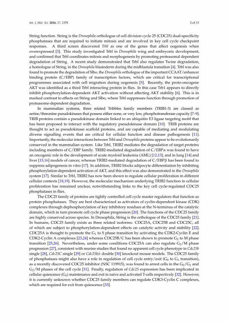
International Journal of Molecular Sciences Article Human TRIB2 Oscillates during the Cell Cycle and Promotes Ubiquitination and Degradation of CDC25C Kai Ling Liang 1,2 , Roberto Paredes 3 , Ruaidhri Carmody 4 , Patrick A. Eyers 5 , Stefan Meyer 3,6 , Tommie V. McCarthy 2 and Karen Keeshan 1, * 1 Paul O’Gorman Leukemia Research Centre, Institute of Cancer Sciences, College of Medical, Veterinary and Life Sciences, University of Glasgow, Glasgow G12 0ZD, UK; [email protected] 2 School of Biochemistry and Cell Biology, University College Cork, Cork, Ireland; [email protected] 3 Stem Cell and Leukaemia Proteomics Laboratory, University of Manchester, Manchester M20 3LJ, UK; [email protected] (R.P.); [email protected] (S.M.) 4 Institute of Infection, Immunity and Inflammation, College of Medical, Veterinary and Life Sciences, University of Glasgow, Glasgow G12 8TA, UK; [email protected] 5 Department of Biochemistry, Institute of Integrative Biology, University of Liverpool, Liverpool L69 7ZB, UK; [email protected] 6 Paediatric and Adolescent Oncology, Royal Manchester Children’s and Christie Hospital, University of Manchester, Manchester M13 9WL, UK * Correspondence: [email protected]; Tel.: +44-141-301-7895; Fax: +44-141-301-7898 Academic Editors: William Chi-shing Cho and Sanjay K. Srivastava Received: 23 June 2016; Accepted: 18 August 2016; Published: 23 August 2016 Abstract: Tribbles homolog 2 (TRIB2) is a member of the mammalian Tribbles family of serine/threonine pseudokinases (TRIB1-3). Studies of TRIB2 indicate that many of the molecular interactions between the single Drosophila Tribbles (Trbl) protein and interacting partners are evolutionary conserved. In this study, we examined the relationship between TRIB2 and cell division cycle 25 (CDC25) family of dual-specificity protein phosphatases (mammalian homologues of Drosophila String), which are key physiological cell cycle regulators. Using co-immunoprecipitation we demonstrate that TRIB2 interacts with CDC25B and CDC25C selectively. Forced overexpression of TRIB2 caused a marked decrease in total CDC25C protein levels. Following inhibition of the proteasome, CDC25C was stabilized in the nuclear compartment. This implicates TRIB2 as a regulator of nuclear CDC25C turnover. In complementary ubiquitination assays, we show that TRIB2-mediated degradation of CDC25C is associated with lysine-48-linked CDC25C polyubiquitination driven by the TRIB2 kinase-like domain. A cell cycle associated role for TRIB2 is further supported by the cell cycle regulated expression of TRIB2 protein levels. Our findings reveal mitotic CDC25C as a new target of TRIB2 that is degraded via the ubiquitin proteasome system. Inappropriate CDC25C regulation could mechanistically underlie TRIB2 mediated regulation of cellular proliferation in neoplastic cells. Keywords: TRIB2; pseudokinase; CDC25B; CDC25C; dual-specificity phosphatase; ubiquitin proteasome; degradation; cell cycle 1. Introduction The Tribbles (Trbl) gene was discovered in three independent Drosophila genetic screens. Two screens [1,2] were designed to identify mutations that affect gastrulation, the formation of ventral furrow by mesodermal precursor cells during Drosophila embryo development. In Trbl mutants, the precursor cells exhibit premature mitosis, leading to defective gastrulation. These pioneering studies identified Trbl as an inhibitor of mitosis and implicated Trbl as a direct regulator of fly Int. J. Mol. Sci. 2016, 17, 1378; doi:10.3390/ijms17091378 www.mdpi.com/journal/ijms
Transcript of Human TRIB2 Oscillates during the Cell Cycle and …eprints.gla.ac.uk/123001/1/123001.pdf ·...

International Journal of
Molecular Sciences
Article
Human TRIB2 Oscillates during the Cell Cycle andPromotes Ubiquitination and Degradationof CDC25CKai Ling Liang 1,2, Roberto Paredes 3, Ruaidhri Carmody 4, Patrick A. Eyers 5, Stefan Meyer 3,6,Tommie V. McCarthy 2 and Karen Keeshan 1,*
1 Paul O’Gorman Leukemia Research Centre, Institute of Cancer Sciences, College of Medical,Veterinary and Life Sciences, University of Glasgow, Glasgow G12 0ZD, UK; [email protected]
2 School of Biochemistry and Cell Biology, University College Cork, Cork, Ireland; [email protected] Stem Cell and Leukaemia Proteomics Laboratory, University of Manchester, Manchester M20 3LJ, UK;
[email protected] (R.P.); [email protected] (S.M.)4 Institute of Infection, Immunity and Inflammation, College of Medical, Veterinary and Life Sciences,
University of Glasgow, Glasgow G12 8TA, UK; [email protected] Department of Biochemistry, Institute of Integrative Biology, University of Liverpool,
Liverpool L69 7ZB, UK; [email protected] Paediatric and Adolescent Oncology, Royal Manchester Children’s and Christie Hospital,
University of Manchester, Manchester M13 9WL, UK* Correspondence: [email protected]; Tel.: +44-141-301-7895; Fax: +44-141-301-7898
Academic Editors: William Chi-shing Cho and Sanjay K. SrivastavaReceived: 23 June 2016; Accepted: 18 August 2016; Published: 23 August 2016
Abstract: Tribbles homolog 2 (TRIB2) is a member of the mammalian Tribbles family ofserine/threonine pseudokinases (TRIB1-3). Studies of TRIB2 indicate that many of the molecularinteractions between the single Drosophila Tribbles (Trbl) protein and interacting partners areevolutionary conserved. In this study, we examined the relationship between TRIB2 and celldivision cycle 25 (CDC25) family of dual-specificity protein phosphatases (mammalian homologuesof Drosophila String), which are key physiological cell cycle regulators. Using co-immunoprecipitationwe demonstrate that TRIB2 interacts with CDC25B and CDC25C selectively. Forced overexpressionof TRIB2 caused a marked decrease in total CDC25C protein levels. Following inhibition of theproteasome, CDC25C was stabilized in the nuclear compartment. This implicates TRIB2 as a regulatorof nuclear CDC25C turnover. In complementary ubiquitination assays, we show that TRIB2-mediateddegradation of CDC25C is associated with lysine-48-linked CDC25C polyubiquitination driven by theTRIB2 kinase-like domain. A cell cycle associated role for TRIB2 is further supported by the cell cycleregulated expression of TRIB2 protein levels. Our findings reveal mitotic CDC25C as a new targetof TRIB2 that is degraded via the ubiquitin proteasome system. Inappropriate CDC25C regulationcould mechanistically underlie TRIB2 mediated regulation of cellular proliferation in neoplastic cells.
Keywords: TRIB2; pseudokinase; CDC25B; CDC25C; dual-specificity phosphatase; ubiquitinproteasome; degradation; cell cycle
1. Introduction
The Tribbles (Trbl) gene was discovered in three independent Drosophila genetic screens.Two screens [1,2] were designed to identify mutations that affect gastrulation, the formation of ventralfurrow by mesodermal precursor cells during Drosophila embryo development. In Trbl mutants,the precursor cells exhibit premature mitosis, leading to defective gastrulation. These pioneeringstudies identified Trbl as an inhibitor of mitosis and implicated Trbl as a direct regulator of fly
Int. J. Mol. Sci. 2016, 17, 1378; doi:10.3390/ijms17091378 www.mdpi.com/journal/ijms
Int. J. Mol. Sci. 2016, 17, 1378 2 of 13
String function. String is the Drosophila orthologue of cell division cycle 25 (CDC25) dual-specificityphosphatases that are required to initiate mitosis and are involved in key cell cycle checkpointresponses. A third screen discovered Trbl as one of the genes that affect oogenesis whenoverexpressed [3]. This study investigated Trbl in Drosophila wing and embryonic development,and confirmed that Trbl coordinates mitosis and morphogenesis by promoting proteasomal dependentdegradation of String. A recent study demonstrated that Trbl also regulates Twine degradation,a homologue of String, in the Drosophila blastoderm during the midblastula transition [4]. Trbl was alsofound to promote the degradation of Slbo, the Drosophila orthologue of the important CCAAT/enhancerbinding protein (C/EBP) family of transcription factors, which are critical for transcriptionalprogrammes associated with cell migration during oogenesis [5]. Recently, the proto-oncogeneAKT was identified as a third Trbl interacting protein in flies. In this case Trb1 appears to directlyinhibit phosphorylation-dependent AKT activation without affecting AKT stability [6]. This is inmarked contrast to effects on String and Slbo, where Trbl suppresses function through promotion ofproteasome-dependent degradation.
In mammalian systems, three related Tribbles family members (TRIB1-3) are classed asserine/threonine pseudokinases that possess either none, or very low, phosphotransferase capacity [7–9].TRIB proteins contain a pseudokinase domain linked to an ubiquitin E3 ligase targeting motif thathas been proposed to interact with the regulatory pseudokinase domain [10]. TRIB proteins arethought to act as pseudokinase scaffold proteins, and are capable of mediating and modulatingdiverse signalling events that are critical for cellular function and disease pathogenesis [11].Importantly, the molecular interactions between Trbl and Drosophila proteins appear to be evolutionarilyconserved in the mammalian system. Like Trbl, TRIB2 mediates the degradation of target proteinsincluding members of C/EBP family. TRIB2-mediated degradation of C/EBPα was found to havean oncogenic role in the development of acute myeloid leukemia (AML) [12,13], and in lung [14] andliver [15,16] models of cancer, whereas TRIB2-mediated degradation of C/EBPβ has been found tosuppress adipogenesis in vitro [17]. In addition, TRIB2 blocks adipocyte differentiation by inhibitingphosphorylation-dependent activation of AKT, and this effect was also demonstrated in the Drosophilasystem [17]. Similar to Trbl, TRIB2 has now been shown to regulate cellular proliferation in differentcellular contexts [18,19]. However, the molecular mechanism underlying TRIB2 function in cellularproliferation has remained unclear, notwithstanding links to the key cell cycle-regulated CDC25phosphatases in flies.
The CDC25 family of proteins are tightly controlled cell cycle master regulators that function asprotein phosphatases. They are best characterized as activators of cyclin-dependent kinase (CDK)complexes through dephosphorylation of key inhibitory residues at the N-terminus of the catalyticdomain, which in turn promote cell cycle phase progression [20]. The functions of the CDC25 familyare highly conserved across species. In Drosophila, String is the orthologue of the CDC25 family [21].In humans, CDC25 family exists as three related isoforms: CDC25A, CDC25B and CDC25C, allof which are subject to phosphorylation-dependent effects on catalytic activity and stability [22].CDC25A is thought to promote the G1 to S phase transition by activating the CDK2-Cyclin E andCDK2-Cyclin A complexes [23,24] whereas CDC25B/C has been shown to promote G2 to M phasetransition [25,26]. Nevertheless, under some conditions CDC25A can also regulate G2/M phaseprogression [27], consistent with murine studies that found no apparent cell cycle phenotype in Cdc25bsingle [28], Cdc25C single [29] or Cdc25b/c double [30] knockout mouse models. The CDC25 familyof phosphatases might also have a role in regulation of cell cycle entry/exit (G0 to G1 transition),as a recently discovered CDC25 inhibitor (NSC 119915), was found to arrest cells in the G0/G1 andG2/M phases of the cell cycle [31]. Finally, regulation of Cdc25 expression has been implicated incellular quiescence (G0) maintenance and exit in naïve and activated T-cells respectively [32]. However,it is currently unknown whether CDC25 family members can regulate CDK3-Cyclin C complexes,which are required for exit from quiescence [33].

Int. J. Mol. Sci. 2016, 17, 1378 3 of 13
In this study, we evaluated human TRIB2-interacting proteins and investigated if theTrbl-mediated degradation of String is likely to be functionally conserved in vertebrate TRIB2pseudokinase, which have been linked to a variety of cancer-associated signaling pathways [34].Here, we show that mammalian CDC25B and CDC25C are novel interacting partners of TRIB2.Consistently, the overexpression of TRIB2 promotes polyubiquitination and proteasome-dependentdegradation of CDC25C. Our data suggests TRIB2-mediated degradation of CDC25C takes place inthe nucleus. In accordance with a potential functional role of TRIB2 in cell cycle regulation, we alsofind that TRIB2 protein expression is tightly regulated during the cell cycle.
2. Results
2.1. TRIB2 Interacts with CDC25B/C But Not CDC25A
To examine the interaction of TRIB2 in human cells with all the three isoforms in the CDC25family, we overexpressed epitope-tagged versions of each protein (FLAG-tagged CDC25A/B/Cand MYC-tagged TRIB2) in HeLa cells and performed co-immunoprecitation experiments.Upon immunoprecipitation with anti-FLAG antibody and Western blotting with anti-MYC antibody,we found that TRIB2 co-immunoprecipitated with both human CDC25B and CDC25C but not humanCDC25A (Figure 1A) proteins. Hence, TRIB2 interaction with CDC25 family shows some selectivityand, in marked contrast to TRIB3, does not appear to interact with CDC25A [35]. We also demonstratedthat TRIB2 binds to both human and mouse CDC25C orthologues (Figure 1B). The weak signal suggeststhat this is a transient and highly unstable interaction.
Int. J. Mol. Sci. 2016, 17, 1378 3 of 13
Here, we show that mammalian CDC25B and CDC25C are novel interacting partners of TRIB2. Consistently, the overexpression of TRIB2 promotes polyubiquitination and proteasome-dependent degradation of CDC25C. Our data suggests TRIB2-mediated degradation of CDC25C takes place in the nucleus. In accordance with a potential functional role of TRIB2 in cell cycle regulation, we also find that TRIB2 protein expression is tightly regulated during the cell cycle.
2. Results
2.1. TRIB2 Interacts with CDC25B/C But Not CDC25A
To examine the interaction of TRIB2 in human cells with all the three isoforms in the CDC25 family, we overexpressed epitope-tagged versions of each protein (FLAG-tagged CDC25A/B/C and MYC-tagged TRIB2) in HeLa cells and performed co-immunoprecitation experiments. Upon immunoprecipitation with anti-FLAG antibody and Western blotting with anti-MYC antibody, we found that TRIB2 co-immunoprecipitated with both human CDC25B and CDC25C but not human CDC25A (Figure 1A) proteins. Hence, TRIB2 interaction with CDC25 family shows some selectivity and, in marked contrast to TRIB3, does not appear to interact with CDC25A [35]. We also demonstrated that TRIB2 binds to both human and mouse CDC25C orthologues (Figure 1B). The weak signal suggests that this is a transient and highly unstable interaction.
Figure 1. Tribbles homolog 2 (TRIB2) interacts with isoform B and C of cell division cycle 25 (CDC25) family proteins physically. (A) Interaction of MYC-tagged TRIB2 with different isoforms of FLAG-tagged CDC25 proteins was examined by co-immunoprecipitation (co-IP) in HeLa cells. IP, immunoprecipitation. Co-immunoprecipitated MYC-tagged TRIB2 signals were quantified by densitometry analyses and normalized to the respective immunoprecipitated FLAG-tagged CDC25 signals. The normalized values are indicated below the sub-panel; (B) interaction of MYC-tagged TRIB2 with human and mouse orthologues of FLAG-tagged CDC25C was examined by co-IP.
2.2. TRIB2 Promotes Ubiquitination and Proteasomal Degradation of CDC25C
To determine if TRIB2 regulates CDC25C through a mechanism related to that described in flies [1–3], we examined the effect of TRIB2 overexpression on CDC25C protein expression levels.TRIB2 overexpression has potent leukaemogenic effects, and is associated with proliferation in vitro [18]. Interestingly, overexpression of TRIB2 in HeLa cells led to a decrease in endogenous CDC25C protein expression (Figure 2A), suggesting TRIB2 could regulate CDC25C turnover. In contrast to CDC25A, which primarily resides in the nucleus, CDC25B/C are held inactive in the cytoplasm, and translocate to the nucleus on activation by phosphorylation, in order to promote cell cycle progression [20]. To test the stability of endogenous CDC25C in both nuclear and cytoplasmic compartments in the presence of ectopic TRIB2, we employed the proteasome inhibitor MG132,
Figure 1. Tribbles homolog 2 (TRIB2) interacts with isoform B and C of cell division cycle 25(CDC25) family proteins physically. (A) Interaction of MYC-tagged TRIB2 with different isoformsof FLAG-tagged CDC25 proteins was examined by co-immunoprecipitation (co-IP) in HeLa cells.IP, immunoprecipitation. Co-immunoprecipitated MYC-tagged TRIB2 signals were quantified bydensitometry analyses and normalized to the respective immunoprecipitated FLAG-tagged CDC25signals. The normalized values are indicated below the sub-panel; (B) interaction of MYC-taggedTRIB2 with human and mouse orthologues of FLAG-tagged CDC25C was examined by co-IP.
2.2. TRIB2 Promotes Ubiquitination and Proteasomal Degradation of CDC25C
To determine if TRIB2 regulates CDC25C through a mechanism related to that described inflies [1–3], we examined the effect of TRIB2 overexpression on CDC25C protein expression levels.TRIB2overexpression has potent leukaemogenic effects, and is associated with proliferation in vitro [18].Interestingly, overexpression of TRIB2 in HeLa cells led to a decrease in endogenous CDC25C proteinexpression (Figure 2A), suggesting TRIB2 could regulate CDC25C turnover. In contrast to CDC25A,which primarily resides in the nucleus, CDC25B/C are held inactive in the cytoplasm, and translocate

Int. J. Mol. Sci. 2016, 17, 1378 4 of 13
to the nucleus on activation by phosphorylation, in order to promote cell cycle progression [20]. To testthe stability of endogenous CDC25C in both nuclear and cytoplasmic compartments in the presenceof ectopic TRIB2, we employed the proteasome inhibitor MG132, which prevents degradation ofproteasomal substrates. Subcellular fractionation showed that endogenous CDC25C was locatedprimarily in the cytoplasm in untransfected vehicle-cells (Figure 2B). Overexpression of TRIB2 didnot affect nuclear translocation of CDC25C proteins in vehicle-treated cells, as expression in thenucleus remained similar when compared to empty vector-transfected cells (Figure 2B). Our data alsoshows that the treatment of untransfected and empty vector transfected cells with MG132 leads to anaccumulation of CDC25C in the nucleus in the absence of a decrease in cytoplasmic CDC25C levels.This strongly argues against the inhibition of CDC25C nuclear to cytoplasmic translocation by MG132but instead suggests that in the steady state the predominant localization of CDC25C in the cytoplasmmay be due to a rapid turnover of nuclear CDC25C. Importantly, in MG132-treated cells, overexpressionof TRIB2 led to a marked increase in the level of nuclear CDC25C compared to control untransfected orempty-vector transfected cells (Figure 2B). These data resemble BRCA1 mediated polyubiquitinationand proteasomal degradation of CDC25C in response to DNA damage [36]. BRCA1-mediated CDC25Cdegradation is dependent on nuclear proteasome activity and inhibition of the proteasome in BRCA1overexpressing cells leads to CDC25C accumulation in the nucleus [36]. Consistently, we foundincreased CDC25C in the nuclear compartment upon MG132 treatment, suggesting that TRIB2specifically regulates the turnover of CDC25C in the nucleus. Intriguingly, our data also suggests thatTRIB2 stability is also regulated through a similar mechanism (Figure 2B).
Int. J. Mol. Sci. 2016, 17, 1378 4 of 13
which prevents degradation of proteasomal substrates. Subcellular fractionation showed that endogenous CDC25C was located primarily in the cytoplasm in untransfected vehicle-cells (Figure 2B). Overexpression of TRIB2 did not affect nuclear translocation of CDC25C proteins in vehicle-treated cells, as expression in the nucleus remained similar when compared to empty vector-transfected cells (Figure 2B). Our data also shows that the treatment of untransfected and empty vector transfected cells with MG132 leads to an accumulation of CDC25C in the nucleus in the absence of a decrease in cytoplasmic CDC25C levels. This strongly argues against the inhibition of CDC25C nuclear to cytoplasmic translocation by MG132 but instead suggests that in the steady state the predominant localization of CDC25C in the cytoplasm may be due to a rapid turnover of nuclear CDC25C. Importantly, in MG132-treated cells, overexpression of TRIB2 led to a marked increase in the level of nuclear CDC25C compared to control untransfected or empty-vector transfected cells (Figure 2B). These data resemble BRCA1 mediated polyubiquitination and proteasomal degradation of CDC25C in response to DNA damage [36]. BRCA1-mediated CDC25C degradation is dependent on nuclear proteasome activity and inhibition of the proteasome in BRCA1 overexpressing cells leads to CDC25C accumulation in the nucleus [36]. Consistently, we found increased CDC25C in the nuclear compartment upon MG132 treatment, suggesting that TRIB2 specifically regulates the turnover of CDC25C in the nucleus. Intriguingly, our data also suggests that TRIB2 stability is also regulated through a similar mechanism (Figure 2B).
Figure 2. TRIB2 promotes proteasome-dependent degradation of CDC25C in the nucleus. (A) Whole cell lysates from FLAG-TRIB2-transfected HeLa cells and controls (untransfected and empty vector-transfected) were analyzed by Western blotting; (B) cells were treated with dimethylsulfoxide (DMSO-vehicle) or 10 µM of MG132 for 4 h before subcellular fractionation for Western blotting analysis. α-Tubulin and HDAC1 are cytoplasmic and nuclear markers respectively. CDC25C signals were quantified by densitometry analyses and normalized to the respective loading control signals. The normalized values were indicated below the sub-panel.
TRIB2 has been shown to drive the degradation of proteins via ubiquitination and subsequent recognition of targets through the ubiquitin proteasome system [37]. We therefore sought to
Figure 2. TRIB2 promotes proteasome-dependent degradation of CDC25C in the nucleus. (A) Wholecell lysates from FLAG-TRIB2-transfected HeLa cells and controls (untransfected and emptyvector-transfected) were analyzed by Western blotting; (B) cells were treated with dimethylsulfoxide(DMSO-vehicle) or 10 µM of MG132 for 4 h before subcellular fractionation for Western blottinganalysis. α-Tubulin and HDAC1 are cytoplasmic and nuclear markers respectively. CDC25C signalswere quantified by densitometry analyses and normalized to the respective loading control signals.The normalized values were indicated below the sub-panel.

Int. J. Mol. Sci. 2016, 17, 1378 5 of 13
TRIB2 has been shown to drive the degradation of proteins via ubiquitination and subsequentrecognition of targets through the ubiquitin proteasome system [37]. We therefore sought to determineif TRIB2 has an effect on the ubiquitination of CDC25C in cells. We found that TRIB2 overexpressionleads to increased ubiquitination of endogenous CDC25C proteins (Figure 3A). Immunoblotting withan antibody specific for lysine K48-linked polyubiquitin chains demonstrated that TRIB2 promotesthe K48-linked polyubiquitination of endogenous CDC25C (Figure 3B). Protein ubiquitination viaK48-linked ubiquitin chains is a well-characterised cellular signal for protein elimination through26S proteasomal degradation [38]. Hence, our results provide the first evidence that mammalianTRIB2 has the ability to promote polyubiquitination of CDC25C, which in turn increases proteasomaldependent degradation of CDC25C phosphatase. TRIB2-mediated degradation of CEBPα requiresboth an intact kinase-like domain and a C-terminal COP1-binding motif [39]. To determine if specificTRIB2 domains are essential for promotion of endogenous CDC25C ubiquitination, we performeda structure-function analysis of TRIB2 in HeLa cells. As demonstrated with full-length (FL) TRIB2protein, overexpression of the TRIB2 kinase-like domain (KD) alone was sufficient to drive CDC25Cubiquitination, whereas deletion of either the N or C-terminal regions (dN or dC) had little effect(Figure 3C). Our results confirm that TRIB2 pseudokinase domain is sufficient for TRIB2-mediatedubiquitination and degradation of CDC25C.
Int. J. Mol. Sci. 2016, 17, 1378 5 of 13
determine if TRIB2 has an effect on the ubiquitination of CDC25C in cells. We found that TRIB2 overexpression leads to increased ubiquitination of endogenous CDC25C proteins (Figure 3A). Immunoblotting with an antibody specific for lysine K48-linked polyubiquitin chains demonstrated that TRIB2 promotes the K48-linked polyubiquitination of endogenous CDC25C (Figure 3B). Protein ubiquitination via K48-linked ubiquitin chains is a well-characterised cellular signal for protein elimination through 26S proteasomal degradation [38]. Hence, our results provide the first evidence that mammalian TRIB2 has the ability to promote polyubiquitination of CDC25C, which in turn increases proteasomal dependent degradation of CDC25C phosphatase. TRIB2-mediated degradation of CEBPα requires both an intact kinase-like domain and a C-terminal COP1-binding motif [39]. To determine if specific TRIB2 domains are essential for promotion of endogenous CDC25C ubiquitination, we performed a structure-function analysis of TRIB2 in HeLa cells. As demonstrated with full-length (FL) TRIB2 protein, overexpression of the TRIB2 kinase-like domain (KD) alone was sufficient to drive CDC25C ubiquitination, whereas deletion of either the N or C-terminal regions (dN or dC) had little effect (Figure 3C). Our results confirm that TRIB2 pseudokinase domain is sufficient for TRIB2-mediated ubiquitination and degradation of CDC25C.
Figure 3. TRIB2 promotes K48-linked polyubiquitination of CDC25C. (A) Effect of overexpression of MYC-tagged TRIB2 on ubiquitination of endogenous CDC25C in HeLa cells. Effect of overexpression of (B) MYC-tagged TRIB2 wild type and (C) different mutants on K48-linked ubiquitination of endogenous CDC25C in HeLa cells. FL, full length; dN, N-terminal deleted; KD, only kinase domain expressed; dC, C-terminal deleted. For (A,C), all samples were treated with 10 µM of MG132 for 7 h prior cell lysis. Ub-HA, HA-tagged ubiquitin.
Figure 3. TRIB2 promotes K48-linked polyubiquitination of CDC25C. (A) Effect of overexpression ofMYC-tagged TRIB2 on ubiquitination of endogenous CDC25C in HeLa cells. Effect of overexpression of(B) MYC-tagged TRIB2 wild type and (C) different mutants on K48-linked ubiquitination of endogenousCDC25C in HeLa cells. FL, full length; dN, N-terminal deleted; KD, only kinase domain expressed;dC, C-terminal deleted. For (A,C), all samples were treated with 10 µM of MG132 for 7 h prior celllysis. Ub-HA, HA-tagged ubiquitin.

Int. J. Mol. Sci. 2016, 17, 1378 6 of 13
2.3. TRIB2 Protein Levels Are Regulated in a Cell Cycle Dependent Manner
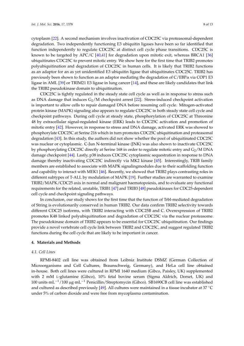
We have previously shown that TRIB2 inhibition either through TRIB2 knockdown or viainhibition of an E2F1-TRIB2 regulatory loop results in perturbation of the cell cycle and cell death [18],and TRIB2 level was shown to be under the control of the β-TRCP ubiquitin E3 ligase in liver cancercells [16]. Given the new potential role of TRIB2 in the regulation of vertebrate CDC25B/C, we assessedthe expression of TRIB2 during cell cycle phases and cell cycle progression. We measured relativeTRIB2 protein expression at specific cell cycle phases using a myeloid leukaemia cell line, SB1690CB.G0/G1-S phase border block was achieved by mimosine treatment and G2/M phase border block bynocodazole treatment (Figure 4A). This revealed elevated TRIB2 protein levels in the G2/M phasewhich co-incided with elevated phosphorylated histone H3 (pSer 10) levels which is a marker ofmitosis (Figure 4B).
Int. J. Mol. Sci. 2016, 17, 1378 6 of 13
2.3. TRIB2 Protein Levels Are Regulated in a Cell Cycle Dependent Manner
We have previously shown that TRIB2 inhibition either through TRIB2 knockdown or via inhibition of an E2F1-TRIB2 regulatory loop results in perturbation of the cell cycle and cell death [18], and TRIB2 level was shown to be under the control of the β-TRCP ubiquitin E3 ligase in liver cancer cells [16]. Given the new potential role of TRIB2 in the regulation of vertebrate CDC25B/C, we assessed the expression of TRIB2 during cell cycle phases and cell cycle progression. We measured relative TRIB2 protein expression at specific cell cycle phases using a myeloid leukaemia cell line, SB1690CB. G0/G1-S phase border block was achieved by mimosine treatment and G2/M phase border block by nocodazole treatment (Figure 4A). This revealed elevated TRIB2 protein levels in the G2/M phase which co-incided with elevated phosphorylated histone H3 (pSer 10) levels which is a marker of mitosis (Figure 4B).
Figure 4. TRIB2 is regulated at the protein level at cell cycle specific phases. Asynchronous (1) SB1690CB cells were G0/G1 (2) arrested by Mimosine followed by release to allow them to progress into S cell cycle phase (3) synchronously and G2/M (4) arrested by Nocodozole followed by release to allow M phase (5) progression. (A) Histogram representation of cell cycle analysis by flow cytometry; (B) Western blotting analysis with TRIB2 and p-histone H3 signal serves as a mitosis marker. Histone H3 indicated protein loading. TRIB2 signals were quantified by densitometry analyses and normalized to the respective Histone H3 signals. The normalized values were indicated below the sub-panel.
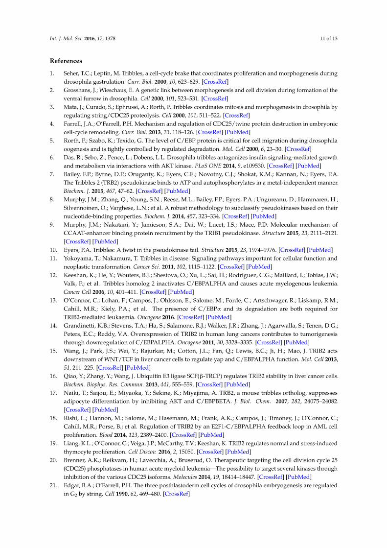
To further evaluate the cyclic TRIB2 expression, we synchronized RPMI-8402, a T-cell acute lymphoblastic leukemia (T-ALL) cell line by a single thymidine block, and monitored the level of TRIB2 mRNA andTRIB2 protein levels as the synchronized cells progressed through the cell cycle following thymidine removal. DNA staining and flow cytometry analysis of samples confirmed the successful synchronization of cells at the G1/S boundary; after removal of thymidine, cells entered and progressed through S phase (S12 and S15) synchronously (Figure 5A). At 20 h after thymidine removal, specific populations of cells were present in G2/M phase (Figure 5A). This was confirmed by detection of increased levels of phosphorylated histone H3 (pSer 10) in cell populations collected at 22 h (S22) (Figure 5B). Remarkably, we observed highly cyclic expression of TRIB2 protein in the absence of significant changes in TRIB2 mRNA levels (Figure 5B,C). Increased levels of TRIB2
Figure 4. TRIB2 is regulated at the protein level at cell cycle specific phases. Asynchronous (1)SB1690CB cells were G0/G1 (2) arrested by Mimosine followed by release to allow them to progressinto S cell cycle phase (3) synchronously and G2/M (4) arrested by Nocodozole followed by release toallow M phase (5) progression. (A) Histogram representation of cell cycle analysis by flow cytometry;(B) Western blotting analysis with TRIB2 and p-histone H3 signal serves as a mitosis marker. Histone H3indicated protein loading. TRIB2 signals were quantified by densitometry analyses and normalized tothe respective Histone H3 signals. The normalized values were indicated below the sub-panel.
To further evaluate the cyclic TRIB2 expression, we synchronized RPMI-8402, a T-cell acutelymphoblastic leukemia (T-ALL) cell line by a single thymidine block, and monitored the level ofTRIB2 mRNA andTRIB2 protein levels as the synchronized cells progressed through the cell cyclefollowing thymidine removal. DNA staining and flow cytometry analysis of samples confirmed thesuccessful synchronization of cells at the G1/S boundary; after removal of thymidine, cells enteredand progressed through S phase (S12 and S15) synchronously (Figure 5A). At 20 h after thymidineremoval, specific populations of cells were present in G2/M phase (Figure 5A). This was confirmed bydetection of increased levels of phosphorylated histone H3 (pSer 10) in cell populations collected at 22 h(S22) (Figure 5B). Remarkably, we observed highly cyclic expression of TRIB2 protein in the absence

Int. J. Mol. Sci. 2016, 17, 1378 7 of 13
of significant changes in TRIB2 mRNA levels (Figure 5B,C). Increased levels of TRIB2 protein weredetected in S12, S17 and S22 where cells were synchronized cells in G1/S phase, S phase and M-phase,respectively (Figure 5B). Consistent with our findings, we see reduced total levels of CDC25C at S17through to S24 where the majority of cells are in S and M-Phase. Hence, TRIB2 protein expressionappears to vary cyclically during the cell cycle, which might be relevant to its ability to modulate thestability of CDC25 isoforms.
Int. J. Mol. Sci. 2016, 17, 1378 7 of 13
protein were detected in S12, S17 and S22 where cells were synchronized cells in G1/S phase, S phase and M-phase, respectively (Figure 5B). Consistent with our findings, we see reduced total levels of CDC25C at S17 through to S24 where the majority of cells are in S and M-Phase. Hence, TRIB2 protein expression appears to vary cyclically during the cell cycle, which might be relevant to its ability to modulate the stability of CDC25 isoforms.
Figure 5. TRIB2 is regulated at the protein level during cell cycle phase progression. Asynchronous (AS) RPMI-8402 cells were arrested by single thymidine block (S0) followed by release (S12–S24) to allow them to progress into different cell cycle phase synchronously. (A) Cell cycle analysis by flow cytometry. Histograms of cell cycle profile for all samples were staggered offset; (B) Western blotting analysis with p-histone H3 signal serves as a mitosis marker. TRIB2 and CDC25C signals were quantified by densitometry analyses and normalized to the respective β-actin signals. The normalized values were indicated below each sub-panel; (C) expression of TRIB2 mRNA measured by quantitative reverse transcription-polymerase chain reaction (RT-PCR).
3. Discussion
In this study, we established a novel relationship between mammalian TRIB2 and CDC25 protein family members, and found that TRIB2 interacts physically with CDC25B/C. We showed that TRIB2 promotes CDC25C proteasomal degradation, confirming that this functional relationship has been conserved between Drosopila Trbl [1–3] and human TRIB2, which is extremely highly conserved in vertebrates homologues [7]. We found that an increase in CDC25C turnover can be driven by TRIB2-triggered K48-linked polyubiquitination. In addition, our data suggest that TRIB2 degrades a nuclear population of CDC25C, and therefore, TRIB2 may target the key mitotic regulatory fraction of CDC25C. Our analysis reveals cyclic expression of TRIB2 during the cell cycle in-line with an oscillatory cell cycle relationship between TRIB2 and CDC25C.
Owing to the critical roles of CDC25 family of proteins in cell cycle regulation, the expression and activity of CDC25 proteins is very tightly regulated. CDC25C activity is controlled primarily by two mechanisms at the post-translational level. The first mechanism involves activating or inhibitory phosphorylation of CDC25C, which either leads to CDC25C activation (e.g., through PLK1), or results in binding of CDC25C with 14-3-3 protein and CDC25C sequestration in the cytoplasm [22]. A second mechanism involves inactivation of CDC25C via proteasomal-dependent degradation. Two independently functioning E3 ubiquitin ligases have been so far identified that function independently to regulate CDC25C at distinct cell cycle phase transitions. CDC25C is known to be
Figure 5. TRIB2 is regulated at the protein level during cell cycle phase progression. Asynchronous (AS)RPMI-8402 cells were arrested by single thymidine block (S0) followed by release (S12–S24) to allowthem to progress into different cell cycle phase synchronously. (A) Cell cycle analysis by flow cytometry.Histograms of cell cycle profile for all samples were staggered offset; (B) Western blotting analysiswith p-histone H3 signal serves as a mitosis marker. TRIB2 and CDC25C signals were quantified bydensitometry analyses and normalized to the respective β-actin signals. The normalized values wereindicated below each sub-panel; (C) expression of TRIB2 mRNA measured by quantitative reversetranscription-polymerase chain reaction (RT-PCR).
3. Discussion
In this study, we established a novel relationship between mammalian TRIB2 and CDC25 proteinfamily members, and found that TRIB2 interacts physically with CDC25B/C. We showed that TRIB2promotes CDC25C proteasomal degradation, confirming that this functional relationship has beenconserved between Drosopila Trbl [1–3] and human TRIB2, which is extremely highly conservedin vertebrates homologues [7]. We found that an increase in CDC25C turnover can be driven byTRIB2-triggered K48-linked polyubiquitination. In addition, our data suggest that TRIB2 degradesa nuclear population of CDC25C, and therefore, TRIB2 may target the key mitotic regulatory fraction ofCDC25C. Our analysis reveals cyclic expression of TRIB2 during the cell cycle in-line with an oscillatorycell cycle relationship between TRIB2 and CDC25C.
Owing to the critical roles of CDC25 family of proteins in cell cycle regulation, the expressionand activity of CDC25 proteins is very tightly regulated. CDC25C activity is controlled primarilyby two mechanisms at the post-translational level. The first mechanism involves activating orinhibitory phosphorylation of CDC25C, which either leads to CDC25C activation (e.g., throughPLK1), or results in binding of CDC25C with 14-3-3 protein and CDC25C sequestration in the
Int. J. Mol. Sci. 2016, 17, 1378 8 of 13
cytoplasm [22]. A second mechanism involves inactivation of CDC25C via proteasomal-dependentdegradation. Two independently functioning E3 ubiquitin ligases have been so far identified thatfunction independently to regulate CDC25C at distinct cell cycle phase transitions. CDC25C isknown to be targeted by APC/C [40,41] for degradation upon mitotic exit, whereas BRCA1 [36]ubiquitinates CDC25C to prevent mitotic entry. We show here for the first time that TRIB2 promotespolyubiquitination and degradation of CDC25C in human cells. It is likely that TRIB2 functionsas an adaptor for an as yet unidentified E3 ubiquitin ligase that ubiquitinates CDC25C. TRIB2 haspreviously been shown to function as an adaptor mediating the degradation of C/EBPα via COP1 E3ligase in AML [39] or TRIM21 E3 ligase in lung cancer [14], and these are likely candidates that linkthe TRIB2 pseudokinase domain to ubiquitination.
CDC25C is tightly regulated in the steady state cell cycle as well as in response to stress suchas DNA damage that induces G2/M checkpoint arrest [22]. Stress-induced checkpoint activationis important to allow cells to repair damaged DNA before resuming cell cycle. Mitogen-activatedprotein kinase (MAPK) signaling also appears to regulate CDC25C in both steady state cell cycle andcheckpoint pathways. During cell cycle at steady state, phosphorylation of CDC25C at Threonine48 by extracellular signal-regulated kinase (ERK) leads to CDC25C activation and promotion ofmitotic entry [42]. However, in response to stress and DNA damage, activated ERK was showed tophosphorylate CDC25C at Serine 216 which in turn promotes CDC25C ubiquitination and proteasomaldegradation [43]. In this study, the authors did not show whether the pool of ubiquitinated CDC25Cwas nuclear or cytoplasmic. C-Jun N-terminal kinase (JNK) was also shown to inactivate CDC25Cby phosphorylating CDC25C directly at Serine 168 in order to regulate mitotic entry and G2/M DNAdamage checkpoint [44]. Lastly, p38 induces CDC25C cytoplasmic sequestration in response to DNAdamage thereby inactivating CDC25C indirectly via MK2 kinase [45]. Interestingly, TRIB familymembers are established to associate with MAPK signalingmodules due to their scaffolding functionand capability to interact with MEK1 [46]. Recently, we showed that TRIB2 plays contrasting roles indifferent subtypes of T-ALL by modulation of MAPK [19]. Further studies are warranted to examineTRIB2/MAPK/CDC25 axis in normal and malignant haematopoiesis, and to evaluate any functionalrequirements for the related, unstable, TRIB1 [47] and TRIB3 [48] pseudokinases for CDC25-dependentcell cycle and checkpoint signaling pathways.
In conclusion, our study shows for the first time that the function of Trbl-mediated degradationof String is evolutionarily conserved in human TRIB2. Our data confirm TRIB2 selectivity towardsdifferent CDC25 isoforms, with TRIB2 interacting with CDC25B and C. Overexpression of TRIB2promotes K48 linked polyubiquitination and degradation of CDC25C via the nuclear proteasome.The pseudokinase domain of TRIB2 appears to be essential for CDC25C ubiquitination. Our findingsprovide a novel vertebrate cell cycle link between TRIB2 and CDC25C, and suggest regulated TRIB2functions during the cell cycle that are likely to be important in cancer.
4. Materials and Methods
4.1. Cell Lines
RPMI-8402 cell line was obtained from Leibniz Institute DSMZ (German Collection ofMicroorganisms and Cell Cultures, Braunschweig, Germany), and HeLa cell line obtainedin-house. Both cell lines were cultured in RPMI 1640 medium (Gibco, Paisley, UK) supplementedwith 2 mM L-glutamine (Gibco), 10% fetal bovine serum (Sigma Aldrich, Dorset, UK) and100 units·mL−1/100 µg·mL−1 Penicillin/Streptomycin (Gibco). SB1690CB cell line was establishedand cultured as described previously [49]. All cultures were maintained in a tissue incubator at 37 ◦Cunder 5% of carbon dioxide and were free from mycoplasma contamination.

Int. J. Mol. Sci. 2016, 17, 1378 9 of 13
4.2. Plasmids and Transfection
Plasmids that encode Ub, TRIB2 wild type and mutants have been described [13,39]. CDC25A/Binserts from pCMV6-CDC25A/B-MYC-FLAG (OriGene, Rockville, MD, USA: RC200496 and RC207409)were sub-cloned into MluI/AsiSI digested pCMV6-AC-FLAG empty vector (OriGene). Human CDC25Cinsert was PCR-amplified from pcDNA3-HisA-CDC25C [50] (Addgene plasmid #10964) and was ligatedinto MluI/AsiSI digested pCMV6-AC-FLAG. Cells were transfected with plasmids indicated in therelated figures using X-tremeGENE™ HP DNA transfection reagent (Roche, Dorset, UK).
4.3. Antibodies and Western Blotting
The following antibodies were used for Western blotting: Anti-K48-Ub (Apu2 clone;Millipore, Watford, UK), anti-phospho-Histone H3 (D2C8 clone; Cell Signaling Technology, Leiden,The Netherlands and #06-570; Millipore), anti-Histone H3 (#9715; Cell Signaling Technology)anti-α-tubulin (B-5-1-2 clone; Sigma Aldrich), anti-β-actin (AC-15 clone; Sigma Aldrich), anti-FLAG(M2 clone; Sigma Aldrich), anti-HA (HA-7 clone; Sigma Aldrich), anti-CDC25C (C-20 clone; Santa CruzBiotechnology, Heidelberg, Germany), anti-HDAC1 (H-51 clone; Santa Cruz Biotechnology), anti-MYC(9E10 clone; Santa Cruz Biotechnology) and anti-TRIB2 (B-06 clone; Santa Cruz Biotechnology).Unless indicated otherwise, Western blotting was performed as described previously [19].Densitometry analyses were performed using Image J (NIH, Bethesda, MD, USA) [51].
4.4. Co-Immunoprecipitation
Cells were lysed in protease inhibitors-supplemented Tris lysis buffer (50 mM Tris buffer (pH 7.4),150 mM NaCl, 0.5% IGEPAL® CA-630, 5% glycerol and 1 mM EDTA) after 24 h of transfection.Prior to immunoprecipitation, protein lysates were pre-cleared by incubation with Protein G Agarosebeads (Millipore, Cork, Ireland) at 4 ◦C for 30 min. For immunoprecipitation, pre-cleared lysateswere incubated with the indicated antibody and Protein G Agarose beads at 4 ◦C for overnight.All incubations were done on a rotary tube mixer. Immunoprecipitated proteins were eluted fromwashed beads by boiling at 95 ◦C for 5 min in Laemmli buffer and analyzed by Western blotting.
4.5. Subcellular Fractionation
Following transfection, cells were treated with DMSO (Sigma Aldrich) or 10 µM MG132(Sigma Aldrich) for 4 h at 37 ◦C before subcellular fractionated by Active Motif Nuclear ExtractKit (La Hulpe, Belgium). Nuclear and cytoplasmic lysates were analyzed by Western blotting.
4.6. Ubiquitination Assay
Following transfection, cells were treated with dimethylsulfoxide (DMSO) or 10 µM MG132 for7 h at 37 ◦C followed by 10 mM N-ethylmaleimide (Sigma Aldrich) for 30 s at room temperature.Cells were then lysed in 1% (wt/vol) sodium dodecyl sulphate (Sigma Aldrich) solution by boilingat 95 ◦C for 5 min. The crude cell lysate was sonicated prior centrifugation to derive cleared proteinlysate. Protein lysate was diluted in protease inhibitors-supplemented Tris lysis buffer (50 mM Trisbuffer (pH 7.4), 150 mM NaCl, 0.5% IGEPAL® CA-630, 5% glycerol and 1 mM EDTA). Pre-clearing,immunoprecipitation and elution of immunoprecipitated proteins were performed, as described inSection 4.4. Protein ubiquitination was analyzed by Western blotting.
4.7. Cell Cycle Synchronization
SB1690CB cells were arrested in late G1 phase with 0.2 mM mimosine (Sigma Aldrich) for 24 h orin G2/M phase with 200 ng/mL of nocodazole (Sigma Aldrich) for 18 h. G1 and G2/M phase-arrestedcells were released from block by replacing medium and culturing them further at 37 ◦C for 7 h.Samples were collected at different points of the cell cycle for flow cytometry and Western blottinganalyses. For Western blotting, cells were lysed in protease inhibitors-supplemented CHAPS/Bicine

Int. J. Mol. Sci. 2016, 17, 1378 10 of 13
buffer (20 mM bicine, 0.6% CHAPS, 2 mM MgCl2, 1 µM ZnCl2, 420 mM NaCl and 500 U/mL PierceUniversal Nuclease).
RPMI-8402 cells were sub-cultured a day before they were treated with 0.75 mM thymidine(Sigma Aldrich) and cultured further at 37 ◦C for 21 h. Synchronous cells (S0) were then released fromthymidine block by washing twice in phosphate buffered saline (PBS) before resuspended in freshcomplete medium for further culture. Samples were collected at different time points (S12, S15, S17, S20,S22 and S24) for flow cytometry, Western blotting and quantitative reverse transcription-polymerasechain reaction (RT-PCR) analyses. For Western blotting, cells were lysed directly in Laemmli buffer.
4.8. Cell Cycle Analysis
Cells were fixed in 70% ice-cold ethanol and stored at −20 ◦C. To analyze cell cycle, cells werewashed twice in PBS before they were resuspended in BD Pharmingen™ Propidium Iodide/RNaseStaining buffer (BD Biosciences, Oxford, UK). Flow cytometry was performed using BD FACSCantoII system (BD Biosciences). Aggregates were excluded by gating of forward scatter height versusarea signals.
4.9. Quantitative RT-PCR
RNA was extracted from cells using RNeasy® Mini Kit (Qiagen, Manchester, UK) and convertedto cDNA using High Capacity cDNA Reverse Transcription Kit (Applied Biosystems, Paisley, UK).TRIB2 and β2M mRNA expressions were measured using Fast SYBR® Green Master Mix (AppliedBiosystems). Primer sequences have been described [19]. Each target was measured in triplicatereactions. TRIB2 expression was normalized to β2M and was calculated using the 2−∆∆Ct method.
Acknowledgments: Kai Ling Liang was supported by Health Research Board Ireland (Ph.D. Scholars Programin Cancer Biology). Karen Keeshan was supported by The Howat Foundation Ltd., and Children with CancerUK and acknowledges funding from Bloodwise project grant 13011. Patrick A. Eyers acknowledges fundingfrom the North West Cancer Development Fund and project grant CR1088. Roberto Paredes and Stefan Meyerare supported by the Kay Kendall Leukemia Fund and Bloodwise. Ruaidhri Carmody is supported by MedicalResearch Council grant MR/M010694/1 and Biotechnology and Biological Research Sciences Research Councilgrant BB/M003671/1. The authors declare no competing financial interests.
Author Contributions: Karen Keeshan and Kai Ling Liang conceived the experiments; Kai Ling Liang andRoberto Paredes performed the experiments; Kai Ling Liang, Tommie V. McCarthy and Karen Keeshan designedexperiments and analysed the data; Kai Ling Liang made the figures; Ruaidhri Carmody contributed materialsand analysis tools; Patrick A. Eyers, Ruaidhri Carmody and Stefan Meyer provided expert advice on experimentaldesign, data interpretation and critical reading. All authors edited the paper.
Conflicts of Interest: The authors declare no conflict of interest. The founding sponsors had no role in the designof the study; in the collection, analyses, or interpretation of data; in the writing of the manuscript, and in thedecision to publish the results.
Abbreviations
AML Acute myeloid leukemiaCDC25 Cell division cycle 25C/EBP CCAAT/enhancer binding proteinDMSO DimethylsulfoxideERK Extracellular signal-regulated kinaseJNK c-Jun N-terminal kinaseKD kinase-like domainMAPK Mitogen-activated protein kinasePBS Phosphate buffered salineRT-PCR Reverse transcription-polymerase chain reactionT-ALL T-cell acute lymphoblastic leukemiaTrbl Drosophila TribblesTRIB2 Tribbles homolog 2Ub-HA Ub-tagged ubiquitin
Int. J. Mol. Sci. 2016, 17, 1378 11 of 13
References
1. Seher, T.C.; Leptin, M. Tribbles, a cell-cycle brake that coordinates proliferation and morphogenesis duringdrosophila gastrulation. Curr. Biol. 2000, 10, 623–629. [CrossRef]
2. Grosshans, J.; Wieschaus, E. A genetic link between morphogenesis and cell division during formation of theventral furrow in drosophila. Cell 2000, 101, 523–531. [CrossRef]
3. Mata, J.; Curado, S.; Ephrussi, A.; Rorth, P. Tribbles coordinates mitosis and morphogenesis in drosophila byregulating string/CDC25 proteolysis. Cell 2000, 101, 511–522. [CrossRef]
4. Farrell, J.A.; O’Farrell, P.H. Mechanism and regulation of CDC25/twine protein destruction in embryoniccell-cycle remodeling. Curr. Biol. 2013, 23, 118–126. [CrossRef] [PubMed]
5. Rorth, P.; Szabo, K.; Texido, G. The level of C/EBP protein is critical for cell migration during drosophilaoogenesis and is tightly controlled by regulated degradation. Mol. Cell 2000, 6, 23–30. [CrossRef]
6. Das, R.; Sebo, Z.; Pence, L.; Dobens, L.L. Drosophila tribbles antagonizes insulin signaling-mediated growthand metabolism via interactions with AKT kinase. PLoS ONE 2014, 9, e109530. [CrossRef] [PubMed]
7. Bailey, F.P.; Byrne, D.P.; Oruganty, K.; Eyers, C.E.; Novotny, C.J.; Shokat, K.M.; Kannan, N.; Eyers, P.A.The Tribbles 2 (TRB2) pseudokinase binds to ATP and autophosphorylates in a metal-independent manner.Biochem. J. 2015, 467, 47–62. [CrossRef] [PubMed]
8. Murphy, J.M.; Zhang, Q.; Young, S.N.; Reese, M.L.; Bailey, F.P.; Eyers, P.A.; Ungureanu, D.; Hammaren, H.;Silvennoinen, O.; Varghese, L.N.; et al. A robust methodology to subclassify pseudokinases based on theirnucleotide-binding properties. Biochem. J. 2014, 457, 323–334. [CrossRef] [PubMed]
9. Murphy, J.M.; Nakatani, Y.; Jamieson, S.A.; Dai, W.; Lucet, I.S.; Mace, P.D. Molecular mechanism ofCCAAT-enhancer binding protein recruitment by the TRIB1 pseudokinase. Structure 2015, 23, 2111–2121.[CrossRef] [PubMed]
10. Eyers, P.A. Tribbles: A twist in the pseudokinase tail. Structure 2015, 23, 1974–1976. [CrossRef] [PubMed]11. Yokoyama, T.; Nakamura, T. Tribbles in disease: Signaling pathways important for cellular function and
neoplastic transformation. Cancer Sci. 2011, 102, 1115–1122. [CrossRef] [PubMed]12. Keeshan, K.; He, Y.; Wouters, B.J.; Shestova, O.; Xu, L.; Sai, H.; Rodriguez, C.G.; Maillard, I.; Tobias, J.W.;
Valk, P.; et al. Tribbles homolog 2 inactivates C/EBPALPHA and causes acute myelogenous leukemia.Cancer Cell 2006, 10, 401–411. [CrossRef] [PubMed]
13. O’Connor, C.; Lohan, F.; Campos, J.; Ohlsson, E.; Salome, M.; Forde, C.; Artschwager, R.; Liskamp, R.M.;Cahill, M.R.; Kiely, P.A.; et al. The presence of C/EBPα and its degradation are both required forTRIB2-mediated leukaemia. Oncogene 2016. [CrossRef] [PubMed]
14. Grandinetti, K.B.; Stevens, T.A.; Ha, S.; Salamone, R.J.; Walker, J.R.; Zhang, J.; Agarwalla, S.; Tenen, D.G.;Peters, E.C.; Reddy, V.A. Overexpression of TRIB2 in human lung cancers contributes to tumorigenesisthrough downregulation of C/EBPALPHA. Oncogene 2011, 30, 3328–3335. [CrossRef] [PubMed]
15. Wang, J.; Park, J.S.; Wei, Y.; Rajurkar, M.; Cotton, J.L.; Fan, Q.; Lewis, B.C.; Ji, H.; Mao, J. TRIB2 actsdownstream of WNT/TCF in liver cancer cells to regulate yap and C/EBPALPHA function. Mol. Cell 2013,51, 211–225. [CrossRef] [PubMed]
16. Qiao, Y.; Zhang, Y.; Wang, J. Ubiquitin E3 ligase SCF(β-TRCP) regulates TRIB2 stability in liver cancer cells.Biochem. Biophys. Res. Commun. 2013, 441, 555–559. [CrossRef] [PubMed]
17. Naiki, T.; Saijou, E.; Miyaoka, Y.; Sekine, K.; Miyajima, A. TRB2, a mouse tribbles ortholog, suppressesadipocyte differentiation by inhibiting AKT and C/EBPBETA. J. Biol. Chem. 2007, 282, 24075–24082.[CrossRef] [PubMed]
18. Rishi, L.; Hannon, M.; Salome, M.; Hasemann, M.; Frank, A.K.; Campos, J.; Timoney, J.; O’Connor, C.;Cahill, M.R.; Porse, B.; et al. Regulation of TRIB2 by an E2F1-C/EBPALPHA feedback loop in AML cellproliferation. Blood 2014, 123, 2389–2400. [CrossRef] [PubMed]
19. Liang, K.L.; O’Connor, C.; Veiga, J.P.; McCarthy, T.V.; Keeshan, K. TRIB2 regulates normal and stress-inducedthymocyte proliferation. Cell Discov. 2016, 2, 15050. [CrossRef] [PubMed]
20. Brenner, A.K.; Reikvam, H.; Lavecchia, A.; Bruserud, O. Therapeutic targeting the cell division cycle 25(CDC25) phosphatases in human acute myeloid leukemia—The possibility to target several kinases throughinhibition of the various CDC25 isoforms. Molecules 2014, 19, 18414–18447. [CrossRef] [PubMed]
21. Edgar, B.A.; O’Farrell, P.H. The three postblastoderm cell cycles of drosophila embryogenesis are regulatedin G2 by string. Cell 1990, 62, 469–480. [CrossRef]
Int. J. Mol. Sci. 2016, 17, 1378 12 of 13
22. Boutros, R.; Lobjois, V.; Ducommun, B. CDC25 phosphatases in cancer cells: Key players? Good targets?Nat. Rev. Cancer 2007, 7, 495–507. [CrossRef] [PubMed]
23. Blomberg, I.; Hoffmann, I. Ectopic expression of CDC25A accelerates the G1/S transition and leads topremature activation of cyclin E- and cyclin A-dependent kinases. Mol. Cell. Biol. 1999, 19, 6183–6194.[CrossRef] [PubMed]
24. Hoffmann, I.; Draetta, G.; Karsenti, E. Activation of the phosphatase activity of human CDC25A bya CDK2-cyclin E dependent phosphorylation at the G1/S transition. EMBO J. 1994, 13, 4302–4310. [PubMed]
25. Millar, J.B.; Blevitt, J.; Gerace, L.; Sadhu, K.; Featherstone, C.; Russell, P. P55CDC25 is a nuclear proteinrequired for the initiation of mitosis in human cells. Proc. Natl. Acad. Sci. USA 1991, 88, 10500–10504.[CrossRef]
26. Gabrielli, B.G.; de Souza, C.P.; Tonks, I.D.; Clark, J.M.; Hayward, N.K.; Ellem, K.A. Cytoplasmic accumulationof CDC25B phosphatase in mitosis triggers centrosomal microtubule nucleation in hela cells. J. Cell Sci. 1996,109, 1081–1093. [PubMed]
27. Mailand, N.; Podtelejnikov, A.V.; Groth, A.; Mann, M.; Bartek, J.; Lukas, J. Regulation of G2/M events byCDC25A through phosphorylation-dependent modulation of its stability. EMBO J. 2002, 21, 5911–5920.[CrossRef] [PubMed]
28. Lincoln, A.J.; Wickramasinghe, D.; Stein, P.; Schultz, R.M.; Palko, M.E.; de Miguel, M.P.; Tessarollo, L.;Donovan, P.J. CDC25B phosphatase is required for resumption of meiosis during oocyte maturation.Nat. Genet. 2002, 30, 446–449. [CrossRef] [PubMed]
29. Chen, M.S.; Hurov, J.; White, L.S.; Woodford-Thomas, T.; Piwnica-Worms, H. Absence of apparent phenotypein mice lacking CDC25C protein phosphatase. Mol. Cell. Biol. 2001, 21, 3853–3861. [CrossRef] [PubMed]
30. Ferguson, A.M.; White, L.S.; Donovan, P.J.; Piwnica-Worms, H. Normal cell cycle and checkpoint responsesin mice and cells lacking CDC25B and CDC25C protein phosphatases. Mol. Cell. Biol. 2005, 25, 2853–2860.[CrossRef] [PubMed]
31. Lavecchia, A.; Di Giovanni, C.; Pesapane, A.; Montuori, N.; Ragno, P.; Martucci, N.M.; Masullo, M.;de Vendittis, E.; Novellino, E. Discovery of new inhibitors of CDC25B dual specificity phosphatases bystructure-based virtual screening. J. Med. Chem. 2012, 55, 4142–4158. [CrossRef] [PubMed]
32. Yusuf, I.; Fruman, D.A. Regulation of quiescence in lymphocytes. Trends Immunol. 2003, 24, 380–386.[CrossRef]
33. Ren, S.; Rollins, B.J. Cyclin C/CDK3 promotes RB-dependent G0 exit. Cell 2004, 117, 239–251. [CrossRef]34. Bailey, F.P.; Byrne, D.P.; McSkimming, D.; Kannan, N.; Eyers, P.A. Going for broke: Targeting the human
cancer pseudokinome. Biochem. J. 2015, 465, 195–211. [CrossRef] [PubMed]35. Sakai, S.; Ohoka, N.; Onozaki, K.; Kitagawa, M.; Nakanishi, M.; Hayashi, H. Dual mode of regulation of cell
division cycle 25 a protein by TRB3. Biol. Pharm. Bull. 2010, 33, 1112–1116. [CrossRef] [PubMed]36. Shabbeer, S.; Omer, D.; Berneman, D.; Weitzman, O.; Alpaugh, A.; Pietraszkiewicz, A.; Metsuyanim, S.;
Shainskaya, A.; Papa, M.Z.; Yarden, R.I. BRCA1 targets G2/M cell cycle proteins for ubiquitination andproteasomal degradation. Oncogene 2013, 32, 5005–5016. [CrossRef] [PubMed]
37. Salome, M.; Campos, J.; Keeshan, K. TRIB2 and the ubiquitin proteasome system in cancer.Biochem. Soc. Trans. 2015, 43, 1089–1094. [CrossRef] [PubMed]
38. Komander, D. The emerging complexity of protein ubiquitination. Biochem. Soc. Trans. 2009, 37, 937–953.[CrossRef] [PubMed]
39. Keeshan, K.; Bailis, W.; Dedhia, P.H.; Vega, M.E.; Shestova, O.; Xu, L.W.; Toscano, K.; Uljon, S.N.;Blacklow, S.C.; Pear, W.S. Transformation by tribbles homolog 2 (TRIB2) requires both the TRIB2 kinasedomain and COP1 binding. Blood 2010, 116, 4948–4957. [CrossRef] [PubMed]
40. Chen, F.; Zhang, Z.; Bower, J.; Lu, Y.; Leonard, S.S.; Ding, M.; Castranova, V.; Piwnica-Worms, H.;Shi, X. Arsenite-induced CDC25C degradation is through the KEN-box and ubiquitin–proteasome pathway.Proc. Natl. Acad. Sci. USA 2002, 99, 1990–1995. [CrossRef] [PubMed]
41. Pfleger, C.M.; Kirschner, M.W. The KEN box: An APC recognition signal distinct from the D box targeted byCDH1. Genes Dev. 2000, 14, 655–665. [PubMed]
42. Wang, R.; He, G.; Nelman-Gonzalez, M.; Ashorn, C.L.; Gallick, G.E.; Stukenberg, P.T.; Kirschner, M.W.;Kuang, J. Regulation of CDC25C by ERK-map kinases during the G2/M transition. Cell 2007, 128, 1119–1132.[CrossRef] [PubMed]
Int. J. Mol. Sci. 2016, 17, 1378 13 of 13
43. Eymin, B.; Claverie, P.; Salon, C.; Brambilla, C.; Brambilla, E.; Gazzeri, S. P14ARF triggers G2 arrest throughERK-mediated CDC25C phosphorylation, ubiquitination and proteasomal degradation. Cell Cycle 2006, 5,759–765. [CrossRef] [PubMed]
44. Gutierrez, G.J.; Tsuji, T.; Cross, J.V.; Davis, R.J.; Templeton, D.J.; Jiang, W.; Ronai, Z.A. JNK-mediatedphosphorylation of CDC25C regulates cell cycle entry and G2/M DNA damage checkpoint. J. Biol. Chem.2010, 285, 14217–14228. [CrossRef] [PubMed]
45. Manke, I.A.; Nguyen, A.; Lim, D.; Stewart, M.Q.; Elia, A.E.; Yaffe, M.B. MAPKAP kinase-2 is a cell cyclecheckpoint kinase that regulates the G2/M transition and S phase progression in response to UV irradiation.Mol. Cell 2005, 17, 37–48. [CrossRef] [PubMed]
46. Yokoyama, T.; Kanno, Y.; Yamazaki, Y.; Takahara, T.; Miyata, S.; Nakamura, T. TRIB1 links the MEK1/ERKpathway in myeloid leukemogenesis. Blood 2010, 116, 2768–2775. [CrossRef] [PubMed]
47. Soubeyrand, S.; Martinuk, A.; Lau, P.; McPherson, R. TRIB1 is regulated post-transcriptionally by proteasomaland non-proteasomal pathways. PLoS ONE 2016, 11, e0152346. [CrossRef] [PubMed]
48. Salazar, M.; Lorente, M.; Orea-Soufi, A.; Davila, D.; Erazo, T.; Lizcano, J.; Carracedo, A.; Kiss-Toth, E.;Velasco, G. Oncosuppressive functions of tribbles pseudokinase 3. Biochem. Soc. Trans. 2015, 43, 1122–1126.[CrossRef] [PubMed]
49. Meyer, S.; Fergusson, W.D.; Oostra, A.B.; Medhurst, A.L.; Waisfisz, Q.; de Winter, J.P.; Chen, F.; Carr, T.F.;Clayton-Smith, J.; Clancy, T.; et al. A cross-linker-sensitive myeloid leukemia cell line from a 2-year-old boywith severe Fanconi anemia and biallelic FANCD1/BRCA2 mutations. Genes Chromosomes Cancer 2005, 42,404–415. [CrossRef] [PubMed]
50. Savitsky, P.A.; Finkel, T. Redox regulation of CDC25C. J. Biol. Chem. 2002, 277, 20535–20540. [CrossRef][PubMed]
51. Schneider, C.A.; Rasband, W.S.; Eliceiri, K.W. NIH Image to ImageJ: 25 years of image analysis. Nat. Methods2012, 9, 671–675. [CrossRef] [PubMed]
© 2016 by the authors; licensee MDPI, Basel, Switzerland. This article is an open accessarticle distributed under the terms and conditions of the Creative Commons Attribution(CC-BY) license (http://creativecommons.org/licenses/by/4.0/).