Human Muscle Cultured in Monolayer and Cocultured … · Human Muscle Cultured in Monolayer and...
11
The Journal of Neuroscience, October 1987, 7(10): 31313141 Human Muscle Cultured in Monolayer and Cocultured with Fetal Rat Spinal Cord: Importance of Dorsal Root Ganglia for Achieving Successful Functional Innervation Takayoshi Kobayaship Valerie Askanas, and W. King Engel USC Neuromuscular Center, Department of Neurology, University of Southern California School of Medicine, Los Angeles, California 90077 Adult human muscle cultured in monolayer was cocultured with explants of 13-14-d-old rat embryo using (a) ventral spinal cord (VSC), (b) transverse section of whole spinal cord (WSC), and (c) WSC with dorsal root ganglia (DRG) attached (WSC + DRG). AChR clusters and AChE-positive patches, both at the nerve-muscle contacts, were studied at 5, 12, and 21 d of coculture with each of the 3 spinal cord preparations. In addition, AChE-positive patches were stud- ied after 31-64 d of coculture with WSC + DRG to evaluate further organization of those patches. Compared to VSC and WSC cocultures, WSC + DRG in- duced significantly more AChR clusters per muscle fiber at the nerve-muscle contacts at 5 d of coculture, and the per- centage of muscle fibers containing AChR clusters was higher at all 3 time points quantitated. The number of AChE-positive sites was the same with all 3 spinal cord preparations in early (day 5) cocultures. Between 12 and 21 d of coculture, the number of muscle fibers containing AChE patches in- creased significantly only with WSC + DRG, correlating with the increased number of contracting muscle fibers in that coculture system. Only in human muscle cocultured with WSC + DRG was successful innervation of the cultured muscle fibers achieved, as manifested by (1) contractions in a continuous rhythm of large groups of muscle fibers that were reversibly blocked by 1 mM d-tubocurarine (aneurally cultured human muscle does not spontaneously contract); (2) well-developed cross- striations throughout the fiber; (3) well-organized AChE-pos- itive sites; and (4) a trend from multifocal toward unifocal innervation of those muscle fibers. Our studies demonstrate that adult human muscle cultured in monolayer can be innervated by fetal rat spinal cord and that, in our system, DRG are essential for achieving func- tional innervation. De novo formation of neuromuscular junctions in monolayer cultures has beendemonstratedpreviously in embryonic animal muscleand animal muscle cell lines cocultured with embryonic Received Sept. 23, 1986; revised Mar. 25, 1987; accepted Apr. 1, 1987. This work was supported by U.S. Public Health Service Grant NS 18661 to V.A. T.K. was a Muscular Dystrophy Association Postdoctoral Research Fellow. Correspondence should be addressed to Valerie Askanas, USC Neuromuscular Center, Hospital of the Good Samaritan, 637 South Lucas Avenue, Los Angeles, CA 90017-2395. a Present address: Department of Neurology, Tokyo Medical and Dental Uni- versity, l-4-45 Ushima, Bunkyo-ku, Tokyo 113, Japan. Copyright 0 1987 Society for Neuroscience 0270-6474/87/103131-l 1$02.00/O animal spinal cord (Shimada et al., 1969; Fischbach, 1972; Fischbach et al., 1979;Nakajima et al., 1980;Davey and Cohen, 1986).Recently, we have developed a new model in which adult human muscle cultured in monolayer can be cocultured with fetal rat spinal cord. In this system,nicotinic AChR and AChE accumulate at the nerve-muscle contacts, presumably young neuromuscular junctions that have developed entirely de novo in culture (Kobayashi and Askanas, 1985).Only when the dorsal root ganglia(DRG) were attached to the spinalcord explant did large areas of cross-striated human muscle fibers in closeprox- imity to the ventral part of spinal cord explant contract in a continuous rhythm, and those contractions could be reversibly blocked by 1 mM of d-tubocurarine (Askanaset al., 1985; Ko- bayashi et al., 1985). Coculture with whole transverse explants of the spinal cord without DRG attached, or with the explants of the ventral part of spinal cord (VSC), caused accumulation of AChR clustersat the nerve-muscle contacts and survival of the cultured human muscle longerthan when cultured aneurally; but only occasionally did those 2 types of spinal cord coculture induce contractile activity of the muscle fibers. Unlike aneurally cultured embryonic animal muscle, which practically always spontaneouslycontracts, human muscle fibers cultured aneu- rally in monolayer virtually never spontaneously contract (As- kanas and Engel, 1979). Our previous report (Kobayashi and Askanas, 1985) de- scribed only the phenomena of AChR and AChE accumulation at the nerve-muscle contacts. In the presentstudy we describe quantitative analyses of AChR clusters and AChE-positive patches formed at nerve-muscle contacts at various stages of innervation of human musclecultured in monolayer and then cocultured with 3 different kinds of spinalcord explants, ventral spinal cord (VSC), whole spinal cord (WSC), and WSC with DRG attached (WSC + DRG). Spatial and quantitative rela- tionships betweenAChR clustersand AChE patches were also studied at various developmental stages of neuromuscular junc- tion formation, applying a technique we developed of double- labeling AChR and AChE on the basisof autoradiography of 125alpha-bungarotoxin and histochemistry of AChE. The lengths and widths of innervated-contracting and innervated-noncon- tracting control musclefibers were also measured. Materials and Methods Coculture of humanmuscle grownin monolayer withfetal rat spinal cord. Human muscle cultures were established, using our explant-reex- plantation technique (Askanas and Engel, 1975), from diagnostic muscle biopsies of patients considered, after all studies were performed, not to have an intrinsicmuscle disease. Cultures were initiated from 1 mm3
Transcript of Human Muscle Cultured in Monolayer and Cocultured … · Human Muscle Cultured in Monolayer and...

The Journal of Neuroscience, October 1987, 7(10): 31313141
Human Muscle Cultured in Monolayer and Cocultured with Fetal Rat Spinal Cord: Importance of Dorsal Root Ganglia for Achieving Successful Functional Innervation
Takayoshi Kobayaship Valerie Askanas, and W. King Engel
USC Neuromuscular Center, Department of Neurology, University of Southern California School of Medicine, Los Angeles, California 90077
Adult human muscle cultured in monolayer was cocultured with explants of 13-14-d-old rat embryo using (a) ventral spinal cord (VSC), (b) transverse section of whole spinal cord (WSC), and (c) WSC with dorsal root ganglia (DRG) attached (WSC + DRG). AChR clusters and AChE-positive patches, both at the nerve-muscle contacts, were studied at 5, 12, and 21 d of coculture with each of the 3 spinal cord preparations. In addition, AChE-positive patches were stud- ied after 31-64 d of coculture with WSC + DRG to evaluate further organization of those patches.
Compared to VSC and WSC cocultures, WSC + DRG in- duced significantly more AChR clusters per muscle fiber at the nerve-muscle contacts at 5 d of coculture, and the per- centage of muscle fibers containing AChR clusters was higher at all 3 time points quantitated. The number of AChE-positive sites was the same with all 3 spinal cord preparations in early (day 5) cocultures. Between 12 and 21 d of coculture, the number of muscle fibers containing AChE patches in- creased significantly only with WSC + DRG, correlating with the increased number of contracting muscle fibers in that coculture system.
Only in human muscle cocultured with WSC + DRG was successful innervation of the cultured muscle fibers achieved, as manifested by (1) contractions in a continuous rhythm of large groups of muscle fibers that were reversibly blocked by 1 mM d-tubocurarine (aneurally cultured human muscle does not spontaneously contract); (2) well-developed cross- striations throughout the fiber; (3) well-organized AChE-pos- itive sites; and (4) a trend from multifocal toward unifocal innervation of those muscle fibers.
Our studies demonstrate that adult human muscle cultured in monolayer can be innervated by fetal rat spinal cord and that, in our system, DRG are essential for achieving func- tional innervation.
De novo formation of neuromuscular junctions in monolayer cultures has been demonstrated previously in embryonic animal muscle and animal muscle cell lines cocultured with embryonic
Received Sept. 23, 1986; revised Mar. 25, 1987; accepted Apr. 1, 1987. This work was supported by U.S. Public Health Service Grant NS 18661 to
V.A. T.K. was a Muscular Dystrophy Association Postdoctoral Research Fellow. Correspondence should be addressed to Valerie Askanas, USC Neuromuscular
Center, Hospital of the Good Samaritan, 637 South Lucas Avenue, Los Angeles, CA 90017-2395.
a Present address: Department of Neurology, Tokyo Medical and Dental Uni- versity, l-4-45 Ushima, Bunkyo-ku, Tokyo 113, Japan.
Copyright 0 1987 Society for Neuroscience 0270-6474/87/103131-l 1$02.00/O
animal spinal cord (Shimada et al., 1969; Fischbach, 1972; Fischbach et al., 1979; Nakajima et al., 1980; Davey and Cohen, 1986). Recently, we have developed a new model in which adult human muscle cultured in monolayer can be cocultured with fetal rat spinal cord. In this system, nicotinic AChR and AChE accumulate at the nerve-muscle contacts, presumably young neuromuscular junctions that have developed entirely de novo in culture (Kobayashi and Askanas, 1985). Only when the dorsal root ganglia (DRG) were attached to the spinal cord explant did large areas of cross-striated human muscle fibers in close prox- imity to the ventral part of spinal cord explant contract in a continuous rhythm, and those contractions could be reversibly blocked by 1 mM of d-tubocurarine (Askanas et al., 1985; Ko- bayashi et al., 1985). Coculture with whole transverse explants of the spinal cord without DRG attached, or with the explants of the ventral part of spinal cord (VSC), caused accumulation of AChR clusters at the nerve-muscle contacts and survival of the cultured human muscle longer than when cultured aneurally; but only occasionally did those 2 types of spinal cord coculture induce contractile activity of the muscle fibers. Unlike aneurally cultured embryonic animal muscle, which practically always spontaneously contracts, human muscle fibers cultured aneu- rally in monolayer virtually never spontaneously contract (As- kanas and Engel, 1979).
Our previous report (Kobayashi and Askanas, 1985) de- scribed only the phenomena of AChR and AChE accumulation at the nerve-muscle contacts. In the present study we describe quantitative analyses of AChR clusters and AChE-positive patches formed at nerve-muscle contacts at various stages of innervation of human muscle cultured in monolayer and then cocultured with 3 different kinds of spinal cord explants, ventral spinal cord (VSC), whole spinal cord (WSC), and WSC with DRG attached (WSC + DRG). Spatial and quantitative rela- tionships between AChR clusters and AChE patches were also studied at various developmental stages of neuromuscular junc- tion formation, applying a technique we developed of double- labeling AChR and AChE on the basis of autoradiography of 125alpha-bungarotoxin and histochemistry of AChE. The lengths and widths of innervated-contracting and innervated-noncon- tracting control muscle fibers were also measured.
Materials and Methods Coculture of human muscle grown in monolayer with fetal rat spinal cord. Human muscle cultures were established, using our explant-reex- plantation technique (Askanas and Engel, 1975), from diagnostic muscle biopsies of patients considered, after all studies were performed, not to have an intrinsic muscle disease. Cultures were initiated from 1 mm3
3132 Kobayashi et al. * Monolayer Human Muscle Cocultured with Fetal Rat Spinal Cord
muscle explants, 5 of which were placed in each 35 mm petri dish (Falcon) coated with a gelatin-human plasma mixture. After an abun- dant growth of cells had emerged from the explants, the explants were removed and transferred to collagen-coated petri dishes. After another abundant growth of cells had emerged from the reexplanted explants, those explants were removed and the monolayer muscle cultures con- tinued. About 5-7 d after myoblast fusion, 1 mm3 explants of spinal cord derived from 13-l 4-d-old Sprague-Dawley rat embryos (8-l 0 mm in length) were placed on the myotube monolayer cultures, 5 explants per petri dish.
Seventeen different experiments were established, comprising a total of 680 cultures, with 3 different types of spinal cord, viz:, VSC, WCS, and WCS + DRG. An additional 23 experiments, comprising a total of 660 cultures, served for coculture of human muscle and WSC + DRG for studies of contracting muscle.
Before coculture with spinal cord, the muscle alone was cultured in F,, medium (Gibco) (Vogel et al., 1972) supplemented with 10% fetal bovine serum (FBS), 50 ng fibroblast growth factor (FGF), 10 ng epi- - . dermal growth factor (EGF), and 10 pg insulin/ 1 ml medium (Askanas and Gallez-Hawkins. 1985). After coculture with sninal cord exnlants. cultures were maintained in F,, medium supplemented with 10% FBS: All cultures were fed twice a week, and living cultures were examined every or every other day by phase-contrast inverted microscopy.
Quantitative analysis of AChR clusters on human muscle jibers co- cultured with direrent types of spinal cord preparations. Seven indepen- dent experiments were established to study the presence of AChR clus- ters at nerve-muscle contacts of human muscle cocultured with VSC, WSC, and WSC + DRG for 5, 12, and 2 1 d. Six to twelve culture dishes were established for each type of spinal cord explant. In each experiment, AChR clusters were visualized by autoradiography of 1251-alpha-bun- garotoxin (alpha-BT). Autoradiography of alpha-BT was performed as described previously (Kobayashi and Askanas, 1985). Large areas of muscle fibers contacted by neurites were chosen for quantitative analysis of AChR clusters. AChR clusters were counted in 2-4 fields per culture dish using a 6.3 x bright-field objective. The total number of muscle fibers that appeared to be contacted by neurites and the total number of AChR clusters were counted. Accordingly, the number of AChR clusters per individual muscle fiber was calculated. Since a few of the muscle fibers that appeared, under the light microscopy, to be contacted by neurites may have only had neurites trespassing them, the total number of muscle fibers counted in each type of coculture was large, in an attempt to compensate for this unavoidable error. The number of noncontracting muscle fibers counted in each type of coculture at 5, 12, and 21 d was 923, 1077, and 676, respectively;& VSC, 619, 1267;and 604 for WSC: and 1609. 1041. and 884 for WSC + DRG. The same calculations were performed on 271 continuously contracting muscle fibers at 21 d of WSC + DRG coculture.
Quantative analysis of AChE patches on human muscle$bers cocul- tured with dtzerent types of spinal cord explants. AChE staining was performed as previously described (Kobayashi and Askanas, 1985) according to a modification of the method of Kamowsky and Roots (1964) using rubeanic acid for enhancement (Nakamura and Torigoe, 1979; Kobayashi et al., 1982). Five independent experiments with hu- man muscle cocultured with 3 different kinds of spinal cord explants were studied at 5, 12, and 21 d of coculture. The number of muscle fibers counted at those 3 time points was 587,54 1, and 443, respectively, for VSC; 640,463, and 5 15 for WSC, and 577,508, and 538 for WSC + DRG. In addition, AChE-positive patches on 201 contracting muscle fibers and on 163 noncontracting but net&e-contacted muscle fibers were counted in 7 culture dishes from 5 experiments at 3 l-64 d after coculture, since well-organized AChE-positive patches were easily iden- tifiable in the older cocultures. Quantitative analysis of the AChE patches was performed in the same way as analysis of AChR clusters.
Double-labeling of AChR clusters and AChE patches by combining autoradiography of 1Z5Z-alpha-BT and AChE histochemisty. To study the relationship between AChR clusters and AChE patches on the same muscle fiber, cultures were “stained” with a double-labeling method we developed. Living cultures were first incubated at 37°C with lZ51-alpha- BT for 1 hr. After incubation, cultures were rinsed several times in F,, medium with 0.2% BSA and 0.1 M phosphate buffer, fixed for 5 min in 1% glutaraldehyde in 0.1 M phosphate buffer, and rinsed 3 times in 0.1
M phosphate buffer. After fixation, cultures were incubated at 37°C for 60 min in a histochemical medium for AChE containing 5 mg acetyl- thiocholine iodide, 6.3 ml of 0.2 M phosphate buffer (pH 5.8), 0.7 ml of 0.1 M sodium citrate, 1 .O ml of 30 mM copper sulfate, 1 .O ml of 0.2
mM iso-octamethyl pyrophosphoramide (OMPA), and 1 .O ml of 5 rnr.4 potassium ferricyanide. After incubation, cultures were thoroughly rinsed, refixed again with 2.5% glutaraldehyde + 1% tannic acid for 30 min, and rinsed several times with 0.1 M phosphate buffer. AChE-positive patches were then photographed in bright- and phase-contrast micros- copy. After being photographed, the cultures were dehydrated, covered with NTB II emulsion, and exposed for 9 d in the dark at 4°C. After the autoradiographs were developed, the exact areas of those autora- diographs were matched with the previously taken AChE pictures, and the autoradiographs were photographed at the same magnification as were the AChE stains.
The double-labeling method was performed after 13-4 1 d of coculture with WSC + DRG on cultures containing both large areas of contracting muscle fibers and, for comparison, areas of muscle fibers contacted by neurites but not contracting.
The total number of AChR clusters and AChE patches, and the length of AChR clusters and AChE patches, were measured on the same in- dividual muscle fibers. The relationship between AChR clusters and AChE patches was studied on 632 contracting and 247 noncontracting muscle fibers.
Quantitative analysis of the length and the width of contracting and noncontracting musclejibers contacted by rat spinal cord neurons. Con- tracting muscle fibers were present only in cocultures with WSC + DRG. Irregular contractions were visible after 7-14 d of muscle-spinal cord coculture. About 2 weeks after the appearance of the first contractions, the contracting muscle fibers developed morphologic features strikingly different from those of muscle fibers contacted by neurites but not contracting. Mainly, the contracting fibers were considerably longer and thinner and were more densely packed in parallel to one another (Fig. 2, a, d, e). Measurements of the length and width of contracting and noncontracting muscle fibers were performed as follows: In 6 different experiments in which muscle was cocultured with the spinal cord ex- plants for 28-57 d, overlapping photographs were taken in each culture dish of 8 contracting areas (in which individual muscle fibers could be well-visualized) and of 8 sister noncontracting areas in the same dish. After a montage (final magnification, 320x) was made, the length of the muscle fibers was measured, and the width of the fibers was measured at 4 points per muscle fiber, which was arbitrarily divided into 5 equal parts. A total of 160 contracting muscle fibers and 174 muscle fibers contacted by neurites but not contracting was measured. The length and mean width of each muscle fiber were calculated. For the width, the following additional calculations were made, separately for the con- tracting and noncontracting muscle fibers: (a) coefficient of variation (CV) (SD/mean x 100) for each individual muscle fiber; (b) the mean of all individual fiber means in one experiment; (c) CV of all fibers in one experiment; and (d) the mean of the CVs between the 8 experiments (calculation I). The means of “d” were compared between contracting and noncontracting fibers. Also calculated for the width was the “super- mean” of the individual fiber CVs, taking all 8 experiments together (calculation II), and comparison was made between contracting and noncontracting fibers.
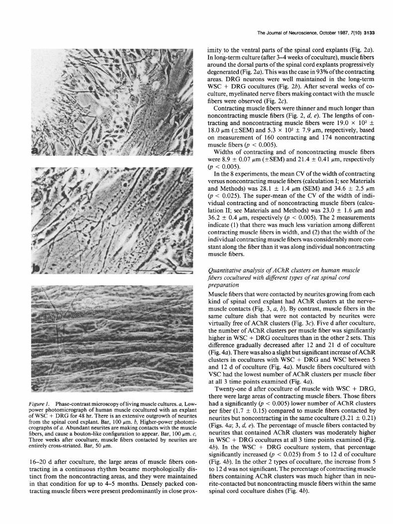
Results Observation of living cultures The beginning of coculture, or “innervation,” was arbitrarily considered to be the day the neural explants were added. Two to three days after coculture, long neurites had emerged from the spinal cord explants, and bouton-like structures were present at the nerve-muscle contacts (Fig. 1, a, b). Neurites emerged earlier from the explants of WSC + DRG (24 hr of coculture) than from VSC or WSC explants (46-64 hr of coculture). How- ever, after 5-6 d of coculture, the outgrowth of neurites from all 3 kinds of spinal cord preparation appeared to be the same.
In WSC + DRG cocultures, occasional asynchronous con- tractions of a few individual muscle fibers were observed as early as 4-6 d after coculture. Usually, though, synchronous contractions of several muscle fibers were not observed sooner than lo-12 d after coculture.
About 14-16 d after coculture, the number of contracting muscle fibers progressively increased, and the majority of con- tracting fibers had become entirely cross-striated (Fig. lc). About
The Journal of Neuroscience, October 1987, 7(10) 3133
Figure I. Phase-contrast microscopy of living muscle cultures. a, Lew- power photomicrograph of human muscle cocultured with an explant of WSC + DRG for 48 hr. There is an extensive outgrowth of neurites from the spinal cord explant. Bar, 100 pm. b, Higher-power photomi- crographs of a. Abundant neurites are making contacts with the muscle fibers, and cause a bouton-like configuration to appear. Bar, 100 pm. c, Three weeks after coculture, muscle fibers contacted by neurites are entirely cross-striated. Bar, 50 pm.
16-20 d after coculture, the large areas of muscle fibers con- tracting in a continuous rhythm became morphologically dis- tinct from the noncontracting areas, and they were maintained in that condition for up to 4-5 months. Densely packed con- tracting muscle fibers were present predominantly in close prox-
imity to the ventral parts of the spinal cord explants (Fig. 2~). In long-term culture (after 3-4 weeks of coculture), muscle fibers around the dorsal parts of the spinal cord explants progressively degenerated (Fig. 2~). This was the case in 93% ofthe contracting areas. DRG neurons were well maintained in the long-term WSC + DRG cocultures (Fig. 2b). After several weeks of co- culture, myelinated nerve fibers making contact with the muscle fibers were observed (Fig. 2~).
Contracting muscle fibers were thinner and much longer than noncontracting muscle fibers (Fig. 2, d, e). The lengths of con- tracting and noncontracting muscle fibers were 19.0 x lo* + 18.0 pm (*SEM) and 5.3 x lo* f 7.9 Mm, respectively, based on measurement of 160 contracting and 174 noncontracting muscle fibers (p < 0.005).
Widths of contracting and of noncontracting muscle fibers were 8.9 + 0.07 pm (+SEM) and 21.4 f 0.41 pm, respectively @ < 0.005).
In the 8 experiments, the mean CV of the width of contracting versus noncontracting muscle fibers (calculation I; see Materials and Methods) was 28.1 + 1.4 pm (SEM) and 34.6 f 2.5 pm (p < 0.025). The super-mean of the CV of the width of indi- vidual contracting and of noncontracting muscle fibers (calcu- lation II; see Materials and Methods) was 23.0 + 1.6 pm and 36.2 f 0.4 pm, respectively 0, < 0.005). The 2 measurements indicate (1) that there was much less variation among different contracting muscle fibers in width, and (2) that the width of the individual contracting muscle fibers was considerably more con- stant along the fiber than it was along individual noncontracting muscle fibers.
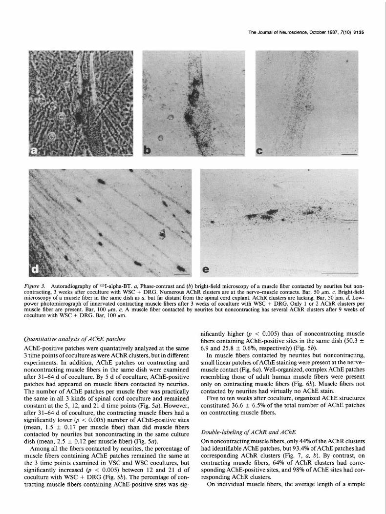
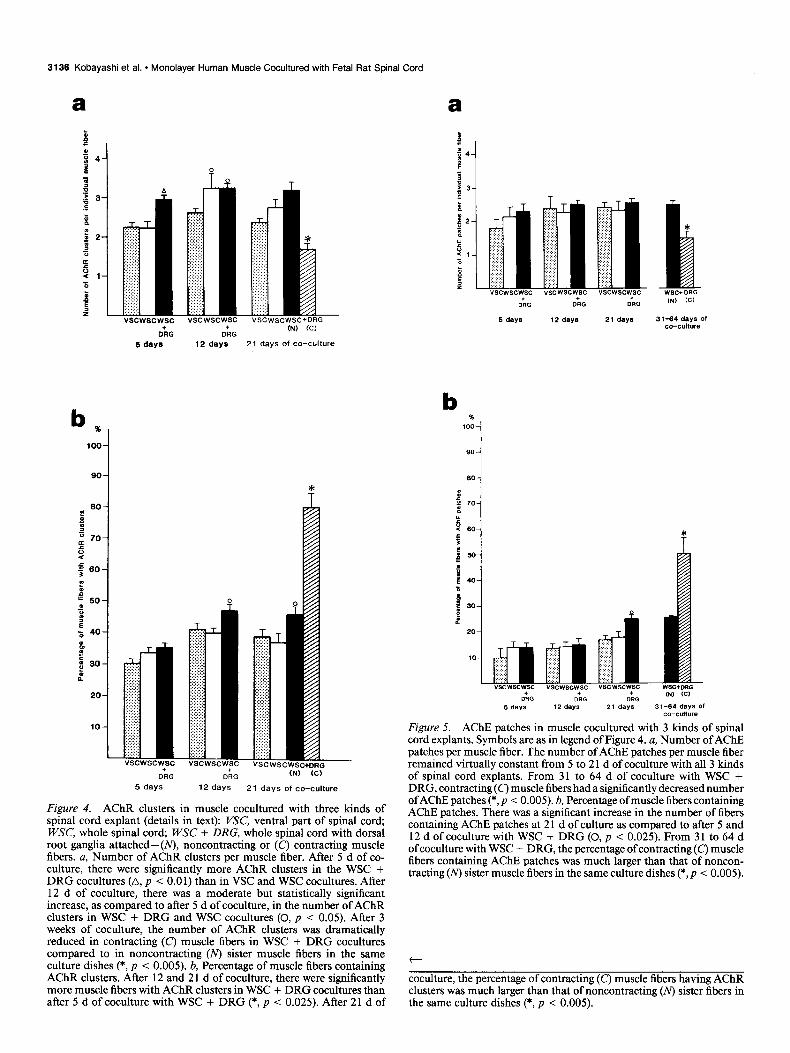
Quantitative analysis of AChR clusters on human muscle fibers cocultured with d@erent types of rat spinal cord preparation Muscle fibers that were contacted by neurites growing from each kind of spinal cord explant had AChR clusters at the nerve- muscle contacts (Fig. 3, a, b). By contrast, muscle fibers in the same culture dish that were not contacted by neurites were virtually free of AChR clusters (Fig. 3~). Five d after coculture, the number of AChR clusters per muscle fiber was significantly higher in WSC + DRG cocultures than in the other 2 sets. This difference gradually decreased after 12 and 2 1 d of coculture (Fig. 4~). There was also a slight but significant increase ofAChR clusters in cocultures with WSC + DRG and WSC between 5 and 12 d of coculture (Fig. 4~). Muscle fibers cocultured with VSC had the lowest number of AChR clusters per muscle fiber at all 3 time points examined (Fig. 4~).
Twenty-one d after coculture of muscle with WSC + DRG, there were large areas of contracting muscle fibers. Those fibers had a significantly 0, < 0.005) lower number of AChR clusters per fiber (1.7 -+ 0.15) compared to muscle fibers contacted by neurites but noncontracting in the same coculture (3.21 + 0.21) (Figs. 4a; 3, d, e). The percentage of muscle fibers contacted by neurites that contained AChR clusters was moderately higher in WSC + DRG cocultures at all 3 time points examined (Fig. 4b). In the WSC + DRG coculture system, that percentage significantly increased (p < 0.025) from 5 to 12 d of coculture (Fig. 4b). In the other 2 types of coculture, the increase from 5 to 12 d was not significant. The percentage of contracting muscle fibers containing AChR clusters was much higher than in neu- rite-contacted but noncontracting muscle fibers within the same spinal cord coculture dishes (Fig. 4b).

3134 Kobayashi et al. l Monolayer Human Muscle Cocultured with Fetal Rat Spinal Cord
Figure 2. Phase-contrast microscopy of long-term cocultures of human muscle and WSC + DRG. a, Low-power photomicrograph of 9-week-old coculture. Abundant muscle fibers in close proximity to the presumably ventral part of the spinal cord explant are densely packed, parallel to each other, and are continuously contracting. Muscle fibers in proximity to the other (presumably dorsal) part of the spinal cord explant have degenerated. Bar, 1 mm. b, Well-maintained DRG neurons in 4-week-old coculture. Bar, 50 pm. c, Myelinated nerve fibers branching over muscle fibers (from left to right) in 5-week-old coculture. Bar, 50 pm. d, e, Contracting (d) and noncontracting (e) muscle fibers in 3-week-old coculture. Contracting muscle fibers are thinner, longer, and more uniform in their individual diameters than noncontracting sister muscle fibers. Bar, 100 pm.
The Journal of Neuroscience, October 1987, 7(10) 3135
Figure 3. Autoradiography of lZ51-alpha-BT. a, Phase-contrast and (b) bright-field microscopy of a muscle fiber contacted by neurites but non- contracting, 3 weeks after coculture with WSC + DRG. Numerous AChR clusters are at the nerve-muscle contacts. Bar, 50 Wm. c, Bright-field microscopy of a muscle fiber in the same dish as a, but far distant from the spinal cord explant. AChR clusters are lacking. Bar, 50 pm. d, Low- power photomicrograph of innervated contracting muscle fibers after 3 weeks of coculture with WSC + DRG. Only 1 or 2 AChR clusters per muscle fiber are present. Bar, 100 pm. e, A muscle fiber contacted by neurites but noncontracting has several AChR clusters after 9 weeks of coculture with WSC + DRG. Bar, 100 pm.
Quantitative analysis of AChE patches AChE-positive patches were quantatively analyzed at the same 3 time points of coculture as were AChR clusters, but in different experiments. In addition, AChE patches on contracting and noncontracting muscle fibers in the same dish were examined after 3 l-64 d of coculture. By 5 d of coculture, AChE-positive patches had appeared on muscle fibers contacted by neurites. The number of AChE patches per muscle fiber was practically the same in all 3 kinds of spinal cord coculture and remained constant at the 5, 12, and 21 d time points (Fig. 5~). However, after 31-64 d of coculture, the contracting muscle fibers had a significantly lower (p < 0.005) number of AChE-positive sites (mean, 1.5 f 0.17 per muscle fiber) than did muscle fibers contacted by neurites but noncontracting in the same culture dish (mean, 2.5 + 0.12 per muscle fiber) (Fig. 5~).
Among all the fibers contacted by neurites, the percentage of muscle fibers containing AChE patches remained the same at the 3 time points examined in VSC and WSC cocultures, but significantly increased (p < 0.005) between 12 and 21 d of coculture with WSC + DRG (Fig. 5b). The percentage of con- tracting muscle fibers containing AChE-positive sites was sig-
nificantly higher (p < 0.005) than of noncontracting muscle fibers containing AChE-positive sites in the same dish (50.3 + 6.9 and 25.8 + 0.6%, respectively) (Fig. 5b).
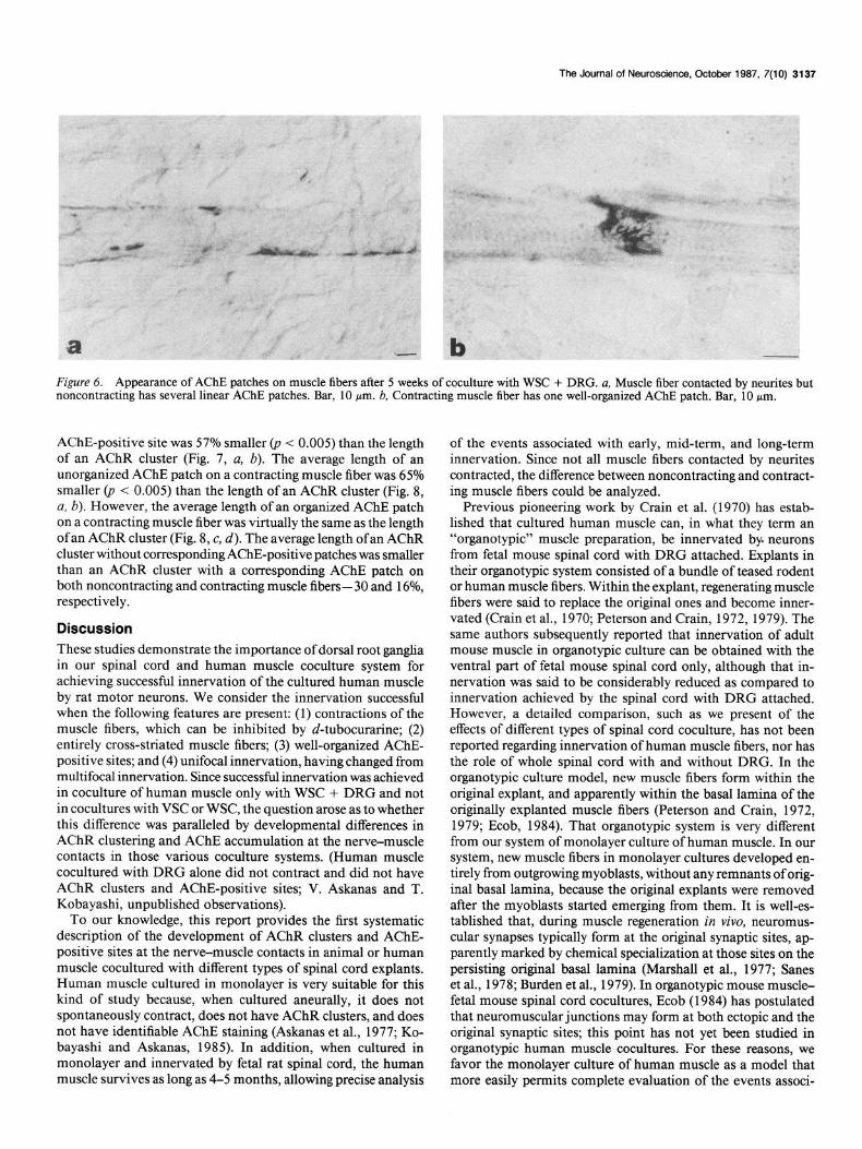
In muscle fibers contacted by neurites but noncontracting, small linear patches of AChE staining were present at the nerve- muscle contact (Fig. 6~). Well-organized, complex AChE patches resembling those of adult human muscle fibers were present only on contracting muscle fibers (Fig. 6b). Muscle fibers not contacted by neurites had virtually no AChE stain.
Five to ten weeks after coculture, organized AChE structures constituted 36.6 f 6.5% of the total number of AChE patches on contracting muscle fibers.
Double-labeling of AChR and AChE On noncontracting muscle fibers, only 44% of the AChR clusters had identifiable AChE patches, but 93.4% of AChE patches had corresponding AChR clusters (Fig. 7, a, b). By contrast, on contracting muscle fibers, 64% of AChR clusters had corre- sponding AChE-positive sites, and 98% of AChE sites had cor- responding AChR clusters.
On individual muscle fibers, the average length of a simple
3136 Kobayashi et al. * Monolayer Human Muscle Cocultured with Fetal Rat Spinal Cord
a f I 54 7
: s B 3 .E z 0
52 J
! i 0 e 1 z
1 z
vscwscwsc vscwscwsc VSCwscWSC+DRG
oh oh (N) (Cl
5 days 12 days 2 1 days of co-culture
ORG D+RG 04) (C)
5 days 12 days 2 1 days of co-culture
Figure 4. AChR clusters in muscle cocultured with three kinds of spinal cord explant (details in text): VSC, ventral part of spinal cord; WSC, whole spinal cord; WSC + DRG, whole spinal cord with dorsal root ganglia attached-( noncontracting or (C’) contracting muscle fibers. a, Number of AChR clusters per muscle fiber. After 5 d of co- culture, there were significantly more AChR clusters in the WSC + DRG cocultures (A, p < 0.0 1) than in VSC and WSC cocultures. After 12 d of coculture, there was a moderate but statistically significant increase, as compared to after 5 d of coculture, in the number of AChR clusters in WSC + DRG and WSC cocultures (0, p < 0.05). After 3 weeks of coculture, the number of AChR clusters was dramatically reduced in contracting (C) muscle fibers in WSC + DRG cocultures compared to in noncontracting (N) sister muscle fibers in the same culture dishes (*, p < 0.005). b, Percentage of muscle fibers containing AChR clusters. After 12 and 2 1 d of coculture, there were significantly more muscle fibers with AChR clusters in WSC + DRG cocultures than after 5 d of coculture with WSC + DRG (*, p < 0.025). After 21 d of
a
21 days 31-64 days Of co-culture
b
Figure 5. AChE patches in muscle cocultured with 3 kinds of spinal cord explants. Symbols are as in legend of Figure 4. a, Number of AChE patches per muscle fiber. The number of AChE patches per muscle fiber remained virtually constant from 5 to 2 1 d of coculture with all 3 kinds of spinal cord explants. From 31 to 64 d of coculture with WSC + DRG, contracting(c) muscle fibers had a significantly decreased number ofAChE patches (*, p < 0.005). b, Percentage of muscle fibers containing AChE patches. There was a significant increase in the number of fibers containing AChE patches at 2 1 d of culture as compared to after 5 and 12 d of coculture with WSC + DRG (0, p < 0.025). From 31 to 64 d of coculture with WSC + DRG, the percentage of contracting (C) muscle fibers containing AChE patches was much larger than that of noncon- tracting (N) sister muscle fibers in the same culture dishes (*,p < 0.005).
t
coculture, the percentage of contracting (C’) muscle fibers having AChR clusters was much larger than that of noncontracting (N) sister fibers in the same culture dishes (*, p < 0.005).
The Journal of Neuroscience, October 1987, 7(10) 3137
Figure 6. Appearance of AChE patches on muscle fibers after 5 weeks of coculture with WSC + DRG. a, Muscle fiber contacted by neurites but noncontracting has several linear AChE patches. Bar, 10 Wm. b, Contracting muscle fiber has one well-organized AChE patch. Bar, 10 pm.
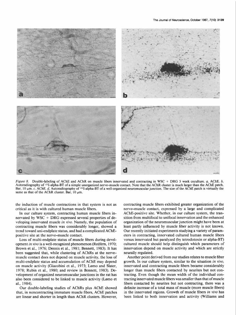
AChE-positive site was 57% smaller (p < 0.005) than the length of an AChR cluster (Fig. 7, a, b). The average length of an unorganized AChE patch on a contracting muscle fiber was 65% smaller (p < 0.005) than the length of an AChR cluster (Fig. 8, a, b). However, the average length of an organized AChE patch on a contracting muscle fiber was virtually the same as the length of an AChR cluster (Fig. 8, c, d). The average length of an AChR cluster without corresponding AChE-positive patches was smaller than an AChR cluster with a corresponding AChE patch on both noncontracting and contracting muscle fibers- 30 and 16%, respectively.
Discussion These studies demonstrate the importance of dorsal root ganglia in our spinal cord and human muscle coculture system for achieving successful innervation of the cultured human muscle by rat motor neurons. We consider the innervation successful when the following features are present: (1) contractions of the muscle fibers, which can be inhibited by d-tubocurarine; (2) entirely cross-striated muscle fibers; (3) well-organized AChE- positive sites; and (4) unifocal innervation, having changed from multifocal innervation. Since successful innervation was achieved in coculture of human muscle only with WSC + DRG and not in cocultures with VSC or WSC, the question arose as to whether this difference was paralleled by developmental differences in AChR clustering and AChE accumulation at the nerve-muscle contacts in those various coculture systems. (Human muscle cocultured with DRG alone did not contract and did not have AChR clusters and AChE-positive sites; V. Askanas and T. Kobayashi, unpublished observations).
To our knowledge, this report provides the first systematic description of the development of AChR clusters and AChE- positive sites at the nerve-muscle contacts in animal or human muscle cocultured with different types of spinal cord explants. Human muscle cultured in monolayer is very suitable for this kind of study because, when cultured aneurally, it does not spontaneously contract, does not have AChR clusters, and does not have identifiable AChE staining (Askanas et al., 1977; Ko- bayashi and Askanas, 1985). In addition, when cultured in monolayer and innervated by fetal rat spinal cord, the human muscle survives as long as 4-5 months, allowing precise analysis
of the events associated with early, mid-term, and long-term innervation. Since not all muscle fibers contacted by neurites contracted, the difference between noncontracting and contract- ing muscle fibers could be analyzed.
Previous pioneering work by Crain et al. (1970) has estab- lished that cultured human muscle can, in what they term an “organotypic” muscle preparation, be innervated by. neurons from fetal mouse spinal cord with DRG attached. Explants in their organotypic system consisted of a bundle of teased rodent or human muscle fibers. Within the explant, regenerating muscle fibers were said to replace the original ones and become inner- vated (Crain et al., 1970; Peterson and Crain, 1972, 1979). The same authors subsequently reported that innervation of adult mouse muscle in organotypic culture can be obtained with the ventral part of fetal mouse spinal cord only, although that in- nervation was said to be considerably reduced as compared to innervation achieved by the spinal cord with DRG attached. However, a detailed comparison, such as we present of the effects of different types of spinal cord coculture, has not been reported regarding innervation of human muscle fibers, nor has the role of whole spinal cord with and without DRG. In the organotypic culture model, new muscle fibers form within the original explant, and apparently within the basal lamina of the originally explanted muscle fibers (Peterson and Crain, 1972, 1979; Ecob, 1984). That organotypic system is very different from our system of monolayer culture of human muscle. In our system, new muscle fibers in monolayer cultures developed en- tirely from outgrowing myoblasts, without any remnants of orig- inal basal lamina, because the original explants were removed after the myoblasts started emerging from them. It is well-es- tablished that, during muscle regeneration in vivo, neuromus- cular synapses typically form at the original synaptic sites, ap- parently marked by chemical specialization at those sites on the persisting original basal lamina (Marshall et al., 1977; Sanes et al., 1978; Burden et al., 1979). In organotypic mouse muscle- fetal mouse spinal cord cocultures, Ecob (1984) has postulated that neuromuscular junctions may form at both ectopic and the original synaptic sites; this point has not yet been studied in organotypic human muscle cocultures. For these reasons, we favor the monolayer culture of human muscle as a model that more easily permits complete evaluation of the events associ-

3138 Kobayashi et al. * Monolayer Human Muscle Cocultured with Fetal Rat Spinal Cord
Figure 7. Double-labeling of AChRs and AChE patches on muscle fibers contacted by neurites but noncontracting in WSC + DRG 3 week cocultures. a, Histochemistry of AChE. b, Autoradiography of 1251-alpha-BT. AChR clusters in b are more numerous and larger than the AChE patches; an AChR cluster and an AChE patch are present together in only one area (arrow). Bar, 50 pm. c, Histochemistry ofAChE. d, Autoradiography of 12SI-alpha-BT. In this area all AChR clusters have corresponding AChE patches. Bar, 10 km.
ated with de nova formation of neuromuscular junctions. In our studies, as compared to VSC or WSC cocultures, ex-
plants of WSC + DRG-induced more AChR clusters per in- dividual innervated muscle fiber after 5 d of coculture, and the percentage of muscle fibers containing one or more AChR clus- ters was moderately higher in WSC + DRG cocultures at all 3 time points quantitated.
While the great majority (93.4%) of AChE patches on non- contracting muscle fibers and virtually all (98%) of AChE patches on contracting muscle fibers had corresponding AChR clusters, a much lower percentage (44% on noncontracting and 64% on contracting muscle fibers) of AChR clusters had corresponding AChE patches on the same muscle fibers. This, and the fact that there were significantly more AChR clusters than AChE patches on noncontracting muscle fibers, indicates that detectable AChR clusters are more easily induced at the nerve-muscle contacts than are AChE accumulations. Interestingly, while the number of muscle fibers containing AChR clusters was significantly higher in WSC + DRG coculture at all 3 time points examined, the number of muscle fibers containing AChE-positive sites in- creased significantly only between 12 and 2 1 d of coculture with WSC + DRG. This finding, and the fact that there was also a significant increase in the number of contracting muscle fibers between 12 and 21 d of coculture with WSC + DRG, suggests that in this period of time there may be an increase in acetyl- choline release inducing both the muscle contractions and the AChE-positive sites.
The mechanism(s) by which DRG contribute to motor neu-
ron-induced contractions in our culture system is not clear. From the literature it is known that DRG have a trophic inllu- ence on spinal motor neurons in vivo. Early removal of DRG during embryogenesis of tadpoles caused reduction of surviving motor neurons (Davis et al., 1983). In the chick embryo, re- moval of DRG afferent fibers resulted in increased loss of motor neurons during the final stages of natural motor neuron death (Okado and Oppenheim, 1984). Efferent terminals of DRG neu- rons form synapses with both motor neuron dendrites and motor neuron somas in the spinal cord of frog and cat (McLaughlin, 1972; Liuzzi et al., 1984). In the developing tadpole larva, DRG afferents contact migrating motor neurons (Liuzzi et al., 1983). Recently it has been reported that chick motor neurons in ho- mogeneous cultures can be kept viable for a longer period of time if DRG neurites make physical contact with them (Bennett, 1986). From these reports, it can be concluded that DRG exert a trophic (survival?) influence on motor neurons. Exactly how DRG exert their seemingly necessary trophic or/and excitatory influence on motor neurons in our culture system (and in the other systems) remains to be studied.
In monolayer cultures of embryonic chick muscle, functional nerve-muscle contacts were formed de novo in cocultures with spinal cord without DRG, and spontaneous and stimulus-evoked action potentials were detected in myotubes adjacent to the spinal cord explants within 24 hr after coculture with the spinal cord explants (Fischbach, 1972; Frank and Fischbach, 1977). Since cultured chick embryo muscle fibers spontaneously con- tract even in aneural cultures, the influence of innervation on
The Journal of Neuroscience, October 1987, 7(10) 3139
Figure 8. Double-labeling of AChE and AChR on muscle fibers innervated and contracting in WSC + DRG 5 week coculture. a, AChE. b, Autoradiography of lZSI-alpha-BT of a simple unorganized nerve-muscle contact. Note that the AChR cluster is much larger than the AChE patch. Bar, 10 pm. c, AChE. d, Autoradiography of V-alpha-BT of a well-organized neuromuscular junction. The size of the AChE patch is virtually the same as that of the AChR cluster. Bar, 10 pm.
the induction of muscle contractions in that system is not as critical as it is with cultured human muscle fibers.
In our culture system, contracting human muscle fibers in- nervated by WSC + DRG expressed several properties of de- veloping innervated muscle in vivo. Namely, the population of contracting muscle fibers was considerably longer, showed a trend toward uni-endplate status, and had a complicated AChE- positive site at the nerve-muscle contact.
Loss of multi-endplate status of muscle fibers during devel- opment in vivo is a well-recognized phenomenon, (Redfern, 1970; Brown et al., 1976; Dennis et al., 198 1; Bennett, 1983). It has been suggested that, while clustering of AChRs at the nerve- muscle contact does not depend on muscle activity, the loss of multi-endplate status and accumulation of AChE may depend on muscle activity (Giacobini et al., 1973; Lomo and Slater, 1978; Rubin et al., 1980; and review in Bennett, 1983). De- velopment of organized neuromuscular junctions in the rat has also been considered to be linked to muscle activity (Lomo et al., 1984).
Our double-labeling studies of AChRs plus AChE showed that, in noncontracting immature muscle fibers, AChE patches are linear and shorter in length than AChR clusters. However,
contracting muscle fibers exhibited greater organization of the nerve-muscle contact, expressed by a large and complicated AChE-positive site. Whether, in our culture system, the tran- sition from multifocal to unifocal innervation and the enhanced organization of the neuromuscular junction might have been at least partly influenced by muscle fiber activity is not known. Our recently initiated experiments studying a variety of param- eters in contracting, innervated cultured human muscle fibers versus innervated but paralyzed (by tetrodotoxin or alpha-BT) cultured muscle should help distinguish which parameters of innervation depend on muscle activity and which are strictly neurally regulated.
Another point derived from our studies relates to muscle fiber growth. In our culture system, similar to the situation in vivo, innervated and contracting muscle fibers became considerably longer than muscle fibers contacted by neurites but not con- tracting. Even though the mean width of the individual con- tracting innervated muscle fibers was smaller than that of muscle fibers contacted by neurites but not contracting, there was a definite increase of a total mass of muscle (more muscle fibers) in the innervated regions. Growth of muscle fibers in vivo has been linked to both innervation and activity (Williams and
3140 Kobayashi et al. * Monolayer Human Muscle Cocultured with Fetal Rat Spinal Cord
Goldspink, 197 1). Recently, the nerve-muscle contact and the subsequent activity have been emphasized as the important steps in vivo in the formation of secondary myotubes, from which a majority of the adult muscle fibers generate (Harris, 1986). Whether the increase of muscle mass in our culture sys- tem reflects the development of secondary myotubes or is due to other phenomena remains unknown.
Bennett, M. R. (1986) Lower motoneurone afferent trophic factors and LMN death: Effect and mechanism. Muscle Nerve (Suppl.) 9: 15.
Bennett, M. R., and A. G. Pettigrew (1974) The formation of synapses in striated muscle during development. J. Physiol. (Lond.) 241: 5 15- 545.
Conclusion and prospects
The developmental pattern of innervation of human muscle cocultured in monolayer with fetal rat spinal cord having DRG attached resembles that of innervated muscle in vivo. Our culture system provides a useful experimental model, in which short-, mid-, and long-term events associated with de novo innervation of human muscle can be precisely studied. Since human fetal material is practically not available for this kind of systematic study, tissue culture of human muscle is becoming an important model for multidisciplinary analyses of human muscle devel- opment and innervation. Recent studies from our laboratory have demonstrated that expression of genes for the muscle- specific isozymes of creatine kinase, glycogen phosphorylase, and phosphoglycerate mutase was significantly increased in hu- man muscle cultured in monolayer and innervated by WSC + DRG (Martinuzzi et al., 1986, 1987). Histoenzymatic matu- ration of innervated cultured human muscle was also much more advanced than that of the noninnervated controls (Vita et al., 1987). In addition, the innervated cultured human muscle fibers had increased resting membrane potentials compared to the noninnervated cultured human fibers, and they exhibited curare-sensitive miniature endplate and endplate potentials, as demonstrated by microelectrode studies (Saito et al., 1986; K. Saito, T. Kobayashi, V. Askanas, W. K. Engel, and K. Ishikawa, unpublished observations).
Brown, M. C., J. K. S. Jansen, and D. V. Essen (1976) Polyneural innervation of skeletal muscle in new-born rats and its elimination during maturation. J. Physiol. (Lond.) 261: 387-422.
Burden, S. J., P. B. Sargent, and U. J. McMahan (1979) Acetylcholine receptors in regenerating muscle accumulate at original synaptic sites in the absence of the nerve. J. Cell Biol. 182: 412-425.
Crain, S. M., L. Alfei, and E. R. Peterson (1970) Neuromuscular transmission in cultures of adult human and rodent skeletal muscle after innervation in vitro by fetal rodent spinal cord. J. Neurobiol. 1: 471-489.
Davey, D. F., and M. W. Cohen (1986) Localization of acetylcholine receptors and cholinesterase on nerve-contacted and noncontacted muscle cells grown in the presence of agents that block action poten- tials. J. Neurosci. 6: 673-680.
Davis, M. R., M. Constantine-Paton, and D. Shorr (1983) Dorsal root ganglia removal in Rana pipiens produces fewer motoneurons. Brain Res. 265: 283-288.
Dennis, M. J., L. Ziskind-Conhaim, and A. J. Harris (1981) Devel- opment of neuromuscular junctions in rat embryo. Dev. Biol. 81: 266-279.
Ecob, M. (1984) The location of neuromuscular junctions on regen- erating adult mouse muscle in culture. J. Neurol. Sci. 64: 175-l 82.
Fischbach, G. D. (1972) Synapse formation between dissociated nerve and muscle cells in low density cell culture. Dev. Biol. 28: 407-429.
Fischbach, G. D., E. Frank, T. M. Jessel, L. L. Rubin, and S. M. Scheutze (1979) Accumulation of acetylcholine receptors and acetylcholin- esterase at newly formed nerve-muscle synapses. Pharmacol. Rev. 30: 41 l-428.
Frank, E., and G. D. Fischbach (1977) ACh receptors accumulate at newly formed nerve-muscle synapses in vitro. In Cell and Tissue Interactions, J. W. Lash and M. M. Burner. eds., VP. 285-292, Raven, New York.
_, .__
Aneural monolayer cultures of diseased adult human muscle have already provided important information regarding patho- geneses of some neuromuscular disorders (Askanas, 1984; Wit- kowski, 1986a, b). Our new model of de novo innervation of human muscle cultured in monolayer can now be applied to study diseased human muscle, especially those diseases that may require innervation for phenotypic manifestation of muscle-cell abnormalities.
Giacobini, G., G. Filogamo, M. Weber, P. Boquet, and J. P. Changeux (1973) Effects of a snake aluha-neurotoxin on the development of innervated skeletal muscles in chick embryo. Proc. Natl. Acad. Sci. USA 70: 1708-1712.
Harris, A. J. (1986) Transfer of synaptic terminals from primary to secondary myotubes. Muscle Nerve (Suppl.) 9: 44.
Kamovskyi M.-J., and L. Roots (1964) Direct-coloring of thiocholine method for cholinesterase. J. Histochem. Cvtochem. 12: 2 19-22 1.
Kobayashi, T., and V. Askanas (1985) Acetycholine receptors and acetylcholinesterase accumulate at the nerve-muscle contacts of de- novo grown human monolayer muscle co-cultured with fetal rat spinal cord. Exp. Neurol. 88: 327-335.
References Askanas, V. (1984) Human muscle and Schwann cells in tissue culture
as a tool in studying pathogenesis and treatment of neuromuscular disorders. In Neuromuscular Diseases, G. Serratrice et al., eds., pp. 373-319, Raven, New York.
Askanas, V., and W. K. Engel (1975) New program for investigating adult human skeletal muscle grown aneurally in tissue culture. Neu- rology 25: 58-67.
Askanas, V., and W. K. Engel (1979) Normal and diseased human muscle in tissue culture. In Handbook of Clinical Neurology, Vol. 40, P. J. Vinken and G. W. Bryun, eds., pp. 183-196, North-Holland, New York.
Askanas, V., and G. Gallez-Hawkins (1985) Synergistic influence of polypeptide growth factors on cultured human muscle. Arch. Neurol. 18: 716-719.
Kobayashi, T., V. Askanas, and W. K. Engel (1985) Innervation of human muscle cultured in monolayer by rat spinal cord is influenced by dorsal root ganglia (DRG). J. Cell Biol. (Suppl.) 101: 13 la.
Kobayashi, T., H. Tsukagoshi, and Y. Shimizu (1982) Trophic effects of sympathetic ganglia on normal and dystrophic chicken skeletal muscles in tissue culture. Exp. Neurol. 77: 241-253.
Liuzzi, F. J., M. S. Beattie, and J. C. Bresnahan (1983) Dorsal root afferents contact migrating motoneurons in the developing frog spinal cord. Brain Res. 262: 299-302.
Liuzzi, F. J., M. S. Beattie, and J. C. Bresnahan (1984) The relationship of dorsal root afferents to motoneuron somata and dendrites in the adult bullfrog: A light and electron microscopic study using horse- radish peroxidase. Neuroscience 4: 95 l-96 1.
Askanas, V., W. K. Engel, S. Ringel, and A. Bender (1977) Acetyl- choline receptors in aneurally cultured human and animal muscle. Neurology 27: 1019-1022.
Askanas, V., W. K. Engel, and T. Kobayashi (1985) TRH enhances motor-neuron-evoked contractions of cultured human muscle. Ann. Neurol. 18: 716-719.
Lomo, T., and C. R. Slater (1978) Control of acetylcholine sensitivity and svnantic formation bv muscle activitv. J. Phvsiol. (Land.) 275: 39 l-402:
I . I
Lomo, T., R. Mirsky, and S. Pockett (1984) Formation of neuro- muscular junctions in adult rats: Role of postsynaptic impulse activ- ity. In Neuromuscular Diseases, G. Serratrice et al., eds., pp. 393- 399, Raven, New York.
Marshall, L. M., J. R. Sanes, and U. J. McMahan (1977) Reinnervation of original synaptic sites on muscle fiber basement membrane after disruption of the muscle cells. Proc. Natl. Acad. Sci. USA 74: 3073- 3077.
Bennett, M. R. (1983) Development of neuromuscular syapses. Phys- Martinuzzi, A., V. Askanas, T. Kobayashi, W. K. Engel, and S. DiMauro iol. Rev. 63: 915-1048. (1986) Expression of muscle-gene specific isoenzymes of phosphor-

The Journal of Neuroscience, October 1987, 7(10) 3141
ylase and creatine kinase in innervated cultured human muscle. J. Cell Biol. 103: 1423-1429.
Martinuzzi, A., V. Askanas, T. Kobayashi, W. K. Engel, and J. Gorsky ( 1987) Developmental expression of the muscle-specific isozyme of phosphoglycerate mutase in human muscle cultured in monolayer and innervated by fetal rat spinal cord. Exp. Neurol. 96: 365-372.
McLaughlin, B. J. (1972) Dorsal root projections to the motor nuclei in cai spinal cord: J. Cbmp. Neurol. 14k: 46 l-474.
Nakaiima. Y.. Y. Kidokoro. and F. G. Klier (1980) The develooment of functional neuromuscular junctions in vi&o: An ultrastructu;e and physiological study. Dev. Biol. 77: 52-72.
Nakamura, T., and K. Torigoe (1979) Simultaneous visualization of catecholamine fluorescence and cholinesterase activity in the periph- eral autonomic nerve. Acta Histochem. Cytochem. 12: 569.
Okado, N., and R. W. Oppenheim (1984) Cell death of motoneurons in the chick embryo spinal cord. IV. In the loss of motoneurons following removal of afferent input. J. Neurosci. 4: 1639-1652.
Peterson, E. R., and S. M. Crain. (1972) Regeneration and innervation in cultures of adult mammalian skeletal muscle coupled with fetal rodent spinal cord. Exp. Neurol. 36: 136-l 59.
Peterson, E. R., and S. M. Crain (1979) Maturation of human muscle after innervation by fetal mouse spinal cord explants in long-term cultures. In Muscle Regeneration, A. Maruo, ed., pp. 429-44 1, Raven, New York.
Redfem, P. (1970) Neuromuscular transmission in new-born rat. J. Physiol. (Lond.) 209: 701-709.
Rubin, L., L. S. M. Schuetze, C. L. Weill, and G. D. Fischbach (1980)
Regulation of acetylcholinesterase appearance at neuromuscular junc- tions in vitro. Nature 238: 264-267.
Saito, K., T. Kobayashi, V. Askanas, W. K. Engel, and K. Ishikawa (1986) Electrical parameters of human muscle cultured in monolayer aneurally and innervated by rat spinal cord. Muscle Nerve (Suppl.) 9: 162.
Sanes, J. R., L. M. Marshall, and U. J. McMahan (1978) Reinnervation of muscle fiber basal lamina after removal of myofibers. Differentia- tion of regenerating axons at original synaptic sites. J. Cell Biol. 78: 176-198.
Shimada, Y., D. A. Fishman, and A. A. Moscona (1969) Formation of neuromuscular junction in embryonic cell cultures. Proc. Natl. Acad. Sci. 62: 7 15-72 1.
Vita, G., V. Askanas, A. Martinuzzi, and W. K. Engel (1987) His- toenzymatic profile of human muscle cultured in monolayer and in- nervated de novo by fetal rat spinal cord. Muscle Nerve (in press).
Vogel, Z., A. J. Sytkowsky, and M. W. Nierenberg (1972) Acetylcho- line recenters of muscle erown in vitro. Proc. Natl. Acad. Sci. USA 69: 31863184. -
Williams, P. E., and G. Goldspink (1971) Longitudinal growth of striated muscle fibers. J. Cell Sci. 9: 75 l-767.
Witkowski, J. A. (1986a) Tissue culture studies of muscle disorders: Part 1. Techniques, cell growth, morphology, cell surface. Muscle Nerve 9: 19 l-207.
Witkowski, J. A. (1986b) Tissue culture studies of muscle disorders: Part 2. Biochemical studies, nerve-muscle culture, metabolic myopa- thies and animal models. Muscle Nerve 9: 283-298.