HUMAN MACHINE INTERFACE THROUGH ELECTROMYOGRAPHY MINOR PROJECT FULL REPORT
30
i A PROJECT REPORT ON HUMAN MACHINE INTERFACE THROUGH ELECTROMYOGRAPHY Submitted by ARJUN RAM GAUTAM NATH HITESH DEWASI UNDER THE GUIDANCE OF PROF. RITESH SARASHWAT in partial fulfillment for the award of BACHELOR OF TECHNOLOGY IN ELECTRONICS AND COMMUNICATION ENGINEERING FROM RAJASTHAN TECHNICAL UNIVERSITY DEPARTMENT OF ELECTRONICS AND COMMUNICATION ENGINEERING JODHPUR INSTITUTE OF ENGINEERING AND TECHNOLOGY MOGRA, N. H. 62, PALI ROAD,JODHPUR-342802 NOVEMBER-2015 SESSION 2015-16
-
Upload
gautam221094 -
Category
Engineering
-
view
218 -
download
2
Transcript of HUMAN MACHINE INTERFACE THROUGH ELECTROMYOGRAPHY MINOR PROJECT FULL REPORT

i
A PROJECT REPORT ON
HUMAN MACHINE INTERFACE THROUGH
ELECTROMYOGRAPHY
Submitted by
ARJUN RAM
GAUTAM NATH
HITESH DEWASI
UNDER THE GUIDANCE OF PROF.
RITESH SARASHWAT
in partial fulfillment for the award of
BACHELOR OF TECHNOLOGY IN
ELECTRONICS AND COMMUNICATION ENGINEERING
FROM
RAJASTHAN TECHNICAL UNIVERSITY
DEPARTMENT OF
ELECTRONICS AND COMMUNICATION ENGINEERING
JODHPUR INSTITUTE OF ENGINEERING AND TECHNOLOGY MOGRA,
N. H. 62, PALI ROAD,JODHPUR-342802
NOVEMBER-2015
SESSION 2015-16

ii
JODHPUR INSTITUTE OF ENGINEERING & TECHNOLOGY
DEPARTMENT OF ELECTRONICS & COMMUNICATION
ENGINEERING
CERTIFICATE
This is to certify that the following students ARJUN RAM, GAUTAM NATH & HITESH
DEWASI have successfully completed the project Titled " HUMAN MACHINE INTERFACE
THROUGH ELECTROMYOGRAPHY” towards the partial fulfillment of Bachelor of Technology
in Electronics and Communications engineering of the Rajasthan Technical University during
academic year 2015 – 2016.
…..….………………… ………………………..
Project Associate Guide
(Prof. Lakshmi Chaudhary) (Prof. Ritesh Saraswat)
INTERNAL EXAMINER EXTERNAL EXAMINER
(Signature with Date) (Signature with Date)

iii
ACKNOWLEDGEMENT
At the very outset, I take this opportunity to express my sincere gratitude to all those who helped me
in the successful completion of my Project report.
We would like to make a number of acknowledgements to those who have helped us to prepare this
report.
We are highly grateful to Prof. O. P. VYAS, Dean (Engineering), JIET for proving us this opportunity to
carry out independent study on this topic.
The divine support given by our guide Prof. Ritesh Saraswat (B. Tech) , Prof. K. K. ARORA, HOD (M.
Tech) & Prof. (Dr.) HEMANT PUROHIT, HOD (B. Tech) Department of Electronics and
Communication Engineering, J.I.E.T, Jodhpur, without them the work would not be possible.
NAME ROLL NO
ARJUN RAM 12EJIEC705
GAUTAM NATH 12EJIEC714
HITESH DEWASI 12EJIEC720

iv
TABLE OF CONTENTS
CHAPTER PAGE NO.
Title Page…………………………………………………………………….i
Certificate………………………………….………………………………...ii
Acknowledgement…………………………….………………………….…iii
List Of figure………………………………………………………………..vi
Abstract……………………………………………………………….…….vii
Market Survey……………………………………………………………..viii
1. INTRODUCTION
1.1 Project Overview……………………………………….…………...1
1.2 Hardware Specification……………………………….…………….2
1.2.1 EMG portable Device……………………………….…...……..…21.2.2 EMG Electrode Type………………………………..…………….2
1.2.3 IC-INA 122 Op-Amp……………………………….……………..3
1.2.3.1 Description……………………………………….……………...41.2.3.2 Specification…………………………………………………….6
1.2.3.3 Application……………………………………….……………...6
1.2.4 IC- OPA 2241 Op-amp………………….…………………………7
1.2.4.1 Description……………………………………………………….7
1.2.4.2 Specification……………………………………………………...8
1.2.4.3 Application………………………………………………………..81.2.5 Battery………………………………………………………………9
1.2.5.1 Specification…………………………………………..…………..91.3 Software Specification………………………………………..……...10
2. STUDY & ANALYSIS
2.1 Need of Study………………….…………………………..………….11
2.2 Signal Recording………………………………………………..……..11
3. DESIGN
3.1 Methodology……………………………………….…………………12..
3.2 Signal Processing……………………………………….…….………13

v
3.3 Circuit Design…………………………………………...………......15
3.4 PCB Layout…………………………………………..………..……15
3.5 Working……………………………………………….……….……16
4. APPLICATION…………………………………………………………..…18
5.1 ADVANTAGE……………………………………………………………..19
5.2 DISADVANTAGE……………………………………………………..….19
6. FUTURE SCOPE…………………………………………………….……..20
7. CONCLUSION……………………………………………………………..21
8. REFERENCE……………………………………………………….……....22

vi
LIST OF FIGURE
FIGURE NO. FIGURE NAME PAGE NO.CHAPTER 11.2.1 EMG Portable Device 2
1.2.2.1 NMES Electrode 31.2.2.2 Metallic Disc Electrode 3
1.2.2.3 Nor axon’s gel Electrode 3
1.2.2.4 Use of Electrode 4
1.2.2.5 Fine Wire 4
1.2.3.1 Symbol Of Op-Amp 5
1.2.3.2 IC-INA 122 Op-Amp Pin Configuration 5
1.2.3.3 IC-INA 122 Op-Amp 6
1.2.4.1 IC-OPA 2241Pin Configuration 7
1.2.4.2 IC-OPA 2241 Op-Amp 8
1.2.5.1 Battery-9 Volt 9
1.3.1 Software Processing Analysis 10
1.3.2 EOG Signal Characteristics 10
CHAPTER 2
2.2.1 Electrode Placement 11
CHAPTER 3
3.2.1 System Design 13
3.2.2 Signal Processing 13
3.3.1 CIRCUIT DIAGRAM 15
3.4.1 PCB LAYOUT 15
3.4.2 Single Side PCB 16
3.5.1 EMG Signal 17
3.5.2 Electrode Placement 17

vii
ABSTRACT
Electromyography (EMG) signals can be used for clinical/biomedical applications, Evolvable
Hardware Chip (EHW) development, and modern human computer interaction. EMG signals acquired
from muscles require advanced methods for detection, decomposition, processing, and classification.
The purpose of this paper is to illustrate the various methodologies and algorithms for EMG signal
analysis to provide efficient and effective ways of understanding the signal and its nature. We further
point up some of the hardware implementations using EMG focusing on applications related to
prosthetic hand control, grasp recognition, and human computer interaction. A comparison study is also
given to show performance of various EMG signal analysis methods. This paper provides researchers
a good understanding of EMG signal and its analysis procedures. This knowledge will help them
develop more powerful, flexible, and efficient applications.

viii
MARKET SURVEY
SR.NO.
COMPONENT NAME
(MAJOR & MINOR) SPECIFICATION PRICE
1 IC 1 INA 122 Op-Amp 160
2 IC 2 OPA 2241 Op-Amp 160
3
Resistor
R1 (100 K ) 1
R2, R3 (10 K ) 1
R4 (1 K ) 1
4 Capacitor 1 uF,25 volt electrolytic 5
5 Battery 9 Volt 20
6 Ultrasound Gel ------- 25
7 Switch On/Off 10
8 Connector 3/2 Pin Connector 130
9 Electrode ECG Metallic/Disposal Electrode 700

1
CHAPTER 1
INTRODUCTION
1.1 PROJECT OVERVIEW:
Electromyography (EMG) is defined as the study of the muscular function through the
Analysis of the generated electric signals during muscular contractions. The potential
difference obtained in the fibers can be registered in the surface of the human body through
surface electrodes due to the biological tissues conducting properties. Our project studies the
muscular activity as an input
In order to control applications. A large set of target muscles are available so we can
interact widely with the computer. The main goal of the project is to provide tetraplegia
individuals the capability to control a portable device (especially to be able to write and
send SMS). In order to accomplish this task we monitories’ muscle activity through an
Electromyography
The recurrent and increasing electromyography study in medicine related areas led to a great
Scientific investment improve the myographic signal acquisition and analysis process.
These advances culminate with the possibility to use portable electromyography devices that
Communicate via wireless with a processing system. Portability makes it possible for any
individual the transport and use of an EMG device with great social acceptance (Costanzia et
al., 2004). EMG devices portability and reduced size easily conducted to its use in HCI
with work carried through in the area of Accessibility, Robotics, Mobile Computation and
Recognition of Gestures, among others. (Roy et al, 1994) present a gesture-based person
Portable device, process the digital signal and emulate certain events accordingly to the
Features detected. Being able to detect and to evaluate muscular activity in an individual
gives us the possibility to associate it with determined interface commands, thus having the
myographic signal as input. This kind of interaction can also be useful to full capable
individuals in a hands-busy situation, such as in a presentation or in a mobility context.
Machine interface for people with serious motor limitations due to cerebral paralysis. This
work, based on gestural recognition with biomechanic and bioelectric sensors, present
many motivating results being capable to differentiate gestures through the use of neural
networks. In the same scope, (Barreto et al., 1999) introduce a system that tries to offer the
users with serious motor limitations the possibility to use the traditional interfaces to

2
Point and select. This system associates face movements to mouse control, being sufficiently
similar to the system ”Tongue Point” (Salem and Zhai, 1997) but using myographic
signals. It is still in the accessibility context that (Erikssonet al., 1998) lean over prosthesis
control. As discussed in the introduction, a person’s brain wave -*patterns and muscle
stimulation can be measured and recorded using almost identical measurement techniques.
With accurate results, researches will soon be able to show a detailed direct Relation between
mental thought and physical motion, improve communications with lacking motor Skills, and
much more. But what are these measurement procedures and how are they carried out?
1.2 HARDWARE SPECIFICAION:
1.2.1 EMG Portable Device:
Our electromyography device collects samples at a 1000Hz sampling rate in 5
independent channels. It has a 110dB CMRR amplifier and a band pass filter between
25 and 500 Hz with gain 1000. It is a relatively small device (14cm * 8cm * 4cm) that can
be carried in a belt or pocket. It is a portable device which communicates by a Bluetooth
interface within a 100 meters range. To col- lect the signals we use surface differential
electrodes, with 1.5 cm radius (Gam boa et al., 2004)
Figure 1.2.1 EMG Portable Device
1.2.2 EMG ELECTRODE TYPES:
Most major limb and trunk muscle activity can be measured using surface electrode
techniques. For deeper, smaller, or overlaid muscles fine wire electrodes need to be used
to acquire intramuscular activities. For surface electrodes, simple platinum or silver disc
electrodes, pre-gelled Ag-AgCl electrodes, and wet-gel electrodes are commonly used.
The disc electrodes are reusable while the gel electrodes are single use. Distinction exists
between electrodes used for SEMG and those used for NMES and ETS. Whenever an
electrical stimulation is applied, the electrodes used must be properly designed to deliver

3
Such electrical stimulations otherwise the power density generated at the skin contact can
result in patient injury. Also, for the evaluation and treatment of incontinence, special
vaginal and anal probes are used to measure the pelvic floor muscle activities.
Fig 1.2.2.1 - NMES electrodes:
Fig 1.2.2.2:- Metallic disc electrodes
Fig 1.2.2.3 :- Noraxon’s gel electrodes: pre-gelled AgCl (1, 2) and wet-gels (3, 4)

4
Fig 1.2.2.4:-Use of electrode
Fig 1.2.2.5:-Fine wire electrode
For all electrode types, additional measures can be taken to affix the electrode cabling to the
patient body to minimize movement artifacts. Regular adhesive tape, hook and loop fasteners,
and elastic straps are commonly used to secure cabling onto the body, but never the electrodes
as this will affect the readings when intramuscular EMG is required to look into the electrical
activity of deeper or overlaid muscles, thin and flexible fine wire electrodes are used.
The fine wire electrode is inserted into the muscle site of interest. The needle or steel
cannula is removed, and the electrode wires are connected to the steel spring adapters to
minimize movement artifacts.
1.2.3 IC-INA 122 OPAMP:
1.2.3.1 Description:
The INA122 is a precision instrumentation amplifier for accurate, low noise differential
signal acquisition. Its two-op-amp design provides excellent performance with very low
quiescent current, and is ideal for portable instrumentation and data acquisition systems. The
INA122 can be operated with single power supplies from 2.2V to 36V and
quiescent current is a mere 60mA. It can also be operated from dual supplies. By utilizing
an input level-shift network, input commonmode range extends to 0.1V below negative rail
(single supply ground). A single external resistor sets gain from 5V/V to
10000V/V. Laser trimming provides very low offset

5
Voltage (250mV max), offset voltage drift (3mV/°C max) and excellent common-mode
rejection.
Package options include 8-pin plastic DIP and SO-8 surface-mount packages. Both are
specified for the –40°C to +85°C extended industrial temperature range.
Fig 1.2.3.1:- Symbol of Op-amp
Fig 1.2.3.2:- IC-INA 122 Op-amp pin configuration

6
Fig1.2.3.3:-IC-INA 122 Op-amp
1.2.3.2 SPECIFICATION:
LOW QUIESCENT CURRENT: 60mA
WIDE POWER SUPPLY RANGE
Single Supply: 2.2V to 36V
Dual Supply: –0.9/+1.3V to ±18V
COMMON-MODE RANGE TO (V–)–0.1V
RAIL-TO-RAIL OUTPUT SWING
LOW OFFSET VOLTAGE: 250mV max
LOW OFFSET DRIFT: 3mV/°C max
LOW INPUT BIAS CURRENT: 25nA max
8-PIN DIP AND SO-8 SURFACE-MOUNT
1.2.3.3 APPLICATIONS:
PORTABLE, BATTERY OPERATED SYSTEMS
INDUSTRIAL SENSOR AMPLIFIER:
Bridge, RTD, Thermocouple
PHYSIOLOGICAL AMPLIFIER:
USE IN ECG, EEG, EMG
MULTI-CHANNEL DATA ACQUISITION

7
1.2.4 IC-OPA 2241 OPAMP:
1.2.4.1 DESCRIPTION:
The OPA241 series and OPA251 series are specifically designed for battery powered,
Portable applications. In addition to very low power consumption (25mA), these amplifiers
feature low offset voltage, rail-to-rail output swing, high common-mode rejection, and
high open-loop gain.
The OPA241 series is optimized for operation at low power supply voltage while the
OPA251 series is optimized for high power supplies. Both can operate from either single
(+2.7V to +36V) or dual supplies (±1.35V to ±18V). The input common-mode voltage
range extends 200mV below
The negative supply—ideal for single-supply applications. They are unity-gain stable and
can drive large capacitive loads. Special design considerations assure that these products
are easy to use. High performance is maintained as the amplifiers swing to their specified
limits. Because the initial Offset voltage (±250mV max) is so low, user adjustment is
usually not required. However, external trim pins are provided for special applications
(single versions only).
Fig 1.2.4.1:-Pin configuration of OPA2241 Op-amp

8
Fig 1.2.4.2:-IC-OPA 2241 OP-AMP
The OPA241 and OPA251 (single versions) are available in standard 8-pin DIP and SO-
8 surface-mount packages. The OPA2241 and OPA2251 (dual versions) come in 8-pin
DIP and SO-8 surface-mount packages. The OPA4241 and OPA4251 (quad versions) are
available in 14-pin DIP and SO-14 surface-mount packages. All are fully specified from
–40°C to +85°C and operate from –55°C to +125°C.
1.2.4.2 SPECIFICATION:
Micro POWER: IQ = 25mA
Single Supply Voltage
Rail-to-Rail Output within 50mV
Low Offset Voltage: 250uV max
High Common-Mode Rejection: 124dB
High Open-Loop Gain: 128dB
Input Bias Current: -4nA
Input Offset Current: 0.1nA
Input Voltage Noise, f = 0.1Hz to 10Hz: 1uVp-p
Common-Mode Voltage Range: -0.2V to (V+)-0.8V
1.2.4.3 APPLICATIONS:
Battery Operated Instruments
Portable Devices
Use in Medical Instruments
Use in Test Equipment
9
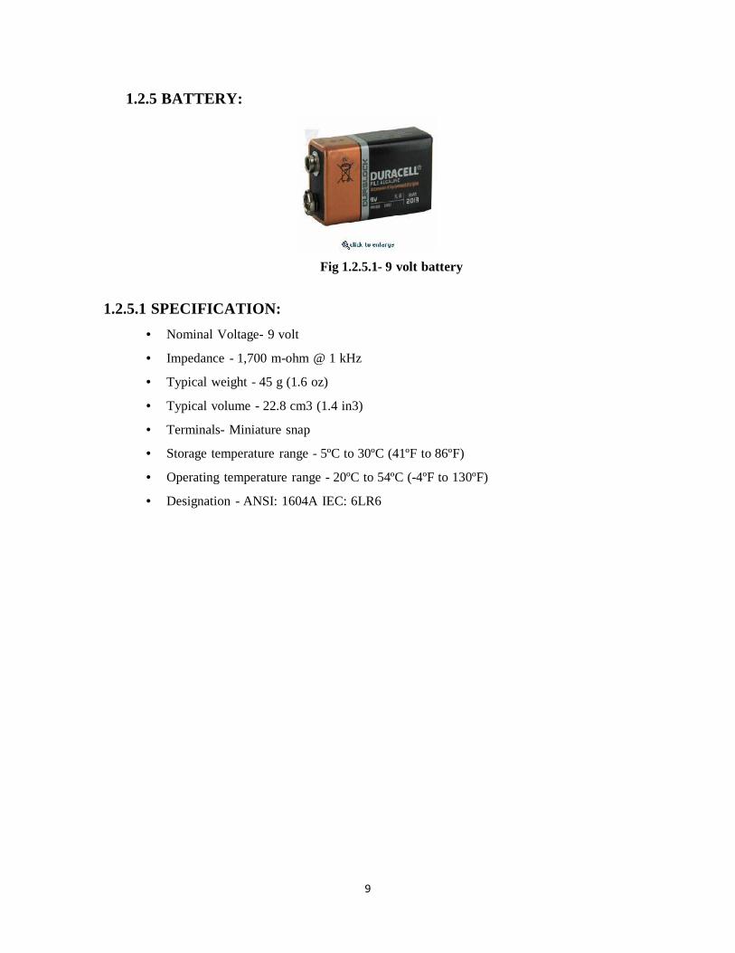
1.2.5 BATTERY:
Fig 1.2.5.1- 9 volt battery
1.2.5.1 SPECIFICATION:
Nominal Voltage- 9 volt
Impedance - 1,700 m-ohm @ 1 kHz
Typical weight - 45 g (1.6 oz)
Typical volume - 22.8 cm3 (1.4 in3)
Terminals- Miniature snap
Storage temperature range - 5ºC to 30ºC (41ºF to 86ºF)
Operating temperature range - 20ºC to 54ºC (-4ºF to 130ºF)
Designation - ANSI: 1604A IEC: 6LR6

1010
1.3 SOFTWARE SPECIFICATION:
The software application analyzes the amplitude of the incoming signals, as the criteria
of an occurred event (opening/closing of the hand) are the threshold changes of the
first derivative. When this happens, an In order a processing by a computer to be
enabled, the analog data needs to be converted in a digital form. А multifunctional
USB device of National Instruments− NI USB-6009 (Fig. 6-3), has been used. The
EMG signal is fed to one of the analog inputs. The visualization and the data
processing are done through software
Fig. 1.3.1. Software for processing, analysis and gripper control
Similarly, the amplified signal is fed to an analog input of NI USB-6009 for
conversion and then the data are analyzed by Lab View software. As can be seen in
Fig. 1.3.2 -. Fragment of software for processing and analysis of EOG: EOG signalcharacteristic (above); animation showing the actual position of the eyes (below)

1111
CHAPTER-2
STUDY & ANALYSIS
2.1 NEED OF STUDY
Despite of the scope, all the projects refereed in- tend to present EMG as an input interface. In
this article we present our results and present user studies to go further and validate
electromyography de- vices as daily wearable interfaces. Our prototype ac- complishes the
control of various computer Applications and introduces a synergy between applications that ease
the text-entry task. In the next section we present our approach. The other sections are focused in
the user evaluations, results and discussion. In the last section we present the conclusions taken by
the user evaluations along with the work still to be done.
2.2 SIGNAL RECORDING:
In order to get useful information concerning the muscular activity it is necessary to carefully
analyze some aspects, from technical details at the electrode placement in the surface of the
human body to the points where this placement must be done. Several aspects influence the signal
quality: skin preparation, electrodes placement position, electrodes fixation, electrodes distance
and outside interferences (De Luca, 1997).We have discarded all the skin preparation techniques
since we don’t think they are appropriate to a user interface. Besides, after several tests we
observed good signal quality with small interference. However, to reinforce the surface electrodes
adherence we created an elastic band for the neck and two elastic bands for the forearm. We used
the 2cm distance between electrodes which guarantees a solution of commitment (Figure 2),
collecting the signal of a significant portion of the muscle and restricting, simultaneously, the
undesired signals to insignificant values (De Luca, 1997).
Figure 2.2.1: Electrodes placement

1212
CHAPTER 3
DESIGN
3.1 METHODOLOGY
We conducted several experiments to assess the us- ability of myographic activity as as interaction
modal- ity. For that purpose, besides the experiments to vali- date the interaction speed and
accuracy, we focused our attention in the interface robustness as a daily wearable interface.
a) Clicking Start Button, where a timer is activated;
b) Moving the cursor towards the Stop button, with any trajectory;
c) Clicking Stop Button, and the time is presented to the user and saved. The Start Button
dimensions are always 8, 5 x 8,5mm but there are four Stop Button dimensions (8.5 x 8.5mm; 12.5 x
12.5mm; 17 x 17mm; 22 x 22mm). We made 80 evaluations, 20 of each for every Stop Button
size. The Start Button changed between the four corners. The users were equipped with two pairs
of electrodes in each forearm (four directions) and another pair near one eye to detect blinking

1313
3.2 SIGNAL PROCESSING:
Figure 3.2.1: System Design
The pre-processing is composed by some basic procedures that prepare the signal to be smoothed.
The signal received from the electromyography device has
Figure 3.2.2: Signal Processing

1414
As mentioned before, the useful frequency range is between 0 Hz and 500 Hz. So filtering the raw
signal is recommended to limit the signal’s frequency range to the desired range. Van Boxtel in
[vB08] found that a band-pass frequency range of 20 Hz to 500 Hz appeared to be adequate
because there was a negligible contribution of higher frequency components to the EMG signal.
In [CJDLR10] De Luca et al. are recommending to set the low-pass filter corner frequency to 400-
450 Hz, where the high end of the SEMG frequency spectrum is expected. Various
recommendations for the high-pass filter corner frequency can be found in literature. All the
recommendations are between 5 Hz and 20 Hz. SENIAM recommends to set the high-pass filter
corner frequency to 10 - 20Hz For several decades it has been commonly accepted that the
preferred manner for processing the EMG signal after filtering was to calculate the integrated
rectified signal. This was done by full-wave rectifying the EMG signal, integrating the signal over
a specified interval of time and subsequently forming a time series of the integrated values. Full-
wave rectification means the conversion of the input waveform to one of constant polarity (positive
or negative) at its output. This approach of rectifying and integrating became widespread since it
is possible to make these calculations accurately and inexpensively with the limited electronics
technology of earlier decades. Equation (2.2) shows how the calculation of the integrated rectified
signal is

1515
3.3 CIRCUIT DIAGRAM:
Fig 3.3.1:-Circuit diagram of Human Machine Interface through Electromyography
3.4 PCB LAYOUT
Fig. 3.4.1: Component layout for the PCB

1616
Fig. 3.4.2: An actual-size, single-side PCB for HMI through EMG
3.5 WORKING
The circuit diagram of HMI through EMG is shown in Fig. 3. Starting from the left, two electrodes
attached to a muscle are connected to a single-supply instrumentation amplifier INA122 (IC1),
which amplifies the voltage difference between the two electrodes, rejecting any signal that is
common to both—thereby removing much of the noise and 50Hz AC EMI. Resistor R1 determines
the gain of the output of IC1.
It is difficult to deal with the bi-polar nature of the bio-signals. To overcome this problem, we use
virtual-ground topology or split-rail topology, which splits the main power rail into two rails—one
having Vcc and the other with half of Vcc (virtual ground). The created virtual ground acts as the
pseudo-zero voltage and thus makes the output to swing between 0 and Vcc with virtual ground
as zero volts, which makes the entire portion of the EMG wave available and also in the positive
range only, thus making this topology ideal for interfacing with digital systems or microcontrollers.
IC1 is specifically designed for single-supply operations, for which it includes voltage translators
and overvoltage protection. The electrodes are directly attached to the muscle. Prior to attaching
the electrodes, clean the patient’s skin with alcohol for removing dirt and apply an ultrasound gel.
Two electrodes are attached over the muscle of interest and the third one (reference) is connected

1717
to an area preferably in the joints where there are less muscles. The reference electrode should
only be connected at pin 3 of CON1. It is vital as it provides the common-mode signal.
The reference is connected to the virtual ground and pin 5 (Vref) of IC1, so care must be taken to
isolate the patient from ground. The patient must not touch any grounded metal, which can cause
drainage of common-mode voltage and thus raise the amplitude of the output and inject a lot of
noise. The output from instrumentation amplifier IC1 is fed to a non-inverting amplifier built
around OPA2241 (IC2) via capacitor C1, which removes the DC offset from the electrodes. The
amplified output signal from IC2 is sent to the PC oscilloscope via an audio jack to get the display
of the waveform.
Fig.3.5.1-EMG Signal on the Pc based oscilloscope, Fig 3.5.2 :-Electrode Placement

1818
CHAPTER 4
APPLICATION
1. Skin placement.
2. Avoid movement of electrodes by using straps or tape to
firmly secure electrode I place.
3. Avoid bending of leads, place leads pointing in the
direction that you want the wire to Continue in. (e.g., for
electrodes placed on an extremity, have the lead pointing
towards The proximal end of the extremity so that the wire
will not have to be bent in order to go In the proximal
direction.)
4. Avoid any stress on the wires by making sure that the
wires are loose underneath the Tape or wrap that is holding
them in place. Be sure to check when the wires cross the
Joint that once the joint is fully extended the wires are not
drawn taunt.
5. Avoid placing electrodes over scars.
6. Measured potential.
7. Measuring skeletal muscle electrical output during physical activity
8. Sports medicine
9. EMG signals can be used for variety of applications like clinical/biomedical
10. EMG can be used to sense isometric muscular activity11. To measure the force of the isometric muscle contraction, a surface EMG was used
12. The idea of controlling machines with levers and knobs. Instead, they plan to have machines
respond directly to human gestures.

1919
CHAPTER 5
5.1 ADVANTAGE
Extremely sensitive
Record single muscle activity
Access to deep musculature
Little cross-talk concern
Surface EMG Recording Provide a safe.
Easy and noninvasive method
5.2DISADVANTAGE
Extremely sensitive
Requires medical personnel, certification
Repositioning nearly impossible
Detection area may not be representative of entire muscle

2020
CHAPTER 6
FUTURE SCOPE
It is intended to develop software environment for computer’s mouse control and cursor
movement, done by eye movements. Such a development could be useful for people with
disabilities, allowing them access to a computer. The application can easily be extended to allow
control of various electromechanical devices (wheelchairs). At the date of submission of the paper
a project was launched for realization of artificial, five-finger hand which will be controlled by
registered EMG potentials. In the near future we will see higher function prosthetics, brain
computer interfaces with more control, voice recognition and camera gesture recognition being
used more. Although not quite the death of the everyday mouse and keyboard, we will dentally
start to see new types of technology integrated into our everyday lives. Portable devices are
becoming smaller and more complex, so we should start seeing growth in wearable interfaces.
Robots, and the way we interact with them is already beginning to change, we are in the computer
era, but soon we will be in the robotic era.
Nano technology has provided a new wave of advancements, but these have yet to be fully utilized
in HMI, Nano technology has an important future role to play. Nano machines and super batteries
haven't fully materialized, so we have something to look forward to. There is also the potential of
Quantum Computing which will unleash an entirely new level of processor, with amazing speeds.
HMI technology is impressive now, but in the future there will be nothing like it. It won't matter
who you are, what language you speak, or what disability you have, the variety of technology will
cater for everyone. This document has provided an overview of what HMI , and has shown you a
glimpse of what the future might hold. One thing that is certain is that technologies will begin to
converge, devices will combine functionality, new levels of sensor fusions will be created, and all
this for one purpose, to enhance our Human Machine Interaction. The technology involved in HMI

2121
CHAPTER 7
CONCLUSION
The EMG/EEG-based Human–Computer Interaction system presented represents a potential alternative for
the communication of individuals with severe motor disabilities and their computers. Because the system
commands cursor movements on the basis of detection and classification of EMG signals, its operation is
relatively simple for the user. Use of the interface only requires the voluntary contraction of a set of cranial
muscles, requiring little training on the part of the subject. The operation of the system is possible due to
the difference in the spectral composition of EMG signals generated by contraction of different cranial
muscles. Thus, the novel classification approach allows the on-line separation of EMG signals caused by
the contraction of different muscles, based on the real-time estimation of their power spectra. In our
evaluation of the interface, six experimental subjects were able to drive the computer cursor from the
Screen corners to the center and perform a click at each end of that trajectory in an average of 16 seconds.
This level of performance achieved by untrained subjects reveals that this interface can be employed for
such applications as web browsing and appliance control through appropriate hardware/software setups.
In comparison with other unassisted interfaces for users with disabilities, the EMG/EEG interface proposed
has the potential to be more affordable and portable then others, such as eye-gaze tracking devices

2222
REFERENCES
1. http://www.kevinwarwick.com/
2. Basmajian JV, et al. Computers in Electromyography. Butterworths, 1975
3. Shavi R, Green N. Ensemble averaging of locomotors electromyography patterns
using interpolation. Med & Biol Eng & Comput, 21: 573-578, 1983.
4. De Luca. C.J. (1997) The Use of Surface Electromyography in Biomechanics. Journal
of Applied Biomechanics, Volume 13, Pages 135-163, 1997
5. www.datasheets4u.com
6. http://www.electronicsforyou.com
7. Kuiken, A. & Lipschutz, R. (2008) A strategy for identifying locomotion modes using
surface electromyography
8. https://www.grasstelefactor.com/products/clinsystems/cmeeg1.html
9. http://www.futurehealth.org/a-2.htm
10. http://www.biosemi.com/publications/artikel4.htm