HUMAN HEAD PHANTOM MATERIAL CHARACTERIZATION...
34
HUMAN HEAD PHANTOM MATERIAL CHARACTERIZATION FOR MICROWAVE IMAGING SYSTEM MOHD SOLLEHUDIN BIN MD SAID UNIVERSITI TEKNOLOGI MALAYSIA
Transcript of HUMAN HEAD PHANTOM MATERIAL CHARACTERIZATION...
HUMAN HEAD PHANTOM MATERIAL CHARACTERIZATION FOR
MICROWAVE IMAGING SYSTEM
MOHD SOLLEHUDIN BIN MD SAID
UNIVERSITI TEKNOLOGI MALAYSIA
HUMAN HEAD PHANTOM MATERIAL CHARACTERIZATION FOR
MICROWAVE IMAGING SYSTEM
MOHD SOLLEHUDIN BIN MD SAID
A thesis submitted in fulfilment of the
requirements for the award of the degree of
Master of Engineering (Electrical)
Faculty of Electrical Engineering
Universiti Teknologi Malaysia
MARCH 2015
iii
Dedicated to my beloved family, mother and father
and
To my honourable supervisor, Dr Norhudah Binti Seman

iv
ACKNOWLEDGEMENT
Alhamdulillah, In the name of Allah, all praise and gratitude to Allah S.W.T
the Lord of the Universe, Most Gracious and Most Merciful for giving me strength to
finish my research project and a good health for the last four semesters. Without
blessed from Him, I would not have been able to be at this stage.
I would like to take this opportunity to express my deepest appreciation and
gratefulness to my supervisor, Dr. Norhudah Seman for his valuable guidance,
suggestion and supportive in all aspect throughout the completion of my research
project. I am also indebted to Universiti Teknologi Malaysia (UTM) for funding my
Master study by UTM Zamalah Award. To all technicians and staff of Microwave
Laboratory and Wireless Communication Center (WCC) also deserve special thanks
for their helps and support in the laboratory and provide certain equipment needed
for this project.
My fellow postgraduate students should also be recognized for their support. My
sincere appreciation is further extended to all my friends for their support and ideas.
Finally, special appreciations give to who have involved and helped me in finishing
my thesis. Last but not least, my earnest appreciation and thanks to my family
members, especially to my beloved parents who have always been very
understanding and provide the motivation throughout finishing and completing this
project. Special thanks to all who participated.
v
ABSTRAK
Barah payudara dan otak tersenarai sebagai barah yang menjadi penyebab
utama kematian di seluruh dunia. Baru-baru ini, pengimejan gelombang mikro telah
dicadangkan untuk pengimejan dan diagnosis barah payudara, dan boleh diteruskan
untuk pengimejan barah otak. Walau bagaimanapun, tisu dan sel kepala manusia
berbeza daripada tisu dan sel payudara manusia terutama daripada sifat-sifat
dielektrik justeru itu penghasilan fantom kepala yang sesuai diperlukan. Fantom
yang perlu bagi sistem pengesanan barah otak mesti mempunyai spesifikasi tertentu
untuk menjadikannya sesuai dengan sistem pengimejan. Tesis ini membentangkan
siasatan terhadap sifat-sifat dielektrik bahan fantom kepala manusia untuk sistem
pengimejan gelombang mikro. Dalam penyiasatan, sampel-sampel fantom dibuat
menggunakan air dan gelatin dalam menghasilkan fantom yang kos efektif. Skop
penyiasatan adalah pada sifat air, tisu fantom, faktor perubahan dielektrik,
pengawetan fantom dan jangka hayat fantom. Siasatan ini memberi tumpuan kepada
sifat dielektrik yang terdiri daripada ketelusan relatif dan kekonduksian di seluruh
frekuensi gelombang mikro daripada 1-6 GHz. Semua ukuran diperoleh
menggunakan Rangkaian Penganalisis Vektor dengan prob dielektrik untuk
mendapatkan ketelusan kompleks. Siasatan ke atas ciri-ciri air menunjukkan bahawa
sebarang jenis air putih mempunyai sifat dielektrik yang hampir sama. Lima
komposisi bahan berasaskan gelatin dibentangkan dalam siasatan ini menunjukkan ia
mempunyai sifat dielektrik hampir sama dengan lima tisu kepala manusia iaitu
perkara kelabu (kompososi 5g gelatin, 20g air dan 0.5 gula), perkara putih
(kompososi 5g gelatin dan 14g air), cecair tulang belakang serebrum (kompososi 10g
gelatin dan 50g air), darah (kompososi 10g gelatin dan 30g air) dan kulit (kompososi
10g gelatin dan 20g air). Selain nisbah antara air dan gelatin, tiga faktor-faktor lain
iaitu suhu, garam dan gula mampu mengubah sifat dielektrik bahan. Pengawetan
bahan berasaskan gelatin telah dicadangkan menggunakan cuka dan ia mampu
memanjangkan jangka hayat fantom. Hasil dalam tesis ini berguna dalam
meningkatkan pengetahuan mengenai sifat dielektrik bahan yang digunakan dalam
fantom kepala manusia yang mana penting apabila menghasilkan, memperbaiki dan
mengawal sifat dielektrik fantom.
vi
ABSTRACT
Breast and brain cancers are stated as the most common causes of cancer-
related deaths around the world. Recently, microwave imaging has been proposed for
breast cancer imaging and diagnosis, and can be extended for brain cancer imaging.
However, tissues and cells for human head are different from human breast
especially in terms of dielectric properties thus requiring the development of an
appropriate head phantom. The required phantom for brain cancer detection system
must have particular specification to make it compatible with the imaging system.
This thesis presents an investigation on dielectric properties of materials of human
head phantom for microwave imaging system. In the investigation, samples of
phantoms are made using water and gelatin in producing a cost effective phantom.
The scopes of investigation are on the characteristics of water, tissues of phantom,
dielectric variation factors, preservation of phantom and lifespan of phantom. This
study focuses on dielectric properties consisting of relative permittivity and
conductivity across microwave frequency from 1 to 6 GHz. All measurements are
obtained using Vector Network Analyzer with a dielectric probe to obtain complex
permittivity. Investigation on water characteristics indicate that almost any type of
plain water has similar dielectric characteristics. Five compositions of gelatin-based
materials presented in this investigation showed to have similar dielectric properties
with five human head tissues, which are grey matter (composition of 5g gelatin, 20g
water and 0.5g sugar), white matter (composition of 5g gelatin and 14g water),
cerebral spinal fluid (composition of 10g gelatin and 50g water), blood (composition
of 10g gelatin and 30g water), and skin (composition of 10g gelatin and 20g water).
Besides the ratio between water and gelatin, three other factors of temperature, salt
and sugar are discovered to be able to change the dielectric properties of the
materials in the investigation. The preservation of gelatin-based material is proposed
using vinegar and is able to prolong the lifespan of phantom. The outcome in this
thesis is useful in gaining knowledge on dielectric characteristics of material used in
human head phantom which is important in the stage of developing, tuning and
controlling the dielectric properties of the phantom.

vii
TABLE OF CONTENTS
CHAPTER TITLE PAGE
DECLARATION ii
DEDICATION iii
ACKNOWLEDGEMENT iv
ABSTRAK v
ABSTRACT vi
TABLE OF CONTENTS vii
LIST OF TABLES x
LIST OF FIGURES xii
LIST OF APPENDIX
xvi
1 INTRODUCTION
1.1 Introduction
1.2 Problem Statements
1.3 Objective of the Research
1.4 Research Contribution
1.5 Scope of Study
1.6 Thesis Outline
1
1
3
5
5
6
8
2 LITERATURE REVIEW
2.1 Introduction
2.2 Microwave Imaging
2.3 Human Head
2.3.1 Biological Structure of Human Head
10
10
11
13
14

viii
2.3.2 Electrical Properties of Human Head
2.4 Phantom
2.4.1 Previous Research on Human Phantom using
Gelatin Material
2.4 Summary
14
20
23
29
3 METHODOLOGY
3.1 Introduction
3.2 Research Methodology
3.2.1 Experiment Setup, Calibration and
Precision Test
3.2.2 Phantom Material
3.2.2.1 Study on Water Characteristic
3.2.2.2 Phantom Compositions
3.2.3 Dielectric Variation Factors
3.2.4 The Use of Vinegar as Preservative
3.2.5 Gelatin Based Phantom Material with
Addition of Vinegar
3.3 Summary
30
30
31
34
37
37
38
40
42
43
44
4 PHANTOM EXPERIMENTAL RESULTS AND
DISCUSSION
4.1 Introduction
4.2 Investigation of Water Samples
4.3 Human Head Phantom
4.4 Summary
45
45
46
53
65
5 THE DIELECTRIC VARIATION FACTORS AND
PRESERVATION OF PHANTOM
5.1 Introduction
5.2 The effect of temperature level, salt and sugar
5.2.1 Water Samples
5.2.2 Gelatin Based Samples
67
67
68
68
74

ix
5.3 Vinegar as Preservative Material
5.3.1 The Study of Vinegar Characteristic
5.3.2 Phantom Material with Addition of Vinegar
5.4 Phantom Life Span
5.4.1 Phantom without Vinegar
5.4.2 Phantom with Vinegar
5.5 Summary
79
80
86
89
89
93
103
6 CONCLUSION AND RECOMMENDATION
6.1 Introduction
6.2 Conclusion
6.3 Future Works
104
104
106
107
REFERENCES 109
Appendix A 118

x
LIST OF TABLES
TABLE NO. TITLE
PAGE
2.1 Dielectric properties of biological tissues used in the
phantom model at 1GHz
19
2.2 Mixture Used for Skin Fibro Glandular, Transitional and
Fat Phantom
25
2.3 Materials needed to form CSF, grey matter, white matter,
and hemorrhagic stroke (blood)
27
2.4 Summarized table for previous research on gelatin phantom 28
4.1 Five Samples of Tap Water from Different Locations 46
4.2 The lowest and highest variation of relative permittivity and
conductivity shown from the measured tap water from
different locations
48
4.3 Five Water Samples from Different Sources 49
4.4 The lowest and highest variation of relative permittivity and
conductivity shown in the measured different source of
waters
52
4.5 Composition of Grey matter samples for human head
phantom in gram (g)
53
4.6 Composition of white matter samples for human head
phantom in gram (g)
55
4.7 Composition of cerebral spinal fluid samples for human
head phantom in gram (g)
57
4.8 Composition of blood samples for human head phantom in
gram (g)
59
xi
4.9 Composition of skin samples for human head phantom in
gram (g)
60
4.10 The percentage of error calculation between measure and
theoretical data on its relative permittivity for each selected
sample.
63
4.11 The percentage of error calculation between measure and
theoretical data on its conductivity for each selected
sample.
64
4.12 The summarized table for the composition of material to
produce homogeneous head phantom for five head tissues.
66
5.1 Three water samples measured at different temperature
level
69
5.2 Water samples in conjunction with different amount of salt 71
5.3 Water samples in conjunction with different amount of
sugar
73
5.4 Gelatin based samples measured at different temperature
level
75
5.5 Gelatin based samples in conjunction with different amount
of salt
76
5.6 Gelatin based samples in conjunction with different amount
of sugar
78
5.7 Four vinegar samples from different manufacturer 80
5.8 Three vinegar samples measured at different temperature
level
82
5.9 Vinegar samples in conjunction with different amount of
water
84
5.10 Gelatin based phantom material with different ratio of water
and vinegar
87
5.11 Comparison table for Sample VP1 to VP4 102
6.1 Summary of composition for head tissues phantom and its
percentage difference to theoretical value
106

xii
LIST OF FIGURES
FIGURE NO. TITLE
PAGE
1.1 Overview on the scope of research 6
2.1 Radar-based microwave imaging system of (a) monostatic,
and (b) bistatic radars
11
2.2 System setup of microwave imaging for human head 12
2.3 Cross section of human brain covered by protective skin
and skull
14
2.4 Dielectric constant profile of the phantom 19
2.5 Conductivity profiles of the phantom 20
2.6 Resulting breast phantom under measurement 24
2.7 Structure of a heterogeneous breast phantom 26
2.8 Heterogeneous breast phantom 26
3.1 Flow chart of the research methodology 32
3.2 An experimental set up for measuring dielectric properties
with a Vector Network Analyzer (VNA) and dielectric
probe
34
3.3 Precision measurement data of the used measurement
instrument
35
3.4 Flow chart of calibration and precision test. 36
3.5 Schematic representation of the formation of a 3D-network
starting form dissolved gelator molecules
39
4.1 Relative permittivity of tap water from different locations 47
4.2 Conductivity of tap water from different locations 47

xiii
4.3 Relative permittivity of water samples from different
source
49
4.4 Conductivity of water samples from different source 51
4.5 Relative permittivity of Sample 1 and 2 versus theoretical
values (grey matter)
54
4.6 Conductivity plots of Sample 1 and 2 versus theoretical
values (grey matter)
54
4.7 Relative permittivity of Sample 3 and 4 versus theoretical
values (white matter)
56
4.8 Conductivity plots of Sample 3 and 4 versus theoretical
values (white matter)
56
4.9 Relative permittivity of Sample 5 and 6 versus theoretical
values (cerebral spinal fluid (CSF))
58
4.10 Conductivity plots of Sample 5 and 6 versus theoretical
values (cerebral spinal fluid (CSF))
58
4.11 Relative permittivity of Sample 7 and 8 versus theoretical
values (blood)
59
4.12 Conductivity plots of Sample 7 and 8 versus theoretical
values (blood)
60
4.13 Relative permittivity of Sample 9 and 10 versus theoretical
values (skin)
61
4.14 Conductivity plots of Sample 9 and 10 versus theoretical
values (skin)
61
5.1 Relative permittivity of water samples at different
temperature level
69
5.2 Conductivity of water samples at different temperature
level
70
5.3 Relative permittivity of water samples with different
amount of added salt
71
5.4 Conductivity of water samples with different amount of
added salt
72
5.5 Relative permittivity of water samples with different
amount of added sugar
73

xiv
5.6 Conductivity of water samples with different amount of
added sugar
74
5.7 Relative permittivity of gelatin based sample at different
temperature level
75
5.8 Conductivity of gelatin based samples at different
temperature level
76
5.9 Relative permittivity of gelatin based samples with
different amount of added salt
77
5.10 Conductivity of gelatin based samples with different
amount of added salt
77
5.11 Relative permittivity of gelatin based samples with
different amount of added sugar
78
5.12 Conductivity of gelatin based samples with different
amount of added sugar
79
5.13 Relative permittivity of vinegar samples from different
manufacturers
81
5.14 Conductivity of vinegar samples from different
manufacturers
82
5.15 Relative permittivity of vinegar samples at different
temperature level
83
5.16 Conductivity of vinegar samples at different temperature
level
84
5.17 Relative permittivity of vinegar samples with different
amount of water
85
5.18 Conductivity of vinegar samples with different amount of
water
85
5.19 Relative permittivity of vinegar and water 86
5.20 Relative permittivity of gelatin based phantom material in
variation of water to vinegar ratio
87
5.21 Conductivity of gelatin based phantom material in
variation of water to vinegar ratio
88
xv
5.22 Relative permittivity of gelatin based phantom material
without preserved by vinegar measured in 6 weeks time
period
90
5.23 Conductivity of gelatin based phantom material without
preserved by vinegar measured in 6 weeks time period
91
5.24 Sample of gelatin based phantom material without
preserved by vinegar captured in 6 weeks time period.
92
5.25 Relative permittivity of gelatin based phantom material
preserved by 5g of vinegar measured in 6 weeks time
period
93
5.26 Conductivity of gelatin based phantom material preserved
by 5g of vinegar measured in 6 weeks time period
94
5.27 Sample of gelatin based phantom material preserved by 5g
of vinegar captured in 6 weeks time period
95
5.28 Relative permittivity of gelatin based phantom material
preserved by 10g of vinegar measured in 6 weeks time
period
96
5.29 Conductivity of gelatin based phantom material preserved
by 10g of vinegar measured in 6 weeks time period
97
5.30 Sample of gelatin based phantom material preserved by
10g of vinegar captured in 6 weeks time period
98
5.31 Relative permittivity of gelatin based phantom material
preserved by 15g of vinegar measured in 6 weeks time
period
99
5.32 Conductivity of gelatin based phantom material preserved
by 15g of vinegar measured in 6 weeks time period
100
5.33 Sample of gelatin based phantom material preserved by
15g of vinegar captured in 6 weeks time period
101

xvi
LIST OF APPENDIX
APPENDIX TITLE
PAGE
A Compositions of materials prepared for mimic tissue
of human head
118

CHAPTER 1
INTRODUCTION
1.1 Introduction
At the beginning of this research, two most important terms which required to
be understood are ‗Microwave Imaging‘ and ‗Human Head Phantom‘. Referring to
the title of this research, the properties of materials used in human head phantom are
investigated for the purpose to be used in modelling of phantom for microwave
imaging.
Microwave imaging can be defined as a system, which occupied to sketch the
internal structure of an object, so by that the internal structure of that object can be
observed. This system operates by illuminating the object with an antenna that
generates electromagnetic energy at microwave frequencies. This frequency is
depending to the specification of the system. There are two techniques normally used
in microwave imaging, the first one is using antenna at transmitting and receiving
side which the object is illuminated by microwave signal from antenna at
transmitting side, then the signal propagate through the object and collected by
receiving antenna at the other side. While, the second is using reflection technique,
which transmitted microwave signal will be reflected by the object then collected by
the same antenna.
2
In the development of microwave imaging system, modeling of realistic
human phantom is also required. The word of phantom sometimes confusing people
who does not has basic knowledge in this area. Human phantom basically is a model
of human body parts including its cells and tissues, which mimic cells and tissues of
real human. In microwave imaging system, phantom is used to simulate the
interaction of electromagnetic wave with biological tissues [1].
Microwave imaging has been mainly proposed for breast cancer detection,
but some recent reports have also speculated the use of microwave in extremities
imaging, diagnostics of lung cancer, brain imaging and cardiac imaging [2-8].In
previous past decade, microwave imaging attracts attention among researcher, which
breast cancer imaging is frequently focused on[2-5]. Then, microwave imaging start
to focus on brain imaging as reported in [12-18, 78-80], where x-rays
mammography, computed tomography (CT) and magnetic resonance imaging (MRI)
system act as main scanning system to overcome the complexity of brain imaging
especially on brain stroke an others brain cancer diagnosis. But the lack of using
these scanning systems because they do not offer safe, fast, cost effective and
portable screening tools [16]. X-rays generally can kill living tissue due to ionizing
radiation that exposes during screening process that harmful to human body over a
prolonged period of time while MRI is very accurate screening tool but it is costly,
time consuming and not widely available and also not accessible at rural medical
clinics or carried by first response paramedical teams [16, 19].Where, the first
response is important to increase the survival rates. While, the microwave has
potential for imaging can supplement current diagnostic methods as it may provide
fast, cost effective and portable detection systems [6].
Compared to the other medical imaging techniques, microwave imaging is
still in its infancy. One historical reason for this might due to the fact that most
microwave systems-devices originated in military applications, radar being an
obvious example [9-10]. In recent years however, due to the mobile/wireless
revolution, unprecedented progress in high performance microwave hardware have
been witnessed. This opens up a unique opportunity for development of microwave
imaging systems.
3
In order to carry out a research in this area, multi-disciplinary effort is
required. In the case of measurement, the physical human phantom can be developed
and then illuminated by an antenna or array of antennas operating over the desired
microwave frequency band. The reflected and transmitted signals collected by
antennas can be stored for further processing. Depending on the processing technique
chosen, this data can be efficiently used to produce map of dielectric constant in
image body, such a work done on breast phantom is reported in [11].
1.2 Problem Statement
Other than breast cancer, brain cancer also has been noted as the most
common cause of cancer-related deaths around the world. Currently, (MRI) is mostly
used for the screening process. But this MRI is too costly and not widely available
[19] especially in rural medical centre. Early cancer diagnosis and detection are very
important to increase cancer survival rates. Nowadays, microwave imaging has gain
attention among researcher due its potential in breast cancer detection. These
scenarios then lead to the motivation for development of microwave imaging with
the purpose for brain cancer detection, which also causes the study on new phantom
for human head is also required. Although there are availability of phantom in the
market, but it is costly and not specifically meet the requirements of the system
especially in term of operating frequency. Apart from that, the reported study on
phantom of human head for microwave imaging application is very limited. The
previous study on breast phantom also does not provide explanation on the
characteristic and behaviour of material in the phantom. Researcher usually made the
recipe of their phantom without provide the information on the relation of their
recipe with the dielectric properties of the phantom. These reasons motivate for the
investigation on the material of the head phantom.
In fact, high microwave frequency for example 3-11 GHz which is used in
ultra-wideband breast cancer detection offers high resolution. However, the use of
4
high frequency might lower penetration of required signal into the brain. At
frequencies lower than 3 GHz, it would allow for a higher penetration but would be
insensitive to small regions of dielectric changes [15]. Brain imaging is classified as
high difficulty application which mainly due to the complexity as well as the
structural, functional and electrical in homogeneity of the human brain [18].
Therefore, it is important to determine the optimal spectrum in order to couple
electromagnetic energy into the brain matter since the brain is surrounded by a high
contrast dielectric shield comprising of the skin, skull and cerebral spinal fluid
(CSF).
In electrical form, every tissue in human body as well as in human head can
be represented by the electrical properties or also known as dielectric properties. As
reported in [39], there are two properties that define the electrical properties of
human tissue, which are the relative permittivity (εr) and conductivity (σ). These
properties represent as the propagation, reflection, attenuation, and other behavior of
electromagnetic fields in the human body. The relative permeability (μr) of human
body can be assumed as 1, which shows the human body is weakly magnetic [39].
Therefore, the characterization study in this research is focuses only in form of
permittivity and conductivity.
In this research, the characterization of a human head phantom is conducted
based on the study of its electrical properties across 1 to 6 GHz using simple and
common material such as jelly powder, gelatin, water and sugar. This wideband
frequency range is chosen in order to have good trade off between resolution and
penetration in human head imaging since the lower frequency will provide good
penetration and good resolution can be provided by higher microwave frequency [26-
28]. The electrical properties in term of permittivity and conductivity of the chosen
mixtures of materials are obtained through measurement conducted in laboratory
using special dielectric probe connected to a vector network analyser (VNA). The
characteristics of each measured sample are observed through its analyzed data on
relative permittivity and conductivity.
5
1.3 Objective of the research
The objective of the research is to conduct the following theoretical and
experimental investigations through the development of human head phantom which
divided as follows:
1) To study and investigate the characteristic of electrical properties on several
material used in the human head phantom.
2) To specify the simple composition of material that mimic dielectric of head
tissues for microwave imaging system.
3) To improve the lifespan of phantom material by the proposed preservation.
1.4 Research Contribution
Based on the objective in this research, the experimental study performed in
this thesis provides following contributions.
1) The characteristics of materials used in head phantom are obtained through the
investigation based on electrical properties.
i. The investigation on water samples has been conducted, which provides
useful knowledge on the electrical characteristic of water.
ii. The investigation on dielectric variation factors has been conducted,
which provides useful knowledge to tune and control the dielectric
properties using basic material.
2) Compositions of sample material to prepare phantom with similar dielectric
properties for five head tissues which are grey matter, white matter, cerebral
6
spinal fluid (CSF), blood and skin are acquired via the conducted experimental
study.
3) The lifespan of phantom material has been investigated thoroughly that lead to
the finding of the suitable amount of vinegar that can be used in preservation.
Through this proposed preservation, the lifespan of the phantom can be
improved.
1.5 Scope of Study
The scope of study basically represents the boundary of work, which must be
conducted to ensure the effectiveness in achieving each objective of this research.
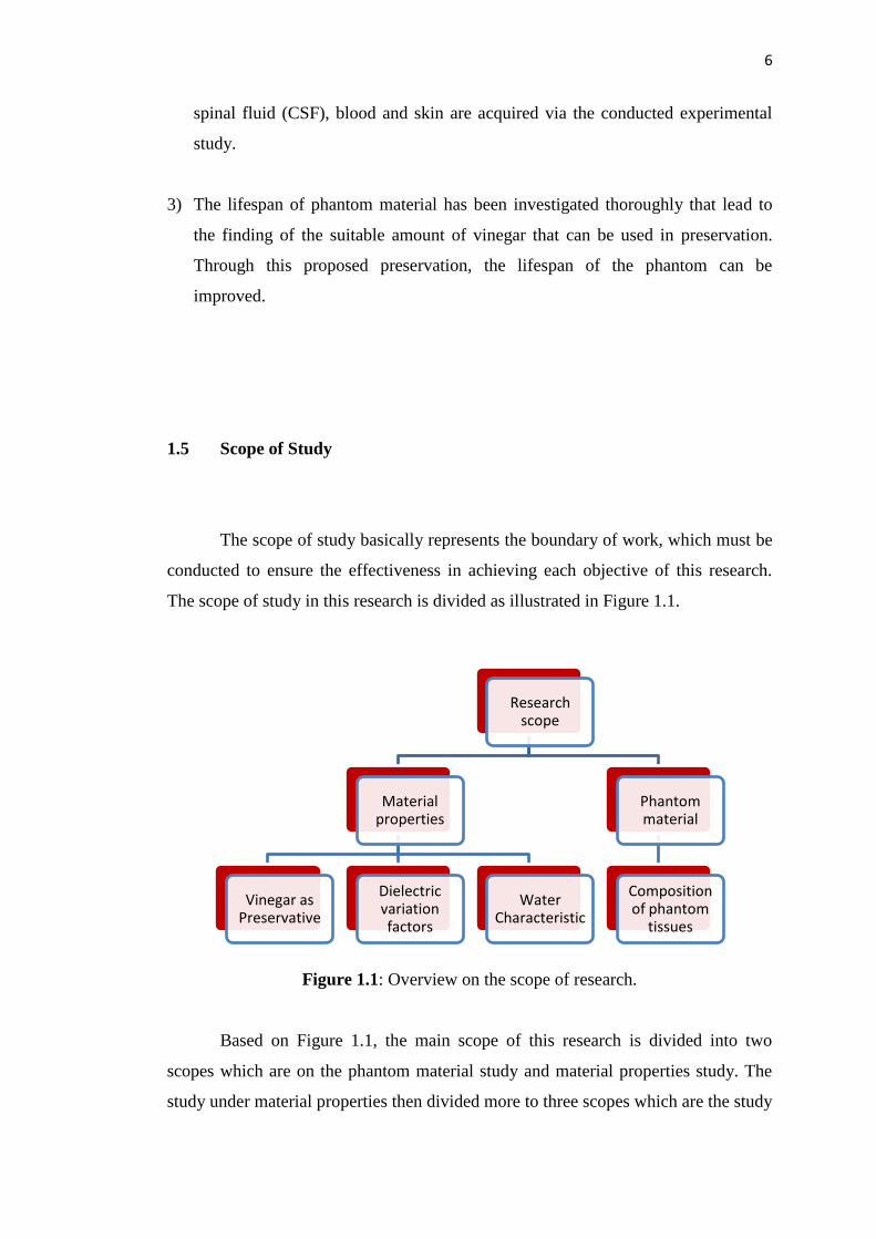
The scope of study in this research is divided as illustrated in Figure 1.1.
Figure 1.1: Overview on the scope of research.
Based on Figure 1.1, the main scope of this research is divided into two
scopes which are on the phantom material study and material properties study. The
study under material properties then divided more to three scopes which are the study
Research scope
Material properties
Vinegar as Preservative
Dielectric variation factors
Water Characteristic
Phantom material
Composition of phantom
tissues

7
on water characteristic, dielectric variation factors and preservation. The detailed
scopes of study in this research are as follows:
1) Investigation on the electrical characteristic of water used in material for head
phantom tissues.
i. In properties study of tap waters taken from different locations, several
samples of tap water are measured to observe dielectric difference due to
location of tap water source.
ii. In properties study of waters that taken from different sources, samples of
water from different sources, which are tap water, distilled water,
mineral(underground) water, filtered water and reverse osmosis water are
investigated in term of their dielectric properties.
2) Investigation on dielectric variation factors on materials for head phantom
tissues.
i. Experimental study is divided into four parts, which to study the
properties of tap waters taken from different locations, the properties of
waters that taken from different sources, the effects of temperature and
the addition of sugar to dielectric measurements.
ii. Other factors which are temperature and addition of sugar or salt is
investigated towards its effects on dielectric properties.
3) Investigation on vinegar as preservative in materials for head phantom tissues.
i. The experimental study is divided into four parts, which are to study the
properties of vinegar that taken from different manufacturers, the effects
of vinegar with temperature and the properties of vinegar in the variation
of water and the effect of vinegar toward phantom life span.
ii. Vinegars from several manufactures are investigated on its dielectric
properties, which might have dissimilarity.

8
iii. In others part, vinegar is investigated towards its dielectric changes due to
temperature. The mixture of vinegar and water in different compositions
are investigated in term of dielectric properties and physical observation.
iv. Furthermore, the investigation on phantom preservation by using vinegar
is performed for a few certain of different periods.
4) Experimental study on phantom material to specify the composition of material
for head phantom tissues.
i. Firstly, the electrical properties of human head tissues and cells data are
collected from database website dielectric properties of body tissues.
ii. In the experimental study, simple and low cost material which is gelatin is
used as main phantom material. Various samples of material with
different compositions are prepared and measured using Vector Network
Analyzer (VNA).Then obtained data is analyzed in term of relative
permittivity and conductivity. This relative permittivity and conductivity
data are then compared with relative permittivity and conductivity of
tissues and cells in real human head. This procedure is conducted
continuously until sample of material which most similar to real human
head obtained.
iii. Sample of phantom material which is mimicking each tissues and cells of
real human head is selected to be used as head phantom tissues.
1.6 Thesis outline
This thesis is divided to six chapters, which are the introduction, literature
review, research methodology, phantom experimental result and discussion,
dielectric variation factors and preservation of phantom and the last chapter is
conclusion and recommendation. In the introduction, brief information is given in
introducing microwave imaging and its phantom. In addition, Chapter 1 also consists
9
of the information that significant to this research, which are problem statement,
objectives, research contributions and scope. In Chapter 2, research background and
literature reviews is provided, which related to ultra wideband microwave imaging
system, human phantom, tissues composition in human head and electric properties
of material. Important equations for dielectric analysis also described in this chapter.
While, in Chapter 3 provides the methodology of this research. This chapter
focusing more on the experimental study in this research starting with the setup of
experiment and then the calibration and precision test for the VNA. Then, this
chapter presents the methodology of the investigation on phantom material, dielectric
variation factors and preservation of material using vinegar. Section of phantom
material discusses the experimental study to find compositions of materials that have
similar properties with head tissues and cells for the development of human head
phantom. While, in section of dielectric variation factors discusses the experimental
study on the factors that cause dielectric changes on material and in the preservation
of material discuss the experimental study about suitability vinegar as preservative in
phantom material.
Chapter 4 in this thesis presents the characteristic for different types of plain
water based on their electrical properties. This chapter also presents the composition
of materials that have similar electrical properties with real head tissues that could be
used as head phantom. The investigation on electrical properties is observed based on
the relative permittivity and conductivity that conducted through the measurement in
the laboratory. Meanwhile, Chapter 5 discusses the results of the investigation on
dielectric variation factors and preservation of phantom. This chapter presents the
factors that able to vary the dielectric properties of material. In this chapter also, the
results on effectiveness of vinegar that proposed as preservation material is presented
Chapter 6, which is the last chapter of this thesis, concerns the conclusion of
the works that have been done in this thesis and provide future recommendation
related to this research works.

109
REFERENCES
[1] M. Lazebnik, E. L. Madsen, G. R. Frank, and S. C. Hagness, ―Tissue-
mimicking phantom material for narrowband and ultra wideband microwave
application,‖ Physics in Medicine and Biology, vol. 50, pp. 4245-4258, 2005.
[2] Alshehri, S. A., S. Khatun, ―UWB imaging for breast cancer detection using
neural networks,‖ Progress In Electromagnetic Research C, vol. 7, 79–93,
2009.
[3] M. Klemm, I.J. Craddock, J.A. Leendertz, A. Preece, D.R. Gibbins, M. Shere
and R. Benjamin, "Clinical trials of a UWB imaging radar for breast cancer,"
2010 Proceedings of the Fourth European Conference on Antennas and
Propagation (EuCAP), pp.1–4, 2010.
[4] MaciejKlemm, Ian J. Craddock, Jack A. Leendertz, Alan Preece, and Ralph
Benjamin, ―Radar-Based Breast Cancer Detection Using a Hemispherical
Antenna Array—Experimental Results‖, IEEE Transactions On Antennas
and Propagation, Vol. 57, No. 6, June 2009.
[5] E. C. Fear, X. Li, S. C. Hagness and M. A. Stuchly, ―Confocal Microwave
Imaging for Breast Cancer Detection: Localization of Tumors in Three
Dimensions‖, IEEE Transactions on Biomedical Engineering, Vol.49, No. 8,
2002.
[6] S. Y. Semenov and D. R. Corfiled, ―Microwave Tomography for Brain
Imaging: Feasibility Assessment for Stroke Detection‖, International Journal
of Antennas and Propagation, vol. 2008, Article ID 254830, 8 pages, 2008.
[7] S. Y. Semenov, R. H. Svenson, V. G. Posukh, A. G. Nazarov, Y. E. Sizov, A.
E. Bulyshev, A. E. Souvorov, W. Chen, J. Kasell, and G. P. Tatsis,
―Dielectrical spectroscopy of canine myocardium during acute ischemia and
110
hypoxia at frequency spectrum From 100 kHz to 6 GHz", IEEE Transactions
on Medical Imaging, Vol. 21, No. 6, 2002.
[8] R. Gagarin, N. Hyoung-sun Youn Celik and M. Iskander, "Noninvasive
microwave technique for hemodynamic assessments," Antennas and
Propagation Society International Symposium (APSURSI), pp.1–4, 2010.
[9] N. Seman and M. E. Bialkowski, "Design of a Wideband Reflectometer for a
Microwave Imaging System," International Conference on Microwaves,
Radar & Wireless Communications (MIKON), pp.25-28, 2006.
[10] V. Zhurbenko, ―Challenges in the Design of Microwave Imaging Systems for
Breast Cancer Detection‖, Advanced in Electrical and Computer
Engineering, Vol. 11, No.1, 2011.
[11] AlShehri, S. Khatun, A. B. Jantan, R. S. A. Raja Abdullah, R. Mahmood, and
Z. Awang, "Experimental breast tumor detection using NN-based UWB
imaging," Progress In Electromagnetics Research, Vol. 111, 447-465, 2011.
[12] I. A. Gouzouasis and I. S. Karanasiou and N. K. Uzunoglu, ―Exploring the
Enhancement of the Imaging Properties of a Microwave Radiometry Sytems
for Possible Functional Imaging Using a Realistic Human Head Model‖, 4th
International Conference Imaging Technology in Bio Medical Sciences,
Medical Images to Clinical Information – Bridging the Gap, 2009.
[13] M. Miyakawa, Y. Kawada and M. Bertero, ―Image Generation in Chirp Pulse
Microwave Computed Tomography (CP-MCT) by Numerical Computational:
Computational of a Human Head Model‖, Electronics and Communications
in Japan, Part 3, Vol. 88, No. 9, 2005.
[14] I. S. Karanasiou, N. K. Uzunogle and A. Garetsos, ―Electromagnetic Analysis
of Non-Invasive 3D passive Microwave Imaging System‖, Progress in
Electromgnetics Research, PIER 44, 287-308; 2004.
[15] D. Ireland and M. Bialkowski, "Feasibility study on microwave stroke
detection using a realistic phantom and the FDTD method," Asia Pacific
Microwave Conference, pp.1360-1363, 2010.
[16] D. Ireland and M. Bialkowski, ―Microwave Head Imaging for Stroke
Detection‖, Progress in Electromagnetics Research M, Vol. 21, 163-175,
2011.
[17] H. Trefna and M. Persson, "Antenna array design for brain monitoring,"
Antennas and Propagation Society International Symposium, pp.1–4, 2008.

111
[18] A. Oikonomou, I. S. Karanasiou and N. K. Uzunoglu, ―Phased-Array Near
Field Radiometry for Brain Intracranial Applications‖, Progress in
Electromagnetics Research, Vol. 109, 345-360; 2010.
[19] W.C. Khor, M.E. Bialkowski, A.M. Abbosh, N. Seman, and S. Crozier, ―An
Ultra Wideband Microwave Imaging System for Breast Cancer Detection,‖
IEICE Transactions on Communications, vol. 90-B, no. 9, pp. 2376-2381,
Sept. 2007.
[20] Q. Fang, Computational methods for microwave medical imaging, Ph.D.
dissertation, Dartmouth College, Hanover (2004).
[21] Bindu, G., S. J. Abraham, A. Lonappan, V. Thomas, C. K. Aanandan, and K.
T. Mathew, Active microwave imaging for breast cancer detection, Progress
in Electromagnetics Research, PIER 58, 149-169, (2006).
[22] H. Zhang, S. Y. Tan, and H. S. Tan, A novel method for microwave breast
cancer detection, Progress In Electromagnetics Research, Vol. 83, 413-434,
(2008).
[23] J. C. Y. Lai, C. B. Soh, E. Gunawan, and K. S. Low, Homogeneous and
Heterogeneous Breast Phantoms for Ultra-Wideband Microwave Imaging
Applications, Progress In Electromagnetics Research, PIER 100, pp. 397-
415, (2010).
[24] M. E. Bialkowski, N. Seman, A.nAbbosh and W. C. Khor, Compact
Reflectometers for a Wideband Microwave Breast Cancer Detection System,
African Journal of Information and Communication Technology, vol. 2, no. 3,
pp. 119 – 125, (2006).
[25] W.C. Khor, Y.H. Foo, M.E. Bialkowski and S. Crozier, ―Investigations into
Microwave Properties of Various Substances to Develop a Breast Phantom
for a UWB Breast Tumour Radar Detecting System‖ Proc. 17th International
Conference on Microwave, Radar and Wireless Communications(MIKON-
08), Krakow, Poland, May 19-21, 2008
[26] Jae Myeong Choi; Heau-Jo Kang; Yong-Seok Choi, A Study on the Wireless
Body Area Network Applications and Channel Models, Future Generation
Communication and Networking, FGCN '08 Second International
Conference, vol.2, no., pp.263-266, (2008).
[27] F. Martell, C. Buratti and R. Verdone, On the performance of an IEEE
802.15.6 Wireless Body Area Network, 11th European Wireless Conference

112
2011 - Sustainable Wireless Technologies (European Wireless), pp.1-6,
(2011).
[28] K. S. Kwak, S. Ullah and N. Ullah, An overview of IEEE 802.15.6 standard,
2010 3rd International Symposium on Applied Sciences in Biomedical and
Communication Technologies (ISABEL), pp.1-6, (2010).
[29] B. Mohammed, D. Ireland, and A. Abbosh, ―Experimental investigations into
detection of breast tumour using microwave system with planar array,‖ IET
Microw. Antennas Propag., vol. 6, no. 12, pp. 1311–1317, Sep. 2012.
[30] B. Mohammed, A. Abbosh, and D. Ireland, ―Circular antenna array for brain
imaging systems,‖ Proc. IEEE Antennas Propag. Soc. Int. Symp., Chicago,
IL, USA, pp. 1–2, Jul. 2012.
[31] B. Mohammed, A. Abbosh, and D. Ireland, ―Stroke detection based on
variations in reflection coefficients of wideband antennas,‖ in Proc. IEEE
Antennas Propag. Soc. Int. Symp., Chicago, IL, USA, pp. 1–2, Jul. 2012.
[32] L. Catarinucci, P. Palazzari, and L. Tarricone, ―On the use of numerical
phantoms in the study of the human-antenna interaction problem,‖ IEEE
Antennas Wireless Propag. Lett., vol. 2, no. 1, pp. 43–45, Feb. 2003.
[33] S. Mustafa, A. Abbosh, B. Henin, and D. Ireland, ―Brain stroke detection
using continuous wavelets transform matching filters,‖ in Proc. Int. Biomed.
Eng. Conf., Cairo, Egypt, pp. 194–197, Dec. 2012.
[34] J. C. Lin and J. M. Clarke, ―Microwave imaging of cerebral edema,‖ Proc.
IEEE, vol. 70, no. 5, pp. 523–524, May 1982.
[35] M. Sperandio, M. Guermandi, and R. Guerrieri, ―A four-shell diffusion
phantom of the head for electrical impedance tomography,‖ IEEE Trans.
Biomed. Eng., vol. 59, no. 2, pp. 383–389, Feb. 2012.
[36] M. Akter, T. Hirai, Y. Hiai, M. Kitajima, M. Komi, R. Murakami, H.
Fukuoka, A. Sasao, R. Tya, and E. M. Haacke, ―Detection of hemorrhagic
hypointense foci in the brain on susceptibility-weighted imaging: Clinical and
phantom studies,‖ Acad. Radiol., vol. 14, no. 9, pp. 1011–1019, 2007.
[37] K. Karathanasis, I. Gouzouasis, I. Karanasiou, and N. Uzunoglu,
―Experimental study of a hybrid microwave radiometry—Hyperthermia
apparatus with the use of an anatomical head phantom,‖ IEEE Trans. Inf.
Technol. Biomed., vol. 16, no. 2, pp. 241–247, Mar. 2012.
113
[38] B. Mohammed, A. Abbosh, B. Henin, and P. Sharpe, ―Head phantom for
testing microwave systems for head imaging,‖ Proc. Int. Biomed.Eng. Conf.,
Cairo, Egypt, pp. 191–193, Dec. 2012.
[39] Taylor and Francis Group, LLC, ―Apendix A : Electrical Properties of the
Human Body‖, 2009.
[40] Gabriel, C. Compilation of the dielectric properties of body tissues at RF and
microwave frequencies, Final technical report, Occupational and
Environmental Health Directorate Radiofrequency Radiation Division,
Brooks Air Force Base, TX, 1996.
[41] Damijan Miklavcic, Natasa Pavselj ―Electric Properties of Tissues‖
University of Ljubljana, Slovenia, Wiley Encyclopaedia of Biomedical
Engineering, John Wiley & Sons 2006.
[42] Dielectric properties of body tissues in the frequency range of 10Hz –
100GHz, ," Available: http://niremf.ifac.cnr.it/tissprop/, [Accessed:
December 3rd, 2013]
[43] Fong P M, Keil D C, Does M D and Gore J C, ―Polymer gels for magnetic
resonance imaging of radiation dosedistributions at normal room atmosphere‖
Phys. Med. Biol. 46 3105–13, 2001.
[44] Madsen E L, Zagzebski J A, Frank G R, Greenleaf J F and Carson P L,
―Anthropomorphic breast phantoms for assessing ultrasonic imaging system
performance and for training ultrasonographers: part I‖, J. Clin. Ultrasound
10 67–75, 1982
[45] Madsen E L, Kelly-Fry E and Frank G R, ―Anthropomorphic phantoms for
assessing systems used in ultrasound imaging of the compressed breast‖,
Ultrasound Med. Biol. 14 182–201, 1988.
[46] Surry K J, Austin H J B, Fenster A and Peters T M, ―Poly(vinyl alcohol)
cryogel phantoms for use in ultrasound and MR imaging‖, Phys. Med. Biol.
49 5529–46, 2004.
[47] Guy AW, ―Analysis of electromagnetic fields induced in biological tissue by
thermographic studies on equivalent phantom models‖, IEEE Trans. Microw.
Theory Tech. 19 189–217, 1971.
[48] Chou C-K, Chen G-W, Guy A W and Luk K H, ―Formulas for preparing
phantom muscle tissue at various radio frequencies‖, Bioelectromagnetics 5
435–41, 1984.
114
[49] Cheung A Y and Koopman D W, ―Experimental development of simulated
biomaterials for dosimetry studies of hazardous microwave radiation‖, IEEE
Trans. Microw. Theory Tech. 24 669–73, 1976.
[50] Lagendijk J J W and Nilsson P, ―Hyperthermia dough: a fat and bone
equivalent phantom to test microwave/radiofrequency hyperthermia heating
systems‖, Phys. Med. Biol. 30 709–12, 1985.
[51] Bini M G, Ignesti A, Millanta L, Olmi R, Rubino N and Vanni R, ―The
polyacrylamide as a phantom material for electromagnetic hyperthermia
studies‖, IEEE Trans. Biomed. Eng. 31 317–22, 1984.
[52] Andreuccetti D, Bini MG, IgnestiA,Olmi R, Rubino Nand Vanni R, ―Use of
polyacrylamide as a tissue-equivalent material in the microwave range‖,
IEEE Trans. Microw. Theory Tech. 35 275–7, 1988.
[53] Surowiec A, Shrivastava P N, Astrahan M and Petrovich Z, ―Utilization of a
multilayer polyacrylamide phantom for evaluation of hyperthermia
applicators‖, Int. J. Hyperthermia 8 795–807, 1992.
[54] McCann C, Kumaradas J C, Gertner M R, Davidson S R H, Dolan A M and
Sherar M D, ‖Feasibility of salvage interstitial microwave thermal therapy for
prostate carcinoma following failed brachytherapy: studies in a tissue
equivalent phantom‖, Phys. Med. Biol. 48 1041–52, 2003.
[55] Davidson S R H and Sherar M D, ―Measurement of the thermal conductivity
of polyacrylamide tissue-equivalent phantom‖, Int. J. Hyperthermia 19 551–
62, 2003.
[56] Robinson M J, Richardson M J, Green J L and Preece A W, ―New materials
for dielectric simulation of tissues‖, Phys. Med. Biol. 36 1565–71, 1991.
[57] Nikawa Y, Chino M and Kikuchi K, ―Soft and dry phantom modeling
material using silicone rubber with carbon fibre‖, IEEE Trans. Microw.
Theory Tech. 44 1949–53. 1996.
[58] Chang J T, Fanning M W, Meaney P M and Paulsen K D, ―A conductive
plastic for simulating biological tissue at microwave frequencies‖, IEEE
Trans. Electromagn. Compat. 42 76–81, 2000.
[59] Youngs I J, Treen A S, Fixter G and Holden S, ―Design of solid broadband
human tissue simulant materials‖, IEEE Proc.-Sci. Meas. Technol. 149 232–
8, 2002.
115
[60] Hagness S C, Taflove A and Bridges J E, ―Two-dimensional FDTD analysis
of a pulsed microwave confocal system for breast cancer detection: fixed-
focus and antenna-array sensors‖, IEEE Trans. Biomed. Eng. 45 1470–9,
1998.
[61] Li X and Hagness S C, ―A confocal microwave imaging algorithm for breast
cancer detection‖, IEEE Microw. Wireless Comp. Lett. 11 130–2, 2001.
[62] Bond E J, Li X, Hagness S C and Van Veen B D, ‖Microwave imaging via
space-time beam forming for early detection of breast cancer‖, IEEE Trans.
Antennas Propagat. 51 1690–705, 2003.
[63] Hernandez-Lopez M A, Quintillan-Gonzalez M, Garcia S G, Bretones A R
and Martin R G, ―A rotating array of antennas for confocal microwave breast
imaging‖, Microw. Opt. Technol. Lett. 39 307–11, 2003.
[64] Nilavalan R, Gbedemah A, Craddock I J, Li X and Hagness S C, ―Numerical
investigation of breast tumor detection using multi-static radar‖, Electron.
Lett. 39 1787–8, 2003.
[65] El-ShenaweeM, ―Resonant spectra of malignant breast cancer tumors using
the three-dimensional electromagnetic fast multipole model‖, IEEE Trans.
Biomed. Eng. 51 35–44, 2004.
[66] Huo Y, Bansal R and Zhu Q, ‖Modeling of noninvasive microwave
characterization of breast tumors‖, IEEE Trans. Biomed. Eng. 51 1089–94,
2004.
[67] Li X, Davis S K, Hagness S C, van der Weide D W and Van Veen B D,
―Microwave imaging via spacetime beamforming: experimental investigation
of tumor detection in multi-layer breast phantoms‖, IEEE Trans. Microw.
Theory Tech. 52 1856–65, 2004.
[68] Davis S K, Tandradinata H, Hagness S C and Van Veen B D, ―Ultrawideband
microwave breast cancer detection: a detection-theoretic approach using the
generalized likelihood ratio test‖, IEEE Trans. Biomed. Eng. 52 1237–50,
2005.
[69] Miyakawa, M.; Takata, S.; Inotsume, K., "Development of non-uniform
breast phantom and its microwave imaging for tumor detection by CP-MCT,"
Engineering in Medicine and Biology Society, 2009. EMBC 2009. Annual
International Conference of the IEEE , vol., no., pp.2723,2726, 3-6 Sept.
2009.

116
[70] Marchal C, Nadi M, Tosser A J, Roussey C and Gaulard M L, ―Dielectric
properties of gelatin phantoms used for simulations of biological tissues
between 10 and 50 MHz‖, Int. J. Hyperthermia 5 725–32, 1989.
[71] J. Z. Bao, M. L. Swicord, and C. C. Davis, ―Microwave dielectric
characterization of binary mixtures of water, methanol, and ethanol,‖ Journal
of Chemical Physics, vol. 104, no. 12, pp. 4441–4450, 1996.
[72] Sunaga T, IkehiraH, Furukawa S, Tamura M, Yoshitome E, Obata T,
ShinkaiH, Tanada S,MurataHand SasakiY, ―Development of a dielectric
equivalent gel for better impedance matching for human skin‖,
Bioelectromagnetics 24 214–17, 2003.
[73] M. J. Schroeder, S. Anupama, and R. M. Nelson, ―An analysis on the role of
water content and state on effective permittivity using mixing formulas,‖
Journal of Biomechanics, Biomedical, and Biophysical Engineering, vol. 2,
no. 1, 2008.
[74] M. Ostadrahimi, R. Reopelle, S. Noghanian, S. Pistorius, A. Vahedi, and F.
Safari, ―A heterogeneous breast phantom for microwave breast imaging,‖ in
Proceedings of the 31st Annual International Conference of the IEEE
Engineering in Medicine and Biology Society: Engineering the Future of
Biomedicine (EMBC ’09), pp. 2727–2730, Minneapolis, Minn, USA,
September 2009.
[75] Aslina Abu Bakar, Amin Abbosh, Philip Sharpe and Marek Bialkowski,
―Artificial Breast Phantom for Microwave Imaging Modality‖, 2010 IEEE
EMBS Conference on Biomedical Engineering & Sciences (IECBES 2010),
School of ITEE, University of Queensland, Australia, 2010.
[76] Camerin Hahn and Sima Noghanian, ―Heterogeneous Breast Phantom
Development for Microwave Imaging Using Regression Models‖,
International Journal of Biomedical Imaging, Article ID 803607, Department
of Electrical Engineering, University of North Dakota, USA, 2012.
[77] Younis M. Abbosh, ―Breast cancer diagnosis using microwave and hybrid
imaging methods‖, International Journal of Computer Science &
Engineering Survey (IJCSES), Vol.5, No.3, June 2014
[78] Mohammed, B.; Abbosh, A.; Henin, B.; Sharpe, P., "Head phantom for
testing microwave systems for head imaging," 2012 Cairo International
117
Biomedical Engineering Conference (CIBEC), vol., no., pp.191,193, 20-22
Dec. 2012.
[79] Mustafa, S.; Mohammed, B.; Abbosh, A., "Novel Preprocessing Techniques
for Accurate Microwave Imaging of Human Brain," IEEE Antennas and
Wireless Propagation Letters, vol.12, no., pp.460,463, 2013.
[80] Mohammed, B.J.; Abbosh, A.M.; Mustafa, S.; Ireland, D. "Microwave
System for Head Imaging", IEEE Transactions on Instrumentation and
Measurement, On page(s): 117 - 123 Volume: 63, Issue: 1, Jan. 2014.
[81] Drinking water quality standard, Available: http://kmam.moh.gov.my/public-
user/drinking-water-quality-standard.html , [Accessed: January 2015].
[82] Kimmo Leivo, ―Gelation and Gel Properties of Two- and Three-Component
Pyrene Based Low Molecular Weight Organogelators”, Ph.D. dissertation,
Department of Chemistry, University Of Jyväskylä, 25th
March 2011.
[83] Lech RUSINIAK, ―Electric Properties of Water‖, Institute of Geophysics,
Polish Academy of Sciences, Poland, Vol. 52, No. 1, 2004.
[84] ―Formaldehyde‖, Environmental and Occupational Health and Safety
Services (EOHSS), University of Medical and Dentistry of New Jersey
(UMDNJ), 2004.