Ectoparasites of Bats in Mongolia (Ischnopsyllidae, Nycteribiidae ...
Human ectoparasites and the spread of plague inEurope during the Second PandemicKatharine R. Deana,1, Fabienne Krauera, Lars Walløeb, Ole Christian Lingjærdec, Barbara Bramantia,d,Nils Chr. Stensetha,1, and Boris V. Schmida,1
aCentre for Ecological and Evolutionary Synthesis (CEES), Department of Biosciences, University of Oslo, N-0316 Oslo, Norway; bDepartment of Physiology,Institute of Basic Medical Sciences, University of Oslo, N-0317 Oslo, Norway; cDepartment of Computer Science, University of Oslo, N-0316 Oslo, Norway;and dDepartment of Biomedical and Specialty Surgical Sciences, Faculty of Medicine, Pharmacy and Prevention, University of Ferrara, 35-441221Ferrara, Italy
Contributed by Nils Chr. Stenseth, December 4, 2017 (sent for review September 4, 2017; reviewed by Xavier Didelot and Kenneth L. Gage)
Plague, caused by the bacterium Yersinia pestis, can spread throughhuman populations by multiple transmission pathways. Today, mosthuman plague cases are bubonic, caused by spillover of infected fleasfrom rodent epizootics, or pneumonic, caused by inhalation of infec-tious droplets. However, little is known about the historical spread ofplague in Europe during the Second Pandemic (14–19th centuries),including the Black Death, which led to high mortality and recurrentepidemics for hundreds of years. Several studies have suggested thathuman ectoparasite vectors, such as human fleas (Pulex irritans) orbody lice (Pediculus humanus humanus), caused the rapidly spreadingepidemics. Here, we describe a compartmental model for plaguetransmission by a human ectoparasite vector. Using Bayesian infer-ence, we found that this model fits mortality curves from nine out-breaks in Europe better than models for pneumonic or rodenttransmission. Our results support that human ectoparasites were pri-mary vectors for plague during the Second Pandemic, including theBlack Death (1346–1353), ultimately challenging the assumption thatplague in Europe was predominantly spread by rats.
Yersinia pestis | Black Death | SIR modeling | Bayesian analysis | MonteCarlo Markov chain
Plague, caused by the bacterium Yersinia pestis, has been exten-sively studied due to its modern and historical significance. In
the past, plague has famously caused at least three pandemics inhuman history: the First Pandemic beginning with the JustinianicPlague (6th to 8th centuries), the Second Pandemic beginning withthe “Black Death” (14th to 19th centuries), and the Third Pandemic(beginning in the 19th century) (1). Today, plague persists primarilyin rodent reservoirs in Asia, Africa, and the Americas, where itposes a recurrent threat to nearby human settlements (2).The most common forms of plague infection are bubonic and
pneumonic (2). Bubonic plague occurs when bacteria enter theskin, usually from the bite of an infected flea vector. The bacteriaare then transported to the lymph nodes, causing characteristicswelling, or “buboes.” Bubonic plague is typically transmitted tohumans from wild or commensal rodents (3), but transmissionbetween people is also thought to occur by human ectoparasites,such as human fleas (Pulex irritans) or body lice (Pediculushumanus humanus) (4). Primary pneumonic plague occurs whenaerosolized bacteria enter and infect the lungs. Pneumonic plaguecan also arise as a complication of bubonic or septicemic infections(2), known as secondary pneumonic plague. Individuals withpneumonic plague can transmit the disease through the respiratoryroute, although outbreaks of pneumonic plague are typically smallbecause infected persons die rapidly without treatment (5). Septi-cemic plague occurs when bacteria infect the bloodstream, com-monly from a primary pneumonic or bubonic infection (2).A central focus of historical plague research has been to un-
derstand the spread and persistence of plague in Europe. Little isknown about the transmission of plague in Europe, the MiddleEast, and North Africa during the Second Pandemic, includingthe Black Death, when the disease killed an estimated one-third
of the population. Many studies (4, 6, 7) have suggested that humanectoparasites, like human fleas and body lice, were more likely thancommensal rats to have caused the rapidly spreading epidemics.Proponents of the “human ectoparasite hypothesis” argue thatplague epidemics during the Second Pandemic differ from the rat-associated epidemics that occurred later, during the Third Pan-demic. Specifically, the geographic spread and total mortality of theBlack Death far exceeds that of modern plague epidemics (8).While contemporaneous accounts of symptoms during the SecondPandemic are consistent with those of plague (7), there are no de-scriptions of rat epizootics, or “rat falls,” that often precede epi-demics in the Third Pandemic (7–9). Some have noted that theclimate of northern Europe could not have fostered the widespreaddistribution of Rattus rattus (10), a claim that is supported by ascarcity of rats in the archaeological record (6). Finally, epidemio-logical characteristics of plague in Europe, such as a high rate ofhousehold transmission (11), are suggestive of a more direct trans-mission route (12).Despite support for human ectoparasite transmission, it has
been difficult to assess their historical contribution because theirrole in modern plague epidemics appears to be relatively minor.Today, human ectoparasite diseases have declined in most de-veloped countries, but they are still associated with poverty andunhygienic conditions (13). In the past, human ectoparasites
Significance
Plague is infamous as the cause of the Black Death (1347–1353)and later Second Pandemic (14th to 19th centuries CE), whendevastating epidemics occurred throughout Europe, the MiddleEast, and North Africa. Despite the historical significance of thedisease, the mechanisms underlying the spread of plague inEurope are poorly understood. While it is commonly assumedthat rats and their fleas spread plague during the SecondPandemic, there is little historical and archaeological supportfor such a claim. Here, we show that human ectoparasites, likebody lice and human fleas, might be more likely than rats tohave caused the rapidly developing epidemics in pre-IndustrialEurope. Such an alternative transmission route explains manyof the notable epidemiological differences between historicaland modern plague epidemics.
Author contributions: K.R.D., N.C.S., and B.V.S. designed research; K.R.D. performed re-search; K.R.D, F.K., and B.V.S. analyzed data; and K.R.D., F.K., L.W., O.C.L., B.B., N.C.S., andB.V.S. wrote the paper.
Reviewers: X.D., Imperial College London; and K.L.G., Centers for Disease Controland Prevention.
The authors declare no conflict of interest.
This open access article is distributed under Creative Commons Attribution-NonCommercial-NoDerivatives License 4.0 (CC BY-NC-ND).1To whom correspondence may be addressed. Email: [email protected], [email protected], or [email protected].
This article contains supporting information online at www.pnas.org/lookup/suppl/doi:10.1073/pnas.1715640115/-/DCSupplemental.
www.pnas.org/cgi/doi/10.1073/pnas.1715640115 PNAS Early Edition | 1 of 6
ECOLO
GY
have been efficient vectors for diseases such as epidemic typhus(14) and relapsing fever (15). In 1941, plague-infected body liceand human fleas were found on septicemic patients during anoutbreak in Morocco (16), indicating that humans can transmitthe disease to lice and human fleas. In addition, recent experi-mental studies have demonstrated that body lice can transmit thebacteria to naive rabbits (4, 17–19). However, the transmissionfrom body lice and human fleas to humans has not yet beendocumented, and thus the importance of human ectoparasitetransmission in current and historic settings remains an openquestion. Our theoretical analysis demonstrates that human ec-toparasites may indeed play such a role.Mathematical modeling can provide strong insight into mecha-
nisms of plague transmission for past epidemics. Previous epide-miological models of plague during the Second Pandemic arefocused mainly on modeling the spread of the disease by commensalrats during a single outbreak (20, 21). In this study, we developed asusceptible–infectious–recovered (SIR) model for plague trans-mission with a human ectoparasite vector and compared it tomodels for pneumonic and rat–flea transmission. We applied thesemodels to nine outbreaks during the Second Pandemic, to gain a
broad understanding of the transmission dynamics of plague inEuropean epidemics. We identified the best-fitting model for eachoutbreak and estimated the basic reproduction number, R0.
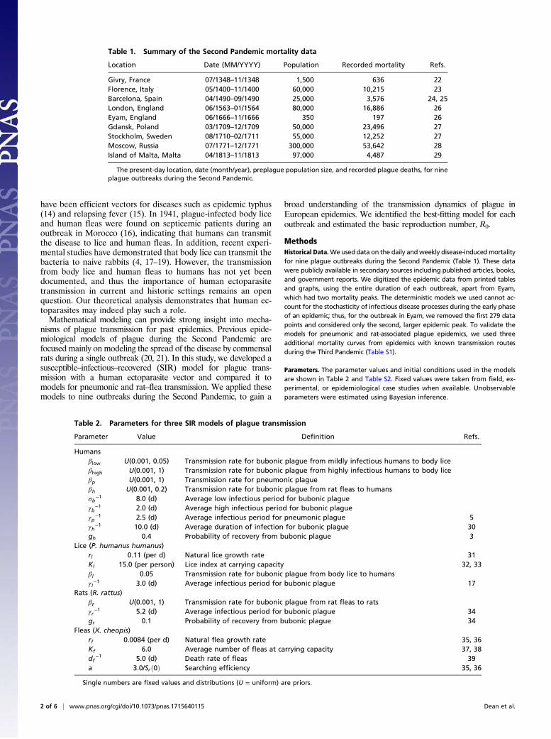
MethodsHistorical Data.Weused data on the daily andweekly disease-inducedmortalityfor nine plague outbreaks during the Second Pandemic (Table 1). These datawere publicly available in secondary sources including published articles, books,and government reports. We digitized the epidemic data from printed tablesand graphs, using the entire duration of each outbreak, apart from Eyam,which had two mortality peaks. The deterministic models we used cannot ac-count for the stochasticity of infectious disease processes during the early phaseof an epidemic; thus, for the outbreak in Eyam, we removed the first 279 datapoints and considered only the second, larger epidemic peak. To validate themodels for pneumonic and rat-associated plague epidemics, we used threeadditional mortality curves from epidemics with known transmission routesduring the Third Pandemic (Table S1).
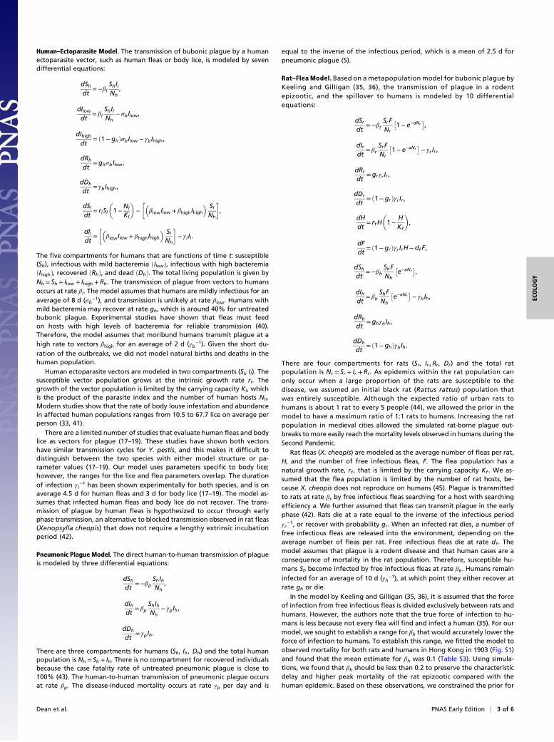
Parameters. The parameter values and initial conditions used in the modelsare shown in Table 2 and Table S2. Fixed values were taken from field, ex-perimental, or epidemiological case studies when available. Unobservableparameters were estimated using Bayesian inference.
Table 1. Summary of the Second Pandemic mortality data
Location Date (MM/YYYY) Population Recorded mortality Refs.
Givry, France 07/1348–11/1348 1,500 636 22Florence, Italy 05/1400–11/1400 60,000 10,215 23Barcelona, Spain 04/1490–09/1490 25,000 3,576 24, 25London, England 06/1563–01/1564 80,000 16,886 26Eyam, England 06/1666–11/1666 350 197 26Gdansk, Poland 03/1709–12/1709 50,000 23,496 27Stockholm, Sweden 08/1710–02/1711 55,000 12,252 27Moscow, Russia 07/1771–12/1771 300,000 53,642 28Island of Malta, Malta 04/1813–11/1813 97,000 4,487 29
The present-day location, date (month/year), preplague population size, and recorded plague deaths, for nineplague outbreaks during the Second Pandemic.
Table 2. Parameters for three SIR models of plague transmission
Parameter Value Definition Refs.
Humansβlow U(0.001, 0.05) Transmission rate for bubonic plague from mildly infectious humans to body liceβhigh U(0.001, 1) Transmission rate for bubonic plague from highly infectious humans to body liceβp U(0.001, 1) Transmission rate for pneumonic plagueβh U(0.001, 0.2) Transmission rate for bubonic plague from rat fleas to humansσb
−1 8.0 (d) Average low infectious period for bubonic plagueγb
−1 2.0 (d) Average high infectious period for bubonic plagueγp
−1 2.5 (d) Average infectious period for pneumonic plague 5γh
−1 10.0 (d) Average duration of infection for bubonic plague 30gh 0.4 Probability of recovery from bubonic plague 3
Lice (P. humanus humanus)rl 0.11 (per d) Natural lice growth rate 31Kl 15.0 (per person) Lice index at carrying capacity 32, 33βl 0.05 Transmission rate for bubonic plague from body lice to humansγl
−1 3.0 (d) Average infectious period for bubonic plague 17Rats (R. rattus)
βr U(0.001, 1) Transmission rate for bubonic plague from rat fleas to ratsγr
−1 5.2 (d) Average infectious period for bubonic plague 34gr 0.1 Probability of recovery from bubonic plague 34
Fleas (X. cheopis)rf 0.0084 (per d) Natural flea growth rate 35, 36Kf 6.0 Average number of fleas at carrying capacity 37, 38df
−1 5.0 (d) Death rate of fleas 39a 3.0/Srð0Þ Searching efficiency 35, 36
Single numbers are fixed values and distributions (U = uniform) are priors.
2 of 6 | www.pnas.org/cgi/doi/10.1073/pnas.1715640115 Dean et al.
Human–Ectoparasite Model. The transmission of bubonic plague by a humanectoparasite vector, such as human fleas or body lice, is modeled by sevendifferential equations:
dShdt
=−βlShIlNh
,
dIlowdt
= βlShIlNh
− σbIlow,
dIhighdt
= ð1−ghÞσbIlow − γbIhigh,
dRh
dt=ghσbIlow,
dDh
dt= γbIhigh,
dSldt
= rlSl
�1−
Nl
Kl
�−��
βlowIlow + βhighIhigh� SlNh
�,
dIldt
=��
βlowIlow + βhighIhigh� SlNh
�− γl Il .
The five compartments for humans that are functions of time t: susceptible(Sh), infectious with mild bacteremia ðIlowÞ, infectious with high bacteremiaðIhighÞ, recovered ðRhÞ, and dead ðDhÞ. The total living population is given byNh = Sh + Ilow + Ihigh +Rh. The transmission of plague from vectors to humansoccurs at rate βl. The model assumes that humans are mildly infectious for anaverage of 8 d (σb−1), and transmission is unlikely at rate βlow. Humans withmild bacteremia may recover at rate gh, which is around 40% for untreatedbubonic plague. Experimental studies have shown that fleas must feedon hosts with high levels of bacteremia for reliable transmission (40).Therefore, the model assumes that moribund humans transmit plague at ahigh rate to vectors βhigh for an average of 2 d (γb
−1). Given the short du-
ration of the outbreaks, we did not model natural births and deaths in thehuman population.
Human ectoparasite vectors are modeled in two compartments (Sl, Il). Thesusceptible vector population grows at the intrinsic growth rate rl. Thegrowth of the vector population is limited by the carrying capacity Kl, whichis the product of the parasite index and the number of human hosts Nh.Modern studies show that the rate of body louse infestation and abundancein affected human populations ranges from 10.5 to 67.7 lice on average perperson (33, 41).
There are a limited number of studies that evaluate human fleas and bodylice as vectors for plague (17–19). These studies have shown both vectorshave similar transmission cycles for Y. pestis, and this makes it difficult todistinguish between the two species with either model structure or pa-rameter values (17–19). Our model uses parameters specific to body lice;however, the ranges for the lice and flea parameters overlap. The durationof infection γl
−1 has been shown experimentally for both species, and is onaverage 4.5 d for human fleas and 3 d for body lice (17–19). The model as-sumes that infected human fleas and body lice do not recover. The trans-mission of plague by human fleas is hypothesized to occur through earlyphase transmission, an alternative to blocked transmission observed in rat fleas(Xenopsylla cheopis) that does not require a lengthy extrinsic incubationperiod (42).
Pneumonic Plague Model. The direct human-to-human transmission of plagueis modeled by three differential equations:
dShdt
=−βpShIhNh
,
dIhdt
= βpShIhNh
− γpIh,
dDh
dt= γpIh.
There are three compartments for humans (Sh, Ih, Dh) and the total humanpopulation is Nh = Sh + Ih. There is no compartment for recovered individualsbecause the case fatality rate of untreated pneumonic plague is close to100% (43). The human-to-human transmission of pneumonic plague occursat rate βp. The disease-induced mortality occurs at rate γp per day and is
equal to the inverse of the infectious period, which is a mean of 2.5 d forpneumonic plague (5).
Rat–Flea Model. Based on a metapopulation model for bubonic plague byKeeling and Gilligan (35, 36), the transmission of plague in a rodentepizootic, and the spillover to humans is modeled by 10 differentialequations:
dSrdt
=−βrSrFNr
�1− e−aNr
,
dIrdt
= βrSrFNr
�1− e−aNr
− γr Ir ,
dRr
dt=grγr Ir ,
dDr
dt= ð1−grÞγr Ir ,
dHdt
= rfH�1−
HKf
�,
dFdt
= ð1−grÞγr IrH−df F,
dShdt
=−βhShFNh
�e−aNr
,
dIhdt
= βhShFNh
�e−aNr
− γhIh,
dRh
dt=ghγhIh,
dDh
dt= ð1−ghÞγhIh.
There are four compartments for rats (Sr, Ir ,Rr, Dr) and the total ratpopulation is Nr = Sr + Ir +Rr. As epidemics within the rat population canonly occur when a large proportion of the rats are susceptible to thedisease, we assumed an initial black rat (Rattus rattus) population thatwas entirely susceptible. Although the expected ratio of urban rats tohumans is about 1 rat to every 5 people (44), we allowed the prior in themodel to have a maximum ratio of 1:1 rats to humans. Increasing the ratpopulation in medieval cities allowed the simulated rat-borne plague out-breaks to more easily reach the mortality levels observed in humans during theSecond Pandemic.
Rat fleas (X. cheopis) are modeled as the average number of fleas per rat,H, and the number of free infectious fleas, F. The flea population has anatural growth rate, rf , that is limited by the carrying capacity Kf . We as-sumed that the flea population is limited by the number of rat hosts, be-cause X. cheopis does not reproduce on humans (45). Plague is transmittedto rats at rate βr by free infectious fleas searching for a host with searchingefficiency a. We further assumed that fleas can transmit plague in the earlyphase (42). Rats die at a rate equal to the inverse of the infectious period
γr−1, or recover with probability gr. When an infected rat dies, a number of
free infectious fleas are released into the environment, depending on theaverage number of fleas per rat. Free infectious fleas die at rate df . Themodel assumes that plague is a rodent disease and that human cases are aconsequence of mortality in the rat population. Therefore, susceptible hu-mans Sh become infected by free infectious fleas at rate βh. Humans remain
infected for an average of 10 d (γh−1), at which point they either recover at
rate gh or die.In the model by Keeling and Gilligan (35, 36), it is assumed that the force
of infection from free infectious fleas is divided exclusively between rats andhumans. However, the authors note that the true force of infection to hu-mans is less because not every flea will find and infect a human (35). For ourmodel, we sought to establish a range for βh that would accurately lower theforce of infection to humans. To establish this range, we fitted the model toobserved mortality for both rats and humans in Hong Kong in 1903 (Fig. S1)and found that the mean estimate for βh was 0.1 (Table S3). Using simula-tions, we found that βh should be less than 0.2 to preserve the characteristicdelay and higher peak mortality of the rat epizootic compared with thehuman epidemic. Based on these observations, we constrained the prior for
Dean et al. PNAS Early Edition | 3 of 6
ECOLO
GY
the transmission rate to humans βh to 0.0–0.2, which enabled us to use thismodel for outbreaks where only human mortality was available.
Bayesian Inference and Markov Chain Monte Carlo.We fitted the deterministicmodels to the observed data using Bayesian inference and estimated un-observable parameters of interest. The models had a time-step of 1 d andwere fitted to daily mortality or weekly mortality. Denoting the set of modelparameters as Θ= fS0, β, . . .g, the probability p of the observed data D1...m
given Θ is calculated as the product of a series of Poisson random variableswith mean λT equal to the human mortality in the model at times T1...m:
pðDjΘÞ= ∏m
T=1e−λT
ðλT ÞDT
DT !.
The parameters that we fitted were the transmission rates for each model(βlow, βhigh, βp, βr, βh) and the size of the initial primary host population that
was at risk [Sð0Þh, Sð0Þr] or infected [Ið0Þh, Ið0Þr]. We assumed uniformlydistributed priors and obtained posterior distributions using Markov chainMonte Carlo (MCMC) simulations with an adaptive Metropolis–Hastings al-gorithm implemented in PyMC2 (46) (for examples of the implementation,see https://zenodo.org/record/1043924). We ran the MCMC simulations for180,000 iterations with a burn-in of 80,000 iterations and a thinning of 10.We assessed convergence for each model by running three independentMCMC chains and verifying that the Gelman–Rubin statistic (47) was<1.05 for each parameter. We performed model comparison using theBayesian information criterion (BIC) from the maximum-likelihood esti-mates of the model parameters (48). The model with the lowest BIC valuewas the unique preferred model if the second-best model had a BIC valueof at least 10 larger (49).
Estimation of the Basic Reproduction Number. We estimated the basic re-production number in each model for the primary host using the next-generation matrix method (50).
Reporting Error.We conducted the analysis again considering different levelsof underreporting (10%, 25%, and 50%) for each outbreak. To do so, weincorporated a constant probability of reporting into the likelihood function.
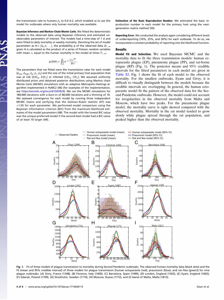
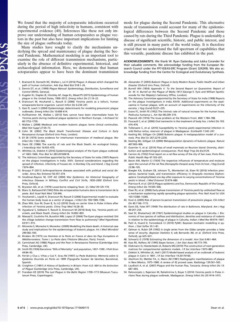
ResultsModel Fit and Selection. We used Bayesian MCMC and themortality data to fit the three transmission models: human ec-toparasite plague (EP), pneumonic plague (PP), and rat-borneplague (RP) (Fig. 1). The posterior means and 95% credibleintervals for the fitted parameters in each model are given inTable S3. Fig. 1 shows the fit of each model to the observedmortality. For the smallest outbreaks, Eyam and Givry, it isdifficult to visually distinguish between the models because thecredible intervals are overlapping. In general, the human ecto-parasite model fit the pattern of the observed data for the Sec-ond Pandemic outbreaks. However, the model could not accountfor irregularities in the observed mortality from Malta andMoscow, which have two peaks. For the pneumonic plaguemodel, the mortality curve is right skewed compared with theobserved mortality. Mortality in the rat model tended to growslowly while plague spread through the rat population, andpeaked higher than the observed mortality.
A
D
G H I
E F
B C
Fig. 1. Fit of three models of plague transmission to mortality during Second Pandemic outbreaks. The observed human mortality data (black dots) and thefit (mean and 95% credible interval) of three models for plague transmission [human ectoparasite (red), pneumonic (blue), and rat–flea (green)] for nineplague outbreaks: (A) Givry, France (1348), (B) Florence, Italy (1400), (C) Barcelona, Spain (1490), (D) London, England (1563), (E) Eyam, England (1665),(F) Gdansk, Poland (1709), (G) Stockholm, Sweden (1710), (H) Moscow, Russia (1772), and (I) Island of Malta, Malta (1813).
4 of 6 | www.pnas.org/cgi/doi/10.1073/pnas.1715640115 Dean et al.
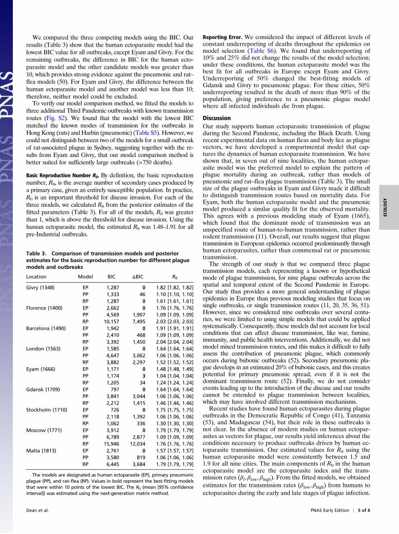
We compared the three competing models using the BIC. Ourresults (Table 3) show that the human ectoparasite model had thelowest BIC value for all outbreaks, except Eyam and Givry. For theremaining outbreaks, the difference in BIC for the human ecto-parasite model and the other candidate models was greater than10, which provides strong evidence against the pneumonic and rat–flea models (50). For Eyam and Givry, the difference between thehuman ectoparasite model and another model was less than 10;therefore, neither model could be excluded.To verify our model comparison method, we fitted the models to
three additional Third Pandemic outbreaks with known transmissionroutes (Fig. S2). We found that the model with the lowest BICmatched the known modes of transmission for the outbreaks inHong Kong (rats) andHarbin (pneumonic) (Table S5). However, wecould not distinguish between two of the models for a small outbreakof rat-associated plague in Sydney, suggesting together with the re-sults from Eyam and Givry, that our model comparison method isbetter suited for sufficiently large outbreaks (>750 deaths).
Basic Reproduction Number R0. By definition, the basic reproductionnumber, R0, is the average number of secondary cases produced bya primary case, given an entirely susceptible population. In practice,R0 is an important threshold for disease invasion. For each of thethree models, we calculated R0 from the posterior estimates of thefitted parameters (Table 3). For all of the models, R0 was greaterthan 1, which is above the threshold for disease invasion. Using thehuman ectoparasite model, the estimated R0 was 1.48–1.91 for allpre-Industrial outbreaks.
Reporting Error. We considered the impact of different levels ofconstant underreporting of deaths throughout the epidemics onmodel selection (Table S6). We found that underreporting of10% and 25% did not change the results of the model selection;under these conditions, the human ectoparasite model was thebest fit for all outbreaks in Europe except Eyam and Givry.Underreporting of 50% changed the best-fitting models ofGdansk and Givry to pneumonic plague. For these cities, 50%underreporting resulted in the death of more than 90% of thepopulation, giving preference to a pneumonic plague modelwhere all infected individuals die from plague.
DiscussionOur study supports human ectoparasite transmission of plagueduring the Second Pandemic, including the Black Death. Usingrecent experimental data on human fleas and body lice as plaguevectors, we have developed a compartmental model that cap-tures the dynamics of human ectoparasite transmission. We haveshown that, in seven out of nine localities, the human ectopar-asite model was the preferred model to explain the pattern ofplague mortality during an outbreak, rather than models ofpneumonic and rat–flea plague transmission (Table 3). The smallsize of the plague outbreaks in Eyam and Givry made it difficultto distinguish transmission routes based on mortality data. ForEyam, both the human ectoparasite model and the pneumonicmodel produced a similar quality fit for the observed mortality.This agrees with a previous modeling study of Eyam (1665),which found that the dominant mode of transmission was anunspecified route of human-to-human transmission, rather thanrodent transmission (11). Overall, our results suggest that plaguetransmission in European epidemics occurred predominantly throughhuman ectoparasites, rather than commensal rat or pneumonictransmission.The strength of our study is that we compared three plague
transmission models, each representing a known or hypotheticalmode of plague transmission, for nine plague outbreaks across thespatial and temporal extent of the Second Pandemic in Europe.Our study thus provides a more general understanding of plagueepidemics in Europe than previous modeling studies that focus onsingle outbreaks, or single transmission routes (11, 20, 35, 36, 51).However, since we considered nine outbreaks over several centu-ries, we were limited to using simple models that could be appliedsystematically. Consequently, these models did not account for localconditions that can affect disease transmission, like war, famine,immunity, and public health interventions. Additionally, we did notmodel mixed transmission routes, and this makes it difficult to fullyassess the contribution of pneumonic plague, which commonlyoccurs during bubonic outbreaks (52). Secondary pneumonic pla-gue develops in an estimated 20% of bubonic cases, and this createspotential for primary pneumonic spread, even if it is not thedominant transmission route (52). Finally, we do not considerevents leading up to the introduction of the disease and our resultscannot be extended to plague transmission between localities,which may have involved different transmission mechanisms.Recent studies have found human ectoparasites during plague
outbreaks in the Democratic Republic of Congo (41), Tanzania(53), and Madagascar (54), but their role in these outbreaks isnot clear. In the absence of modern studies on human ectopar-asites as vectors for plague, our results yield inferences about theconditions necessary to produce outbreaks driven by human ec-toparasite transmission. Our estimated values for R0 using thehuman ectoparasite model were consistently between 1.5 and1.9 for all nine cities. The main components of R0 in the humanectoparasite model are the ectoparasite index and the trans-mission rates (βl, βlow, βhigh). From the fitted models, we obtainedestimates for the transmission rates (βlow, βhigh) from humans toectoparasites during the early and late stages of plague infection.
Table 3. Comparison of transmission models and posteriorestimates for the basic reproduction number for different plaguemodels and outbreaks
Location Model BIC ΔBIC R0
Givry (1348) EP 1,287 0 1.82 [1.82, 1.82]PP 1,333 46 1.10 [1.10, 1.10]RP 1,287 0 1.61 [1.61, 1.61]
Florence (1400) EP 2,662 0 1.76 [1.76, 1.76]PP 4,569 1,907 1.09 [1.09, 1.09]RP 10,157 7,495 2.03 [2.03, 2.03]
Barcelona (1490) EP 1,942 0 1.91 [1.91, 1.91]PP 2,410 468 1.09 [1.09, 1.09]RP 3,392 1,450 2.04 [2.04, 2.04]
London (1563) EP 1,585 0 1.64 [1.64, 1.64]PP 4,647 3,062 1.06 [1.06, 1.06]RP 3,882 2,297 1.52 [1.52, 1.52]
Eyam (1666) EP 1,171 0 1.48 [1.48, 1.49]PP 1,174 3 1.04 [1.04, 1.04]RP 1,205 34 1.24 [1.24, 1.24]
Gdansk (1709) EP 797 0 1.64 [1.64, 1.64]PP 3,841 3,044 1.06 [1.06, 1.06]RP 2,212 1,415 1.46 [1.46, 1.46]
Stockholm (1710) EP 726 0 1.75 [1.75, 1.75]PP 2,118 1,392 1.06 [1.06, 1.06]RP 1,062 336 1.30 [1.30, 1.30]
Moscow (1771) EP 3,912 0 1.79 [1.79, 1.79]PP 6,789 2,877 1.09 [1.09, 1.09]RP 15,946 12,034 1.76 [1.76, 1.76]
Malta (1813) EP 2,761 0 1.57 [1.57, 1.57]PP 3,580 819 1.06 [1.06, 1.06]RP 6,445 3,684 1.79 [1.79, 1.79]
The models are designated as human ectoparasite (EP), primary pneumonicplague (PP), and rat–flea (RP). Values in bold represent the best-fitting modelsthat were within 10 points of the lowest BIC. The R0 (mean [95% confidenceinterval]) was estimated using the next-generation matrix method.
Dean et al. PNAS Early Edition | 5 of 6
ECOLO
GY
We found that the majority of ectoparasite infections occurredduring the period of high infectivity in humans, consistent withexperimental evidence (40). Inferences like these not only im-prove our understanding of human ectoparasites as plague vec-tors in the past but also have important implications for limitingthe size of plague outbreaks today.Many studies have sought to clarify the mechanisms un-
derlying the spread and maintenance of plague during the Sec-ond Pandemic. Mathematical modeling is an important tool toexamine the role of different transmission mechanisms, partic-ularly in the absence of definitive experimental, historical, andarchaeological information. Here, we demonstrate that humanectoparasites appear to have been the dominant transmission
mode for plague during the Second Pandemic. This alternativemode of transmission could account for many of the epidemio-logical differences between the Second Pandemic and thosecaused by rats during the Third Pandemic. Plague is undeniably adisease of significant scientific, historic, and public interest, andis still present in many parts of the world today. It is thereforecrucial that we understand the full spectrum of capabilities thatthis versatile, pandemic disease has exhibited in the past.
ACKNOWLEDGMENTS. We thank W. Ryan Easterday and Jukka Corander fortheir valuable comments. We acknowledge funding from the European Re-search Council under the FP7-IDEAS-ERC Program (Grant 324249). We also ac-knowledge funding from the Centre for Ecological and Evolutionary Synthesis.
1. Bramanti B, Stenseth NC, Walløe L, Lei X (2016) Plague: A disease which changed thepath of human civilization. Adv Exp Med Biol 918:1–26.
2. Dennis DT, et al. (1999) Plague Manual: Epidemiology, Distribution, Surveillance andControl (WHO, Geneva).
3. Kugeler KJ, Staples JE, Hinckley AF, Gage KL, Mead PS (2015) Epidemiology of humanplague in the United States, 1900–2012. Emerg Infect Dis 21:16–22.
4. Drancourt M, Houhamdi L, Raoult D (2006) Yersinia pestis as a telluric, humanectoparasite-borne organism. Lancet Infect Dis 6:234–241.
5. Gani R, Leach S (2004) Epidemiologic determinants for modeling pneumonic plagueoutbreaks. Emerg Infect Dis 10:608–614.
6. Hufthammer AK, Walløe L (2013) Rats cannot have been intermediate hosts forYersinia pestis during medieval plague epidemics in Northern Europe. J Archaeol Sci40:1752–1759.
7. Walløe L (2008) Medieval and modern bubonic plague: Some clinical continuities.Med Hist Suppl 27:59–73.
8. Cohn SK (2002) The Black Death Transformed: Disease and Culture in EarlyRenaissance Europe (Oxford Univ Press, London).
9. Ell SR (1979) Some evidence for interhuman transmission of medieval plague. RevInfect Dis 1:563–566.
10. Davis DE (1986) The scarcity of rats and the Black Death: An ecological history.J Interdiscip Hist 16:455–470.
11. Whittles LK, Didelot X (2016) Epidemiological analysis of the Eyam plague outbreakof 1665–1666. Proc Biol Sci 283:20160618.
12. The Advisory Committee appointed by the Secretary of State for India (1907) Reportson the plague investigations in India. XXIV. General considerations regarding thespread of infection, infectivity of houses etc. in Bombay City and Island. J Hyg (Lond)7:874–894.
13. Brouqui P (2011) Arthropod-borne diseases associated with political and social dis-order. Annu Rev Entomol 56:357–374.
14. Smallman-Raynor M, Cliff AD (2004) War Epidemics: An Historical Geography ofInfectious Diseases in Military Conflict and Civil Strife, 1850–2000 (Oxford UnivPress, Oxford).
15. Bryceson AD, et al. (1970) Louse-borne relapsing fever. Q J Med 39:129–170.16. Blanc G, Baltazard M (1942) Rôle des ectoparasites humains dans la transmission de la
peste. Bull Acad Natl Med 126:446.17. Houhamdi L, Lepidi H, Drancourt M, Raoult D (2006) Experimental model to evaluate
the human body louse as a vector of plague. J Infect Dis 194:1589–1596.18. Zhao WH, Guo M, Duan B, Su LQ (2016) Study on carrier time in Pulex irritans after
infection of Yersinia pestis. China Trop Med 16:28–30.19. Ayyadurai S, Sebbane F, Raoult D, Drancourt M (2010) Body lice, Yersinia pestis ori-
entalis, and Black Death. Emerg Infect Dis 16:892–893.20. Massad E, Coutinho FA, Burattini MN, Lopez LF (2004) The Eyam plague revisited: Did
the village isolation change transmission from fleas to pulmonary? Med Hypotheses63:911–915.
21. Monecke S, Monecke H, Monecke J (2009) Modelling the black death. A historical casestudy and implications for the epidemiology of bubonic plague. Int J Med Microbiol299:582–593.
22. Biraben JN (1975) Les Hommes et la Peste en France et dans les Pays Européens etMéditerranéens. Tome I. La Peste dans l’Histoire (Mouton, Paris). French.
23. Carmichael AG (1986) Plague and the Poor in Renaissance Florence (Cambridge UnivPress, Cambridge, UK).
24. Smith RS (1936) Barcelona “Bills of Mortality” and population, 1457–1590. J Polit Econ44:84–93.
25. Ferrán y Clua J, Viñas y Cusí F, Grau Rd (1907) La Peste Bubónica: Memoria sobre laEpidemia Ocurrida en Porto en 1899 (Tipografia Sucesor de Sánchez, Barcelona).Spanish.
26. Creighton C (1891) A History of Epidemics in Britain: From A.D. 664 to the Extinctionof Plague (Cambridge Univ Press, Cambridge, UK).
27. Frandsen KE (2010) The Last Plague in the Baltic Region 1709–1713 (Museum Tuscu-lanum Press, Copenhagen).
28. Alexander JT (2003) Bubonic Plague in Early Modern Russia: Public Health and UrbanDisaster (Oxford Univ Press, Oxford).
29. Burrell WH (1854) Appendix V. To the Second Report on Quarantine: Report ofDr. W. H. Burrell on the Plague of Malta 1813 (George E. Eyre and William Spottis-woode for Her Majesty’s Sationery Office, London).
30. The Advisory Committee appointed by the Secretary of State for India (1908) Reportson the plague investigations in India XXVIII. Additional experiments on the septi-caemia in human plague, with an account of experiments on the infectivity of theexcreta. J Hyg (Lond) 8:221–235.
31. Evans FC, Smith FE (1952) The intrinsic rate of natural increase for the human louse,Pediculus humanus L. Am Nat 86:299–310.
32. Peacock AD (1916) The louse problem at the Western Front. BMJ 1:784–788.33. Foucault C, et al. (2006) Oral ivermectin in the treatment of body lice. J Infect Dis 193:
474–476.34. Tollenaere C, et al. (2010) Susceptibility to Yersinia pestis experimental infection in
wild Rattus rattus, reservoir of plague in Madagascar. EcoHealth 7:242–247.35. Keeling MJ, Gilligan CA (2000) Bubonic plague: A metapopulation model of a zoo-
nosis. Proc Biol Sci 267:2219–2230.36. Keeling MJ, Gilligan CA (2000) Metapopulation dynamics of bubonic plague. Nature
407:903–906.37. Guernier V, et al. (2014) Fleas of small mammals on Reunion Island: Diversity, distri-
bution and epidemiological consequences. PLoS Negl Trop Dis 8:e3129.38. Carrion AL (1932) Final report on a rat-flea survey of the city of San Juan, Porto Rico.
Public Health Rep 47:193–201.39. Bacot AW, Martin CJ (1924) The respective influences of temperature and moisture
upon the survival of the rat flea (Xenopsylla cheopis) away from its host. J Hyg (Lond)23:98–105.
40. Boegler KA, Graham CB, Johnson TL, Montenieri JA, Eisen RJ (2016) Infection prev-alence, bacterial loads, and transmission efficiency in Oropsylla montana (Siphon-aptera: Ceratophyllidae) one day after exposure to varying concentrations of Yersiniapestis in blood. J Med Entomol 53:674–680.
41. Piarroux R, et al. (2013) Plague epidemics and lice, Democratic Republic of the Congo.Emerg Infect Dis 19:505–506.
42. Eisen RJ, et al. (2006) Early-phase transmission of Yersinia pestis by unblocked fleas asa mechanism explaining rapidly spreading plague epizootics. Proc Natl Acad Sci USA103:15380–15385.
43. Kool JL (2005) Risk of person-to-person transmission of pneumonic plague. Clin InfectDis 40:1166–1172.
44. Davis DE, Fales WT (1949) The distribution of rats in Baltimore, Maryland. Am J Hyg49:247–254.
45. Seal SC, Bhatacharji LM (1961) Epidemiological studies on plague in Calcutta. I. Bio-nomics of two species of ratfleas and distribution, densities and resistance of rodentsin relation to the epidemiology of plague in Calcutta. Indian J Med Res 49:974–1007.
46. Patil A, Huard D, Fonnesbeck CJ (2010) PyMC: Bayesian stochastic modelling in py-thon. J Stat Softw 35:1–81.
47. Gelman A, Rubin DR (1992) A single series from the Gibbs sampler provides a falsesense of security. Bayesian Statistics 4, eds Bernardo JM, et al. (Oxford Univ Press,Oxford), pp 625–631.
48. Schwartz G (1978) Estimating the dimension of a model. Ann Stat 6:461–464.49. Kass RE, Raftery AE (1995) Bayes factors. J Am Stat Assoc 90:773–795.50. Diekmann O, Heesterbeek JA, Roberts MG (2010) The construction of next-generation
matrices for compartmental epidemic models. J R Soc Interface 7:873–885.51. Didelot X, Whittles LK, Hall I (2017) Model-based analysis of an outbreak of bubonic
plague in Cairo in 1801. J R Soc Interface 14:20170160.52. Alsofrom DJ, Mettler FA, Jr, Mann JM (1981) Radiographic manifestations of plaque
in New Mexico, 1975–1980. A review of 42 proved cases. Radiology 139:561–565.53. Laudisoit A, et al. (2007) Plague and the human flea, Tanzania. Emerg Infect Dis 13:
687–693.54. Ratovonjato J, Rajerison M, Rahelinirina S, Boyer S (2014) Yersinia pestis in Pulex ir-
ritans fleas during plague outbreak, Madagascar. Emerg Infect Dis 20:1414–1415.
6 of 6 | www.pnas.org/cgi/doi/10.1073/pnas.1715640115 Dean et al.