Analysis of Noncanonical Calcium-Dependent Protein Kinases in ...
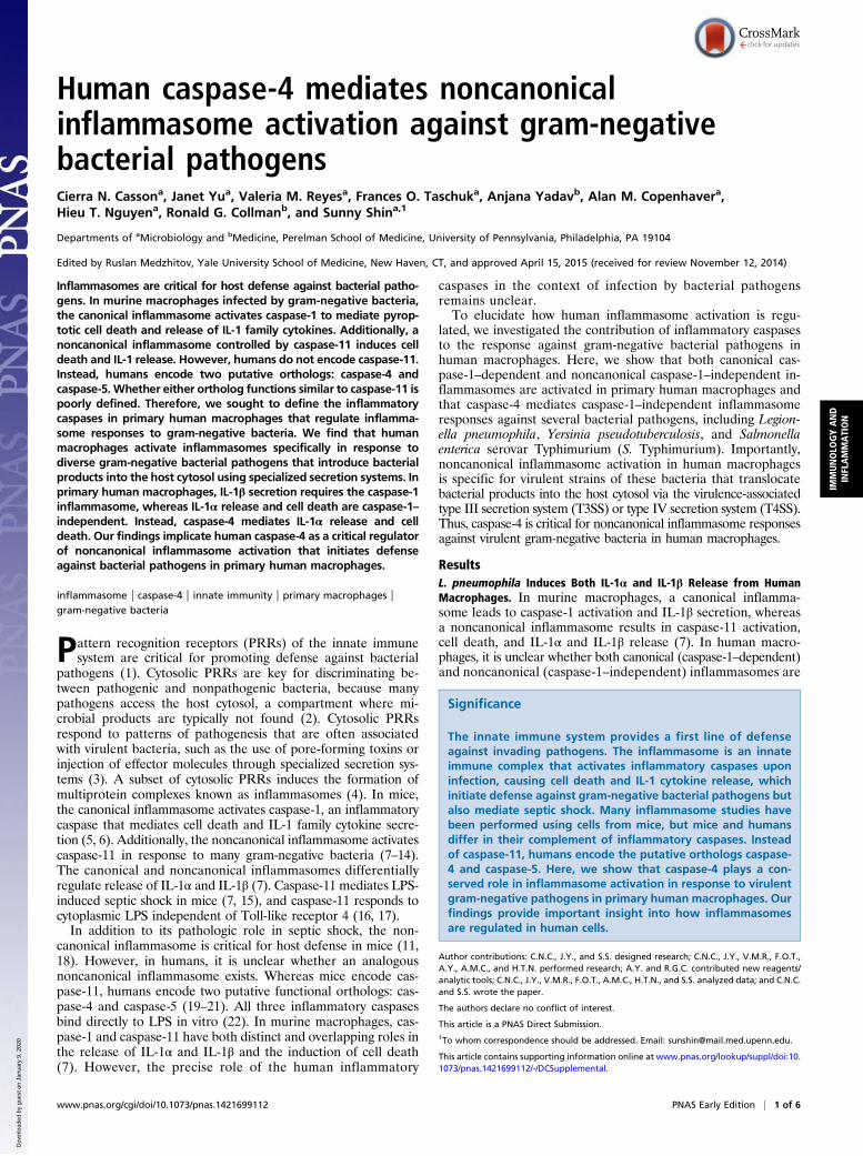
Human caspase-4 mediates noncanonicalinflammasome activation against gram-negativebacterial pathogensCierra N. Cassona, Janet Yua, Valeria M. Reyesa, Frances O. Taschuka, Anjana Yadavb, Alan M. Copenhavera,Hieu T. Nguyena, Ronald G. Collmanb, and Sunny Shina,1
Departments of aMicrobiology and bMedicine, Perelman School of Medicine, University of Pennsylvania, Philadelphia, PA 19104
Edited by Ruslan Medzhitov, Yale University School of Medicine, New Haven, CT, and approved April 15, 2015 (received for review November 12, 2014)
Inflammasomes are critical for host defense against bacterial patho-gens. In murine macrophages infected by gram-negative bacteria,the canonical inflammasome activates caspase-1 to mediate pyrop-totic cell death and release of IL-1 family cytokines. Additionally, anoncanonical inflammasome controlled by caspase-11 induces celldeath and IL-1 release. However, humans do not encode caspase-11.Instead, humans encode two putative orthologs: caspase-4 andcaspase-5. Whether either ortholog functions similar to caspase-11 ispoorly defined. Therefore, we sought to define the inflammatorycaspases in primary human macrophages that regulate inflamma-some responses to gram-negative bacteria. We find that humanmacrophages activate inflammasomes specifically in response todiverse gram-negative bacterial pathogens that introduce bacterialproducts into the host cytosol using specialized secretion systems. Inprimary human macrophages, IL-1β secretion requires the caspase-1inflammasome, whereas IL-1α release and cell death are caspase-1–independent. Instead, caspase-4 mediates IL-1α release and celldeath. Our findings implicate human caspase-4 as a critical regulatorof noncanonical inflammasome activation that initiates defenseagainst bacterial pathogens in primary human macrophages.
inflammasome | caspase-4 | innate immunity | primary macrophages |gram-negative bacteria
Pattern recognition receptors (PRRs) of the innate immunesystem are critical for promoting defense against bacterial
pathogens (1). Cytosolic PRRs are key for discriminating be-tween pathogenic and nonpathogenic bacteria, because manypathogens access the host cytosol, a compartment where mi-crobial products are typically not found (2). Cytosolic PRRsrespond to patterns of pathogenesis that are often associatedwith virulent bacteria, such as the use of pore-forming toxins orinjection of effector molecules through specialized secretion sys-tems (3). A subset of cytosolic PRRs induces the formation ofmultiprotein complexes known as inflammasomes (4). In mice,the canonical inflammasome activates caspase-1, an inflammatorycaspase that mediates cell death and IL-1 family cytokine secre-tion (5, 6). Additionally, the noncanonical inflammasome activatescaspase-11 in response to many gram-negative bacteria (7–14).The canonical and noncanonical inflammasomes differentiallyregulate release of IL-1α and IL-1β (7). Caspase-11 mediates LPS-induced septic shock in mice (7, 15), and caspase-11 responds tocytoplasmic LPS independent of Toll-like receptor 4 (16, 17).In addition to its pathologic role in septic shock, the non-
canonical inflammasome is critical for host defense in mice (11,18). However, in humans, it is unclear whether an analogousnoncanonical inflammasome exists. Whereas mice encode cas-pase-11, humans encode two putative functional orthologs: cas-pase-4 and caspase-5 (19–21). All three inflammatory caspasesbind directly to LPS in vitro (22). In murine macrophages, cas-pase-1 and caspase-11 have both distinct and overlapping roles inthe release of IL-1α and IL-1β and the induction of cell death(7). However, the precise role of the human inflammatory
caspases in the context of infection by bacterial pathogensremains unclear.To elucidate how human inflammasome activation is regu-
lated, we investigated the contribution of inflammatory caspasesto the response against gram-negative bacterial pathogens inhuman macrophages. Here, we show that both canonical cas-pase-1–dependent and noncanonical caspase-1–independent in-flammasomes are activated in primary human macrophages andthat caspase-4 mediates caspase-1–independent inflammasomeresponses against several bacterial pathogens, including Legion-ella pneumophila, Yersinia pseudotuberculosis, and Salmonellaenterica serovar Typhimurium (S. Typhimurium). Importantly,noncanonical inflammasome activation in human macrophagesis specific for virulent strains of these bacteria that translocatebacterial products into the host cytosol via the virulence-associatedtype III secretion system (T3SS) or type IV secretion system (T4SS).Thus, caspase-4 is critical for noncanonical inflammasome responsesagainst virulent gram-negative bacteria in human macrophages.
ResultsL. pneumophila Induces Both IL-1α and IL-1β Release from HumanMacrophages. In murine macrophages, a canonical inflamma-some leads to caspase-1 activation and IL-1β secretion, whereasa noncanonical inflammasome results in caspase-11 activation,cell death, and IL-1α and IL-1β release (7). In human macro-phages, it is unclear whether both canonical (caspase-1–dependent)and noncanonical (caspase-1–independent) inflammasomes are
Significance
The innate immune system provides a first line of defenseagainst invading pathogens. The inflammasome is an innateimmune complex that activates inflammatory caspases uponinfection, causing cell death and IL-1 cytokine release, whichinitiate defense against gram-negative bacterial pathogens butalso mediate septic shock. Many inflammasome studies havebeen performed using cells from mice, but mice and humansdiffer in their complement of inflammatory caspases. Insteadof caspase-11, humans encode the putative orthologs caspase-4 and caspase-5. Here, we show that caspase-4 plays a con-served role in inflammasome activation in response to virulentgram-negative pathogens in primary human macrophages. Ourfindings provide important insight into how inflammasomesare regulated in human cells.
Author contributions: C.N.C., J.Y., and S.S. designed research; C.N.C., J.Y., V.M.R., F.O.T.,A.Y., A.M.C., and H.T.N. performed research; A.Y. and R.G.C. contributed new reagents/analytic tools; C.N.C., J.Y., V.M.R., F.O.T., A.M.C., H.T.N., and S.S. analyzed data; and C.N.C.and S.S. wrote the paper.
The authors declare no conflict of interest.
This article is a PNAS Direct Submission.1To whom correspondence should be addressed. Email: [email protected].
This article contains supporting information online at www.pnas.org/lookup/suppl/doi:10.1073/pnas.1421699112/-/DCSupplemental.
www.pnas.org/cgi/doi/10.1073/pnas.1421699112 PNAS Early Edition | 1 of 6
IMMUNOLO
GYAND
INFLAMMATION
Dow
nloa
ded
by g
uest
on
Janu
ary
9, 2
020

activated during bacterial infection (12). To determine if bothcanonical and noncanonical inflammasomes are activated, wefirst used L. pneumophila, a pathogen that triggers robust in-flammasome activation in murine macrophages and causes asevere form of pneumonia, Legionnaires’ disease, in humans(23). To replicate within macrophages, L. pneumophila uses aT4SS to inject effector proteins into the host cell cytosol (24–26).Because L. pneumophila activates both the canonical and non-canonical inflammasomes in murine macrophages (13, 14), weexamined whether the bacterium induces IL-1α and IL-1β re-lease from human macrophages as well. First, we differentiatedand infected the THP-1 monocytic cell line. Upon infection,THP-1 cells underwent death and released IL-1α and IL-1β in amanner requiring the presence of the bacterial T4SS (Fig. 1A).THP-1 cells infected with bacterial mutants lacking a functionalT4SS [T4SS− L. pneumophila (Lp)] did not activate the inflam-masome but still up-regulated pro–IL-1β, suggesting that thecells are capable of sensing T4SS− Lp (Fig. 1B).Inflammasome activation has been extensively analyzed in
murine macrophages and in murine and human monocytic andepithelial cell lines. In human epithelial cell lines, noncanonicalinflammasome activation leads to IL-18 release and cell deathduring S. Typhimurium infection (27). Additionally, noncanonicalinflammasome activation occurs in a number of transformed celllines in response to intracellular LPS (22, 27). However, we havelimited understanding of inflammasome biology in primary humaninnate immune cells, particularly with respect to pathways thatregulate IL-1α and IL-1β release. We therefore infected primaryhuman monocyte-derived macrophages (MDMs) from healthyhuman donors with L. pneumophila. Importantly, cell death and
IL-1 release in primary human macrophages required the pres-ence of the bacterial T4SS (Fig. 1C). Although T4SS− bacteriadid not elicit IL-1β secretion, pro–IL-1β was up-regulated in allinfected donor cells (Fig. 1D). Mature IL-1β was detected in thesupernatants of cells infected with WT L. pneumophila (WT Lp)but not with T4SS− Lp (Fig. S1). The absence of inflammasomeactivation in T4SS− Lp-infected cells was not due to a lack ofpriming, because MDMs that were first directly primed withLPS and then infected with T4SS− Lp also did not activate theinflammasome (Fig. 1 E and F). Thus, L. pneumophila triggersrobust T4SS-dependent inflammasome responses in primaryhuman macrophages.
Caspase-1–Dependent and –Independent Inflammasome PathwaysAre Activated During Infection of Human Macrophages. To deter-mine whether both canonical (caspase-1–dependent) and non-canonical (caspase-1–independent) inflammasomes are triggeredin human macrophages, we first examined the contribution ofcaspase-1 to inflammasome responses. We infected primary humanMDMs and found that L. pneumophila infection induced caspase-1cleavage into a 10-kDa (p10) subunit that was released into thesupernatant (Fig. 2A). Caspase-1 was processed in response to WTLp and not T4SS− Lp, suggesting that cytosolic sensing of T4SSactivity controls caspase-1 cleavage into its active form. We nextasked whether caspase-1 catalytic activity is required for inflam-masome activation in human macrophages. We pretreated primaryhuman MDMs with Ac-YVAD-cmk, a chemical inhibitor of cas-pase-1 activity, and examined inflammasome responses. In primaryMDMs, caspase-1 activity played a major role in controlling IL-1β
Fig. 1. L. pneumophila induces both IL-1α and IL-1β release from humanmacrophages. Phorbol 12-myristate 13-acetate (PMA)-differentiated THP-1cells (A and B) or primary human MDMs (C and D) were infected with WT Lp,infected with T4SS− Lp, or mock-infected with PBS (Mock) for 20 h. (E and F)Primary human MDMs were primed with LPS and infected with WT Lp orT4SS− Lp or mock-infected for 4 h. Cell death (% cytotoxicity) was measuredusing a lactate dehydrogenase release assay and normalized to mock-infected cells. IL-1α and IL-1β levels in the supernatants were measured byELISA. Immunoblot analysis was performed on lysates for full-length IL-1β(pro–IL-1β), and blots were reprobed for β-actin as a loading control. West-ern blots (B, D, and F) are representative of at least three independent ex-periments. Shown are the pooled results of four independent experiments inTHP-1 cells (A) or the pooled results of six independent infections of cellsfrom different healthy human donors (C and E). Each data point shows themean of triplicate infected wells. For A, *P < 0.05 and **P < 0.01 by unpairedt test. For C and E, ***P < 0.001, **P < 0.01, and *P < 0.05 by paired t test.The dashed line is the limit of detection.
Fig. 2. Caspase-1–dependent and –independent inflammasomes are acti-vated during infection of human macrophages. (A) Primary human MDMswere infected with WT Lp, infected with T4SS− Lp, or mock-infected for 20 h.Immunoblot analysis was performed on supernatants (sup) for cleaved caspase-1(casp1 p10) and on lysates for full-length caspase-1 (pro-casp1). Lysates werereprobed for β-actin. Primary humanMDMs (B) or PMA-differentiated THP-1 cells(C) were pretreated with 40 μM caspase-1 inhibitor [Ac-YVAD-cmk (YVAD)]or vehicle control (DMSO) and infected with WT Lp or mock-infected for20 h. (D and E) PMA-differentiated THP-1 cells were transfected with controlsiRNA (CNTRL) or siRNA against caspase-1 and infected with WT Lp or mock-infected for 20 h. Immunoblot analysis was performed on lysates for pro-casp1,pro-casp4, and pro–IL-1β, and blots were reprobed for β-actin. Cell death wasmeasured using a lactate dehydrogenase release assay and normalized tomock-infected cells. IL-1α, IL-1β, and TNF levels in the supernatants weremeasured by ELISA. (A and D) Western blots are representative of three in-dependent experiments. Shown are the pooled results of four independentinfections of cells from different donors (B) or the pooled results of four (C) orthree (E) independent experiments in THP-1 cells. Each data point shows themean of triplicate infected wells. *P < 0.05 by paired t test (B and E) or un-paired t test (C). NS, not significant. The dashed line is the limit of detection.
2 of 6 | www.pnas.org/cgi/doi/10.1073/pnas.1421699112 Casson et al.
Dow
nloa
ded
by g
uest
on
Janu
ary
9, 2
020
secretion in response to WT Lp (Fig. 2B). Inhibition of caspase-1catalytic activity had no significant effect on cell death or IL-1αrelease. These data imply that as in murine cells, caspase-1–independent pathways contribute to IL-1α release in human cells,and these results were consistent with what was observed in THP-1cells (Fig. 2C). Importantly, the caspase-1–independent cytokineTNF was unaffected by inhibitor treatment. To strengthen the linkbetween caspase-1 and IL-1β secretion in human macrophages, wealso knocked down caspase-1 in THP-1 cells (Fig. 2D). Caspase-1knockdown did not affect expression of caspase-4 or pro–IL-1βduring infection with WT Lp but significantly reduced IL-1βsecretion (Fig. 2E). Thus, caspase-1 mediates IL-1β release butnot IL-1α release, suggesting that another inflammatory cas-pase may mediate IL-1α release during bacterial infection ofhuman macrophages.
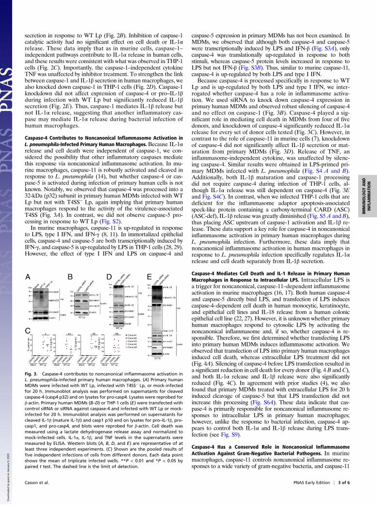
Caspase-4 Contributes to Noncanonical Inflammasome Activation inL. pneumophila-Infected Primary Human Macrophages. Because IL-1αrelease and cell death were independent of caspase-1, we con-sidered the possibility that other inflammatory caspases mediatethis response via noncanonical inflammasome activation. In mu-rine macrophages, caspase-11 is robustly activated and cleaved inresponse to L. pneumophila (14), but whether caspase-4 or cas-pase-5 is activated during infection of primary human cells is notknown. Notably, we observed that caspase-4 was processed into a32-kDa (p32) subunit in primary human MDMs infected with WTLp but not with T4SS− Lp, again implying that primary humanmacrophages respond to the activity of the virulence-associatedT4SS (Fig. 3A). In contrast, we did not observe caspase-5 pro-cessing in response to WT Lp (Fig. S2).In murine macrophages, caspase-11 is up-regulated in response
to LPS, type I IFN, and IFN-γ (8, 11). In immortalized epithelialcells, caspase-4 and caspase-5 are both transcriptionally induced byIFN-γ, and caspase-5 is up-regulated by LPS in THP-1 cells (28, 29).However, the effect of type I IFN and LPS on caspase-4 and
caspase-5 expression in primary MDMs has not been examined. InMDMs, we observed that although both caspase-4 and caspase-5were transcriptionally induced by LPS and IFN-β (Fig. S3A), onlycaspase-4 was translationally up-regulated in response to bothstimuli, whereas caspase-5 protein levels increased in response toLPS but not IFN-β (Fig. S3B). Thus, similar to murine caspase-11,caspase-4 is up-regulated by both LPS and type I IFN.Because caspase-4 is processed specifically in response to WT
Lp and is up-regulated by both LPS and type I IFN, we inter-rogated whether caspase-4 has a role in inflammasome activa-tion. We used siRNA to knock down caspase-4 expression inprimary human MDMs and observed robust silencing of caspase-4and no effect on caspase-1 (Fig. 3B). Caspase-4 played a sig-nificant role in mediating cell death in MDMs from four of fivedonors, and knockdown of caspase-4 significantly reduced IL-1αrelease for every set of donor cells tested (Fig. 3C). However, incontrast to the role of caspase-11 in murine cells (7), knockdownof caspase-4 did not significantly affect IL-1β secretion or mat-uration from primary MDMs (Fig. 3D). Release of TNF, aninflammasome-independent cytokine, was unaffected by silenc-ing caspase-4. Similar results were obtained in LPS-primed pri-mary MDMs infected with L. pneumophila (Fig. S4 A and B).Additionally, both IL-1β maturation and caspase-1 processingdid not require caspase-4 during infection of THP-1 cells, al-though IL-1α release was still dependent on caspase-4 (Fig. 3Eand Fig. S4C). In contrast, when we infected THP-1 cells that aredeficient for the inflammasome adaptor apoptosis-associatedspeck-like protein containing a carboxy-terminal CARD (ASC)(ASC-def), IL-1β release was greatly diminished (Fig. S5 A and B),thus placing ASC upstream of caspase-1 activation and IL-1β re-lease. These data support a key role for caspase-4 in noncanonicalinflammasome activation in primary human macrophages duringL. pneumophila infection. Furthermore, these data imply thatnoncanonical inflammasome activation in human macrophages inresponse to L. pneumophila infection specifically regulates IL-1αrelease and cell death separately from IL-1β secretion.
Caspase-4 Mediates Cell Death and IL-1 Release in Primary HumanMacrophages in Response to Intracellular LPS. Intracellular LPS isa trigger for noncanonical, caspase-11–dependent inflammasomeactivation in murine macrophages (16, 17). Both human caspase-4and caspase-5 directly bind LPS, and transfection of LPS inducescaspase-4–dependent cell death in human monocytic, keratinocyte,and epithelial cell lines and IL-18 release from a human colonicepithelial cell line (22, 27). However, it is unknown whether primaryhuman macrophages respond to cytosolic LPS by activating thenoncanonical inflammasome and, if so, whether caspase-4 is re-sponsible. Therefore, we first determined whether transfecting LPSinto primary human MDMs induces inflammasome activation. Weobserved that transfection of LPS into primary human macrophagesinduced cell death, whereas extracellular LPS treatment did not(Fig. 4A). Silencing of caspase-4 before LPS transfection resulted ina significant reduction in cell death for every donor (Fig. 4 B and C),and both IL-1α release and IL-1β release were also significantlyreduced (Fig. 4C). In agreement with prior studies (4), we alsofound that primary MDMs treated with extracellular LPS for 20 hinduced cleavage of caspase-5 but that LPS transfection did notincrease this processing (Fig. S6A). These data indicate that cas-pase-4 is primarily responsible for noncanonical inflammasome re-sponses to intracellular LPS in primary human macrophages;however, unlike the response to bacterial infection, caspase-4 ap-pears to control both IL-1α and IL-1β release during LPS trans-fection (see Fig. S9).
Caspase-4 Has a Conserved Role in Noncanonical InflammasomeActivation Against Gram-Negative Bacterial Pathogens. In murinemacrophages, caspase-11 controls noncanonical inflammasome re-sponses to a wide variety of gram-negative bacteria, and caspase-11
Fig. 3. Caspase-4 contributes to noncanonical inflammasome activation inL. pneumophila-infected primary human macrophages. (A) Primary humanMDMs were infected with WT Lp, infected with T4SS− Lp, or mock-infectedfor 20 h. Immunoblot analysis was performed on supernatants for cleavedcaspase-4 (casp4 p32) and on lysates for pro-casp4. Lysates were reprobed forβ-actin. Primary human MDMs (B–D) or THP-1 cells (E) were transfected withcontrol siRNA or siRNA against caspase-4 and infected with WT Lp or mock-infected for 20 h. Immunoblot analysis was performed on supernatants forcleaved IL-1β (mature IL-1β) and casp1 p10 and on lysates for pro–IL-1β, pro-casp1, and pro-casp4, and blots were reprobed for β-actin. Cell death wasmeasured using a lactate dehydrogenase release assay and normalized tomock-infected cells. IL-1α, IL-1β, and TNF levels in the supernatants weremeasured by ELISA. Western blots (A, B, D, and E) are representative of atleast three independent experiments. (C) Shown are the pooled results offive independent infections of cells from different donors. Each data pointshows the mean of triplicate infected wells. **P < 0.01 and *P < 0.05 bypaired t test. The dashed line is the limit of detection.
Casson et al. PNAS Early Edition | 3 of 6
IMMUNOLO
GYAND
INFLAMMATION
Dow
nloa
ded
by g
uest
on
Janu
ary
9, 2
020

is activated in response to bacteria that introduce bacterial productsinto the host cytosol via virulence-associated secretion systems (8–10, 14). We thus hypothesized that caspase-4 has a conserved role ininflammasome activation against other pathogens that use special-ized secretion systems to deliver bacterial components into the hostcytosol. Similar to the L. pneumophila T4SS, other gram-negativepathogens, including S. Typhimurium and Y. pseudotuberculosis, usea T3SS to inject bacterial effectors that modify host signaling (30).Although the T3SS is evolutionarily quite distinct from the T4SS,both secretion systems perform analogous functions to introducebacterial products into the host cytosol. Therefore, we infectedprimary human MDMs with S. Typhimurium and Y. pseudotuber-culosis to test whether these bacteria also activate noncanonicalinflammasome responses in human cells. S. Typhimurium triggeredrobust cell death and IL-1 release in a manner requiring the Sal-monella pathogenicity island I (SPI-1) T3SS, because bacterialacking SPI-1 [T3SS− S. Typhimurium (St)] induced little inflam-masome activation (Fig. 5A). Because Y. pseudotuberculosis encodeseffectors that block inflammasome activation (31, 32), we infectedmacrophages with a strain of Y. pseudotuberculosis lacking thesix known secreted effectors (Δ6 Yp), as Δ6 Yp induces robustcaspase-11–dependent inflammasome activation in murine macro-phages (14). For Y. pseudotuberculosis, robust inflammasomeactivation in human macrophages also required the T3SS (Fig. 5B).For both bacteria, the inflammasome-independent cytokine TNFwas secreted independent of the presence of a T3SS.We next tested whether caspase-4 has a conserved role in
inflammasome responses against Y. pseudotuberculosis andS. Typhimurium by using siRNA to knock down caspase-4 inprimary human MDMs (Fig. S7 A and B). Indeed, caspase-4silencing in MDMs significantly reduced IL-1α release (Fig. 5D and E). Similar to infection with L. pneumophila, IL-1βsecretion was caspase-4–independent during infection withY. pseudotuberculosis, and IL-1β secretion and maturation andcaspase-1 processing were also caspase-4–independent duringS. Typhimurium infection (Fig. S8). Additionally, infection ofASC-def THP-1 cells showed that ASC is required for IL-1βrelease during both Y. pseudotuberculosis and S. Typhimuriuminfection, again placing ASC upstream of caspase-1 (Fig. S5C).Caspase-4 knockdown also significantly reduced cell death uponY. pseudotuberculosis infection (Fig. S7C), although cell deathduring S. Typhimurium infection was independent of caspase-4,implying that at least two distinct pathways that mediate celldeath are activated in human cells during infection (Fig. S7D).Furthermore, upon infection with WT S. Typhimurium, caspase-4was processed into the p32 subunit and released into the super-
natant (Fig. 5C). Caspase-4 processing requires the presence ofthe T3SS, because infection with T3SS− St did not induce caspase-4cleavage. Unlike caspase-4, caspase-5 was not processed during ei-ther Y. pseudotuberculosis or S. Typhimurium infection (Fig. S6B).Thus, supporting our findings with L. pneumophila, caspase-4 me-diates noncanonical, caspase-1–independent inflammasome activa-tion during infection with Y. pseudotuberculosis and S. Typhimuriumas well. Collectively, these data implicate caspase-4 as a criticalmediator of inflammasome activation against gram-negative bacte-rial pathogens that translocate bacterial products into the cytosol ofprimary human macrophages.
DiscussionOur data demonstrate that caspase-4 regulates noncanonicalinflammasome responses against gram-negative bacterial path-ogens in primary human macrophages. Recent findings also in-dicate a role for caspase-4 in nonhematopoietic cells, becausecaspase-4 mediates secretion of IL-18, another IL-1 family cy-tokine, in epithelial cell lines infected with S. Typhimurium (27).Interestingly, Shigella flexneri encodes an effector protein thatblocks caspase-4 activity (33), and overexpression of caspase-4 incell lines restricts growth of L. pneumophila (18), supporting acritical role for caspase-4 in defense against bacterial pathogens.Further studies will be important for understanding how caspase-4responds specifically to virulent bacteria. Because human caspase-4can bind LPS directly and enhances inflammasome activation inresponse to LPS when ectopically expressed in mouse macro-phages (22, 34), caspase-4 may respond to LPS that is somehowreleased into the cytosol during infection with virulent bacteria.In murine cells, IFN-inducible guanylate-binding proteins (GBPs)enhance disruption of phagosomes carrying bacterial cargo andallow bacterial products to enter the host cell cytosol, thus pro-moting caspase-11 activation (35, 36). It would be of interest todetermine if GBPs enhance caspase-4 activation in human mac-rophages as well.Because L. pneumophila and S. Typhimurium reside within
pathogen-containing vacuoles and Y. pseudotuberculosis has anextracellular lifestyle, it is still unclear if caspase-4 is also activatedby gram-negative bacterial pathogens that reside within the mac-rophage cytosol. Presumably, because virulent strains of thesepathogens reside and replicate within the cytosol, their LPS couldbe sensed directly via caspase-4. However, many of these patho-gens may have also evolved to evade caspase-4–mediated sensingby blocking caspase-4 activity, as demonstrated by S. flexneri (33),or by encoding LPS that is not readily detectable, as is the case forFrancisella novicida in murine macrophages (16).
Fig. 4. Caspase-4 mediates inflammasome activation in primary human macrophages in response to intracellular LPS. (A) Primary human MDMs were primedwith Pam3CSK4 (Invivogen) and treated with extracellular LPS at the indicated concentrations, mock-transfected with Fugene HD (Promega) alone, ortransfected with Fugene HD and LPS at the indicated concentrations for 20 h. (B and C) Primary human MDMs were transfected with control siRNA or siRNAagainst caspase-4, primed with Pam3CSK4, and mock-transfected with Fugene HD alone or transfected with Fugene HD and 2 μg/mL LPS for 20 h. Immunoblotanalysis was performed on lysates for pro-casp1, pro-casp4, and pro–IL-1β, and blots were reprobed for β-actin. Cell death was measured using a lactatedehydrogenase release assay and normalized to LPS alone (A) or mock-transfected cells (C). IL-1α, IL-1β, and TNF levels in the supernatants were measured byELISA. (B) Western blots are representative of at least four independent experiments. (A and C) Shown are the pooled results of five independent infections ofcells from different donors. Each data point shows the mean of triplicate infected wells. *P < 0.05 by paired t test. The dashed line is the limit of detection.
4 of 6 | www.pnas.org/cgi/doi/10.1073/pnas.1421699112 Casson et al.
Dow
nloa
ded
by g
uest
on
Janu
ary
9, 2
020

We observed that although caspase-11 contributes to IL-1βrelease from murine macrophages in response to both cytosolicLPS and bacterial infection, caspase-4 contributes to IL-1β re-lease in response to cytosolic LPS but does not play a major rolein controlling IL-1β release from human macrophages duringbacterial infection. We found that human ASC is upstream ofcaspase-4–independent caspase-1 activation and IL-1β secretionin response to bacterial infection. We speculate that ASC isworking in conjunction with NLRP3 to activate caspase-1 duringgram-negative bacterial infection, because NLRP3 is recruited toASC foci during infection of THP-1 cells with S. Typhimurium(37) and both ASC and NLRP3 contribute to IL-1β secretion(38). It is possible that during bacterial infection, another cyto-solic pathogen-associated molecular pattern dominantly triggersa canonical NLRP3/ASC/caspase-1 inflammasome and IL-1βrelease independent of caspase-4. Alternatively, even though wedo not detect caspase-5 processing, it is possible that caspase-5contributes to caspase-1 activation and IL-1β secretion inde-pendent of caspase-4 during bacterial infection (Fig. S9).Intriguingly, in addition to our finding that caspase-4 is acti-
vated during bacterial infection, caspase-4 is activated in re-sponse to endoplasmic reticulum (ER) stress (39) and UVBirradiation (40). These data imply that both exogenous and en-dogenous stressors may trigger caspase-4 activation and that acommon mechanism may be involved. Both UVB irradiation and
ER stress result in elevated cytoplasmic calcium, which has beenlinked to inflammasome activation (41–43). Because L. pneu-mophila intercepts ER-derived vesicles to establish its replicativevacuole (44, 45), it is possible that perturbations to ER andcalcium homeostasis during bacterial infection provide commonsignals that induce caspase-4 activation.Overall, our data implicate caspase-4 as a critical mediator of
host defense against virulent gram-negative bacteria in primaryhuman macrophages and reveal unexpected differences in theregulation of noncanonical inflammasome pathways in murineand human cells. Caspase-4 plays an important role as an innateimmune effector for discrimination between pathogenic andnonpathogenic bacteria in humans, and further studies will ex-amine the basis for differences in how noncanonical inflamma-somes function in different organisms. Like caspase-11 in mice,caspase-4 may play a dual role in humans both to protect thehost and to mediate septic shock during bacterial infection.Therefore, studying caspase-4 is critical for our understanding ofhow the human immune system coordinates an appropriate re-sponse during bacterial infection.
Materials and MethodsPrimary Human Samples. All studies on human peripheral blood mononuclearcells (PBMCs) were performed in compliance with the requirements of the USDepartment of Health and Human Services and the principles expressed inthe Declaration of Helsinki. Samples obtained from the University of Penn-sylvania Human Immunology Core are considered to be a secondary use ofdeidentified human specimens and are exempt via Title 55 Part 46, SubpartA of 46.101 (b) of the Code of Federal Regulations.
Bacterial Strains. All experiments using L. pneumophila were performed withL. pneumophila serogroup 1 strains. The strain Lp02 (thyA), a thymidineauxotroph derived from Lp01 (25), and the ΔdotA (T4SS−) isogenic mutantstrain (46) were used as previously described (14). For 48 h before infection, L.pneumophila strains were grown in a stationary patch on charcoal yeast ex-tract agar plates at 37 °C. All experiments using S. Typhimurium were per-formed with S. Typhimurium SL1344 strains. The strain SL1344 and the ΔsipB(T3SS−) isogenic mutant strain were used. S. Typhimurium strains were grownovernight in LB broth with aeration at 37 °C. Three hours before infection, S.Typhimurium strains were diluted into fresh LB with 300 mM NaCl and thengrown for 3 h standing at 37 °C to induce SPI-1 expression. All experimentsusing Y. pseudotuberculosis were performed with Y. pseudotuberculosisIP2666 strains. The strain IP2666 ΔyopHOJMEK (Δ6 Yp) (47) and the ΔyopB(T3SS−) isogenic mutant (48) strain were used. Y. pseudotuberculosis strainswere grown overnight in 2× yeast extract tryptone (YT) broth with aeration at26 °C. Three hours before infection, Y. pseudotuberculosis strains were dilutedinto fresh 2× YT with 20 mM sodium oxalate and 20 mM MgCl2, and thengrown for 1 h with aeration at 26 °C followed by 2 h with aeration at 37 °C.
Macrophage Infections. THP-1 cells (TIB-202; American Type Culture Collec-tion) were maintained in RPMI supplemented with 10% (vol/vol) heat-inac-tivated FBS, 0.05 mM β-mercaptoethanol, 100 IU/mL penicillin, and 100 μg/mLstreptomycin at 37 °C in a humidified incubator. One day before infection,THP-1 cells were replated in media without antibiotics at a concentrationof 2.0 × 10⁵ cells per well of a 48-well plate and incubated overnight with200 nM phorbol 12-myristate 13-acetate to induce differentiation intomacrophages. The media were replaced with warm media without anti-biotics on the day of infection.
Primary human PBMCs from deidentified healthy human donors wereobtained from the University of Pennsylvania Human Immunology Core.PBMCs were pelleted at 200 × g for 12 min and washed two times with PBScontaining 0.5% BSA and 2 mM EDTA. Monocytes were negatively selectedusing the Pan Monocyte Isolation Kit, Human (Miltenyi), which enrichesfor both CD14- and CD16-expressing human monocytes. After selection,monocytes were cultured in RPMI supplemented with 10% (vol/vol) heat-inactivated FBS, 2 mM L-glutamine, 100 IU/mL penicillin, 100 μg/mL strep-tomycin, and 50 ng/mL recombinant human M-CSF (Gemini Bio Products). Cellswere cultured for 4 d in 10 mL of media in 10-cm dishes at 0.5 × 10⁶ cells permilliliter; fresh media with 50 ng/mL M-CSF was then added, and cells werecultured for an additional 2 d to complete differentiation into macrophages.One day before infection, MDMs were gently detached and replated in mediawithout antibiotics andwith 25 ng/mLM-CSF at a concentration of 1.25× 10⁵ cells
Fig. 5. Caspase-4 has a conserved role in noncanonical inflammasome ac-tivation against gram-negative bacterial pathogens. (A and C) Primary hu-man MDMs were primed with LPS and infected with WT S. Typhimurium (WTSt), infected with T3SS− St, or mock-infected for 4 h. Immunoblot analysiswas performed on supernatants for casp4 p32 and on lysates for pro-casp4.Blots were reprobed for β-actin. (B) Primary human MDMs were primed withLPS and infected with T3SS-expressing effectorless Y. pseudotuberculosis(Δ6 Yp) or T3SS− Yp or mock-infected for 4 h. (D and E) Primary human MDMswere transfected with control siRNA or siRNA against caspase-4, primed withLPS, and infected with Δ6 Yp (D) or WT St (E) or mock-infected for 4 h. Celldeath was measured using a lactate dehydrogenase release assay and nor-malized to mock-infected cells. IL-1α, IL-1β, and TNF levels in the superna-tants were measured by ELISA. (C) Western blots are representative of atleast three independent experiments. Shown are the pooled results of four(A and E), five (D), or six (B) independent infections of cells from differentdonors. Each data point shows the mean of triplicate infected wells. *P <0.05 and **P < 0.01 by paired t test. The dashed line is the limit of detection.
Casson et al. PNAS Early Edition | 5 of 6
IMMUNOLO
GYAND
INFLAMMATION
Dow
nloa
ded
by g
uest
on
Janu
ary
9, 2
020
per well of a 48-well plate. LPS pretreatment, siRNA knockdown experi-ments, transfection of intracellular LPS, cytotoxicity assays, ELISA, immu-noblot analysis, quantitative RT-PCR analysis, and statistical analysis wereperformed as described in SI Materials and Methods. Differences were con-sidered statistically significant if the P value was <0.05.
ACKNOWLEDGMENTS. We thank the Human Immunology Core of the PennCenter for AIDS Research and Abramson Cancer Center for primary humanPBMCs. We thank Igor Brodsky for critical reading of the manuscript, the
Brodsky laboratory for valuable feedback on experimental design and foruse of the Y. pseudotuberculosis and S. Typhimurium strains, and LeighKnodler for valuable technical advice. This work was supported, in part, byNIH Grants K99/R00AI087963 (to S.S.) and T32GM007229 (to A.M.C.), as wellas by American Lung Association Grant RG-268528-N (to S.S.), the Universityof Pennsylvania University Research Foundation (S.S.), American Heart Asso-ciation Grant 13BGIA14780070 (to S.S.), and a University of PennsylvaniaInstitute for Immunology Pilot Grant (to S.S.). This material is based on worksupported by the National Science Foundation under Grant DGE-0822 (toC.N.C., graduate research fellowship).
1. Janeway CA, Jr, Medzhitov R (2002) Innate immune recognition. Annu Rev Immunol20:197–216.
2. Harton JA, Linhoff MW, Zhang J, Ting JP-Y (2002) Cutting edge: CATERPILLER: A largefamily of mammalian genes containing CARD, pyrin, nucleotide-binding, and leucine-rich repeat domains. J Immunol 169(8):4088–4093.
3. Vance RE, Isberg RR, Portnoy DA (2009) Patterns of pathogenesis: Discrimination ofpathogenic and nonpathogenic microbes by the innate immune system. Cell HostMicrobe 6(1):10–21.
4. Martinon F, Burns K, Tschopp J (2002) The inflammasome: A molecular platformtriggering activation of inflammatory caspases and processing of proIL-beta. Mol Cell10(2):417–426.
5. Li P, et al. (1995) Mice deficient in IL-1 beta-converting enzyme are defective inproduction of mature IL-1 beta and resistant to endotoxic shock. Cell 80(3):401–411.
6. Kuida K, et al. (1995) Altered cytokine export and apoptosis in mice deficient ininterleukin-1 beta converting enzyme. Science 267(5206):2000–2003.
7. Kayagaki N, et al. (2011) Non-canonical inflammasome activation targets caspase-11.Nature 479(7371):117–121.
8. Rathinam VAK, et al. (2012) TRIF licenses caspase-11-dependent NLRP3 inflammasomeactivation by gram-negative bacteria. Cell 150(3):606–619.
9. Gurung P, et al. (2012) Toll or interleukin-1 receptor (TIR) domain-containing adaptorinducing interferon-β (TRIF)-mediated caspase-11 protease production integrates Toll-like receptor 4 (TLR4) protein- and Nlrp3 inflammasome-mediated host defenseagainst enteropathogens. J Biol Chem 287(41):34474–34483.
10. Broz P, et al. (2012) Caspase-11 increases susceptibility to Salmonella infection in theabsence of caspase-1. Nature 490(7419):288–291.
11. Aachoui Y, et al. (2013) Caspase-11 protects against bacteria that escape the vacuole.Science 339(6122):975–978.
12. Lamkanfi M, Dixit VM (2014) Mechanisms and functions of inflammasomes. Cell157(5):1013–1022.
13. Case CL, et al. (2013) Caspase-11 stimulates rapid flagellin-independent pyroptosis inresponse to Legionella pneumophila. Proc Natl Acad Sci USA 110(5):1851–1856.
14. Casson CN, et al. (2013) Caspase-11 activation in response to bacterial secretion sys-tems that access the host cytosol. PLoS Pathog 9(6):e1003400.
15. Wang S, et al. (1998) Murine caspase-11, an ICE-interacting protease, is essential forthe activation of ICE. Cell 92(4):501–509.
16. Hagar JA, Powell DA, Aachoui Y, Ernst RK, Miao EA (2013) Cytoplasmic LPS activatescaspase-11: Implications in TLR4-independent endotoxic shock. Science 341(6151):1250–1253.
17. Kayagaki N, et al. (2013) Noncanonical inflammasome activation by intracellular LPSindependent of TLR4. Science 341(6151):1246–1249.
18. Akhter A, et al. (2012) Caspase-11 promotes the fusion of phagosomes harboringpathogenic bacteria with lysosomes by modulating actin polymerization. Immunity37(1):35–47.
19. Kamens J, et al. (1995) Identification and characterization of ICH-2, a novel memberof the interleukin-1 beta-converting enzyme family of cysteine proteases. J Biol Chem270(25):15250–15256.
20. Munday NA, et al. (1995) Molecular cloning and pro-apoptotic activity of ICErelII andICErelIII, members of the ICE/CED-3 family of cysteine proteases. J Biol Chem 270(26):15870–15876.
21. Kamada S, Funahashi Y, Tsujimoto Y (1997) Caspase-4 and caspase-5, members of theICE/CED-3 family of cysteine proteases, are CrmA-inhibitable proteases. Cell DeathDiffer 4(6):473–478.
22. Shi J, et al. (2014) Inflammatory caspases are innate immune receptors for intra-cellular LPS. Nature 514(7521):187–192.
23. McDade JE, et al. (1977) Legionnaires’ disease: Isolation of a bacterium and demon-stration of its role in other respiratory disease. N Engl J Med 297(22):1197–1203.
24. Marra A, Blander SJ, Horwitz MA, Shuman HA (1992) Identification of a Legionellapneumophila locus required for intracellular multiplication in human macrophages.Proc Natl Acad Sci USA 89(20):9607–9611.
25. Berger KH, Isberg RR (1993) Two distinct defects in intracellular growth complemented
by a single genetic locus in Legionella pneumophila. Mol Microbiol 7(1):7–19.26. Isberg RR, O’Connor TJ, Heidtman M (2009) The Legionella pneumophila replication
vacuole: Making a cosy niche inside host cells. Nat Rev Microbiol 7(1):13–24.27. Knodler LA, et al. (2014) Noncanonical inflammasome activation of caspase-4/caspase-11
mediates epithelial defenses against enteric bacterial pathogens. Cell Host Microbe
16(2):249–256.28. Ossina NK, et al. (1997) Interferon-gamma modulates a p53-independent apoptotic
pathway and apoptosis-related gene expression. J Biol Chem 272(26):16351–16357.29. Lin XY, Choi MS, Porter AG (2000) Expression analysis of the human caspase-1 sub-
family reveals specific regulation of the CASP5 gene by lipopolysaccharide and
interferon-gamma. J Biol Chem 275(51):39920–39926.30. Galán JE, Lara-Tejero M, Marlovits TC, Wagner S (2014) Bacterial type III secretion
systems: Specialized nanomachines for protein delivery into target cells. Annu Rev
Microbiol 68:415–438.31. Brodsky IE, et al. (2010) A Yersinia effector protein promotes virulence by preventing
inflammasome recognition of the type III secretion system. Cell Host Microbe 7(5):
376–387.32. LaRock CN, Cookson BT (2012) The Yersinia virulence effector YopM binds caspase-1
to arrest inflammasome assembly and processing. Cell Host Microbe 12(6):799–805.33. Kobayashi T, et al. (2013) The Shigella OspC3 effector inhibits caspase-4, antagonizes
inflammatory cell death, and promotes epithelial infection. Cell Host Microbe 13(5):
570–583.34. Kajiwara Y, et al. (2014) A critical role for human caspase-4 in endotoxin sensitivity.
J Immunol 193(1):335–343.35. Meunier E, et al. (2014) Caspase-11 activation requires lysis of pathogen-containing
vacuoles by IFN-induced GTPases. Nature 509(7500):366–370.36. Pilla DM, et al. (2014) Guanylate binding proteins promote caspase-11-dependent
pyroptosis in response to cytoplasmic LPS. Proc Natl Acad Sci USA 111(16):6046–6051.37. Man SM, et al. (2014) Inflammasome activation causes dual recruitment of NLRC4 and
NLRP3 to the samemacromolecular complex. Proc Natl Acad Sci USA 111(20):7403–7408.38. Li H, Willingham SB, Ting JP-Y, Re F (2008) Cutting edge: Inflammasome activation by
alum and alum’s adjuvant effect are mediated by NLRP3. J Immunol 181(1):17–21.39. Hitomi J, et al. (2004) Involvement of caspase-4 in endoplasmic reticulum stress-
induced apoptosis and Abeta-induced cell death. J Cell Biol 165(3):347–356.40. Sollberger G, Strittmatter GE, Kistowska M, French LE, Beer H-D (2012) Caspase-4 is
required for activation of inflammasomes. J Immunol 188(4):1992–2000.41. Feldmeyer L, et al. (2007) The inflammasome mediates UVB-induced activation and
secretion of interleukin-1beta by keratinocytes. Curr Biol 17(13):1140–1145.42. Sano R, Reed JC (2013) ER stress-induced cell death mechanisms. Biochim Biophys Acta
1833(12):3460–3470.43. Murakami T, et al. (2012) Critical role for calcium mobilization in activation of the
NLRP3 inflammasome. Proc Natl Acad Sci USA 109(28):11282–11287.44. Horwitz MA, Silverstein SC (1980) Legionnaires’ disease bacterium (Legionella pneu-
mophila) multiples intracellularly in human monocytes. J Clin Invest 66(3):441–450.45. Swanson MS, Isberg RR (1995) Association of Legionella pneumophila with the
macrophage endoplasmic reticulum. Infect Immun 63(9):3609–3620.46. Berger KH, Merriam JJ, Isberg RR (1994) Altered intracellular targeting properties
associated with mutations in the Legionella pneumophila dotA gene. Mol Microbiol
14(4):809–822.47. Lilo S, Zheng Y, Bliska JB (2008) Caspase-1 activation in macrophages infected with
Yersinia pestis KIM requires the type III secretion system effector YopJ. Infect Immun
76(9):3911–3923.48. Palmer LE, Hobbie S, Galán JE, Bliska JB (1998) YopJ of Yersinia pseudotuberculosis is
required for the inhibition of macrophage TNF-alpha production and down-
regulation of the MAP kinases p38 and JNK. Mol Microbiol 27(5):953–965.
6 of 6 | www.pnas.org/cgi/doi/10.1073/pnas.1421699112 Casson et al.
Dow
nloa
ded
by g
uest
on
Janu
ary
9, 2
020