Human brain networks of auditory attention and working memory
61
Human brain networks of auditory attention and working memory Juha Salmitaival Department of Psychology University of Helsinki, Finland Academic dissertation to be publicly discussed by due permission of the Faculty of Behavioural Sciences at the University of Helsinki in Auditorium XII, Fabianinkatu 33, on the 7 th of December, 2009, at 12 o’clock. UNIVERSITY OF HELSINKI Department of Psychology Studies 62: 2009
Transcript of Human brain networks of auditory attention and working memory

Human brain networks of auditory attention and working memory
Juha Salmitaival
Department of Psychology
University of Helsinki, Finland
Academic dissertation to be publicly discussed
by due permission of the Faculty of Behavioural Sciences
at the University of Helsinki in Auditorium XII, Fabianinkatu 33,
on the 7th of December, 2009, at 12 o’clock.
UNIVERSITY OF HELSINKI
Department of Psychology
Studies 62: 2009

2
Supervisors Professor Kimmo Alho Department of Psychology University of Helsinki, Finland Dr. Teemu Rinne Department of Psychology University of Helsinki, Finland Reviewers Professor Erich Schröger Department of Psychology University of Leipzig, Germany Associate Professor Wolfgang Teder-Sälejärvi Department of Psychology North Dakota State University, North Dakota, USA Opponent Professor Jari Hietanen Department of Psychology University of Tampere, Finland
ISSN 0781-8254 ISBN 978-952-10-5905-6 (paperback)
ISBN 978-952-10-5906-3 (PDF) http:/ethesis.helsinki.fi
Helsinki University Printing House Helsinki 2009

3
Contents
Abstract……………...…………………………………………………………….…........ 4Tiivistelmä……………………...……………………………………………….…....…… 5Acknowledgements…………………..……………………………..……………….….. List of original publications………………………………………………………….… 81 Introduction…………….………………………………………………....................... 9
1.1 Attention and working memory…….………..…………………………….... 91.2 Attention-related modulation of brain activity in the auditory and
visual modalities……………………………..............................................101.3 Brain networks involved in bottom-up triggered and top-down
controlled attention………………………………………………................. 121.4 Brain networks involved in working memory……………..…………..…...17
2 Aims of the study…………….……………………………………………………….. 183 Methods and Results.……………………………………………….........................
3.1 Brain research methods used in Studies I-IV...………………………...... 3.1.1 MRI techniques………...………….………………………………............ 3.1.2 EEG………………………………………………………….…….............. 223.2 Participants in Studies I-IV…..……………….…………………………….. 233.3 Stimuli in Studies I-IV……….…………….………………………………... 233.4 MRI data acquisition and data-analysis (Studies I, III and IV).……….... 253.5 EEG data acquisition and data-analysis (Study II).……………………... 263.6 Study I. Orienting and maintenance of spatial attention in audition
and vision: multimodal and modality-specific brain activations………... 273.6.1 Details of the experimental design…..…………………………….... 273.6.2 Results…………………………………………..……………………...28
3.7 Study II. Orienting and maintenance of spatial attention in auditionand vision: an event-related brain potential study..................................3.7.1 Details of the experimental design………………………………......303.7.2 Results………………………………………………………………….
3.8 Study III. Bottom-up triggered and top-down controlled shifting ofauditory attention…….………………………………………………..……. 3.8.1 Details of the experimental design………………………………….. 3.8.2 Results……………………………………………………………….....34
3.9 Study IV. Cognitive and motor loops of the cerebro-cerebellarsystem activated by an auditory working memory task andsensory-motor task……………………………………………..………...... 373.9.1 Details of the experimental design…………………………………. 373.9.2 Results……………………………………………………………….... 38
4 Discussion….………………………………………………………………….…........ 414.1 Top-down controlled shifts of auditory attention...….………………….... 424.2 Bottom-up triggered auditory attention……...……………………………. 444.3 Auditory working memory and attention………………………………….. 454.4 Conclusions……….……………………………………………………….... 47
5 References.………………………………………………………………..….............. Original publications..………………………………………..….…..……………….... 62
6
21
2121
30
30
3333
50

4
Abstract
This thesis examines brain networks involved in auditory attention and auditory
working memory using measures of task performance, brain activity, and
neuroanatomical connectivity. Auditory orienting and maintenance of attention were
compared with visual orienting and maintenance of attention, and top-down controlled
attention was compared to bottom-up triggered attention in audition. Moreover, the
effects of cognitive load on performance and brain activity were studied using an
auditory working memory task. Corbetta and Shulman’s (2002) model of visual
attention suggests that what is known as the dorsal attention system (intraparietal
sulcus/superior parietal lobule, IPS/SPL and frontal eye field, FEF) is involved in the
control of top-down controlled attention, whereas what is known as the ventral attention
system (temporo-parietal junction, TPJ and areas of the inferior/middle frontal gyrus,
IFG/MFG) is involved in bottom-up triggered attention. The present results show that
top-down controlled auditory attention also activates IPS/SPL and FEF. Furthermore, in
audition, TPJ and IFG/MFG were activated not only by bottom-up triggered attention,
but also by top-down controlled attention. In addition, the posterior cerebellum and
thalamus were activated by top-down controlled attention shifts and the ventromedial
prefrontal cortex (VMPFC) was activated by to-be-ignored, but attention-catching
salient changes in auditory input streams. VMPFC may be involved in the evaluation of
environmental events causing the bottom-up triggered engagement of attention.
Auditory working memory activated a brain network that largely overlapped with the
one activated by top-down controlled attention. The present results also provide further
evidence of the role of the cerebellum in cognitive processing: During auditory working
memory tasks, both activity in the posterior cerebellum (the crus I/II) and reaction speed
increased when the cognitive load increased. Based on the present results and earlier
theories on the role of the cerebellum in cognitive processing, the function of the
posterior cerebellum in cognitive tasks may be related to the optimization of response
speed.

5
Tiivistelmä
Tässä väitöskirjatutkimuksessa tutkittiin kuulotarkkaavaisuuteen ja kuulonvaraiseen
työmuistiin liittyviä aivoverkostoja mittaamalla tehtäväsuoriutumista, aivojen
aktivaatiota ja aivoalueiden välisiä anatomisia yhteyksiä. Ääniin kohdistuvan
tarkkaavaisuuden suuntaamista ja ylläpitoa verrattiin kuviin kohdistuvan
tarkkaavaisuuden suuntaamiseen ja ylläpitoon, sekä ääniin tavoitteellisesti kohdistettua
tarkkaavaisuutta verrattiin niihin tahattomasti kohdistuvaan tarkkaavaisuuteen. Lisäksi
tutkittiin kognitiivisen kuormituksen vaikutuksia tehtäväsuoriutumiseen ja aivojen
aktivaatioon kuulonvaraisessa työmuistitehtävässä. Corbettan ja Shulmanin (2002)
mallin mukaan niin sanottu dorsaalinen tarkkaavaisuusjärjestelmä (päälaen- ja
otsalohkon yläosien taaemmat alueet) säätelee tavoitteellista, ”ylhäältä alaspäin”
kontrolloitua näkötarkkaavaisuutta, kun taas niin sanottu ventraalinen
tarkkaavaisuusjärjestelmä (päälaenlohkon alaosan ja etuotsalohkon sivun alaosan
taaemmat alueet) osallistuu näkökohteiden ”alhaalta ylöspäin” käynnistämään
tarkkaavaisuuteen. Osoitimme, että myös tavoitteellinen kuulotarkkaavaisuuden
suuntaaminen aktivoi samoja päälaenlohkon ja etuotsalohkon yläosien taaempia alueita
kun näkötarkkaavaisuuden suuntaaminen. Kuulojärjestelmässä päälaenlohkon alaosan
ja etuotsalohkon sivun taaempien alaosan alueiden aktivaation kasvu ei sen sijaan
liittynyt vain äänten käynnistämään tahattomaan tarkkaavaisuuteen, vaan myös
tavoitteelliseen kuulotarkkaavaisuuden suuntaamiseen. Lisäksi tavoitteelliseen
tarkkaavaisuuden suuntaamiseen liittyi myös pikkuaivojen takaosan ja talamuksen
aktivaation kasvu, ja etuotsalohkon sisäpinnan alaosa puolestaan aktivoitui ei-
tarkkailtavien äänten joukossa esiintyneiden muita hieman voimakkaampien äänten
vaikutuksesta. Tämä etuotsalohkojen sisäpinnan alue saattaa osallistua
tarkkaavaisuuden puoleensa vetävien äänien merkityksen arviointiin. Tulokset osoittivat
myös, että kuulonvaraisen työmuistin tehtävä aktivoi pääosin samoja aivoalueita kuin
tavoitteellinen tarkkaavaisuuden suuntaaminen. Kun työmuistitehtävän aikana esiintyvä
pikkuaivojen taka-osan aktivaatio kasvoi, koehenkilöiden reaktioajat lyhenivät. Näiden
tulosten ja aiempien teorioiden perusteella tämä pikkuaivojen alue saattaa osallistua
reaktionopeuden optimointiin kognitiivisessa tehtävässä.

6
Acknowledgements
This study was carried out in the Attention and Memory Networks (AMN) research
group at the Department of Psychology, University of Helsinki. I wish to express my
uttermost gratitude to my supervisors Professor Kimmo Alho and Dr. Teemu Rinne, for
giving me the opportunity to work in the AMN. Thank you for sharing your knowledge
with me and for supporting me throughout this work. I especially thank you for your
incredible patience with regards to reading and commenting on numerous versions of
the manuscripts. I also wish to thank other members of the AMN. Working with you
was both interesting and fun. Special thanks to Dr. Alexander Degerman for teaching
me many important skills related to brain research and for being my good friend during
my thesis work.
I am indebted to Professor Synnöve Carlson, the senior researcher in Study IV who
has taught me a great deal about research work. I am also very grateful to my other co-
authors: Docent Oili Salonen, Dr. Antti Korvenoja, Dr. Elvira Brattico, Dr. Johanne
Pallesen, Mr. Tuomas Neuvonen, and Ms. Sonja Koistinen. Without you, completion of
this thesis would have been impossible. Thanks to Dr. Ilkka Linnankoski for his help in
revising the language of the manuscript of Study IV and to Professor Veijo Virsu for his
comments on the manuscripts of Studies I and II. I also want to thank the official
reviewers of this thesis, Professor Erich Schröger and Associate Professor Wolfgang
Teder-Sälejärvi for constructive comments on the manuscript, Professor Jari Hietanen
for agreeing to serve as my opponent, and Professor Christina Krause for agreeing to
serve in the grading committee.
The data of the present studies were collected in the Advanced Magnetic Imaging
(AMI) Centre, Helsinki University of Technology (fMRI, DW-MRI), Cognitive Brain
Research Unit (CBRU), Department of Psychology, University of Helsinki (EEG) and
Helsinki Medical Imaging Center, the Helsinki University Central Hospital (fMRI).
Thanks to all the employees of these laboratories, and especially to Ms. Marita Kattelus
(AMI-centre) and Mr. Miika Leminen (CBRU).
This work was financially supported by the funding from the University of Helsinki
(Studies I-II), the Finnish Cultural Foundation (Studies I-III), the Finnish Graduate
School of Psychology (Studies III-IV), the Academy of Finland (Studies I-III) and the
Nordic Center of Excellence in Cognitive Control established by the Nordic Council

7
(Studies III-IV). I am deeply grateful for the opportunity to be a full-time doctoral
student during most of my thesis work.
I want to extend my gratitude to my good friend Mr. Miikka Miettinen. Scientific
discussions with you were crucially important for the development of my thoughts and
ideas. I also express my special gratitude to my sister Johanna Salmi, my father Kalevi
Salmi, and my mother Tarja Salmi. Thank you for your encouragement, understanding,
and faith in me. Finally, with overwhelming gratitude, I thank my wife Kaisa
Salmitaival and my daughter Sara Salmitaival. You are the true inspiration of my life
and to you this work is dedicated.
Kirkkonummi, November, 2009 Juha Salmitaival (a.k.a. Salmi)

8
List of original publications
Study I Salmi, J., Rinne, T., Degerman, A., Salonen, O., & Alho, K. (2007). Orienting
and maintenance of spatial attention in audition and vision: multimodal and modality-
specific activations. Brain Structure and Function, 212, 181-294.
Study II Salmi, J., Rinne, T., Degerman, A., & Alho, K. (2007). Orienting and
maintenance of spatial attention in audition and vision: an ERP study. European
Journal of Neuroscience, 25, 3725-3733.
Study III Salmi, J., Rinne, T., Koistinen, S., Salonen, O., & Alho, K. (2009). Brain
networks of bottom-up triggered and top-down controlled shifting of auditory attention.
Brain Research, 1286, 155-164.
Study IV Salmi, J., Pallesen, K.J., Neuvonen, T., Brattico, E., Korvenoja, A., Salonen,
O., & Carlson, S. Cognitive and motor loops of the human cerebro-cerebellar system.
Journal of Cognitive Neuroscience, in press.

9
1 Introduction
1.1 Attention and working memory
Attention is a theoretical construct used to describe how relevant information is selected
for further processing and how irrelevant information is ignored (selective attention;
Broadbent, 1958; see also Lachter et al., 2004). Modern theories of attention typically
assume that, instead of one bottleneck for the selection of information (e.g., early
selection theories, e.g., Broadbent, 1958; late selection theories, e.g., Deutch and
Deutch, 1963), attention can affect several stages of processing depending on the
current goals and sensory inputs (Johnston and Heinz, 1978; see also Underwood,
1993). Attention may affect information processing via two routes: Salient changes in
the environment may trigger attention in a bottom-up manner, while attention based on
the current goals of behavior can be termed top-down controlled attention (Cherry,
1953; see also Wood and Cowan, 1995). Top-down modulations of attention serve to
actively maintain attention to a specific target (maintenance of attention), such as a
particular speaker during a conversation, or to shift attention voluntarily from one target
to another (shifting or orienting of attention; Cherry, 1953; see also Treisman, 1971). In
addition, selective attention is thought to play a significant role in ‘higher level’
cognitive processes, such as working memory (Posner and Rorthbart, 2006).
Miller and colleagues (1960) suggested that the storage of a limited amount of
information at a time (probably 4-7 units, see Cowan, 2001; Miller, 1956) and the
manipulating of this information is based on working memory (see also Baddeley and
Hitch, 1974). The concept of working memory emphasizes the role of manipulation of
information in the mind (Baddeley and Hitch, 1974) instead of merely storing
information in short-term memory (see Miller, 1956). The manipulation of information
in the mind refers to the mental processing (e.g., calculation or transformation) of
information contents that are currently in working memory. A working memory model
developed by Baddeley and Hitch (1974) suggests that the storage and manipulation of
information in working memory is based on two slave systems, a phonological loop and
a visuo-spatial sketch pad. The model also suggests that these systems are controlled by
a central executive system responsible for directing attention to the relevant contents
and coordinating working memory function when, for example, multiple tasks are
conducted at the same time. More recent models of working memory (e.g., Barouillet et

10
al., 2007; Cowan, 2001) have attempted to clarify the role of attention in working
memory. Cowan (2001) suggested that working memory is not a distinct system, but
can be explained in terms of long-term memory and attention (i.e., that working
memory is the part of long-term memory that is activated by attention). In Cowan’s
model, as well as in that of Barouillet et al. (2007), attention causes the bottleneck that
limits the capacity of working memory. There is also empirical evidence suggesting a
link between working memory and attention (see Awh et al., 2006; Cowan and Morey,
2006). For example, Vogel et al. (2005) showed that participants who could remember
more objects from a spatial array also more efficiently excluded irrelevant objects. In
other words, working memory and attention seem to be largely overlapping concepts.
At the level of brain networks, however, the overlap or segregation of working memory
and attention remains largely unclear (see Corbetta et al., 2002).
1.2 Attention-related modulation of brain activity in the auditory
and visual modalities
Research on the brain mechanisms of auditory attention has often used the dichotic
listening paradigm introduced by Cherry (1953). In this paradigm, the participant is
presented with different (spoken) messages to the left and right ears, and the task of the
participant is to attend to the input delivered to one ear and to ignore the input to the
other ear. By applying a dichotic paradigm where series of tones instead of speech were
delivered to the two ears during collection of the scalp-recorded electroencephalogram
(EEG), Hillyard and colleagues (1973) were the first to show reliably (see Näätänen,
1975) the effect of selective attention on event-related brain potentials (ERPs)
reflecting time-locked changes in EEG (see 3.1.2). They reported that ERPs to the to-
be-attended tones were negatively displaced at around 100 ms from tone onset at the
fronto-central scalp areas in relation to similar tones when they were to be ignored and
concurrent tones of different pitch delivered to the opposite ear were to be attended. The
authors suggested that this negative difference (Nd; Hansen and Hillyard, 1980) was
related to the selection of relevant sounds on the basis of their location and pitch.
Auditory attention studies applying EEG (see Alho, 1992), magnetoencephalography
(MEG; Degerman et al., 2008; Hari et al., 1989; Rif et al., 1991), positron emission
tomography (PET; Alho et al., 1999; Alho et al., 2003; O’Leary et al., 1997; Tzourio et
al., 1997; Zatorre et al., 1999), and functional magnetic resonance imaging (fMRI;

11
Degerman et al., 2006) have often applied experimental conditions similar to those used
by Cherry (1953) and Hillyard et al. (1973), with the attended and unattended sounds
differing from each other in location, pitch, or both. Visual attention, in turn, has often
been studied by using the covert visual attention paradigm in which the participant
fixes his/her gaze at one location and attends to another location. This paradigm was
originally developed by von Helmholz (1909). EEG, MEG, PET, and fMRI studies have
shown that covert visual attention to a particular location results in location-specific
attention-related modulations in the activity of the extrastriate visual cortex (e.g.,
Corbetta et al., 1993; Heinze et al., 1994; Hopf et al., 2000; Mangun et al., 1998;
Martinez et al., 2006; Noesselt et al., 2002; Tootell et al., 1998). Location-specific
attention effects occur in specific regions of the visual cortex due to the spatiotopic
organization of the visual system that is apparent from the retina to the visual cortex
(Tootell et al., 1998). Some visual studies have reported that visual attention to the
color, shape, or velocity of the visual stimuli causes modulations of brain activity in
distinct extrastriate areas (Corbetta et al., 1990). Electrophysiological recordings in
animals suggest that at the cellular level, these attention effects can be observed as
specific tunings of the neuronal receptive fields (Moran and Desimone, 1985; see also
Fritz et al., 2007).
In the auditory system, there is no evidence for spatiotopic organization in the
cortical or subcortical auditory structures of primates or felines (Brugge et al., 2001;
Furukawa et al., 2000; Stecker and Middlebrooks, 2003). Instead, spectral auditory
information is encoded in tonotopically organized representations in the auditory cortex
of non-human primates (Merzenich and Brugge, 1973), canines (Tunturi and Barrett,
1977), and humans (Woods et al., 2009). Tonotopy is apparent throughout the auditory
pathway from the inner ear to the auditory cortex (Fettiplace and Fuchs, 1999; Kaas and
Hackett, 1998; Lee et al., 2004). Most likely due to the lack of spatiotopic organization
in the auditory cortex, auditory attention studies typically show no spatiotopic attention
effects (see, e.g., Alho et al., 1994; Degerman et al., 2006; Petkov et al., 2004; Zatorre
et al., 1999). Location-specific effects that have been observed in some auditory studies
(Alho et al., 1999; Rinne et al., 2008; Woldorff and Hillyard, 1991; Woods et al., 1992)
probably reflect the contralateral organization of the auditory pathways (Upadhyay et
al., 2007). Consistently, with dominantly contralateral projections from the ears to the
left and right auditory cortices, these studies suggest that the attention-related
modulations are stronger in the hemisphere contralateral to the location of the attended

12
sounds than in the ipsilateral hemisphere. Based on electrophysiological recordings in
non-human primates (Kaas and Hackett, 1999; Rauschecker and Tian, 2000) and felines
(Lomber and Malhotra, 2008), researchers have suggested that sound location and
sound pitch are processed separately in the auditory cortex. However, related results
from human brain imaging studies are not homogenous. Some studies have reported
that the attention-related activations for selective attention to sounds in a particular
location differ in their auditory cortex distribution from those for selective attention to
sounds with a particular pitch (see, e.g., Barrett and Hall, 2006; Degerman et al., 2006;
Krumbholz et al., 2007), while other studies have observed no such difference
(Degerman et al., 2008; Zatorre et al., 1999).
The aforementioned studies described the structure of the auditory and visual
systems, and how this structure is linked to attention-related modulations in the auditory
and visual cortices. In addition to the auditory and visual cortices, attention-related
modulations are observed in several other brain structures, as theories of attention
suggest (Johnston and Heinz, 1978; Underwood, 1993). In the auditory system, for
example, attention effects have been observed in the auditory pathway in the inferior
colliculus (Rinne et al., 2008) and in the thalamic medial geniculate nucleus (von
Kriegstein et al., 2008). In addition, attention affects neuronal activity in at least the
prefrontal and parietal cortex (e.g., Alho et al., 1999; Alho et al., 2003; Degerman et al.,
2006; Zatorre et al., 1999). Moreover, visual attention has also been shown to affect the
thalamus (La Berge and Bushbaum, 1990) and posterior cerebellum (Allen et al., 1997;
Le et al., 1998).
1.3 Brain networks involved in top-down controlled and bottom-up triggered attention
Posner (1980) introduced the cue paradigm that may serve to characterize the
chronometry of visual attention shifting and its deterioration in neuropsychological
patients. In one condition (i.e., endogenous attention condition), the participants are
required to focus their attention on a centrally presented visual cue (e.g., an arrow)
designating the to-be-attended location. On a trial-by-trial basis, cues are followed by
target stimuli either at the to-be-attended location (valid cue) or at the to-be-ignored
location (invalid cue). The valid cue is assumed to guide top-down controlled attention,
thus facilitating target detection at the cued location, whereas invalid cue is followed by

13
the re-orienting of attention to the location of the target. In another condition (i.e.,
exogenous attention condition), the cue is presented at the location that is the same as
(valid) or opposite (invalid) to the location of the following target. It is assumed that
valid cues trigger attention in a bottom-up manner, and that target detection therefore
benefits from preceding information at the cue location.
Neuropsychological studies applying the cue paradigm in brain-damaged patients
have suggested that the parietal cortex (Posner et al., 1984), thalamus (Rafal and
Posner, 1987), and cerebellum (Townsend et al., 1999) are critical for the endogenous
or top-down controlled spatial orienting of attention. The results of brain imaging
experiments in healthy participants are largely consistent with these results, at least with
regard to the cerebro-cortical areas associated with the endogenous orienting of spatial
attention (e.g., Corbetta et al., 2000; Hopfinger et al., 2000, for a review see, Corbetta
and Shulman, 2002). However, while brain imaging studies have suggested that the top-
down controlled spatial orienting of visual attention activates primarily the superior
areas of the parietal and prefrontal cortex (Hopfinger et al., 2000), visual hemispatial
neglect (a deficit in attention to one side of space, typically to the left hemispace in
patients with a right hemisphere lesion) is typically observed in patients with lesions to
the inferior parietal or prefrontal cortex (Corbetta and Shulman, 2002).
Based on findings of numerous patient and brain imaging studies, Corbetta and
Shulman (2002) suggested that two distinct brain networks are involved in the control
of spatial attention (Figure 1): the dorsal attention system, consisting of the superior
parietal lobule (SPL)/intraparietal sulcus (IPS) and frontal eye field (FEF), is involved
in goal-directed or top-down controlled attention shifting, and the ventral attention
system, consisting of the temporo-parietal junction (TPJ) and inferior/medial frontal
gyrus (IFG/MFG), mediates stimulus-driven or bottom-up triggered attention.

14
Figure 1. The model of attention by Corbetta and Shulman (2002). a) Dorsal (blue) and ventral (orange)
frontoparietal networks of attention (left) and regions involved with unilateral spatial neglect (right). FEF,
frontal eye field; IPS/SPL, intraparietal sulcus/superior parietal lobule; TPJ, temporoparietal junction
(IPL/STG, inferior parietal lobule/superior temporal gyrus); VFC, ventral frontal cortex (IFG/MFG,
inferior frontal gyrus/middle frontal gyrus). b) The model of top-down controlled and bottom-up
triggered attention. The IPS–FEF network is involved in the top-down control of attention (blue arrows).
The TPJ–VFC network is involved in bottom-up triggered attention (orange arrows). Nature
Neuroscience, 3, 201-215. Copyright (2002) Nature Publishing Group. Printed with permission.
FMRI studies of top-down controlled and bottom-up triggered visual attention (e.g.,
Kim et al., 1999; Kincade et al., 2005; Peelen et al., 2004; Rosen et al., 1999; Serences
and Yantis, 2007) support the model by Corbetta and Shulman (2002) with respect to
the areas activated by top-down controlled and bottom-up triggered attention. However,
this model bears some limitations: (1) Most of these studies (Kim et al., 1999; Peelen et
al., 2004; Rosen et al., 1999; Serences and Yantis, 2007) suggest no clear segregation,
but an overlap between the brain systems activated by top-down controlled and bottom-
up triggered attention; (2) the model is based on studies of visual attention only.
Whether the same brain areas are activated by top-down controlled and bottom-up
triggered attention in audition and vision remains unknown; (3) the thalamic nuclei and

15
posterior cerebellum are not included in the model, although many studies in brain
damaged patients (Hugdahl et al., 1991; Mesulam, 1981; Rafal and Posner, 1987;
Townsend et al., 1996, Townsend et al., 1999) and brain imaging in healthy participants
(Allen et al., 1997; Gitelman et al., 1999; Le et al., 1998; Yantis et al., 2002) have
suggested that these brain areas are important in the control of voluntary attention.
The model by Corbetta and Shulman (2002) is strongly based on results obtained
using the cue paradigm. The studies applying this paradigm typically examine brain
activity associated with a cue that is followed by a target. As explained above, a
centrally presented arrow cue serves to direct attention in a top-down manner towards
the to-be-attended location, and a cue presented at the to-be attended location is serves
to trigger bottom-up attention. However, both kinds of cues are relevant and require
(top-down controlled) attention. One can therefore argue that it is difficult to separate
activations associated with top-down controlled and bottom-up triggered attention from
each other or from activations associated with other task-related processes, such as the
selection of relevant information, using the cue paradigm (Serences and Yantis, 2007).
For example, previous ERP studies using the cue paradigm have examined brain
activity associated with the spatial orienting of visual attention (Harter et al., 1989;
Hopf and Mangun, 2000; Nobre et al., 2000; see also Green et al. 2005). These studies
have shown that endogenous cues elicit two successive ERP responses at 200–700 ms
after the cue onset. Some have suggested that these responses may reflect activity in the
parietal areas related to the orienting of attention (Harter et al., 1989; Hopf and
Mangun, 2000) and subsequent modulation of visual-cortex activity (Harter et al.,
1989). However, selective attention, anticipation of the target and processing of the cue
stimulus may affect these responses. Thus, whether ERPs following an attention-
shifting cue actually reflect the orienting of attention or other task-related processes
remains unclear. Moreover, ERPs to an attention-shifting cue have been studied mainly
in vision while auditory studies have focused on the Nd effects elicited by cued target
sounds (Schröger and Eimer, 1993; 1996). In addition to the cue paradigm, other experimental designs have also been used to
study the orienting of attention (see, Shomstein and Yantis, 2006; Vandenberghe et al.,
2001; Yantis et al., 2002). For example, Yantis and colleagues (2002) used a
modification of a rapid serial visual presentation (RSVP) task. In this task, participants
attended to either a letter stream presented to the left of the fixation point or to a
concurrent stream presented to the right of the fixation point. In some trials, the

16
participants were to maintain their attention at the current location (maintenance of
attention), and in others, they were to shift their attention to the letter stream at the
opposite side (orienting of attention). With respect to stimulation, target detection, and
demands for selective attention, these trials were similar. Therefore, in contrast to
studies using the cue paradigm, Yantis and colleagues (2002) were able to separate
maintenance and orienting attention from these other task-related factors that could
potentially interfere with the interpretation of results. While the studies using the cue
paradigm typically report brain activity in widely distributed brain networks including,
in addition to the dorsal and ventral attention systems, for example, the visual and motor
cortices (e.g., Corbetta et al., 2000; Hopfinger et al., 2000), Yantis and colleagues
reported that the top-down controlled orienting of visual attention is associated with
activity in specifically in SPL, MFG, and the superior frontal gyrus (SFG). Later,
Shomstein and Yantis (2006) conducted a similar rapid serial presentation study with
speech sounds. This fMRI study revealed activations associated with the top-down
controlled orienting of auditory attention mainly in the same SPL and frontal areas as in
the previous visual study, suggesting that mainly the same brain areas are involved in
visual and auditory spatial shifts of attention (see also Wu et al., 2007).
In audition, the distractor paradigm (Schröger and Wolff, 1998) has been used to
study the effects of bottom-up triggered attention on behavior and brain activity. In this
paradigm, participants are required to press a button in response to each stimulus based
on a forced choice detection task (e.g., a shorter or longer tone). Occasionally, task-
irrelevant changes occur in these sounds (e.g., their pitch changes during tone-duration
discrimination). These task-irrelevant sound changes trigger attention in a bottom-up
manner, as indicated by distraction from task performance. Attention and target
processing are required for each trial in this paradigm. Therefore, comparison of brain
responses to distractor and other sounds reveals brain activity specifically involved in
bottom-up triggered attention. Studies using the distractor paradigm (Rinne et al., 2007)
or novel sounds among to-be-ignored sounds (Baudena et al., 1995; Dominguez-Borras
et al., 2009; Halgren et al., 1995a; Halgren et al., 1995b; Molholm et al., 2005; Rinne et
al., 2005), suggest that auditory change detection and subsequent bottom-up triggered
attention shifting includes the superior temporal, inferior prefrontal, and inferior parietal
cortices (for a review see, Näätänen et al., 2007; see also Yago et al., 2003). Thus, the
findings of these studies on auditory change detection and bottom-up triggered attention
are mainly consistent with the ventral attention system suggested by the model by

17
Corbetta and Shulman (2002). However, to date no previous auditory studies have
compared top-down controlled attention to bottom-up triggered attention.
1.4 Brain networks involved in working memory
By showing that the activity of neurons in the prefrontal cortex is maintained when non-
human primates maintain or manipulate information in the mind, Goldman and Rosvold
(1970) provided evidence for neuronal mechanisms of working memory. Subsequent
studies have shown that distinct prefrontal neurons are specific to auditory or visual
stimulation and even to specific features of the stimuli during working memory
processing (Fuster, 1989; Goldman-Rakic, 1987). The role of the prefrontal cortex in
working memory is further supported by deficits in working memory tasks in patients
with prefrontal lesions (Milner, 1982; see also Müller and Knight, 2006). Based on
these findings, research on working memory has traditionally focused on the prefrontal
cortex. Later on, however, brain imaging studies have implicated a broader brain
network of frontal and parietal areas in working memory (for a review see, Smith and
Jonides, 1998).
Braver and colleagues (1997) developed a working memory task for the purpose of
brain imaging that enables the parametric manipulation of working memory load while
keeping stimulation and motor responses similar. In these n-back tasks, participants
focus on a sequence of stimuli. In the 1-back task, the participants are instructed to
press a response button if the stimulus is the same as the one presented in the previous
trial. In the 2-back task, participants are instructed to respond if the stimulus is the same
as the one presented two trials before. The increase in working memory load during the
n-back tasks activates not only the prefrontal cortex, but also the widely distributed
brain networks in the superior frontal and parietal cortices, which largely overlap with
those activated by the top-down controlled attention studied with the cue-paradigm or
RSVP tasks (Carlson et al., 1998; Martinkauppi et al., 2000; for a review, see Smith and
Jonides, 1998). Working memory studies, however, typically postulate different
functions than do attention studies for these areas: some have suggested, for instance,
that the superior parietal cortex contributes to the manipulation of information in
working memory, whereas the superior frontal cortex is associated with the monitoring
of information that is being manipulated (Champod and Petrides, 2007).

18
Besides IPS, FEF, and SMA (e.g., Carlson et al., 1998; Martinkauppi et al., 2000),
the posterior cerebellum (Chen and Desmond, 2005a,b; Desmond et al., 1997; Hayter et
al., 2007; Kirschen et al., 2005) also shows load-dependent activity during working
memory tasks. Due to the role of the cerebellum in motor processing (Ito, 2002), it is
critical that motor activity be taken into account when studying cerebellar activity
associated with cognitive processing. Research shows that with this kind of control
working memory tasks partly activate different areas of the cerebellum than do simple
motor tasks, such as finger tapping (Desmond et al., 1997; see also Allen et al., 1997).
While a simple motor task mainly activates areas of the anterior cerebellum ipsilateral
to the hand of response, cognitive tasks cause enhanced activity in posterior cerebellar
areas, such as the crus I/II, bilaterally. This dissociation of cognitive and motor
cerebellar activity suggests that the cerebellum may contribute to cognitive processing.
In keeping with this proposal, studies in non-human primates have shown that the
prefrontal and parietal areas involved in working memory are connected to the crus I/II
via cerebro-ponto-cerebellar feed-forward projections (Allen et al., 1978; Schmahmann
and Pandya, 1989; 1995), and that the crus I/II areas are connected to the prefrontal and
parietal areas via cerebello-thalamo-cerebral feedback projections (Middleton and
Strick, 1994; 2000). Recent diffusion weighted MRI (DW-MRI) studies suggest that the
tracts between the cerebral cortex and cerebellum can also be studied in humans based
on tracing of the diffusivity of water in the brain (see 3.1.1, Jissendi et al., 2008;
Ramnani et al., 2006).
2 Aims of the study
The aim of the present thesis was to examine the brain networks involved in auditory
top-down controlled attention, auditory bottom-up triggered attention, and auditory
working memory. Auditory top-down controlled attention was compared with visual
top-down controlled attention to reveal if the same brain networks underlie top-down
controlled attention in the two modalities (Studies I and II). Auditory top-down
controlled attention was also compared with auditory bottom-up triggered attention to
reveal the overlap and segregation of the brain networks involved in these processes
(Study III). Finally, we studied the brain networks involved in auditory working
memory (Study IV) and compared these to brain networks involved in auditory top-
down controlled attention (Studies I, II and III).

19
In more detail, Study I utilized fMRI to examine the brain activity associated with
the top-down controlled orienting and maintenance of spatial attention in audition.
These activations were then compared to those associated with the top-down controlled
orienting and maintenance of spatial attention in vision. Three issues were addressed:
(1) Equally demanding (as measured with reaction times and hit rates) auditory and
visual orienting and maintenance tasks were designed to compare the modality-specific
and multimodal effects of orienting and maintenance attention in audition and vision;
(2) in contrast to trial-by-trial studies focusing on rapid activity changes (e.g., those
using the cue paradigm; Corbetta et al., 2000; Hopfinger et al., 2000; Kim et al., 1999;
Kincade et al., 2005; Peelen et al., 2004; Rosen et al., 1999), possible sustained brain
activations during the orienting of attention were also analyzed and predicted that this
could reveal activity in the posterior cerebellum and thalamus during the orienting of
attention tasks; and (3) the effects caused by differences in sensory stimulation and task
demands were minimized (for the methods of Study I, see 3.6.1) by comparing orienting
of attention tasks to maintenance of attention tasks that shared similar sensory inputs
and the same number of targets.
In Study II, participants performed during EEG recordings auditory and visual
orienting and maintenance tasks similar to those in Study I that applied fMRI. Although
previous EEG studies on visual attention have examined ERP effects associated with
the orienting of attention (Harter et al., 1989; Hopf and Mangun, 2000; Nobre et al.,
2000), they failed to separate these effects from those related to the maintenance of
attention. Therefore, whether the reported effects were specifically related to the
orienting of attention remains unclear. Moreover, the activity sources underlying these
ERP effects are also unclear. Study II compared ERPs and performance during the
orienting and maintenance of auditory and visual attention in order to separate
orienting-related attention effects from those related to the maintenance of attention.
The hypothesis was that comparison of the ERPs to the attended sounds and pictures in
the orienting conditions to those in the maintenance conditions could reveal specific
effects of orienting of attention on brain activity.
In Study III, brain activity associated with bottom-up triggered and top-down
controlled attention in audition was examined with fMRI. Occasional task-irrelevant
louder tones among the to-be-ignored and to-be-attended streams of tones served to
induce bottom-up triggered shifts of attention. The advantage of using salient sounds
instead of exogenous cues to trigger attention in a bottom-up manner is that they can be

20
made task-irrelevant and independent of target events that demand voluntary attention.
Therefore, one can probably separate between top-down controlled and bottom-up
triggered attention more effectively by using task-irrelevant salient changes than by
using exogenous cues followed by targets. Top-down controlled attention shifts were
studied by using centrally presented visual cues (arrows) that occasionally guided
participants to shift their attention from the left auditory stream to the right one, or vice
versa. By using visual cues, the involvement of auditory bottom-up triggered attention
during the top-down controlled attention condition was avoided. In Study III, a
distinction between bottom-up triggered modulations caused by changes in to-be-
ignored and to-be-attended stimulus streams was also made to examine the role of task
relevance in bottom-up triggered attention, and the effect of bottom-up triggered
attention on top-down controlled attention shifts was studied. Periods of maintained
attention with a similar number of targets and with no attention-catching louder tones
served as a baseline to eliminate activations associated with selective attention and
target processing. Based on previous experiments, the hypothesis predicted that top-
down controlled and bottom-up triggered auditory attention activate, at least partly,
overlapping areas of the parietal and frontal cortices (Rinne et al., 2007; Watkins et al.,
2007).
Study IV examined brain networks activated by non-verbal auditory working
memory, and especially the role of the posterior cerebellum in these networks. To
reveal the effects of cognitive load increase on performance and brain activity,
participants performed working memory tasks of three difficulty-levels during fMRI. In
addition, DW-MRI data were collected and tractography analysis tracing the neuronal
tracts was performed to investigate the anatomical connectivity within the cerebro-
ponto-cerebellar and cerebello-thalamo-cerebral networks. The hypothesis predicted
that non-verbal auditory working memory activates networks that overlap with those
activated by the orienting of attention (Corbetta et al., 2002). Based on previous studies
on verbal working memory (Chen and Desmond, 2005a,b; Desmond et al., 1997) the
posterior cerebellum was expected to be activated during auditory non-verbal working
memory. Tract-tracing studies in non-human primates (Middleton and Strick, 2000;
Schmahmann and Pandya, 1997) suggest that the human cerebellum may be connected
with cerebro-cortical areas involved in working memory. Therefore, tractography using
the cerebellar activity clusters activated by working memory and those activated by a

21
sensory-motor control task as starting points was performed, and the anatomical
connections between these areas and the cerebral cortex were examined.
3 Methods and Results
3.1 Brain research methods used in Studies I-IV
3.1.1 MRI techniques MRI utilizes the magnetic properties of particles to create a contrast between different
structures, such as human brain tissues (Mansfield and Maudsley, 1977). Contrast in
structural MRI is typically based on the paramagnetism of hydrogen atoms. The
magnetic field of hydrogen atoms aligns with the strong magnetic field (B0 field) in the
MRI scanner. In MRI, a radiofrequency (RF) pulse is directed into the magnetic field at
a specific frequency, which rotates a magnetic vector of protons against the main
magnetic field, and hydrogen atoms absorb energy. When the RF pulse ends, the
hydrogen atoms align again with the B0 field and release energy. This energy release
(relaxation) has a tissue-specific effect on the MRI signal, which is the basis of the MRI
contrast. Spatial location is coded into the MRI signal by using gradient magnetic fields.
In fMRI, the contrast between activated and non-activated tissue is based on changes
in the magnetic properties of hemoglobin (Ogawa et al., 1990). The magnetism of
hemoglobin decreases in deoxygenation. Increased neuronal energy consumption in
activated tissues causes prolific deoxygenation, resulting in a difference between the
MRI signals obtained from activated and non-activated tissues. This difference is
termed the blood oxygenation level-dependent (BOLD) signal. A major part of the
increase in neuronal energy consumption is related to the post-synaptic potentials in the
dendrites (Attwell and Iadecola, 2002). Consistently, Logothetis and colleagues (2001)
showed that the BOLD signal correlates strongly with local post-synaptic potentials.
The MRI technique enables accurate localization of the fMRI signal reflecting these
localized metabolic changes, thus providing a spatial resolution of up to a few
millimeters. The BOLD signal follows stimulation with a delay of several hundred
milliseconds and typically reaches its peak in 4-6 s. Despite the inherent sluggishness of
the BOLD signal, fMRI may in some cases serve to separate events in time with a
resolution of a few hundreds of milliseconds. In the traditional fMRI-data analysis
(Turner et al., 1998), data is fitted to a regression model that is conducted based on

22
timing of the events or blocks of the experiment. Statistical testing is then used to reveal
if the signal related to task manipulation differs significantly from the selected baseline.
With typical effect sizes (percentage of signal change from 0.1 to 2) the eliciting event
has to be repeated for maybe tens of times to get significant effects.
DW-MRI is an application of MRI that serve to determine bundles of white matter
tracts between brain regions (Mori and van Zijl, 2002). In DW-MRI, the contrast is
based on the anisotropic diffusion of water molecules in brain tissue. Cell membranes
restrict diffusion, thus causing stronger molecule movement more parallel rather than
perpendicular to the axonal bundles. Diffusion orientation information collected from
various angles in DW-MRI may be used to reconstruct the neuronal tracts by using
tractography. A DW-MRI signal has a much lower contrast-to-noise ratio than does a
structural MRI signal (Behrens et al., 2003). Due to high uncertainties in the signal,
probabilistic techniques have proved effective in tracing the neuronal tracts (Behrens et
al., 2003). Probabilistic tractography analysis may be performed by first defining
specific seed regions (starting points) in the brain, and then determining tracts that
connect these seed regions to other brain areas by using a probabilistic algorithm with
predefined tracking parameters.
3.1.2 EEG EEG records the potential difference between two scalp locations as a function of time.
EEG measures synchronous activity in large neuronal populations generated mainly by
post-synaptic potentials in the dendrites (Rugg and Coles, 1995). As EEG measures
electric currents that are directly related to neuronal activity, EEG reflects closely, at a
millisecond scale, the time course of activity in synchronously active neuronal
populations. Thus, EEG has a far better temporal resolution than the sluggish fMRI
signal. The limitation of EEG, in turn, is that the signal does not carry exact information
about the source location, which has to be estimated from EEG signals that are recorded
with electrodes at the scalp. The inverse problem, as well as attenuation and distortion
of EEG signal by tissues between the source and the electrodes, complicates source
localization (Picton et al., 1995). However, localization accuracy may be improved by
using higher number of recording sites, prior anatomical or neurophysiological
knowledge, and advanced source modeling methods.
ERPs are EEG changes time-locked to a certain event, such as the presentation of a
sound or picture. However, the eliciting event must be repeated for tens, hundreds or

23
even thousand of times depending on the effect size. The averaging of epochs following
each event is required to reveal the signal (i.e., the ERP) and to attenuate “noise” (i.e.,
EEG activity not time-locked to the event of interest).
3.2 Participants in studies I-IV
In Studies I-IV, participants were healthy right-handed adults with normal hearing,
normal or corrected-to-normal vision, and no history of psychiatric or neurological
problems. All participants provided their written informed consent prior to testing in
accordance with the experimental protocol approved by the Ethics Committee of the
Hospital District of Helsinki and Uusimaa that also approved Studies I, III, and IV,
which applied fMRI. The Ethics Committee of the Department of Psychology,
University of Helsinki approved Study II. Table I summarizes the participants in
Studies I-IV. Table 1. Participants in studies I-IV.
Study N Females Age (mean) in years
I 10 5 24-35 (28)
II 13 8 22-36 (28)
III 20 9 21-42 (27)
IV: Exp 1
IV: Exp 2
10
10
5
1
22-31 (25)
23-38 (28)
Eight of the participants in Study I also participated in Study II, and two of the
participants in Experiment 1 of Study IV also participated in Experiment 2 of Study IV.
In Studies I and III, one participant, and in Study II three participants were rejected
from the final analyses based on the specific rejection criteria reported in the articles.
3.3 Stimuli in Studies I-IV
In each experiment, the auditory stimuli were spectrally complex enough to produce
strong activations in the auditory cortices (Zatorre et al., 2002). The effective intensity
of the tones at the eardrum was about 70 dB SPL. Studies I and II used band-limited (-3
dB bandwidth of the sounds was at 120–180 Hz) normally distributed noise bursts of
100 ms in duration. The center frequency of the noise bursts glided either downwards
from 280 to 70 Hz (standards, p = 0.88) or upwards from 70 to 280 Hz (deviants, p =

24
0.12). Study III used monoaural iterated rippled noise (IRN) tones (16 iterations, delay
4.1 ms, perceived pitch at 244 Hz) of 40 or 100 ms in duration. In addition, loudness-
deviating IRN tones (LDT; 15 dB increment) of 70 ms in duration were occasionally
(1.8 % of all tones) presented among the other tones. In Study IV, the auditory stimuli
included nine sound combinations (chords) belonging to three different chord categories
according to Western tonal music theory (“major”, “minor” and “dissonant”), each
spanning three frequency levels (high, middle, low) separated by an octave. The major
chords played over three octaves consisted of the pitches A, C#, E, A, and C#. Thus,
each of the three major chords was played with the same pitches and with a specific
frequency (high, middle, or low). Consequently, the major chord was characterized
mostly by consonant intervals. The three minor chords played over three octaves
consisted of A, C, E, A, and C, thus including the minor third interval. The three
dissonant chords, also played over three octaves, consisted of A, Bb, G, Ab, and C, thus
including the minor second and several other dissonant intervals. The duration of each
chord was 870 ms.
Visual stimuli were presented in Studies I, II, and III. In Studies I and II the visual
stimuli consisted of open thin-rimmed “large” and “small” circles presented on a black
background for 100 ms. The diameter of the large circle was 4.2° (standard, p = 0.88),
and the diameter of the small circle was 3.1° (deviant, p = 0.12). Central visual stimuli
were presented at the center of the screen, and lateralized visual stimuli on the
horizontal meridian 5.1° to the left or right of the center of the screen. In Study III, the
visual stimulus was a black fixation cross (size 1.5° x 1.5°) surrounded by visual cue
that was composed of two arrowheads: one pointing to the left, and the other to the
right. During the task blocks, one of the arrowheads was green, indicating the direction
where the sounds were to be attended to, and the other was red, indicating the direction
where the sounds were to be ignored. It was confirmed before the experiment that all
participants could easily distinguish between the green and red colors of the visual cue.
During the breaks between the blocks, both arrowheads in the visual cue were black. In
all studies, participants fixated on a centrally presented fixation mark during both the
tasks and the rest periods between them.
In Studies I and II, the stimuli were presented in independent streams at random 400-
to 1400-ms (mean 660 ms) intervals (from offset to onset) between the stimuli within
each modality. In Study III, the offset-to-onset interval of the tones varied randomly

25
from 120 ms to 460 ms within each ear, and in Study IV, the offset-to-onset interval of
sounds was always 2790 ms.
3.4 MRI acquisition and data analysis (Studies I, III, and IV)
MRI scanning in Studies I and III and in Experiment II of Study IV were performed
using a 3T GE Signa scanner. Experiment II of Study IV was performed using a 1.5T
Siemens Sonata scanner. Each fMRI study used a head coil around each participants
head (also called a birdcage coil). In Studies I and III, fMRI images were collected
continuously throughout the experiment. In Study IV (Experiment I), stimulus
presentation was interleaved with image acquisition (stimuli were presented during
scanner silence). A T1-weighted inversion recovery spin-echo volume was acquired to
anatomically align fMRI images of each participant. T1 image acquisition used the
same slice prescription as did functional image acquisition, except for a denser in-plane
resolution (matrix 256 x 256).
Table 2. Summary of the fMRI and DW-MRI acquisition parameters.
Study Time of
repet. (s)
Slice thickness
(mm)
In-plane
resolution
Number
of slices
Number
of vol.
I 2.8 4 3.4 x 3.4 mm2 28 1050
III 2.0 4 3.4 x 3.4 mm2 28 1254
IV Exp I
IV Exp II
3.66
10
4
3
3.5 x 3.5 mm2
1.88 x 1.88 mm2
36
54
896
128
Data analysis was performed with fMRI Expert Analysis Tool software from the
Functional Magnetic Resonance Imaging of the Brain Center (FMRIB) software library
(FSL, www.fmrib.ox.ac.uk/fsl, Smith et al., 2004). In order to allow for the initial
stabilization of the fMRI signal, the first five to six volumes of each scan were excluded
from the analysis. The data were motion corrected, spatially smoothed (Gaussian kernel
of 5–7 mm), and high-pass filtered (cutoff 150–300 s). The hemodynamic response was
modeled using a gamma (Studies I and IV) or double-gamma (Study III) function. For
group analyses, Z-statistic images for each participant were transformed into a standard
space (MNI152; Montreal Neurological Institute).
For the time-series analysis, the raw data were motion corrected and high-pass
filtered (cutoff 60 s), and the region of interest (ROI) data were transferred to percent

26
signal change values relative to the mean ROI signal across all volumes. The time
points (volumes) were then sorted by time relative to the onset of the block, and the
ROI time series was linearly interpolated and temporally smoothed using a low-pass
Butterworth filter. Finally, the baseline of the ROI time series was set to the mean of the
event during a time window of 5–0 s before block onset.
Probabilistic tractography was performed with FMRIB Diffusion Toolkit (FDT)
software. The following analysis parameters served for iteration of the tracts: step
length = 0.5 mm; number of steps = 2000; number of pathways = 5000; curvature
threshold = 0.2 (corresponding to a minimum angle of approximately ± 80º). Moreover,
in all analyses, anisotropy constraints implemented in FDT and masks served to exclude
non-physiological paths and thresholding to eliminate the most improbable tracts. The
results of the tractography analyses were thresholded with a voxel connectivity
threshold value of 50 at the individual level. Values between 300 and 4500 were used in
the group analysis, depending on the size of the seed, distance between the seed and
target region, and lateralization of the tract.
3.5 EEG acquisition and data analysis (Study II)
EEG was recorded at a sampling rate of 500 Hz and a bandwidth from direct current
(DC) to 100 Hz. In offline analysis, the auditory and visual events were sampled into
epochs beginning 50 ms before and ending 700 ms after each stimulus onset. Mean
amplitude over the 50-ms prestimulus period was used as the baseline. Epochs with
changes exceeding ± 100 µV were rejected, as were the epochs for the first five stimuli
of each stimulus sequence and those followed by a target. For the statistical testing of
attention effects on ERPs, electrode matrices were selected from fronto-central
locations for auditory ERPs, and from parieto-occipital locations for visual ERPs. These
electrode matrices included 3 (anterior to posterior) x 5 (left to right) electrodes for the
early responses and 5 (anterior to posterior) x 5 (left to right) electrodes for the late
responses. For each electrode, mean amplitudes over 50-ms time windows for the early
responses and those over 300-ms time windows for the late responses, centered at the
peak latency of the response in the grand-average ERP, were calculated and used in
repeated-measures analyses of variance (ANOVAs). Greenhouse–Geisser correction
was applied when appropriate, although, the original degrees of freedom are reported
together with their correction factor ε.

27
3.6 Study I: Orienting and maintenance of attention in audition
and vision: modality-specific and multimodal activations
3.6.1 Details of the experimental design Study I utilized fMRI to examine brain activity associated with the top-down controlled
orienting and maintenance of attention in audition and vision. The experiment consisted
of auditory and visual tasks in which the participants selectively attended to sounds or
pictures presented at a central location (maintenance task) or alternated the focus of
their auditory or visual attention between opposite lateral locations (orienting task). For
details of the experimental design, see Figure 2. During auditory and visual tasks, and
rest periods between them, the participants were asked to focus on a small (size 0.7° x
0.7°) yellow fixation cross-presented at the center of the screen. In order to control for
sensory and motor activations, similar series of sounds and pictures were delivered, and
the same number of responses were required in tasks that were compared to each other.
Moreover, auditory and visual stimuli were also presented in unattended locations to
decrease the predictability of the next stimulus location and to increase the demands for
selective attention. A separate control experiment was conducted to investigate the
possible differences in activations related to auditory and visual attention to central vs.
lateral locations.

28
Figure 2. Task design in Studies I and II. Independent sequences of sounds and pictures (open circles)
with a duration of 100 ms and an offset-to-onset interval of 400–1400 ms in each sequence were
presented in three different spatial locations (left, center, or right). Left and right stimulus locations
alternated within the auditory and visual modality. Half of the stimuli appeared randomly in the center
location. In the orienting tasks, participants were required to focus their gaze on a fixation cross and to
attend to the stimuli presented at the left or right locations (and to ignore the stimuli in the center). After a
lateral stimulus had appeared, the participants were to shift their attention to the opposite side and to wait
for the next stimulus. In the maintenance tasks, the participants were instructed to attend to the sounds or
pictures at the center and to ignore the other stimuli. In the control experiment, the participants
maintained their attention either at the left, center, or right. In both experiments, the participants were
required to respond to infrequent targets (deviants in the attended modality and at the attended location)
in each task. Deviant sounds had an upward frequency glide, and the standard sounds had a downward
frequency glide. Deviant visual stimuli were smaller than the standard visual stimuli. F indicates the focus
of attention (left, center, or right) on each trial. Study I. Copyright (2007) Springer-Verlag. Printed with
permission.
3.6.2 Results
Task performance. There were no significant within-modality differences in hit rates
(HRs), false alarm rates (FaRs), or reaction times (RTs) between the orienting and
maintenance tasks, or between-modality differences in HRs. RTs were slower in the
auditory tasks than in the visual tasks (main effect of modality, F(1, 9) = 47.7, p <
0.001).
Brain activity in the orienting and maintenance tasks. Auditory orienting and
maintenance of attention activated the supratemporal auditory cortices and the inferior
parietal and prefrontal cortices more strongly than did the respective visual tasks
(Figure 3, top left), whereas only the occipital visual cortex and the superior parietal
cortex showed stronger activity during the visual tasks than during the auditory tasks
(Figure 3, top right). Auditory and visual orienting of attention tasks activated superior

29
and inferior parietal areas, as well as the middle frontal gyrus, anterior prefrontal cortex,
and the posterior cerebellum, when these tasks were compared to the corresponding
maintenance tasks (Figure 3, bottom). A comparison between auditory and visual
orienting-related activations showed no significant differences (Z > 1.64). Moreover, no
area showed greater activity for maintenance than orienting tasks in the auditory or
visual modality.
Figure 3. Auditory and visual modality-specific brain activations (top; auditory orienting and auditory
maintenance task vs. visual orienting and visual maintenance task), and multimodal orienting-related
brain activations (bottom; auditory orienting and visual orienting task vs. auditory maintenance and visual
maintenance task). N = 9, voxel-corrected p < 0.01; SPL, superior parietal lobule; TPJ, temporoparietal
junction; MFG, middle frontal gyrus; VPFC, ventral prefrontal cortex. Medial view is of the right
hemisphere. Study I. Copyright (2007) Springer-Verlag. Printed with permission.
These results indicate that mainly the same parietal, frontal and posterior cerebellar
areas are activated by the orienting of attention in audition and vision (Figure 3,
bottom). Modality-specific differences were found, however, when combined data from
both auditory tasks were compared to data from both visual tasks (this comparison
shows both the auditory and visual attention-related modulations and modality-specific
effects for the orienting and maintenance of attention). In addition to supratemporal
auditory cortices, auditory tasks produced stronger activity than did the respective
visual tasks in the inferior parietal and prefrontal cortices (Figure 3, top).

30
3.7 Study II: Orienting and maintenance of spatial attention in
audition and vision: an event-related brain potential study
3.7.1 Details of the experimental design
Study II used an experimental design similar to the main condition of Study I. For each
modality, there were 12 Maintenance Center conditions, 6 Maintenance Left conditions,
6 Maintenance Right conditions, and 12 Orienting conditions. As the same stimuli and
the same target detection task were applied both in Orienting and Maintenance
conditions, it was assumed that comparisons of behavioral performance and ERPs to the
attended stimuli in the two conditions would reveal effects specifically related to the
orienting of attention.
3.7.2 Results
Task performance. No significant differences in RTs, HRs, or FaRs were observed
between the Auditory Maintenance Left, Center, and Right conditions. However, RTs in
the Auditory Orienting condition were significantly slower than the mean RTs
calculated for each participant over the three Auditory Maintenance conditions (F(1,9) =
24.3, p < 0.001), whereas HRs and FaRs showed no difference between the Auditory
Orienting and Maintenance conditions. Nor were any significant differences in RTs,
HRs, or FaRs evident between the Visual Maintenance Left, Center, and Right
conditions. However, HRs were significantly lower (F(1,9) = 26.7, p < 0.001) and FaRs
higher (F(1,9) = 30.4, p < 0.001) in the Visual Orienting condition than in the Visual
Maintenance conditions. RTs showed no significant difference between the Visual
Orienting and Maintenance conditions. As in Study I, RTs were slower in the auditory
tasks than in the visual tasks (see article for details).
Auditory and visual ERPs. As Figure 4 (top) shows, ERPs to attended sounds in the
Auditory Maintenance Center, Left, and Right conditions showed a negative difference
(Nd) over the fronto-central scalp areas in relation to ERPs to similar sounds when
participants attended to visual stimuli at the center of the screen (Visual Maintenance
Center condition). Attended sounds appearing to the right or left also elicited a
significant Nd in the Auditory Orienting conditions in comparison to similar but
unattended sounds in the Visual Maintenance Center condition. For the attended sounds
at the right, the Nd was stronger in the Auditory Orienting condition than in the

31
Auditory Maintenance condition at frontal and central midline sites, which resulted in a
significant Condition x Electrode interaction (F(14,126) = 1.86, p < 0.05, ε = 0.18). In
both Auditory Orienting and Auditory Maintenance conditions, the Nds to the sounds at
the left and the Nds to the sounds at the right were stronger over the hemisphere
contralateral to the location of the attended sounds than over the ipsilateral hemisphere
as revealed by repeated-measures ANOVA for mean Nd amplitudes over 150–200 ms
from sound onset with factors Condition (orienting, maintenance), Attended location
(left, right), Anterior-to-posterior (three electrode rows) electrodes, and Left-to-right
electrodes (five electrode columns; Attended location x Left–to–right interaction,
F(4,36) = 5.29, p < 0.01, ε = 0.15).
Figure 4. Grand-average (N = 10) ERPs to auditory and visual stimuli (non-targets) at selected electrodes
(auditory ERPs: a central electrode corresponding approximately to the Cz location in the 10-20 system;
visual ERPs: an occipital electrode corresponding approximately to the Oz location in the 10-20 system).
The upper part of the figure shows auditory ERPs, and the lower part shows visual ERPs to the attended
and unattended sounds and pictures at the left, center and right in the Maintenance Left, Maintenance
Center, Maintenance Right, and Orienting conditions. ERPs to attended sounds showed a negative
difference (Nd) in relation to ERPs to similar sounds when the participants attended to visual stimuli.
ERPs to attended visual stimuli showed Nd and a late positive response (LPR) in relation to ERPs to
similar visual stimuli when the participants attended to sounds. Study II. Copyright (2007) Blackwell
Publishing Ltd. Printed with permission.
Attended visual stimuli in the Visual Maintenance Center, Left, and Right conditions
elicited an Nd (Figure 4, bottom) as compared to similar but unattended visual stimuli
in the Auditory Maintenance Center condition. No significant amplitude differences in
the Nds to the lateral visual stimuli were evident between the Visual Maintenance Left
or Visual Maintenance Right and Visual Orienting conditions (Figure 4, bottom left and

32
bottom right). In each of these conditions, the Nd began at approximately 100 ms and
lasted until 300 ms from stimulus onset. In the Maintenance Left and Right conditions,
Nds were stronger over the hemisphere contralateral to the attended location than over
the ipsilateral hemisphere. Attended visual stimuli at the right elicited a late positive
response (LPR, Figure 4, right bottom) in the Visual Maintenance Right and Visual
Orienting conditions relative to similar visual stimuli in the Auditory Maintenance
Center condition. The LPR began at around 400 ms, lasted until 700 ms from stimulus
onset, and peaked over the parietal areas (Figure 4, right bottom). The LPR was larger
for the attended visual stimuli at the right in the Visual Maintenance condition than for
similar stimuli in the Visual Orienting condition, as repeated measures ANOVA for
mean LPR amplitudes over a 400–700-ms time window (Attention x Electrode rows
and columns interaction, F(24,216) =1.72, p < 0.05, ε = 0.13) shows. As seen in Figure
4 (bottom), attended visual stimuli at the left also appeared to elicit an LPR at 400–700
ms from stimulus onset in the Visual Maintenance Left and Visual Orienting conditions.
However, these LPRs were not statistically significant (p > 0.1). Moreover, the LPR
amplitudes showed no significant difference (p > 0.1) between the Visual Maintenance
Left and Visual Orienting conditions.
In contrast to our fMRI findings (Study I), ERPs revealed only minor differences
between the orienting and maintenance of attention in audition and vision. The main
finding in the auditory modality was that the Nd for the attended sounds at the right was
larger in the Auditory Orienting than in the Auditory Maintenance condition (Figure 4,
top right), suggesting enhanced activity in the auditory cortex due to increased attention
demands. In the visual modality, LPR to the attended visual stimuli at the right, in turn,
was larger in the Visual Maintenance condition than in the Visual Orienting condition
(bottom right), perhaps due to weaker activity in the extrastriate visual cortex associated
with preparation to the stimulus (Harter et al., 1989).
33
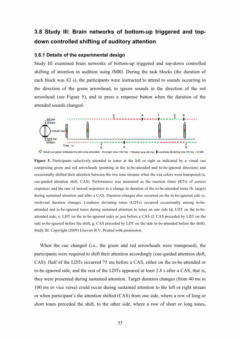
3.8 Study III: Brain networks of bottom-up triggered and top-
down controlled shifting of auditory attention
3.8.1 Details of the experimental design Study III examined brain networks of bottom-up triggered and top-down controlled
shifting of attention in audition using fMRI. During the task blocks (the duration of
each block was 82 s), the participants were instructed to attend to sounds occurring in
the direction of the green arrowhead, to ignore sounds in the direction of the red
arrowhead (see Figure 5), and to press a response button when the duration of the
attended sounds changed.
Figure 5. Participants selectively attended to tones at the left or right as indicated by a visual cue
comprising green and red arrowheads (pointing in the to-be-attended and to-be-ignored direction) and
occasionally shifted their attention between the two tone streams when the cue colors were transposed (a,
cue-guided attention shift, CAS). Performance was measured as the reaction times (RTs) of correct
responses and the rate of missed responses to a change in duration of the to-be-attended tones (b, target)
during sustained attention and after a CAS. Duration changes also occurred on the to-be-ignored side (c,
irrelevant duration change). Loudness deviating tones (LDTs) occurred occasionally among to-be-
attended and to-be-ignored tones during sustained attention to tones on one side (d, LDT on the to-be-
attended side; e, LDT on the to-be-ignored side) or just before a CAS (f, CAS preceded by LDT on the
side to-be-ignored before the shift; g, CAS preceded by LDT on the side to-be-attended before the shift).
Study III. Copyright (2009) Elsevier B.V. Printed with permission.
When the cue changed (i.e., the green and red arrowheads were transposed), the
participants were required to shift their attention accordingly (cue-guided attention shift,
CAS). Half of the LDTs occurred 75 ms before a CAS, either on the to-be-attended or
to-be-ignored side, and the rest of the LDTs appeared at least 2.8 s after a CAS; that is,
they were presented during sustained attention. Target duration changes (from 40 ms to
100 ms or vice versa) could occur during sustained attention to the left or right stream
or when participant’s the attention shifted (CAS) from one side, where a row of long or
short tones preceded the shift, to the other side, where a row of short or long tones,

34
respectively, occurred. Half of the LDTs in the to-be-attended or to-be-ignored stream
during sustained attention to one ear, half of the pre-shift LDTs in the to-be-ignored
stream and in the to-be-attended stream before the shift, and half of the CASs without
LDTs were followed by a target to examine the effect of LDT and CAS on reaction
times (RTs) to the targets and the rate of missed responses (Miss%, percentage of
missed responses to the targets).
3.8.2 Results Task performance. During sustained-attention with no LDTs the mean RT to targets
was 1161 ms (s.e.m. 47 ms) and the mean miss rate was 5.1% (s.e.m. 1.6%). RTs to the
targets decreased (paired samples t-test, t(18) = 6.8, p < 0.0001) when a target was
preceded by an LDT in the same to-be-attended tone stream, but increased (t(18) = 2.9,
p < 0.01) when a target was preceded by an LDT in the to-be-ignored stream, as
compared with targets during the sustained attention preceded by no LDT. LDTs in the
to-be-ignored stream also raised the miss rate (t(18) = 2.9, p < 0.01) for the subsequent
targets as compared to those during sustained attention preceded by no LDT. For targets
preceded by a CAS with no preceding LDT, the mean RT was 1297 ms (s.e.m. 57 ms),
which was significantly longer (t(18) = 3.9, p < 0.001) than for targets not preceded by
an LDT during sustained attention. RTs decreased significantly (t(18) = 3.7, p < 0.01)
for targets preceded by a CAS, when the CAS was preceded by an LDT in the to-be-
ignored stream before the CAS and attended after the CAS as compared with targets
following a CAS preceded by no LDT.
Brain activity associated with bottom-up triggered and top-down controlled shifts of
attention. Comparison of brain activity associated with these LDT events to brain
activity associated with sustained attention without LDTs revealed LDT-related activity
in the right TPJ, ventromedial parts of the SPL, posterior parts of the left IFG/MFG, and
left FEF/premotor cortex (PMC; Figure 6, top). Comparisons of brain activity following
CASs with activity during sustained attention revealed CAS-related activity in the
bilateral IPS/SPL, FEF/PMC, TPJ, IFG/MFG, cingulate/medial frontal gyrus
(CG/medFG), lateral and ventromedial occipital cortex (OC), and crus I/II of the
posterior cerebellum (Post. Cb; center). Thus, both LDTs and CASs activated the
ventromedial SPL, left IFG/MFG, left FEF/PMC, and right TPJ. Analysis of activation
time-series (Figure 6, bottom) suggested that activity in the right SPL (F(1,18) = 8.6, p
< 0.01) and posterior cerebellum (F(1,18) = 6.9, p < 0.05) ROI was lower when a CAS

35
was preceded by an LDT in the to-be-attended stream before the CAS (green line) as
compared to CASs with no preceding LDT (white line), as indicated by repeated-
measures ANOVA for mean percentual signal changes with factors Event type (CAS
with no LDT, and CAS preceded by an LDT in the to-be-attended stream) and Time
from the event onset (4–6, 6–8, and 8–10 s).
Figure 6. Top: Bottom-up influences on brain activity caused by LDTs during auditory sustained
attention (blue, Z > 3.0, p < 0.01). Middle: Top-down controlled CAS contrasted with auditory sustained
attention (red, Z > 5.0, p < 0.01, and yellow, Z > 3.0, p < 0.01). Bottom: Activation time-series in the
right SPL, right TPJ and posterior cerebellum (Post. Cb) ROIs during different events. Note that the top
middle figure shows activity at the posterior temporal cortex. Below the brain surface, however, this
activity cluster reached TPJ. Study III. Copyright (2009) Elsevier B.V. Printed with permission.
The effect of locus of attention on bottom-up modulations of brain activity during
sustained attention and attention shifting. Brain activity associated with LDTs in the to-
be-ignored stream was higher (Z > 1.64, cluster corrected p < 0.05) than activity
associated with the to-be-attended stream in the ventromedial prefrontal cortex
(VMPFC) both during sustained attention and when followed by a CAS (a conjunction
of the to-be-ignored vs. to-be-attended LDT contrasts analyzed separately for LDTs
occurring during sustained attention and for LDTs preceding a CAS).

36
Brain activations associated with duration changes in the to-be-attended and to-be-
ignored stream during sustained attention. To reveal activity specifically related to the
processing of target duration changes occurring in the to-be-attended stream and the
processing of similar changes in the to-be-ignored stream, activations were analyzed
separately for these changes when they occurred with no adjacent LDT or CAS. These
target duration changes activated (Z > 5.0, cluster corrected p < 0.01) distributed areas
of the superior temporal cortex as compared to sustained auditory attention (Figure 7,
top, red/yellow). Outside the temporal cortex, target duration changes activated areas of
the posterior IFG/MFG, IPS, CG/medFG, left precentral gyrus, and FEF/PMC,
bilaterally (top, red/yellow). Activity in the left precentral, central, and postcentral
gyrus (contralateral to the hand of response), right IFG and CG/medFG was observed
when target duration changes were compared to duration changes in the to-be-ignored
stream (Z > 3.0, cluster corrected p < 0.01, bottom, red). Duration changes in the to-be-
ignored stream activated (Z > 5.0, cluster corrected p < 0.01) areas of STG, IPS,
CG/midFG, TPJ, posterior IFG/MFG, and thalamus when they were compared to
auditory sustained attention (Figure 7, top, green/yellow). Comparison of brain activity
to duration changes in the to-be-ignored and to-be-attended streams, in turn, revealed
activation (Z > 3.0, cluster corrected p < 0.01) in SPL and FEF/PMC bilaterally (Figure
7, bottom, green).
Thus, the present results suggest that bottom-up triggered and top-down controlled
auditory attention activates largely overlapping brain areas that correspond to the dorsal
and ventral attention systems in vision (Corbetta and Shulman, 2002), as well as in
CG/midFG, crus I/II of the posterior cerebellum, and VMPFC. The finding that IPS and
superior parts of SPL are activated specifically by top-down controlled attention,
whereas the ventromedial SPL is activated by LDTs and to-be-ignored duration
changes, suggests that different areas of SPL may play different roles in auditory
attention.

37
Figure 7. Areas activated (top, Z > 5.0, cluster corrected p < 0.01; bottom, Z > 3.0, cluster corrected p <
0.01) by duration changes in the to-be-attended (red) and to-be-ignored (green) tone streams during
sustained attention as compared to sustained attention with no duration changes (top) or with each other
(bottom). Areas that overlap between to-be-ignored and to-be-attended duration changes appear in
yellow. The section at the top right is from the medial surface, and the white horizontal line at the bottom
left marks the level of the section shown at the bottom right. Study III. Copyright (2009) Elsevier B.V.
Printed with permission.
3.9 Study IV: Cognitive and motor loops of the human cerebro-
cerebellar system activated by an auditory working memory
task and sensory-motor task
3.9.1 Details of the experimental design
Study IV used n-back tasks (Braver et al., 1997) during fMRI to examine the brain
networks involved in auditory working memory, and specifically the role of the
cerebellum in these networks. The participants pressed one of two buttons in response
to each sound. In the 1-back task, the participants pressed the left button when a sound
was the same as the previous sound (1-back target), and in the 2-back task, when the
sound was the same as the one that occurred two sounds back (2-back target). If the
sounds were different, they pressed the right button (non-target). In the sensory-motor

38
task, the participants pressed the right button for each sound. Each block began with a
4-s centrally presented written instruction indicating the task. The duration of each task
block was 73.2 s (containing 20 trials) followed by an 18.3-s period during which the
fixation cross was presented with no sounds. Twelve blocks for each task condition
were presented in staircase order (e.g., …sensory-motor, 1-back, 2-back, 2-back, 1-
back, sensory-motor, sensory-motor, 1-back, 2-back…). The n-back tasks differed only
with respect to the working memory load.
3.9.2. Results
Figure 8 shows the behavioral results of Study IV. An increase in the cognitive load
from the sensory-motor task to the 1-back task and further to the 2-back task resulted in
longer RTs and higher rates of incorrect responses (IR).
Figure 8. Task performance. An increase in
cognitive resulted in longer reaction times (RT,
red) and higher rates of incorrect responses (IR,
blue). SM = sensory-motor task. *p < 0.05, **p
< 0.01, ***p < 0.001.
fMRI results. The across-participants studied 2-back vs. 1-back contrast showed
memory-load-dependent activity bilaterally in the IPS/SPL, dPMC, pre-supplementary
motor area (preSMA), lateral PFC, basal ganglia, and crus I/II (Figure 9). Moreover,
unilateral load-dependent activity was observed in the right lobule VIIB and left lobule
VIII of the cerebellum. The contrast between the sensory-motor task and the rest
periods revealed activity (Z > 2.5, uncorrected) bilaterally in the motor/somatosensory
cortices (MC/SC), medial PFC, STG, posterior cingulate gyrus (PCG), and lobules
V/VI of the right anterior cerebellum, as revealed by across-participants analysis.

39
Figure 9. Cognitive-load-dependent (red clusters, 2-back vs. 1-back, Z > 3.1, cluster corrected p < 0.01)
and sensory-motor (blue clusters, sensory-motor task vs. rest, Z > 2.5, uncorrected) cerebro-cerebellar
activity networks across all participants (N = 10). Color-coded vertical lines mark the level of the sections
of the images at the bottom. lPFC, lateral prefrontal cortex; dPMC, dorsal premotor cortex; MC/SC,
motor/somatosensory cortex; IPS, intraparietal sulcus; STG, superior temporal gyrus; PCG, posterior
cingulate gyrus; mPFC, medial prefrontal cortex; SPL, superior parietal lobule; BG, basal ganglia;
MedFG, medial frontal gyrus. L = left.
Tractography results. Analysis using the activated cerebellar areas as starting points
and pons as the endpoint suggested that the tracts between the pons and the cerebellar
areas activated by the cognitive load, especially crus I/II, project mainly via rostral areas
of the basilar pons (Figure 10a), whereas the tracts between the pons and cerebellar
lobule V/VI that showed sensory-motor activity project mainly via the more caudal
nuclei of the pons (Figure 10a).
After determining the subareas of the pons with the highest probability of
connectivity to the cerebellum, it was examined which cerebral areas are linked to the
activated cerebellar areas via the pons. Probabilistic tractography using crus I/II as a
starting point showed tracts that linked the anterior PFC (aPFC), dPMC, M1, SC, and
SPL with the cerebellum. When lobule VIIB served as a starting point, tracts connecting
lobule VIIB with the aPFC, M1, and dPMC were observed. The lobule V/VI area that
was activated by the sensory-motor task showed tracts with the M1 and dPMC. Each of

40
the observed pontine bundles projected via the capsula interna, cerebral peduncle, and
dentate nucleus.
Analysis using the cerebellar activity clusters as starting points and the thalamus as
an endpoint suggested that the tracts from the activated area in crus I/II were connected
mainly to the mediodorsal thalamic nuclei (Figure 10b). Activated areas in lobules V/VI
showed tracts connecting these areas mainly with the ventral posterior lateral thalamic
nuclei (Figure 10b). Tractography analysis using the activated area in the lobule VIIB as
a starting point suggested minor tracts connecting lobule VIIB with the mediodorsal
thalamic nuclei, while the activated area in lobule VIII did not have tracts with thalamic
endpoints. Crus I/II and lobule VIIB showed tracts connecting these areas via the
thalamus with widely distributed areas of the cerebral cortex, including the PFC, SPL,
dPMC, and M1. Corresponding analysis using the V/VI activity cluster as a starting
point, in turn, revealed tracts connecting only lobule V/VI with the MC/SC, and SPL.
The observed cerebello-thalamo-cortical tracts projected from the dentate nucleus to the
superior cerebellar peduncles and red nucleus.
Figure 10. a) Pontine classification of the cerebro-ponto-cerebellar tracts and b) thalamic classification of the cerebello-thalamo-cerebral tracts across all participants (N = 10, the reference image is the individual “Colin” brain of the Montreal Neurological Institute). The positions of the slices in the z-axis are indicated by the colored lines in the sagittal sections of the brain (slices are in order from top-to-bottom). The endpoints of the tracts traced beginning from the right crus I/II activity cluster appear in red, and the endpoints of the tracts traced from the right V/VI activity cluster appear in blue. Areas that include the endpoints of both the tracts traced from crus I/II and lobules V/VI appear in purple. The figure shows ipsilateral and contralateral tracts traced from the crus I/II activity cluster, and only the ipsilateral tracts traced from the lobule V/VI activity cluster (as we were unable to trace the fibers crossing the hemispheres from the activity cluster in lobules V/VI). CPN, caudal pontine nuclei; RPN, rostral pontine nuclei; MD, mediodorsal thalamic nuclei; VPL, ventral posterior lateral thalamic nuclei; L = left.

41
Correlation between brain activity and performance. Between-participants
analysis showed a negative correlation between the load-dependent change in the
cerebellar activity and RT. In 2-back vs. 1-back tasks, the correlation between the
relative signal change in the left crus I/II and RT was r = -0.735 (p < 0.05), and the
relative signal change in the right crus I/II and RT r = -0.760, (p < 0.05). In 2-back
vs. sensory-motor tasks, the relative signal change/RT correlation was r = -0.540, (p
= 0.107) in the left crus I/II, and r = -0.847, (p < 0.001) in the right crus I/II. Activity
in the anterior cerebellar ROI showed no correlation with performance in any task
condition.
4 Discussion
The present Studies I-IV investigated brain networks involved in attention and
working memory. The top-down controlled orienting of attention was compared to
the maintenance of attention in audition in studies employing fMRI (Study I) and
ERPs (Study II). Study I revealed that the auditory and visual top-down controlled
orienting of attention activates similar brain regions. The multimodal brain network
activated by the orienting of attention consisted of the SPL/IPS, FEF/PMC, crus I/II
of the cerebellum, and the pulvinar nucleus of the thalamus. Modality-specific
differences between the orienting and maintenance of attention tasks were observed
in the inferior parietal and inferior frontal cortices. These areas were more strongly
activated by the auditory tasks than by the visual tasks. Study II further characterized
these auditory and visual attention-related modulations. In audition, the top-down
controlled orienting of attention was associated with stronger ERP effects (i.e., Nds)
than was the maintenance of attention. These Nds were probably generated in the
superior temporal cortex (Giard et al., 1988; Rif et al., 1991; Woldorff et al., 1993)
and were stronger over the hemisphere contralateral to the attended location.
However, ERPs appeared to reflect no orienting-specific activity in the parieto-
fronto-cerebellar network observed with fMRI in Study I. Thus, although ERPs
provide important temporal information on the selection of attended inputs, they may
be less useful for studying the top-down controlled orienting of attention as fMRI. In
Study III, bottom-up triggered and top-down controlled auditory attention was
studied with fMRI. The results suggested that in audition, top-down controlled and
bottom-up triggered attention activates largely overlapping temporo-parietal,

42
superior parietal, and frontal areas. Only IPS, superior parts of SPL, and crus I/II
were activated specifically by top-down attention, and VMPFC was specifically
activated by bottom-up attention. In study IV, the effects of a cognitive load increase
were examined with fMRI in an auditory working memory task. A cognitive load
increase in the auditory working memory task (2-back vs. 1-back) resulted in
enhanced activity in brain networks that overlapped with those activated by top-
down controlled attention in Studies I and III. Study IV also showed that the effect of
increasing the cognitive load on the cerebellar crus I/II showed higher activity for
shorter reaction times. Moreover, the DW-MRI results showed that crus I/II was
connected with those cerebral areas that were also activated by an increase in
cognitive load.
4.1 Top-down controlled shifts of auditory attention
The present results suggest that auditory and visual top-down controlled spatial shifts
of attention activate mainly the same brain networks (Study I; see also Study III).
These results are consistent with those of an earlier neuropsychological study (Farah
et al., 1989) and of brain imaging studies that have separately examined visual or
auditory top-down controlled spatial shifts of attention (Shomstein and Yantis, 2006;
Yantis et al., 2002). It should be noted, however, that auditory and visual top-down
controlled attention shifting may be based on different neuronal populations within
the same brain area and that these differences may be too small to be detected by
neuropsychological tests or non-invasive brain imaging studies. Indeed,
neurophysiological recordings in non-human primates suggest that the superior
parietal cortex in particular contains mainly neurons that respond to unimodal
(visual, somatosensory, or auditory) input (Cohen et al., 2005).
The present results are also in line with those of a wealth of studies conducted in
visual modality, suggesting that a network that includes SPL/IPS and FEF/PMC is
activated by top-down controlled spatial shifts of attention (Corbetta et al., 1995;
Corbetta et al., 2005; Hopfinger et al., 2000; Kim et al., 1999; Kincade et al., 2005;
Peelen et al., 2004; Rosen et al., 1999; Shomstein and Yantis, 2006; Serences and
Yantis, 2007; Vandenberghe et al., 2001; Yantis et al., 2002). In addition to the top-
down controlled orienting of spatial attention, this network is activated by other
types of tasks, such as the shifting of attention between two sensory modalities

43
(Shomstein and Yantis, 2004), the switching of the tasks (Chiu and Yantis, 2009),
and working memory (Corbetta et al., 2002; for further information on the overlap of
brain networks for attention and working memory, see Chapter 4.3). To cover these
divergent findings, Shomstein and Yantis (2006) suggested that this network may be
activated by various types of tasks that require the reconfiguration of attention.
Recent studies have further examined the specific roles of the superior parietal and
superior frontal cortex. These studies suggest that the superior parietal cortex
contributes to the binding of information from different sources, which is needed in
controlling voluntary attention (Gottlieb, 2007), whereas the superior frontal cortex
is involved in the execution of top-down controlled attention shifts (Desmurget et al.,
2009; see also Mesulam, 1981). Future studies, would do well to investigate how
top-down controlled attention works in more naturalistic situations. For example,
studies with cues based on the direction of the another person’s gaze suggest that
neuronal activity in the dorsal attention system is weaker when attention is shifted
according to gaze cues than when it is shifted according to arrow cues (Hietanen et
al., 2006; Hietanen et al., 2008). Thus, it is possible that the brain networks involved
in shifting spatial attention are somewhat differently activated in conditions that are
more naturalistic than those of the present experiments.
In addition to SPL/IPS and FEF/PMC, top-down controlled shifts of spatial
attention in audition also activated other brain areas. The results of Studies I and III
also suggest that top-down controlled attention in audition activate TPJ and
IFG/MFG as well. These results are consistent with those of previous studies
reporting activity in these areas during top-down controlled attention or active sound
localization (Alain et al., 2008; Alho et al., 1999; Maeder et al., 2001). However,
these findings are discrepant with the model of Corbetta and Shulman (2002), thus
suggesting that TPJ and IFG/MFG are specifically involved in bottom-up triggered
attention. The discrepancy may be due to differences between auditory and visual
attention or between the auditory and visual experimental designs, or both. Still,
some visual studies report activity in TPJ and IFG/MFG during top-down controlled
attention, also (Peelen et al., 2004; Rosen et al., 1999). Finally, Studies I and III
suggested that the posterior cerebellum is activated by top-down controlled auditory
attention. These results are consistent with those of previous studies reporting on
cerebellar activity in attention shifting tasks (Allen et al., 1997; Le et al., 1998).
However, many studies of visual top-down controlled attention using the cue

44
paradigm have observed no activity in the posterior cerebellum (e.g., Corbetta et al.,
2000; Hopfinger et al., 2000; Kim et al., 1999; Kincade et al., 2005; Peelen et al.,
2004; Rosen et al., 1999). The activity of the cerebellum may not have been
observed in these studies because they used a trial-by-trial design and because the
cerebellum may have shown typically more sustained modulatory activity (Leiner et
al., 1991). Study IV further examined the anatomical and functional role of the
cerebellum in cognitive processing (see 4.3).
Although strong effects were found in Study I using fMRI, Study II showed no
activity specifically associated with top-down controlled attention. This suggests that
attention-related ERPs in this paradigm are not strongly affected by activations of the
parieto-frontal network associated with top-down controlled attention. Early negative
and late positive ERP components associated with top-down controlled attention
shifts in earlier studies (Harter et al., 1989; Hopf and Mangun, 2000; Nobre et al.,
2000) were observed in both the orienting of attention and the maintenance of
attention conditions in Study II. Therefore, even in conditions demanding top-down
controlled orienting attention, the effects of attention on ERPs may be associated
with selection of the attended input for further processing after an attention shift
rather than with the actual attention shifting. Based on these results, the cue
paradigm seems rather limited in ERP studies of top-down controlled attention.
4.2 Bottom-up triggered auditory attention
Study III suggested that auditory change detection and subsequent bottom-up
triggered attention activate a brain network that includes TPJ, posterior parts of
IFG/MFG, ventromedial parts of SPL, and FEF/PMC. Based on the findings of
Studies I and III, none of these regions are activated by bottom-up triggered
attention, but also by top-down controlled attention alone. Earlier studies have
repeatedly reported the involvement of TPJ and posterior parts of the IFG/MFG in
auditory change detection and bottom-up triggered attention (Baudena et al., 1995;
Halgren et al., 1995a,b; Knight et al., 1989; Molholm et al., 2005; Woods et al.,
1992). Consistent with the present findings, some previous studies have suggested
that auditory change detection and bottom-up triggered attention also activate SPL
and FEF/PMC (Rinne et al., 2007; Watkins et al., 2006). The activity of SPL and
FEF/PMC in Study III, as well as in earlier studies by Watkins and colleagues (2006)
45
and Rinne and colleagues (2007), may be partially explained by the voluntary
orienting of attention back to the task after temporary distraction.
In contrast to previous fMRI studies of auditory bottom-up triggered attention,
Study III made a distinction between bottom-up modulations in the to-be-ignored
and to-be-attended streams. LDTs in the to-be-ignored tone stream were expected to
draw attention to this stream, so comparison between LTDs in the to-be-ignored and
to-be-attended streams should reflect bottom-up triggered spatial attention shifting.
This comparison revealed no changes in the activity of TPJ and IFG/MFG, which
may be involved in the bottom-up triggered shifting of spatial attention (Corbetta and
Shulman, 2002). Based on the results of Study III, one could propose that, at least in
audition, TPJ and IFG/MFG are involved in the detection and processing of changes
and not specifically in bottom-up triggered spatial attention shifting. Furthermore,
these same results suggest that the relevancy of TPJ and IFG/MFG are not
significantly modulated by the relevancy of the event that triggers attention.
Comparison of brain activity associated with LTDs in the to-be-ignored and to-be-
attended LDTs did, however, reveal activity in VMPFC. Previous studies have
suggested that VMPFC is involved in the inhibition of irrelevant stimuli as well as in
releasing this inhibition (Bechara et al., 2000; Rolls, 2000; Rule et al., 2002).
VMPFC activity observed in Study III in the comparison of processing LDTs in the
to-be-ignored and to-be-attended streams may be related to active suppression of the
processing of to-be-ignored LDTs during sustained attention as well as to the release
of this suppression in order to facilitate attention shifting (to-be-ignored LDT
followed by CAS).
4.3 Auditory working memory and attention
Comparison of the results of Study IV with those of Studies I and III suggests that
auditory working memory activates brain networks that largely overlap with those
activated by auditory top-down controlled attention. On the one hand, the same brain
networks may actually be involved in both top-down controlled attention and
working memory, as would be suggested by working memory models by Barouillet
et al. (2004) and Cowan (2001), which claim that attention and working memory are
not distinct but largely overlapping functions. On the other hand, the present
experimental designs may have been unable to distinguish between attention and

46
working memory. Because Study IV used a block design that was not intended to
control for attention, it is especially likely that the working memory tasks used in this
study required top-down controlled attention. Studies using parametric design for
manipulating the load of attention and working memory separately would probably
help to clarify this issue. A recent study with such an experimental design suggested
that at least in the auditory cortex, attention and working memory activates distinct
areas (Rinne et al., 2009).
Study IV in particular investigated the role of the cerebellum in working memory.
Our results suggest that working memory load increase and sensory-motor
processing activate distinct cerebro-cerebellar networks. In keeping with previous
studies that used n-back tasks (Carlson et al., 1999; Martinkauppi et al., 2000; Rämä
et al., 2001), an increase in working memory load activated IPS/SPL, FEF/PMC, and
various areas of the prefrontal cortex. In addition, as some previous working memory
studies have reported (Chen et al., 2005a,b; Desmond et al., 1997; Hayter et al.,
2007; Kirchen et al., 2005), activations in the posterior cerebellum are related to
working memory load. Study IV further showed that the load-dependent activity in
the cerebellar crus I/II correlated with shorter reaction times. This finding is in
agreement with those of previous studies that reported longer reaction times after
temporary (Desmond et al., 2005) or permanent (Nixon and Passingham, 2000;
Townsend et al., 1999) cerebellar dysfunction.
Consistent with previous studies (Allen et al., 1997; Desmond et al., 1997), Study
IV showed that cognitive processing and sensory-motor processing activate specific
regions of the cerebellum. These activated regions served as starting points for
tractography analysis in examining the segregation of the cognitive and motor loops
of the cerebro-cerebellar system, previously studied in non-human primates (see
Middleton and Strick, 2000). In line with studies of non-human primates (Brodal,
1978; Kelly and Strick, 2003; Middleton and Strick, 2000; Schmahmann, 1996),
Study IV showed that the cerebellar areas activated by cognitive and motor
processing are connected with distinct cerebro-cortical regions via specific pontine
and thalamic nuclei. Comparison of the fMRI results of Study IV on auditory
working memory to those of Studies I and III on top-down controlled attention
indicate that the present working memory tasks and attention-shifting tasks activated
largely overlapping areas in the posterior cerebellum. The cerebellar activity in
working memory tasks may therefore be involved in attention.

47
4.4 Conclusions
Figure 11 summarizes the main results of Studies I-IV. The figure shows the areas
that were activated by top-down controlled attention, bottom-up triggered attention,
and working memory in audition. Brain networks activated by top-down controlled
attention in audition include the auditory cortex (AC), IPS/SPL, FEF/PMC, DLPFC,
TPJ, IFG/MFG, thalamus (pulvinar), and crus I/II of the posterior cerebellum. As
earlier studies have suggested, the role of the AC in auditory attention is probably
related to the selective enhancement of attended contents (e.g., Petkov et al., 2004).
Auditory working memory activates brain networks that largely overlap with those
brain networks activated by top-down controlled attention. Based on the present
results, the main difference between the brain networks of auditory top-down
controlled attention and auditory working memory is that, in contrast to top-down
controlled attention, working memory showed no activity in TPJ and IFG/MFG. In
addition, working memory tasks did show activity in widespread areas of DLPFC,
whereas top-down controlled attention showed relatively little activity in DLPFC.
Note, however, that due to completely different task designs and different
participants, the results of Studies I and IV should be compared with caution.
Auditory change detection and bottom-up triggered attention activated brain
networks that include IPS/SPL, FEF/PMC, TPJ, IFG/MFG, and VMPFC. Thus, the
DLPFC, thalamus, and crus I/II of the posterior cerebellum were activated by top-
down controlled attention and working memory, but not by bottom-up triggered
attention, whereas VMPFC was specifically activated by bottom-up triggered
attention.

48
Figure 11. A graphical summary of the results of Studies I-IV. The figure shows the areas involved in
top-down controlled attention (red), bottom-up triggered attention (blue), and working memory in
audition (green). Colored lines surrounding the boxes outline the areas involved in specific networks.
IPS/SPL, intraparietal sulcus/superior parietal lobule; FEF/PMC, frontal eye field/premotor cortex;
DLPFC, dorsolateral prefrontal cortex; TPJ, temporoparietal junction; AC, auditory cortex; IFG/MFG
inferior frontal gyrus/middle frontal gyrus; VMPFC, ventromedial prefrontal cortex.
By comparing the results of the present and other recent studies, the roles of these
areas can be evaluated in even greater detail. (1) Present and previous neuroimaging
studies show that IPS/SPL and FEF/PMC are activated by various tasks requiring
attention or working memory (Carlson et al., 1998; Chiu and Yantis, 2009; Corbetta
et al., 2002; Martinkauppi et al., 2000; Shomstein and Yantis, 2004; Shomstein and
Yantis, 2006; Yantis et al., 2002). Studies on attention typically demonstrate rather
good control of working memory processing (e.g., Chiu and Yantis, 2009; Shomstein
and Yantis, 2004; Shomstein and Yantis, 2006; Yantis et al., 2002), whereas studies
of working memory seldom control for attention (e.g., Carlson et al., 1998; Corbetta
et al., 2002; Martinkauppi et al., 2000). Moreover, even some working memory
studies suggest that the activity in these regions may be involved in attention rather
than working memory, as increase in working memory load boost activity in there
regions only marginally (Linden et al., 2003). Based on these results, one could
propose that IPS/SPL and FEF/PMC are involved mainly in attention and not in
working memory. (2) Findings from strictly controlled experiments showing that
DLPFC is seldom activated by top-down controlled attention (Chiu and Yantis,
2009; Shomstein and Yantis, 2006; Yantis et al., 2002) suggest that DLPFC may be
more involved in working memory rather than top-down controlled attention. This
suggestion is supported by neurophysiological studies that propose DLPFC as a key

49
area in working memory (Levy and Goldman-Rakic, 2000). (3) Our results may be
insufficient to claim that TPJ and IFG/MFG are involved in top-down controlled
attention in audition. The results of Studies I and III, which show activity in these
regions during top-down controlled attention, could be attributed to the effect of top-
down controlled attention on brain regions (TPJ and IFG/MFG) that may actually be
involved in bottom-up triggered attention (see Corbetta and Shulman, 2002). Seldom
observed in visual studies, this modulation could be result from stronger dependence
on bottom-up triggered attention in audition that in vision (see Kubovy and van
Valkenburg, 2001; Mayer et al., 2009), for example, because the auditory signal
typically changes more rapidly in time than does the visual signal. (4) The results of
Study III, which show activity in VMPFC associated with loudness increments in the
stream of to-be-ignored tones, together suggest that VMPFC may be involved in
suppression of the processing of irrelevant changes in stimuli as well as in releasing
this suppression (see also Rule et al., 2002). (5) Studies I, III, and IV showed activity
in the posterior cerebellum during auditory top-down controlled attention and
auditory working memory, thus suggesting that the posterior cerebellum is strongly
involved in auditory attention and working memory (see also Petacci et al., 2005).
The results of Study IV further suggest that the posterior cerebellum may contribute
to optimization of the processing speed when the cognitive load increases. In
summary, the results of Studies I-IV show that auditory top-down controlled
attention, auditory bottom-up triggered attention, and auditory working memory
activate distinct but largely overlapping brain networks in the human brain.

50
5 References
Allen, G., Buxton, R. B., Wong, E. C., & Courchesne, E. (1997). Attentional activation of
the cerebellum independent of motor involvement. Science, 275, 1940-1943.
Allen, G. I., Gilbert, P. F., & Yin, T. C. (1978). Convergence of cerebral inputs onto dentate
neurons in monkey. Experimental Brain Research, 32, 151-170.
Attwell, D., & Iadecola, C. (2002). The neural basis of functional brain imaging signals.
Trends in Neurosciences, 25, 621-625.
Awh, E., Vogel, E. K., & Oh, S. H. (2006). Interactions between attention and working
memory. Neuroscience, 28, 201-208.
Baddeley, A.D., & Hitch, G.J.L. (1974). Working Memory, In G.A. Bower (Ed.), The
psychology of learning and motivation: advances in research and theory (Vol. 8, pp. 47-
89). Academic Press, New York.
Barrett, D. J., & Hall, D. A. (2006). Response preferences for "what" and "where" in human
non-primary auditory cortex. NeuroImage, 32, 968-977.
Barrouillet, P., Bernardin, S., Portrat, S., Vergauwe, E., & Camos, V. (2007). Time and
cognitive load in working memory. Journal of Experimental Psychology: Learning,
Memory, and Cognition, 33, 570-585.
Baudena, P., Halgren, E., Heit, G., & Clarke, J. M. (1995). Intracerebral potentials to rare
target and distractor auditory and visual stimuli. III. Frontal cortex.
Electroencephalography and Clinical Neurophysiology, 94, 251-264.
Bechara, A., Tranel, D., & Damasio, H. (2000). Characterization of the decision-making
deficit of patients with ventromedial prefrontal cortex lesions. Brain, 123, 2189-2202.
Behrens, T. E., Woolrich, M. W., Jenkinson, M., Johansen-Berg, H., Nunes, R. G., Clare, S.,
Matthews, P. M., Brady, J. M., & Smith, S. M. (2003). Characterization and propagation
of uncertainty in diffusion-weighted MR imaging. Magnetic Resonance in Medicine, 50,
1077-1088.
Braver, T. S., Cohen, J. D., Nyström, L. E., Jonides, J., Smith, E. E., & Noll, D. C. (1997). A
parametric study of prefrontal cortex involvement in human working memory.
NeuroImage, 5, 49-62.
Broadbent, D. E. (1958). Perception and Communication. Pergamon Press, New York.
Brodal, P. (1978). The corticopontine projection in the rhesus monkey. origin and principles
of organization. Brain, 101, 251-283.
Brugge, J. F., Reale, R. A., Jenison, R. L., & Schnupp, J. (2001). Auditory cortical spatial
receptive fields. Audiology and Neuro-Otology, 6, 173-177.
Carlson, S., Martinkauppi, S., Rämä, P., Salli, E., Korvenoja, A., & Aronen, H. J. (1998).
Distribution of cortical activation during visuospatial n-back tasks as revealed by

51
functional magnetic resonance imaging. Cerebral Cortex, 8, 743-752.
Chen, S. H., & Desmond, J. E. (2005a). Cerebrocerebellar networks during articulatory
rehearsal and verbal working memory tasks. NeuroImage, 24, 332-338.
Chen, S. H., & Desmond, J. E. (2005b). Temporal dynamics of cerebro-cerebellar network
recruitment during a cognitive task. Neuropsychologia, 43, 1227-1237.
Cherry, E. C. (1953). Some experiments on the recognition of speech, with one and with two
ears. The Journal of the Acoustical Society of America, 25, 975-979.
Chiu, Y. C., & Yantis, S. (2009). A domain-independent source of cognitive control for task
sets: Shifting spatial attention and switching categorization rules. The Journal of
Neuroscience, 29, 3930-3938.
Cohen, Y. E., Russ, B. E., & Gifford, G. W.,3rd. (2005). Auditory processing in the posterior
parietal cortex. Behavioral and Cognitive Neuroscience Reviews, 4, 218-231.
Corbetta, M., Kincade, J. M., Ollinger, J. M., McAvoy, M. P., & Shulman, G. L. (2000).
Voluntary orienting is dissociated from target detection in human posterior parietal
cortex. Nature Neuroscience, 3, 292-297.
Corbetta, M., Miezin, F. M., Dobmeyer, S., Shulman, G. L., & Petersen, S. E. (1990).
Attentional modulation of neural processing of shape, color, and velocity in humans.
Science, 248, 1556-1559.
Corbetta, M., Miezin, F. M., Shulman, G. L., & Petersen, S. E. (1993). A PET study of
visuospatial attention. Journal of Neuroscience, 13, 1202-1226.
Corbetta, M., & Shulman, G. L. (2002). Control of goal-directed and stimulus-driven
attention in the brain. Nature Reviews Neuroscience, 3, 201-215.
Corbetta, M., Shulman, G. L., Miezin, F. M., & Petersen, S. E. (1995). Superior parietal
cortex activation during spatial attention shifts and visual feature conjunction. Science,
270, 802-805.
Cowan, N. (2001). The magical number 4 in short-term memory: A reconsideration of
mental storage capacity. The Behavioral and Brain Sciences, 24, 87-114.
Cowan, N., & Morey, C. C. (2006). Visual working memory depends on attentional filtering.
Trends in Cognitive Sciences, 10, 139-141.
Degerman, A., Rinne, T., Salmi, J., Salonen, O., & Alho, K. (2006). Selective attention to
sound location or pitch studied with fMRI. Brain Research, 1077, 123-134.
Degerman, A., Rinne, T., Särkkä, A. K., Salmi, J., & Alho, K. (2008). Selective attention to
sound location or pitch studied with event-related brain potentials and magnetic fields.
The European Journal of Neuroscience, 27, 3329-3341.
Desmond, J. E., Chen, S. H., & Shieh, P. B. (2005). Cerebellar transcranial magnetic
stimulation impairs verbal working memory. Annals of Neurology, 58, 553-560.
Desmond, J. E., Gabrieli, J. D., Wagner, A. D., Ginier, B. L., & Glover, G. H. (1997).

52
Lobular patterns of cerebellar activation in verbal working-memory and finger-tapping
tasks as revealed by functional MRI. The Journal of Neuroscience, 17, 9675-9685.
Desmurget, M., Reilly, K. T., Richard, N., Szathmari, A., Mottolese, C., & Sirigu, A. (2009).
Movement intention after parietal cortex stimulation in humans. Science, 324, 811-813.
Deutsch, J. A., & Deutsch, D. (1963). Attention: some theoretical considerations.
Psychological Review, 70, 80–90.
Domínguez-Borràs, J., Trautmann, S. A., Erhard, P., Fehr, T., Herrmann, M., & Escera, C.
(2009). Emotional context enhances auditory novelty processing in superior temporal
gyrus. Cerebral Cortex, 19, 1521-1529.
Farah, M. J., Wong, A. B., Monheit, M. A., & Morrow, L. A. (1989). Parietal lobe
mechanisms of spatial attention: Modality-specific or supramodal? Neuropsychologia, 27,
461-470.
Fettiplace, R., & Fuchs, P. A. (1999). Mechanisms of hair cell tuning. Annual Review of
Physiology, 61, 809-834.
Fritz, J. B., Elhilali, M., David, S. V., & Shamma, S. A. (2007). Auditory attention-focusing
the searchlight on sound. Current Opinion in Neurobiology, 17, 437-455.
Furukawa, S., Xu, L., & Middlebrooks, J. C. (2000). Coding of sound-source location by
ensembles of cortical neurons. The Journal of Neuroscience, 20, 1216-1228.
Fuster, J. M. (1998). Distributed memory for both short and long term. Neurobiology of
Learning and Memory, 70, 268-274.
Giard, M. H., Perrin, F., Pernier, J., & Peronnet, F. (1988). Several attention-related wave
forms in auditory areas: A topographic study. Electroencephalography and Clinical
Neurophysiology, 69, 371-384.
Gitelman, D. R., Nobre, A. C., Parrish, T. B., LaBar, K. S., Kim, Y. H., Meyer, J. R., &
Mesulam, M. (1999). A large-scale distributed network for covert spatial attention:
Further anatomical delineation based on stringent behavioural and cognitive controls.
Brain, 122, 1093-1106.
Goldman, P. S., & Rosvold, H. E. (1970). Localization of function within the dorsolateral
prefrontal cortex of the rhesus monkey. Experimental Neurology, 27, 291-304.
Goldman-Rakic, P. S. (1987). Development of cortical circuitry and cognitive function.
Child Development, 58, 601-622.
Gottlieb, J. (2007). From thought to action: The parietal cortex as a bridge between
perception, action, and cognition. Neuron, 53, 9-16.
Green, J. J., Teder-Sälejärvi, W. A., & McDonald, J. J. (2005). Control mechanisms
mediating shifts of attention in auditory and visual space: A spatio-temporal ERP
analysis. Experimental Brain Research, 166, 358-369.
Halgren, E., Baudena, P., Clarke, J. M., Heit, G., Liegeois, C., Chauvel, P., & Musolino, P.

53
(1995). Intracerebral potentials to rare target and distractor auditory and visual stimuli. I.
Superior temporal plane and parietal lobe. Electroencephalography and Clinical
Neurophysiology, 94, 191-220.
Halgren, E., Baudena, P., Clarke, J. M., Heit, G., Marinkovic, K., Devaux, B., Vignal, J. P.,
& Biraben, A. (1995). Intracerebral potentials to rare target and distractor auditory and
visual stimuli. II. Medial, lateral and posterior temporal lobe. Electroencephalography
and Clinical Neurophysiology, 94, 229-250.
Hansen, J. C., & Hillyard, S. A. (1980). Endogenous brain potentials associated with
selective auditory attention. Electroencephalography and Clinical Neurophysiology, 49,
277-290.
Hari, R., Hämäläinen, M., Kaukoranta, E., Mäkelä, J., Joutsiniemi, S. L., & Tiihonen, J.
(1989). Selective listening modifies activity of the human auditory cortex. Experimental
Brain Research, 74, 463-470.
Harter, M.R., Millter, S.L., Price, N.J., LaLonde, M.E. & Keyes, A.L. (1989). Neural
processes involved in directing attention. Journal of Cognitive Neuroscience, 1, 223-237.
Hayter, A. L., Langdon, D. W., & Ramnani, N. (2007). Cerebellar contributions to working
memory. NeuroImage, 36, 943-954.
Heinze, H. J., Mangun, G. R., Burchert, W., Hinrichs, H., Scholz, M., Münte, T. F., Gös, A.,
Scherg, M., Johannes, S., Hundeshagen, H., et al. (1994). Combined spatial and temporal
imaging of brain activity during visual selective attention in humans. Nature, 372, 543-
546.
von Helmholz, H., (1909). Treatise on Physiological Optics. Voss, Hamburg.
Hietanen, J. K., Leppänen, J. M., Nummenmaa, L., & Astikainen, P. (2008). Visuospatial
attention shifts by gaze and arrow cues: An ERP study. Brain Research, 1215, 123-136.
Hietanen, J. K., Nummenmaa, L., Nyman, M. J., Parkkola, R., & Hämäläinen, H. (2006).
Automatic attention orienting by social and symbolic cues activates different neural
networks: An fMRI study. NeuroImage, 33, 406-413.
Hillyard, S. A., Hink, R. F., Schwent, V. L., & Picton, T. W. (1973). Electrical signs of
selective attention in the human brain. Science, 182, 177-180.
Hopf, J. M., Luck, S. J., Girelli, M., Hagner, T., Mangun, G. R., Scheich, & Heinze, H. J.
(2000). Neural sources of focused attention in visual search. Cerebral Cortex, 10, 1233-
1241.
Hopf, J. M., & Mangun, G. R. (2000). Shifting visual attention in space: An
electrophysiological analysis using high spatial resolution mapping. Clinical
Neurophysiology, 111, 1241-1257.
Hopfinger, J. B., Buonocore, M. H., & Mangun, G. R. (2000). The neural mechanisms of
top-down attentional control. Nature Neuroscience, 3, 284-291.

54
Hugdahl, K., Wester, K., & Asbjornsen, A. (1991). Auditory neglect after right frontal lobe
and right pulvinar thalamic lesions. Brain and Language, 41, 465-473.
Ito, M. (2002). Historical review of the significance of the cerebellum and the role of
Purkinje cells in motor learning. Annals of the New York Academy of Sciences, 978, 273-
288.
Jissendi, P., Baudry, S., & Baleriaux, D. (2008). Diffusion tensor imaging (DTI) and
tractography of the cerebellar projections to prefrontal and posterior parietal cortices: A
study at 3T. Journal of Neuroradiology, 35, 42-50.
Johnston, W., & Heinz, S. (1978). Flexibility and capacity demands of attention. Journal of
Experimental Psychology: General, 107,420-435.
Kaas, J. H., & Hackett, T. A. (1998). Subdivisions of auditory cortex and levels of
processing in primates. Audiology and Neuro-Otology, 3, 73-85.
Kaas, J. H., & Hackett, T. A. (1999). 'What' and 'where' processing in auditory cortex.
Nature Neuroscience, 2, 1045-1047.
Kastner, S., De Weerd, P., Desimone, R., & Ungerleider, L. G. (1998). Mechanisms of
directed attention in the human extrastriate cortex as revealed by functional MRI.
Science, 282, 108-111.
Kelly, R. M., & Strick, P. L. (2003). Cerebellar loops with motor cortex and prefrontal
cortex of a nonhuman primate. The Journal of Neuroscience, 23, 8432-8444.
Kim, Y. H., Gitelman, D. R., Nobre, A. C., Parrish, T. B., LaBar, K. S., & Mesulam, M. M.
(1999). The large-scale neural network for spatial attention displays multifunctional
overlap but differential asymmetry. NeuroImage, 9, 269-277.
Kincade, J. M., Abrams, R. A., Astafiev, S. V., Shulman, G. L., & Corbetta, M. (2005). An
event-related functional magnetic resonance imaging study of voluntary and stimulus-
driven orienting of attention. The Journal of Neuroscience, 25, 4593-4604.
Kirschen, M. P., Chen, S. H., Schraedley-Desmond, P., & Desmond, J. E. (2005). Load- and
practice-dependent increases in cerebro-cerebellar activation in verbal working memory:
An fMRI study. NeuroImage, 24, 462-472.
Knight, R. T., Scabini, D., Woods, D. L., & Clayworth, C. C. (1989). Contributions of
temporal-parietal junction to the human auditory P3. Brain Research, 502, 109-116.
Krumbholz, K., Eickhoff, S. B., & Fink, G. R. (2007). Feature- and object-based attentional
modulation in the human auditory "where" pathway. Journal of Cognitive Neuroscience,
19, 1721-1733.
Kubovy, M., & Van Valkenburg, D. (2001). Auditory and visual objects. Cognition, 80, 97-
126.
LaBerge, D., & Buchsbaum, M. S. (1990). Positron emission tomographic measurements of
pulvinar activity during an attention task. The Journal of Neuroscience, 10, 613-619.

55
Lachter, J., Forster, K. I., & Ruthruff, E. (2004). Forty-five years after broadbent (1958):
Still no identification without attention. Psychological Review, 111, 880-913.
Le, T. H., Pardo, J. V., & Hu, X. (1998). 4 T-fMRI study of nonspatial shifting of selective
attention: Cerebellar and parietal contributions. Journal of Neurophysiology, 79, 1535-
1548.
Lee, C. C., Schreiner, C. E., Imaizumi, K., & Winer, J. A. (2004). Tonotopic and heterotopic
projection systems in physiologically defined auditory cortex. Neuroscience, 128, 871-
887.
Leiner, H. C., Leiner, A. L., & Dow, R. S. (1991). The human cerebro-cerebellar system: its
computing, cognitive, and language skills. Behavioral and Brain Research, 29, 113-128.
Levy, R., & Goldman-Rakic, P. S. (2000). Segregation of working memory functions within
the dorsolateral prefrontal cortex. Experimental Brain Research, 133, 23-32.
Linden, D. E., Bittner, R. A., Muckli, L., Waltz, J. A., Kriegeskorte, N., Goebel, R., Singer,
W., & Munk, M. H. (2003). Cortical capacity constraints for visual working memory:
Dissociation of fMRI load effects in a fronto-parietal network. NeuroImage, 20, 1518-
1530.
Logothetis, N. K., Pauls, J., Augath, M., Trinath, T., & Oeltermann, A. (2001).
Neurophysiological investigation of the basis of the fMRI signal. Nature, 412, 150-157.
Lomber, S. G., & Malhotra, S. (2008). Double dissociation of 'what' and 'where' processing
in auditory cortex. Nature Neuroscience, 11, 609-616.
Maeder, P. P., Meuli, R. A., Adriani, M., Bellmann, A., Fornari, E., Thiran, J. P., Pittet, A.,
& Clarke, S. (2001). Distinct pathways involved in sound recognition and localization: A
human fMRI study. NeuroImage, 14, 802-816.
Mangun, G. R., Buonocore, M. H., Girelli, M., & Jha, A. P. (1998). ERP and fMRI measures
of visual spatial selective attention. Human Brain Mapping, 6, 383-389.
Mansfield, P., Maudsley, A.A. (1977) Medical imaging by NMR. British Journal of
Radiology, 50, 188-194.
Martinez, A., Teder-Sälejärvi, W., Vazquez, M., Molholm, S., Foxe, J. J., Javitt, D. C., Di
Russo, F., Worden, M. S., & Hillyard, S. A. (2006). Objects are highlighted by spatial
attention. Journal of Cognitive Neuroscience, 18, 298-310.
Martinkauppi, S., Rämä, P., Aronen, H. J., Korvenoja, A., & Carlson, S. (2000). Working
memory of auditory localization. Cerebral Cortex, 10, 889-898.
Mayer, A. R., Franco, A. R., & Harrington, D. L. (2009). Neuronal modulation of auditory
attention by informative and uninformative spatial cues. Human Brain Mapping, 30,
1652-1666.
McNab, F., & Klingberg, T. (2008). Prefrontal cortex and basal ganglia control access to
working memory. Nature Neuroscience, 11, 103-107.

56
Merzenich, M. M., & Brugge, J. F. (1973). Representation of the cochlear partition of the
superior temporal plane of the macaque monkey. Brain Research, 28, 275-296.
Mesulam, M. M. (1981). A cortical network for directed attention and unilateral neglect.
Annals of Neurology, 10, 309-325.
Middleton, F. A., & Strick, P. L. (1994). Anatomical evidence for cerebellar and basal
ganglia involvement in higher cognitive function. Science, 266, 458-461.
Middleton, F. A., & Strick, P. L. (2000). Basal ganglia and cerebellar loops: Motor and
cognitive circuits. Brain Research Reviews, 31, 236-250.
Miller, G. A. (1956). The magical number seven, plus or minus two: Some limits on our
capacity for processing information. Psychological Review, 63, 81-97.
Miller, G.A., Galanter, E. & Pribram, K.H. (1960). Plans and the Structure of Behavior.
Holt, Rinehart & Winston, New York.
Milner, B. (1982). Some cognitive effects of frontal-lobe lesions in man. Philosophical
Transactions of the Royal Society of London. Series B, 298, 211-226.
Molholm, S., Martinez, A., Ritter, W., Javitt, D. C., & Foxe, J. J. (2005). The neural circuitry
of pre-attentive auditory change-detection: An fMRI study of pitch and duration
mismatch negativity generators. Cerebral Cortex, 15, 545-551.
Moran, J., & Desimone, R. (1985). Selective attention gates visual processing in the
extrastriate cortex. Science, 23, 782-784.
Mori, S., & van Zijl, P. C. (2002). Fiber tracking: Principles and strategies - a technical
review. NMR in Biomedicine, 15, 468-480.
Müller, N. G., & Knight, R. T. (2006). The functional neuroanatomy of working memory:
Contributions of human brain lesion studies. Neuroscience, 139(1), 51-58.
Näätänen, R. (1975). Selective attention and evoked potentials in humans--a critical review.
Biological Psychology, 2, 237-307.
Näätänen, R., Paavilainen, P., Rinne, T., & Alho, K. (2007). The mismatch negativity
(MMN) in basic research of central auditory processing: A review. Clinical
Neurophysiology, 118, 2544-2590.
Nixon, P. D., & Passingham, R. E. (2000). The cerebellum and cognition: Cerebellar lesions
impair sequence learning but not conditional visuomotor learning in monkeys.
Neuropsychologia, 38, 1054-1072.
Nobre, A. C., Sebestyen, G. N., & Miniussi, C. (2000). The dynamics of shifting visuospatial
attention revealed by event-related potentials. Neuropsychologia, 38, 964-974.
Noesselt, T., Hillyard, S. A., Woldorff, M. G., Schoenfeld, A., Hagner, T., Jäncke, L.,
Tempelmann, C., Hinrichs, H., & Heinze, H. J. (2002). Delayed striate cortical activation
during spatial attention. Neuron, 35, 575-587.
O’Leary, D. S., Andreasen, N. C., Hurtig, R. R., Torres, I. J., Flashman, L. A., Kesler, M. L.,

57
Arndt, S. V., Cizadlo, T. J., Ponto, L. L. B., Watkins, G. L. & Hichwa, R. D. (1997).
Auditory and visual attention assessed with PET. Human Brain Mapping, 5, 422-436.
Ogawa, S., Lee, T. M., Kay, A. R., & Tank, D. W. (1990). Brain magnetic resonance
imaging with contrast dependent on blood oxygenation. Proceedings of the National
Academy of Sciences of the United States of America, 87, 9868-9872.
Peelen, M. V., Heslenfeld, D. J., & Theeuwes, J. (2004). Endogenous and exogenous
attention shifts are mediated by the same large-scale neural network. NeuroImage, 22,
822-830.
Petacchi, A., Laird, A. R., Fox, P. T., & Bower, J. M. (2005). Cerebellum and auditory
function: An ALE meta-analysis of functional neuroimaging studies. Human Brain
Mapping, 25, 118-128.
Petkov, C. I., Kang, X., Alho, K., Bertrand, O., Yund, E. W., & Woods, D. L. (2004).
Attentional modulation of human auditory cortex. Nature Neuroscience, 7, 658-663.
Picton, T. W., Lins, O. G., & Scherg, M. (1995). The recording and analysis of event-related
potentials. In Boller, F., & Grafman, J. (eds), Handbook of Neuropsychology. Elsevier
Science B. V., Amsterdam, pp. 3-73.
Posner, M. I. (1980). Orienting of attention. The Journal of Experimental Psychology, 32, 3-
25.
Posner, M. I., & Rothbart, M. K. (2007). Research on attention networks as a model for the
integration of psychological science. Annual Review of Psychology, 58, 1-23.
Posner, M. I., Walker, J. A., Friedrich, F. J., & Rafal, R. D. (1984). Effects of parietal injury
on covert orienting of attention. The Journal of Neuroscience, 4, 1863-1874.
Rafal, R. D., & Posner, M. I. (1987). Deficits in human visual spatial attention following
thalamic lesions. Proceedings of the National Academy of Sciences of the United States of
America, 84, 7349-7353.
Rämä, P., Martinkauppi, S., Linnankoski, I., Koivisto, J., Aronen, H. J., & Carlson, S.
(2001). Working memory of identification of emotional vocal expressions: An fMRI
study. NeuroImage, 13, 1090-1101.
Ramnani, N., Behrens, T. E., Johansen-Berg, H., Richter, M. C., Pinsk, M. A., Andersson, J.
L., Rudebeck, P., Ciccarelli, O., Richter, W., Thompson, A. J., Gross, C. G., Robson, M.
D., Kastner, S., & Matthews, P. M. (2006). The evolution of prefrontal inputs to the
cortico-pontine system: Diffusion imaging evidence from macaque monkeys and humans.
Cerebral Cortex, 16, 811-818.
Rauschecker, J. P., & Tian, B. (2000). Mechanisms and streams for processing of "what" and
"where" in auditory cortex. Proceedings of the National Academy of Sciences of the
United States of America, 97, 11800-11806.
Rif, J., Hari, R., Hämäläinen, M. S., & Sams, M. (1991). Auditory attention affects two

58
different areas in the human supratemporal cortex. Electroencephalography and Clinical
Neurophysiology, 79, 464-472.
Rinne, T., Balk, M. H., Koistinen, S., Autti, T., Alho, K., Sams, M. (2008). Auditory
selective attention modulates activation of human inferior colliculus. Journal of
Neurophysiology, 100, 3323-3327.
Rinne, T., Degerman, A., & Alho, K. (2005). Superior temporal and inferior frontal cortices
are activated by infrequent sound duration decrements: An fMRI study. NeuroImage, 26,
66-72.
Rinne, T., Kirjavainen, S., Salonen, O., Degerman, A., Kang, X., Woods, D. L., & Alho, K.
(2007). Distributed cortical networks for focused auditory attention and distraction.
Neuroscience Letters, 416, 247-251.
Rinne, T., Koistinen, S., Salonen, O., & Alho, K. (2009). Task-dependent activations of
human auditory cortex during pitch discrimination and pitch memory tasks. The Journal
of Neuroscience, 21, 13338-13343.
Rolls, E. T. (2000). The orbitofrontal cortex and reward. Cerebral Cortex, 10, 284-294.
Rosen, A. C., Rao, S. M., Caffarra, P., Scaglioni, A., Bobholz, J. A., Woodley, S. J.,
Hammeke, T. A., Cunningham, J. M., Prieto, T. E., & Binder, J. R. (1999). Neural basis
of endogenous and exogenous spatial orienting. A functional MRI study. Journal of
Cognitive Neuroscience, 11, 135-152.
Rugg, M.D., & Coles, M.G.H. (1995). Electrophysiology of Mind: Event-Related Brain
Potentials and Cognition. Oxford University Press, London.
Rule, R. R., Shimamura, A. P., & Knight, R. T. (2002). Orbitofrontal cortex and dynamic
filtering of emotional stimuli. Cognitive, Affective and Behavioral Neuroscience, 2, 264-
270.
Schmahmann, J. D. (1996). From movement to thought: Anatomic substrates of the
cerebellar contribution to cognitive processing. Human Brain Mapping, 4, 174-198.
Schmahmann, J. D., & Pandya, D. N. (1989). Anatomical investigation of projections to the
basis pontis from posterior parietal association cortices in rhesus monkey. The Journal of
Comparative Neurology, 289, 53-73.
Schmahmann, J. D., & Pandya, D. N. (1995). Prefrontal cortex projections to the basilar
pons in rhesus monkey: Implications for the cerebellar contribution to higher function.
Neuroscience Letters, 199, 175-178.
Schmahmann, J. D., & Pandya, D. N. (1997). The cerebrocerebellar system. International
Review of Neurobiology, 41, 31-60.
Schröger, E., & Eimer, M. (1993). Effects of transient spatial attention on auditory event-
related potentials. NeuroReport, 4, 588-590.
Schröger, E., & Eimer, M. (1996). Effects of lateralized cues on the processing of lateralized

59
auditory stimuli. Biological Psychology, 43, 203-226.
Schröger, E., & Wolff, C. (1998). Behavioral and electrophysiological effects of task-
irrelevant sound change: A new distraction paradigm. Brain Research, 7, 71-87.
Serences, J. T., & Yantis, S. (2007). Spatially selective representations of voluntary and
stimulus-driven attentional priority in human occipital, parietal, and frontal cortex.
Cerebral Cortex, 17, 284-293.
Shomstein, S., & Yantis, S. (2004). Control of attention shifts between vision and audition in
human cortex. The Journal of Neuroscience, 24, 10702-10706.
Shomstein, S., & Yantis, S. (2006). Parietal cortex mediates voluntary control of spatial and
nonspatial auditory attention. The Journal of Neuroscience, 26, 435-439.
Smith, S.M., Jenkinson, M., Woolrich, M.W., Beckmann, C.F., Behrens, T.E., Johansen-
Berg, H., Bannister, P. R., De Luca, M., Drobnjak, I., Flitney, D. E., Niazy, R. K.,
Saunders, J., Vickers, J., Zhang, Y., De Stefano, N., Brady, J. M., & Matthews, P. M.
(2004). Advances in functional and structural MR image analysis and implementation as
FSL. Neuroimage, 23, 208-219.
Smith, E. E., & Jonides, J. (1998). Neuroimaging analyses of human working memory.
Proceedings of the National Academy of Sciences of the United States of America, 95,
12061-12068.
Stecker, G. C., & Middlebrooks, J. C. (2003). Distributed coding of sound locations in the
auditory cortex. Biological Cybernetics, 89, 341-349.
Tootell, R. B., Hadjikhani, N., Hall, E. K., Marrett, S., Vanduffel, W., Vaughan, J. T., &
Dale, A. M. (1998). The retinotopy of visual spatial attention. Neuron, 21, 1409-1422.
Townsend, J., Courchesne, E., Covington, J., Westerfield, M., Harris, N. S., Lyden, P.,
Lowry, T. P., & Press, G. A. (1999). Spatial attention deficits in patients with acquired or
developmental cerebellar abnormality. The Journal of Neuroscience, 19, 5632-5643.
Townsend, J., Harris, N. S., & Courchesne, E. (1996). Visual attention abnormalities in
autism: Delayed orienting to location. Journal of the International Neuropsychological
Society, 2, 541-550.
Treisman, A. (1971). Shifting attention between the ears. The Quarterly Journal of
Experimental Psychology, 23, 157-167.
Tunturi, A. R., & Barrett, T. W. (1977). Tonotopic pattern for single neurons in dog cortex,
using elementary signals. Physiological Chemistry and Physics, 9, 81-84.
Turner, R., Howseman, A., Rees, G. E., Josephs, O., & Friston, K. (1998). Functional
magnetic resonance imaging of the human brain: data acquisition and analysis.
Experimental Brain Research, 123, 5-12.
Tzourio, N., Massioui, F. E., Crivello, F., Joliot, M., Renault, B., & Mazoyer, B. (1997).
Functional anatomy of human auditory attention studied with PET. NeuroImage, 5, 63-

60
77.
Underwood, G, ed. (1993). The Psychology of Attention. Aldershot, Elgar, UK.
Upadhyay, J., Ducros, M., Knaus, T. A., Lindgren, K. A., Silver, A., Tager-Flusberg, H., &
Kim, D. S. (2007). Function and connectivity in human primary auditory cortex: A
combined fMRI and DTI study at 3 Tesla. Cerebral Cortex, 17, 2420-2432.
Vandenberghe, R., Gitelman, D. R., Parrish, T. B., & Mesulam, M. M. (2001). Functional
specificity of superior parietal mediation of spatial shifting. NeuroImage, 14, 661-673.
Vogel, E. K., McCollough, A. W., & Machizawa, M. G. (2005). Neural measures reveal
individual differences in controlling access to working memory. Nature, 438, 500-503.
von Kriegstein, K., Patterson, R. D., & Griffiths, T. D. (2008). Task-dependent modulation
of medial geniculate body is behaviorally relevant for speech recognition. Current
Biology, 9, 1855-1859.
Watkins, S., Dalton, P., Lavie, N., & Rees, G. (2007). Brain mechanisms mediating auditory
attentional capture in humans. Cerebral Cortex, 17, 1694-1700.
Winkler, I., Teder-Sälejärvi, W. A., Horvath, J., Näätänen, R., & Sussman, E. (2003).
Human auditory cortex tracks task-irrelevant sound sources. NeuroReport, 14, 2053-
2056.
Woldorff, M. G., Gallen, C. C., Hampson, S. A., Hillyard, S. A., Pantev, C., Sobel, D., &
Bloom, F. E. (1993). Modulation of early sensory processing in human auditory cortex
during auditory selective attention. Proceedings of the National Academy of Sciences of
the United States of America, 90, 8722-8726.
Woldorff, M. G., & Hillyard, S. A. (1991). Modulation of early auditory processing during
selective listening to rapidly presented tones. Electroencephalography and Clinical
Neurophysiology, 79, 170-191.
Wood, N. L., & Cowan, N. (1995). The cocktail party phenomenon revisited: Attention and
memory in the classic selective listening procedure of cherry (1953). Journal of
Experimental Psychology: General, 124, 243-262.
Woods, D. L., Alho, K., & Algazi, A. (1992). Intermodal selective attention. I. effects on
event-related potentials to lateralized auditory and visual stimuli.
Electroencephalography and Clinical Neurophysiology, 82, 341-355.
Woods, D. L., Knight, R. T., & Scabini, D. (1993). Anatomical substrates of auditory
selective attention: Behavioral and electrophysiological effects of posterior association
cortex lesions. Brain Research, 1, 227-240.
Woods, D. L., Stecker, G. C., Rinne, T., Herron, T. J., Cate, A. D., Yund, E. W., Liao, I, &
Kang, X. (2009). Functional maps of human auditory cortex: Effects of acoustic features
and attention. PloS One, 4, e5183.
Wu, C. T., Weissman, D. H., Roberts, K. C., & Woldorff, M. G. (2007). The neural circuitry

61
underlying the executive control of auditory spatial attention. Brain Research, 1134, 187-
198.
Yago, E., Escera, C., Alho, K., Giard, M. H., & Serra-Grabulosa, J. M. (2003).
Spatiotemporal dynamics of the auditory novelty-P3 event-related brain potential. Brain
Research Cognitive Brain Research, 16, 383-390.
Yantis, S., Schwarzbach, J., Serences, J. T., Carlson, R. L., Steinmetz, M. A., Pekar, J. J., &
Courtney, S. M. (2002). Transient neural activity in human parietal cortex during spatial
attention shifts. Nature Neuroscience, 5, 995-1002.
Zatorre, R. J., Bouffard, M., Ahad, P., & Belin, P. (2002). Where is 'where' in the human
auditory cortex? Nature Neuroscience, 5, 905-909.
Zatorre, R. J., Mondor, T. A., & Evans, A. C. (1999). Auditory attention to space and
frequency activates similar cerebral systems. NeuroImage, 10, 544-554.