Human Anatomy and Physiology II Laboratoryclassvideos.net/anatomy/pdf/blood.pdf · ·...
24
1 Human Anatomy and Physiology II Laboratory The Blood This lab involves the exercise entitled: “Blood”. Complete the Review Sheet for the exercise and take the related quiz on the blood. Alternately, your instructor may have you turn in drawings of blood cells and an analysis of data. Click on the sound icon for the audio file (mp3 format) for each slide. There is also a link to a dowloadable mp4 video which can be played on an iPod.
Transcript of Human Anatomy and Physiology II Laboratoryclassvideos.net/anatomy/pdf/blood.pdf · ·...

1
Human Anatomy and
Physiology IILaboratory
The Blood
This lab involves the exercise entitled: “Blood”. Complete the Review Sheet for the exercise and take the related quiz on the blood. Alternately, your instructor may have you turn in drawings of blood cells and an analysis of data.Click on the sound icon for the audio file (mp3 format) for each slide. There is also a link to a dowloadable mp4 video which can be played on an iPod.

2
Composition of BloodFormed Elements - cells and their
derivatives - about 45% of the total blood volume (% of formed elements = the hematocrit)
•erythrocytes - red blood cells
•leukocytes - white blood cells
•thrombocytes - plateletsSee Marieb, Figure 18.1
5 to 6 X10⁶/mm3
5 to 10 X103/mm3
150 to 250 X 103/mm3
Blood is composed of two basic parts: the formed elements (cells and cell derivatives) and the plasma (liquid portion). There are four categories of formed elements, shown above.

3
Hematocrit
The hematocrit is the % of formed elements in whole blood.
PCV = Packed Cell Volume
VPRC = Volume of Packed Red Cells.
The hematocrit is determined by centrifuging a blood sample so that the formed elements move to one end of a tube. Their volume is then measured as a % of the total blood volume. The hematocrit is roughly equivalent to the packed cell volume and volume of packed red cells.

4
- the blood’s liquid portion (Table 18.1)
•water 91+%
•protein - albumin, fibrinogens, globulins
•other solutes - nutrients, wastes, blood gases, electrolytes, regulatory molecules
Plasma
Plasma is the liquid portion of blood, and is about 55% of whole blood. It contains electrolytes, proteins, nutrients, wastes, and chemical messengers. Because of these dissolved substances, plasma has an osmolarity which helps it to hold water and maintain a viscosity.

5
Erythrocytes
D=7.8μ
Erythrocytes - red blood cells 5 to 6 million/mm3 Biconcave disks which function in transporting oxygen and carbon dioxide to and from tissues. Their shape facilitates both volume and surface area. Their structure is that of a flexible membrane sack.

6
Leukocytes = White Blood CellsTwo categories:
Agranulocytes - Don’t have observable granules when stained
Lymphocytes 25-30% of all leukocytes; B and T cells provide specific immunity, they are produced in adults in lymph tissue.
Monocytes ~9% of leukocytes, they transform into macrophages in connective tissue; produced in the bone marrow.
White blood cells, or leucocytes, are 5 to 10 thousand/ mm3 of blood. They are in two categories, depending on whether they have observable granules when viewed under the light microscope. Agranulocytes do not have observable granules.

7
Lymphocytes
Small lymphocytes display little cytoplasm
Large lymphocytes are T-cells or Natural Killer Cells
Lymphocytes come in small, medium, and large varying in diameter from 6 to 18m. They make up 25 to 30% of all leukocytes. Most are recirculatingimmunocompetent cells. T-cell lymphocytes are responsible for cell-mediated immunity, while B-cell lymphocytes secrete antibodies (humoralimmunity).

8
Plasma Cells are B-LymphocytesPlasma cells are specific B-cells which have been activated by antigen challenge to generic B-cells. Plasma cells secrete antibodies as part of the specific immune response.
Plasma cells are B lymphocytes which have been activated to produce monoclonal antibodies in either a primary or secondary specific immune reaction.

9
Monocytes
Monocytes exhibit a characteristic horseshoe-shaped nucleus.
Monocytes,about 9% of all leukocytes, originate in bone marrow, spend up to 20 days in the circulation, then travel to the tissues where they become macrophages. Macrophages are the most important phagocyte outside the circulation, and are critical to wound healing by removing debris, bacteria and even spent neutrophils. Macrophages also act as mediators for the T-cell response.

10
Granulocytes- They have observable granules when stained.
Neutrophils are 60-65% of leukocytes, the most common wbc, they perform active and passive phagocytes. A.k.a. polymorphonuclear leucocytesMany-shaped nucleus
Granulocytes have observable granules when stained and viewed under the light microscope. These granules are derived from lysosomes and are important in phagocytosis. Neutrophils are the most abundant circulating phagocytes.
11
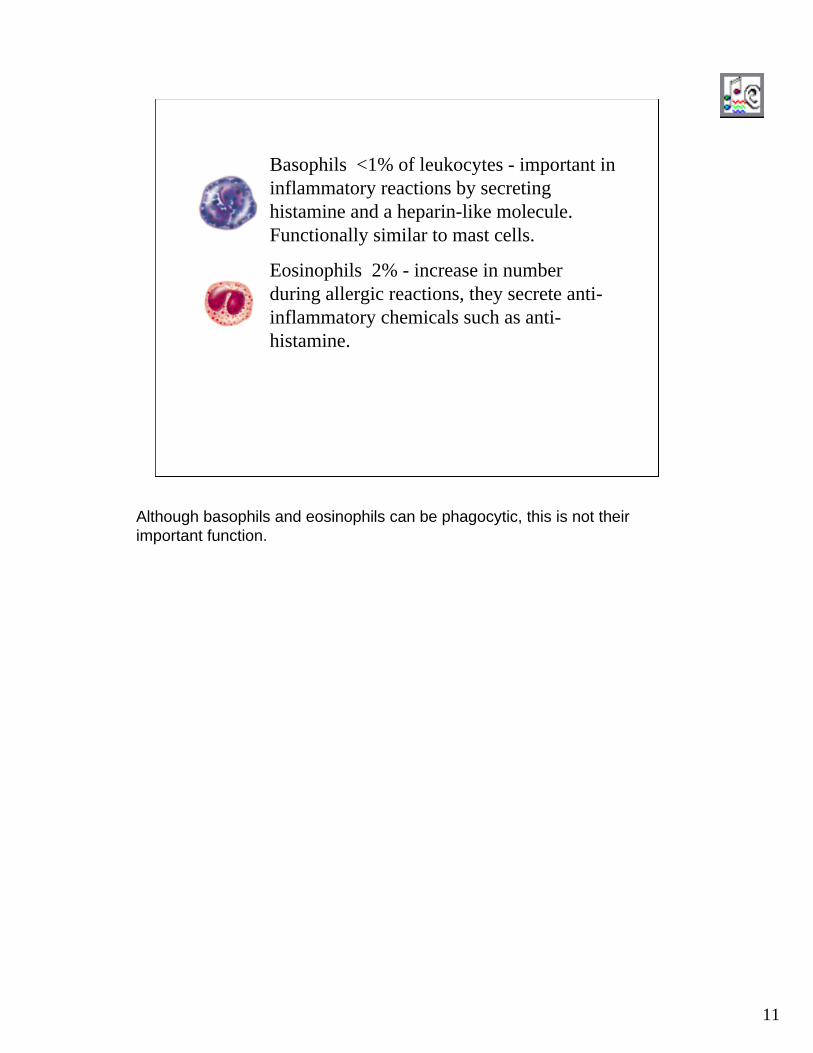
Basophils <1% of leukocytes - important in inflammatory reactions by secreting histamine and a heparin-like molecule. Functionally similar to mast cells.
Eosinophils 2% - increase in number during allergic reactions, they secrete anti-inflammatory chemicals such as anti-histamine.
Although basophils and eosinophils can be phagocytic, this is not their important function.

12
Neutrophils
Nucleus is composed of several lobes connected by thin nuclear strands.
Also called PMN (polymorphonuclear) neutrophils because of their nuclear shape. These cells spend 8 to 10 days in the circulation making their way to sites of infection etc. where they phagocytize bacteria, viruses, infected cells, debris etc. They have two types of granules: most numerous are specific granules which contain bactericidal agents such as lysozyme;azurophilic granules are lysosomes containing peroxidase and other enzymes.

13
Eosinophils
Typical bi-lobed nucleus.
Acidophilic granules
erythrocytes
Eosinophils secrete histaminase which breaks down histamine and reduces inflammatory reactions. They are found in large numbers during allergic reactions, and also when multicellular parasites are present. They may also phagocytize antigen-antibody complexes produced by allergies.

14
BasophilsLarge, basic-staining granules
Large, lobed nucleus.
Large, basic-staining granules contain histamine, SRS of anaphylaxis, heparan sulfate (related to the anti-coagulant heparin) and hydrolytic enzymes. Histamine and SRS cause dilation of small blood vessels, a large cause of inflammation.

15
Determining Blood Type
You would determine ABO blood type by placing one drop of antiserum on each position as indicated below. Rh (D) determination is optional and requires extra mixing and time. The antiserum should be at room temperature.
16
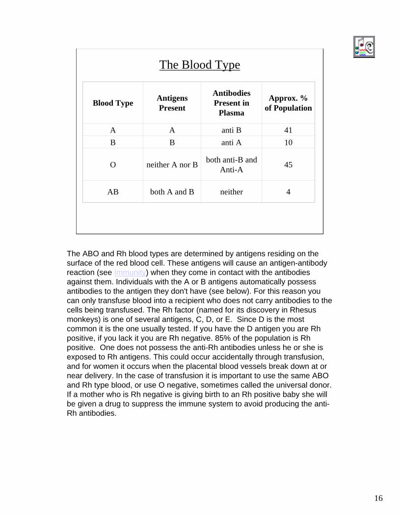
The Blood Type
Blood Type Antigens Present
Antibodies Present in
Plasma
Approx. % of Population
A A anti B 41B B anti A 10
O neither A nor B both anti-B and Anti-A 45
AB both A and B neither 4
The ABO and Rh blood types are determined by antigens residing on the surface of the red blood cell. These antigens will cause an antigen-antibody reaction (see Immunity) when they come in contact with the antibodies against them. Individuals with the A or B antigens automatically possess antibodies to the antigen they don't have (see below). For this reason you can only transfuse blood into a recipient who does not carry antibodies to the cells being transfused. The Rh factor (named for its discovery in Rhesus monkeys) is one of several antigens, C, D, or E. Since D is the most common it is the one usually tested. If you have the D antigen you are Rhpositive, if you lack it you are Rh negative. 85% of the population is Rhpositive. One does not possess the anti-Rh antibodies unless he or she is exposed to Rh antigens. This could occur accidentally through transfusion, and for women it occurs when the placental blood vessels break down at or near delivery. In the case of transfusion it is important to use the same ABO and Rh type blood, or use O negative, sometimes called the universal donor. If a mother who is Rh negative is giving birth to an Rh positive baby she will be given a drug to suppress the immune system to avoid producing the anti-Rh antibodies.

17
Here are the four blood types after reaction with Anti-A and Anti-B antiserum. A reaction is indicated by clumps of agglutinated cells. No reaction remains simply cloudy.In the Review Sheet and in the quiz you will determine the blood type of a sample.

18
The Blood Smear
Here’s how a blood smear is made. In the quiz you will have to identify erythrocytes, leucocytes, and platelets.

1
This is unstained blood. Note the large white blood cell in the center of the field. Without staining, the exact identity of the cell cannot be determined. The most numerous cells are erythrocytes. En masse or stained they appear red, but when spread apart in this way you can see they are not truly red.

2
Oil Immersion – 1000X
This is a neutrophil at 1000x magnification. When stained the lobes of the nucleus appear connected by thin bands. Because the nucleus can take many shapes neutrophils are called “polymorphonuclear” leucocytes. Note the red cells appear donut-shaped since the center is thinner and takes less stain than the edges.

3
The cell in center field is a lymphocyte. Note the solid nucleus which nearly fills the cell leaving only a small amount of cytoplasm visible.
4
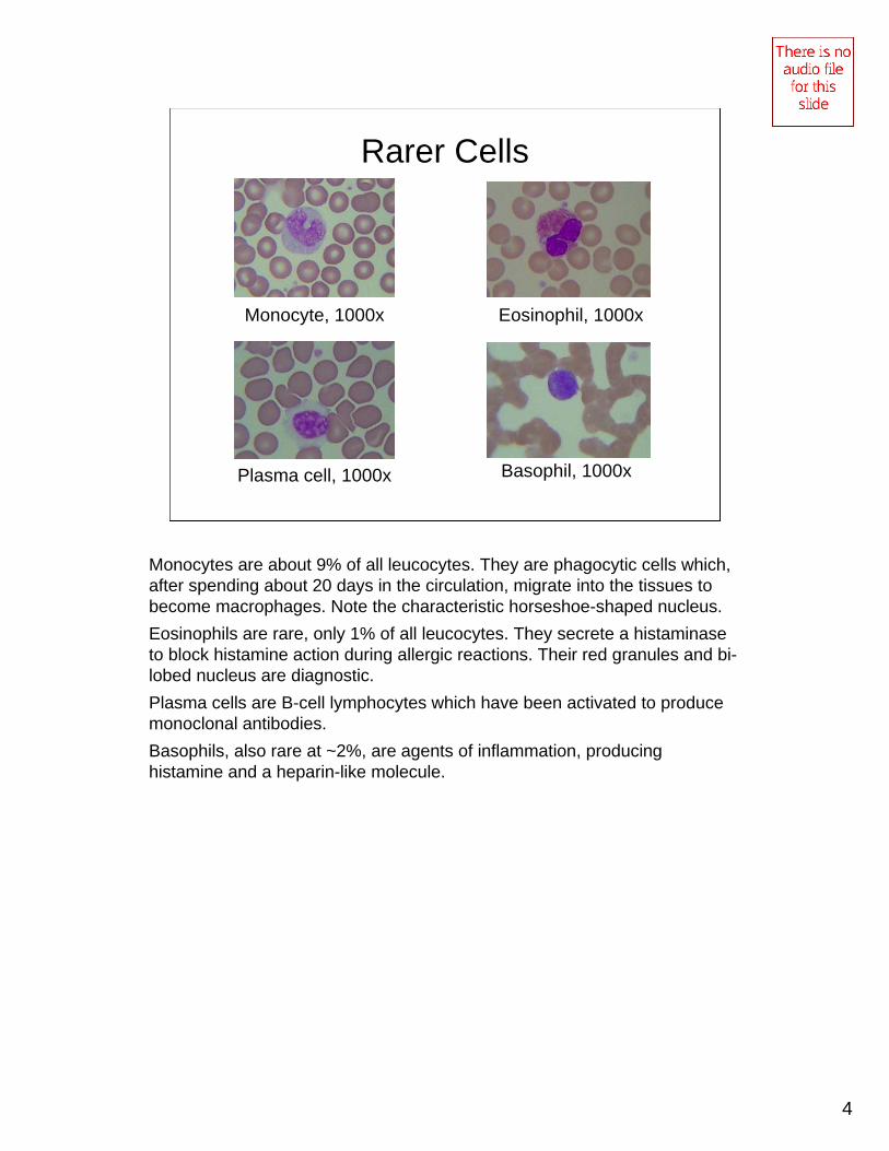
Rarer Cells
Monocyte, 1000x Eosinophil, 1000x
Plasma cell, 1000x Basophil, 1000x
Monocytes are about 9% of all leucocytes. They are phagocytic cells which, after spending about 20 days in the circulation, migrate into the tissues to become macrophages. Note the characteristic horseshoe-shaped nucleus.Eosinophils are rare, only 1% of all leucocytes. They secrete a histaminaseto block histamine action during allergic reactions. Their red granules and bi-lobed nucleus are diagnostic.Plasma cells are B-cell lymphocytes which have been activated to produce monoclonal antibodies.Basophils, also rare at ~2%, are agents of inflammation, producing histamine and a heparin-like molecule.

19
The Hematocrit
1.
2.
3.
4. After cetrifuging for 5 minutes read % of formed elements in hematocrit reader.
Here is how the hematocrit is determined. Using a clean lancet you would draw blood from an prepared finger and fill, or nearly fill, a heparinizedcapillary tube. [Question: why is the tube heparinized?] You would then use a hematocrit scale or measure with a ruler to determine the hematocrit. In the Review Sheet and in the quiz you will determine the hematocrit.

20
Lab Protocol for The Blood
1) Complete the Review Sheet for the Blood. 2) Submit drawings and/or data from PhysioEX as indicated by your instructor.
3) Take the related quiz for the Blood.