Human 3D vascularized organotypic microfluidic assays to ... · Human 3D vascularized organotypic...
7
Correction MEDICAL SCIENCES, ENGINEERING Correction for “Human 3D vascularized organotypic micro- fluidic assays to study breast cancer cell extravasation,” by Jessie S. Jeon, Simone Bersini, Mara Gilardi, Gabriele Dubini, Joseph L. Charest, Matteo Moretti, and Roger D. Kamm, which appeared in issue 1, January 6, 2015, of Proc Natl Acad Sci USA (112:214–219; first published December 18, 2014; 10.1073/ pnas.1417115112). The authors note that the following statement should be added to the Acknowledgments: “We would like to thank Dr. Yoneda for the original MDA-231-BO breast cancer cell line, and Dr. Broggini (Istituto di Ricerche Farmacologiche Mario Negri) for providing these cells modified to express a fluorescent marker.” www.pnas.org/cgi/doi/10.1073/pnas.1501426112 E818 | PNAS | February 17, 2015 | vol. 112 | no. 7 www.pnas.org
-
Upload
trinhthien -
Category
Documents
-
view
221 -
download
2
Transcript of Human 3D vascularized organotypic microfluidic assays to ... · Human 3D vascularized organotypic...

Correction
MEDICAL SCIENCES, ENGINEERINGCorrection for “Human 3D vascularized organotypic micro-fluidic assays to study breast cancer cell extravasation,” byJessie S. Jeon, Simone Bersini, Mara Gilardi, Gabriele Dubini,Joseph L. Charest, Matteo Moretti, and Roger D. Kamm, whichappeared in issue 1, January 6, 2015, of Proc Natl Acad Sci USA(112:214–219; first published December 18, 2014; 10.1073/pnas.1417115112).The authors note that the following statement should be
added to the Acknowledgments: “We would like to thankDr. Yoneda for the original MDA-231-BO breast cancer cell line,and Dr. Broggini (Istituto di Ricerche Farmacologiche Mario Negri)for providing these cells modified to express a fluorescent marker.”
www.pnas.org/cgi/doi/10.1073/pnas.1501426112
E818 | PNAS | February 17, 2015 | vol. 112 | no. 7 www.pnas.org

Human 3D vascularized organotypic microfluidic assaysto study breast cancer cell extravasationJessie S. Jeona,1, Simone Bersinib,c,1, Mara Gilardic,d, Gabriele Dubinie, Joseph L. Charestf, Matteo Morettic,2,3,and Roger D. Kamma,g,2,3
aDepartment of Mechanical Engineering, Massachusetts Institute of Technology, Cambridge, MA 02139; bDepartment of Electronics, Information andBioengineering, Politecnico di Milano, 20133 Milan, Italy; cCell and Tissue Engineering Laboratory, Istituto di Ricovero e Cura a Carattere Scientifico (IRCCS)Istituto Ortopedico Galeazzi, 20161 Milan, Italy; dPhD School in Life Sciences, Department of Biotechnology and Biosciences, University of Milano–Bicocca,20126 Milan, Italy; eDepartment of Chemistry, Materials and Chemical Engineering, Politecnico di Milano, 20133 Milan, Italy; fCharles Stark DraperLaboratory, Cambridge, MA 02139; and gDepartment of Biological Engineering, Massachusetts Institute of Technology, Cambridge, MA 02139
Edited by David A. Weitz, Harvard University, Cambridge, MA, and approved November 24, 2014 (received for review September 5, 2014)
A key aspect of cancer metastases is the tendency for specific cancercells to home to defined subsets of secondary organs. Despite theseknown tendencies, the underlying mechanisms remain poorlyunderstood. Here we develop a microfluidic 3D in vitro model toanalyze organ-specific human breast cancer cell extravasation intobone- and muscle-mimicking microenvironments through a micro-vascular network concentrically wrapped with mural cells. Extrav-asation rates and microvasculature permeabilities were significantlydifferent in the bone-mimicking microenvironment compared withunconditioned or myoblast containing matrices. Blocking breastcancer cell A3 adenosine receptors resulted in higher extravasationrates of cancer cells into the myoblast-containingmatrices comparedwith untreated cells, suggesting a role for adenosine in reducingextravasation. These results demonstrate the efficacy of our modelas a drug screening platform and a promising tool to investigatespecific molecular pathways involved in cancer biology, with poten-tial applications to personalized medicine.
microenvironment | breast cancer | metastasis | extravasation |microfluidics
Dissemination of cancer cells from a primary tumor to sec-ondary loci is responsible for more than 90% of cancer-
related mortality (1). Despite significant advances in diagnosticsand therapy, most of the available treatments are not effective,because the disseminated cells are resistant to conventionalagents (2). Invasion and metastasization are complex and mul-tistep processes guided by a wide spectrum of genetic and bio-chemical determinants (3). A key aspect of metastases is reflectedin the interactions between specific cancer cell types and differentsecondary organs. Although circulatory patterns and flow ratesmay play some role in cancer cell dissemination, it appears thatthe organ specificity of metastases is primary due to the cross-talkbetween specific cancer cells and biologically unique tissues: theseed-and-soil paradigm (4). For example, breast and prostatecancers are known to metastasize frequently to bone (5), with 70%of advanced breast cancer patients affected by skeletal metastases,leading to high rates of morbidity and mortality (6). Moreover,it has been recently demonstrated that breast cancer cells canreseed from bone to other sites including the breast, furtheremphasizing the key role of the bone microenvironment in themetastatic process (7).Metastasis organ specificity and extravasation appear to be
tightly coupled because specific chemo-attractant molecules aresecreted by organ-specific stromal cells (8). Furthermore, posi-tive interactions with circulating noncancer cells, e.g., platelets,leukocytes, and monocytes/macrophages, promote cancer celltransendothelial migration into surrounding tissues (9).In vivo and ex vivo studies have been performed to investigate
cancer cell extravasation in mouse models through liver sinusoidsand pulmonary circulation (10) or in zebrafish embryos (11).Recently, Schumacher et al. have shown the influence of platelet-secreted nucleotides playing a crucial role in the transendothelial
migration of cancer cells in the lung of mouse models (12). Invivo models have been developed to study breast cancer metas-tases to bone by means of i.v. and skeletal injection of breastcancer cells in mice (13). Although in vivo models play an es-sential role in replicating physiological conditions, they lack thepossibility to analyze highly specific interactions between humancancer cells, human blood vessels, and tissues, and they are notwell suited to perform reproducible parametric studies. Toremedy this, several in vitro models have been developed toanalyze cell migration mechanisms and particularly the invasivepotential of cancer cells (14). However, these models are oftenhighly simplified, such as the Boyden chamber or wound assays(15), which fail to allow the analysis of complex cell–cell and cell–matrix interactions, are limited in their capability to tightly controlthe local microenvironment, and offer only limited imaging.Microfluidics overcomes some of the technical limitations of
traditional assays (16), allowing the study of cancer metastasesunder biochemically and biophysically controlled 3D microen-vironments coupled with high-resolution real-time imaging (17).Various microfluidic models have been developed for studyingtumor angiogenesis (18), transition to invasion (19), intravasation(20), the role of interstitial flow (21) and matrix stiffness (22) on
Significance
The cancer biology seed-and-soil paradigm recognizes the ex-istence of organ-specific patterns of metastasization that drivethe spread of selected primary tumors toward specific sec-ondary loci. However, despite efforts to model the organotypicmicroenvironment, the organ specificity of cancer metastasesneeds to be elucidated. The relevance of this study lies in thegeneration of a human vascularized organ-specific microenvi-ronment, which can be used to investigate and tune the ex-travasation process of metastatic tumor cells. Furthermore,beyond mimicking the pro- or antimetastatic signatures ofdifferent microenvironments, our microfluidic model providesinsights into different properties of organ-specific endothelia.This study paves the way toward advanced in vitro models toscreen for highly tailored organ-specific therapeutics and in-vestigate key molecular pathways involved in organ-specificmetastases.
Author contributions: J.S.J., S.B., G.D., J.L.C., M.M., and R.D.K. designed research; J.S.J.,S.B., and M.G. performed research; J.S.J., S.B., M.G., M.M., and R.D.K. analyzed data; andJ.S.J., S.B., M.M., and R.D.K. wrote the paper.
The authors declare no conflict of interest.
This article is a PNAS Direct Submission.1J.S.J. and S.B. contributed equally to this work.2M.M. and R.D.K. contributed equally to this work.3To whom correspondence may be addressed. Email: [email protected] or [email protected].
This article contains supporting information online at www.pnas.org/lookup/suppl/doi:10.1073/pnas.1417115112/-/DCSupplemental.
214–219 | PNAS | January 6, 2015 | vol. 112 | no. 1 www.pnas.org/cgi/doi/10.1073/pnas.1417115112
cancer cell migration, adhesion (23), and extravasation (24, 25).Recently, our group presented a microfluidic model to in-vestigate the specificity of breast cancer metastasis to bone,providing quantitative data on cancer cell extravasation rate andreproducing the effects of the CXCL5–CXCR2 interaction be-tween bone cells and metastatic breast cancer cells observed invivo (26). However, in that system, the vascular wall was repre-sented by an endothelial monolayer on the side of a central gelregion. With the recent attempts in inducing vasculogenesis (27,28), vascular networks have been generated inside the gel regioneither by coculture with human lung fibroblasts in separate gelregions or by interstitial flow. Despite the tremendous advancesin modeling angiogenesis and vasculogenesis, these models havenot previously been used to study metastasis organ specificity andinvestigate the role of human organ-specific microenvironments.Here we present an organ-specific human 3D microfluidic
model that enables the study of human metastatic breast cancercell extravasation within a perfusable human microvascularizedbone-mimicking (BMi) microenvironment. The resulting modelrepresents a functional human quad-culture in which breastcancer cells flow into, adhere to, and metastasize through humanmicrovascular networks. These networks are supported by pri-mary human bone marrow-derived mesenchymal stem cells(hBM-MSCs) that have undergone phenotypical transition to-ward the smooth muscle cell lineage, embedded in a BMi micro-environment with homogeneously distributed osteo-differentiated(OD) primary hBM-MSCs.
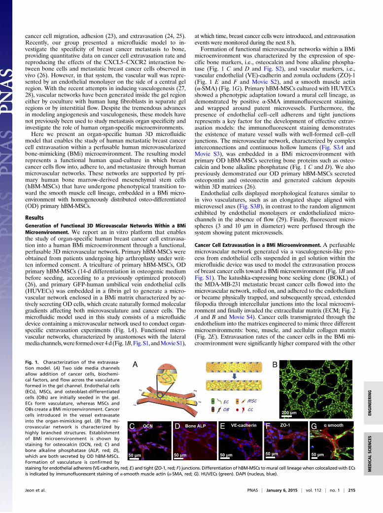
ResultsGeneration of Functional 3D Microvascular Networks Within a BMiMicroenvironment. We report an in vitro platform that enablesthe study of organ-specific human breast cancer cell extravasa-tion into a human BMi microenvironment through a functional,perfusable 3D microvascular network. Primary hBM-MSCs wereobtained from patients undergoing hip arthroplasty under writ-ten informed consent. A triculture of primary hBM-MSCs, ODprimary hBM-MSCs (14-d differentiation in osteogenic mediumbefore seeding, according to a previously optimized protocol)(26), and primary GFP-human umbilical vein endothelial cells(HUVECs) was embedded in a fibrin gel to generate a micro-vascular network enclosed in a BMi matrix characterized by ac-tively secreting OD cells, which create naturally formed moleculargradients affecting both microvasculature and cancer cells. Themicrofluidic model used in this study consists of a microfluidicdevice containing a microvascular network used to conduct organ-specific extravasation experiments (Fig. 1A). Functional micro-vascular networks, characterized by anastomoses with the lateralmediachannels,were formedover4d(Fig. 1B,Fig.S1, andMovieS1),
at which time, breast cancer cells were introduced, and extravasationevents were monitored during the next 8 h.Formation of functional microvascular networks within a BMi
microenvironment was characterized by the expression of spe-cific bone markers, i.e., osteocalcin and bone alkaline phospha-tase (Fig. 1 C and D and Fig. S2), and vascular markers, i.e.,vascular endothelial (VE)-cadherin and zonula occludens (ZO)-1(Fig. 1 E and F and Movie S2), and α smooth muscle actin(α-SMA) (Fig. 1G). Primary hBM-MSCs cultured with HUVECsshowed a phenotypic adaptation toward a mural cell lineage, asdemonstrated by positive α-SMA immunofluorescent staining,and wrapped around patent microvessels. Furthermore, thepresence of endothelial cell–cell adherens and tight junctionsrepresents a key factor for the development of effective extrav-asation models: the immunofluorescent staining demonstratesthe existence of mature vessel walls with well-formed cell–celljunctions. The microvascular network, characterized by complexinterconnections and continuous hollow lumens (Fig. S3A andMovie S3), was embedded in a BMi microenvironment withprimary OD hBM-MSCs secreting bone proteins such as osteo-calcin and bone alkaline phosphatase (Fig. 1 C and D). We alsopreviously demonstrated our OD primary hBM-MSCs secretedosteopontin and osteonectin and generated calcium depositswithin 3D matrices (26).Endothelial cells displayed morphological features similar to
in vivo vasculatures, such as an elongated shape aligned withmicrovessel axes (Fig. S3B), in contrast to the random alignmentexhibited by endothelial monolayers or endothelialized micro-channels in the absence of flow (29). Finally, fluorescent micro-spheres (3 and 10 μm in diameter) were perfused through thesystem showing patent microvessels.
Cancer Cell Extravasation in a BMi Microenvironment. A perfusablemicrovascular network generated via a vasculogenesis-like pro-cess from endothelial cells suspended in gel solution within themicrofluidic device was used to model the extravasation processof breast cancer cells toward a BMi microenvironment (Fig. 1B andFig. S1). The katushka-expressing bone seeking clone (BOKL) ofthe MDA-MB-231 metastatic breast cancer cells flowed into themicrovascular network, rolled on, and adhered to the endotheliumor became physically trapped, and subsequently spread, extendedfilopodia through intercellular junctions into the local microenvi-ronment and finally invaded the extracellular matrix (ECM; Fig. 2A and B and Movie S4). Cancer cells transmigrated through theendothelium into the matrices engineered to mimic three differentmicroenvironments: bone, muscle, and acellular collagen matrix(Fig. 2E). Extravasation rates of the cancer cells in the BMi mi-croenvironment were significantly higher compared with the other
Fig. 1. Characterization of the extravasa-tion model. (A) Two side media channelsallow addition of cancer cells, biochemi-cal factors, and flow across the vasculatureformed in the gel channel. Endothelial cells(ECs), MSCs, and osteoblast-differentiatedcells (OBs) are initially seeded in the gel.ECs form vasculature, whereas MSCs andOBs create a BMi microenvironment. Cancercells introduced in the vessel extravasateinto the organ-mimicking gel. (B) The mi-crovascular network is characterized byhighly branched structures. Establishmentof BMi microenvironment is shown bystaining for osteocalcin (OCN, red; C ) andbone alkaline phosphatase (ALP, red; D),which are both secreted by OD hBM-MSCs.Formation of vasculature is confirmed bystaining for endothelial adherens (VE-cadherin, red; E) and tight (ZO-1, red; F) junctions. Differentiation of hBM-MSCs tomural cell lineagewhen colocalized with ECsis indicated by immunofluorescent staining of α-smooth muscle actin (α-SMA, red; G). HUVECs (green). DAPI (nucleus, blue).
Jeon et al. PNAS | January 6, 2015 | vol. 112 | no. 1 | 215
MED
ICALSC
IENCE
SEN
GINEE
RING

conditions such as the microenvironment conditioned with themyoblast cell line C2C12, mimicking a muscle microenvironment,or control matrix without any cells. BOKL average extravasationrate was 56.5 ± 4.8% in the presence of OD hBM-MSCs com-pared with 8.2 ± 2.3% with C2C12 myoblasts and 14.7 ± 3.6%without stromal cell addition (control matrix; Fig. 2C). The av-erage extravasation rate within the BMi microenvironment wasthen 3.8-fold higher than without stromal cell addition, which ishigher compared with our previous work analyzing the extrava-sation of cancer cells through a simple and less physiologicallyrelevant endothelial monolayer (2.06). It is noteworthy that thepresence of actively secreting mural cell-like hBM-MSCs thatenabled the generation of a more in vivo-like microenvironmentcould have also played a role in the increased extravasation ratio.No significant differences were quantified comparing cancer cellmigration distance following extravasation in BMi microenviron-ments and control matrices (23.26 ± 2.73 vs. 30.63 ± 6.07 μm).Vessel permeability was quantified by analyzing 70-kDa dextran
diffusion through the microvascular wall and compared among ex-perimental conditions (Fig. S3A). Permeability values were signifi-cantly higher in BMi microfluidic devices 4.12 × 10⁻⁶ ± 0.75 × 10⁻⁶
cm/s compared with control matrices 0.89 × 10⁻⁶ ± 0.31 × 10⁻⁶ cm/s(Fig. 2D). Finally, the highest permeability values were found in the
presence of C2C12 myoblasts 8.37 × 10⁻⁶ ± 1.53 × 10⁻⁶ cm/s, re-sulting in a 2.0-fold increase compared with the BMi systems (Fig.2D). It is surprising that the most leaky environment C2C12 gaverise to the lowest extravasation rate. This demonstrates that per-meability is only one of multiple factors affecting extravasation.We also explored the capability of the assay to distinguish
between the extravasation potentials of different cell types andcellular environments. First, an inflammatory model was set upwith the addition of a macrophage cell line. RAW264.7 macro-phages were homogeneously dispersed within the fibrin gel. Theaverage extravasation rate within macrophage conditioned BMimicroenvironment was significantly higher (32.4 ± 4.3%) com-pared with matrices without addition of stromal cells (14.7 ±3.6%) but lower with respect to standard BMi microenvironment(56.5 ± 4.8%; Fig. S4A). The promising nature of these datawere confirmed by microvessel permeability values, which werehigher (6.77 × 10⁻⁶ ± 1.56 × 10⁻⁶ cm/s) in presence of macrophagescompared with BMi matrices (4.12 × 10⁻⁶ ± 0.75 × 10⁻⁶ cm/s;Fig. S4B). We also performed a control experiment introducingnonmetastatic mammary epithelial cells (MCF-10A) within theBMi microenvironment. MCF-10A extravasation rate was signifi-cantly lower (5.2 ± 2.5%) compared with BOKL (56.5 ± 4.8%; Fig.S5), thus demonstrating the specificity of the interaction betweenmetastatic cancer cells and a BMi microenvironment.
The Role of Adenosine in Cancer Cell Extravasation. As we foundstriking differences between cancer cell extravasation within BMiand muscle-mimicking microenvironments, we asked which fac-tors secreted by muscle might be responsible. It is reported thatthe skeletal muscle microenvironment reduces cancer cell tu-morigenicity and elicits paracrine-mediated cytotoxic and cyto-static responses from metastatic cancer cells (30). In particular,several studies identified the A3 adenosine receptor (A3AR),which is expressed by multiple cancer cell types (31), as key in theantimetastatic and protective effect of skeletal muscle cells (32).Before investigating the role of A3AR in extravasation, we firstdemonstrated through immunofluorescence that BOKL intro-duced in our system expressed the A3AR (Fig. 3A and Fig. S6).Then, we introduced the A3AR antagonist PSB-10 within C2C12containing microfluidic devices (Fig. 3H) and preincubatedcancer cells with A3AR before seeding (33). With the addition ofthe antagonist, cancer cell extravasation rate significantly in-creased (32.4 ± 7.7%) compared with nontreated C2C12 ma-trices (8.2 ± 2.3%; Fig. 3E), whereas microvessel permeabilitywas not significantly affected (7.4 × 10⁻⁶ ± 2.61 × 10⁻⁶ vs. 8.04 ×10⁻⁶ ± 1.72 × 10⁻⁶ cm/s; Fig. 3G). These data indirectly dem-onstrate the presence of C2C12-secreted adenosine in the sys-tem, as well as the protective role of this molecule against cancermetastases. Mass spectrometry (MS) analyses performed on cellculture supernatants provided further evidence of adenosine se-cretion by the C2C12-conditioned microenvironment (Fig. 3B andFig. S7), whereas immunofluorescence staining demonstrated theexpression of the adenosine converter CD73 (Fig. 3C), which cat-alyzes the conversion of AMP to adenosine (34).Supported by our results with untreated or conditioned
(A3AR antagonist) C2C12 microenvironments, we investigatedthe role of adenosine on cancer cell extravasation within a BMimicroenvironment using our model as drug screening platform(Fig. 3H). The presence of adenosine significantly reduced can-cer cell extravasation (12.7 ± 2.8%) compared with the untreatedBMi microenvironment (56.5 ± 4.8%; Fig. 3D). Furthermore, wefound a dramatic increase in microvasculature permeability(8.22 × 10⁻⁶ ± 1.76 × 10⁻⁶ cm/s) compared with BMi control(4.12 × 10⁻⁶ ± 0.75 × 10⁻⁶ cm/s; Fig. 3F). It is interesting to notethat treating BMi microenvironments with adenosine led to ex-travasation rate and permeability values comparable to C2C12-conditioned matrices, suggesting this molecule could be theoreticallyused to tune a specific microenvironment with antimetastatic
Fig. 2. Cancer cell extravasation. (A) Extravasation of cancer cells (red) in-troduced into the vascular network (HUVECs, green) is monitored in realtime. (B) Magnified images of white dotted box in A show extravasation ofcancer cells. (C) Percent of cancer cells extravasated varies significantlyamong the vascular networks embedded in different microenvironments,i.e., acellular and bone or muscle-mimicking microenvironment, respectively.(D) Permeability values increased when cells are added to mimic the twoorgan-specific microenvironments compared with HUVEC only condition. (E)Schematic of HUVEC only, osteo-cell, and C2C12 cell added systems. HUVECsare shown as green cells that form vessel, osteo-cells are blue colored cellsand secrete bone matrix as shown in yellow, and C2C12 cells are depicted asyellow cells. Cancer cells are colored in red and seen both in the vessels aswell as extravasated out in the surrounding matrix.
216 | www.pnas.org/cgi/doi/10.1073/pnas.1417115112 Jeon et al.

properties. Finally, Live/Dead and MTT assays were performedat different time points on cancer cells treated with adenosine,confirming that the A3AR antagonist action in reducing extrava-sation was not due to a cytotoxic effect on BOKL (Figs. S8 and S9).
Effect of Shear Stress on Microvasculature and Cancer Cell Extravasation.Because extravasation normally occurs during blood flow, wenext examined the effects on microvascular networks condi-tioned by physiological levels of shear stress. In these experi-ments, we preconditioned microvascular networks with flowovernight before cancer cell seeding and analyzed the effects offlow on extravasation and microvessel permeability. We selecteda flow rate of 2 μL/min in the channel, which resulted in anaverage velocity in the vasculature of 220 μm/s and a wall shearstress of 0.25 dyne/cm2 (35).Cancer cell extravasation within the flow conditioned BMi
microenvironment occurred at a rate of 38.6 ± 4.8% (Fig. 4A),significantly lower than under static conditions (56.5 ± 4.8%).We also found that microvessel permeability decreased 2.4-foldcompared with static conditions (1.72 × 10⁻⁶ ± 0.53 × 10⁻⁶ cm/scompared with 4.12 × 10⁻⁶ ± 0.75 × 10⁻⁶ 10−6 cm/s; Fig. 4B).Endothelial cells exposed to a laminar flow were characterizedby an elongated morphology and a clear actin filament alignmentin the flow direction as shown in Fig. 4 E (flow) and D (staticcontrol) and measured using anisotropy score (Fig. S10 and SIMaterials and Methods), with compelling stress fibers at cell–celljunctions (Fig. S3B), thus displaying morphological featuresresembling in vivo microvessels (36). Finally, it is worth notingthat flow-conditioned breast cancer cells migrated further intothe surrounding matrix (33.7 ± 4.3 μm) compared with staticexperiments (23.3 ± 2.7 μm; Fig. 4C).
DiscussionAn organ-specific 3D microfluidic model was created to studyhuman breast cancer cell extravasation into an actively secretingBMi microenvironment generated with OD hBM-MSCs throughperfusable human microvascular networks composed of endo-thelial and mural-like cells. The relevance of the present work liesin the application of a complex model to investigate and sub-sequently tune a specific step of the metastatic cascade withindifferent organ-specific microenvironments with implications for
the screening of new therapeutics. The efficacy and potentiality ofour microfluidic model go beyond the established role of adeno-sine as a promising anticancer molecule to specifically demon-strate its involvement in the extravasation process.The microfluidic model presented is based on the coculture of
endothelial and hBM-MSC–derived mural cells to create physi-ologically relevant highly branched structures, characterized byreduced microvessel diameter and increased branch numbercompared with microvascular networks made of endothelial cellalone (27). Particularly, this model overcomes some of the lim-itations of previously developed elegant models that showedimpaired perfusability when supportive stromal cells were addedto the system (37). It is known that a wide panel of molecules isinvolved in vessel wall and network maturation including vas-cular endothelial growth factor (VEGF), platelet derived growthfactor (PDGF-B), and members of the TGF-β family. Moreover,it is reported that angiopoietin-1 (Ang-1) represents a key
Fig. 3. Percent of cancer cell extravasation andvascular permeability in bone and muscle-mimickingmicroenvironment with addition of stimulating orblocking molecules. Cancer cells express the A3
adenosine receptor (A), whereas C2C12 embeddedmatrices secrete adenosine as shown by MS data(268 m/z peak) (B) and CD73 immunofluorescentstaining (C). Percentage of cancer cells that extrav-asate (D) and permeability of the vasculature (F) inthe BMi microenvironment with OD hBM-MSCSs,with and without adenosine. Extravasation ratedecreased significantly with addition of adenosine,whereas the permeability increased with adeno-sine. Percentage of cancer cells that extravasate(E ) and permeability of the vasculature (G) in themuscle-mimicking microenvironment with C2C12cells, with and without PSB10. Although block-ing of A3AR with the addition of PSB10 did notalter the permeability of the vasculature, cancercell extravasation rate increased significantly. (H)Schematic of osteo-cell and C2C12 cell added sys-tems with adenosine, A3 adenosine receptor andits antagonist PSB10.
Fig. 4. Cancer cell extravasation and endothelial cell (EC) permeabilitychange in the presence of flow through the vasculature. Extravasation ofcancer cells (A) and permeability of the vasculature (B) decreased signifi-cantly with the addition of flow. (C) Extravasated cancer cells migratedfurther in the flow condition vs. the static condition. Actin (yellow) withinECs in static condition (D) and under conditions when flow was added in thevasculature (E). DAPI (nucleus, blue).
Jeon et al. PNAS | January 6, 2015 | vol. 112 | no. 1 | 217
MED
ICALSC
IENCE
SEN
GINEE
RING

molecule in vessel stabilization and could play a role in the re-cruitment of mesenchymal cells (38). As we recently demon-strated, the addition of Ang-1 within HUVEC + hBM-MSCcocultures significantly induces mesenchymal cell expressionof common markers displayed by mural cells, e.g., α-SMA,SM22α, and NG-2 (27), which contribute to network stabiliza-tion. Taken together, these findings provide evidence that thecombination of endothelial-mesenchymal stem cell cocultureand the external addition of stabilizing molecules promote thegeneration of functional microvascular networks, which repre-sent a suitable model to study cancer cell extravasation withinan actively secreting BMi microenvironment.Cancer cell extravasation rates were significantly higher in the
BMi microenvironment compared with control matrices withoutstromal cells or muscle-mimicking microenvironments. Althoughthe higher permeability in the BMi microenvironment is likelydue to a combination of factors, it may also contribute to theincrease in extravasation. Indeed, matrix-specific cell-secretedchemokines can both generate molecular gradients affectingcancer cell transendothelial migration (39) and alter endothelialpermeability (40). Of particular note is the increase in micro-vasculature permeability when adenosine is added. It has beenreported that adenosine can interact with endothelial cellsexpressing A2B adenosine receptors, promoting the release ofproangiogenic factors including VEGF and IL-8, which couldlead to the increased permeability (41). Although A2B adenosinereceptors are preferentially expressed by human microvascularendothelial cells compared with HUVECs, the external additionof adenosine in the BMi microenvironment played a role in theincreased microvessel permeability compared with untreatedmatrices, despite reducing cancer cell extravasation. These datahighlight that permeability represents only one of the key factorsdriving cancer cell extravasation. C2C12-secreted adenosinecould also partially explain permeability differences detectedcomparing no stromal cell addition, BMi microenvironment, andC2C12 conditioned matrices. Moreover, the presence of highvalues of microvessel permeability with the addition of the A3ARantagonist within C2C12-embedded microenvironments, whichare comparable to data obtained with untreated C2C12 matrices,suggests the A3AR antagonist did not adversely affect the me-chanical properties of the endothelium, confirming previous stud-ies demonstrating low expression levels of A3AR by endothelialcells (41). However, the addition of the antagonist did promotean increase in the extravasation rate compared with untreatedmicroenvironments, suggesting a specific interaction with cancercells. Indeed, it should also be considered the A3AR antagonistcould have affected endothelial cell secretory activity or the ex-pression of surface markers contributing to cancer cell adhesion/extravasation, thus leading to increased extravasation rates.Substantial clinical and experimental evidence indicates a key
role of macrophages in multiple steps of the metastatic cascade.Based on previous experiments performed in our laboratory oncancer cell intravasation (20), we suggest the increased micro-vasculature permeability could be due to the macrophage secre-tion of the inflammatory cytokine TNF-α. However, it is knownthat TNF-α inhibits osteoblast differentiation and bone-specificprotein expression, particularly osteocalcin, while inducing osteo-clastogenesis (42). Then, we can speculate the macrophage-secreted TNF-α could have conditioned the BMi microenvironment,limiting the prometastatic effect induced by OD cell secretome.Further analyses are required to in-depth investigate specific roleand impact of macrophages and particularly macrophage-se-creted TNF-α on breast cancer bone metastases.Several works have recently shown that when endothelial cells
are subjected to laminar flow shear stress, transendothelial elec-trical resistance increases, microvasculature permeability decrea-ses, actin filaments become more aligned, and cytoskeletal tensionand intercellular forces between endothelial cells increase
compared with static conditions. Moreover, a significant over-expression has been highlighted for β-catenin (adherens junc-tions) and ZO-1 (tight junctions), with respect to disturbedflow or static conditions (43). Overall, these studies suggest thatexposure of endothelial cells to controlled, laminar flows resultsin cell–cell junction tightening with a corresponding decrease invasculature permeability. These findings agree with our resultsshowing decreased microvessel permeability due to flow shearstress in the same BMi microenvironment. It is likely that thereduced extravasation rate within the flow conditioned BMimicroenvironment could be due to enhanced endothelial cellintercellular forces and cell–cell junction tightening. Further-more, we can speculate the presence of an interstitial flow withinthe BMi matrix during flow conditioned experiments couldpartially explain the increased migration distance shown bybreast cancer cells compared with static controls, as previouslydemonstrated by several studies highlighting the link betweenflow-induced mechanical stimulation and cell migration (21).Our data demonstrate that generation of a more physiologicalmicroenvironment with the addition of endothelial wall shearstress can dramatically affect extravasation potential, whichcould have critical implications on drug screening tests and invitro model development. We would like to emphasize that, inaddition to endothelial cell preconditioning, our platform ena-bles real time monitoring of cancer cell extravasation under flow,thus allowing the in situ analysis of extravasation in the micro-environment where both endothelial and cancer cells are ex-posed to physiological stimuli.In conclusion, an organ-specific vascularized 3D microfluidic
model was created to study human breast cancer cell extrava-sation into an actively secreting human BMi microenvironmentthrough perfusable human microvascular networks composed ofendothelial and mural-like cells. We found clear evidence of theseed (breast cancer cells) and soil (BMi microenvironment)coupling and provided quantitative results regarding the anti-metastatic and protective role played by skeletal muscle cells.Particularly, we demonstrated the effectiveness of the model asa drug screening assay, being able to investigate both the effectsof A3AR antagonist on cancer cell extravasation in a C2C12myoblast conditioned matrix and the antimetastatic role ofadenosine in a human BMi microenvironment, thus overcominglimitations of traditional in vitro models. These assays contributeto the continued evolution of 3D in vitro models, improving ondrawbacks of animal studies, e.g., differences in drug metabolismthat affect the outcome of therapeutic treatments, and capable ofbridging highly specific human in vitro cultures with physiologicalin vivo conditions. Success in these modeling efforts will help tofoster a more effective screening of tailored anticancer therapiesin the context of personalized medicine and promote the study ofkey molecular pathways involved in cancer biology in controlled,organ-specific, physiological-like conditions.
Materials and MethodsMicrofluidic System. The microfluidic system contains a 1.3-mm-wide centralhydrogel region flanked by two lateral media channels, as previously used inour group for other studies (27). The microfabrication process of the devicewas documented in detail for other configurations (26). Briefly, the micro-fluidic device was fabricated with poly-dimethyl-siloxane (PDMS, Silgard 184;Dow Chemical) using soft lithography techniques from patterned SU-8 sili-con wafers. Inlet and outlet ports were created with biopsy punches, anda cover glass was bonded to the PDMS after 60-s oxygen plasma treatmentto generate 200-μm-deep microchannels. A thrombin solution (4 U/mL) wasused to resuspend cells, and 10-μL aliquots were mixed with 10 μL fibrinogensolution (5 mg/mL) to create a fibrin gel. After gelation, media channelswere filled with cell culture medium, and microfluidic devices were culturedfor 4 d. The two channels system enabled easy access to the hydrogel regionfor cancer cell extravasation studies under static or dynamic conditions.
218 | www.pnas.org/cgi/doi/10.1073/pnas.1417115112 Jeon et al.

Immunofluorescent Staining and Image Acquisition. All devices were washedwith PBS (Invitrogen), fixed with 4% paraformaldehyde (PFA) for 15 min andpermeabilized with 0.1% Triton-X 100 solution for 5 min at room temper-ature. Samples were treated with 5% BSA + 3% (wt/vol) goat serum solutionfor at least 3 h at 4 °C before incubation with primary antibody. Mousepolyclonal α-SMA antibody (abcam; dilution 1:100), rabbit polyclonal VE-cadherin antibody (abcam; dilution 1:100), mouse polyclonal ZO-1 antibody(Invitrogen; dilution 1:100), rabbit polyclonal osteocalcin antibody (BTI; di-lution 1:50), and mouse monoclonal bone alkaline phosphatase (abcam;dilution 1:40) were used for staining. Red fluorescently labeled secondaryantibodies (Invitrogen) were used at 1:200 dilution (Invitrogen). Cell nucleiwere stained with DAPI (5 mg/mL; Invitrogen) at 1:500 dilution, and F-actinfilaments were stained with AlexaFluor633 phalloidin (Invitrogen) at 1:100dilution. All images were captured using a confocal microscope (OlympusIX81) and processed with Imaris software (Bitplane Scientific Software).
Addition of Adenosine and Antagonist for the Adenosine Receptor. Additionalexperiments were performed to test the role of the muscle-secretedmoleculeadenosine as an antimetastatic agent. Adenosine (R&D Systems) was dilutedat a final concentration of 10 μM and added to BMi microfluidic devices for24 h before cancer cell addition. Cancer cells were preincubated withadenosine 4 h before the seeding. Extravasation events were monitored in
presence of adenosine. To confirm the presence and effect of adenosinewithin C2C12 myoblast conditioned matrices, the highly specific A3AR an-tagonist (PSB-10; R&D Systems) at 10 μM was introduced into the micro-fluidic devices for 24 h before cancer cell addition, and cancer cells werepreincubated with A3AR for 4 h before injection. Extravasation events weremonitored in presence of A3AR antagonist.
Statistics. All extravasation percentages are reported as averages ± standarderror of the mean (SEM). Measurements are obtained from a minimum of 10regions of interest (ROIs) from three or more independent devices, whereasall permeability values are averages of five (minimum) to seven (maximum)measurements from two (minimum) to four (maximum) independent de-vices. Measurements were compared using an unpaired Student t test. Sta-tistical tests were performed with SigmaPlot12. All tests with P < 0.05 andP < 0.005 were assumed to be statistically significant.
ACKNOWLEDGMENTS. We thank Prof. Andrea Mele and Walter Panzeri(Politecnico di Milano) for MS experiments and support during data analysis.This work as supported by National Cancer Institute Grants R33 CA174550-01and R21 CA140096 and the Italian Ministry of Health. S.B. was supported bythe Fondazione Fratelli Agostino and Enrico Rocca through a Progetto Roccadoctoral fellowship, and J.S.J. was supported by a Draper fellowship.
1. Valastyan S, Weinberg RA (2011) Tumor metastasis: Molecular insights and evolvingparadigms. Cell 147(2):275–292.
2. Chambers AF, Groom AC, MacDonald IC (2002) Dissemination and growth of cancercells in metastatic sites. Nat Rev Cancer 2(8):563–572.
3. Hanahan D, Weinberg RA (2011) Hallmarks of cancer: The next generation. Cell144(5):646–674.
4. Fidler IJ (2003) The pathogenesis of cancer metastasis: The ‘seed and soil’ hypothesisrevisited. Nat Rev Cancer 3(6):453–458.
5. Bussard KM, Gay CV, Mastro AM (2008) The bone microenvironment in metastasis:What is special about bone? Cancer Metastasis Rev 27(1):41–55.
6. Coleman RE (2012) Bone cancer in 2011: Prevention and treatment of bone metas-tases. Nat Rev Clin Oncol 9(2):76–78.
7. KimMY, et al. (2009) Tumor self-seeding by circulating cancer cells. Cell 139(7):1315–1326.8. Nguyen DX, Bos PD, Massagué J (2009) Metastasis: From dissemination to organ-
specific colonization. Nat Rev Cancer 9(4):274–284.9. Reymond N, d’Água BB, Ridley AJ (2013) Crossing the endothelial barrier during
metastasis. Nat Rev Cancer 13(12):858–870.10. Al-Mehdi AB, et al. (2000) Intravascular origin of metastasis from the proliferation
of endothelium-attached tumor cells: A new model for metastasis. Nat Med 6(1):100–102.
11. Stoletov K, et al. (2010) Visualizing extravasation dynamics of metastatic tumor cells.J Cell Sci 123(Pt 13):2332–2341.
12. Schumacher D, Strilic B, Sivaraj KK, Wettschureck N, Offermanns S (2013) Platelet-derived nucleotides promote tumor-cell transendothelial migration and metastasisvia P2Y2 receptor. Cancer Cell 24(1):130–137.
13. Kuperwasser C, et al. (2005) A mouse model of human breast cancer metastasis tohuman bone. Cancer Res 65(14):6130–6138.
14. Lauffenburger DA, Horwitz AF (1996) Cell migration: A physically integrated molec-ular process. Cell 84(3):359–369.
15. Simpson KJ, et al. (2008) Identification of genes that regulate epithelial cell migrationusing an siRNA screening approach. Nat Cell Biol 10(9):1027–1038.
16. Bersini S, Jeon JS, Moretti M, Kamm RD (2014) In vitro models of the metastaticcascade: From local invasion to extravasation. Drug Discov Today 19(6):735–742.
17. Shin Y, et al. (2012) Microfluidic assay for simultaneous culture of multiple cell typeson surfaces or within hydrogels. Nat Protoc 7(7):1247–1259.
18. Vickerman V, Kamm RD (2012) Mechanism of a flow-gated angiogenesis switch: Earlysignaling events at cell-matrix and cell-cell junctions. Integr Biol (Camb) 4(8):863–874.
19. Sung KE, et al. (2011) Transition to invasion in breast cancer: A microfluidic in vitromodel enables examination of spatial and temporal effects. Integr Biol (Camb) 3(4):439–450.
20. Zervantonakis IK, et al. (2012) Three-dimensional microfluidic model for tumor cellintravasation and endothelial barrier function. Proc Natl Acad Sci USA 109(34):13515–13520.
21. Polacheck WJ, Charest JL, Kamm RD (2011) Interstitial flow influences direction oftumor cell migration through competing mechanisms. Proc Natl Acad Sci USA 108(27):11115–11120.
22. Pathak A, Kumar S (2012) Independent regulation of tumor cell migration by matrixstiffness and confinement. Proc Natl Acad Sci USA 109(26):10334–10339.
23. Song JW, et al. (2009) Microfluidic endothelium for studying the intravascular ad-hesion of metastatic breast cancer cells. PLoS ONE 4(6):e5756.
24. Jeon JS, Zervantonakis IK, Chung S, Kamm RD, Charest JL (2013) In vitro model oftumor cell extravasation. PLoS ONE 8(2):e56910.
25. Zhang Q, Liu T, Qin J (2012) A microfluidic-based device for study of transendothelialinvasion of tumor aggregates in realtime. Lab Chip 12(16):2837–2842.
26. Bersini S, et al. (2014) A microfluidic 3D in vitro model for specificity of breast cancermetastasis to bone. Biomaterials 35(8):2454–2461.
27. Jeon JS, et al. (2014) Generation of 3D functional microvascular networks with humanmesenchymal stem cells in microfluidic systems. Integr Biol (Camb) 6(5):555–563.
28. Moya ML, Hsu YH, Lee AP, Hughes CC, George SC (2013) In vitro perfused humancapillary networks. Tissue Eng Part C Methods 19(9):730–737.
29. Chrobak KM, Potter DR, Tien J (2006) Formation of perfused, functional microvasculartubes in vitro. Microvasc Res 71(3):185–196.
30. Parlakian A, et al. (2010) Skeletal muscle phenotypically converts and selectively in-hibits metastatic cells in mice. PLoS ONE 5(2):e9299.
31. Madi L, et al. (2004) The A3 adenosine receptor is highly expressed in tumor versusnormal cells: Potential target for tumor growth inhibition. Clin Cancer Res 10(13):4472–4479.
32. Fishman P, et al. (2004) An agonist to the A3 adenosine receptor inhibits colon car-cinoma growth in mice via modulation of GSK-3 beta and NF-kappa B. Oncogene23(14):2465–2471.
33. Müller CE (2003) Medicinal chemistry of adenosine A3 receptor ligands. Curr Top MedChem 3(4):445–462.
34. Regateiro FS, Cobbold SP, Waldmann H (2013) CD73 and adenosine generation in thecreation of regulatory microenvironments. Clin Exp Immunol 171(1):1–7.
35. Evani SJ, Prabhu RG, Gnanaruban V, Finol EA, Ramasubramanian AK (2013) Mono-cytes mediate metastatic breast tumor cell adhesion to endothelium under flow.FASEB J 27(8):3017–3029.
36. He P, Adamson RH (1995) Visualization of endothelial clefts and nuclei in living mi-crovessels with combined reflectance and fluorescence confocal microscopy. Micro-circulation 2(3):267–276.
37. Kim S, Lee H, Chung M, Jeon NL (2013) Engineering of functional, perfusable 3Dmicrovascular networks on a chip. Lab Chip 13(8):1489–1500.
38. Jain RK (2003) Molecular regulation of vessel maturation. Nat Med 9(6):685–693.39. Hsu YL, Hou MF, Kuo PL, Huang YF, Tsai EM (2013) Breast tumor-associated osteoblast-
derived CXCL5 increases cancer progression by ERK/MSK1/Elk-1/snail signaling pathway.Oncogene 32(37):4436–4447.
40. Borsig L, Wolf MJ, Roblek M, Lorentzen A, Heikenwalder M (2014) Inflammatorychemokines and metastasis—tracing the accessory. Oncogene 33(25):3217–3224.
41. Feoktistov I, et al. (2002) Differential expression of adenosine receptors in humanendothelial cells: Role of A2B receptors in angiogenic factor regulation. Circ Res 90(5):531–538.
42. Abbas S, Zhang YH, Clohisy JC, Abu-Amer Y (2003) Tumor necrosis factor-alpha in-hibits pre-osteoblast differentiation through its type-1 receptor. Cytokine 22(1-2):33–41.
43. Colgan OC, et al. (2007) Regulation of bovine brain microvascular endothelial tightjunction assembly and barrier function by laminar shear stress. Am J Physiol HeartCirc Physiol 292(6):H3190–H3197.
Jeon et al. PNAS | January 6, 2015 | vol. 112 | no. 1 | 219
MED
ICALSC
IENCE
SEN
GINEE
RING