
HTA-Report of MRgFUS - DIMDI · HTA-Report of MRgFUS ... Monika Neumann, Barbara Priebe, Dr....
80
HTA-Report of MRgFUS 06/02/2004 Page 1/80 Health Technology Assessment Report Magnetic Resonance Guided Focused Ultrasound Surgery for the Treatment of Uterine Fibroids Version 1.0 February 6 th , 2004 Prepared by IMOR-Analytica GmbH Institute for Medical Outcome Research, Germany Dr. Hubertus Rosery MPH Principal Investigator Dr. Frank Thieleke Dr. Marion Thieme et al IMOR-Analytica Projekt Team Members: Helena Adamis, Dr. Rito Bergemann, Dr. Andrea Kempf-Mueller, Dr. Stefanie Maxion-Bergemann, Inka Mueller-Welte, Monika Neumann, Barbara Priebe, Dr. Hubertus Rosery, Fred Sorenson, Dr. Frank Thieleke, Dr. Marion Thieme
Transcript of HTA-Report of MRgFUS - DIMDI · HTA-Report of MRgFUS ... Monika Neumann, Barbara Priebe, Dr....

HTA-Report of MRgFUS
06/02/2004 Page 1/80
Health Technology Assessment Report
Magnetic Resonance Guided Focused Ultrasound Surgery for the Treatment of Uterine Fibroids
Version 1.0
February 6th, 2004
Prepared by IMOR-Analytica GmbH
Institute for Medical Outcome Research, Germany
Dr. Hubertus Rosery MPH Principal Investigator
Dr. Frank Thieleke Dr. Marion Thieme
et al
IMOR-Analytica Projekt Team Members: Helena Adamis, Dr. Rito Bergemann, Dr. Andrea Kempf-Mueller, Dr. Stefanie Maxion-Bergemann, Inka Mueller-Welte, Monika Neumann, Barbara Priebe, Dr. Hubertus Rosery, Fred Sorenson, Dr. Frank Thieleke, Dr. Marion Thieme
HTA-Report of MRgFUS
06/02/2004 Page 2/80
Content List of Abbreviations ....................................................................................................................... 5 Executive Summary........................................................................................................................ 6 Scientific Summary ......................................................................................................................... 6 1 Introduction ............................................................................................................................ 8
1.1 Background Information ................................................................................................ 8 1.2 Epidemiology of Uterine Fibroids................................................................................... 9 1.3 Common Treatments................................................................................................... 10
1.3.1 Hysterectomy .......................................................................................................... 10 1.3.2 Myomectomy........................................................................................................... 11 1.3.3 Uterine Artery Embolization .................................................................................... 11 1.3.4 Other Treatment Options ........................................................................................ 11
1.4 Epidemiology of Service .............................................................................................. 12 1.5 Technical Information .................................................................................................. 13 1.6 Policy Question............................................................................................................ 15
2 Methodology......................................................................................................................... 15 2.1 Project Plan ................................................................................................................. 15 2.2 Pivotal Clinical Study................................................................................................... 16
2.2.1 Patient Population ................................................................................................... 16 2.2.2 Investigational Plan ................................................................................................. 17 2.2.3 Statistical Analysis Plan .......................................................................................... 19
2.3 Literature Review......................................................................................................... 20 2.3.1 Sources of Data ...................................................................................................... 20 2.3.2 Search Strategy ...................................................................................................... 21 2.3.3 Selection Criteria..................................................................................................... 22 2.3.4 Data Extraction........................................................................................................ 23 2.3.5 Quality Control ........................................................................................................ 23
2.4 Cost Assessment......................................................................................................... 24 2.4.1 Assessed Cost Items .............................................................................................. 24 2.4.2 Data Sources .......................................................................................................... 24 2.4.3 Costing .................................................................................................................... 25
3 Results ................................................................................................................................. 27 3.1 Sources of Data........................................................................................................... 27
3.1.1 Bibliographic Databases ......................................................................................... 27 3.1.2 Pivotal Study ........................................................................................................... 30 3.1.3 Additional Sources .................................................................................................. 30
3.2 Focused therapy.......................................................................................................... 31 3.2.1 Baseline Statistics ................................................................................................... 31 3.2.2 Appraisal of Safety .................................................................................................. 34 3.2.3 Appraisal of Efficacy................................................................................................ 40 3.2.4 Appraisal of Costs ................................................................................................... 43
3.3 Comparators................................................................................................................ 45 3.3.1 Appraisal of Safety .................................................................................................. 45 3.3.2 Appraisal of Efficacy................................................................................................ 51 3.3.3 Appraisal of Costs ................................................................................................... 56
3.4 Synthesis of Evidence ................................................................................................. 63 4 Discussion............................................................................................................................ 65
4.1 Methods of Assessment .............................................................................................. 65 4.2 Quality of Evidence...................................................................................................... 66 4.3 Uncertainties/ Lack of Information ............................................................................... 67 4.4 Generalizability/ Applicability ....................................................................................... 68
5 Conclusion ........................................................................................................................... 69
HTA-Report of MRgFUS
06/02/2004 Page 3/80
6 Recommendation ................................................................................................................. 70 6.1 Political Authority ......................................................................................................... 70 6.2 Third-Party Payer ........................................................................................................ 71 6.3 Management/ Administration....................................................................................... 71 6.4 Clinician....................................................................................................................... 71 6.5 Citizen/ Patient ............................................................................................................ 72
Appendix A Literature Review A.1 History of Literature Search A.2 User Guide for Microsoft ® Access Data Base A.3 Extracted Literature Data for HTA-Report (Full) A.4 Extracted Literature Data for HTA-Report (Executive Summary) A.5 Abstracts from OVID-Search B Pivotal Study B.1 Study Protocol B.2 Case Report Forms (CRFs) B.3 Statistical Analysis Plan - Baseline Section, V.11 B.4 Statistical Analysis Plan – Efficacy Section, V.10 B.5 Analysis Results – Baseline Section B.6 Analysis Results – Safety Section B.7 Analysis Results – Efficacy Section
C Electronic Documents C.1 MRgFUS – Introduction in German (Microsoft ® PowerPoint)) C.2 MRgFUS – Medical Film in English (Elecard MPEG2 Player) C.3 MRgFUS – Medical Film in German (Elecard MPEG2-Player) C.4 Reference List - Citations (Reference Manager ®) C.5 Reference List – Identified Articles per Literature Search (Reference Manager ®) C.6 Extracted Data of Eligible Publications (Microsoft ® Access) C.7 MRgFUS - Cost Calculation (Microsoft ® Excel)
List of Figures Figure 1 Characterization of Fibroids.............................................................................................. 8 Figure 2 Main Treatment Options for Uterine Fibroids.................................................................. 10 Figure 3 Volunteer is Placed on the MR Bed................................................................................ 13 Figure 4 Schematic Illustration of the Working Principle............................................................... 13 Figure 5 Schematic Illustration of the Principle of Focused Ultrasound........................................ 14 Figure 6 Study Sites ..................................................................................................................... 17 Figure 7 Study Flow Chart – Test and Control Arms .................................................................... 18 Figure 8 Examples of Screen Masks ............................................................................................ 23 Figure 9 General Study Selection Process................................................................................... 29 Figure 10 NHS-References Selection Process............................................................................. 30 Figure 11 Patient Flow up to Month 6........................................................................................... 31 Figure 12 Number of Patients per 10-Point Cluster of Change in Symptom Severity Score ........ 40
HTA-Report of MRgFUS
06/02/2004 Page 4/80
List of Tables Table 1 Frequency of Hospital Diagnoses “Leiomyoma of the uterus” (ICD 10-code D25)............ 9 Table 2 Internet-Domains of INAHTA-members........................................................................... 20 Table 3 Internet-Domains of Treatment Guideline Databases...................................................... 21 Table 4 Search Terms and Concepts ........................................................................................... 22 Table 5 MRgFUS-Treatment Cost Calculation Assumptions........................................................ 26 Table 6 Number of Identified Records by Database..................................................................... 27 Table 7 Results of Abstract Screening ......................................................................................... 28 Table 8 Results of Kappa Test ..................................................................................................... 28 Table 9 Results of Full-Text Screening......................................................................................... 29 Table 10 Procedural Information about Test Arm......................................................................... 32 Table 11 Patient Demographic Characteristics within and between Treatment Groups............... 32 Table 12 Baseline UFS-QOL Descriptive Statistics ...................................................................... 33 Table 13 Baseline SF-36 Descriptive Statistics ............................................................................ 33 Table 14 Adverse Events per Patient ........................................................................................... 34 Table 15 Adverse Events per Test Arm and Control Arm by Body System.................................. 34 Table 16 Pain and Overall Discomfort per Pre-, Intra- and Post-procedure – Test Arm............... 35 Table 17 Physician-Reported Clinically Significant Findings at Week 1 ....................................... 35 Table 18 Physician-Reported Clinically Significant Findings at Month 1 ...................................... 36 Table 19 Physician-Reported Clinically Significant Findings at Month 3 ...................................... 36 Table 20 Physician-Reported Clinically Significant Findings at Month 6 ...................................... 37 Table 21 Patient-Reported Clinically Significant Findings at Week 1 ........................................... 37 Table 22 Patient-Reported Clinically Significant Findings at Month 1 .......................................... 37 Table 23 Serious Adverse Events in Test Arm during 6 Months .................................................. 38 Table 24 Serious Adverse Events in Control Arm during 6 Months.............................................. 39 Table 25 Number of Occurrences of Significant Clinical Complications ....................................... 39 Table 26 ITT Analysis of Treatment Arm: UFS-QOL Baseline to Month 3 and 6 ......................... 40 Table 27 SF-36 Baseline to Month 6 ............................................................................................ 41 Table 28 SF-36 Change Scores Baseline to Month 6 .................................................................. 42 Table 29 Overall Treatment Effect at Month 3 by Test and Control Arm ...................................... 42 Table 30 Satisfaction with Treatment at Month 6 by Test and Control Arm.................................. 43 Table 31 Cost Accounting of one MRgFUS Procedure ................................................................ 44 Table 32 Lost Working Days prior to Treatment, Month 1, 3, 6 by Test and Control Arm ............ 44 Table 33 Evidence of Safety of Hysterectomy.............................................................................. 46 Table 34 Evidence of Safety of Myomectomy .............................................................................. 48 Table 35 Evidence of Safety of UAE ............................................................................................ 50 Table 36 Evidence of Efficacy of Hysterectomy ........................................................................... 51 Table 37 Evidence of Efficacy of Myomectomy ............................................................................ 53 Table 38 Evidence of Efficacy of UAE .......................................................................................... 55 Table 39 Evidence of Cost of Hysterectomy................................................................................. 56 Table 40 Further Evidence of Cost of Hysterectomy .................................................................... 58 Table 41 Further Evidence of Cost of Hysterectomy .................................................................... 59 Table 42 Evidence of Cost of Myomectomy ................................................................................. 61 Table 43 Evidence of Cost of UAE ............................................................................................... 62
HTA-Report of MRgFUS
06/02/2004 Page 5/80
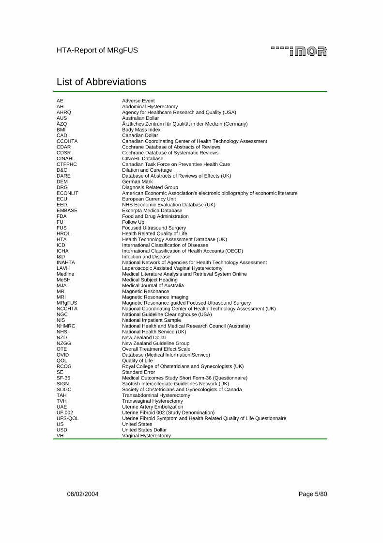
List of Abbreviations AE Adverse Event AH Abdominal Hysterectomy AHRQ Agency for Healthcare Research and Quality (USA) AUS Australian Dollar ÄZQ Ärztliches Zentrum für Qualität in der Medizin (Germany) BMI Body Mass Index CAD Canadian Dollar CCOHTA Canadian Coordinating Center of Health Technology Assessment CDAR Cochrane Database of Abstracts of Reviews CDSR Cochrane Database of Systematic Reviews CINAHL CINAHL Database CTFPHC Canadian Task Force on Preventive Health Care D&C Dilation and Curettage DARE Database of Abstracts of Reviews of Effects (UK) DEM German Mark DRG Diagnosis Related Group ECONLIT American Economic Association's electronic bibliography of economic literature ECU European Currency Unit EED NHS Economic Evaluation Database (UK) EMBASE Excerpta Medica Database FDA Food and Drug Administration FU Follow Up FUS Focused Ultrasound Surgery HRQL Health Related Quality of Life HTA Health Technology Assessment Database (UK) ICD International Classification of Diseases ICHA International Classification of Health Accounts (OECD) I&D Infection and Disease INAHTA National Network of Agencies for Health Technology Assessment LAVH Laparoscopic Assisted Vaginal Hysterectomy Medline Medical Literature Analysis and Retrieval System Online MeSH Medical Subject Heading MJA Medical Journal of Australia MR Magnetic Resonance MRI Magnetic Resonance Imaging MRgFUS Magnetic Resonance guided Focused Ultrasound Surgery NCCHTA National Coordinating Center of Health Technology Assessment (UK) NGC National Guideline Clearinghouse (USA) NIS National Impatient Sample NHMRC National Health and Medical Research Council (Australia) NHS National Health Service (UK) NZD New Zealand Dollar NZGG New Zealand Guideline Group OTE Overall Treatment Effect Scale OVID Database (Medical Information Service) QOL Quality of Life RCOG Royal College of Obstetricians and Gynecologists (UK) SE Standard Error SF-36 Medical Outcomes Study Short Form-36 (Questionnaire) SIGN Scottish Intercollegiate Guidelines Network (UK) SOGC Society of Obstetricians and Gynecologists of Canada TAH Transabdominal Hysterectomy TVH Transvaginal Hysterectomy UAE Uterine Artery Embolization UF 002 Uterine Fibroid 002 (Study Denomination) UFS-QOL Uterine Fibroid Symptom and Health Related Quality of Life Questionnaire US United States USD United States Dollar VH Vaginal Hysterectomy

HTA-Report of MRgFUS
06/02/2004 Page 6/80
Executive Summary On the basis of the health technology assessment detailed in the full report, it can be concluded that „Magnetic Resonance guided Focused Ultra Sound Surgery“ (MRgFUS) – as an alternative to hysterectomy, myomectomy and uterine artery embolization (UAE) in women with uterine fibroids (myoma) – is clearly more beneficial to the patient and almost equally important, its implementation would result in a substantial economic advantage, e.g. the reduction in hospital stay and sick-leave days, compared to the usual standard treatments. Additionally, noticeable improvement in each of the eight subscales of the Medical Outcomes Study Short Form-36 (SF 36) as well as in each of the seven subscales of Uterine-Fibroid-Symptom Severity Score can be expected. Since MRgFUS is a non-invasive treatment, it is associated with substantially less use of antibiotics, analgesics and anesthetics. A low rate of adverse events is reported. There is also evidence that MRgFUS therapy is associated with less pain compared to hysterectomy and myomectomy and even more so with respect to UAE. The procedure can be repeated at any time with the added advantage that it preserves the uterus and therefore enabling the chances for future fertility if so desired. Finally, patients are generally in the position to leave the hospital on the same day.
Scientific Summary The scientific summary is a comprehensive summary of the health technology assessment (HTA) report in order to allow for a quick assessment of the report’s relevance, quality, and main findings to determine it’s future consideration. The purpose of this report was, to compare the innovative treatment approach using „Magnetic Resonance guided Focused Ultrasound Surgery“ (MRgFUS) in women with uterine fibroids (myoma) with the established treatments hysterectomy, myomectomy, and the new procedure Uterine Artery Embolization (UAE). The main considerations were efficacy, safety, as well as economic aspects between the treatment approaches under examination based on a pivotal clinical study and a systematic literature review. The prospective, non-randomized clinical study was performed in eight centres of five countries in order to compare the safety and efficacy of MRgFUS (N=109) versus abdominal hysterectomy (N=83). Follow-Up visits were completed at week 1 and month 1, 3, and 6 after treatment to evaluate the patient’s overall health status and to report any complications that may or may not be related to the intervention as a measure of safety. The OTE, SF-36, and UFS-QOL were administered to assess Quality of Life. The systematic literature review used six databases Medline, Embase, Cinahl, Econlit, CDAR, and CDSR in order to accumulate information that was available up until the cut-off point of June 2003. An extensive search strategy involving 138 search steps was undertaken that yielded 1053 articles, of which 542 articles were considered eligible for abstract screening, 131 articles eligible for full-text screening, and finally 47 articles suitable for the review. Data from these 47 articles were extracted and entered into a specially designed electronic database that allowed for the possibility of entering not only general information about the conducted studies, but information on the specific treatment groups as well. A systematic search of the HTA database of NHS Center for Reviews and Dissemination identified 179 articles including 40 articles that were relevant for cost assessment of the comparative treatment regimes hysterectomy, myomectomy, and UAE. Costs for MRgFUS therapy were calculated using an analytical model. Treatment with MRgFUS was associated with a remarkable improvement of uterine fibroid symptoms and eight SF-36 subcategories. The mean change of the main symptom severity score changed by 24 points (p-value < 0.0001) at month 3 and remained stable up to month 6. All SF-

HTA-Report of MRgFUS
06/02/2004 Page 7/80
36 subscales (physical function, role physical, bodily pain, general health, vitality, role-emotional, social functioning, mental health) increased, with up to 17.3 points (bodily pain) at month one and even by 27.3 points (role-physical) at month 6. Immediately after the treatment with MRgFUS the majority of patients had no pain (75 %) or discomfort (68 %) whereas most of rest reported only mild pain (18 %) and discomfort (25 %). At each follow up visit (week 1, month 1, 3, 6) about 94 % of the MRgFUS patients had no or only mild physician-reported clinically significant findings in all subcategories (redness of abdominal wall, scaring/dimpling/retraction of abdomen, ulceration of abdomen, swelling in treatment area, firmness in treatment area, internal bleeding, external bleeding, abnormal vaginal discharge, bowel symptoms, bladder symptoms, nausea, vomiting, fever, pain at treatment side). Similarly, approximately 90 % of MRgFUS-patients did not report or had only mild status in the categories discomfort, pain, abdominal tenderness, paresthesis, other. Eleven/ 26 publications were chosen for review that focused on 19/ 39 treatment patterns with respect to efficacy and safety of hysterectomy/ myomectomy. Sixteen publications about UAE were reviewed for the assessment of efficacy and safety. The assessment of this literature showed that inconsistencies existed between them in terms of their quality and means of reporting of the severity of symptoms, uterine and fibroid anatomy, definition of adverse events, inclusion or exclusion criteria, inhomogeneous application of antibiotics, analgesics, preoperative hormonal treatment, and/or anaesthetics, as well as the recruitment periods and follow up periods. Therefore, it is difficult to assemble a comprehensive, definitive summary of the literature findings. Most studies about the efficacy of hysterectomy as well as most studies about the efficacy of myomectomy primarily reported transfusion rates as efficacy parameters, ranging from 3% to 23% for hysterectomy and 0% to 50% for myomectomy. Studies about the efficacy of UAE focused on the shrinkage of the fibroid, ranging from 23 % up to 86 %, although these figures are heavily dependent on the time of follow-up. Thus, the comparative assessment of efficacy is again complicated by the divergent study designs. With respect to the appraisal of safety – again – heterogeneous study designs constitute a substantial bias when attempting to make a comparative assessment, firstly with respect how the respective adverse events were defined and the time of follow up. Generally, hysterectomy/ myomectomy is associated with a mean blood loss of 200 up to 600 ml, and a fever rate of up to 50 %/ 56 %, while UAE is associated with a higher rate of post-operative pain. According to available information, hysterectomy/ myomectomy/ UAE caused a mean time for hospitalization between 2-10/ 1-8/ 1-4 days respectively. In addition, the mean time for recuperation was reported as lasting 29/ 20/ 7 days for the 3 comparative treatment approaches. The most empirical data were found concerning the costs of hysterectomy, ranging from 1,900 USD up to 19,393 USD (reference year 2003). Corresponding information about myomectomy-related costs and UAE-induced costs was much more difficult to obtain and restricts making general cost estimates for these procedures. This HTA report was initiated and sponsored by INSIGHTEC, the manufacturer of the Exablate2000 device, who had no direct influence on the contents of the report. The HTA report was conducted by IMOR GmbH, which is an independent Institute for Medical Outcome Research with long-term experience in producing HTA reports to the accepted scientific standards. The report is mainly addressed to medical decision makers, but could be useful for patients and clinicians as well. Overall, it can be concluded that MRgFUS is associated with remarkable improvement of quality of life, a low rate of adverse events, and lower associated costs due to low treatment duration and a shorter recovery time in women with uterine fibroids (myoma) compared with the usual treatment regimes.

HTA-Report of MRgFUS
06/02/2004 Page 8/80
1 Introduction
1.1 Background Information Uterine fibroids (leiomyoma, myoma) are the most common neoplasms of the female pelvis. The size of these benign tumors varies from that of a pinhead to larger than a melon (1). Fibroids generate from the diseased wall of the uterine and are classified in three ways, depending on their location (see Figure 1).
Figure 1 Characterization of Fibroids
Intramural fibroids grow within the myometrium. They are the most common fibroids. Subserosal fibroids grow out from the serosa. They can be either stalk-like (pedunculated) or broad-based (sessile). These are the second most common fibroids. Submucous fibroids grow from the endometrium. They can also be stalk-like or broad-based. Only about 5% of fibroids are submucous (2).
The exact cause of fibroids is unknown. Fibroids are most common among women between 30 to 40 years. Being Afro-American and having high exposure to estrogen increase the risk for fibroids. A family history and being overweight tend to increase the risk slightly, whereas giving birth and being athletic seem to lower the risk. In addition, the formation of fibroids may be attributable to abnormalities in substances called growth factors. Less than 25% of patients with fibroids experience symptoms (3). If fibroids trigger symptoms, it is generally associated with their size, number or location. The most common symptom for which women seek treatment is abdominal uterine bleeding (menorrhagia). Other complaints associated with uterine fibroids may include pelvic discomfort and pain, pressure on the bladder or bowels, leading to increased urinary frequency, incontinence or constipation. Infertility, miscarriage, and increased risk of complication during pregnancy are additional symptoms of fibroids.
Fibroids
Intramural Subserosal Submucous
Stalk-like Broad-based Stalk-like Broad-based
Fibroids
Intramural Subserosal Submucous
Stalk-like Broad-based Stalk-like Broad-based

HTA-Report of MRgFUS
06/02/2004 Page 9/80
1.2 Epidemiology of Uterine Fibroids Uterine fibroids are one of the most common conditions affecting women at reproductive age. The overall prevalence of fibroids in the population varies depending on the population examined, whether asymptomatic women are included, and the sensitivity and specificity of the methods used to detect fibroids. A recent Scandinavian study using ultrasound in a random sample of 335 asymptomatic women aged 25–40 found an overall prevalence of 5.4 %, with the prevalence increasing with age (3.3 % in women aged 25–32 vs. 7.8 % in women aged 33–40) (4). Kjerulff et al. showed in an analysis of discharge data with more than 53,000 hysterectomies, that black women were more than twice as likely to have a diagnosis of uterine fibroids than white women (65,4% versus 28,5%) (5), while Materia et al. assessed that 41 % of 3,141 hysterectomies were attributed to fibroids (6). The incidence is more difficult to estimate. Most available sources of data are hospital-based. The annual incidence of diagnosed fibroids in a prospective cohort of US women aged 25-44 was 12,8 per 1000 women/years (7). The incidence of fibroids in Germany is based on hospital statistics and is similar to the US. According to the German Federal Bureau of Statistics, 94,066 hospitalizations were recorded in the year 2000 due to fibroids. The incidence was calculated by correlating these figures with the number of female members of the statutory health insurances (Table 1). The statutory health insurances cover 89% of all insured people in Germany (8), therefore the actual incidence may be even higher.
Table 1 Frequency of Hospital Diagnoses “Leiomyoma of the uterus” (ICD 10-code D25)
Age Number of Cases Number of Females
Incidence (Cases per 10,000)/year
< 25 248 9,185,353 0,3
25–30 1,393 1,997,263 7,0
30–35 5,187 2,572,931 20,2
35–40 13,611 3,109,155 43,8
40–45 22,507 2,919,637 77,1
45–50 24,630 2,530,622 97,4
50–55 13,361 2,367,880 56,5
55–60 6,063 1,976,352 30,7
60–65 3,772 2,641,466 14,3
> 65 3,294 8,265,792 4,0
Total 94,066 37,566,451 25,0
Source: German Federal Bureau of Statistics (2000), “Gesundheit VIII A”
HTA-Report of MRgFUS
06/02/2004 Page 10/80
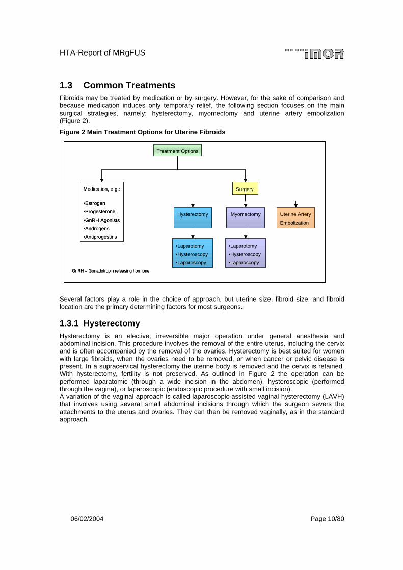
1.3 Common Treatments Fibroids may be treated by medication or by surgery. However, for the sake of comparison and because medication induces only temporary relief, the following section focuses on the main surgical strategies, namely: hysterectomy, myomectomy and uterine artery embolization (Figure 2).
Figure 2 Main Treatment Options for Uterine Fibroids
Several factors play a role in the choice of approach, but uterine size, fibroid size, and fibroid location are the primary determining factors for most surgeons.
1.3.1 Hysterectomy Hysterectomy is an elective, irreversible major operation under general anesthesia and abdominal incision. This procedure involves the removal of the entire uterus, including the cervix and is often accompanied by the removal of the ovaries. Hysterectomy is best suited for women with large fibroids, when the ovaries need to be removed, or when cancer or pelvic disease is present. In a supracervical hysterectomy the uterine body is removed and the cervix is retained. With hysterectomy, fertility is not preserved. As outlined in Figure 2 the operation can be performed laparatomic (through a wide incision in the abdomen), hysteroscopic (performed through the vagina), or laparoscopic (endoscopic procedure with small incision). A variation of the vaginal approach is called laparoscopic-assisted vaginal hysterectomy (LAVH) that involves using several small abdominal incisions through which the surgeon severs the attachments to the uterus and ovaries. They can then be removed vaginally, as in the standard approach.
Treatment Options
Medication, e.g.:
•Estrogen
•Progesterone
•GnRH Agonists
•Androgens
•Antiprogestins
Surgery
GnRH = Gonadotropin releasing hormone
Hysterectomy Myomectomy Uterine Artery
Embolization
•Laparotomy
•Hysteroscopy
•Laparoscopy
•Laparotomy
•Hysteroscopy
•Laparoscopy
Treatment Options
Medication, e.g.:
•Estrogen
•Progesterone
•GnRH Agonists
•Androgens
•Antiprogestins
Surgery
GnRH = Gonadotropin releasing hormone
Hysterectomy Myomectomy Uterine Artery
Embolization
•Laparotomy
•Hysteroscopy
•Laparoscopy
•Laparotomy
•Hysteroscopy
•Laparoscopy
HTA-Report of MRgFUS
06/02/2004 Page 11/80
1.3.2 Myomectomy For women seeking an alternative to hysterectomy that would allow them to retain their uterus (in order to bear children or for cultural, psychological, or sexual reasons), myomectomy removes only the visible and accessible fibroids, leaving the uterus in place. Myomectomy may also help to regulate abnormal uterine bleeding caused by fibroids. This procedure has certain limitations including those involving numerous, large fibroids or cancer. In these cases, conversion to a full hysterectomy may be necessary. Like hysterectomy, this procedure may be accomplished by laparotomy or less invasive means, such as hysteroscopy or laparoscopy, respectively. Laparotomy is used for subserosal or intramural, very large or numerous fibroids. After the fibroids are removed, careful reconstruction of the uterine wall is critical in both laparotomy and laparoscopy, so that bleeding and infection do not occur. A hysteroscopic myomectomy may be used for submucous fibroids found in the uterine cavity. A hysteroscopic resectoscope (a thin scope that contains surgical and viewing instruments) is passed up into the uterine cavity through the vagina and cervical canal. A wire loop, conducting electrical current, is then used to shave off the fibroid. Women whose uterus is no larger than it would be at a six-weeks pregnancy and who have a small number of subserous fibroids may be eligible for treatment with laparoscopy as well as with hysteroscopy.
1.3.3 Uterine Artery Embolization Uterine Artery Embolization (UAE), also called uterine fibroid embolization, is a radiological alternative to surgery. It destroys fibroids by depriving them of their blood supply. The procedure is typically performed by the insertion of a catheter into a uterine artery. Small particles are injected at the point where the artery feeds the blood vessels leading to the uterine fibroid. They can be made of organic compounds (e.g. polyvinyl alcohol particles) or acrylic materials (e.g. embosphere microspheres). The particles block the blood supply to the tiny arteries that feed abnormal fibroid cells and the tissue eventually dies. Circulation to normal uterine tissue, however, is usually restored. In general, UAE is an option only for those who have finished childbearing. Although UAE may protect fertility in many women, the procedure does pose some risk for ovarian failure and infertility.
1.3.4 Other Treatment Options Medical treatment with synthetic hormones, such as gonadotropin agonists and progestins, results in a variable and temporary reduction in size of the uterine. Thermal ablation techniques induce thermal coagulation of the fibroid(s), while producing a minimal disturbance to the endometrium, uterine wall or surrounding abdominal anatomy.

HTA-Report of MRgFUS
06/02/2004 Page 12/80
1.4 Epidemiology of Service The prevalence of fibroid-related hospitalization based on the Nationwide Inpatient Sample (NIS) were 26 to 28 admissions per 1000 cases. These numbers included women between the ages of 15 and 64 years. The highest rate of fibroids diagnosis was seen in women aged 35-54 years (e.g. 70% for women aged between 40 and 44 years in 1992) (9). Hysterectomy is the second most frequently performed surgery in premenopausal women (Caesarean sections are first). By the age of 60, about a third of American women have undergone this procedure. Luoto et al. report an increase in the incidence of hysterectomies within the Finnish female population from 311/100,000 women to 400/100,000 from 1979 to 1986, whereas half of the hysterectomies were performed for fibroids (10). Similarly, fibroids are the most commonly listed discharge diagnosis for hysterectomy in the US, accounting for a third of all hysterectomies, or 140,000 cases annually (11). In a major 2002 Government report, 68% of fibroid-related hysterectomies were performed on Afro-American women, 33% in Caucasians, and 45% among women of other ethnic groups (7). Abdominal hysterectomy is the most common procedure and is used in over 80% of hysterectomies on Afro-American women and about 60% in Caucasian and other ethnic groups. Vaginal hysterectomy is used in less than 20% of the cases on Afro-American women and slightly under 40% on Caucasian and other groups (7). In comparison to white women, black women having hysterectomy were found to have an increased risk of one or more complications of surgical or medial care (odds ratio 1.4, 95% CI 1.3-1.5) and more than three times the in-hospital mortality rate (odds ratio 3.1, 95% CI 2.0-4.8) (5). According to NIS data at least 37,000 myomectomies, are performed annually (1). A literature review shows that 50% of patients undergoing myomectomy become pregnant, but face a higher risk of caesarean section or miscarriage (12). The cumulative recurrence rate for fibroid growth after myomectomy, severe enough to need additional treatment, has been reported at 51% after 5 years (13). UAE for the treatment of fibroids is a more recent development. The number of UAE procedures in the US increased from 50 in 1996 to more than 4,000 in 1999 (14). The success rate for this treatment ranges between 85% and 94% (15-17). Forman et al. reported that 2/192 women undergoing UAE became pregnant, however, it should be pointed out that only 17 patients from their study population was <40 years (12). Further treatment was required in 29% of patients who underwent UAE (18). However, it was reported that a less invasive approach was necessary in these cases. Up to now there is no Germany-specific data on the frequency of the available treatment. In addition to the outlined literature review process several other institutions were approached including the German Federal Bureau of Statistics, the German Hospital Institute, and the German Hospital Society.
HTA-Report of MRgFUS
06/02/2004 Page 13/80
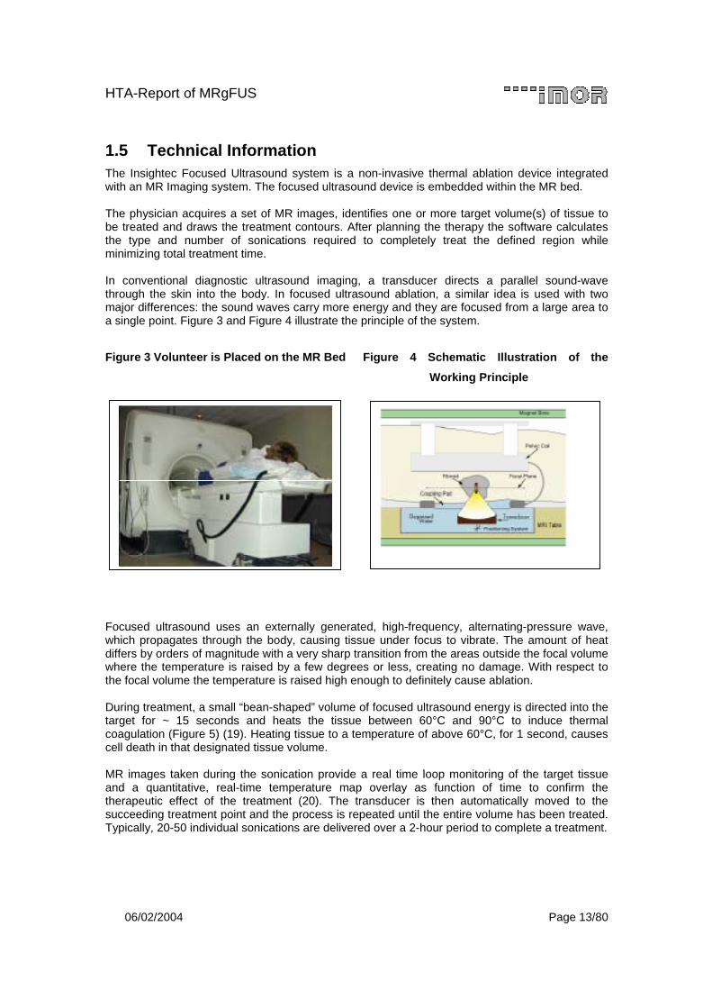
1.5 Technical Information The Insightec Focused Ultrasound system is a non-invasive thermal ablation device integrated with an MR Imaging system. The focused ultrasound device is embedded within the MR bed. The physician acquires a set of MR images, identifies one or more target volume(s) of tissue to be treated and draws the treatment contours. After planning the therapy the software calculates the type and number of sonications required to completely treat the defined region while minimizing total treatment time. In conventional diagnostic ultrasound imaging, a transducer directs a parallel sound-wave through the skin into the body. In focused ultrasound ablation, a similar idea is used with two major differences: the sound waves carry more energy and they are focused from a large area to a single point. Figure 3 and Figure 4 illustrate the principle of the system.
Figure 3 Volunteer is Placed on the MR Bed Figure 4 Schematic Illustration of the
Working Principle
Focused ultrasound uses an externally generated, high-frequency, alternating-pressure wave, which propagates through the body, causing tissue under focus to vibrate. The amount of heat differs by orders of magnitude with a very sharp transition from the areas outside the focal volume where the temperature is raised by a few degrees or less, creating no damage. With respect to the focal volume the temperature is raised high enough to definitely cause ablation. During treatment, a small “bean-shaped” volume of focused ultrasound energy is directed into the target for ~ 15 seconds and heats the tissue between 60°C and 90°C to induce thermal coagulation (Figure 5) (19). Heating tissue to a temperature of above 60°C, for 1 second, causes cell death in that designated tissue volume. MR images taken during the sonication provide a real time loop monitoring of the target tissue and a quantitative, real-time temperature map overlay as function of time to confirm the therapeutic effect of the treatment (20). The transducer is then automatically moved to the succeeding treatment point and the process is repeated until the entire volume has been treated. Typically, 20-50 individual sonications are delivered over a 2-hour period to complete a treatment.
HTA-Report of MRgFUS
06/02/2004 Page 14/80
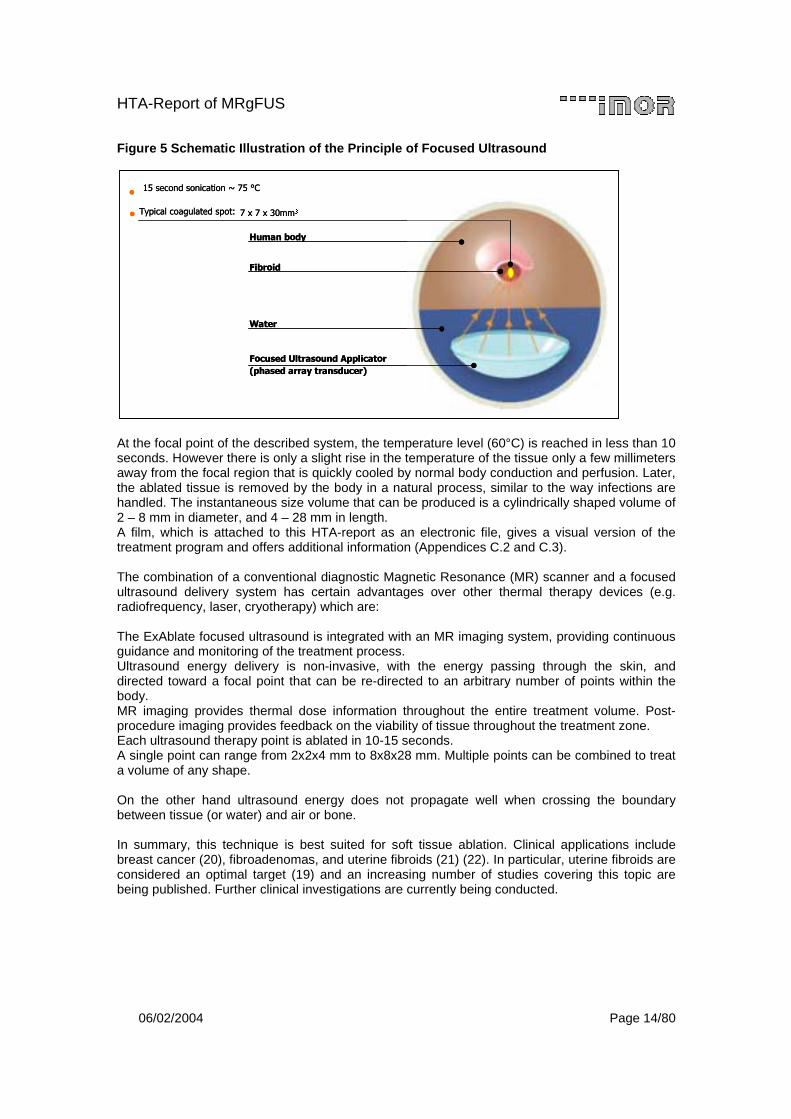
Figure 5 Schematic Illustration of the Principle of Focused Ultrasound
At the focal point of the described system, the temperature level (60°C) is reached in less than 10 seconds. However there is only a slight rise in the temperature of the tissue only a few millimeters away from the focal region that is quickly cooled by normal body conduction and perfusion. Later, the ablated tissue is removed by the body in a natural process, similar to the way infections are handled. The instantaneous size volume that can be produced is a cylindrically shaped volume of 2 – 8 mm in diameter, and 4 – 28 mm in length. A film, which is attached to this HTA-report as an electronic file, gives a visual version of the treatment program and offers additional information (Appendices C.2 and C.3). The combination of a conventional diagnostic Magnetic Resonance (MR) scanner and a focused ultrasound delivery system has certain advantages over other thermal therapy devices (e.g. radiofrequency, laser, cryotherapy) which are: The ExAblate focused ultrasound is integrated with an MR imaging system, providing continuous guidance and monitoring of the treatment process. Ultrasound energy delivery is non-invasive, with the energy passing through the skin, and directed toward a focal point that can be re-directed to an arbitrary number of points within the body. MR imaging provides thermal dose information throughout the entire treatment volume. Post-procedure imaging provides feedback on the viability of tissue throughout the treatment zone. Each ultrasound therapy point is ablated in 10-15 seconds. A single point can range from 2x2x4 mm to 8x8x28 mm. Multiple points can be combined to treat a volume of any shape. On the other hand ultrasound energy does not propagate well when crossing the boundary between tissue (or water) and air or bone. In summary, this technique is best suited for soft tissue ablation. Clinical applications include breast cancer (20), fibroadenomas, and uterine fibroids (21) (22). In particular, uterine fibroids are considered an optimal target (19) and an increasing number of studies covering this topic are being published. Further clinical investigations are currently being conducted.
��������
��� ��
����
���������� ��������������� ���������� ��� ������ �
�������� �������� �����
����������������������� �������������
��������
��� ��
����
���������� ��������������� ���������� ��� ������ �
�������� �������� �����
����������������������� �������������
HTA-Report of MRgFUS
06/02/2004 Page 15/80
1.6 Policy Question The HTA-Report on hand is addressed to one key research question: What are the advantages and what are the disadvantages of using Magnetic Resonance guided Focused Ultrasound Surgery (MRgFUS) for the treatment of uterine fibroids? Assessment criteria are:
• Safety • Efficacy • Costs.
Comparative interventions are:
• Hysterectomy • Myomectomy • Uterine Artery Embolization.
Focused perspectives are: • Patient • Healthcare Provider • Third Party Payers.
2 Methodology
2.1 Project Plan Due to a broad, multi-national target audience (healthcare authorities, healthcare providers, clinicians, patients and their associated advocates) we based our report on the proposed framework “Best practice in undertaking and reporting HTA” conducted by the EUR-ASSESS Working Group 4 (23). The policy question implicates two approaches, namely the assessment of the new MRgFUS technology and its comparison with established interventions. The assessment of MRgFUS was mainly based on the results of “A pivotal clinical study to evaluate the safety and effectiveness of MRgFUS in the treatment of uterine fibroids”. Information about the comparative interventions was captured by means of a systematic literature review. Each step of the literature review was planned carefully a priori and documented to ensure transparency in the systematic process.
HTA-Report of MRgFUS
06/02/2004 Page 16/80
2.2 Pivotal Clinical Study This section describes the basic methodology used to conduct and analyze a study, designated as UF002 that was designed and carried out for the purpose of obtaining the final regulatory approval in the US. The objective of the trial was to evaluate the safety and effectiveness of MagneticResonance guided Focused UltraSound Surgery thermal ablation in the treatment of uterine fibroids (MRgFUS) relative to the current standard of care: abdominal hysterectomy. The effectiveness was assessed using the Uterine Fibroid Symptom and Health-Related Quality-of-Life questionnaire (UFS-QOL). The UFS-QOL was designed as a uterine-fibroid specific questionnaire to evaluate the symptoms of uterine fibroids and their impact on HRQL (24). The UFS-QOL was designed as two scales: a symptom severity scale (UFS) and a health-related quality-of-life scale (QOL) with six dimensions (concern, activities, energy/mood, control, self-consciousness, and sexual function). Both scales were measured with scores ranging from 0 to 100. Higher scores were indicative of worse UFS or better QOL. Additional outcomes were measured using the Medical Outcomes Study Short Form-36® (SF-36) to assess a patient’s general health status and recovery trajectory (25). The SF-36 consists of eight subscales (Physical Functioning, Role-Physical, Bodily Pain, Vitality, General Health Perception, Social Functioning, Role-Emotional, Mental Health) with scores from 0 to 100. Higher scores on the scale indicate better constitution. As an additional assessment the Overall Treatment Effect scale (OTE) was used. At month 3, patients were asked about the categorical change in their uterine fibroid symptoms. At month 6, patients were asked about their satisfaction and about the effectiveness of the respective treatment pattern. Physicians and subjects were asked about the existence and severity of treatment-related signs and symptoms. Physicians had 15 indications from which to choose, compared to patients who had 6 indications. All Case Report Forms (CRFs) are presented in Appendix B.2.
2.2.1 Patient Population In both non-randomized treatment arms, women had to be pre- or peri-menopausal within 12 months of last menstrual period. The raw score on the UFS-QOL Symptom Severity Screener had to be 21 or greater. Additional inclusion/exclusion criteria are quoted in the clinical study protocol, which is provided in Appendix B.1. In the test arm (MRgFUS) 176 women were enrolled while the control arm (hysterectomy) consists of 108 women.
HTA-Report of MRgFUS
06/02/2004 Page 17/80
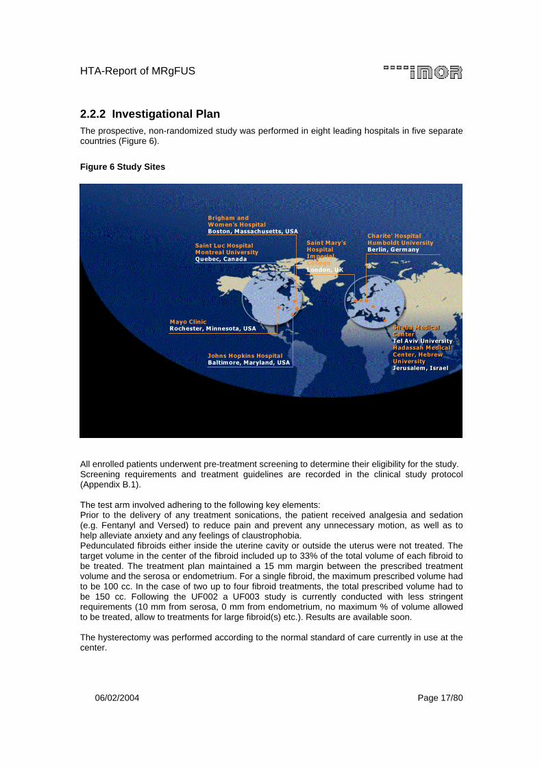
2.2.2 Investigational Plan The prospective, non-randomized study was performed in eight leading hospitals in five separate countries (Figure 6).
Figure 6 Study Sites
All enrolled patients underwent pre-treatment screening to determine their eligibility for the study. Screening requirements and treatment guidelines are recorded in the clinical study protocol (Appendix B.1). The test arm involved adhering to the following key elements: Prior to the delivery of any treatment sonications, the patient received analgesia and sedation (e.g. Fentanyl and Versed) to reduce pain and prevent any unnecessary motion, as well as to help alleviate anxiety and any feelings of claustrophobia. Pedunculated fibroids either inside the uterine cavity or outside the uterus were not treated. The target volume in the center of the fibroid included up to 33% of the total volume of each fibroid to be treated. The treatment plan maintained a 15 mm margin between the prescribed treatment volume and the serosa or endometrium. For a single fibroid, the maximum prescribed volume had to be 100 cc. In the case of two up to four fibroid treatments, the total prescribed volume had to be 150 cc. Following the UF002 a UF003 study is currently conducted with less stringent requirements (10 mm from serosa, 0 mm from endometrium, no maximum % of volume allowed to be treated, allow to treatments for large fibroid(s) etc.). Results are available soon. The hysterectomy was performed according to the normal standard of care currently in use at the center.
� ��������������������������������������������������
������ ��������������� �������!� ���"�������#����
�������� �����������$��� ����#������ �������%
#�� ���� �������������������!� ����� �����&� ���
'��������(������������������� ����� ��������
����#�����)������� ���������������� �������������
#���� *����!�!����!� ������������������#���� ����� �+����!� ���'� ��������$� ���
�������������#���� *����!�!����!� ������������������#���� ����� �+����!� ���'� ��������$� ���
HTA-Report of MRgFUS
06/02/2004 Page 18/80
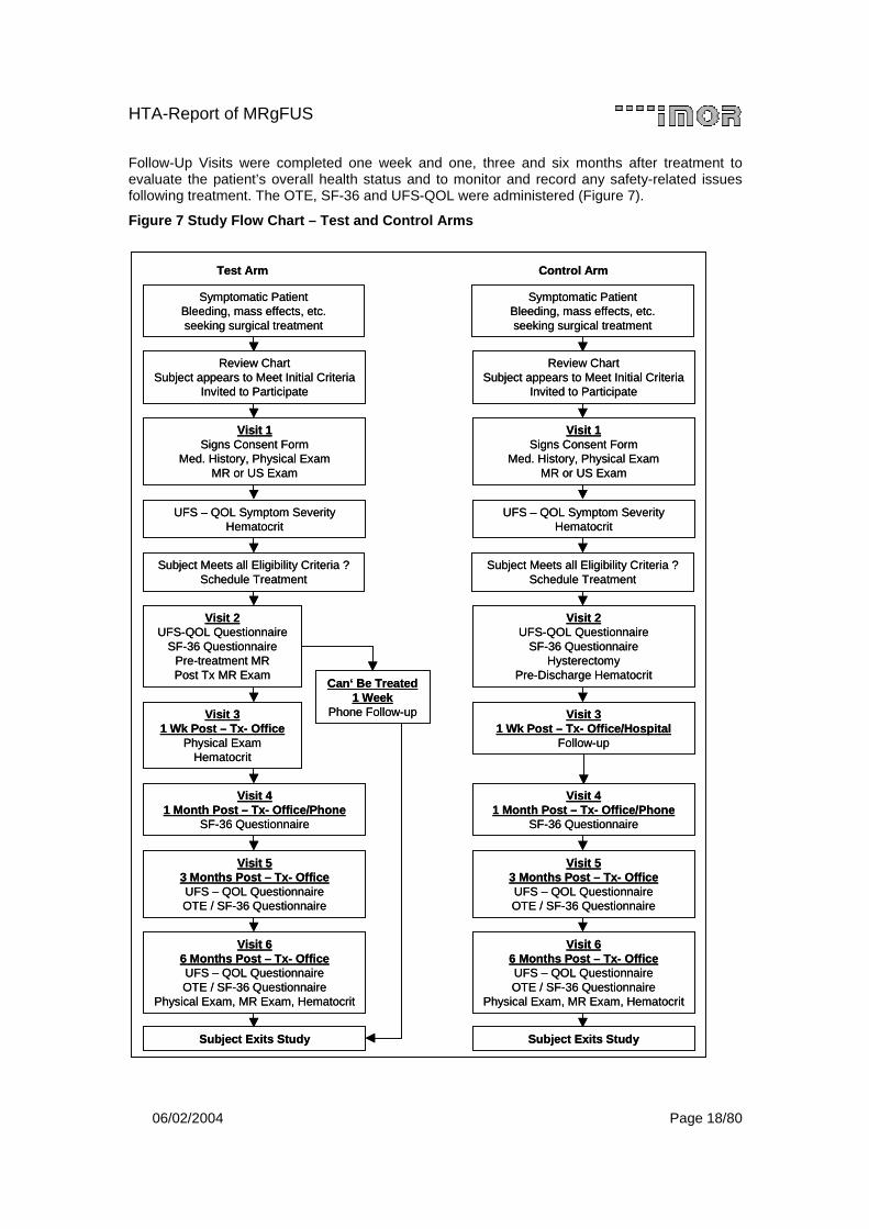
Follow-Up Visits were completed one week and one, three and six months after treatment to evaluate the patient’s overall health status and to monitor and record any safety-related issues following treatment. The OTE, SF-36 and UFS-QOL were administered (Figure 7).
Figure 7 Study Flow Chart – Test and Control Arms
Test Arm Control Arm
Symptomatic PatientBleeding, mass effects, etc. seeking surgical treatment
Review ChartSubject appears to Meet Initial Criteria
Invited to Participate
Visit 1Signs Consent Form
Med. History, Physical ExamMR or US Exam
UFS – QOL Symptom SeverityHematocrit
Subject Meets all Eligibility Criteria ? Schedule Treatment
Visit 2UFS-QOL Questionnaire
SF-36 QuestionnairePre-treatment MRPost Tx MR Exam
Visit 31 Wk Post – Tx- Office
Physical ExamHematocrit
Visit 41 Month Post – Tx- Office/Phone
SF-36 Questionnaire
Visit 53 Months Post – Tx- OfficeUFS – QOL Questionnaire OTE / SF-36 Questionnaire
Visit 66 Months Post – Tx- OfficeUFS – QOL Questionnaire OTE / SF-36 Questionnaire
Physical Exam, MR Exam, Hematocrit
Subject Exits Study
Can‘ Be Treated1 Week
Phone Follow-up
Symptomatic PatientBleeding, mass effects, etc. seeking surgical treatment
Review ChartSubject appears to Meet Initial Criteria
Invited to Participate
Visit 1Signs Consent Form
Med. History, Physical ExamMR or US Exam
UFS – QOL Symptom SeverityHematocrit
Subject Meets all Eligibility Criteria ? Schedule Treatment
Visit 2UFS-QOL Questionnaire
SF-36 QuestionnaireHysterectomy
Pre-Discharge Hematocrit
Visit 31 Wk Post – Tx- Office/Hospital
Follow-up
Visit 41 Month Post – Tx- Office/Phone
SF-36 Questionnaire
Visit 53 Months Post – Tx- OfficeUFS – QOL Questionnaire OTE / SF-36 Questionnaire
Visit 66 Months Post – Tx- OfficeUFS – QOL Questionnaire OTE / SF-36 Questionnaire
Physical Exam, MR Exam, Hematocrit
Subject Exits Study
HTA-Report of MRgFUS
06/02/2004 Page 19/80
2.2.3 Statistical Analysis Plan The primary efficacy endpoint was to achieve an improvement in the symptom severity subscale of the UFS-QOL instrument in the MRgFUS patients. To determine the degree of improvement, change scores were calculated by subtracting the pre-treatment scores from the 6-month scores. This difference was evaluated statistically by means of the one-sample paired t-test. Additionally, a success rate was calculated by tabulating the patients who achieved a 10-point improvement in the total HRQOL score at month 6. The percentage of patients who achieved this level of improvement was calculated and reported with their 95% confidence interval. One-sample paired t-tests were used to evaluate the within-group differences between pre-treatment and post-treatment. Two-sample t-tests were used to compare the differences between the treatment groups. The trajectory of change was analyzed descriptively by regression slopes. Adverse events were recorded and reported along with 95% confidence intervals. The severity of each separate event was categorized as being mild, moderate or severe as well as the suspected relationship of the treatment pattern to the event. To facilitate a suitable comparison of the relative risks of the MRgFUS test arm versus the hysterectomy control arm, a common set of “significant complications” was defined. The primary statistical comparison between treatment groups was conducted on the basis of the reported incidence of these complications. The rate of major adverse events was compared between treatment groups using the Fisher’s exact test. Significant Clinical Complications were defined as
• Fever (i.e. oral temperature > 100.4�������������� ���������������������������st 24 hours post-treatment)
• Antibiotic treatment started > 24 hours after treatment • Intra-operative or post-operative blood transfusion • Unintended major surgical procedures related to treatment (i.e. laparotomy, repair of
perforated viscus, repair of major blood vessels, bowel, or bladder intraoperatively or post-operatively during the same hospitalization; repair of skin burn)
• Discharge requiring referral to a rehabilitation facility, visiting nurse or home health care follow-up
• Life-threatening cardiac or respiratory arrest or other life threatening event • Re-hospitalization longer than 24 hours • Interventional treatment within 42 days of treatment
(hematoma drainage, wound I&D, radiographic embolization, D&C) • Outpatient treatment of significant new medical problem believed to be related to
treatment (i.e. anticoagulation therapy for DVT) • Death within 42 days of treatment
The statistical analysis plan for the Baseline and Efficacy Sections are given in Appendices B.3 and B.4.

HTA-Report of MRgFUS
06/02/2004 Page 20/80
2.3 Literature Review A comprehensive review of the literature, from identification of sources in databases through the screening and extraction of individual articles, was undertaken as an iterative, sequential process. This section describes the basic methodology used to conduct the literature search, screening, and data extraction process.
2.3.1 Sources of Data In order to complete a thorough literature review, 5 different types of sources were searched namely one pivotal study, computerized bibliographical databases, NHS databases, internet domains of the members of INAHTA, and specific medicinal societies. Six of the most widely used computerized bibliographical reference databases served as the primary sources for the literature review, namely
Database Period under Review
MEDLINE 1966 to July Week 1 2003 EMBASE 1980 to 2003 Week 27 CINAHL 1982 to July Week 1 2003 Econlit 1969 to June 2003 CDAR (Cochrane Database of Abstracts of Reviews) 2nd Quarter 2003 CDSR (Cochrane Database of Systematic Reviews) 2nd Quarter 2003 The Cancerlit database was excluded due to the poor quality of results as reported in another HTA report on this topic (26). The final search was completed July 10th 2003. Additionally, the HTA database of the NHS Center for Reviews and Dissemination (including DARE, NHS EED, and HTA) was searched July 18th 2003 with the objective to identify any HTA-reports, systematic reviews and health-economic publications about the management of uterine fibroids. Finally, a search was made addressing the Internet-domains of all INAHTA members as detailed in Table 2.
Table 2 Internet-Domains of INAHTA-members
Institution/Society URL Agency for Healthcare Research and Quality (AHRQ) http://www.ahcpr.gov Canadian Coordinating Office for Health Technology Assessment (CCOHTA) http://www.ccohta.ca British Columbia Office of Health Technology Assessment (BCOHTA) http://www.chspr.uc.ca/bcohta Danish Institute for Health Technology Assessment http://www.dsi.dk Danish Center for Evaluation and HTA (DACEHTA) http://ww.dacehta.dk Finnish Office for Health Care Technology Assessment http://www.stakes.fi/finohta/ The Swedish Council on Technology Assessment in Health Care (SBU) http://www.sbu.se Center for Medical Technology Assessment (CMT) http://www.cmt.liu.se The Norwegian Center for Health Technology Assessment (SMM) http://www.sintef.no/smm National Coordinating Centre for Health Technology Assessment (NCCHTA) http://www.hta.nhsweb.nhs.uk National Institute for Clinical Excellence (NICE/CRD) http://www.nice.org.uk Health Technology Board for Scotland (HTBS) http://www.htbs.co.uk HTA Unit of the Institute of Technology Assessment http://www.oeaw.ac.at/ita/hta German Agency for Health Technology Assessment (DAHTA) http://www.dahta.dimdi.de New Zealand Health Technology Assessment http://www.nzhta.chmeds.ac.nz Swiss Science and Technology Council/ Technology http://www.ta-swiss.ch Basque Office for Health Technology Assessment, (OSTEBA) http://www.euskadi.net/sanidad/osteba Catalan Agency for Health Technology Assessment and Research (CAHTA) http://www.aatm.es
HTA-Report of MRgFUS
06/02/2004 Page 21/80
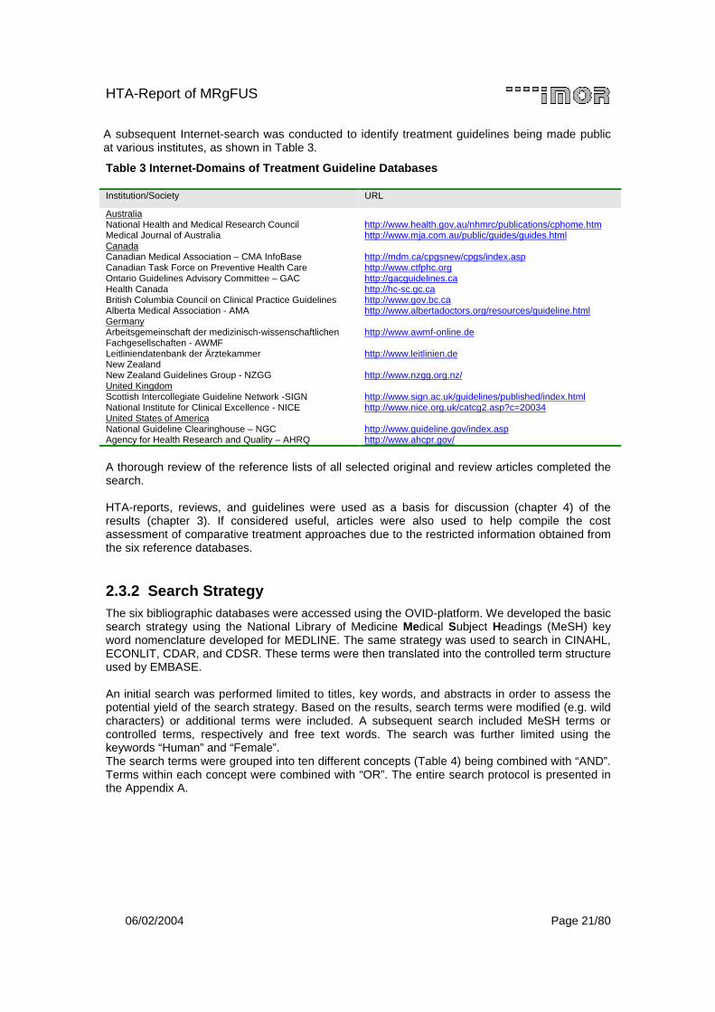
A subsequent Internet-search was conducted to identify treatment guidelines being made public at various institutes, as shown in Table 3.
Table 3 Internet-Domains of Treatment Guideline Databases
Institution/Society URL
Australia National Health and Medical Research Council http://www.health.gov.au/nhmrc/publications/cphome.htm Medical Journal of Australia http://www.mja.com.au/public/guides/guides.html Canada Canadian Medical Association – CMA InfoBase http://mdm.ca/cpgsnew/cpgs/index.asp Canadian Task Force on Preventive Health Care http://www.ctfphc.org Ontario Guidelines Advisory Committee – GAC http://gacguidelines.ca Health Canada http://hc-sc.gc.ca British Columbia Council on Clinical Practice Guidelines http://www.gov.bc.ca Alberta Medical Association - AMA http://www.albertadoctors.org/resources/guideline.html Germany Arbeitsgemeinschaft der medizinisch-wissenschaftlichen Fachgesellschaften - AWMF
http://www.awmf-online.de
Leitliniendatenbank der Ärztekammer http://www.leitlinien.de New Zealand New Zealand Guidelines Group - NZGG http://www.nzgg.org.nz/ United Kingdom Scottish Intercollegiate Guideline Network -SIGN http://www.sign.ac.uk/guidelines/published/index.html National Institute for Clinical Excellence - NICE http://www.nice.org.uk/catcg2.asp?c=20034 United States of America National Guideline Clearinghouse – NGC http://www.guideline.gov/index.asp Agency for Health Research and Quality – AHRQ http://www.ahcpr.gov/
A thorough review of the reference lists of all selected original and review articles completed the search. HTA-reports, reviews, and guidelines were used as a basis for discussion (chapter 4) of the results (chapter 3). If considered useful, articles were also used to help compile the cost assessment of comparative treatment approaches due to the restricted information obtained from the six reference databases.
2.3.2 Search Strategy The six bibliographic databases were accessed using the OVID-platform. We developed the basic search strategy using the National Library of Medicine Medical Subject Headings (MeSH) key word nomenclature developed for MEDLINE. The same strategy was used to search in CINAHL, ECONLIT, CDAR, and CDSR. These terms were then translated into the controlled term structure used by EMBASE. An initial search was performed limited to titles, key words, and abstracts in order to assess the potential yield of the search strategy. Based on the results, search terms were modified (e.g. wild characters) or additional terms were included. A subsequent search included MeSH terms or controlled terms, respectively and free text words. The search was further limited using the keywords “Human” and “Female”. The search terms were grouped into ten different concepts (Table 4) being combined with “AND”. Terms within each concept were combined with “OR”. The entire search protocol is presented in the Appendix A.

HTA-Report of MRgFUS
06/02/2004 Page 22/80
Table 4 Search Terms and Concepts
Concept Search Terms
1 Uterine Fibroids, (Uterus) Myoma, (Uterus) Leiomyoma, Fibromyoma
2 Hysterectomy, Uterus Extirpation
3 Myomectomy
4 Uterine Artery Embolization
5 High Intensity Ultrasound
6 Exablate, Focused Ultrasound Surgery
7 Incidence, Prevalence, Morbidity, Mortality, Frequency
8 Cost?, Quality of Life, Length of Stay, Hospitalization, Transfusion Units, SF 36
9
Treatment Outcome, Uterine Volume, Urinary Frequency, Haemoglobin, Hemoglobin, Haematocrit, Hematocrit, Pregnancy, Fertility, Recurrence Rate, Recurrence Risk, Complication?, Complication?, Postoperative, Complication?, Intraoperative, Side Effect?, Adverse Event?
10 Germany
Additionally DARE, NHS EED, and HTA were searched with the key words hysterectomy (title, abstract), uterine AND fibroids (text), myomectomy (text), and uterine AND artery AND embolization (text) using the Website http://144.32.228.3/scripts/WEBC.EXE/nhscrd. Most of the Internet-Domains of Guideline-Databases provided titles of the respective guidelines. If search masks were used, we inserted the search item “fibroids”.
2.3.3 Selection Criteria Two researchers assessed independently the resulting abstracts using eight hierarchically targeted selection criteria: a) Full text in English or German b) Case report > 20 cases or RCT c) Original research (no comment, no letter) d) Focus on uterine fibroids and women e) Relevant for research question f) Treatment, not diagnosis g) Outcomes or costs are reported h) Treatment not during pregnancy or caesarean section When no abstract was available titles, source, and key words were reviewed. The decisions were recorded and compared. When necessary, the reviewers reconciled differences of opinion. Exclusion criteria were documented. At this stage, articles were included if requested by one of the two reviewers. The thus obtained full text of the remaining articles was peer-reviewed with the same eight criteria plus one additional: Articles reporting unspecific or multiple indications – such as dysfunctional bleeding, pelvic pain, adnexal mass or menorrhagia – were excluded. If additional articles were suggested during the peer-review process, they went through the same screening process as the original articles. Articles found in the HTA database were checked for suitability by one reviewer using the above listed selection criteria d to h.
HTA-Report of MRgFUS
06/02/2004 Page 23/80
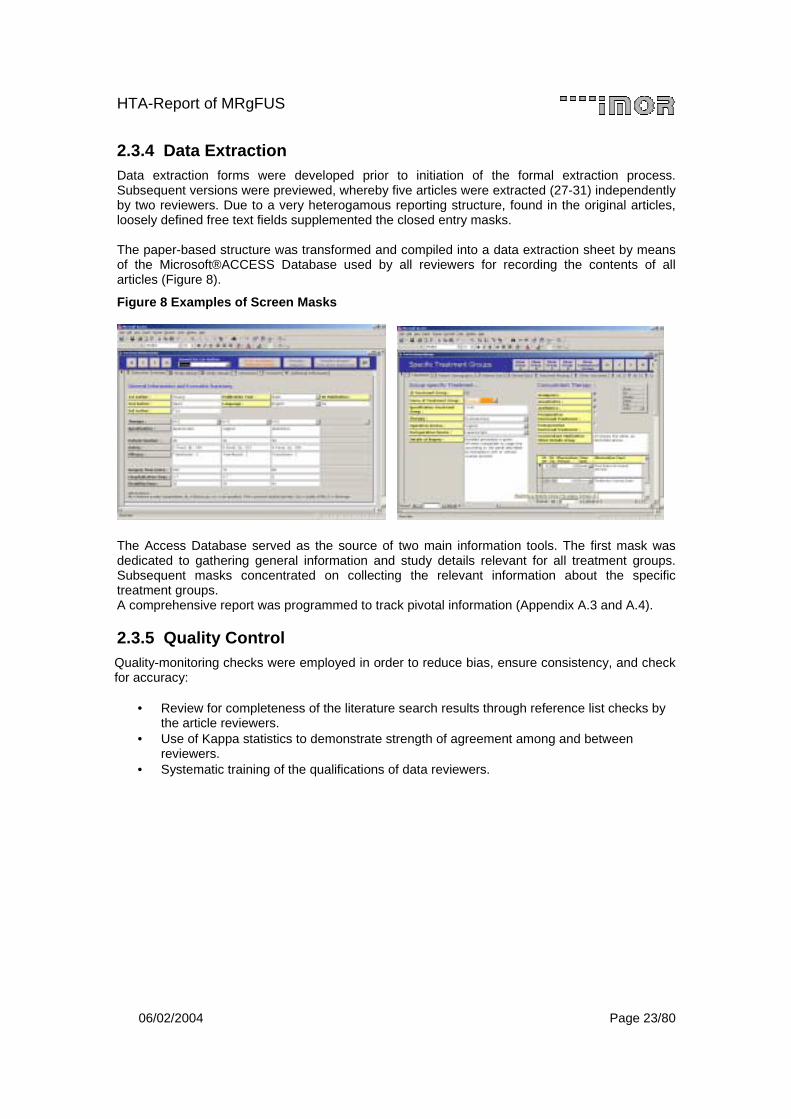
2.3.4 Data Extraction Data extraction forms were developed prior to initiation of the formal extraction process. Subsequent versions were previewed, whereby five articles were extracted (27-31) independently by two reviewers. Due to a very heterogamous reporting structure, found in the original articles, loosely defined free text fields supplemented the closed entry masks. The paper-based structure was transformed and compiled into a data extraction sheet by means of the Microsoft®ACCESS Database used by all reviewers for recording the contents of all articles (Figure 8).
Figure 8 Examples of Screen Masks
The Access Database served as the source of two main information tools. The first mask was dedicated to gathering general information and study details relevant for all treatment groups. Subsequent masks concentrated on collecting the relevant information about the specific treatment groups. A comprehensive report was programmed to track pivotal information (Appendix A.3 and A.4).
2.3.5 Quality Control Quality-monitoring checks were employed in order to reduce bias, ensure consistency, and check for accuracy:
• Review for completeness of the literature search results through reference list checks by the article reviewers.
• Use of Kappa statistics to demonstrate strength of agreement among and between reviewers.
• Systematic training of the qualifications of data reviewers.

HTA-Report of MRgFUS
06/02/2004 Page 24/80
2.4 Cost Assessment Due to the non-invasive nature of the procedure with no corresponding anaesthesia risk and infection risk, it was expected that MRgFUS would be associated with reduced consequential treatment costs. This section describes the methodology of cost assessment, including the identification of relevant cost items, the measurement of resources, and cost valuation of MRgFUS with that of comparative interventions.
2.4.1 Assessed Cost Items Several direct and indirect cost items were distinguished that originated from the International Classification for Health Accounts (ICHA) (http://www.oecd.org/dataoecd/3/40/1896840.xls). With respect to the three perspectives of the policy issue (Patient, Health Care Provider, Third-Party Payer) the intention was to find sufficient information to allow us to distinguish between modes of care (such as in-patient care, day-care, out-patient care, home-care) combined with functions or goods (such as curative care, rehabilitative care, nursing care, clinical laboratory, diagnostic imaging, patient transport, or medical goods). During the examination of health-economic publications available on the treatment of fibroids, a significant degree of differentiation existed between the reported cost items, which subsequently led to recatagorizing the literature findings in a more homogeneous format. Wherever possible, the physical units were reported first (e.g. days of hospital care), before multiplying them with the corresponding unit costs/ prices to obtain total costs in order to help interpret results and be able to adapt them more easily to other settings. Intangible costs such as somatic, mental, psychological or social factors are not reported because they can be characterized as outcome values.
2.4.2 Data Sources We referenced three types of data sources for the economic assessment. Firstly, any selected study of the literature review (Chapter 2.3), which might be considered relevant to the economic review, was flagged and forwarded to the economic reviewer. Secondly, thanks to the comprehensive health-economic, companion questionnaire that was part of the pivotal clinical study UF002 (Section 2.2), original data were available for the calculation of treatment costs and consequential costs of MRgFUS and Hysterectomy. Finally, when examining prices instead of resources used, the reimbursement visits that comprised one treatment case were investigated. Germany was chosen as the reference country because of the well-differentiated and accessible reimbursement codes, already in place. Subsequently the treatment procedure was broken down as thoroughly as possible to obtain equivalent prices.

HTA-Report of MRgFUS
06/02/2004 Page 25/80
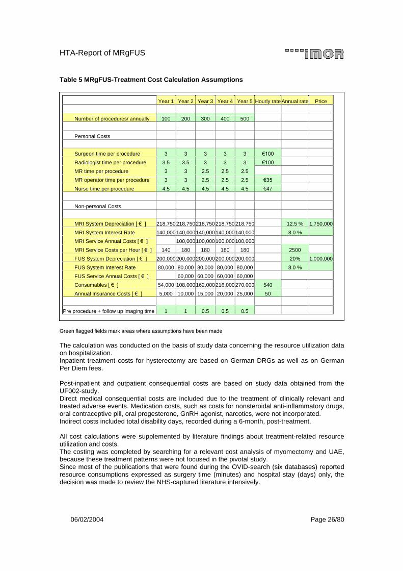
2.4.3 Costing A bottom-up approach for costing was used by adding the various cost components to obtain a complete disease-management program for a representative patient undergoing MRgFUS. Resources expended and the corresponding unit costs were reported separately. Pre-inpatient treatment costs were calculated as cost savings using MRgFUS due to requiring fewer histopathologic laboratory testing and less anesthetic and analgesic support. These cost items normally form part of the standard flat rates or DRGs. The private liquidation rates of practitioners with cottage-hospital affiliations can be taken in future cost analyses. Inpatient treatment costs for MRgFUS were based on a formula that calculated costs when assuming an amortization schedule of five years. It was assumed that the facility uses a brand new MRI with 12.5 % annual across-the board depreciation rate and 8 % interest rate, having annual MRI service agreement to the value of EUR 100,000.00 from year two and approx. EUR 1,750,000.00 in costs for the MR and facility. Average fixed costs were calculated at an assumed rate of 2,500 hours of use per annum. These were considered fixed costs because the MRI system was used for other treatment indications as well, thus working at full capacity. The FUS system costs EUR 1,000,000.00, with a 20 % across-the-board depreciation rate, 8 % interest rate, and EUR 60,000.00 in annual service costs starting at year two. In contrast to the MRI, the annual depreciation rates and service costs are patient-related and therefore considered to be variable cost components. Based on the empirical data resulting from the study UF-002 (mean procedure time = 220 minutes, SD = 56.5; range = 90.0 – 370.0) we assumed not more than three hours for the procedure and the corresponding hours and hourly rates for surgeon (3 h at EUR 100.00/h), radiologist (3 h at EUR 100.00/h), MRI technician (3 hours at a rate of EUR 35.00/h) and nurse (4.5 h at EUR 47.00/h) (see Table 5). Each patient was associated with EUR 540.00 for consumables and EUR 50.00 for insurance costs. Consumables are composed of contrast material (EBM No. 6070 = EUR 8.00), general medication (EUR 60.00), Exablate Kid/Gel Pad (EUR 150.00), Pregnancy Test (EBM No. 3850-3859 = EUR 15.00), anesthetics (EUR 35.00), and antisedativa (EUR 165.00). Facility costs (150-200 m2 accommodation use, reception, cleaning, air condition etc.) are estimated to be EUR 107.00). In the first year, 100 women were assumed to have been treated, with an additional hundred patients each subsequent year, eventually totaling 500 patients in the fifth year. Costs for physician and nursing care, provided by a department with beds (department costs) and accommodation and administrative costs of hospital (basic per diem costs), were not included because MRgFUS is characterized as being undertaken in an outpatient treatment setting.
HTA-Report of MRgFUS
06/02/2004 Page 26/80
Table 5 MRgFUS-Treatment Cost Calculation Assumptions
Green flagged fields mark areas where assumptions have been made The calculation was conducted on the basis of study data concerning the resource utilization data on hospitalization. Inpatient treatment costs for hysterectomy are based on German DRGs as well as on German Per Diem fees. Post-inpatient and outpatient consequential costs are based on study data obtained from the UF002-study. Direct medical consequential costs are included due to the treatment of clinically relevant and treated adverse events. Medication costs, such as costs for nonsteroidal anti-inflammatory drugs, oral contraceptive pill, oral progesterone, GnRH agonist, narcotics, were not incorporated. Indirect costs included total disability days, recorded during a 6-month, post-treatment. All cost calculations were supplemented by literature findings about treatment-related resource utilization and costs. The costing was completed by searching for a relevant cost analysis of myomectomy and UAE, because these treatment patterns were not focused in the pivotal study. Since most of the publications that were found during the OVID-search (six databases) reported resource consumptions expressed as surgery time (minutes) and hospital stay (days) only, the decision was made to review the NHS-captured literature intensively.
Year 1 Year 2 Year 3 Year 4 Year 5 Hourly rate Annual rate Price
Number of procedures/ annually 100 200 300 400 500
Personal Costs
Surgeon time per procedure 3 3 3 3 3 ����
Radiologist time per procedure 3.5 3.5 3 3 3 ����
MR time per procedure 3 3 2.5 2.5 2.5
MR operator time per procedure 3 3 2.5 2.5 2.5 ���
Nurse time per procedure 4.5 4.5 4.5 4.5 4.5 ��
Non-personal Costs
MRI System Depreciation [ ���! 218,750 218,750 218,750 218,750 218,750 12.5 % 1,750,000
MRI System Interest Rate 140,000 140,000 140,000 140,000 140,000 8.0 %
MRI Service Annual Costs [ ���! 100,000 100,000 100,000 100,000
MRI Service Costs per Hour [ ���! 140 180 180 180 180 2500
FUS System Depreciation [ ���! 200,000 200,000 200,000 200,000 200,000 20% 1,000,000
FUS System Interest Rate 80,000 80,000 80,000 80,000 80,000 8.0 %
FUS Service Annual Costs [ ���! 60,000 60,000 60,000 60,000
Consumables [ ���! 54,000 108,000 162,000 216,000 270,000 540
Annual Insurance Costs [ ���! 5,000 10,000 15,000 20,000 25,000 50
Pre procedure + follow up imaging time 1 1 0.5 0.5 0.5
HTA-Report of MRgFUS
06/02/2004 Page 27/80
3 Results
3.1 Sources of Data
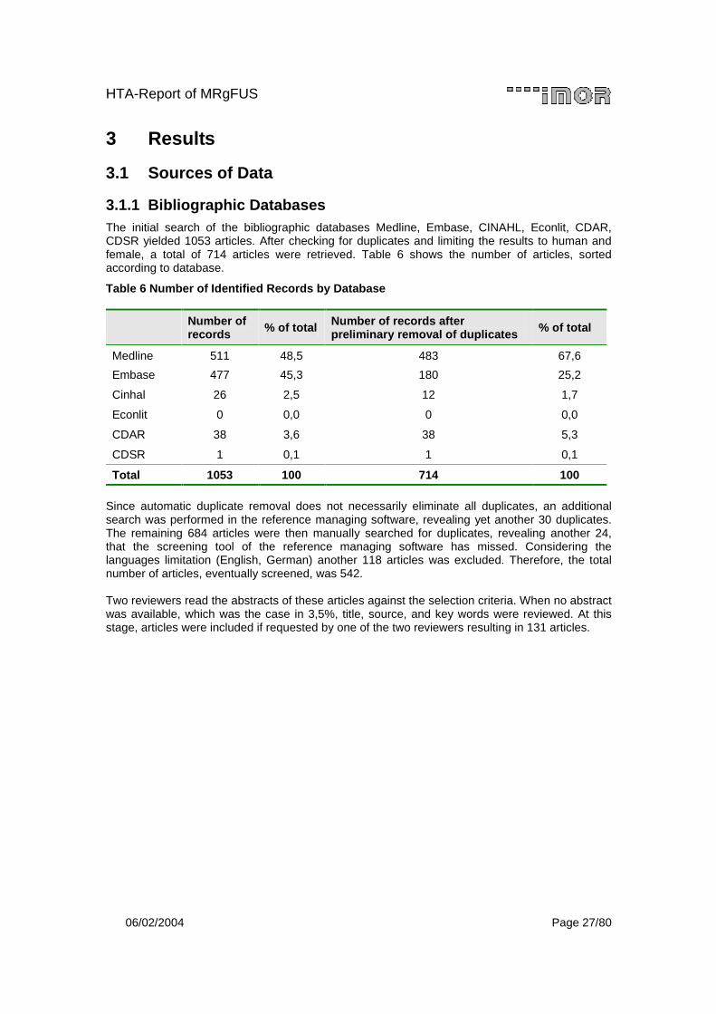
3.1.1 Bibliographic Databases The initial search of the bibliographic databases Medline, Embase, CINAHL, Econlit, CDAR, CDSR yielded 1053 articles. After checking for duplicates and limiting the results to human and female, a total of 714 articles were retrieved. Table 6 shows the number of articles, sorted according to database.
Table 6 Number of Identified Records by Database
Number of records % of total Number of records after
preliminary removal of duplicates % of total
Medline 511 48,5 483 67,6
Embase 477 45,3 180 25,2
Cinhal 26 2,5 12 1,7
Econlit 0 0,0 0 0,0
CDAR 38 3,6 38 5,3
CDSR 1 0,1 1 0,1
Total 1053 100 714 100
Since automatic duplicate removal does not necessarily eliminate all duplicates, an additional search was performed in the reference managing software, revealing yet another 30 duplicates. The remaining 684 articles were then manually searched for duplicates, revealing another 24, that the screening tool of the reference managing software has missed. Considering the languages limitation (English, German) another 118 articles was excluded. Therefore, the total number of articles, eventually screened, was 542. Two reviewers read the abstracts of these articles against the selection criteria. When no abstract was available, which was the case in 3,5%, title, source, and key words were reviewed. At this stage, articles were included if requested by one of the two reviewers resulting in 131 articles.
HTA-Report of MRgFUS
06/02/2004 Page 28/80
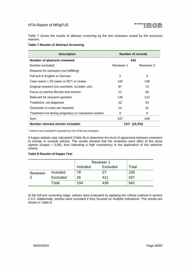
Table 7 shows the results of abstract screening by the two reviewers sorted by the exclusion reasons.
Table 7 Results of Abstract Screening
Description Number of records
Number of abstracts reviewed: 542
Number excluded: Reviewer 1 Reviewer 2
Reasons for exclusion (not fulfilling):
Full text in English or German 2 3
Case report > 20 cases or RCT or review 142 148
Original research (no comment, no letter, etc) 87 72
Focus on uterine fibroids and women 12 36
Relevant for research question 146 112
Treatment, not diagnosis 22 24
Outcomes or costs are reported 14 31
Treatment not during pregnancy or caesarean section 9 9
Sum 437 438
Number relevant articles included: 131* (22,3%) * Articles were included if requested by one of the two reviewers A kappa statistic was calculated (Table 8) to determine the level of agreement between reviewers to include or exclude articles. The results showed that the reviewers were often of the same opinion (Kappa = 0,69), thus indicating a high consistency in the application of the selection criteria.
Table 8 Results of Kappa Test
Reviewer 1 Included Excluded Total
Included 78 27 105 Reviewer 2 Excluded 26 411 437
Total 104 438 542 At the full-text screening stage, articles were evaluated by applying the criteria outlined in section 2.3.3. Additionally, articles were excluded if they focused on multiple indications. The results are shown in Table 9.
HTA-Report of MRgFUS
06/02/2004 Page 29/80
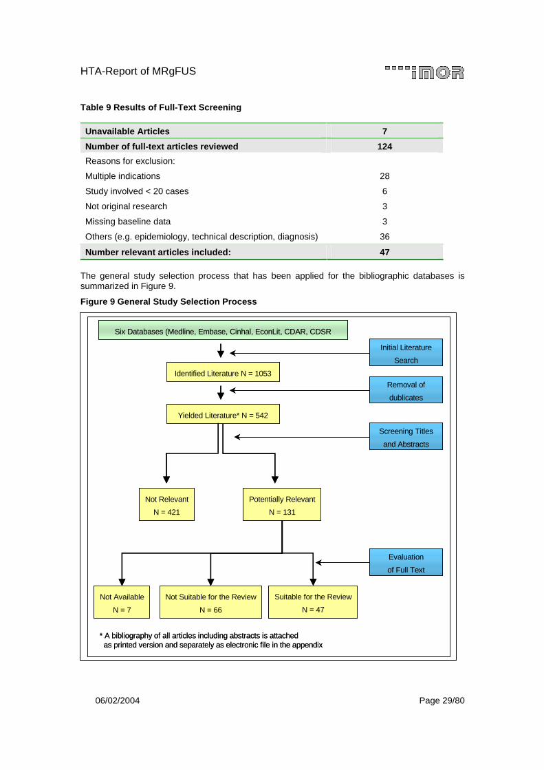
Table 9 Results of Full-Text Screening
Unavailable Articles 7
Number of full-text articles reviewed 124
Reasons for exclusion:
Multiple indications 28
Study involved < 20 cases 6
Not original research 3
Missing baseline data 3
Others (e.g. epidemiology, technical description, diagnosis) 36
Number relevant articles included: 47
The general study selection process that has been applied for the bibliographic databases is summarized in Figure 9.
Figure 9 General Study Selection Process
* A bibliography of all articles including abstracts is attached as printed version and separately as electronic file in the appendix
Six Databases (Medline, Embase, Cinhal, EconLit, CDAR, CDSR
Yielded Literature* N = 542
Screening Titles
and Abstracts
Initial Literature
Search
Potentially Relevant
N = 131
Not Relevant
N = 421
Suitable for the Review
N = 47
Not Suitable for the Review
N = 66
Evaluation
of Full Text
Not Available
N = 7
Removal of
dublicates
Identified Literature N = 1053
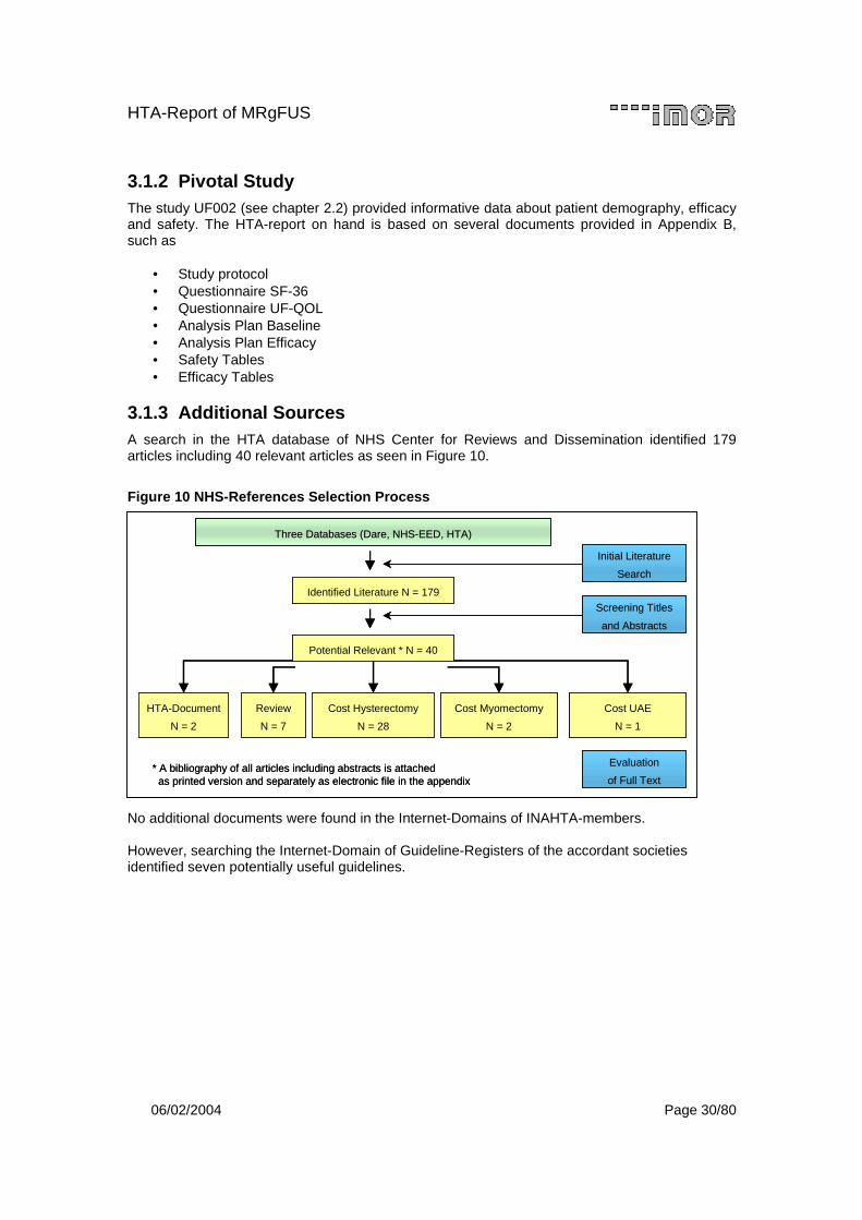
HTA-Report of MRgFUS
06/02/2004 Page 30/80
3.1.2 Pivotal Study The study UF002 (see chapter 2.2) provided informative data about patient demography, efficacy and safety. The HTA-report on hand is based on several documents provided in Appendix B, such as
• Study protocol • Questionnaire SF-36 • Questionnaire UF-QOL • Analysis Plan Baseline • Analysis Plan Efficacy • Safety Tables • Efficacy Tables
3.1.3 Additional Sources A search in the HTA database of NHS Center for Reviews and Dissemination identified 179 articles including 40 relevant articles as seen in Figure 10.
Figure 10 NHS-References Selection Process
No additional documents were found in the Internet-Domains of INAHTA-members. However, searching the Internet-Domain of Guideline-Registers of the accordant societies identified seven potentially useful guidelines.
Three Databases (Dare, NHS-EED, HTA)
Potential Relevant * N = 40
Initial Literature
Search
Review
N = 7
HTA-Document
N = 2
Evaluation
of Full Text
Screening Titles
and Abstracts
Identified Literature N = 179
Cost Hysterectomy
N = 28
Cost Myomectomy
N = 2
Cost UAE
N = 1
* A bibliography of all articles including abstracts is attached as printed version and separately as electronic file in the appendix

HTA-Report of MRgFUS
06/02/2004 Page 31/80
3.2 Focused therapy The following text incorporates the most significant data about baseline characteristics, efficacy, safety, and costs of the MRgFUS-therapy. The data were taken from the FDA-submission report, issued in January 2004. The full tables are given in Appendix B.5 – B.7.
3.2.1 Baseline Statistics The test arm included 109 women receiving MRgFUS. Three patients that withdrew and four treatment failures were recorded; hence 102 patients completed the study. Twenty patients were unable to be evaluated due to exceeding the window allowed for the respective visits. The control arm included 83 patients undergoing hysterectomy. 68 patients completed the study while 2 patients were withdrawn and 13 patients were lost to follow up. Eleven patients were non-evaluable due to exceeding of the time window allowed in the visit schedule or because the UFS-QOL Severity Score was too low.
Figure 11 Patient Flow up to Month 6
N = 176
P atients Enro lled
N = 67
S creen Fa ilures
N = 108
P atients Enro lled
N = 176
S creen Fa ilures
N = 109
P atients R ece ived FUS (ITT)
N = 83
Patients R ece ived H ysterectom y (ITT )
N = 102
C om pleted
N = 3
W ithdraw n
N = 4
Treated Fa ilure
N = 68
Com pleted
N = 2
W ithdraw n
N = 13
Lost to Fo llow -up
N = 82
E va luab le P atients
N = 57
E va luab le P atients
N = 20
N on-eva luab le patients
N = 11
N on-eva luab le patients
Table 10 (respective Appendix B.5, Table B.14) gives more details about the procedure in terms of location and volume of treated fibroids as well as region of treatment, thermal dose volume and non-perfused volume.
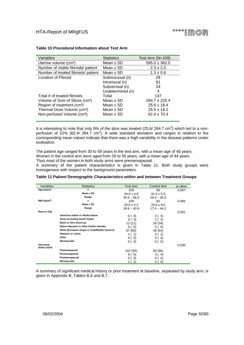
HTA-Report of MRgFUS
06/02/2004 Page 32/80
Table 10 Procedural Information about Test Arm
Variables Statistics Test Arm (N=109) Uterine volume (cm³) Mean ± SD 595.0 ± 362.5 Number of visible fibroids/ patient Mean ± SD 2.3 ± 2.0 Number of treated fibroids/ patient Mean ± SD 1.3 ± 0.6 Location of Fibroid Submucosal (n) 28 Intramural (n) 81 Subserosal (n) 24 Undetermined (n) 4 Total # of treated fibroids Total 137 Volume of Sum of Slices (cm³) Mean ± SD 284.7 ± 225.4 Region of treatment (cm³) Mean ± SD 25.6 ± 18.4 Thermal Dose Volume (cm³) Mean ± SD 25.5 ± 18.2 Non-perfused Volume (cm³) Mean ± SD 62.4 ± 70.4
It is interesting to note that only 9% of the slice was treated (25.6/ 284.7 cm3) which led to a non-perfusion of 22% (62.4/ 284.7 cm3). A wide standard deviation and ranges in relation to the corresponding mean values indicate that there was a high variability in the disease patterns under evaluation. The patient age ranged from 30 to 58 years in the test arm, with a mean age of 45 years. Women in the control arm were aged from 29 to 55 years, with a mean age of 44 years. Thus most of the women in both study arms were premenopausal. A summary of the patient characteristics is given in Table 11. Both study groups were homogenous with respect to the background parameters.
Table 11 Patient Demographic Characteristics within and between Treatment Groups
Variables Statistics Test Arm Control Arm p-value Age (years) n 109 83 0.597 Mean ± SD 44.8 ± 4.9 44.3 ± 5.6 Range 30.0 – 58.0 29.0 – 55.0 BMI (kg/m2) n 109 82 0.000 Mean ± SD 25.8 ± 5.2 29.6 ± 6.0 Range 18.6 – 43.9 17.4 – 44.2 Race (n (%)) 0.001 American Indian or Alaska Native 0 ( 0) 3 ( 4) Asian (including South Asian) 3 ( 3) 2 ( 2) Black or Afro-American 12 (11) 28 (34) Native Hawaiian or other Pacific Islander 0 ( 0) 0 ( 0) White (European Origin or Arab/Middle Eastern) 87 (80) 45 (54) Hispanic or Latino 1 ( 1) 2 ( 2) Other 6 ( 6) 3 ( 4) Missing data 0 ( 0) 0 ( 0) Hormonal Status (n(%)
0.530
Premenopausal 102 (94) 80 (96) Perimenopausal 6 ( 6) 3 ( 4) Postmenopausal 0 ( 0) 0 ( 0) Missing data 1 ( 1) 0 ( 0)
A summary of significant medical history or prior treatment at baseline, separated by study arm, is given in Appendix B, Tables B.6 and B.7.
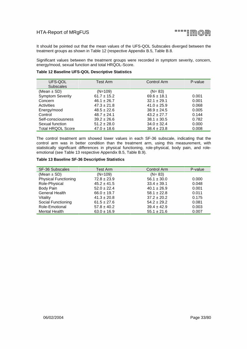
HTA-Report of MRgFUS
06/02/2004 Page 33/80
It should be pointed out that the mean values of the UFS-QOL Subscales diverged between the treatment groups as shown in Table 12 (respective Appendix B.5, Table B.8. Significant values between the treatment groups were recorded in symptom severity, concern, energy/mood, sexual function and total HRQOL-Score.
Table 12 Baseline UFS-QOL Descriptive Statistics
UFS-QOL Subscales
Test Arm Control Arm P-value
(Mean ± SD) (N=109) (N= 83) Symptom Severity 61.7 ± 15.2 69.6 ± 18.1 0.001 Concern 46.1 ± 26.7 32.1 ± 29.1 0.001 Activities 47.3 ± 21.8 41.0 ± 25.9 0.068 Energy/mood 48.5 ± 22.6 38.9 ± 24.5 0.005 Control 48.7 ± 24.1 43.2 ± 27.7 0.144 Self-consciousness 39.2 ± 26.6 38.1 ± 30.5 0.782 Sexual function 51.2 ± 28.0 34.0 ± 32.4 0.000 Total HRQOL Score 47.0 ± 18.6 38.4 ± 23.8 0.008
The control treatment arm showed lower values in each SF-36 subscale, indicating that the control arm was in better condition than the treatment arm, using this measurement, with statistically significant differences in physical functioning, role-physical, body pain, and role-emotional (see Table 13 respective Appendix B.5, Table B.9).
Table 13 Baseline SF-36 Descriptive Statistics
SF-36 Subscales Test Arm Control Arm P-value (Mean ± SD) (N=109) (N= 83) Physical Functioning 72.8 ± 23.9 56.1 ± 30.0 0.000 Role-Physical 45.2 ± 41.5 33.4 ± 39.1 0.048 Body Pain 52.0 ± 22.4 40.1 ± 26.9 0.001 General Health 66.0 ± 19.7 58.1 ± 22.8 0.011 Vitality 41.3 ± 20.8 37.2 ± 20.2 0.175 Social Functioning 61.5 ± 27.6 54.2 ± 29.2 0.081 Role-Emotional 57.8 ± 40.2 39.4 ± 42.9 0.003 Mental Health 63.0 ± 16.9 55.1 ± 21.6 0.007
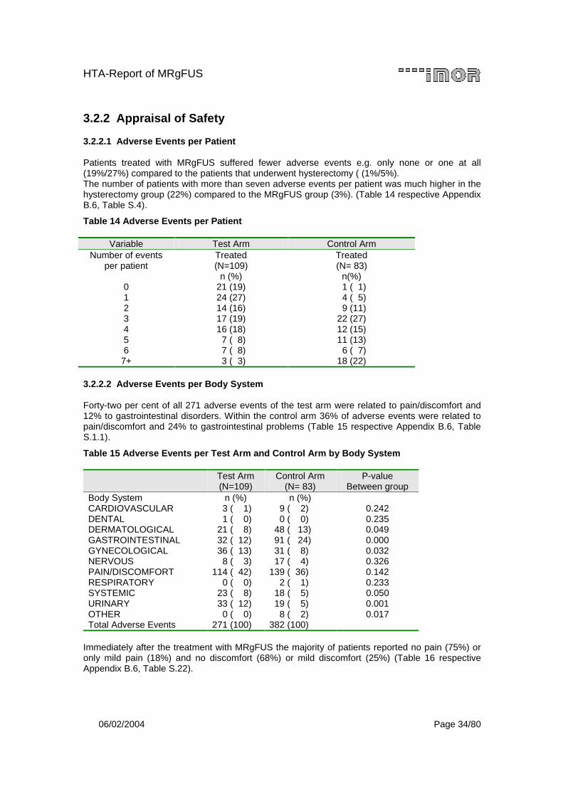
HTA-Report of MRgFUS
06/02/2004 Page 34/80
3.2.2 Appraisal of Safety
3.2.2.1 Adverse Events per Patient
Patients treated with MRgFUS suffered fewer adverse events e.g. only none or one at all (19%/27%) compared to the patients that underwent hysterectomy ( (1%/5%). The number of patients with more than seven adverse events per patient was much higher in the hysterectomy group (22%) compared to the MRgFUS group (3%). (Table 14 respective Appendix B.6, Table S.4).
Table 14 Adverse Events per Patient
Variable Test Arm Control Arm Number of events
per patient
Treated (N=109)
n (%)
Treated (N= 83)
n(%) 0 21 (19) 1 ( 1) 1 24 (27) 4 ( 5) 2 14 (16) 9 (11) 3 17 (19) 22 (27) 4 16 (18) 12 (15) 5 7 ( 8) 11 (13) 6 7 ( 8) 6 ( 7)
7+ 3 ( 3) 18 (22)
3.2.2.2 Adverse Events per Body System
Forty-two per cent of all 271 adverse events of the test arm were related to pain/discomfort and 12% to gastrointestinal disorders. Within the control arm 36% of adverse events were related to pain/discomfort and 24% to gastrointestinal problems (Table 15 respective Appendix B.6, Table S.1.1).
Table 15 Adverse Events per Test Arm and Control Arm by Body System
Test Arm (N=109)
Control Arm (N= 83)
P-value Between group
Body System n (%) n (%) CARDIOVASCULAR 3 ( 1) 9 ( 2) 0.242 DENTAL 1 ( 0) 0 ( 0) 0.235 DERMATOLOGICAL 21 ( 8) 48 ( 13) 0.049 GASTROINTESTINAL 32 ( 12) 91 ( 24) 0.000 GYNECOLOGICAL 36 ( 13) 31 ( 8) 0.032 NERVOUS 8 ( 3) 17 ( 4) 0.326 PAIN/DISCOMFORT 114 ( 42) 139 ( 36) 0.142 RESPIRATORY 0 ( 0) 2 ( 1) 0.233 SYSTEMIC 23 ( 8) 18 ( 5) 0.050 URINARY 33 ( 12) 19 ( 5) 0.001 OTHER 0 ( 0) 8 ( 2) 0.017 Total Adverse Events 271 (100) 382 (100)
Immediately after the treatment with MRgFUS the majority of patients reported no pain (75%) or only mild pain (18%) and no discomfort (68%) or mild discomfort (25%) (Table 16 respective Appendix B.6, Table S.22).
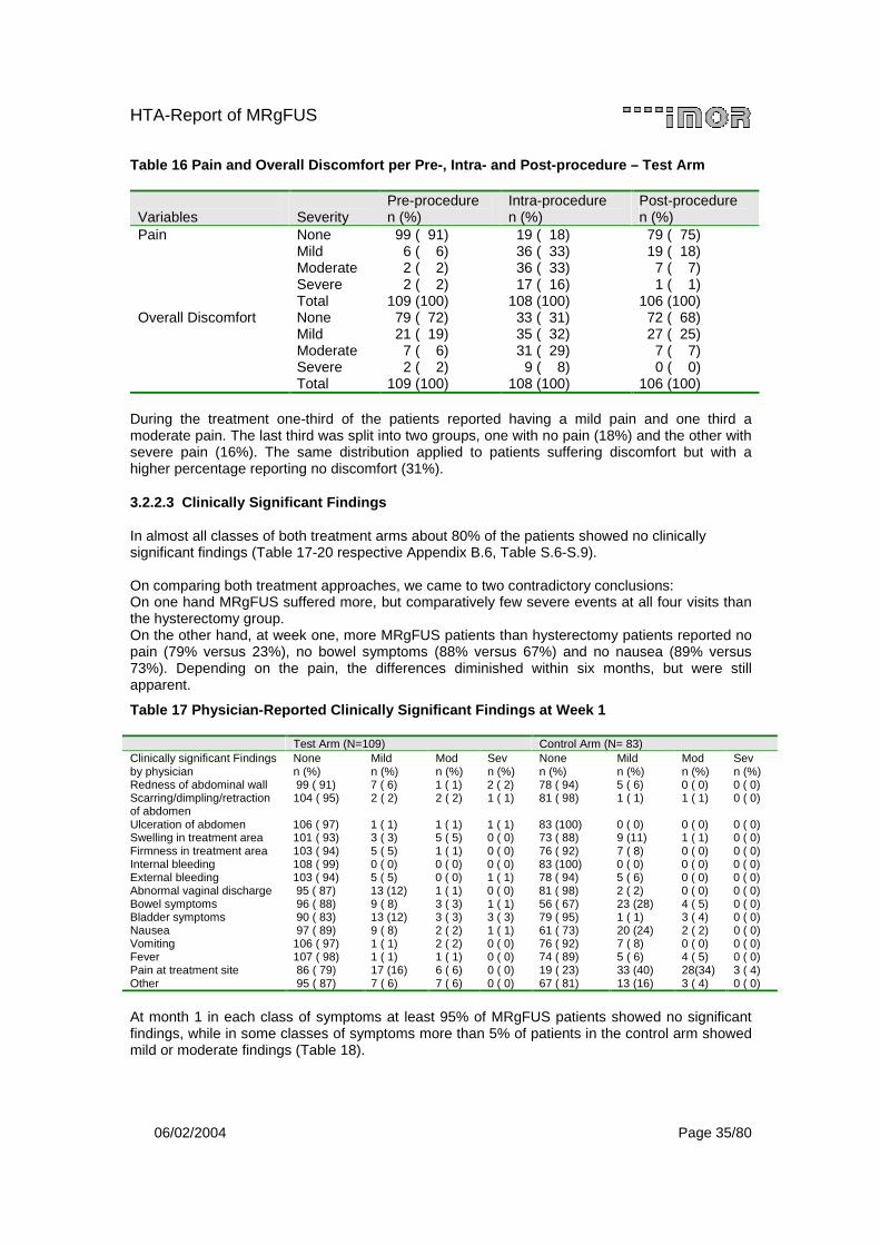
HTA-Report of MRgFUS
06/02/2004 Page 35/80
Table 16 Pain and Overall Discomfort per Pre-, Intra- and Post-procedure – Test Arm
Variables
Severity
Pre-procedure n (%)
Intra-procedure n (%)
Post-procedure n (%)
Pain None 99 ( 91) 19 ( 18) 79 ( 75) Mild 6 ( 6) 36 ( 33) 19 ( 18) Moderate 2 ( 2) 36 ( 33) 7 ( 7) Severe 2 ( 2) 17 ( 16) 1 ( 1) Total 109 (100) 108 (100) 106 (100) Overall Discomfort None 79 ( 72) 33 ( 31) 72 ( 68) Mild 21 ( 19) 35 ( 32) 27 ( 25) Moderate 7 ( 6) 31 ( 29) 7 ( 7) Severe 2 ( 2) 9 ( 8) 0 ( 0) Total 109 (100) 108 (100) 106 (100)
During the treatment one-third of the patients reported having a mild pain and one third a moderate pain. The last third was split into two groups, one with no pain (18%) and the other with severe pain (16%). The same distribution applied to patients suffering discomfort but with a higher percentage reporting no discomfort (31%).
3.2.2.3 Clinically Significant Findings
In almost all classes of both treatment arms about 80% of the patients showed no clinically significant findings (Table 17-20 respective Appendix B.6, Table S.6-S.9). On comparing both treatment approaches, we came to two contradictory conclusions: On one hand MRgFUS suffered more, but comparatively few severe events at all four visits than the hysterectomy group. On the other hand, at week one, more MRgFUS patients than hysterectomy patients reported no pain (79% versus 23%), no bowel symptoms (88% versus 67%) and no nausea (89% versus 73%). Depending on the pain, the differences diminished within six months, but were still apparent.
Table 17 Physician-Reported Clinically Significant Findings at Week 1
Test Arm (N=109) Control Arm (N= 83) Clinically significant Findings by physician
None n (%)
Mild n (%)
Mod n (%)
Sev n (%)
None n (%)
Mild n (%)
Mod n (%)
Sev n (%)
Redness of abdominal wall 99 ( 91) 7 ( 6) 1 ( 1) 2 ( 2) 78 ( 94) 5 ( 6) 0 ( 0) 0 ( 0) Scarring/dimpling/retraction of abdomen
104 ( 95) 2 ( 2) 2 ( 2) 1 ( 1) 81 ( 98) 1 ( 1) 1 ( 1) 0 ( 0)
Ulceration of abdomen 106 ( 97) 1 ( 1) 1 ( 1) 1 ( 1) 83 (100) 0 ( 0) 0 ( 0) 0 ( 0) Swelling in treatment area 101 ( 93) 3 ( 3) 5 ( 5) 0 ( 0) 73 ( 88) 9 (11) 1 ( 1) 0 ( 0) Firmness in treatment area 103 ( 94) 5 ( 5) 1 ( 1) 0 ( 0) 76 ( 92) 7 ( 8) 0 ( 0) 0 ( 0) Internal bleeding 108 ( 99) 0 ( 0) 0 ( 0) 0 ( 0) 83 (100) 0 ( 0) 0 ( 0) 0 ( 0) External bleeding 103 ( 94) 5 ( 5) 0 ( 0) 1 ( 1) 78 ( 94) 5 ( 6) 0 ( 0) 0 ( 0) Abnormal vaginal discharge 95 ( 87) 13 (12) 1 ( 1) 0 ( 0) 81 ( 98) 2 ( 2) 0 ( 0) 0 ( 0) Bowel symptoms 96 ( 88) 9 ( 8) 3 ( 3) 1 ( 1) 56 ( 67) 23 (28) 4 ( 5) 0 ( 0) Bladder symptoms 90 ( 83) 13 (12) 3 ( 3) 3 ( 3) 79 ( 95) 1 ( 1) 3 ( 4) 0 ( 0) Nausea 97 ( 89) 9 ( 8) 2 ( 2) 1 ( 1) 61 ( 73) 20 (24) 2 ( 2) 0 ( 0) Vomiting 106 ( 97) 1 ( 1) 2 ( 2) 0 ( 0) 76 ( 92) 7 ( 8) 0 ( 0) 0 ( 0) Fever 107 ( 98) 1 ( 1) 1 ( 1) 0 ( 0) 74 ( 89) 5 ( 6) 4 ( 5) 0 ( 0) Pain at treatment site 86 ( 79) 17 (16) 6 ( 6) 0 ( 0) 19 ( 23) 33 (40) 28(34) 3 ( 4) Other 95 ( 87) 7 ( 6) 7 ( 6) 0 ( 0) 67 ( 81) 13 (16) 3 ( 4) 0 ( 0)
At month 1 in each class of symptoms at least 95% of MRgFUS patients showed no significant findings, while in some classes of symptoms more than 5% of patients in the control arm showed mild or moderate findings (Table 18).
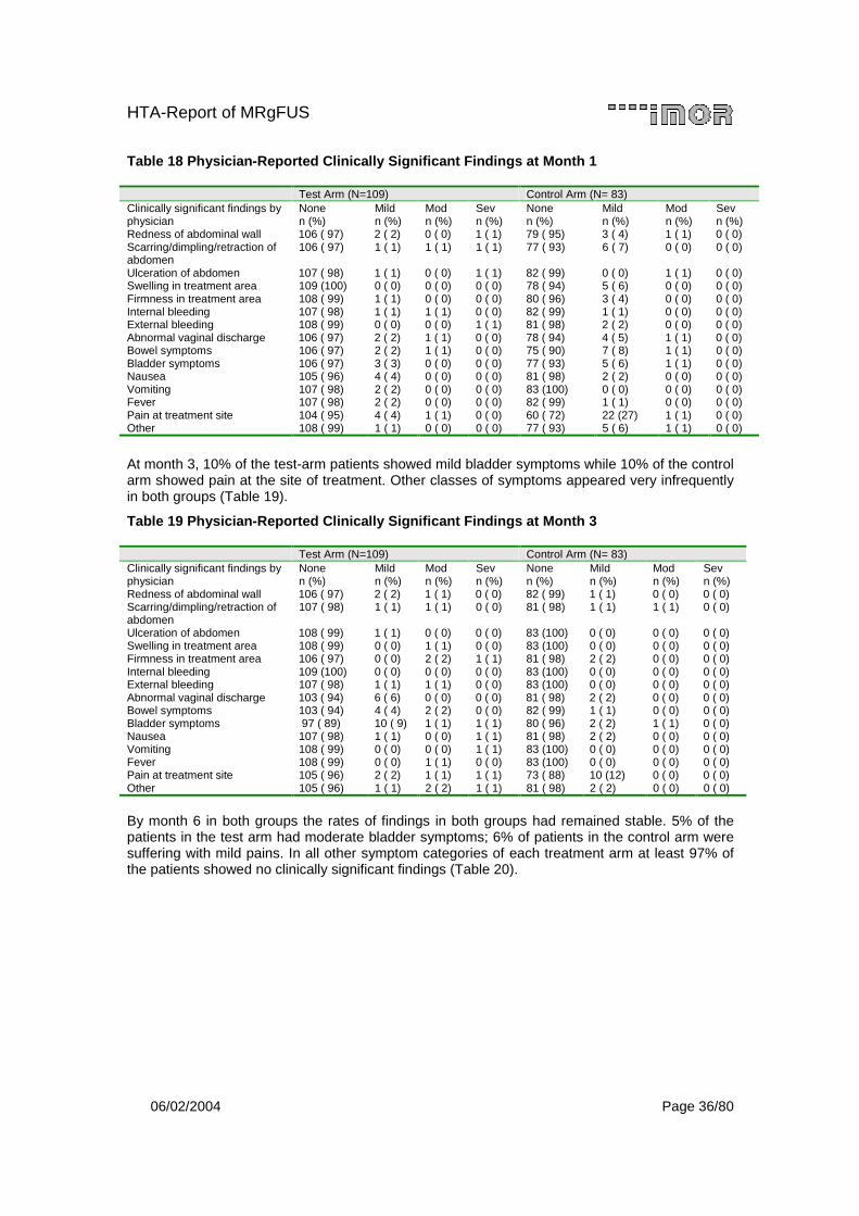
HTA-Report of MRgFUS
06/02/2004 Page 36/80
Table 18 Physician-Reported Clinically Significant Findings at Month 1
Test Arm (N=109) Control Arm (N= 83) Clinically significant findings by physician
None n (%)
Mild n (%)
Mod n (%)
Sev n (%)
None n (%)
Mild n (%)
Mod n (%)
Sev n (%)
Redness of abdominal wall 106 ( 97) 2 ( 2) 0 ( 0) 1 ( 1) 79 ( 95) 3 ( 4) 1 ( 1) 0 ( 0) Scarring/dimpling/retraction of abdomen
106 ( 97) 1 ( 1) 1 ( 1) 1 ( 1) 77 ( 93) 6 ( 7) 0 ( 0) 0 ( 0)
Ulceration of abdomen 107 ( 98) 1 ( 1) 0 ( 0) 1 ( 1) 82 ( 99) 0 ( 0) 1 ( 1) 0 ( 0) Swelling in treatment area 109 (100) 0 ( 0) 0 ( 0) 0 ( 0) 78 ( 94) 5 ( 6) 0 ( 0) 0 ( 0) Firmness in treatment area 108 ( 99) 1 ( 1) 0 ( 0) 0 ( 0) 80 ( 96) 3 ( 4) 0 ( 0) 0 ( 0) Internal bleeding 107 ( 98) 1 ( 1) 1 ( 1) 0 ( 0) 82 ( 99) 1 ( 1) 0 ( 0) 0 ( 0) External bleeding 108 ( 99) 0 ( 0) 0 ( 0) 1 ( 1) 81 ( 98) 2 ( 2) 0 ( 0) 0 ( 0) Abnormal vaginal discharge 106 ( 97) 2 ( 2) 1 ( 1) 0 ( 0) 78 ( 94) 4 ( 5) 1 ( 1) 0 ( 0) Bowel symptoms 106 ( 97) 2 ( 2) 1 ( 1) 0 ( 0) 75 ( 90) 7 ( 8) 1 ( 1) 0 ( 0) Bladder symptoms 106 ( 97) 3 ( 3) 0 ( 0) 0 ( 0) 77 ( 93) 5 ( 6) 1 ( 1) 0 ( 0) Nausea 105 ( 96) 4 ( 4) 0 ( 0) 0 ( 0) 81 ( 98) 2 ( 2) 0 ( 0) 0 ( 0) Vomiting 107 ( 98) 2 ( 2) 0 ( 0) 0 ( 0) 83 (100) 0 ( 0) 0 ( 0) 0 ( 0) Fever 107 ( 98) 2 ( 2) 0 ( 0) 0 ( 0) 82 ( 99) 1 ( 1) 0 ( 0) 0 ( 0) Pain at treatment site 104 ( 95) 4 ( 4) 1 ( 1) 0 ( 0) 60 ( 72) 22 (27) 1 ( 1) 0 ( 0) Other 108 ( 99) 1 ( 1) 0 ( 0) 0 ( 0) 77 ( 93) 5 ( 6) 1 ( 1) 0 ( 0)
At month 3, 10% of the test-arm patients showed mild bladder symptoms while 10% of the control arm showed pain at the site of treatment. Other classes of symptoms appeared very infrequently in both groups (Table 19).
Table 19 Physician-Reported Clinically Significant Findings at Month 3
Test Arm (N=109) Control Arm (N= 83) Clinically significant findings by physician
None n (%)
Mild n (%)
Mod n (%)
Sev n (%)
None n (%)
Mild n (%)
Mod n (%)
Sev n (%)
Redness of abdominal wall 106 ( 97) 2 ( 2) 1 ( 1) 0 ( 0) 82 ( 99) 1 ( 1) 0 ( 0) 0 ( 0) Scarring/dimpling/retraction of abdomen
107 ( 98) 1 ( 1) 1 ( 1) 0 ( 0) 81 ( 98) 1 ( 1) 1 ( 1) 0 ( 0)
Ulceration of abdomen 108 ( 99) 1 ( 1) 0 ( 0) 0 ( 0) 83 (100) 0 ( 0) 0 ( 0) 0 ( 0) Swelling in treatment area 108 ( 99) 0 ( 0) 1 ( 1) 0 ( 0) 83 (100) 0 ( 0) 0 ( 0) 0 ( 0) Firmness in treatment area 106 ( 97) 0 ( 0) 2 ( 2) 1 ( 1) 81 ( 98) 2 ( 2) 0 ( 0) 0 ( 0) Internal bleeding 109 (100) 0 ( 0) 0 ( 0) 0 ( 0) 83 (100) 0 ( 0) 0 ( 0) 0 ( 0) External bleeding 107 ( 98) 1 ( 1) 1 ( 1) 0 ( 0) 83 (100) 0 ( 0) 0 ( 0) 0 ( 0) Abnormal vaginal discharge 103 ( 94) 6 ( 6) 0 ( 0) 0 ( 0) 81 ( 98) 2 ( 2) 0 ( 0) 0 ( 0) Bowel symptoms 103 ( 94) 4 ( 4) 2 ( 2) 0 ( 0) 82 ( 99) 1 ( 1) 0 ( 0) 0 ( 0) Bladder symptoms 97 ( 89) 10 ( 9) 1 ( 1) 1 ( 1) 80 ( 96) 2 ( 2) 1 ( 1) 0 ( 0) Nausea 107 ( 98) 1 ( 1) 0 ( 0) 1 ( 1) 81 ( 98) 2 ( 2) 0 ( 0) 0 ( 0) Vomiting 108 ( 99) 0 ( 0) 0 ( 0) 1 ( 1) 83 (100) 0 ( 0) 0 ( 0) 0 ( 0) Fever 108 ( 99) 0 ( 0) 1 ( 1) 0 ( 0) 83 (100) 0 ( 0) 0 ( 0) 0 ( 0) Pain at treatment site 105 ( 96) 2 ( 2) 1 ( 1) 1 ( 1) 73 ( 88) 10 (12) 0 ( 0) 0 ( 0) Other 105 ( 96) 1 ( 1) 2 ( 2) 1 ( 1) 81 ( 98) 2 ( 2) 0 ( 0) 0 ( 0)
By month 6 in both groups the rates of findings in both groups had remained stable. 5% of the patients in the test arm had moderate bladder symptoms; 6% of patients in the control arm were suffering with mild pains. In all other symptom categories of each treatment arm at least 97% of the patients showed no clinically significant findings (Table 20).
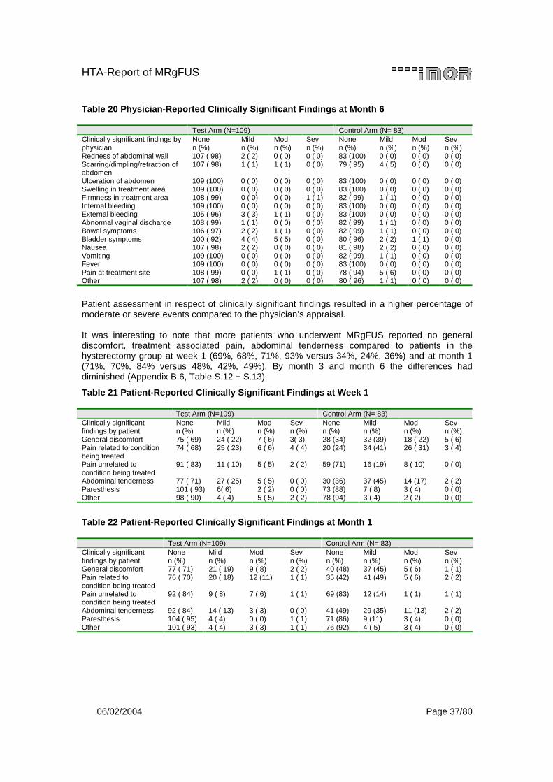
HTA-Report of MRgFUS
06/02/2004 Page 37/80
Table 20 Physician-Reported Clinically Significant Findings at Month 6
Test Arm (N=109) Control Arm (N= 83) Clinically significant findings by physician
None n (%)
Mild n (%)
Mod n (%)
Sev n (%)
None n (%)
Mild n (%)
Mod n (%)
Sev n (%)
Redness of abdominal wall 107 ( 98) 2 ( 2) 0 ( 0) 0 ( 0) 83 (100) 0 ( 0) 0 ( 0) 0 ( 0) Scarring/dimpling/retraction of abdomen
107 ( 98) 1 ( 1) 1 ( 1) 0 ( 0) 79 ( 95) 4 ( 5) 0 ( 0) 0 ( 0)
Ulceration of abdomen 109 (100) 0 ( 0) 0 ( 0) 0 ( 0) 83 (100) 0 ( 0) 0 ( 0) 0 ( 0) Swelling in treatment area 109 (100) 0 ( 0) 0 ( 0) 0 ( 0) 83 (100) 0 ( 0) 0 ( 0) 0 ( 0) Firmness in treatment area 108 ( 99) 0 ( 0) 0 ( 0) 1 ( 1) 82 ( 99) 1 ( 1) 0 ( 0) 0 ( 0) Internal bleeding 109 (100) 0 ( 0) 0 ( 0) 0 ( 0) 83 (100) 0 ( 0) 0 ( 0) 0 ( 0) External bleeding 105 ( 96) 3 ( 3) 1 ( 1) 0 ( 0) 83 (100) 0 ( 0) 0 ( 0) 0 ( 0) Abnormal vaginal discharge 108 ( 99) 1 ( 1) 0 ( 0) 0 ( 0) 82 ( 99) 1 ( 1) 0 ( 0) 0 ( 0) Bowel symptoms 106 ( 97) 2 ( 2) 1 ( 1) 0 ( 0) 82 ( 99) 1 ( 1) 0 ( 0) 0 ( 0) Bladder symptoms 100 ( 92) 4 ( 4) 5 ( 5) 0 ( 0) 80 ( 96) 2 ( 2) 1 ( 1) 0 ( 0) Nausea 107 ( 98) 2 ( 2) 0 ( 0) 0 ( 0) 81 ( 98) 2 ( 2) 0 ( 0) 0 ( 0) Vomiting 109 (100) 0 ( 0) 0 ( 0) 0 ( 0) 82 ( 99) 1 ( 1) 0 ( 0) 0 ( 0) Fever 109 (100) 0 ( 0) 0 ( 0) 0 ( 0) 83 (100) 0 ( 0) 0 ( 0) 0 ( 0) Pain at treatment site 108 ( 99) 0 ( 0) 1 ( 1) 0 ( 0) 78 ( 94) 5 ( 6) 0 ( 0) 0 ( 0) Other 107 ( 98) 2 ( 2) 0 ( 0) 0 ( 0) 80 ( 96) 1 ( 1) 0 ( 0) 0 ( 0)
Patient assessment in respect of clinically significant findings resulted in a higher percentage of moderate or severe events compared to the physician’s appraisal. It was interesting to note that more patients who underwent MRgFUS reported no general discomfort, treatment associated pain, abdominal tenderness compared to patients in the hysterectomy group at week 1 (69%, 68%, 71%, 93% versus 34%, 24%, 36%) and at month 1 (71%, 70%, 84% versus 48%, 42%, 49%). By month 3 and month 6 the differences had diminished (Appendix B.6, Table S.12 + S.13).
Table 21 Patient-Reported Clinically Significant Findings at Week 1
Test Arm (N=109) Control Arm (N= 83) Clinically significant findings by patient
None n (%)
Mild n (%)
Mod n (%)
Sev n (%)
None n (%)
Mild n (%)
Mod n (%)
Sev n (%)
General discomfort 75 ( 69) 24 ( 22) 7 ( 6) 3( 3) 28 (34) 32 (39) 18 ( 22) 5 ( 6) Pain related to condition being treated
74 ( 68) 25 ( 23) 6 ( 6) 4 ( 4) 20 (24) 34 (41) 26 ( 31) 3 ( 4)
Pain unrelated to condition being treated
91 ( 83) 11 ( 10) 5 ( 5) 2 ( 2) 59 (71) 16 (19) 8 ( 10) 0 ( 0)
Abdominal tenderness 77 ( 71) 27 ( 25) 5 ( 5) 0 ( 0) 30 (36) 37 (45) 14 (17) 2 ( 2) Paresthesis 101 ( 93) 6( 6) 2 ( 2) 0 ( 0) 73 (88) 7 ( 8) 3 ( 4) 0 ( 0) Other 98 ( 90) 4 ( 4) 5 ( 5) 2 ( 2) 78 (94) 3 ( 4) 2 ( 2) 0 ( 0)
Table 22 Patient-Reported Clinically Significant Findings at Month 1
Test Arm (N=109) Control Arm (N= 83) Clinically significant findings by patient
None n (%)
Mild n (%)
Mod n (%)
Sev n (%)
None n (%)
Mild n (%)
Mod n (%)
Sev n (%)
General discomfort 77 ( 71) 21 ( 19) 9 ( 8) 2 ( 2) 40 (48) 37 (45) 5 ( 6) 1 ( 1) Pain related to condition being treated
76 ( 70) 20 ( 18) 12 (11) 1 ( 1) 35 (42) 41 (49) 5 ( 6) 2 ( 2)
Pain unrelated to condition being treated
92 ( 84) 9 ( 8) 7 ( 6) 1 ( 1) 69 (83) 12 (14) 1 ( 1) 1 ( 1)
Abdominal tenderness 92 ( 84) 14 ( 13) 3 ( 3) 0 ( 0) 41 (49) 29 (35) 11 (13) 2 ( 2) Paresthesis 104 ( 95) 4 ( 4) 0 ( 0) 1 ( 1) 71 (86) 9 (11) 3 ( 4) 0 ( 0) Other 101 ( 93) 4 ( 4) 3 ( 3) 1 ( 1) 76 (92) 4 ( 5) 3 ( 4) 0 ( 0)

HTA-Report of MRgFUS
06/02/2004 Page 38/80
3.2.2.4 Serious Adverse Events
Nine patients in the Test Arm of the study experienced a total of 10 SAEs. Of these 9 patients, one experienced an event that was considered to be related to the device (Patient 09019), and one experienced an event that was considered to be procedure related (Patient 02018). All other SAEs experienced by patients in the Test Arm were considered by the investigator to be either unlikely related or completely unrelated to the device. A by patient listing of SAEs experienced by patients in the Test Arm of the study is provided in Table 23 and Appendix B.6, Table S.5.).
Table 23 Serious Adverse Events in Test Arm during 6 Months
Patient #
Date of MRgFUS treatment
SAE Start/Stop
Date Event Sa Rb Oc
12001 09 May 02 02 Jul/ 15 Jul 02
PostTx, Continued fibroid symptoms (bleeding and abdominal pain), Heavy Menses: Hysterectomy.
3 3 1
01 Sep/ 19 Sep 02
UTI postTx, overnight hospitalization 3 4 1
05009 23 May 02 01 Jan/ 19 Sep
02
continued heavy menses post FUS. Xfusion 14 wks post-
TxAnemia 2 4 1
05015 07 Jun 02 28 Jun/ 01 Jul
02
Sever menorrhagia:a, hospitalization 6 wks post-Tx.
Transfusion. Hysterectomy 3 5 1
02018 25 Jun 02 25 Jun/ 26 Jun
02 Nausea. Patient Elected
Overnight Hosp. 2 4 1
06020 30 Jul 02 13 Nov/ 18 Nov 02
Patient had a total Abdominal Hysterectomy 2 4 1
06021 30 Jul 02 15 Aug/ 17 Aug
02
Concurrent disease. bleeding and anemia post-Tx, Hosp for
xfusion. Patient elected Dialation and curettageHeavy Menses
3 4 2
08023 23 Aug 02 13 Nov/ 24 Nov 02
Continuation of pain and bleeding post Tx. Patient elected
for UAE. 1 5 1
09019 15 Oct 02 15 Oct/ open Nerve injury 2 1 3
05021 15 Aug 02 Ongoing* Pre-existing brain tumor Unknown 5 3 a Severity: 1=mild; 2=moderate; 3=severe; 4=life threatening b Relationship:1=definite; 2=probable; 3=possible; 4=unlikely; 5=unrelated c Outcome: 1=event resolved/patient recovered; 2=patient alive with sequelae (no current
treatment for event); 3=event still under treatment; 4= patient died; 5=insufficient follow-up/ event still being followed
* Onset and end dates unknown
It is evident, that most of the serious adverse events happened some weeks after the date of MRgFUS-therapy. All serious adverse events were resolved without sequelae with the exception of the last two patients (still ongoing). Eight patients in the Control Arm experienced a total of 10 SAEs. Only one event was considered unrelated or unlikely related to treatment; all others were considered by the investigator to have at least a possible relationship to treatment. Table 24 presents a by patient listing of SAEs, experienced by patients in the Control Arm of the study. This information is also available in Appendix B.6, Table S.5.1.

HTA-Report of MRgFUS
06/02/2004 Page 39/80
Table 24 Serious Adverse Events in Control Arm during 6 Months
Patient # Date of TAH
treatment SAE Start/Stop
Date Event Sa Rb Oc
20013 24 Jan 03 10 Apr 03/ 24 Apr
03 Foreign body in
bladder 2 3 1
17025 20 Dec 02 11 Jan/ 20 Jan 03 Small bowel obstruction
3 3 1
17 Oct/ 18 Oct 03 Surgery
cancelled NA 3 1
17026 18 Oct 02 18 Oct/ 18 Oct 02 Unsutured facia 3 3 1
17004 15 Jul 02 20 Jul/ 20 Jul 02 Ventricle ectopy 2 5 1, 3
17019 3 Sep 02 13 Sep 02/ unknown*
Superficial wound
separation and infection
2 1 1
18048 23 Jan 03 12 Feb/ 18 Feb 03 Pelvic
hematoma 3 1 1
27 Jul/ 28 Jul 02 Intestinal
obstruction 2 3 1
13012 16 Jul 02 18 Nov/ 22 Nov 02
Intestinal obstruction
2 3 1
19001 27 Nov 02 12 Jun/ 10 Jul 2003 Hernia 3 1 1 a Severity: 1=mild; 2=moderate; 3=severe; 4=life threatening b Relationship: 1=definite; 2=probable; 3=possible; 4=unlikely; 5=unrelated c Outcome: 1=event resolved/patient recovered; 2=patient alive with sequelae (no current treatment
for event); 3=event still under treatment; 4= patient died; 5=insufficient follow-up/ event still being followed
* Patient lost to follow-up; stop date of SAE unknown
It is interesting to note that the control arm had more clinical occurrences (63) with health-economic relevance compared than the test arm (17) as summarized in table 25.
Table 25 Number of Occurrences of Significant Clinical Complications
Test Arm Control Arm Fever > 38°C on any 2 Post Tx days excl 1st 24h 3 12 Antibiotics started > 24 hrs post-Tx 3 30 Transfusion 3 6 Unintended Surgical Procedure related to Tx 0 4 Referral to Rehabilitation Facility 0 0 Discharge w/Appliance 0 1 Life-threatening Event 0 0 Re-hospitalization > 24hrs 8 8 Interventional Tx 0 2 Death 0 0 Total # of occurrences 17 63
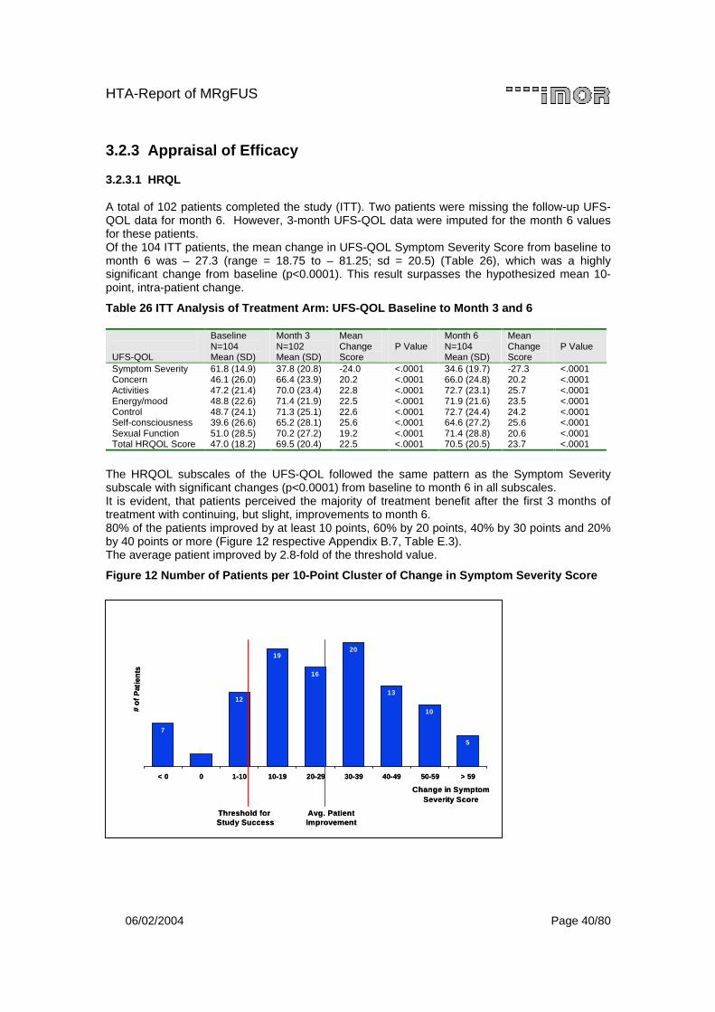
HTA-Report of MRgFUS
06/02/2004 Page 40/80
3.2.3 Appraisal of Efficacy
3.2.3.1 HRQL
A total of 102 patients completed the study (ITT). Two patients were missing the follow-up UFS-QOL data for month 6. However, 3-month UFS-QOL data were imputed for the month 6 values for these patients. Of the 104 ITT patients, the mean change in UFS-QOL Symptom Severity Score from baseline to month 6 was – 27.3 (range = 18.75 to – 81.25; sd = 20.5) (Table 26), which was a highly significant change from baseline (p<0.0001). This result surpasses the hypothesized mean 10-point, intra-patient change.
Table 26 ITT Analysis of Treatment Arm: UFS-QOL Baseline to Month 3 and 6
UFS-QOL
Baseline N=104 Mean (SD)
Month 3 N=102 Mean (SD)
Mean Change Score
P Value
Month 6 N=104 Mean (SD)
Mean Change Score
P Value
Symptom Severity 61.8 (14.9) 37.8 (20.8) -24.0 <.0001 34.6 (19.7) -27.3 <.0001 Concern 46.1 (26.0) 66.4 (23.9) 20.2 <.0001 66.0 (24.8) 20.2 <.0001 Activities 47.2 (21.4) 70.0 (23.4) 22.8 <.0001 72.7 (23.1) 25.7 <.0001 Energy/mood 48.8 (22.6) 71.4 (21.9) 22.5 <.0001 71.9 (21.6) 23.5 <.0001 Control 48.7 (24.1) 71.3 (25.1) 22.6 <.0001 72.7 (24.4) 24.2 <.0001 Self-consciousness 39.6 (26.6) 65.2 (28.1) 25.6 <.0001 64.6 (27.2) 25.6 <.0001 Sexual Function 51.0 (28.5) 70.2 (27.2) 19.2 <.0001 71.4 (28.8) 20.6 <.0001 Total HRQOL Score 47.0 (18.2) 69.5 (20.4) 22.5 <.0001 70.5 (20.5) 23.7 <.0001
The HRQOL subscales of the UFS-QOL followed the same pattern as the Symptom Severity subscale with significant changes (p<0.0001) from baseline to month 6 in all subscales. It is evident, that patients perceived the majority of treatment benefit after the first 3 months of treatment with continuing, but slight, improvements to month 6. 80% of the patients improved by at least 10 points, 60% by 20 points, 40% by 30 points and 20% by 40 points or more (Figure 12 respective Appendix B.7, Table E.3). The average patient improved by 2.8-fold of the threshold value.
Figure 12 Number of Patients per 10-Point Cluster of Change in Symptom Severity Score
7
12
19
16
20
13
10
52
< 0 0 1-10 10-19 20-29 30-39 40-49 50-59 > 59
Change in Symptom Severity Score
# o
f P
atie
nts
Threshold for Study Success
Threshold for Study Success
Avg. PatientImprovement
Avg. PatientImprovement
7
12
19
16
20
13
10
52
< 0 0 1-10 10-19 20-29 30-39 40-49 50-59 > 59
Change in Symptom Severity Score
# o
f P
atie
nts
Threshold for Study Success
Threshold for Study Success
Avg. PatientImprovement
Avg. PatientImprovement
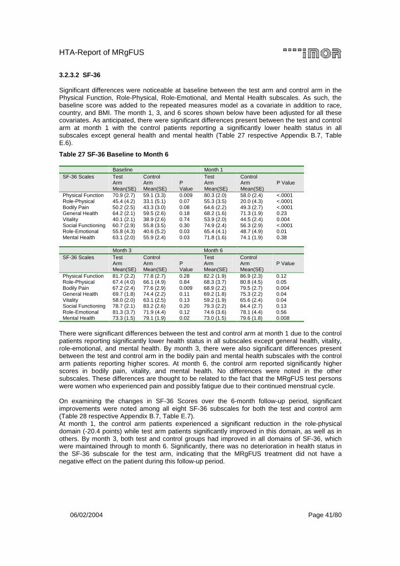
HTA-Report of MRgFUS
06/02/2004 Page 41/80
3.2.3.2 SF-36
Significant differences were noticeable at baseline between the test arm and control arm in the Physical Function, Role-Physical, Role-Emotional, and Mental Health subscales. As such, the baseline score was added to the repeated measures model as a covariate in addition to race, country, and BMI. The month 1, 3, and 6 scores shown below have been adjusted for all these covariates. As anticipated, there were significant differences present between the test and control arm at month 1 with the control patients reporting a significantly lower health status in all subscales except general health and mental health (Table 27 respective Appendix B.7, Table E.6).
Table 27 SF-36 Baseline to Month 6
Baseline Month 1 SF-36 Scales Test
Arm Mean(SE)
Control Arm Mean(SE)
P Value
Test Arm Mean(SE)
Control Arm Mean(SE)
P Value
Physical Function 70.9 (2.7) 59.1 (3.3) 0.009 80.3 (2.0) 58.0 (2.4) <.0001 Role-Physical 45.4 (4.2) 33.1 (5.1) 0.07 55.3 (3.5) 20.0 (4.3) <.0001 Bodily Pain 50.2 (2.5) 43.3 (3.0) 0.08 64.6 (2.2) 49.3 (2.7) <.0001 General Health 64.2 (2.1) 59.5 (2.6) 0.18 68.2 (1.6) 71.3 (1.9) 0.23 Vitality 40.1 (2.1) 38.9 (2.6) 0.74 53.9 (2.0) 44.5 (2.4) 0.004 Social Functioning 60.7 (2.9) 55.8 (3.5) 0.30 74.9 (2.4) 56.3 (2.9) <.0001 Role-Emotional 55.8 (4.3) 40.6 (5.2) 0.03 65.4 (4.1) 48.7 (4.9) 0.01 Mental Health 63.1 (2.0) 55.9 (2.4) 0.03 71.8 (1.6) 74.1 (1.9) 0.38 Month 3 Month 6 SF-36 Scales Test
Arm Mean(SE)
Control Arm Mean(SE)
P Value
Test Arm Mean(SE)
Control Arm Mean(SE)
P Value
Physical Function 81.7 (2.2) 77.8 (2.7) 0.28 82.2 (1.9) 86.9 (2.3) 0.12 Role-Physical 67.4 (4.0) 66.1 (4.9) 0.84 68.3 (3.7) 80.8 (4.5) 0.05 Bodily Pain 67.2 (2.4) 77.6 (2.9) 0.009 68.9 (2.2) 79.5 (2.7) 0.004 General Health 69.7 (1.8) 74.4 (2.2) 0.11 69.2 (1.8) 75.3 (2.2) 0.04 Vitality 58.0 (2.0) 63.1 (2.5) 0.13 59.2 (1.9) 65.6 (2.4) 0.04 Social Functioning 78.7 (2.1) 83.2 (2.6) 0.20 79.3 (2.2) 84.4 (2.7) 0.13 Role-Emotional 81.3 (3.7) 71.9 (4.4) 0.12 74.6 (3.6) 78.1 (4.4) 0.56 Mental Health 73.3 (1.5) 79.1 (1.9) 0.02 73.0 (1.5) 79.6 (1.8) 0.008
There were significant differences between the test and control arm at month 1 due to the control patients reporting significantly lower health status in all subscales except general health, vitality, role-emotional, and mental health. By month 3, there were also significant differences present between the test and control arm in the bodily pain and mental health subscales with the control arm patients reporting higher scores. At month 6, the control arm reported significantly higher scores in bodily pain, vitality, and mental health. No differences were noted in the other subscales. These differences are thought to be related to the fact that the MRgFUS test persons were women who experienced pain and possibly fatigue due to their continued menstrual cycle. On examining the changes in SF-36 Scores over the 6-month follow-up period, significant improvements were noted among all eight SF-36 subscales for both the test and control arm (Table 28 respective Appendix B.7, Table E.7). At month 1, the control arm patients experienced a significant reduction in the role-physical domain (-20.4 points) while test arm patients significantly improved in this domain, as well as in others. By month 3, both test and control groups had improved in all domains of SF-36, which were maintained through to month 6. Significantly, there was no deterioration in health status in the SF-36 subscale for the test arm, indicating that the MRgFUS treatment did not have a negative effect on the patient during this follow-up period.
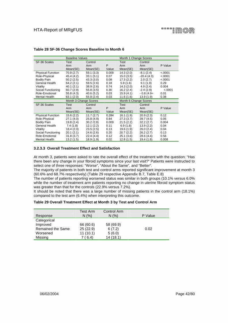
HTA-Report of MRgFUS
06/02/2004 Page 42/80
Table 28 SF-36 Change Scores Baseline to Month 6
Baseline Values Month 1 Change Scores SF-36 Scales Test
Arm Mean(SE)
Control Arm Mean(SE)
P Value
Test Arm Mean(SE)
Control Arm Mean(SE)
P Value
Physical Function 70.9 (2.7) 59.1 (3.3) 0.009 14.3 (2.0) -8.1 (2.4) <.0001 Role-Physical 45.4 (4.2) 33.1 (5.1) 0.07 15.0 (3.5) -20.4 (4.3) <.0001 Bodily Pain 50.2 (2.5) 43.3 (3.0) 0.08 17.3 (2.2) 2.0 (2.7) <.0001 General Health 64.2 (2.1) 59.5 (2.6) 0.18 5.9 (1.6) 9.1 (1.9) 0.29 Vitality 40.1 (2.1) 38.9 (2.6) 0.74 14.3 (2.0) 4.9 (2.4) 0.004 Social Functioning 60.7 (2.9) 55.8 (3.5) 0.30 16.2 (2.4) -2.4 (2.9) <.0001 Role-Emotional 55.8 (4.3) 40.6 (5.2) 0.03 15.9 (4.1) - 0.8 (4.9= 0.01 Mental Health 63.1 (2.0) 55.9 (2.4) 0.03 11.6 (1.6) 13.9 (1.9) 0.38 Month 3 Change Scores Month 6 Change Scores SF-36 Scales Test
Arm Mean(SE)
Control Arm Mean(SE)
P Value
Test Arm Mean(SE)
Control Arm Mean(SE)
P Value
Physical Function 15.6 (2.2) 11.7 (2.7) 0.284 16.1 (1.9) 20.9 (2.3) 0.12 Role-Physical 27.1 (4.0) 25.8 (4.9) 0.84 27.3 (3.7) 39.7 (4.5) 0.05 Bodily Pain 19.8 (2.4) 30.2 /2.9) 0.009 21.5 (2.2) 32.2 (2.7) 0.004 General Health 7.4 (1.8) 12.1 (2.2) 0.11 6.9 (1.8) 13.9 (2.2) 0.04 Vitality 18.4 (2.0) 23.5 (2.5) 0.13 19.6 (1.9) 26.0 (2.4) 0.04 Social Functioning 20.1 (2.1) 24.6 (2.6) 0.20 20.7 (2.2) 26.2 (2.7) 0.13 Role-Emotional 31.8 (3.7) 22.4 (4.4) 0.12 25.1 (3.6) 28.6 (4.4) 0.56 Mental Health 13.2 (1.5) 18.9 (1.9) 0.02 12.8 (1.5) 19.4 (1.8) 0.008
3.2.3.3 Overall Treatment Effect and Satisfaction
At month 3, patients were asked to rate the overall effect of the treatment with the question: “Has there been any change in your fibroid symptoms since your last visit?” Patients were instructed to select one of three responses: “Worse”, “About the Same”, and “Better”. The majority of patients in both test and control arms reported significant improvement at month 3 (60.6% and 68.7% respectively) (Table 29 respective Appendix B.7, Table E.8) The number of patients reporting worsened status was similar in both groups (10.1% versus 6.0% while the number of treatment arm patients reporting no change in uterine fibroid symptom status was greater than that for the controls (22.9% versus 7.2%). It should be noted that there was a large number of missing patients in the control arm (18.1%) compared to the test arm (6.4%) when interpreting this outcome.
Table 29 Overall Treatment Effect at Month 3 by Test and Control Arm
Response
Test Arm N (%)
Control Arm N (%)
P Value
Categorical Improved 66 (60.6) 58 (69.9) Remained the Same 25 (22.9) 6 (7.2) Worsened 11 (10.1) 5 (6.0) Missing 7 ( 6.4) 14 (18.1)
0.02

HTA-Report of MRgFUS
06/02/2004 Page 43/80
The data below represents patient responses to two questions on their satisfaction with the treatment. Significantly, over 18% of the data for the control arm (versus 10% of the test arm) are missing at month 6, which makes it difficult to interpret these findings. In the test arm, 76.1% rated some level of satisfaction with their treatment versus 64.6% of the control patients; 13.8% of test arm patients rated some level of dissatisfaction with their treatment versus 79.5% of control patients. The majority of both groups reported that their treatment was effective in relieving symptoms (71.6% versus 80.7%). However, 18.3% of the test arm reported the treatment as ineffective. (See also Appendix B.7, Table E.9)
Table 30 Satisfaction with Treatment at Month 6 by Test and Control Arm
Treatment Benefit
Test Arm N (%)
Control Arm N (%)
P Value
Satisfied with UF treatment Satisfied 83 (76.1) 66 (79.56) Dissatisfied 15 (13.8) 2 (2.4) Missing 11 (10.1) 15 (18.1)
0.01
How effective was this treatment in eliminating your symptoms? Effective 78 (71.6) 67 (80.7) Ineffective 20 (18.3) 1 (1.2) Missing 11 (10.1) 15 (18.1)
0.0003
3.2.4 Appraisal of Costs Based on calculation assumptions described in Section 2.4.3 the 10-year average costs per MRgFUS treatment is valued at EUR 3,565 (= (4,966 + 4,026 + 3,190 + 2,907 + 2,737) /5) (Table 31). The most important influencing factors are the assumptions of the price for the MRI system, the price for the FUS system, the percentage used for depreciation, and schedule, as well as the utilization ratio of the FUS system (Table 5). The assumptions indicate the necessity to perform sensitivity analysis, which can be based on an Excel Calculation Sheet being attached as electronical file to this HTA-Report in Appendix C.7.
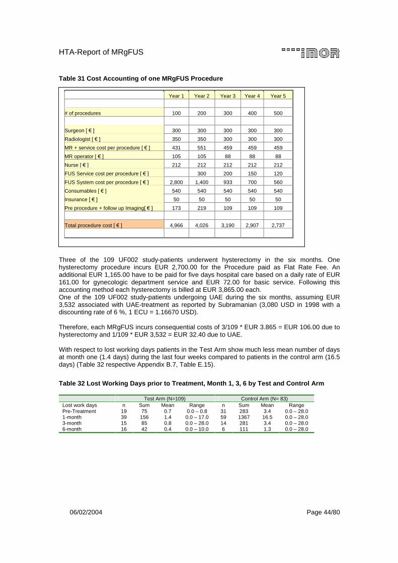
HTA-Report of MRgFUS
06/02/2004 Page 44/80
Table 31 Cost Accounting of one MRgFUS Procedure
Three of the 109 UF002 study-patients underwent hysterectomy in the six months. One hysterectomy procedure incurs EUR 2,700.00 for the Procedure paid as Flat Rate Fee. An additional EUR 1,165.00 have to be paid for five days hospital care based on a daily rate of EUR 161.00 for gynecologic department service and EUR 72.00 for basic service. Following this accounting method each hysterectomy is billed at EUR 3,865.00 each. One of the 109 UF002 study-patients undergoing UAE during the six months, assuming EUR 3,532 associated with UAE-treatment as reported by Subramanian (3,080 USD in 1998 with a discounting rate of 6 %, 1 ECU = 1.16670 USD). Therefore, each MRgFUS incurs consequential costs of 3/109 * EUR 3.865 = EUR 106.00 due to hysterectomy and 1/109 * EUR 3,532 = EUR 32.40 due to UAE. With respect to lost working days patients in the Test Arm show much less mean number of days at month one (1.4 days) during the last four weeks compared to patients in the control arm (16.5 days) (Table 32 respective Appendix B.7, Table E.15).
Table 32 Lost Working Days prior to Treatment, Month 1, 3, 6 by Test and Control Arm
Test Arm (N=109) Control Arm (N= 83) Lost work days n Sum Mean Range n Sum Mean Range Pre-Treatment 19 75 0.7 0.0 – 0.8 31 283 3.4 0.0 – 28.0 1-month 39 156 1.4 0.0 – 17.0 59 1367 16.5 0.0 – 28.0 3-month 15 85 0.8 0.0 – 28.0 14 281 3.4 0.0 – 28.0 6-month 16 42 0.4 0.0 – 10.0 6 111 1.3 0.0 – 28.0
Year 1 Year 2 Year 3 Year 4 Year 5
# of procedures 100 200 300 400 500
Surgeon [ ��! 300 300 300 300 300
Radiologist [ ��! 350 350 300 300 300
MR + service cost per procedure [ ��! 431 551 459 459 459
MR operator [ ��! 105 105 88 88 88
Nurse [ ��! 212 212 212 212 212
FUS Service cost per procedure [ ��! 300 200 150 120
FUS System cost per procedure [ ��! 2,800 1,400 933 700 560
Consumables [ ��! 540 540 540 540 540
Insurance [ ��! 50 50 50 50 50
Pre procedure + follow up Imaging[ ��!� 173 219 109 109 109
Total procedure cost [ ��! 4,966 4,026 3,190 2,907 2,737
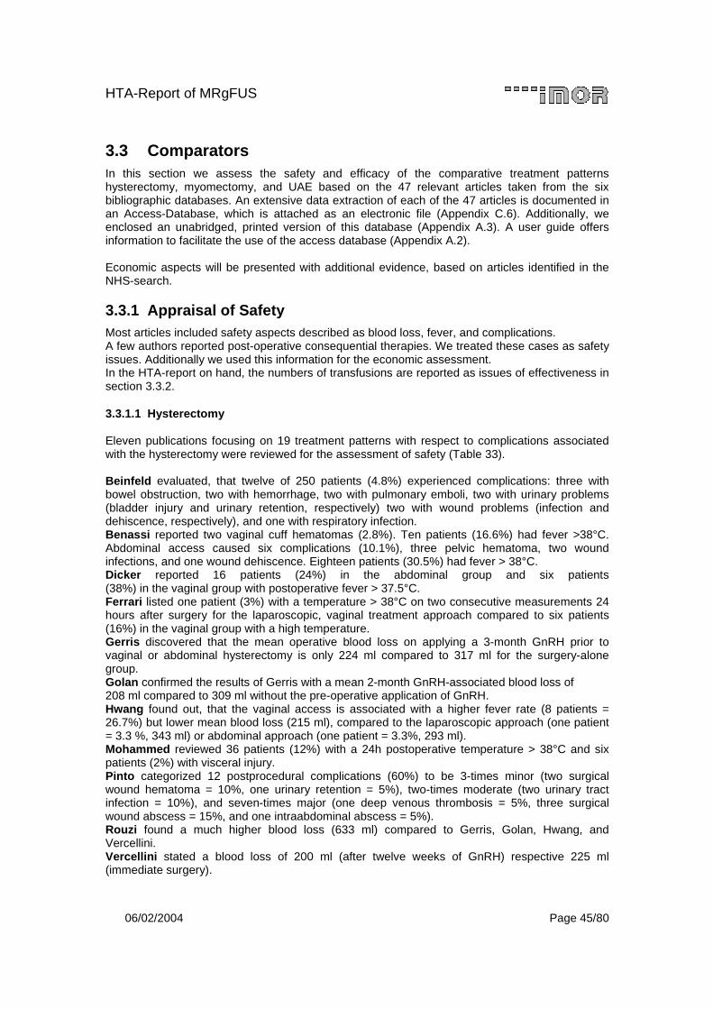
HTA-Report of MRgFUS
06/02/2004 Page 45/80
3.3 Comparators In this section we assess the safety and efficacy of the comparative treatment patterns hysterectomy, myomectomy, and UAE based on the 47 relevant articles taken from the six bibliographic databases. An extensive data extraction of each of the 47 articles is documented in an Access-Database, which is attached as an electronic file (Appendix C.6). Additionally, we enclosed an unabridged, printed version of this database (Appendix A.3). A user guide offers information to facilitate the use of the access database (Appendix A.2). Economic aspects will be presented with additional evidence, based on articles identified in the NHS-search.
3.3.1 Appraisal of Safety Most articles included safety aspects described as blood loss, fever, and complications. A few authors reported post-operative consequential therapies. We treated these cases as safety issues. Additionally we used this information for the economic assessment. In the HTA-report on hand, the numbers of transfusions are reported as issues of effectiveness in section 3.3.2.
3.3.1.1 Hysterectomy
Eleven publications focusing on 19 treatment patterns with respect to complications associated with the hysterectomy were reviewed for the assessment of safety (Table 33). Beinfeld evaluated, that twelve of 250 patients (4.8%) experienced complications: three with bowel obstruction, two with hemorrhage, two with pulmonary emboli, two with urinary problems (bladder injury and urinary retention, respectively) two with wound problems (infection and dehiscence, respectively), and one with respiratory infection. Benassi reported two vaginal cuff hematomas (2.8%). Ten patients (16.6%) had fever >38°C. Abdominal access caused six complications (10.1%), three pelvic hematoma, two wound infections, and one wound dehiscence. Eighteen patients (30.5%) had fever > 38°C. Dicker reported 16 patients (24%) in the abdominal group and six patients (38%) in the vaginal group with postoperative fever > 37.5°C. Ferrari listed one patient (3%) with a temperature > 38°C on two consecutive measurements 24 hours after surgery for the laparoscopic, vaginal treatment approach compared to six patients (16%) in the vaginal group with a high temperature. Gerris discovered that the mean operative blood loss on applying a 3-month GnRH prior to vaginal or abdominal hysterectomy is only 224 ml compared to 317 ml for the surgery-alone group. Golan confirmed the results of Gerris with a mean 2-month GnRH-associated blood loss of 208 ml compared to 309 ml without the pre-operative application of GnRH. Hwang found out, that the vaginal access is associated with a higher fever rate (8 patients = 26.7%) but lower mean blood loss (215 ml), compared to the laparoscopic approach (one patient = 3.3 %, 343 ml) or abdominal approach (one patient = 3.3%, 293 ml). Mohammed reviewed 36 patients (12%) with a 24h postoperative temperature > 38°C and six patients (2%) with visceral injury. Pinto categorized 12 postprocedural complications (60%) to be 3-times minor (two surgical wound hematoma = 10%, one urinary retention = 5%), two-times moderate (two urinary tract infection = 10%), and seven-times major (one deep venous thrombosis = 5%, three surgical wound abscess = 15%, and one intraabdominal abscess = 5%). Rouzi found a much higher blood loss (633 ml) compared to Gerris, Golan, Hwang, and Vercellini. Vercellini stated a blood loss of 200 ml (after twelve weeks of GnRH) respective 225 ml (immediate surgery).
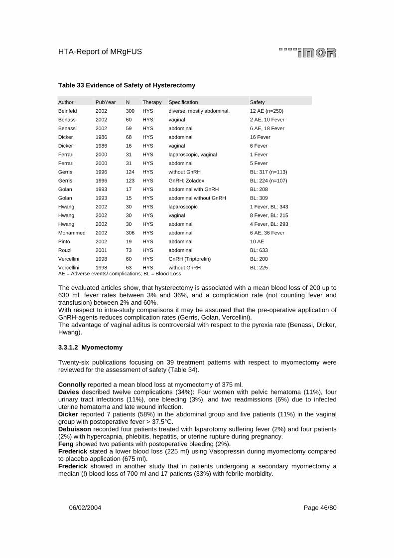
HTA-Report of MRgFUS
06/02/2004 Page 46/80
Table 33 Evidence of Safety of Hysterectomy
Author PubYear N Therapy Specification Safety
Beinfeld 2002 300 HYS diverse, mostly abdominal. 12 AE (n=250)
Benassi 2002 60 HYS vaginal 2 AE, 10 Fever
Benassi 2002 59 HYS abdominal 6 AE, 18 Fever
Dicker 1986 68 HYS abdominal 16 Fever
Dicker 1986 16 HYS vaginal 6 Fever
Ferrari 2000 31 HYS laparoscopic, vaginal 1 Fever
Ferrari 2000 31 HYS abdominal 5 Fever
Gerris 1996 124 HYS without GnRH BL: 317 (n=113)
Gerris 1996 123 HYS GnRH: Zoladex BL: 224 (n=107)
Golan 1993 17 HYS abdominal with GnRH BL: 208
Golan 1993 15 HYS abdominal without GnRH BL: 309
Hwang 2002 30 HYS laparoscopic 1 Fever, BL: 343
Hwang 2002 30 HYS vaginal 8 Fever, BL: 215
Hwang 2002 30 HYS abdominal 4 Fever, BL: 293
Mohammed 2002 306 HYS abdominal 6 AE, 36 Fever
Pinto 2002 19 HYS abdominal 10 AE
Rouzi 2001 73 HYS abdominal BL: 633
Vercellini 1998 60 HYS GnRH (Triptorelin) BL: 200
Vercellini 1998 63 HYS without GnRH BL: 225 AE = Adverse events/ complications; BL = Blood Loss The evaluated articles show, that hysterectomy is associated with a mean blood loss of 200 up to 630 ml, fever rates between 3% and 36%, and a complication rate (not counting fever and transfusion) between 2% and 60%. With respect to intra-study comparisons it may be assumed that the pre-operative application of GnRH-agents reduces complication rates (Gerris, Golan, Vercellini). The advantage of vaginal aditus is controversial with respect to the pyrexia rate (Benassi, Dicker, Hwang).
3.3.1.2 Myomectomy
Twenty-six publications focusing on 39 treatment patterns with respect to myomectomy were reviewed for the assessment of safety (Table 34). Connolly reported a mean blood loss at myomectomy of 375 ml. Davies described twelve complications (34%): Four women with pelvic hematoma (11%), four urinary tract infections (11%), one bleeding (3%), and two readmissions (6%) due to infected uterine hematoma and late wound infection. Dicker reported 7 patients (58%) in the abdominal group and five patients (11%) in the vaginal group with postoperative fever > 37.5°C. Debuisson recorded four patients treated with laparotomy suffering fever (2%) and four patients (2%) with hypercapnia, phlebitis, hepatitis, or uterine rupture during pregnancy. Feng showed two patients with postoperative bleeding (2%). Frederick stated a lower blood loss (225 ml) using Vasopressin during myomectomy compared to placebo application (675 ml). Frederick showed in another study that in patients undergoing a secondary myomectomy a median (!) blood loss of 700 ml and 17 patients (33%) with febrile morbidity.
HTA-Report of MRgFUS
06/02/2004 Page 47/80
Ginsburg reported that the preoperative uterine volume of about 600 cm3 is associated with a higher myomectomy-related blood loss (627 ml) compared to a corresponding uterus volume of less than 600 cm3 (228 ml). Of these, three patients (30%) were febrile, compared to 20% in the low blood-loss group. In contrast to the results of Frederick the use of Vasopressin does not minimize the blood loss (461 versus 369 ml), but Vasopressin seems to reduce the frequency of febrile morbidity (10% versus 40%). Golan stated a mean 2-month GnRH-associated blood loss of 320 ml compared to 476 ml without the pre-operative application of GnRH. Hutchins did not confirm the findings of Golan, showing an average blood loss for the 8-weeks GnRH group of 300 ml versus 257 ml for the comparative group not getting GnRH. Ikpeze reported 12 women with febrile illness (17%) and five women with mild sepsis (7%) and a mean estimated blood loss of 480 ml. Malzoni reported three cases (2%) of coagulation of endometriosis. Mohammed reviewed 6 patients (4%) with a 24h postoperative temperature > 38°C and six patients (4%) with visceral injury. Ou found a higher blood loss using morcellation (378 ml) compared to colpotomy (243 ml). Razzavi exposed seven complications (18%) including wound infection (n=2), adhesion (n=2), readmission for ileus (n=1), chronic pelvic pain (n=1), and chronic incisional pain, and a mean blood loss of 376 ml. Rouzi stated a much higher blood loss as Razzavi being 709 ml. Serachiolli explored the average blood loss associated with laparoscopic myomectomy to be 330 ml (notice: in table 3 of his article 154 ml are stated!). Stringer found differences in the blood loss after laparoscopic myomectomy (110 ml) and open myomectomy (340 ml). The laparoscopic treatment group had five complications (10%): two cannula site bleedings, one paraesthesia of fot, and two intraoperative bradycardia after pitressin injection. Open myomectomy was associated with 14 complications (35%), namely four ileus, six urinary tract infections, one pneumonia, one incisional separation, one pulmonary edema, and one small bowel obstruction. Vercellini stated a mean blood loss of 265 ml and nine patients with febrile morbidity (18%) (with twelve weeks GnRH) compared to 296 ml mean blood loss and ten febrile patients (21%) for those with immediate surgery. Zullo had similar results as Vercellini with 172 ml blood loss for the GnRH-group and 232 ml for the immediate surgery group. Myomectomy is – similar to hysterectomy – associated with a mean blood loss between 175 ml and 670 ml. The complication rate (not counting fever and transfusion) ranged from 2 % (Feng, Malzoni) to 34% (Davies). Four per cent (Mohammed) up to 56% (Dicker) of patients undergoing myomectomy suffer febrile morbidity. The effect of vasopressin is unclear (Frederick, Ginsburg). The laparoscopic approach seems to cause less blood loss compared to the open surgery.

HTA-Report of MRgFUS
06/02/2004 Page 48/80
Table 34 Evidence of Safety of Myomectomy
Author PubYear N Therapy Specification Safety
Clark 2002 37 MYO hysteroscopic n.r.
Connolly 2000 100 MYO abdominal BL: 375 ml
Davies 1999 35 MYO vaginal 12 AE
Dicker 1986 12 MYO abdominal 7 Fever
Dicker 1986 46 MYO vaginal 5 Fever
Dubuisson 1996 213 MYO laparoscopic 8 AE
Feng 2002 99 MYO hysteroscopic 2 AE
Frederick 1994 10 MYO with Vasopressin BL: 225 ml
Frederick 1994 10 MYO without Vasopressin BL: 675 ml
Frederick 2002 58 MYO abdominal, secondary BL: 700 ml, 19 Fever
Ginsburg 1993 10 MYO uterus >= 600 cm3 BL: 627, 3 Fever
Ginsburg 1993 11 MYO uterus < 600 cm3 BL: 228, 2 Fever
Ginsburg 1993 10 MYO Vasopressin BL: 461, 1 Fever
Ginsburg 1993 11 MYO without Vasopressin BL: 379, 4 Fever
Golan 1993 12 MYO with GnRH BL: 320
Golan 1993 9 MYO without GnRH BL: 476
Hart 1999 122 MYO hysteroscopic n.r.
Hutchins 1992 48 MYO with GnRH BL: 300
Hutchins 1992 19 MYO without GnRH BL: 257
Ikpeze 1998 72 MYO abdominal 17 AE, BL: 480
Malzoni 2003 144 MYO laparoscopic 3 AE
Mohammed 2002 135 MYO abdominal 6 AE, 6 Fever
Ostrzenski 1997 32 MYO laparocopic n.r.
Ou 2002 143 MYO laparosc, Colpotomy BL: 243
Ou 2002 22 MYO laparosc, Morcellation BL: 378
Razavi 2003 40 MYO abdominal 7 AE, BL: 376
Rouzi 2001 38 MYO abdominal BL: 709
Seracchioli 2003 34 MYO laparoscopic BL: 330
Soriano 2003 88 MYO laparoscopic n.r.
Soriano 2003 18 MYO laparoconverted n.r.
Steller 1997 130 MYO n.r.
Stringer 1997 49 MYO laparoscopic 5 AE, BL: 110
Stringer 1997 49 MYO abdominal 14 AE, BL: 340
Vercellini 2003 49 MYO GnRH (Triptorelin) BL. 265; 9 Fever
Vercellini 2003 48 MYO without GnRH BL: 296; 10 Fever
Wamsteker 1993 51 MYO fibroid type 0, I, II n.r.
Zollner 2001 77 MYO diverse n.r.
Zullo 1997 35 MYO laparoscopic with GnRH BL: 172
Zullo 1997 32 MYO laparoscopic without GnRH BL: 232 AE = Adverse events/ complications; BL = Blood Loss; n.r. = not reported

HTA-Report of MRgFUS
06/02/2004 Page 49/80
3.3.1.3 Uterine Artery Embolization
Sixteen publications about UAE were reviewed for the assessment of safety (Table 35). Ahmad found two women (6%), who experienced transient ovarian dysfunction manifested by transient amenorrhea and a transient mild elevation of FSH level. Andersen reported one endometriosis, one edema, three skin rash and two hematomas leading to seven complications (11%). One patient changed to laparoscopy and one patient was subsequently hysterectomized. Beinfeld reported one patient with severe pain and another patient who went into respiratory arrest. Goodwin reported six patients with postembolization hysterectomy (10%) during the mean follow up of 16.3 month. Interestingly 46 patients out of the 59 patients had already had a myomectomy. 55 of the 59 patients did not desire fertility (mean age was 43 years). Twenty-two patients (37%) required post-operative pain management. Six out of 20 women with fever (10% respective 34 %) had severe fever that led to delayed admission. Klein reported six complications (17%) including the reasons for the delayed admission of three patients. One patient had a one-day delay in admission because of urinary retention, one patient had two additional hospital days because of nausea and vomiting, and one patient had four days because of pelvic pain, generalized rash, and high fever. Additionally one patient was reported as having a subsequent myomectomy after 8 months, one patient had excessive bleeding after 6 months, and one patient had pelvic pressure. McLucas reported eight patients (32%) requiring overnight observation following embolization for pain management. Seven patients suffered low-grade fever, and two required readmission for further oberservation of their fever. Three patients underwent subsequent hysterectomy, one as a result of the embolization procedure. McLucas recorded in another study 20 post-embolization complications (12%): Two nausea/vomiting, 8 passage of submucous myoma, four premature menopause, and six hysterectomies. Additionally, seven patients had fever (7%). 119 patients (71%) were observed overnight for pain control. Three patients with fever were readmitted to the hospital. Three patients were evaluated by laparoscope for persistent abdominal pain at least 6 month after UAE, because they suffered from filmy pelvic adhesions around pedunculated subserous myomas. Messina listed information about each patient separately. The course of three patients included ovarian failure. Three other patients underwent hysterectomy. Observation time was between 17 and 22 months. Pinto categorized 40 postprocedural complications (95%) of which 20 were considered minor (9 vaginal discharge, 8 postpuncture hematoma, 1 urinary retention, 2 thigh paresthesia), 19 thought to be moderate (10 postembolization syndrom, 3 severe pelvic pain, 2 urinary tract infection, 2 renoureteral clic, 1 vulvovaginitis and 1 anal fissure), and one major (deep venous thrombosis). Notably, multiple complications occurred in nine patients. Additionally, Pinto presented the reason for the 13 patients with department visits: 6 postembolization syndromes, 3 severe pelvic pains, 1 urinary tract infection plus vulvovaginis, 1 renoureteral colic, and one vaginal discharge. Razavi stated a minimal estimated blood loss and seven complications (11%): 1 endometritis requiring readmission for IV antibiotics, 1 readmission for pelvic pain, 1 transient numbness over the groin access site, and 4 menopause with respect to women over 46 years of age. Siskin reported the results of a follow-up phone call 24 hours after discharge. Reported symptoms included pelvic pain/ cramping (n= 41; 84%), fatigue (n= 37; 76%), nausea/vomiting (23; 50%), and a nonpurulent vaginal discharge (n = 9; 18%). During the first week of the recovery period patients had low-grade temperature elevations (n= 6; 32.7%), fever > 38.3°C (n= 5; 10%), or significant constipation (n= 5; 10%).
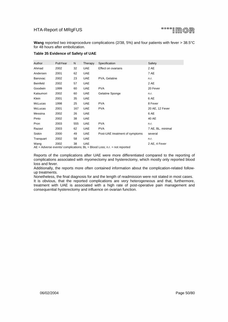
HTA-Report of MRgFUS
06/02/2004 Page 50/80
Wang reported two intraprocedure complications (2/38, 5%) and four patients with fever > 38.5°C for 48 hours after embolization.
Table 35 Evidence of Safety of UAE
Author PubYear N Therapy Specification Safety
Ahmad 2002 32 UAE Effect on ovarians 2 AE
Andersen 2001 62 UAE 7 AE
Banovac 2002 23 UAE PVA, Gelatine n.r.
Beinfeld 2002 57 UAE 2 AE
Goodwin 1999 60 UAE PVA 20 Fever
Katsumori 2002 60 UAE Gelatine Sponge n.r.
Klein 2001 35 UAE 6 AE
McLucas 1998 25 UAE PVA 8 Fever
McLucas 2001 167 UAE PVA 20 AE, 12 Fever
Messina 2002 26 UAE 6 AE
Pinto 2002 38 UAE 40 AE
Pron 2003 555 UAE PVA n.r.
Razavi 2003 62 UAE PVA 7 AE, BL. minimal
Siskin 2000 49 UAE Post-UAE treatment of symptoms several
Tranquart 2002 58 UAE n.r.
Wang 2002 38 UAE 2 AE, 4 Fever AE = Adverse events/ complications; BL = Blood Loss; n.r. = not reported Reports of the complications after UAE were more differentiated compared to the reporting of complications associated with myomectomy and hysterectomy, which mostly only reported blood loss and fever. Additionally, the reports more often contained information about the complication-related follow-up treatments. Nonetheless, the final diagnosis for and the length of readmission were not stated in most cases. It is obvious, that the reported complications are very heterogeneous and that, furthermore, treatment with UAE is associated with a high rate of post-operative pain management and consequential hysterectomy and influence on ovarian function.
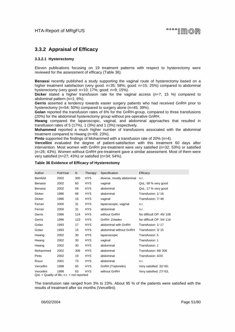
HTA-Report of MRgFUS
06/02/2004 Page 51/80
3.3.2 Appraisal of Efficacy
3.3.2.1 Hysterectomy
Eleven publications focusing on 19 treatment patterns with respect to hysterectomy were reviewed for the assessment of efficacy (Table 36). Benassi recently published a study supporting the vaginal route of hysterectomy based on a higher treatment satisfaction (very good: n=35; 58%; good: n=15; 25%) compared to abdominal hysterectomy (very good: n=10; 17%; good: n=9; 15%). Dicker stated a higher transfusion rate for the vaginal access (n=7; 15 %) compared to abdominal pattern (n=1; 6%). Gerris asserted a tendency towards easier surgery patients who had received GnRH prior to hysterectomy (n=54; 50%) compared to surgery alone (n=45; 39%). Golan reported the transfusion rates of 6% for the GnRH-group, compared to three transfusions (20%) for the abdominal hysterectomy group without pre-operative GnRH. Hwang compared the laparoscopic, vaginal, and abdominal approaches that resulted in transfusion rates of 5 (17%), 1 (3%) and 1 (3%) respectively. Mohammed reported a much higher number of transfusions associated with the abdominal treatment compared to Hwang (n=69; 23%). Pinto supported the findings of Mohammed with a transfusion rate of 20% (n=4). Vercellini evaluated the degree of patient-satisfaction with this treatment 60 days after intervention. Most women with GnRH pre-treatment were very satisfied (n=32; 53%) or satisfied (n=26; 43%). Women without GnRH pre-treatment gave a similar assessment. Most of them were very satisfied (n=27; 43%) or satisfied (n=34; 54%).
Table 36 Evidence of Efficacy of Hysterectomy
Author PubYear N Therapy Specification Efficacy
Beinfeld 2002 300 HYS diverse, mostly abdominal n.r.
Benassi 2002 60 HYS vaginal QoL: 58 % very good
Benassi 2002 59 HYS abdominal QoL: 17 % very good
Dicker 1986 68 HYS abdominal Transfusion: 1/ 16
Dicker 1986 16 HYS vaginal Transfusion: 7/ 48
Ferrari 2000 31 HYS laparoscopic, vaginal n.r.
Ferrari 2000 31 HYS abdominal n.r.
Gerris 1996 124 HYS without GnRH No difficult OP: 45/ 108
Gerris 1996 123 HYS GnRH: Zoladex No difficult OP: 54/ 116
Golan 1993 17 HYS abdominal with GnRH Transfusion: 1/ 17
Golan 1993 15 HYS abdominal without GnRH Transfusion: 3/ 15
Hwang 2002 30 HYS laparoscopic Transfusion: 5
Hwang 2002 30 HYS vaginal Transfusion: 1
Hwang 2002 30 HYS abdominal Transfusion: 1
Mohammed 2002 306 HYS abdominal Transfusion: 69/ 306
Pinto 2002 19 HYS abdominal Transfusion: 4/20
Rouzi 2001 73 HYS abdominal n.r.
Vercellini 1998 60 HYS GnRH (Triptorelin) Very satisfied: 32/ 60;
Vercellini 1998 63 HYS without GnRH Very satisfied: 27/ 63; QoL = Quality of life; n.r. = not reported The transfusion rate ranged from 3% to 23%. About 95 % of the patients were satisfied with the results of treatment after six months (Vercellini).
HTA-Report of MRgFUS
06/02/2004 Page 52/80
3.3.2.2 Myomectomy
Twenty-six publications focusing on 39 treatment patterns with respect to myomectomy were reviewed for the assessment of efficacy (Table 37). Clark asked women at month 6 about satisfaction with treatment and improvement in bleeding symptoms. Twenty-eight patients (78%) had much better or better improvement of bleeding symptoms. Thirty-two women were satisfied (92%). Interestingly only 75% of patients were prepared to undergo treatment again if required. Eighty-one % would recommend treatment to others. Connolly reported that 19 of the women who conceived successfully gave birth, six had miscarriages (24%). Of the live births, six had caesarean sections (36%) and two (11%) were premature. Davies reported four women who received transfusions (11%). 15 women out of 21 women with menorrhagia improved the regularity of their period (71%). Dicker stated a higher transfusion rate for the vaginal approach (n=18; 26%) compared to the abdominal access (n=2; 17%). Debuisson evaluated a failure rate, meaning conversion to laparotomy, of 7.5% (n=16). Feng reported normal menstrual patterns developed in 94 of 99 patients (95%). Unfortunately they did not mention the corresponding date of follow up, which could be up to 1, 3, 6, or 12 month. Frederick (2002) observed five of nine pregnancies after secondary myomectomy carried to term and delivery of a live birth. Ginsburg found 4 women (19%) requiring transfusion. Golan reported a much higher transfusion rate compared to Ginsburg of about 50% (n=11). Hart performed a survival analysis to determine the percentage of patients avoiding or requiring further surgery per cumulated time years in relation to uterus size, fibroid size, position of fibroid, procedure time, age, hormonal endometrial preparation, or number of fibroids excised. Overall, 79% of women at risk avoided further surgery over the first four years after hysteroscopic myomectomy, and none required surgery thereafter during the maximum follow up of eight years. Malzoni registered 21 pregnancies of 38 women (55%) undergoing laparoscopic myomectomy at 12 months. Mohammed recorded 21 transfusions (16%). Ostrzenski reported two out of five infertile patients becoming pregnant and having successful delivery at term by caesarean section. Razzavi focused on several outcomes. Successful improvement of menorrhagia (defined as completely resolved or significantly improved) was observed for 14 patients (64%). 14 patients (54%) had successful pain reduction (same definition as above). Three women (7.5%) received transfusions. Seracchiolli reported, that none of the 33 women in the study needed transfusion. Soriano reported 36 live births out of 44 pregnancies after laparoscopic myomectomy (82%), compared to 4 live births out of 10 pregnancies post laparoconverted myomectomy (40%). The mean follow-up period was 27 and 32 months respectively. Steller examined 51 patients (39%) with a recurrent myoma in almost 80% of all cases within the first two and a half years after myomectomy. 45 patients (59%) who wanted to have children became pregnant after myomectomy and 60% conceived within the first year after the intervention. Stringer found no candidate for transfusion within a group of 49 laparoscopic treated women and three transfusions (6%) following abdominal therapy. Vercellini found only small differences in transfusion rates between preoperative GnRH-treated and not preoperative GnRH-treated women (n=7; 14% versus n=8; 16%). Wamstecker worked out, that the mean number of procedures to achieve complete resection increased with more extensive intramural involved fibroids. Zollner reviewed 49 of 77 patients desiring children with 20 conceptions (41%).
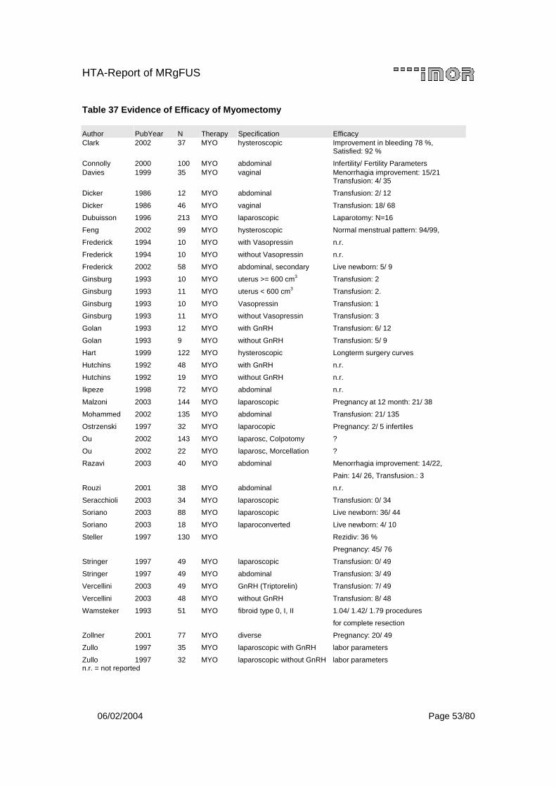
HTA-Report of MRgFUS
06/02/2004 Page 53/80
Table 37 Evidence of Efficacy of Myomectomy
Author PubYear N Therapy Specification Efficacy Clark 2002 37 MYO hysteroscopic Improvement in bleeding 78 %,
Satisfied: 92 %
Connolly 2000 100 MYO abdominal Infertility/ Fertility Parameters Davies 1999 35 MYO vaginal Menorrhagia improvement: 15/21
Transfusion: 4/ 35
Dicker 1986 12 MYO abdominal Transfusion: 2/ 12
Dicker 1986 46 MYO vaginal Transfusion: 18/ 68
Dubuisson 1996 213 MYO laparoscopic Laparotomy: N=16
Feng 2002 99 MYO hysteroscopic Normal menstrual pattern: 94/99,
Frederick 1994 10 MYO with Vasopressin n.r.
Frederick 1994 10 MYO without Vasopressin n.r.
Frederick 2002 58 MYO abdominal, secondary Live newborn: 5/ 9
Ginsburg 1993 10 MYO uterus >= 600 cm3 Transfusion: 2
Ginsburg 1993 11 MYO uterus < 600 cm3 Transfusion: 2.
Ginsburg 1993 10 MYO Vasopressin Transfusion: 1
Ginsburg 1993 11 MYO without Vasopressin Transfusion: 3
Golan 1993 12 MYO with GnRH Transfusion: 6/ 12
Golan 1993 9 MYO without GnRH Transfusion: 5/ 9
Hart 1999 122 MYO hysteroscopic Longterm surgery curves
Hutchins 1992 48 MYO with GnRH n.r.
Hutchins 1992 19 MYO without GnRH n.r.
Ikpeze 1998 72 MYO abdominal n.r.
Malzoni 2003 144 MYO laparoscopic Pregnancy at 12 month: 21/ 38
Mohammed 2002 135 MYO abdominal Transfusion: 21/ 135
Ostrzenski 1997 32 MYO laparocopic Pregnancy: 2/ 5 infertiles
Ou 2002 143 MYO laparosc, Colpotomy ?
Ou 2002 22 MYO laparosc, Morcellation ?
Razavi 2003 40 MYO abdominal Menorrhagia improvement: 14/22,
Pain: 14/ 26, Transfusion.: 3
Rouzi 2001 38 MYO abdominal n.r.
Seracchioli 2003 34 MYO laparoscopic Transfusion: 0/ 34
Soriano 2003 88 MYO laparoscopic Live newborn: 36/ 44
Soriano 2003 18 MYO laparoconverted Live newborn: 4/ 10
Steller 1997 130 MYO Rezidiv: 36 %
Pregnancy: 45/ 76
Stringer 1997 49 MYO laparoscopic Transfusion: 0/ 49
Stringer 1997 49 MYO abdominal Transfusion: 3/ 49
Vercellini 2003 49 MYO GnRH (Triptorelin) Transfusion: 7/ 49
Vercellini 2003 48 MYO without GnRH Transfusion: 8/ 48
Wamsteker 1993 51 MYO fibroid type 0, I, II 1.04/ 1.42/ 1.79 procedures
for complete resection
Zollner 2001 77 MYO diverse Pregnancy: 20/ 49
Zullo 1997 35 MYO laparoscopic with GnRH labor parameters
Zullo 1997 32 MYO laparoscopic without GnRH labor parameters n.r. = not reported
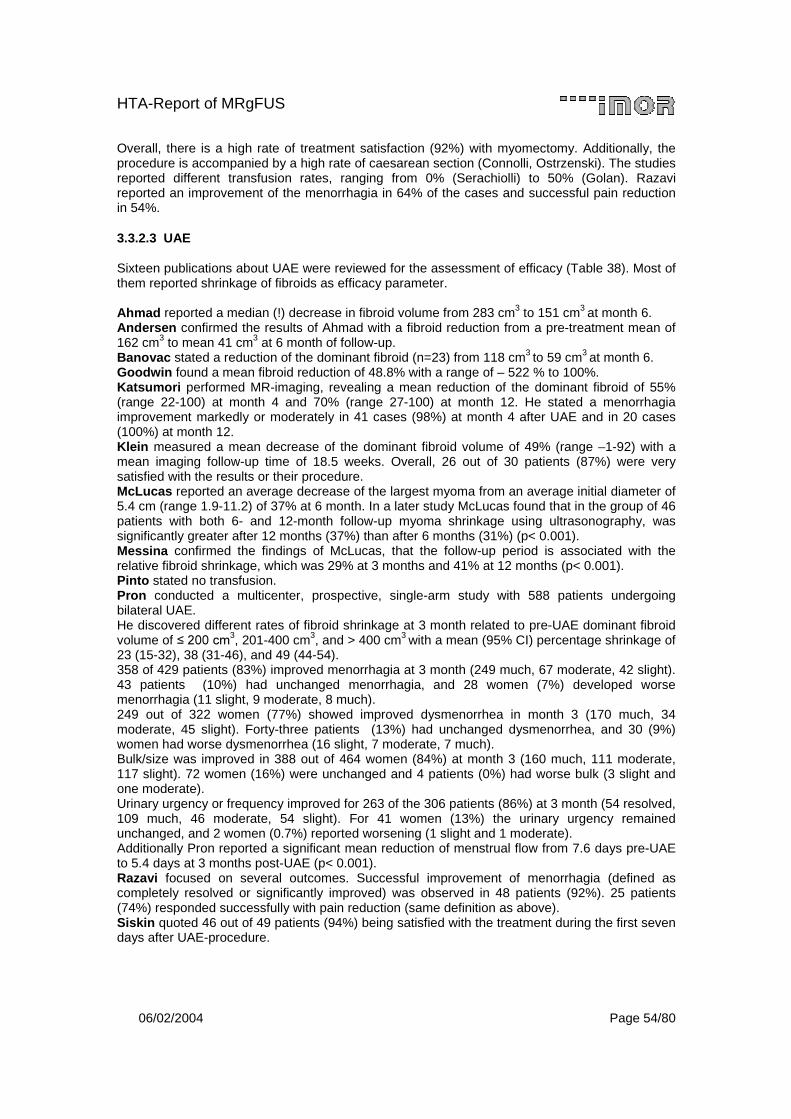
HTA-Report of MRgFUS
06/02/2004 Page 54/80
Overall, there is a high rate of treatment satisfaction (92%) with myomectomy. Additionally, the procedure is accompanied by a high rate of caesarean section (Connolli, Ostrzenski). The studies reported different transfusion rates, ranging from 0% (Serachiolli) to 50% (Golan). Razavi reported an improvement of the menorrhagia in 64% of the cases and successful pain reduction in 54%.
3.3.2.3 UAE
Sixteen publications about UAE were reviewed for the assessment of efficacy (Table 38). Most of them reported shrinkage of fibroids as efficacy parameter. Ahmad reported a median (!) decrease in fibroid volume from 283 cm3 to 151 cm3 at month 6. Andersen confirmed the results of Ahmad with a fibroid reduction from a pre-treatment mean of 162 cm3 to mean 41 cm3 at 6 month of follow-up. Banovac stated a reduction of the dominant fibroid (n=23) from 118 cm3 to 59 cm3 at month 6. Goodwin found a mean fibroid reduction of 48.8% with a range of – 522 % to 100%. Katsumori performed MR-imaging, revealing a mean reduction of the dominant fibroid of 55% (range 22-100) at month 4 and 70% (range 27-100) at month 12. He stated a menorrhagia improvement markedly or moderately in 41 cases (98%) at month 4 after UAE and in 20 cases (100%) at month 12. Klein measured a mean decrease of the dominant fibroid volume of 49% (range –1-92) with a mean imaging follow-up time of 18.5 weeks. Overall, 26 out of 30 patients (87%) were very satisfied with the results or their procedure. McLucas reported an average decrease of the largest myoma from an average initial diameter of 5.4 cm (range 1.9-11.2) of 37% at 6 month. In a later study McLucas found that in the group of 46 patients with both 6- and 12-month follow-up myoma shrinkage using ultrasonography, was significantly greater after 12 months (37%) than after 6 months (31%) (p< 0.001). Messina confirmed the findings of McLucas, that the follow-up period is associated with the relative fibroid shrinkage, which was 29% at 3 months and 41% at 12 months (p< 0.001). Pinto stated no transfusion. Pron conducted a multicenter, prospective, single-arm study with 588 patients undergoing bilateral UAE. He discovered different rates of fibroid shrinkage at 3 month related to pre-UAE dominant fibroid volume of "�������3, 201-400 cm3, and > 400 cm3 with a mean (95% CI) percentage shrinkage of 23 (15-32), 38 (31-46), and 49 (44-54). 358 of 429 patients (83%) improved menorrhagia at 3 month (249 much, 67 moderate, 42 slight). 43 patients (10%) had unchanged menorrhagia, and 28 women (7%) developed worse menorrhagia (11 slight, 9 moderate, 8 much). 249 out of 322 women (77%) showed improved dysmenorrhea in month 3 (170 much, 34 moderate, 45 slight). Forty-three patients (13%) had unchanged dysmenorrhea, and 30 (9%) women had worse dysmenorrhea (16 slight, 7 moderate, 7 much). Bulk/size was improved in 388 out of 464 women (84%) at month 3 (160 much, 111 moderate, 117 slight). 72 women (16%) were unchanged and 4 patients (0%) had worse bulk (3 slight and one moderate). Urinary urgency or frequency improved for 263 of the 306 patients (86%) at 3 month (54 resolved, 109 much, 46 moderate, 54 slight). For 41 women (13%) the urinary urgency remained unchanged, and 2 women (0.7%) reported worsening (1 slight and 1 moderate). Additionally Pron reported a significant mean reduction of menstrual flow from 7.6 days pre-UAE to 5.4 days at 3 months post-UAE (p< 0.001). Razavi focused on several outcomes. Successful improvement of menorrhagia (defined as completely resolved or significantly improved) was observed in 48 patients (92%). 25 patients (74%) responded successfully with pain reduction (same definition as above). Siskin quoted 46 out of 49 patients (94%) being satisfied with the treatment during the first seven days after UAE-procedure.

HTA-Report of MRgFUS
06/02/2004 Page 55/80
Tranquart, as McLucas and Messina, worked out, that the volume reduction of fibroids is associated with the follow-up time. He reported shrinkage of 29% at 3 months, 46% at 6 months, 55% at 12 months, and 86% at 24 months. Wang stated a fibroid shrinkage of 25 %, 49%, and 68% at 1 month, 3 months, and 6 months after embolization respectively.
Table 38 Evidence of Efficacy of UAE
Author PubYear N Therapy Specification Efficacy
Ahmad 2002 32 UAE Effect on ovarians S =130 cm3
Andersen 2001 62 UAE S =120 cm3
Banovac 2002 23 UAE PVA, Gelatine S = 146 cm3
Beinfeld 2002 57 UAE n.r.
Goodwin 1999 60 UAE PVA S = 48 %
Katsumori 2002 60 UAE Gelatine Sponge S = 55 %; Menorrhagia improved: 41/ 42
Klein 2001 35 UAE S = 49 %; Satisfied: 26/ 30
McLucas 1998 25 UAE PVA S = 37 %
McLucas 2001 167 UAE PVA S = 37 %;
Messina 2002 26 UAE S = 29 %/ 41 % at 3/ 12 month
Pinto 2002 38 UAE Transfusion: 0/ 40
Pron 2003 555 UAE PVA S = 23 %/ 38 %/ 49 %,
Menstrual duration shortened
Razavi 2003 62 UAE PVA Menorrhagia improvement: 48/52,
Pain Improvement: 25/ 34
Siskin 2000 49 UAE Patient satisfaction: 46/ 49
Tranquart 2002 58 UAE S = 29 %/ 46 %/ 55 %/ 86 %
at 3/ 6/ 12/ 24 month
Wang 2002 38 UAE S = 25 %/ 49 %/ 68 % S = Shrinkage; PVA = polyvinyl alcohol particles The above results show that the extent of reduction in uterine size is associated with the time of follow up. Additional determinate is the size of fibroid at baseline as Pron worked out. The well-conducted study by Pron showed that, in all likelihood, improvement may be expected in menorrhagia, dysmenorrhea, bulk, and urinary urgency.
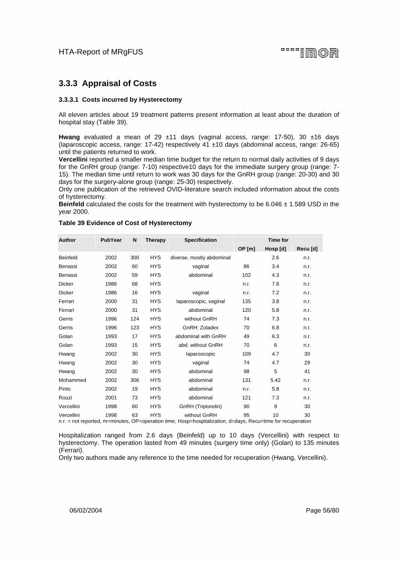
HTA-Report of MRgFUS
06/02/2004 Page 56/80
3.3.3 Appraisal of Costs
3.3.3.1 Costs incurred by Hysterectomy
All eleven articles about 19 treatment patterns present information at least about the duration of hospital stay (Table 39). Hwang evaluated a mean of 29 ±11 days (vaginal access, range: 17-50), 30 ±16 days (laparoscopic access, range: 17-42) respectively 41 ±10 days (abdominal access, range: 26-65) until the patients returned to work. Vercellini reported a smaller median time budget for the return to normal daily activities of 9 days for the GnRH group (range: 7-10) respective10 days for the immediate surgery group (range: 7-15). The median time until return to work was 30 days for the GnRH group (range: 20-30) and 30 days for the surgery-alone group (range: 25-30) respectively. Only one publication of the retrieved OVID-literature search included information about the costs of hysterectomy. Beinfeld calculated the costs for the treatment with hysterectomy to be 6.046 ± 1.589 USD in the year 2000.
Table 39 Evidence of Cost of Hysterectomy
Author PubYear N Therapy Specification Time for
OP [m] Hosp [d] Recu [d]
Beinfeld 2002 300 HYS diverse, mostly abdominal 2.6 n.r.
Benassi 2002 60 HYS vaginal 86 3.4 n.r.
Benassi 2002 59 HYS abdominal 102 4.3 n.r.
Dicker 1986 68 HYS n.r. 7.8 n.r.
Dicker 1986 16 HYS vaginal n.r. 7.2 n.r.
Ferrari 2000 31 HYS laparoscopic, vaginal 135 3.8 n.r.
Ferrari 2000 31 HYS abdominal 120 5.8 n.r.
Gerris 1996 124 HYS without GnRH 74 7.3 n.r.
Gerris 1996 123 HYS GnRH: Zoladex 70 6.8 n.r.
Golan 1993 17 HYS abdominal with GnRH 49 6.3 n.r.
Golan 1993 15 HYS abd. without GnRH 70 6 n.r.
Hwang 2002 30 HYS laparoscopic 109 4.7 30
Hwang 2002 30 HYS vaginal 74 4.7 29
Hwang 2002 30 HYS abdominal 98 5 41
Mohammed 2002 306 HYS abdominal 131 5.42 n.r.
Pinto 2002 19 HYS abdominal n.r. 5.8 n.r.
Rouzi 2001 73 HYS abdominal 121 7.3 n.r.
Vercellini 1998 60 HYS GnRH (Triptorelin) 90 9 30
Vercellini 1998 63 HYS without GnRH 95 10 30 n.r. = not reported, m=minutes, OP=operation time, Hosp=hospitalization, d=days, Recu=time for recuperation Hospitalization ranged from 2.6 days (Beinfeld) up to 10 days (Vercellini) with respect to hysterectomy. The operation lasted from 49 minutes (surgery time only) (Golan) to 135 minutes (Ferrari). Only two authors made any reference to the time needed for recuperation (Hwang, Vercellini).
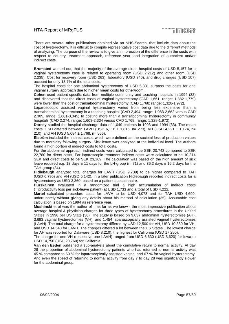
HTA-Report of MRgFUS
06/02/2004 Page 57/80
There are several other publications obtained via an NHS-Search, that include data about the cost of hysterectomy. It is difficult to compile representative cost data due to the different methods of analyzing. The purpose of the review is to give an impression of the difference in the costs with respect to country, treatment approach, reference year, and integration of outpatient and/or indirect costs. Brumsted worked out, that the majority of the average direct hospital costs of USD 5,157 for a vaginal hysterectomy case is related to operating room (USD 2,212) and other room (USD 2,235). Cost for recovery room (USD 263), laboratory (USD 340), and drug charges (USD 107) account for only 13.7% of the total costs. The hospital costs for one abdominal hysterectomy of USD 5,831 surpass the costs for one vaginal surgery approach due to higher mean costs for other/room. Cohen used patient-specific data from multiple community and teaching hospitals in 1994 (32) and discovered that the direct costs of vaginal hysterectomy (CAD 1,661, range: 1,382-1,779) were lower than the cost of transabdominal hysterectomy (CAD 1,768; range: 1,328-1,972). Laparosocopic assisted vaginal hysterectomy varied from being less expensive than a transabdominal hysterectomy in a teaching hospital (CAD 2,494, range: 1,083-2,662 versus CAD 2.305, range: 1,681-3,345) to costing more than a transabdominal hysterectomy in community hospitals (CAD 2,274, range: 1,603-2,334 versus CAD 1,768, range: 1,328-1,972). Dorsey studied the hospital discharge data of 1,049 patients in 1993 and 1994 (33). The mean costs ± SD differed between LAVH (USD 6,116 ± 1,816, n= 273), VH (USD 4,221 ± 1,174, n= 210), and AH (USD 5,084 ± 1,768, n= 566). Elström included the indirect costs, which were defined as the societal loss of production values due to morbidity following surgery. Sick leave was analyzed at the individual level. The authors found a high portion of indirect costs to total costs. For the abdominal approach indirect costs were calculated to be SEK 20,743 compared to SEK 22,780 for direct costs. For laparoscopic treatment indirect costs were calculated to be 10,314 SEK and direct costs to be SEK 23,169. The calculation was based on the high amount of sick leave required e.g. 18 days ± 11 days for the LH-group (n=71) and 36.2 days ± 16.2 days for the TAH-group (34). Hidlebaugh analyzed total charges for LAVH (USD 9,739) to be higher compared to TAH (USD 6,795) and VH (USD 5,142). In a later publication Hidlebaugh reported indirect costs for a hysterectomy as USD 3,360, based on a patient questionnaire. Hurskainen evaluated in a randomized trial a high accumulation of indirect costs (= productivity loss per sick-leave patient) at USD 1,733 and a total of USD 4,222. Martel calculated procedure costs for LAVH to be USD 4,073 and for TAH USD 4,699, unfortunately without giving any details about his method of calculation (35). Assumable cost calculation is based on 1994 as reference year. Mushinski et al was the author of – as far as we know - the most impressive publication about average hospital & physician charges for three types of hysterectomy procedures in the United States in 1998 per US State (36). The study is based on 9.037 abdominal hysterectomies (AH), 3.693 vaginal hysterectomies (VH), and 1.454 laparoscopically assisted vaginal hysterectomies (LAVH). The total charge for a hysterectomy differed by USD 12,500 for AH, USD 10,380 for VH, and USD 14,540 for LAVH. The charges differed a lot between the US States. The lowest charge for AH was reported for Dalaware (USD 8,210), the highest for California (USD 17,250). The charge for one VH (respective one LAVH) ranged from USD 6,630 (USD 8,620) for Iowa to USD 14,750 (USD 20,760) for California. Van den Eeden published a sub-analysis about the cumulative return to normal activity. At day 28 the proportion of abdominal hysterectomy patients who had returned to normal activity was 45 % compared to 60 % for laparoscopically assisted vaginal and 67 % for vaginal hysterectomy. And even the speed of returning to normal activity from day 7 to day 28 was significantly slower for the abdominal group.
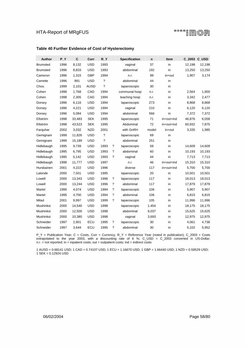
HTA-Report of MRgFUS
06/02/2004 Page 58/80
Table 40 Further Evidence of Cost of Hysterectomy
Author P_Y C Curr R_Y Specification n Item C_2003 C_USD
Brumsted 1996 8,132 USD 1993 vaginal 37 in 12,198 12,198
Brumsted 1996 8,833 USD 1993 abdominal 192 in 13,250 13,250
Cameron 1996 1,315 GBP 1994 n.r. 99 in+out 1,907 3,174
Carrette 1996 891 USD ? abdominal 44 in
Chou 1999 2,101 AUSD ? laparoscopic 30 in
Cohen 1998 1,768 CAD 1994 communal hosp n.r. in 2,564 1,900
Cohen 1998 2,305 CAD 1994 teaching hosp n.r. in 3,342 2,477
Dorsey 1996 6,116 USD 1994 laparoscopic 273 in 8,868 8,868
Dorsey 1996 4,221 USD 1994 vaginal 210 in 6,120 6,120
Dorsey 1996 5,084 USD 1994 abdominal 566 in 7,372 7,372
Ellström 1998 33,483 SEK 1995 laparoscopic 71 in+out+ind 46,876 6,058
Ellström 1998 43,523 SEK 1995 Abdominal 72 in+out+ind 60,932 7,875
Farquhar 2002 3,032 NZD 2001 with GnRH model in+out 3,335 1,985
Gemignani 1999 11,826 USD ? laparoscopic 69 in
Gemignani 1999 15,189 USD ? abdominal 251 in
Hidlebaugh 1995 9,739 USD 1993 ? laparoscopic 59 in 14,609 14,609
Hidlebaugh 1995 6,795 USD 1993 ? abdominal 60 in 10,193 10,193
Hidlebaugh 1995 5,142 USD 1993 ? vaginal 44 in 7,713 7,713
Hidlebaugh 1998 11,777 USD 1997 n.r. 46 in+out+ind 15,310 15,310
Hurskainen 2001 4,222 USD 1996 diverse 117 in+out+ind 5,700 5,700
Lalonde 2000 7,501 USD 1995 laparoscopic 20 in 10,501 10,501
Lowell 2000 13,343 USD 1996 ? laparoscopic 117 in 18,013 18,013
Lowell 2000 13,244 USD 1996 ? abdominal 117 in 17,879 17,879
Martel 1995 4,074 USD 1994 ? laparoscopic 106 in 5,907 5,907
Martel 1995 4,700 USD 1994 ? abdominal 106 in 6,815 6,815
Milad 2001 9,997 USD 1999 ? laparoscopic 105 in 11,996 11,996
Mushinksi 2000 14,540 USD 1998 laparoscopic 1.454 in 18,175 18,175
Mushinksi 2000 12,500 USD 1998 abdominal 9,037 in 15,625 15,625
Mushinksi 2000 10,380 USD 1998 vaginal 3,693 in 12,975 12,975
Schneider 1997 2,901 ECU 1995 ? laparoscopic 30 in 4,061 4,738
Schneider 1997 3,644 ECU 1995 ? abdominal 30 in 5,102 5,952 P_Y = Publication Year; C = Costs, Curr = Currency, R_Y = Reference Year (noted in publication); C_2003 = Costs extrapolated to the year 2003, with a discounting rate of 6 %; C_USD = C_2003 converted in US-Dollar; n.r. = not reported; in = inpatient costs; out = outpatient costs; ind = indirect costs 1 AUSD = 0.68141 USD; 1 CAD = 0.74107 USD; 1 ECU = 1.16670 USD; 1 GBP = 1.66440 USD; 1 NZD = 0.59529 USD; 1 SEK = 0.12924 USD
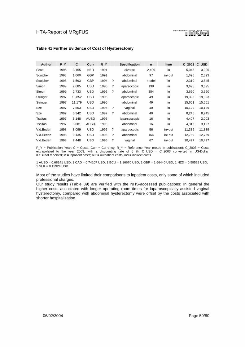
HTA-Report of MRgFUS
06/02/2004 Page 59/80
Table 41 Further Evidence of Cost of Hysterectomy
Author P_Y C Curr R_Y Specification n Item C_2003 C_USD
Scott 1995 3,155 NZD 1991 diverse 2,409 in 5,048 3,005
Sculpher 1993 1,060 GBP 1991 abdominal 97 in+out 1,696 2,823
Sculpher 1998 1,593 GBP 1994 ? abdominal model in 2,310 3,845
Simon 1999 2,685 USD 1996 ? laparsocopic 138 in 3,625 3,625
Simon 1999 2,733 USD 1996 ? abdominal 354 in 3,690 3,690
Stringer 1997 13,852 USD 1995 laparoscopic 49 in 19,393 19,393
Stringer 1997 11,179 USD 1995 abdominal 49 in 15,651 15,651
Sze 1997 7,503 USD 1996 ? vaginal 40 in 10,129 10,129
Sze 1997 6,342 USD 1997 ? abdominal 40 in 8,245 8,245
Tsaltas 1997 3,148 AUSD 1995 laparsoscopic 16 in 4,407 3,003
Tsaltas 1997 3,081 AUSD 1995 abdominal 16 in 4,313 3,197
V.d.Eeden 1998 8,099 USD 1995 ? laparoscopic 56 in+out 11,339 11,339
V.d.Eeden 1998 9,135 USD 1995 ? abdominal 164 in+out 12,789 12,789
V.d.Eeden 1998 7,448 USD 1995 ? vaginal 67 in+out 10,427 10,427 P_Y = Publication Year; C = Costs, Curr = Currency, R_Y = Reference Year (noted in publication); C_2003 = Costs extrapolated to the year 2003, with a discounting rate of 6 %; C_USD = C_2003 converted in US-Dollar; n.r. = not reported; in = inpatient costs; out = outpatient costs; ind = indirect costs 1 AUSD = 0.68141 USD; 1 CAD = 0.74107 USD; 1 ECU = 1.16670 USD; 1 GBP = 1.66440 USD; 1 NZD = 0.59529 USD; 1 SEK = 0.12924 USD Most of the studies have limited their comparisons to inpatient costs, only some of which included professional charges. Our study results (Table 39) are verified with the NHS-accessed publications: In general the higher costs associated with longer operating room times for laparoscopically assisted vaginal hysterectomy, compared with abdominal hysterectomy were offset by the costs associated with shorter hospitalization.
HTA-Report of MRgFUS
06/02/2004 Page 60/80
3.3.3.2 Costs incurred by Myomectomy
Twenty-seven publications, focusing on 39 treatment patterns, with respect to myomectomy were reviewed for the assessment of costs (Table 42). The surgical time ranged from 26 minutes for a laparoscopic myomectomy (Feng) to 258 minutes for an abdominal surgery (Stringer). The mean time for hospital admission varied from 7.5 hours for a laparoscopic intervention to 8.5 days for an abdominal myomectomy (Dicker). Razavi calculated the average number of days required before the patient could return to normal activity for the abdominal access (36 days). Days until recommenced work was analysed by Seracciolli for the laparoscopic approach (20 days). Stringer assessed hospital costs for a laparoscopic myomectomy of USD 13,852 in 1995. Hospital costs for an open myomectomy were ascertained to be USD 11,179 by the year 1995. Clark maintained that treatment costs for a laparoscopic myomectomy was GBP 1,265 in the year 2000. One publication, obtained via NHS-Search, included data pertaining to the cost of a myomectomy. Brumsted generated mean hospitalization costs for a myomectomy of USD 2,070 based on 1993.

HTA-Report of MRgFUS
06/02/2004 Page 61/80
Table 42 Evidence of Cost of Myomectomy
Author PubYear N Therapy Specification Time for
OP [m] Hosp [d] Recu [d]
Clark 2002 37 MYO hysteroscopic n.r. n.r. n.r.
Connolly 2000 100 MYO abdominal n.r. 5 n.r.
Davies 1999 35 MYO vaginal 78 3.9 n.r.
Dicker 1986 12 MYO abdominal n.r. 8.5 n.r.
Dicker 1986 46 MYO vaginal n.r. 1.4 n.r.
Dubuisson 1996 213 MYO laparoscopic 125 2.7 n.r.
Feng 2002 99 MYO hysteroscopic 26 n.r. n.r.
Frederick 1994 10 MYO with Vasopressin n.r. n.r. n.r.
Frederick 1994 10 MYO without Vasopressin n.r. n.r. n.r.
Frederick 2002 58 MYO abdominal, secondary n.r. n.r. n.r.
Ginsburg 1993 10 MYO Uterus #�$������ 77 3.9 n.r.
Ginsburg 1993 11 MYO Uterus < 600 cm3 61 3.7 n.r.
Ginsburg 1993 10 MYO Vasopressin 72 3.6 n.r.
Ginsburg 1993 11 MYO without Vasopressin 66 4 n.r.
Golan 1993 12 MYO with GnRH 80 7.9 n.r.
Golan 1993 9 MYO without GnRH 96 7.3 n.r.
Hart 1999 122 MYO hysteroscopic 28 n.r. n.r.
Hutchins 1992 48 MYO with GnRH n.r. 4.9 n.r.
Hutchins 1992 19 MYO without GnRH n.r. 4.7 n.r.
Ikpeze 1998 72 MYO abdominal 49 8.1 n.r.
Malzoni 2003 144 MYO laparoscopic 85 2.6 n.r.
Mohammed 2002 135 MYO abdominal 127 5.44 n.r.
Ostrzenski 1997 32 MYO laparoscopic 163 < 1 n.r.
Ou 2002 143 MYO laparosc, Colpotomy 144 n.r. n.r.
Ou 2002 22 MYO laparosc, Morcellation 168 n.r. n.r.
Razavi 2003 40 MYO abdominal n.r. 2.9 36
Rouzi 2001 38 MYO abdominal 103 5.8 n.r.
Seracchioli 2003 34 MYO laparoscopic 79 2.25 20
Soriano 2003 88 MYO laparoscopic 150 3 n.r.
Soriano 2003 18 MYO laparoconverted 148 5.5 n.r.
Steller 1997 130 MYO n.r. n.r. n.r.
Stringer 1997 49 MYO laparoscopic 133 0.6 n.r.
Stringer 1997 49 MYO abdominal 258 5.6 n.r.
Vercellini 2003 49 MYO GnRH (Triptorelin) 93 6.1 n.r.
Vercellini 2003 48 MYO without GnRH 90 5.9 n.r.
Wamsteker 1993 51 MYO fibroid type 0, I, II n.r. n.r. n.r.
Zollner 2001 77 MYO diverse n.r. n.r. n.r.
Zullo 1997 35 MYO Laparoscopic with GnRH 99 n.r. n.r.
Zullo 1997 32 MYO laparoscopic without GnRH 113 n.r. n.r. n.r. = not reported, m=minutes, OP=operation time, Hosp=hospitalization, d=days, Recu=time for recuperation

HTA-Report of MRgFUS
06/02/2004 Page 62/80
3.3.3.3 Costs incurred by UAE
Sixteen publications about UAE were reviewed for the assessment of costs (Table 43). The mean time for the procedure ranged from 54 minutes (Klein) to 90 minutes (Ahmad). The mean period of hospitalization ranged from less than one day (Klein, Siskin) to about 4 days (Katsumori). It is interesting to note that more articles report cost for recuperation compared to publications about the cost of a hysterectomy or a myomectomy. Siskin assessed a mean period of time between the procedure and return to employment or school of 7.7 days. Katsumori reported an average of 12 days to reach full recovery. Beinfeld calculated hospital costs for the treatment with UAE to be USD 8,223 ± 1,834 in the year 2000.
Table 43 Evidence of Cost of UAE
Author PubYear N Therapy Specification Time for
OP [m] Hosp [d] Recu [d]
Ahmad 2002 32 UAE effect on ovarians 90 n.r. n.r.
Andersen 2001 62 UAE 75 n.r. n.r.
Banovac 2002 23 UAE PVA, gelatine n.r. n.r. n.r.
Beinfeld 2002 57 UAE n.r. 0.95 n.r.
Goodwin 1999 60 UAE PVA n.r. n.r. n.r.
Katsumori 2002 60 UAE gelatine sponge 54 4.3 12
Klein 2001 35 UAE n.r. < 1 n.r.
McLucas 1998 25 UAE PVA 60 n.r. 7
McLucas 2001 167 UAE PVA n.r. n.r. n.r.
Messina 2002 26 UAE n.r. n.r. n.r.
Pinto 2002 38 UAE n.r. 1.7 9.5
Pron 2003 555 UAE PVA n.r. n.r. n.r.
Razavi 2003 62 UAE PVA n.r. 0 8
Siskin 2000 49 UAE n.r. < 1 7.7
Tranquart 2002 58 UAE n.r. n.r. n.r.
Wang 2002 38 UAE n.r. 3 n.r. n.r. = not reported m=minutes, OP=operation time, Hosp=hospitalization, d=days, Recu=time for recuperation One publication, obtained via NHS-Search, gives data about the cost of a UAE. Subramanian compiled costs of a UAE procedure with average facility costs of USD 3,080 in 1998.
HTA-Report of MRgFUS
06/02/2004 Page 63/80
3.4 Synthesis of Evidence The research question of this HTA report was: What are the advantages and what are the disadvantages of using Magnetic Resonance guided Focused Ultrasound (MRgFUS) for the treatment of uterine fibroids? The principal results are as follows:
• Immediately after the treatment with MRgFUS the majority of patients reported no pain (75%) or only a mild pain (18%) and no discomfort (68%) or just mild discomfort (25%) (Table 16).
• A very small percentage of patients in both treatment arms - MRgFUS as well as abdominal hysterectomy - showed physician-reported clinically significant findings at week 1, month 1, month 3, and month 6 (Table 17-Table 20).
• About 90% of MRgFUS-patients reported no or only a mild condition in each category (discomfort, pain, abdominal tenderness, paresthesis, other) one week after the intervention. In contrast, more patients undergoing hysterectomy suffered moderate or severe discomfort or pain relating to the condition being treated (Table 21).
• MRgFUS-therapy is associated with a low rate of clinical complications. The complication rate is lower compared to the rate of clinical complications with respect to hysterectomy (Table 25). Three patients (6%) developed a high temperature on 2 post-procedure days but not within first 24 hours. Three patients (6%) started antibiotics after 24 hours post-treatment.
• Eighty percent of the patients undergoing MRgFUS improved their uterine fibroid symptoms by about 10 points, 60% by about 20 points, 40 % by about 30 points, and 20% by about 40 points on a maximum scale value of 100 points (Figure 12).
• MRgFUS-patients improved considerably in all eight SF-36 subcategories (Table 28). • Treatment associated costs for one MRgFUS therapy are calculated as EUR 3,565.00 =
USD 4,159.00 (ECU 1.00 = USD 1.16670). • The mean lost working days for patients undergoing MRgFUS were 1.4 days through the
first four weeks after treatment compared to 16.5 days for the patients with hysterectomy. • As a result of the literature review, hysterectomy is associated with a mean blood loss of
200 up to 630 ml, a fever rate between 3% and 36%, and a complication rate (not counting fever and transfusion) between 2% and 50% (Table 33). The transfusion rate ranged from 3% to 23%. A high percentage of hysterectomy patients were satisfied with the treatment (Table 36) (37;38). There are considerable variations in the mean days of admission due to hysterectomy (Table 39). For health-economic analysts it is of interest, that the mean duration until return to work was reported as 30 days (37;39). The costs for one hysterectomy varied with respect to treatment access, country, and reference year (Table 40). Many publications report costs of about USD 5,000.
• It is decisive, whether hysterectomy is accomplished via abdominal, vaginal, or laparoscopic approach with accordant and inconsistent differences in efficacy, safety, and costs.
• The literature review shows, that myomectomy is related to a mean blood loss between 175 ml up to 670 ml, and a complication rate of 2% up to 34% (Table 34). A high rate of treatment satisfaction was determined (40). Improvement of symptoms is reported to be around 70% (40-42). The mean time for hospital admission varied from 7.5 hours for a laparoscopic intervention to 8.5 days for an abdominal myomectomy (Table 42). The mean time until the patient could resume normal activities or return to work was 36 days (41) respective 20 days (43). Information about the costs of myomectomy is scarce and ranges between USD 2,000 (44) and about USD 10,000 (45). The results in respect of efficacy, safety, and costs of myomectomy are different and inconsistent with respect to the treatment approach (abdominal, vaginal, laparoscopic, hysteroscopic).

HTA-Report of MRgFUS
06/02/2004 Page 64/80
• Very detailed reports about the complications following UAE-procedure were found. The treatment is correlated with a high number of post-operative complications (46;47), especially with a high rate of post-operative pain management (46-49) (Table 35). The review of the studies about UAE discloses, that a mean shrinkage of the uterine fibroid of up to 86 % was achieved depending on the length of the follow-up period. One comprehensive study reported that, in all likelihood, improvement could be expected for menorrhagia, sysmenorrhea, bulk, and urinary urgency (50). The mean hospital stay ranged from a treatment within 24 hours (41;47;51;52) up to 4.3 days hospital admission (53). Up to 12 days are needed for recuperation (41;46-48;53). Direct treatment costs are calculated at USD 3,080 (54).
HTA-Report of MRgFUS
06/02/2004 Page 65/80
4 Discussion
4.1 Methods of Assessment This report is a health technology assessment for the new technology Magnetic Resonance guided Focused Ultrasound Surgery (MRgFUS) for the Treatment of Fibroids. Primary analysis data of the study UF002 were available for use in the assessment. The strength of this data is based on well-defined inclusion criteria, the study character as multinational trial, a relatively high number of enrolled patients, the inclusion of a control arm (hysterectomy), multiple recorded items, the use of a health-economic questionnaire, as well as differentiated analysis. The assessment of efficacy and safety of MRgFUS was based on study results already on hand. Intensive communication with the study manager responsible for Insightec-TxSonics, Inc. was used to answer queries. The pivotal study was non-randomized. As a result there are differences in mean BMI, mean SF-36 and mean FUS at baseline in both treatment arms. Thus results of both treatment arms should be interpreted separately. It should be kept in mind, that patients with lower health status at baseline have a higher chance of considerable improvement (points changed). The literature search was based on a search of six established databases to retrieve evidence about efficacy, safety and the cost of comparative treatment patterns hysterectomy, myomectomy, and UAE. We did not focus on pharmacological treatment approaches, either as stand-alone program or pre- and/or post-operative, to avoid complexity. Hysterectomy and myomectomy were chosen as comparators, because they represent common practice in the treatment of uterine fibroids (10;36;55-57). UAE was chosen as a comparator, because this approach plays an increasingly important role in the treatment of uterine fibroids (58). The literature selection and consequential data extraction was performed in a prospective way to avoid bias when selecting the evidence (chapter 2.3). Type of study (randomized controlled trial, cohort study, register) as well as other aspects of study design, like follow-up time, were not used as selection criteria. During the data abstraction of full-text we recognized, that some selected publications are based on less than 20 patients per treatment arm – which was one of the selection criteria. We decided to keep these articles eligible. We did so, because the primary focus was on the assessment of the MRgFUS device and not on an extensive comparison of comparators. We compared our literature references with the selected literature of a recently performed HTA-report about the management of uterine fibroids (59). The authors found 116 articles about the invasive treatment of uterine fibroids with 14 articles congruent to our report, of which 9 articles were selected for our review (47;60-67) and 5 articles being rejected based on full-text-screening (68-72). The difference might be explained in part by our explicit focus on patients with uterine fibroids. Especially for UAE our literature selection produced similar publications to those featured in a systematic review conducted with an assessment program of the American Blue Cross and BlueShield Association in August 2002 (73). We are convinced that our literature search was very thorough, but since no attempt was made to identify unpublished material or to correspond with first authors, some degree of publication bias cannot be completely ruled out. References were documented as printed and electronic versions. Extracted data was entered into a specifically constructed database given in Appendix A.4 and C.6 lending more transparency to the study selection and data extraction.
HTA-Report of MRgFUS
06/02/2004 Page 66/80
4.2 Quality of Evidence Fortunately this HTA-report could be based on a clinical trial UF002 with underlying information about the study protocol, the used questionnaires, the analysis plan, as well as the results. The pivotal study UF002 is a useful, well-conducted, prospective, multi-center trial for the assessment of efficacy and safety of Magnetic Resonance guided Focused Ultrasound Surgery (MRgFUS) compared to abdominal hysterectomy in the treatment of uterine fibroids, as outlined in section 2.2. The sponsor provided this study data. APEX International, MedTrials, and MedTap conducted the data handling and/or the analysis. There was no personal communication with these institutes, due to the fact that study results became available simultaneously with writing this HTA-report. The data are reliable since the results of the study are an integral part of a FDA-submission being submitted in January 2004. The initial literature search in six bibliographic databases produced 47 relevant articles containing useful information about the efficacy and safety of the comparative treatment patterns hysterectomy, myomectomy and UAE (Table 6 - Table 9). A supplementary search in three databases of the NHS Center for Reviews and Dissemination identified 40 useful publications, mainly about hysterectomy-related treatment costs (Figure 10). The internal validity of most studies was limited as they were designed with a a lack of randomization. The majority compared two groups, with inherent, self-selected differences, even before the intervention was carried out. Inconsistency was noticeable in the reporting of the severity of symptoms, as well as uterine and fibroid anatomy. The evidence on the efficacy and safety of the comparative treatment patterns hysterectomy, myomectomy, and UAE is very inhomogeneous, containing limited details, making it difficult to draw conclusions. A lot of the articles do not provide sufficient information on baseline statistics like the patient’s age, weight, the number and size of fibroids, the size of uterus, or clinical anamnesis. For further details, refer to the printed, data-extraction report in Appendix A.4 or access database in Appendix C.6. It is difficult to compare the results of the treatment of MRgFUS with hysterectomy, myomectomy - with vaginal or abdominal access -, and UAE due to the significant differences in the severity of preintervention disease. Furthermore, most articles do not provide information about the definition of adverse events, and about data-handling process. Those studies that define adverse events, offer only inhomogeneous, definition criteria. For example, studies defined fever as a temperature of above 37.5°C (74), 38°C (75), or 38.3°C (39;47). Some studies allot two consecutive measurements (62), a measurement at 24 hours (75), or a record on two of the first 10 postoperative days, excluding the first 24 hours (76). Additionally, some of the studies gave no information as to the inclusion or exclusion criteria (77), whereas others (78;79)had restricted inclusion criteria, e.g. size of uterus (37;38;80), patient age (63;81-83), secondary myomectomy (42;84), the location of fibroids (74;85), size of fibroid (39;86), or number of fibroids (80). Other studies define explicit exclusion criteria, such as patient’s interest in continued fertility (87), pregnancy (82), prior myomectomy (63), numerous fibroids (52), uterus volume (88), or size of fibroids (46). It was the intention of this HTA-report to take into account the inhomogeneous application of antibiotics (40;41;45;48;49;51;52;54;62;65;66;69;77;79;82;85;86;94;99;105), analgesics (41;44;48;49;52;54;65;66;69;82;90-94), preoperative hormonal treatment (37;39;47;48;52;62;67;69;78;80;81;83;95-98), as well as anaesthetics (37;41;42;48-50;54;62;65;66;69;77;82;84;86;87;89;92-94;99-101) within divergent study settings. As a matter of interest, none of the articles reported, that postoperative hormonal treatment was given to patients. Some studies had recruitment periods of about 4 years (37;39;40;45;47;55;63;67;76;77;80;84-86;98-100;102) that may have had an influence on the outcome. In RCTs the dropout of patients (e.g. due to study discontinuation) are not always documented in detail.
HTA-Report of MRgFUS
06/02/2004 Page 67/80
4.3 Uncertainties/ Lack of Information The study UF002 has a follow-up time of six months. Long-term results are eagerly awaited based on a continuation study UF009 with a follow up at 1, 2, and 3 years. The selected articles about the efficacy, safety and costs of hysterectomy, myomectomy and Uterine Artery Embolization are restricted due to inhomogeneous study conditions or missing information. Most of the articles do not mention the location of fibroids. In most reviewed studies the extent of reported adverse events was divergent or lacked transparency. Several publications did not report any adverse events at all. In most cases no information was given about consequential therapies, relating to adverse events and complications. Thus, it is difficult to draw any conclusions about the remedial costs of various kinds of treatment. Associations between postprocedural interventions and clinical failures of primary interventions were not clearly defined. Notably only a few studies recorded quality of life aspects (34) or treatment satisfaction (37;38;40;47;52). Tranquart discovered that the extent of fibroid shrinkage depends on the observation time (100). Thus it became clear, that a long observation time of at least two years is mandatory. We did not focus especially fertility rates or birth rates due to the restricted information on the correlation of these rates with treatment patterns. It is hard to determine the influence the various kinds of treatment or no treatment of uterine fibroids had on infertility rates with respect to multiple confounders such as age, number of childbirths, restricted observation time, or inconsistent family planning. A lot of studies on hysterectomy, myomectomy, or UAE mention preoperative hormonal treatment, while the study protocol of UF002 defines patients previously on GnRH agonist therapy within the last 6 month as an exclusion criterion. Thus it was not clear as to how this would have increased the safety, efficacy and costs of MRgFUS patients that had undergone pre-procedural hormonal therapy. Malzoni reported 32 patients (22%) undergoing one or more surgical procedures associated with laparoscopic myomectomy, namely lysis in 24 patients, tubal plasty in 6 patients, appendectomy in 5 patients, ovarian cystectomy in 4 cases and coagulation of endometriosis in 3 cases (86). Most of the studies did not consider this aspect, namely that supplementary therapies might influence a decision in favor of a specific treatment. The clinical study UF002 disclosed that 60 % of the MRgFUS patients and 85 % of the hysterectomy patients suffered more than one adverse event. Thus, the disparate quote of literature-reported adverse events (Table 33-Table 35) raised the issue on the complexity and differentiation of safety reports. Reports on clinical studies only feature the objective of the study at hand. Studies in general are dedicated – like UF002 - to obtain final regulatory approval and contain inherent detailed information about severity, outcome, and relationship to the device of the respective adverse event. In other words, we can assume the safety of MRgFUS based on clinical data, but we cannot assure safety that is based on published study reports in the same manner.
HTA-Report of MRgFUS
06/02/2004 Page 68/80
4.4 Generalizability/ Applicability The process of synthesizing knowledge from observations involves transcending from the details of a set of observations to the abstract world of a scientific hypothesis or theory. This endeavor is aimed at being completely detached from time and place. In order to assess the possibility of generalizing, we have to distinguish between a pivotal study and a literature review. The study UF002 was performed at eight centers in five countries. A certain patient demography at baseline (defined with parameters such as age, BMI, race, fibroid volume, SF-36, UFS-QOL) make it difficult to adapt to other settings (see summary in chapter 3.2.1 and report in Appendix B.5). The pre-defined treatment procedure (see Study Protocol, Appendix B.1) implements additional restrictions with respect to the possibility of generalization. The literature search included only two constrictions, namely the focus on uterine fibroids and the absence of pregnancy or caesarean section. The limitations arising from the literature articles –e.g. differing inclusion and exclusion criteria, have an important influence on the suitability for generalization, as shown in the following example: Tsaltas reviewed international studies comparing the costs of LAVH and AH and found an inconsistency in as much as some found that LAVH was more expensive and others found TAH was more expensive (102). An intensive analysis would be necessary to detect the bias leading to such conflicting results. The fact that multiparous women have an easier vaginal uterus-access probably reduces costs. Other possible confounders such as uterine size or preliminary caesarean sections, are easily affiliated. With respect to the assessment of treatment costs only some publications reported or included indirect costs incurred by the time required for recuperation. The given hospital charges did not always include the surgeon’s or anesthetist’s fees (103). Publications used different currency and base years (Table 40). The source of cost data varied (e.g. hospital data versus patient questionnaires). However, this assessment report is neither a technical blueprint aimed to provide statistical proof nor does it involve specific target populations. Results should be taken as clues and tendencies and not as facts. Due to the innovative nature of the MRgFUS-technology “real-world experience” is lacking. Thus we do not have sufficient empirical evidence until surgeons and radiologists actually start working with this device. Their practical experience, together with the results of the ongoing study will have an influence on the future assessment.
HTA-Report of MRgFUS
06/02/2004 Page 69/80
5 Conclusion In this HTA-report the advantages and disadvantages of using Magnetic Resonance guided Focused Ultrasound Surgery (MRgFUS) for the treatment of uterine fibroids compared to the common treatment patterns hysterectomy, myomectomy, and UAE were assessed. Based on the evidence at hand, the following conclusions can be drawn:
• The high percentage of MRgFUS patients that improved by more than 10 points (= reduction) in Symptom Severity Score is a good and robust result, particularly with regard to the fact that patients started with a mean of 60 points on a scale of 0-100 (100 is worst case). In other words: Even a patient group with a moderate severity score at baseline showed a great improvement at month 6.
• The mean change in overall health related quality-of-life score of 22.5 points on a scale of 0-100 (100 is best case) at month 3 shows a great improvement in the quality of life of patients undergoing MRgFUS. Since patients started with a mean value of 47 points at baseline, this result can be interpreted as a 50% improvement. Between month 3 and month 6 the patient’s constitution remained stable.
• There are significant differences in the change of SF-36 subscale between patients undergoing MRgFUS as opposed to hysterectomy with a greater improvement of MRgFUS patients. This result is more advantageous regarding the higher mean SF-36 scores at baseline adherent to MRgFUS patients (indicating a better constitution) correlating with a smaller chance of improvement.
• We feel that it is critical to compare the cost evaluations presented in chapter 3.3.3 in any discussion. But one conclusion can already be drawn: The treatment of hysterectomy or myomectomy is found to be more expensive than MRgFUS, irrespective of the kind of treatment given. With respect to a health-economic assessment it has to be pointed out, that the MRgFUS treatment is associated with much less days unfit for work (1.4 days) compared to hysterectomy (16.5 days).
• Obviously the treatment with hysterectomy, myomectomy and UAE is associated with a more frequent dispensing of antibiotics, analgesics as well as anesthetics. It is evident, that UAE is associated with intensive, postoperative pain-management, and sometimes causing delayed discharge. A very impressive schedule of post-embolization medications are presented in the article of Siskin and Pinto (46;47). All this medication is unnecessary when the patient is treated with MRgFUS. Only analgesics during the treatment itself are given in low dosage to avoid anxiety and claustrophobia.
• A reduction of blood loss under MRgFUS therapy may be more relevant where patients with lower than normal haemoglobin levels undergo surgery and where additional blood loss can contribute to the morbidity of the surgery.
• In addition to the low risk of complications there are other arguments in favor of the widespread use of MRgFUS. The shorter hospital stay and recovery period contribute significantly to reduce costs. The low rate of side effects observed in the pivotal study UF002 and the quick return to routine activity is in contrast to the long mean hospital admission of patients undergoing hysterectomy or myomectomy. All patients were able to return to their routine daily activities on the same or next day.
• Overall, MRgFUS for the treatment of uterine fibroids is non-invasive associated with a low infection rate.
• Like UAE, MRgFUS preserves the uterus. It is easily comprehensible - as myomectomy and MRgFUS are used as a fertility enhancing procedure – un-married and nulliparous women are more likely to be treated by this procedure than have to undergo hysterectomy or UAE (which cause a worse ovarian function). Hence, the adverse physical and psychological effects of the loss of the reproductive organ by hysterectomy in the socio-cultural set-up can be avoided by the selective use of myomectomy or MRgFUS.

HTA-Report of MRgFUS
06/02/2004 Page 70/80
6 Recommendation The report endeavors to give appropriate and comprehensive information in a timely fashion. To reduce complexity we would make the following recommendations to the official authorities in Germany. We do so, because each country has initial positions with respect to the subsequent addressees 1) political authorities, 2) third party payers, 3) management/ administration, 4) clinicians, and 5) patients. The evidence available on the management of uterine fibroids is of poor quality. Patients, clinicians, and policymakers do not have the data on which to base decisions about appropriate treatment. Since this condition has a high prevalence and substantial impact on women’s lives, obtaining these data should be given top priority in the field of research. The following sections give a brief summary and are intended as a guide to each individual target group. The recommendations should not be comprehended as logically deduced results in a scientific sense. They should be regarded merely as the results of an evaluation.
6.1 Political Authority The Exablate2000 device has already been given a CE-mark, which is a seal of quality that meets legally required specifications with regard to safety, performance and quality. The CE mark documents that Exablate complies with the essential requirements of the EU guidelines and the German Medical Devices Act (MDA). The device has passed the conformity assessment procedure. The fulfilment of these comprehensive legal requirements guarantees a high level of health protection, performance, and safety for patients, healthcare providers, and third-party payers. Additionally the Exablate device is one of the IST-Grand Prize Winners 2004, the most distinguished European Prize for innovative products, organized by the European Council of Applied Sciences and Engineering (105). MRgFUS is a non-invasive treatment option, eligible for day-treatment. It meets the political dogma “as much outpatient service as possible, as much inpatient service as necessary.” The result of this assessment proves that the MRgFUS – as an alternative to hysterectomy, myomectomy, and UAE – has clear patient benefits and economic advantages, e.g. no hospitalisation and sick–leave. Politicians may be concerned about possibility of the MRgFUS initiating a so-called “offer-induced demand” for this new technology. With respect to nearly 100.000 hospital admissions based on the diagnosis “leiomyoma of the uterus” in Germany in the year 2000 (see section 1.2) MRgFUS will definitely reduce healthcare costs. We recommend initiating consultations about the benefit of the MRgFUS, which can result in a greater acceptance and admission to the market. Although there is already a procedure code TAB-N07-2-5-681.6 “Destruction of Diseased Tissue in the Uterus” within the German DRG-system, the initiation of a specific coding for the MRgFUS-system is advisable, including the initiation of an exclusive code within the DKG-NT I/BG-T tariff, the ambulatory fee schedule EBM (Einheitlicher Bewertungsmaßstab) within the SHI-system, or the ambulatory fee schedule GOÄ (Gebührenordnung für Ärzte) within the private insurance sector. The American Medical Association (AMA) has released officially a CPT III reimbursement code 0071T and 0072T in January 2004 (http://www.ama-assn.org/ama/pub/article/3885-4897.html). Political authorities should encourage medical societies to involve MRgFUS in the consultations with respect to recommendations and guideline development (106;107), as it appears to be a promising alternative to surgery. However, follow-up is needed to evaluate the long-term effects and to determine the women for whom the procedure is most suitable.
HTA-Report of MRgFUS
06/02/2004 Page 71/80
6.2 Third-Party Payer Approximately 89 per cent of the German population is covered by the Social Health Insurances (SHI) system. The insurances within this system are obliged by law to act “collectively and uniformly.” As a consequence, reimbursement decisions (including coverage, coding, and payment) could have a huge impact on the state economy. This HTA-report proves, that the MRgFUS reduces hospitalisation. Thus cutting the costs of treating a patient and consequently the charges per case. We recommend the initiation of a model project according to the Social Code Law §§ 63 SGB V with the purpose of giving the MRgFUS therapy the appropriate coding and payment. This model project should be performed parallel to a fundamental decision of full reimbursement.
6.3 Management/ Administration Since there are no per-case payments or procedural rates for the MRgFUS therapy, hospital payment for this procedure is based on a hospital’s per diem rate. This arrangement often leads to prolonged hospitalization and covering only the cost of the procedure, a strategy that obviously negates the philosophy underlying the non-invasive character of the MRgFUS therapy. The forthcoming German DRG system will have a profound effect on the reimbursement. Two main groups DRG N03B and DRG N04Z are already investigating ways to cover the MRgFUS therapy. The negotiated price per procedure will be decisive for the management and administration of the device. It is of critical importance for healthcare providers, that the MRgFUS therapy can be implemented in an outpatient setting. Assuming a profitable cost-benefit-ratio and considering patients preference for outpatient treatment, competing healthcare providers will soon appear on the market. The MRgFUS procedure is a great benefit to a hospital’s portfolio, intent on ease of planning and performance. The capacity of the acquired MRI-devices can be optimally exploited. Thus the incorporation of the MRgFUS therapy into the healthcare system on the grounds of its economical usage can be recommended.
6.4 Clinician The clinical advantages of the MRgFUS treatment are primarily of a technical nature. The ultrasound is totally integrated with an MR imaging system, which enables the physician an accurate volumetric therapy planning and continuous treatment monitoring during energy inflow. The non-invasive ultrasound energy passes through the skin to a focal point, without damaging overlying tissues. The single point can range from 2x2x4 mm3 to 8x8x28 mm3. Multiple points can be combined to treat a volume of any arbitrary shape. With MRgFUS there is no limit of treatments. Post-Treatment MR contrast images confirm non-perfused regions. Thus treatment success can be verified and documented immediately after the performance of the treatment. Medical advantages could be seen in the avoidance of contamination and infections, and the minimization of adverse events. There is insufficient evidence to recommend the routine use of MRgFUS with fibroid parts enmeshed in the serosa or endometrium.

HTA-Report of MRgFUS
06/02/2004 Page 72/80
6.5 Citizen/ Patient There is evidence, that MRgFUS therapy causes less pain compared to hysterectomy, myomectomy, and most of all UAE. Patients are in a position to leave the hospital on the same day. Good improvements in SF-36 as well as UFS-Symptom Severity Score can be expected. There are also fewer adverse events compared to hysterectomy. Preoperative assessment of uterine size and number, size and location of fibroids is of critical importance in the choice of treatment. Women who wish to preserve fertility and who have been counseled regarding the alternatives and risks, may be offered the MRgFUS as an alternative to hysterectomy. Saving the hospital fee of EUR 10.00/day payable by patients in Germany will be an extra incentive for patients to choose the MRgFUS intervention.

HTA-Report of MRgFUS
06/02/2004 Page 73/80
Reference List
(1) American Accreditation HealthCare Commission. 2003. http://www.morehead.org/wellconnected/000073.html
(2) Vollenhoven B. Introduction: the epidemiology of uterine leiomyomas. [Review] [35 refs]. Baillieres Clinical Obstetrics & Gynaecology 1998; 12(2):169-176.
(3) Lumsden MA, Wallace EM. Clinical presentation of uterine fibroids. [Review] [88 refs]. Baillieres Clinical Obstetrics & Gynaecology 1998; 12(2):177-195.
(4) Borgfeldt C, Andolf E. Transvaginal ultrasonographic findings in the uterus and the endometrium: low prevalence of leiomyoma in a random sample of women age 25-40 years. Acta Obstet Gynecol Scand 2000; 79(3):202-207.
(5) Kjerulff KH, Guzinski GM, Langenberg PW, Stolley PD, Moye NE, Kazandjian VA. Hysterectomy and race. Obstetrics & Gynecology 1993; 82(5):757-764.
(6) Materia E, Rossi L, Spadea T, Cacciani L, Baglio G, Cesaroni G et al. Hysterectomy and socioeconomic position in Rome, Italy. Journal of Epidemiology & Community Health 2002; 56(6):461-465.
(7) Marshall LM, Spiegelman D, Barbieri RL, Goldman MB, Manson JE, Colditz GA et al. Variation in the incidence of uterine leiomyoma among premenopausal women by age and race. Obstet Gynecol 1997; 90(6):967-973.
(8) Schmitt JM. Reimbursement and pricing of medical devices in Germany. HEPAC 2000; 1(2):146-148.
(9) Zhao SZ, Wong JM, Arguelles LM. Hospitalization costs associated with leiomyoma. Clinical Therapeutics 1999; 21(3):563-575.
(10) Luoto R, Rutanen EM, Kaprio J. Five gynecologic diagnoses associated with hysterectomy--trends in incidence of hospitalizations in Finland, 1971-1986. Maturitas 1994; 19(2):141-152.
(11) Lepine LA, Hillis SD, Marchbanks PA, Koonin LM, Morrow B, Kieke BA et al. Hysterectomy surveillance--United States, 1980-1993. MMWR CDC Surveill Summ 1997; 46(4):1-15.
(12) Forman RG, Reidy J, Nott V, Braude P. Fibroids and fertility. Minimally Invasive Therapy & Allied Technologies: Mitat 1999; 8(6):415-419.
(13) Fedele L, Parazzini F, Luchini L, Mezzopane R, Tozzi L, Villa L. Recurrence of fibroids after myomectomy: a transvaginal ultrasonographic study. Hum Reprod 1995; 10(7):1795-1796.
(14) SCVIR. UAE Survey Results. SCVIR News . 14-10-2003. http://www.rehabcity.net.cn/changj/yxxx/20000180001.htm
(15) Goodwin SC, Vedantham S, McLucas B, Forno AE, Perrella R. Preliminary experience with uterine artery embolization for uterine fibroids. Journal of Vascular & Interventional Radiology 1997; 8(4):517-526.

HTA-Report of MRgFUS
06/02/2004 Page 74/80
(16) Worthington-Kirsch RL, Popky GL, Hutchins FL, Jr. Uterine arterial embolization for the management of leiomyomas: quality-of-life assessment and clinical response. Radiology 1998; 208(3):625-629.
(17) Bradley EA, Reidy JF, Forman RG, Jarosz J, Braude PR. Transcatheter uterine artery embolisation to treat large uterine fibroids. British Journal of Obstetrics & Gynaecology 1998; 105(2):235-240.
(18) Broder MS, Goodwin S, Chen G, Tang LJ, Costantino MM, Nguyen MH et al. Comparison of long-term outcomes of myomectomy and uterine artery embolization. Obstet Gynecol 2002; 100(5 Pt 1):864-868.
(19) Silberstein T. MRI-guided focused U/S: novel therapy for leiomyomata. 15-4-2003.
(20) Hynynen K, Pomeroy O, Smith DN, Huber PE, McDannold NJ, Kettenbach J et al. MR imaging-guided focused ultrasound surgery of fibroadenomas in the breast: a feasibility study. Radiology 2001; 219(1):176-185.
(21) Tempany CM, Stewart EA, McDannold N, Quade BJ, Jolesz FA, Hynynen K. MR imaging-guided focused ultrasound surgery of uterine leiomyomas: a feasibility study. Radiology 2003; 226(3):897-905.
(22) Law P, Gedroyc WM, Regan L. Magnetic resonance-guided percutaneous laser ablation of uterine fibroids. J Magn Reson Imaging 2000; 12(4):565-570.
(23) Velasco M, Perleth M, Drummond M, Gurtner F, Jorgensen T, Jovell A et al. Best practice in undertaking and reporting health technology assessments. Working group 4 report 920. Int J Technol Assess Health Care 2002; 18(2):361-422.
(24) Spies JB, Coyne K, Guaou GN, Boyle D, Skyrnarz-Murphy K, Gonzalves SM. The UFS-QOL, a new disease-specific symptom and health-related quality of life questionnaire for leiomyomata. Obstet Gynecol 2002; 99(2):290-300.
(25) Ware JE et al. SF-36 Health Survey: Manual and interpretation guide. New England Medical Center.The Health Institute B, editor. 1996.
(26) AHRQ (Publisher). Management of uterine fibroids 108. 2001. Rockville, MD: Agency for Healthcare Research and Quality (AHRQ). http://www.ahrq.gov/clinic/epcsums/utersumm.htm
(27) Benassi L, Rossi T, Kaihura CT, Ricci L, Bedocchi L, Galanti B et al. Abdominal or vaginal hysterectomy for enlarged uteri: a randomized clinical trial. American Journal of Obstetrics & Gynecology 2002; 187(6):1561-1565.
(28) Devendra K, Tay SK. Laparoscopically-assisted vaginal hysterectomy (LAVH)--an alternative to abdominal hysterectomy. Singapore Medical Journal 2002; 43(3):138-142.
(29) Feng ZC, Shi YP, Liu SP. Hysteroscopic resection of submucous fibroids: Clinical analysis of 99 cases. Gynaecological Endoscopy 2002; 11(2-3):127-130.
HTA-Report of MRgFUS
06/02/2004 Page 75/80
(30) Hwang JL, Seow KM, Tsai YL, Huang LW, Hsieh BC, Lee C. Comparative study of vaginal, laparoscopically assisted vaginal and abdominal hysterectomies for uterine myoma larger than 6 cm in diameter or uterus weighing at least 450 g: a prospective randomized study. Acta Obstetricia et Gynecologica Scandinavica 2002; 81(12):1132-1138.
(31) Ginsburg ES, Benson CB, Garfield JM, Gleason RE, Friedman AJ. The effect of operative technique and uterine size on blood loss during myomectomy: a prospective randomized study. Fertility & Sterility 1993; 60(6):956-962.
(32) Cohen MM, Young W. Costs of hysterectomy: does surgical approach make a difference 64. Journal of Women’s Health 1998; 7(7):885-892.
(33) Dorsey JH, Holtz PM, Griffiths R, I, McGrath MM, Steinberg EP. Costs and charges associated with three alternative techniques of hysterectomy 33. New England Journal of Medicine 1996; 335:476-482.
(34) Ellstrom M, Ferraz-Nunes J, Hahlin M, Olsson JH. A randomized trial with a cost-consequence analysis after laparoscopic and abdominal hysterectomy 60. Obstetrics & Gynecology 1998; 91(1):30-34.
(35) Martel MJ, Gilliland GB. Laparoscopically assisted vaginal hysterectomy:a review of 106 cases 50. Journal of Laparoendoscopic Surgery 1995; 5(6):371-375.
(36) Mushinski M. Average charges for three types of hysterectomy procedures: United States, 1998 91. Statistical Bulletin - Metropolitan Insurance Companies 2000; 81(2):27-36.
(37) Vercellini P, Crosignani PG, Mangioni C, Imparato E, Ferrari A, De Giorgi O. Treatment with a gonadotrophin releasing hormone agonist before hysterectomy for leiomyomas: Results of a multicentre, randomised controlled trial. British Journal of Obstetrics & Gynaecology 1998; 105(11):1148-1154.
(38) Benassi L, Rossi T, Kaihura CT, Ricci L, Bedocchi L, Galanti B et al. Abdominal or vaginal hysterectomy for enlarged uteri: a randomized clinical trial. American Journal of Obstetrics & Gynecology 2002; 187(6):1561-1565.
(39) Hwang JL, Seow KM, Tsai YL, Huang LW, Hsieh BC, Lee C. Comparative study of vaginal, laparoscopically assisted vaginal and abdominal hysterectomies for uterine myoma larger than 6 cm in diameter or uterus weighing at least 450 g: a prospective randomized study. Acta Obstetricia et Gynecologica Scandinavica 2002; 81(12):1132-1138.
(40) Clark TJ, Mahajan D, Sunder P, Gupta JK. Hysteroscopic treatment of symptomatic submucous fibroids using a bipolar intrauterine system: a feasibility study. European Journal of Obstetrics, Gynecology, & Reproductive Biology 2002; 100(2):237-242.
(41) Razavi MK, Hwang G, Jahed A, Modanloo S, Chen B. Abdominal myomectomy versus uterine fibroid embolization in the treatment of symptomatic uterine leiomyomas. AJR American Journal of Roentgenology 2003; 180(6):1571-1575.
(42) Connolly G, Doyle M, Barrett T, Byrne P, De Mello M, Harrison RF. Fertility after abdominal myomectomy. Journal of Obstetrics & Gynaecology 2000; 20(4):418-420.
HTA-Report of MRgFUS
06/02/2004 Page 76/80
(43) Seracchioli R, Colombo FM, Bagnoli A, Govoni F, Missiroli S, Venturoli S. Laparoscopic myomectomy for fibroids penetrating the uterine cavity: is it a safe procedure? BJOG: an International Journal of Obstetrics & Gynaecology 2003; 110(3):236-240.
(44) Brumsted JR, Blackman JA, Badger GJ. Hysteroscopy versus hysterectomy for the treatment of abnormal uterine bleeding - a comparison of cost 19. Fertility & Sterility 1996; 65(2):310-316.
(45) Stringer NH, Walker JC, Meyer PM. Comparison of 49 laparoscopic myomectomies with 49 open myomectomies 49. Journal of the American Association of Gynecologic Laparoscopists 1997; 4(4):457-464.
(46) Pinto I, Chimeno P, Romo A, Paul L, Haya J, de la Cal MA et al. Uterine fibroids: uterine artery embolization versus abdominal hysterectomy for treatment--a prospective, randomized, and controlled clinical trial. Radiology 2003; 226(2):425-431.
(47) Siskin GP, Stainken BF, Dowling K, Meo P, Ahn J, Dolen EG. Outpatient uterine artery embolization for symptomatic uterine fibroids: experience in 49 patients. Journal of Vascular & Interventional Radiology 2000; 11(3):305-311.
(48) McLucas B, Goodwin SC, Perrella R. Embolisation of myomata for pelvic haemorrhage: An alternative to hysterectomy. Minimally Invasive Therapy & Allied Technologies: Mitat 1998; 7(3):261-265.
(49) McLucas B, Adler L, Perrella R. Uterine fibroid embolization: nonsurgical treatment for symptomatic fibroids. Journal of the American College of Surgeons 2001; 192(1):95-105.
(50) Pron G, Bennett J, Common A, Wall J, Asch M, Sniderman K et al. The Ontario Uterine Fibroid Embolization Trial. Part 2. Uterine fibroid reduction and symptom relief after uterine artery embolization for fibroids. Fertility & Sterility 2003; 79(1):120-127.
(51) Beinfeld MT, Bosch JL, Gazelle GS. Hospital costs of uterine artery embolization and hysterectomy for uterine fibroid tumors. Academic Radiology 2002; 9(11):1300-1304.
(52) Klein A, Schwartz ML. Uterine artery embolization for the treatment of uterine fibroids: an outpatient procedure. American Journal of Obstetrics & Gynecology 2001; 184(7):1556-1560.
(53) Katsumori T, Bamba M, Kobayashi TK, Moritani S, Urabe M, Nakajima K et al. Uterine leiomyoma after embolization by means of gelatin sponge particles alone: report of a case with histopathologic features. Annals of Diagnostic Pathology 2002; 6(5):307-311.
(54) Subramanian S, Spies JB. Uterine artery embolization for leiomyomata: resource use and cost estimation 150. Journal of Vascular & Interventional Radiology 2001; 12(5):571-574.
(55) Kjerulff KH, Guzinski GM, Langenberg PW, Stolley PD, Moye NE, Kazandjian VA. Hysterectomy and race. Obstetrics & Gynecology 1993; 82(5):757-764.
(56) Farquhar CM, Steiner CA. Hysterectomy rates in the United States 1990-1997. Obstetrics & Gynecology 2002; 99(2):229-234.
(57) Zhao SZ, Wong JM, Arguelles LM. Hospitalization costs associated with leiomyoma. Clinical Therapeutics 1999; 21(3):563-575.

HTA-Report of MRgFUS
06/02/2004 Page 77/80
(58) James B et al. Uterine artery embolization procedure. In: Uterine artery embolization [database online] . 2003. Georgetown University Hospital. Division of Vasclar and Interventional Radiology. 10-7-2003. http://www.fibroidoptions.com/embol.htm
(59) Myers ER, Barber MD, GustiloAshby T, Couchman G, Matchar DB, McCrory DC. Management of uterine leiomyomata: what do we really know?. [Review] [83 refs]. Obstetrics & Gynecology 2002; 100(1):8-17.
(60) Davies A, Hart R, Magos AL. The excision of uterine fibroids by vaginal myomectomy: a prospective study. Fertility & Sterility 1999; 71(5):961-964.
(61) Dubuisson JB, Chapron C, Levy L. Difficulties and complications of laparoscopic myomectomy. Journal of Gynecologic Surgery 1996; 12(3):159-165.
(62) Frederick J, Fletcher H, Simeon D, Mullings A, Hardie M. Intramyometrial vasopressin as a haemostatic agent during myomectomy. British Journal of Obstetrics & Gynaecology 1994; 101(5):435-437.
(63) Ginsburg ES, Benson CB, Garfield JM, Gleason RE, Friedman AJ. The effect of operative technique and uterine size on blood loss during myomectomy: a prospective randomized study. Fertility & Sterility 1993; 60(6):956-962.
(64) Goodwin SC, McLucas B, Lee M, Chen G, Perrella R, Vedantham S et al. Uterine artery embolization for the treatment of uterine leiomyomata midterm results. Journal of Vascular & Interventional Radiology 1999; 10(9):1159-1165.
(65) Hart R, Molnar BG, Magos A. Long term follow up of hysteroscopic myomectomy assessed by survival analysis. British Journal of Obstetrics & Gynaecology 1999; 106(7):700-705.
(66) Ikpeze OC, Nwosu OB. Features of uterine fibroids treated by abdominal myomectomy at Nnewi, Nigeria. Journal of Obstetrics & Gynaecology 1998; 18(6):569-571.
(67) Wamsteker K, Emanuel MH, de Kruif JH. Transcervical hysteroscopic resection of submucous fibroids for abnormal uterine bleeding: results regarding the degree of intramural extension. Obstetrics & Gynecology 1993; 82(5):736-740.
(68) Nimaroff ML, Dimino M, Maloney S. Laparoscopic-assisted vaginal hysterectomy of large myomatous uteri with supracervical amputation followed by trachelectomy. Journal of the American Association of Gynecologic Laparoscopists 1996; 3(4):585-587.
(69) Brooks PG, Loffer FD, Serden SP. Resectoscopic removal of symptomatic intrauterine lesions. Journal of Reproductive Medicine 1989; 34(7):435-437.
(70) Donnez J, Polet R, Anaf V, Mathieu PE, CasanasRoux F, Nisolle M. Treatment of dysfunctional bleeding and fibroids by advanced endoscopic techniques with the Nd:YAG laser: from the present to the future. Baillieres Clinical Obstetrics & Gynaecology 1995; 9(2):329-345.
(71) Egwuatu VE. Fertility and fetal salvage among women with uterine leiomyomas in a Nigerian Teaching Hospital. International Journal of Fertility 1989; 34(5):341-346.
(72) Gatti D, Falsetti L, Viani A, Gastaldi A. Uterine fibromyoma and sterility: role of myomectomy. Acta Europaea Fertilitatis 1989; 20(1):11-13.

HTA-Report of MRgFUS
06/02/2004 Page 78/80
(73) Blue Cross and Blue Shield Association Medical Advisory Panel. Uterine Artery Embolization for Treatment of Symptomatic Uterine Fibroids. 17[8], 1-20. 2002. Blue Cross and Blue Shield Association in cooperation with Kaiser Foundation Health Plan and Southern California Permanente Medical Group. Technology Evaluation Center. Assessment Program.
(74) Dicker D, Feldberg D, Dekel A, Yeshaya A, Samuel N, Goldman JA. The management of prolapsed submucous fibroids. Australian & New Zealand Journal of Obstetrics & Gynaecology 1986; 26(4):308-311.
(75) Mohammed NB, NoorAli R, AnandaKumar C. Uterine fibroid: clinical presentation and relative morbidity of abdominal myomectomy and total abdominal hysterectomy, in a teaching hospital of Karachi, Pakistan. Singapore Medical Journal 2002; 43(6):289-295.
(76) Vercellini P, Trespidi L, Zaina B, Vicentini S, Stellato G, Crosignani PG. Gonadotropin-releasing hormone agonist treatment before abdominal myomectomy: a controlled trial. Fertility & Sterility 2003; 79(6):1390-1395.
(77) Bornstein SJ. Laparoscopically assisted vaginal hysterectomy at a health maintenance organization: cost-effectiveness and comparison with total abdominal hysterectomy 32. Journal of Reproductive Medicine 1995; 40(6):435-438.
(78) Chapron C, Fernandez B, Dubuisson JB. Total hysterectomy for benign pathologies: direct costs comparison between laparoscopic and abdominal hysterectomy 71. European Journal of Obstetrics, Gynecology, & Reproductive Biology 2000; 89(2):141-147.
(79) Steller J, Husch K, Sudik R, Daume E. Rate of recurrence and pregnancies after myomectomy while preserving the uterus. [German]. Geburtshilfe und Frauenheilkunde 1997; 57(7):383-387.
(80) Zullo E, Pellicano M, De Stefano R, Zupi E, Mastrantonio P, Nappi C et al. GnRH analogues and laparoscopic myomectomy. Italian Journal of Gynaecology & Obstetrics 1997; 9(2):63-67.
(81) Chou DC, Rosen DM, Cario GM, Carlton MA, Lam AM, Chapman M et al. Home within 24 hours of laparoscopic hysterectomy 98. Australian & New Zealand Journal of Obstetrics & Gynaecology 1999; 39(2):234-238.
(82) Gerris J. The place of Zoladexregistered trade mark in deferred surgery for uterine fibroids. Hormone Research 45(6):279-284, 1996.
(83) Soriano D, Dessolle L, Poncelet C, Benifla JL, Madelenat P, Darai E. Pregnancy outcome after laparoscopic and laparoconverted myomectomy. European Journal of Obstetrics, Gynecology, & Reproductive Biology 2003; 108(2):194-198.
(84) Frederick J, Hardie M, Reid M, Fletcher H, Wynter S, Frederick C. Operative morbidity and reproductive outcome in secondary myomectomy: a prospective cohort study. Human Reproduction 2002; 17(11):2967-2971.
(85) Feng ZC, Shi YP, Liu SP. Hysteroscopic resection of submucous fibroids: Clinical analysis of 99 cases. Gynaecological Endoscopy 2002; 11(2-3):127-130.

HTA-Report of MRgFUS
06/02/2004 Page 79/80
(86) Malzoni M, Rotondi M, Perone C, Labriola D, Ammaturo F, Izzo A et al. Fertility after laparoscopic myomectomy of large uterine myomas: Operative technique and preliminary results. European Journal of Gynaecological Oncology 2003; 24(1):79-82.
(87) Carter JE. Laparoscopic myomectomy: time and cost analysis of power vs manual morcellation 145. Journal of Reproductive Medicine 1997; 42(7):383-388.
(88) Ostrzenski A. A new laparoscopic myomectomy technique for intramural fibroids penetrating the uterine cavity. European Journal of Obstetrics, Gynecology, & Reproductive Biology 1997; 74(2):189-193.
(89) Andersen PE, Lund N, Justesen P, Munk T, Elle B, Floridon C. Uterine artery embolization of symptomatic uterine fibroida . Initial success and short-term results. Acta Radiologica 2001; 42(2):234-238.
(90) Chapron C, Dubuisson JB, Aubert V, Morice P, Garnier P, Aubriot FX et al. Total laparoscopic hysterectomy: preliminary results. Human Reproduction 1994; 9(11):2084-2089.
(91) Ferrari MM, Berlanda N, Mezzopane R, Ragusa G, Cavallo M, Pardi G. Identifying the indications for laparoscopically assisted vaginal hysterectomy: a prospective, randomised comparison with abdominal hysterectomy in patients with symptomatic uterine fibroids. BJOG: an International Journal of Obstetrics & Gynaecology 2000; 107(5):620-625.
(92) Katsumori T, Nakajima K, Tokuhiro M. Gadolinium-enhanced MR imaging in the evaluation of uterine fibroids treated with uterine artery embolization. AJR American Journal of Roentgenology 2001; 177(2):303-307.
(93) Wang J, Zhang G, Shi H, Feng Y, Wang W, Wang Y et al. Dextran uterine artery embolization to treat fibroids. Chinese Medical Journal 2002; 115(8):1132-1136.
(94) Golan A, Bukovsky I, Pansky M, Schneider D, Weinraub Z, Caspi E. Pre-operative gonadotrophin-releasing hormone agonist treatment in surgery for uterine leiomyomata. Human Reproduction 1993; 8(3):450-452.
(95) Hutchins FL, Jr. Myomectomy after selective preoperative treatment with a gonadotropin releasing hormone analog. Journal of Reproductive Medicine 1992; 37(8):699-702.
(96) Stringer NH. Laparoscopic myomectomy with the harmonic scalpel: A review of 25 cases. Journal of Gynecologic Surgery 1994; 10(4):241-245.
(97) Zollner U, Classen V, Steck T, Dietl J. Fertility and pregnancy outcome after myomectomy. [German]. Geburtshilfe und Frauenheilkunde 2001; 61(4):213-219.
(98) Ou CS, Harper A, Liu YH, Rowbotham R. Laparoscopic myomectomy technique. Use of colpotomy and the harmonic scalpel. Journal of Reproductive Medicine 2002; 47(10):849-853.
(99) Rouzi AA, AlNoury AI, Shobokshi AS, Jamal HS, Abduljabbar HS. Abdominal myomectomy versus abdominal hysterectomy for symptomatic and big uterine fibroids. Saudi Medical Journal 2001; 22(11):984-986.
(100) Tranquart F, Brunereau L, Cottier JP, Marret H, Gallas S, Lebrun JL et al. Prospective sonographic assessment of uterine artery embolization for the treatment of fibroids. Ultrasound in Obstetrics & Gynecology 2002; 19(1):81-87.
HTA-Report of MRgFUS
06/02/2004 Page 80/80
(101) Chapron C, Fernandez B, Dubuisson JB. Total hysterectomy for benign pathologies: direct costs comparison between laparoscopic and abdominal hysterectomy 71. European Journal of Obstetrics, Gynecology, & Reproductive Biology 2000; 89(2):141-147.
(102) Tsaltas J, Magnus A, Mamers PM, Lawrence AS, Lolatgis N, Healy DL. Laparoscopic and abdominal hysterectomy:a cost comparison 22. Medical Journal of Australia 1997; 166(4):205-207.
(103) Hidlebaugh DA. A health maintenance organization’s initial experience with laparoscopic-assisted vaginal hysterectomy and endometrial ablation 44. Journal of the American Association of Gynecologic Laparoscopists 1995; 2(3):311-318.
(104) Messina ML, Bozzini N, Halbe HW, Pinotti JA. Uterine artery embolization for the treatment of uterine leiomyomata. International Journal of Gynaecology & Obstetrics 2002; 79(1):11-16.
(105) Bonnet H et al. IST-Grand Price Winners 2003. European Council of Applied Sciences and Engineering (EURO-Case), editor. InterNet . 28-4-2003. http://www.euro-case.org/
(106) Lefebvre G, Allaire C, Jeffrey J, Vilos G, Arneja J, Birch C et al. SOGC clinical guidelines. Hysterectomy. Journal of Obstetrics & Gynaecology Canada: JOGC 2002; 24(1):37-61.
(107) Lefebvre G, Vilos G, Allaire C, Jeffrey J, Arneja J, Birch C et al. The management of uterine leiomyomas. Journal of Obstetrics & Gynaecology Canada: JOGC 2003; 25(5):396-418.


