How to Perform a Fetotomy in Cattle: An Illustrated...
4
VM224 How to Perform a Fetotomy in Cattle: An Illustrated Guide 1 Myriam Jimenez, Carlos Risco, and Klibs N. Galvão 2 1. This document is VM224, one of a series of the Veterinary Medicine—Large Animal Clinical Sciences Department, UF/IFAS Extension. Original publication date August 2015. Reviewed October 2018. Visit the EDIS website at http://edis.ifas.ufl.edu. 2. Myriam Jimenez, DVM; Carlos A. Risco, DVM, Dipl. ACT; and Klibs N. Galvão, DVM, MPVM, PhD, Dipl. ACT; Department of Large Animal Clinical Sciences, College of Veterinary Medicine; UF/IFAS Extension, Gainesville, FL 32611. The Institute of Food and Agricultural Sciences (IFAS) is an Equal Opportunity Institution authorized to provide research, educational information and other services only to individuals and institutions that function with non-discrimination with respect to race, creed, color, religion, age, disability, sex, sexual orientation, marital status, national origin, political opinions or affiliations. For more information on obtaining other UF/IFAS Extension publications, contact your county’s UF/IFAS Extension office. U.S. Department of Agriculture, UF/IFAS Extension Service, University of Florida, IFAS, Florida A & M University Cooperative Extension Program, and Boards of County Commissioners Cooperating. Nick T. Place, dean for UF/IFAS Extension. Introduction To perform a fetotomy means to dissect (to cut apart) a dead fetus in utero. Fetotomy is applicable particularly to cows because of the size of the uterus and the opportunity to introduce instruments to the full depth of the fetus. Fetotomy is an obstetric procedure that should only be performed by a trained veterinarian or under a trained veterinarian’s supervision. A fetotomy can be performed under the following circumstances to save the life of the dam: 1. e fetus is dead. 2. e fetus is emphysematous, which decreases the survival rate aſter a C-section. 3. e fetus is too big to be delivered or the dam’s pelvis is too narrow (i.e., feto-maternal disproportion/size mismatch). 4. e fetus has an abnormality that will not allow it to be delivered (such as schistosomus reflexus [http:// www.drostproject.org/en_bovrep/10-46/itemtop68. html]; perosomus horridus [http://www.drostproject. org/en_bovrep/10-46/itemtop23.html]; or perosomus elumbis [http://www.drostproject.org/en_bovrep/10-46/ itemtop25.html]). 5. e fetus and the dam are in a hip-lock that cannot be corrected by fetal rotation. In all cases, there must be sufficient space to perform the cuts. Tools Figure 1 shows the tools required to perform a fetotomy. Figure 1. Obstetric tools required to perform a fetotomy in cattle: (1) fetotome, (2) obstetrical wire threader, (3) obstetrical wire, (4) obstetrical wire handles, (5) wire cutting pliers, (6) obstetric chains, (7) obstetrical handles, (8) Krey’s hook with chain, and (9) obstetrical wire guide. Credits: Myriam Jimenez, edited by Klibs Galvão
Transcript of How to Perform a Fetotomy in Cattle: An Illustrated...
VM224
How to Perform a Fetotomy in Cattle: An Illustrated Guide1
Myriam Jimenez, Carlos Risco, and Klibs N. Galvão2
1. This document is VM224, one of a series of the Veterinary Medicine—Large Animal Clinical Sciences Department, UF/IFAS Extension. Original publication date August 2015. Reviewed October 2018. Visit the EDIS website at http://edis.ifas.ufl.edu.
2. Myriam Jimenez, DVM; Carlos A. Risco, DVM, Dipl. ACT; and Klibs N. Galvão, DVM, MPVM, PhD, Dipl. ACT; Department of Large Animal Clinical Sciences, College of Veterinary Medicine; UF/IFAS Extension, Gainesville, FL 32611.
The Institute of Food and Agricultural Sciences (IFAS) is an Equal Opportunity Institution authorized to provide research, educational information and other services only to individuals and institutions that function with non-discrimination with respect to race, creed, color, religion, age, disability, sex, sexual orientation, marital status, national origin, political opinions or affiliations. For more information on obtaining other UF/IFAS Extension publications, contact your county’s UF/IFAS Extension office.
U.S. Department of Agriculture, UF/IFAS Extension Service, University of Florida, IFAS, Florida A & M University Cooperative Extension Program, and Boards of County Commissioners Cooperating. Nick T. Place, dean for UF/IFAS Extension.
IntroductionTo perform a fetotomy means to dissect (to cut apart) a dead fetus in utero. Fetotomy is applicable particularly to cows because of the size of the uterus and the opportunity to introduce instruments to the full depth of the fetus. Fetotomy is an obstetric procedure that should only be performed by a trained veterinarian or under a trained veterinarian’s supervision. A fetotomy can be performed under the following circumstances to save the life of the dam:
1. The fetus is dead.
2. The fetus is emphysematous, which decreases the survival rate after a C-section.
3. The fetus is too big to be delivered or the dam’s pelvis is too narrow (i.e., feto-maternal disproportion/size mismatch).
4. The fetus has an abnormality that will not allow it to be delivered (such as schistosomus reflexus [http://www.drostproject.org/en_bovrep/10-46/itemtop68.html]; perosomus horridus [http://www.drostproject.org/en_bovrep/10-46/itemtop23.html]; or perosomus elumbis [http://www.drostproject.org/en_bovrep/10-46/itemtop25.html]).
5. The fetus and the dam are in a hip-lock that cannot be corrected by fetal rotation.
In all cases, there must be sufficient space to perform the cuts.
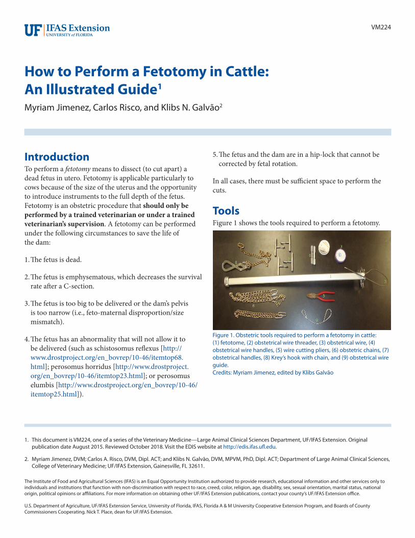
ToolsFigure 1 shows the tools required to perform a fetotomy.
Figure 1. Obstetric tools required to perform a fetotomy in cattle: (1) fetotome, (2) obstetrical wire threader, (3) obstetrical wire, (4) obstetrical wire handles, (5) wire cutting pliers, (6) obstetric chains, (7) obstetrical handles, (8) Krey’s hook with chain, and (9) obstetrical wire guide.Credits: Myriam Jimenez, edited by Klibs Galvão

2How to Perform a Fetotomy in Cattle: An Illustrated Guide
Fetotome Preparation1. Thread the obstetrical wire through the fetotome, coming
from the side of the handle and leaving on the side of the head, then back to the handle. Leave a loop of sufficient length to loop the head or a limb.
2. Cut the ob wire at your arm’s length or longer so you can thread more wire through for each subsequent cut.
3. Connect the wire to the handles.
Anterior Presentation Cut Sequence1. Decapitation and neck amputation: This procedure allows
the shoulders to collapse, and may provide sufficient space for the calf to be delivered or to provide more space for the next cut.
a. Make sure to place the fetotome lateral to the neck and head and as caudal as possible in order to remove the whole neck and the head (Figure 2a). It is useful to have a head snare or an ob chain (Figure 2b) placed around the head and locked in place on the fetotome handle so the wire does not move forward. Once you are comfortable with the placement, use your finger-tips to make sure the ob wire is not twisted (Figure 2b).
b. Start cutting on a steady manner performing wide arm strokes until the head and neck detach from the body.
Usually one person holds the fetotome in place while another person performs the cut (Figure 3).
c. The shoulders can now be collapsed (Figure 4), and you can try to deliver the calf. If the calf still cannot be delivered, proceed to the next step.
2. Thoracic cut and evisceration:
a. You can now place the fetotome as caudal (i.e., toward the tail) as possible to remove the thorax and eviscer-ate (Figure 5).
Figure 2. Beginning of decapitation and neck amputation.Credits: Myriam Jimenez, edited by Klibs Galvão
Figure 3. Decapitation and neck amputation.Credits: Myriam Jimenez, edited by Klibs Galvão
Figure 4. Shoulder collapse after decapitation and neck amputation.Credits: Myriam Jimenez, edited by Klibs Galvão

3How to Perform a Fetotomy in Cattle: An Illustrated Guide
b. Once you have removed the thorax and front legs, eviscerate (Figure 6). If the calf is still too big to be delivered, proceed to next cut.
3. Abdominal cut: Grab the spine with the Krey’s hook, pull the calf toward you as much as possible, place the ob wire as caudal (toward the tail) as possible, and perform the cut (Figure 7).
4. Pelvic bisection:
a. Use the ob wire guide to pass the ob wire around the pelvis, from the dorsal part to the ventral part; then thread the wire into the fetotome, and place a handle on it (Figure 8).
b. Make sure the wire is placed right in between the pelvis by the tuber ischia.
c. Tighten your wire saw and start cutting till you split the legs and pull them out.
Final Comments• Make sure you remove every piece of the fetus
• Examine the uterus for any tears, cuts, or the presence of another calf
• If the calf does not present any abnormality such as schistosomus refelxus or perosomus elumbis, and if the calf is in a caudal presentation and is too large to be delivered vaginally, a fetotomy should be avoided because after cutting one or both rear limbs the thorax will be too large to be delivered and usually the calf cannot be rotated. A C-section would be indicated for these cases.
• Figure 9 shows the delivery of a perosomus elumbis calf. One cut was performed to remove the legs and pelvis, and then a Krey’s hook was hooked to the spine and the calf was delivered by traction.
Figure 5. Thoracic cut.Credits: Myriam Jimenez, edited by Klibs Galvão
Figure 6. Evisceration after thoracic cut.Credits: Myriam Jimenez
Figure 7. Abdominal cut.Credits: Myriam Jimenez, edited by Klibs Galvão
Figure 8. Pelvic bisection.Credits: Myriam Jimenez, edited by Klibs Galvão

4How to Perform a Fetotomy in Cattle: An Illustrated Guide
Figure 9. Delivery of a calf with a perosomus elumbis defect.Credits: Klibs Galvão