How to make spinal motor neurons - Development...How to make spinal motor neurons Brandi N....
11
© 2014. Published by The Company of Biologists Ltd | Development (2014) 141, 491-501 doi:10.1242/dev.097410 491 ABSTRACT All muscle movements, including breathing, walking, and fine motor skills rely on the function of the spinal motor neuron to transmit signals from the brain to individual muscle groups. Loss of spinal motor neuron function underlies several neurological disorders for which treatment has been hampered by the inability to obtain sufficient quantities of primary motor neurons to perform mechanistic studies or drug screens. Progress towards overcoming this challenge has been achieved through the synthesis of developmental biology paradigms and advances in stem cell and reprogramming technology, which allow the production of motor neurons in vitro. In this Primer, we discuss how the logic of spinal motor neuron development has been applied to allow generation of motor neurons either from pluripotent stem cells by directed differentiation and transcriptional programming, or from somatic cells by direct lineage conversion. Finally, we discuss methods to evaluate the molecular and functional properties of motor neurons generated through each of these techniques. KEY WORDS: Motor neuron, Pluripotency, Stem cell Introduction In his memoir, Jean-Dominique Bauby lamented ‘Other than my eye, two things are not paralyzed, my imagination and my memory’ (Bauby, 1998). Such is the plight of those experiencing locked-in syndrome – a condition in which the brain remains relatively intact, but the terminal neurons that connect to all muscles except those servicing the eye are rendered non-functional. As a result, locked-in individuals are left with only their ability to take in visual stimuli and to have thoughts upon which they cannot act. Two broadly defined neuronal types provide the connection between the brain and our musculature: the upper, or cortical spinal motor neurons (CSMNs) and the lower spinal motor neurons. As their name implies, the cell bodies of CSMNs reside in the cortex and transmit motor information down long axons into the spinal cord. Spinal motor neurons receive this information and through axons that project out of the spinal cord to the musculature, actuate muscle contraction through a specialized synapse, the neuromuscular junction (NMJ). Significant injury to the descending spinal cord axons after physical trauma or stroke can result in complete paralysis. Although localized peripheral nerve injury to spinal motor neurons may only result in partial paralysis, conditions such as amyotrophic lateral sclerosis (ALS) and spinal muscular atrophy (SMA) can cause a more global degeneration of motor neurons and, in turn, a locked- in condition (Box 1). Curiosity concerning how the terminal motor circuitry develops and is wired has inspired numerous studies, making spinal motor PRIMER PRIMER SERIES 1 Harvard Stem Cell Institute, Department of Stem Cell and Regenerative Biology, Harvard University, Cambridge, MA 02138, USA. 2 The Howard Hughes Medical Institute, Harvard University, Cambridge, MA 02138, USA. 3 Stanley Center for Psychiatric Research, Broad Institute, Cambridge MA 02138, USA. *Author for correspondence ([email protected]) neurons one of the best-understood neuronal types. A desire to protect and eventually regenerate motor circuitry in the contexts of motor neuron disease and spinal cord injury has motivated attempts to utilize stem cell and reprogramming technologies to produce motor neurons for translational applications, including the modeling of these conditions. In this Primer, we will first review the processes and events that control the specification and differentiation of spinal motor neurons during embryogenesis. We will then discuss how the emerging understanding of motor neuron development has led to methods for directed differentiation of mouse and human pluripotent stem cells (PSCs) into motor neurons. More recent efforts to obtain motor neurons directly from fibroblasts by forced expression of transcription factors important for motor neuron identity will also be outlined. Finally, we consider methods to validate the equivalency of in vitro-derived motor neurons to their bona fide counterparts. In this Primer, we focus our attention exclusively on spinal motor neurons (referred to hereafter simply as MNs) and direct readers interested in CSMN development and reprogramming to recent publications of note (Shoemaker and Arlotta, 2010; Woodworth et al., 2012; Greig et al., 2013). MN development Decades of embryological studies and genetic analyses in model organisms have illuminated the molecular basis of neural induction as well as further differentiation and specification of MNs during Box 1. Motor neuron degeneration in disease Motor neuron diseases (MNDs) result from the progressive degeneration and death of motor neurons (MNs). The two most studied MNDs are the childhood genetic disease spinal muscular atrophy (SMA) and the adult- onset neurodegenerative disease amyotrophic lateral sclerosis (ALS) (Burghes and Beattie, 2009; Ling et al., 2013). Both diseases involve neuromuscular dysfunction progressively leading to fatal paralysis. SMA is an autosomal-recessive disease characterized by the selective loss of spinal MNs. The vast majority of SMA cases are caused by mutations in the ubiquitously expressed survival of motor neuron-1 (SMN1) gene. These mutations lead to the severe reduction in SMN levels, which is thought to affect small nuclear ribonucleoprotein biogenesis, as well as RNA transport in neurons (Burghes and Beattie, 2009). A growing collection of evidence indicates that therapeutics capable of elevating SMN levels could be effective in treating SMA (Passini and Cheng, 2011). Still, precisely how a deficiency in SMN, a ubiquitously expressed protein, causes selective loss of MNs remains unclear. In contrast to SMA, ALS affects both the cortical and spinal MNs. Approximately 10% of ALS cases are classified as familial, leaving the majority of ALS cases to be considered sporadic in origin. Several themes are emerging in the molecular pathologies of ALS (reviewed by Ling et al., 2013). These include dysfunctions in RNA processing and protein homeostasis as well as endoplasmic reticulum stress and problems in axonal transport. Interestingly, in SMA and ALS distinct subtypes of lower MNs are thought to be initially vulnerable to degeneration (Kanning et al., 2010). Thus, studying diverse populations of MNs in vitro could illuminate differential responses and guide the development of new therapeutics. How to make spinal motor neurons Brandi N. Davis-Dusenbery 1,3 , Luis A. Williams 1 , Joseph R. Klim 1,3 and Kevin Eggan 1,2,3, * Development
Transcript of How to make spinal motor neurons - Development...How to make spinal motor neurons Brandi N....
© 2014. Published by The Company of Biologists Ltd | Development (2014) 141, 491-501 doi:10.1242/dev.097410
491
ABSTRACTAll muscle movements, including breathing, walking, and fine motorskills rely on the function of the spinal motor neuron to transmit signalsfrom the brain to individual muscle groups. Loss of spinal motorneuron function underlies several neurological disorders for whichtreatment has been hampered by the inability to obtain sufficientquantities of primary motor neurons to perform mechanistic studies ordrug screens. Progress towards overcoming this challenge has beenachieved through the synthesis of developmental biology paradigmsand advances in stem cell and reprogramming technology, whichallow the production of motor neurons in vitro. In this Primer, wediscuss how the logic of spinal motor neuron development has beenapplied to allow generation of motor neurons either from pluripotentstem cells by directed differentiation and transcriptional programming,or from somatic cells by direct lineage conversion. Finally, we discussmethods to evaluate the molecular and functional properties of motorneurons generated through each of these techniques.
KEY WORDS: Motor neuron, Pluripotency, Stem cell
IntroductionIn his memoir, Jean-Dominique Bauby lamented ‘Other than myeye, two things are not paralyzed, my imagination and my memory’(Bauby, 1998). Such is the plight of those experiencing locked-insyndrome – a condition in which the brain remains relatively intact,but the terminal neurons that connect to all muscles except thoseservicing the eye are rendered non-functional. As a result, locked-inindividuals are left with only their ability to take in visual stimuliand to have thoughts upon which they cannot act.
Two broadly defined neuronal types provide the connectionbetween the brain and our musculature: the upper, or cortical spinalmotor neurons (CSMNs) and the lower spinal motor neurons. Astheir name implies, the cell bodies of CSMNs reside in the cortexand transmit motor information down long axons into the spinalcord. Spinal motor neurons receive this information and throughaxons that project out of the spinal cord to the musculature, actuatemuscle contraction through a specialized synapse, theneuromuscular junction (NMJ).
Significant injury to the descending spinal cord axons afterphysical trauma or stroke can result in complete paralysis. Althoughlocalized peripheral nerve injury to spinal motor neurons may onlyresult in partial paralysis, conditions such as amyotrophic lateralsclerosis (ALS) and spinal muscular atrophy (SMA) can cause amore global degeneration of motor neurons and, in turn, a locked-in condition (Box 1).
Curiosity concerning how the terminal motor circuitry developsand is wired has inspired numerous studies, making spinal motor
PRIMER PRIMER SERIES
1Harvard Stem Cell Institute, Department of Stem Cell and Regenerative Biology,Harvard University, Cambridge, MA 02138, USA. 2The Howard Hughes MedicalInstitute, Harvard University, Cambridge, MA 02138, USA. 3Stanley Center forPsychiatric Research, Broad Institute, Cambridge MA 02138, USA.
*Author for correspondence ([email protected])
neurons one of the best-understood neuronal types. A desire toprotect and eventually regenerate motor circuitry in the contexts ofmotor neuron disease and spinal cord injury has motivated attemptsto utilize stem cell and reprogramming technologies to producemotor neurons for translational applications, including the modelingof these conditions.
In this Primer, we will first review the processes and events thatcontrol the specification and differentiation of spinal motor neuronsduring embryogenesis. We will then discuss how the emergingunderstanding of motor neuron development has led to methods fordirected differentiation of mouse and human pluripotent stem cells(PSCs) into motor neurons. More recent efforts to obtain motorneurons directly from fibroblasts by forced expression oftranscription factors important for motor neuron identity will also beoutlined. Finally, we consider methods to validate the equivalencyof in vitro-derived motor neurons to their bona fide counterparts. Inthis Primer, we focus our attention exclusively on spinal motorneurons (referred to hereafter simply as MNs) and direct readersinterested in CSMN development and reprogramming to recentpublications of note (Shoemaker and Arlotta, 2010; Woodworth etal., 2012; Greig et al., 2013).
MN developmentDecades of embryological studies and genetic analyses in modelorganisms have illuminated the molecular basis of neural inductionas well as further differentiation and specification of MNs during
Box 1. Motor neuron degeneration in diseaseMotor neuron diseases (MNDs) result from the progressive degenerationand death of motor neurons (MNs). The two most studied MNDs are thechildhood genetic disease spinal muscular atrophy (SMA) and the adult-onset neurodegenerative disease amyotrophic lateral sclerosis (ALS)(Burghes and Beattie, 2009; Ling et al., 2013). Both diseases involveneuromuscular dysfunction progressively leading to fatal paralysis.
SMA is an autosomal-recessive disease characterized by the selectiveloss of spinal MNs. The vast majority of SMA cases are caused bymutations in the ubiquitously expressed survival of motor neuron-1(SMN1) gene. These mutations lead to the severe reduction in SMNlevels, which is thought to affect small nuclear ribonucleoproteinbiogenesis, as well as RNA transport in neurons (Burghes and Beattie,2009). A growing collection of evidence indicates that therapeuticscapable of elevating SMN levels could be effective in treating SMA(Passini and Cheng, 2011). Still, precisely how a deficiency in SMN, aubiquitously expressed protein, causes selective loss of MNs remainsunclear.
In contrast to SMA, ALS affects both the cortical and spinal MNs.Approximately 10% of ALS cases are classified as familial, leaving themajority of ALS cases to be considered sporadic in origin. Severalthemes are emerging in the molecular pathologies of ALS (reviewed byLing et al., 2013). These include dysfunctions in RNA processing andprotein homeostasis as well as endoplasmic reticulum stress andproblems in axonal transport. Interestingly, in SMA and ALS distinctsubtypes of lower MNs are thought to be initially vulnerable todegeneration (Kanning et al., 2010). Thus, studying diverse populationsof MNs in vitro could illuminate differential responses and guide thedevelopment of new therapeutics.
How to make spinal motor neuronsBrandi N. Davis-Dusenbery1,3, Luis A. Williams1, Joseph R. Klim1,3 and Kevin Eggan1,2,3,*
Dev
elop
men
t

492
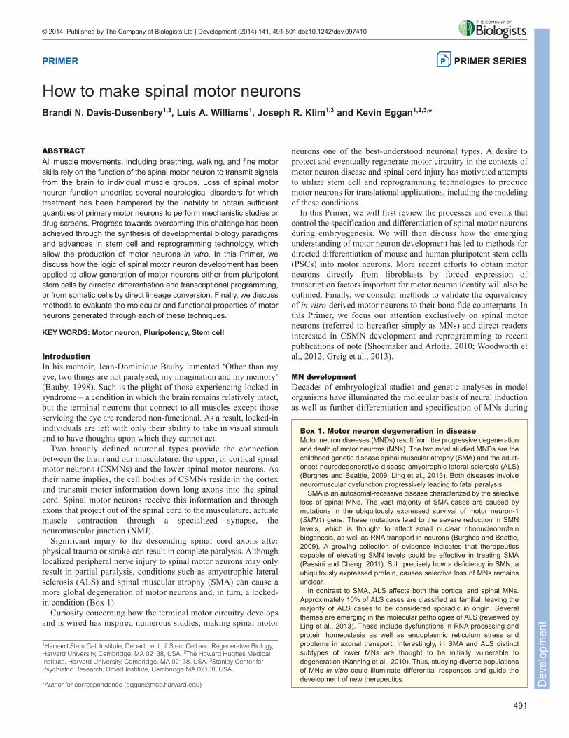
development. In the early 20th century, Spemann and Mangold’swork with amphibian embryos revealed that signals emanating fromthe dorsal lip of the blastopore, now termed the Spemann–Mangold‘organizer’, were required for the induction of neural fate duringgastrulation (Hamburger, 1988). Further studies demonstrated thatrather than providing positive signals to induce neural cell fate, theorganizer was the source of factors that inhibit bone morphogeneticprotein (BMP) signaling, including Chordin, Follistatin and Noggin(De Robertis, 2006). Although the requirement to inhibit BMPsignaling is conserved in higher organisms, additional inductivesignals, including fibroblast growth factors (FGFs), epidermalgrowth factors (EGFs) and Wnts have been identified (Stern, 2005)(Fig. 1A).
Following their initial generation, the cells of the neural tube arespecified along both the rostral-caudal and the dorso-ventral axes.Gradients of signaling molecules along each axis provide a roadmapto guide the differentiation of the emerging neuronal types of eachregion. Progenitor domains are first specified and then refined
through the cooperative action of external signals and downstreamtranscription factors (Jessell, 2000; Alaynick et al., 2011). Along therostral-caudal axis, the neural tube is specified into the majorcomponents of the CNS, including the brain, midbrain, hindbrainand spinal cord (Fig. 1A). Although multiple signals have beenproposed to contribute to the caudalization of the neurons in thespinal cord, chief among them is retinoic acid (RA). Early indevelopment, RA, produced through the activity of retinaldehydedehydrogenase 2 (RALDH-2; also known as ALDH1A2), emanatesfrom the caudal paraxial mesoderm and is crucial for the initialdistinction of neurons of the hindbrain and spinal cord from thosein the forebrain and midbrain (Maden, 2007) (Fig. 1A).
Specification of MN fateWithin the dorso-ventral axis of the neural tube, progenitor cells aredivided into five ventral progenitor domains termed p0, p1, p2, pMNand p3, which in turn give rise to interneuron subtypes V0-3 andmotor neurons (Fig. 1B). A gradient of sonic hedgehog (Shh),
PRIMER Development (2014) doi:10.1242/dev.097410
RA
S
Shh
NC
p3pMNp2p1p0
RP
Irx3
Pax
6
Dbx
2 Dbx
1
Nkx
2.2
Nkx
6.1 Nkx
6.2
Transcription factor code
BMP4Activin
Neural tube
Neuroectoderm
NogginChordinFollistatin
Forebrain
Hindbrain
RA
Spinal Cord
Embryonic CNS
Olig
2
WntsFGF
Fgf8Gdf11
A B
CN
gn3
Blastocyst
Rostral Caudal
Ectoderm
Endoderm
Ventral
Dorsal
RC
ICM
FP
V0
V1
V2
V3
MN
Mesoderm
Cervical
Brachial
Thoracic
Lumbar
Hox
10H
ox11
Hox
c9
Hox
d9
Hox
6 Hox
c4 Hox
5H
oxc8 H
oxc8
BMP/TGFβs
Midbrain
Fig. 1. Spinal cord development and motor neuron specification. (A) In early development, gastrulation results in the specification of the cells of the innercell mass (ICM) into the three germ layers: ectoderm, endoderm and mesoderm. The dorsal region of the ectoderm is further specified into the neuroectodermthrough the inhibition of BMP and activin signaling, and, in higher organisms, enhanced FGF and Wnt signaling. Neuralization proceeds through the formationof a neural plate and subsequent generation of neural folds, which in turn fuse to give rise to the neural tube. The neural tube is then patterned along therostro-caudal axis (anterior-posterior) by a gradient of retinoic acid (RA) generated primarily by the action of Raldh2. In particular, a high level of RA allows theinitial boundary of the spinal cord and hindbrain versus forebrain and hindbrain to be delineated. Fgfs and Gdf11 oppose the activity of RA and allowspecification of more caudal spinal cord cell types. (B) Once the spinal cord is specified, continued release of RA (shaded red) from the somites (S) acts torefine the positional character of neurons along the rostral-caudal axis. The spinal cord is also patterned along the dorso-ventral axis through the combinedaction of sonic hedgehog (Shh; shaded blue) emanating from the notochord (NC) and floor plate (FP) and BMP/TGFβ signaling (shaded yellow) from the roofplate (RP). The ventral spinal cord can be divided into five progenitor domains (p0-p3 and pMN), which give rise to V0-V3 interneurons and motor neurons. Theborders of progenitor domains are established through the cross-repressive action of pairs of transcription factors that are induced by Shh (Class II, in greenand blue) or those that are repressed by Shh (Class I, in yellow and red). The combinatorial action of transcription factors allows the specification of each celltype. For example, pMNs (MN progenitors) express Pax6, Olig2, Nkx6.1 and Nkx6.2. (C) The Hox genes play a crucial role in the specification of MNs alongthe rostral-caudal axis. Similar to patterning along the ventral-dorsal axis, the coordinated interactions between Hox family members allows regionalboundaries to be delineated. Specific expression of Hox accessory factors, such as FoxP1, can further specify MN subtypes. D
evel
opm
ent
secreted from the notochord and cells of the floor plate providesventral topographic information by regulating the expression ofhomeodomain (HD) and basic helix-loop-helix (bHLH) transcriptionfactors (Alaynick et al., 2011). Transcription factors downstream ofShh can be roughly divided into two classes based on theirregulation in response to Shh signaling. Class II proteins, includingNkx6.1, Olig2 and Nkx2.2, are activated by Shh and in turn repressthe expression of class I proteins, including Pax6, Irx3, Dbx1 andDbx2 (Briscoe et al., 2000; Jessell, 2000; Alaynick et al., 2011)(Fig. 1B).
The cross-repressive activity between class II and class I proteinsallows the consolidation of progenitor identity as well as thegeneration of sharp boundaries between adjacent domains (Briscoeet al., 2000). For example, the boundary between p3 and pMN isdelimited by the activity of Pax6 and Nkx2.2. Indeed, in the mouseembryo, mutation of Pax6 results in a dorsal expansion of theNkx2.2 expression boundary (Ericson et al., 1997). Similarly, thedorsal boundary between pMN and p2 is defined by the mutuallyrepressive activities of Irx3 and Olig2 whereas the ventral boundaryis delimited by the expression of Ngn3 (Neurog3) (Novitch et al.,2001; Sugimori et al., 2007). MN progenitors also express the HDtranscription factors Nkx6.1 and Nkx6.2, which act to repress theother progenitor domains (Briscoe et al., 2000) (Fig. 1B).Expression of Olig2 within the pMN domain promotes expressionof Ngn2, which is important for cell cycle exit as well as forinduction of terminal MN transcription factors including Hb9(Mnx1), Isl1, Isl2 and Lhx3 (Novitch et al., 2001). More recently,additional molecular mechanisms, including microRNA pathways,have also been shown to regulate the boundary between someprogenitor domains (Chen et al., 2011).
MN subtype specificationAlthough all MNs derive from a single ventral progenitor domain,further specification of MNs allows the coordinated movement ofhundreds of distinct muscle groups. MNs can be further classifiedbased on their anatomical and functional properties (Box 2). Thepositional identity of MNs along the rostro-caudal axis is determinedby the coordinated action of multiple signaling molecules. Highlevels of RA promote rostral (e.g. cervical and brachial) identitywhereas FGFs and Gdf11 activity give rise to more caudal (e.g.thoracic and lumbar) MNs (Liu et al., 2001). The combined signalsfrom RA, FGFs, Wnts and TGFβ family members are integratedprimarily by the Hox transcription factors to specify MN rostro-caudal subtype identity (Dasen and Jessell, 2009). Mouse andHuman Hox genes are arrayed in four chromosomal clusters (HoxA,HoxB, HoxC and HoxD), each of which harbors a subset of 13paralogous Hox genes (Hox1-Hox13). Within each cluster, theexpression pattern of the Hox genes is spatially and temporallycollinear with their chromosomal organization, such that Hox1 genesare expressed in the rostral region of the organism and Hox13 genesare expressed caudally (Pearson et al., 2005). Consistent with theirconserved role in body patterning, Hox gene expression within thespinal cord determines MN columnar identity and selective muscleinnervation, with Hox4-8 genes expressed at brachial levels, Hox8and Hox9 at the thoracic level and Hox10 and Hox11 in the lumbarregion (Dasen et al., 2005; Dasen and Jessell, 2009) (Fig. 1C).Further specification of individual MN subtypes is provided by fine-tuning the Hox protein expression pattern both spatially andtemporally (Philippidou and Dasen, 2013).
Experimental manipulation of the Hox code in mouse and chickembryos can alter MN subtype and projection pattern (Tiret et al.,1998; Wahba et al., 2001; Vermot et al., 2005; Wu et al., 2008;
Misra et al., 2009; Jung et al., 2010; Philippidou et al., 2012;Lacombe et al., 2013). As a particularly dramatic example, mutationof Hoxc9 in the mouse causes loss of thoracic preganglionic column(PGC) and hypaxial motor column (HMC) MNs and expansion ofthe brachial lateral motor column (LMC) domain (Jung et al., 2010).ChIP-Seq analysis of Hoxc9 revealed that this protein directly bindsnumerous regions within diverse Hox loci and its disruption alteredexpression of multiple Hox genes, suggesting that Hoxc9 mayrepresent a master regulator of MN subtype identity (Jung et al.,2010).
The ability of different Hox genes to play such an important rolein determination of MN identity is perhaps surprising given that the
493
PRIMER Development (2014) doi:10.1242/dev.097410
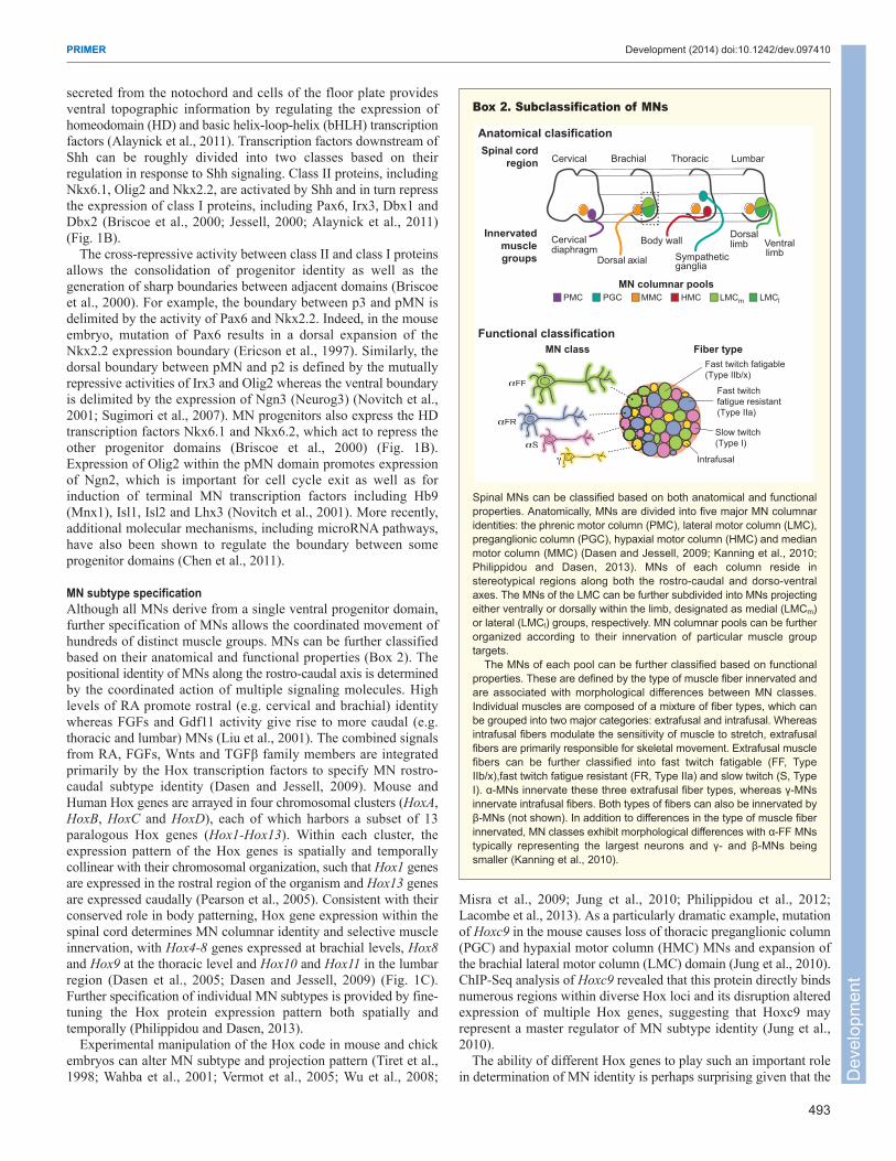
Box 2. Subclassification of MNs
Spinal MNs can be classified based on both anatomical and functionalproperties. Anatomically, MNs are divided into five major MN columnaridentities: the phrenic motor column (PMC), lateral motor column (LMC),preganglionic column (PGC), hypaxial motor column (HMC) and medianmotor column (MMC) (Dasen and Jessell, 2009; Kanning et al., 2010;Philippidou and Dasen, 2013). MNs of each column reside instereotypical regions along both the rostro-caudal and dorso-ventralaxes. The MNs of the LMC can be further subdivided into MNs projectingeither ventrally or dorsally within the limb, designated as medial (LMCm)or lateral (LMCl) groups, respectively. MN columnar pools can be furtherorganized according to their innervation of particular muscle grouptargets.
The MNs of each pool can be further classified based on functionalproperties. These are defined by the type of muscle fiber innervated andare associated with morphological differences between MN classes.Individual muscles are composed of a mixture of fiber types, which canbe grouped into two major categories: extrafusal and intrafusal. Whereasintrafusal fibers modulate the sensitivity of muscle to stretch, extrafusalfibers are primarily responsible for skeletal movement. Extrafusal musclefibers can be further classified into fast twitch fatigable (FF, TypeIIb/x),fast twitch fatigue resistant (FR, Type IIa) and slow twitch (S, TypeI). α-MNs innervate these three extrafusal fiber types, whereas γ-MNsinnervate intrafusal fibers. Both types of fibers can also be innervated byβ-MNs (not shown). In addition to differences in the type of muscle fiberinnervated, MN classes exhibit morphological differences with α-FF MNstypically representing the largest neurons and γ- and β-MNs beingsmaller (Kanning et al., 2010).
MMCMN columnar pools
LMCmPMC HMCPGC LMCl
Cervical Brachial Thoracic Lumbar
Cervical diaphragm
Body wall
Dorsal axial Sympathetic ganglia
Dorsal limb Ventral
limb
Innervated
muscle
groups
Spinal cord
region
Fast twitch fatigable(Type IIb/x)
Fast twitchfatigue resistant(Type IIa)
Slow twitch(Type I)
Intrafusal
αFF
αFR
αSγ
Anatomical clasification
Functional classification
Fiber type
αFF
αFR
αSγγγγγγγγγγ
unctional classification
MN class
Dev
elop
men
t
494
homeodomains of Hox genes are largely conserved betweenparalogs. However, there is accumulating evidence that furtherspecificity of Hox function can be provided by accessory factors,including downstream effectors such as the Forkhead box (Fox)protein P1 and the HD protein Nkx6.1 (Philippidou and Dasen,2013). FoxP1 is expressed at high levels in LMC MNs andFoxP1−/− mouse embryos exhibit disrupted columnar MN identitiesand alterations in MN cell body position and axonal wiring (Dasenet al., 2008; Rousso et al., 2008).
As discussed further below, the body of work describing themolecular underpinnings of MN specification during developmenthas enabled recapitulation of these signals in vitro for the ex vivogeneration of MNs. An exciting consequence of this progress isthat it allows sufficient quantities of MNs to be derived in acontrolled environment to interrogate further the detailedmechanism of MN development and specification. For example,Mazzoni and colleagues performed a series of chromatinimmunoprecipitation assays during MN differentiation in vitro toinvestigate the molecular details regulating Hox gene expression(Mazzoni et al., 2013b). The authors found that addition of RAduring MN differentiation led to recruitment of RA receptors to theHox1-5 chromatin domain that was followed by a rapid domain-wide removal of H3K27me3 and acquisition of cervical MNidentity. Moreover, Cdx2, a transcription factor induced by Wntand FGF, regulated the clearance of H3K27me3 from the Hox1-9chromatin domains, resulting in brachial or thoracic MNspecification (Mazzoni et al., 2013b). Thus, the early modificationof chromatin by patterning factors contributes to the specificationof the rostro-caudal identity of MNs. This type of mechanistic,genome-wide study would surely be impossible with the limitedquantities and mixed populations of cells that can be purified fromthe early embryo. Continued integration of findings fromdevelopmental studies in model organisms and in vitro-derivedMNs will allow a greater understanding of MN specification, andas a consequence further improve strategies to recapitulate MNdevelopment in vitro.
Directed differentiation of PSCs into MNsPSCs, either embryonic stem cells (ESCs) derived from pre-implantation blastocysts (Evans and Kaufman, 1981; Thomson etal., 1998), or induced pluripotent stem cells (iPSCs) obtained byreprogramming of somatic cells with defined transcription factors(Takahashi and Yamanaka, 2006; Takahashi et al., 2007), arecharacterized by their ability to proliferate indefinitely in culturewhile preserving their developmental potential to differentiate intoderivatives of all three embryonic germ layers. By leveragingknowledge of developmental pathways that allow neural inductionand further specification of MNs, stem cell biologists have designedmultiple approaches to direct the differentiation of mouse andhuman PSCs to MNs (Wichterle et al., 2002; Li et al., 2005; SinghRoy et al., 2005; Di Giorgio et al., 2007; Chambers et al., 2009; Huand Zhang, 2009; Karumbayaram et al., 2009; Boulting et al., 2011;Patani et al., 2011; Amoroso et al., 2013). MNs obtained throughthese methods have been shown to possess numerous characteristicsof bona fide MNs, including distinctive electrophysiologicalresponses, the ability to form functional NMJs and the capacity toengraft into the developing spinal cord. Just as the in vivo embryonicdevelopment of spinal MNs can be broken down into distinct stages,so too can the in vitro specification of MNs from PSCs. Specifically,the steps of neural induction followed by caudal and ventralpatterning must all be appropriately executed for MNs to beproduced.
Neural inductionIn the absence of factors that promote pluripotency, such asleukemia inhibitory factor (LIF) and FGF, PSCs spontaneouslydifferentiate into diverse lineages and lose the ability to self-renewor generate chimeric mice (Evans, 2011). Although spontaneousdifferentiation represents a hurdle for the maintenance ofpluripotency, it can be exploited to give rise to differentiated celltypes. Spontaneous differentiation into multiple lineages, includingneurons, can be enhanced by inducing PSCs under non-adherentculture conditions to form multicellular aggregates, termedembryoid bodies (EBs) (Odorico et al., 2001). However, theefficiency of neural induction using these spontaneous approachesis low, and significant cellular heterogeneity within EBs hindersfurther mechanistic studies (Bain et al., 1995). Multiple strategieshave been proposed to improve the production of neural precursorsand cells with neuronal phenotypes from differentiating PSCpopulations. These approaches include treatment of EB cultures withRA (Bain et al., 1995), adherent co-culture of PSCs with PA6 orMS-5 stromal feeder cell lines (Kawasaki et al., 2000; Lee et al.,2007), continued propagation of nestin+ proliferating cells in definedmedia containing mitogens such as FGF2 and EGF (Okabe et al.,1996; Reubinoff et al., 2001; Joannides et al., 2007), lineageselection using genetic reporters (Li et al., 1998), and selectiveenzymatic digestion or manual selection of neural tube-like rosettestructures (Zhang et al., 2001; Hu and Zhang, 2009).
More recently, it has been demonstrated that simultaneousinhibition of the TGFβ/Activin/Nodal and BMP signaling pathways,through the use of either small molecule antagonists or recombinantinhibitors, can induce a rapid and very efficient (>80%) neuralconversion of human PSCs (Smith et al., 2008; Chambers et al.,2009; Zhou et al., 2010; Chambers et al., 2012) (Fig. 2A). Similarto the processes that occur during early development, inhibition ofthe TGFβ and BMP pathways is thought to promote differentiationof PSCs along the neuronal lineage primarily through inhibition ofself-renewal as well as blocking differentiation towards alternativelineages (Chambers et al., 2009). Several other pathways, involvingEGFs, FGFs and Wnts, have been described to regulate neuronaldifferentiation of human and mouse stem cells. In particular, FGF2has been shown to promote induction and survival of neuralprogenitors (Streit et al., 2000; Wilson et al., 2000; Joannides et al.,2007). Therefore, enhancing FGF2 signaling during the neuralinduction phase of differentiation can increase the number of neuralprogenitors, whereas inhibiting it at subsequent stages promotestheir transition into differentiated neurons (Joannides et al., 2007;Chambers et al., 2012).
Caudal and ventral patterningFollowing neural induction of PSCs, neural progenitor cells can bepatterned according to developmental principles. Treatment with RApromotes caudal (spinal cord) identity, while addition of eitherrecombinant Shh or small molecule agonists of the Shh signalingpathway, such as smoothened agonist (SAG) or purmorphamine(PUR), promotes ventralization (Wichterle et al., 2002) (Fig. 2A).Time course studies reveal that differentiation in vitro proceeds withthe same temporal regulation of transcription factors as is observedin vivo, with Sox1+ neural progenitors giving rise to Olig2+ MNprogenitors, which then in turn begin to express Hb9 and Isl1(Fig. 2B) (Wichterle et al., 2002). In the mouse, Hb9+ MNs begin toappear 3-5 days after addition of patterning factors (Wichterle et al.,2002; Di Giorgio et al., 2007). The timing of MN differentiationfrom human PSCs is more protracted and it can, depending on thespecific protocol utilized, require an additional 2-4 weeks after
PRIMER Development (2014) doi:10.1242/dev.097410
Dev
elop
men
t
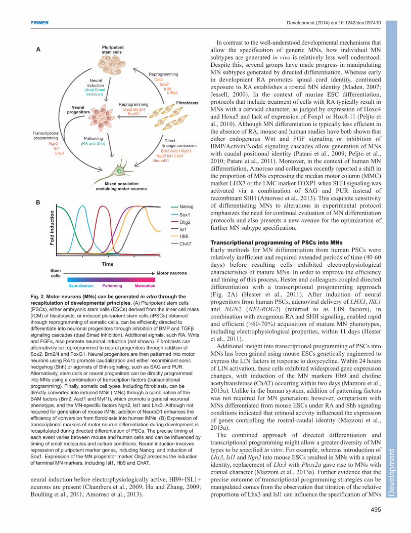
neural induction before electrophysiologically active, HB9+ISL1+neurons are present (Chambers et al., 2009; Hu and Zhang, 2009;Boulting et al., 2011; Amoroso et al., 2013).
In contrast to the well-understood developmental mechanisms thatallow the specification of generic MNs, how individual MNsubtypes are generated in vivo is relatively less well understood.Despite this, several groups have made progress in manipulatingMN subtypes generated by directed differentiation. Whereas earlyin development RA promotes spinal cord identity, continuedexposure to RA establishes a rostral MN identity (Maden, 2007;Jessell, 2000). In the context of murine ESC differentiation,protocols that include treatment of cells with RA typically result inMNs with a cervical character, as judged by expression of Hoxc4and Hoxa5 and lack of expression of Foxp1 or Hox8-11 (Peljto etal., 2010). Although MN differentiation is typically less efficient inthe absence of RA, mouse and human studies have both shown thateither endogenous Wnt and FGF signaling or inhibition ofBMP/Activin/Nodal signaling cascades allow generation of MNswith caudal positional identity (Patani et al., 2009; Peljto et al.,2010; Patani et al., 2011). Moreover, in the context of human MNdifferentiation, Amoroso and colleagues recently reported a shift inthe proportion of MNs expressing the median motor column (MMC)marker LHX3 or the LMC marker FOXP1 when SHH signaling wasactivated via a combination of SAG and PUR instead ofrecombinant SHH (Amoroso et al., 2013). This exquisite sensitivityof differentiating MNs to alterations in experimental protocolemphasizes the need for continual evaluation of MN differentiationprotocols and also presents a new avenue for the optimization offurther MN subtype specification.
Transcriptional programming of PSCs into MNsEarly methods for MN differentiation from human PSCs wererelatively inefficient and required extended periods of time (40-60days) before resulting cells exhibited electrophysiologicalcharacteristics of mature MNs. In order to improve the efficiencyand timing of this process, Hester and colleagues coupled directeddifferentiation with a transcriptional programming approach(Fig. 2A) (Hester et al., 2011). After induction of neuralprogenitors from human PSCs, adenoviral delivery of LHX3, ISL1and NGN2 (NEUROG2) (referred to as LIN factors), incombination with exogenous RA and SHH signaling, enabled rapidand efficient (>60-70%) acquisition of mature MN phenotypes,including electrophysiological properties, within 11 days (Hesteret al., 2011).
Additional insight into transcriptional programming of PSCs intoMNs has been gained using mouse ESCs genetically engineered toexpress the LIN factors in response to doxycycline. Within 24 hoursof LIN activation, these cells exhibited widespread gene expressionchanges, with induction of the MN markers Hb9 and cholineacetyltransferase (ChAT) occurring within two days (Mazzoni et al.,2013a). Unlike in the human system, addition of patterning factorswas not required for MN generation; however, comparison withMNs differentiated from mouse ESCs under RA and Shh signalingconditions indicated that retinoid activity influenced the expressionof genes controlling the rostral-caudal identity (Mazzoni et al.,2013a).
The combined approach of directed differentiation andtranscriptional programming might allow a greater diversity of MNtypes to be specified in vitro. For example, whereas introduction ofLhx3, Isl1 and Ngn2 into mouse ESCs resulted in MNs with a spinalidentity, replacement of Lhx3 with Phox2a gave rise to MNs withcranial character (Mazzoni et al., 2013a). Further evidence that theprecise outcome of transcriptional programming strategies can bemanipulated comes from the observation that titration of the relativeproportions of Lhx3 and Isl1 can influence the specification of MNs
495
PRIMER Development (2014) doi:10.1242/dev.097410
Mixed population
containing motor neurons
Oct4Sox2
Klf4
Fibroblasts
Pluripotentstem cells
Neuralprogenitors
Reprogramming
Directlineage conversion
Brn2 Ascl1 Myt1l Ngn2 Isl1 Lhx3 NeuroD1
Neuralinduction
(dual Smadinhibition)
Patterning(RA and Shh)
Transcriptionalprogramming
Ngn2 Isl1 Lhx3
Sox2 Brn2/4FoxG1
Reprogramming
Fo
ld in
du
cti
on
Time
Nanog
Sox1Olig2Isl1Hb9
A
B
Stem
cellsMotor neurons
Neuralization
ChAT
Patterning Maturation
c-Myc
Fig. 2. Motor neurons (MNs) can be generated in vitro through therecapitulation of developmental principles. (A) Pluripotent stem cells(PSCs), either embryonic stem cells (ESCs) derived from the inner cell mass(ICM) of blastocysts, or induced pluripotent stem cells (iPSCs) obtainedthrough reprogramming of somatic cells, can be efficiently directed todifferentiate into neuronal progenitors through inhibition of BMP and TGFβsignaling cascades (dual Smad inhibition). Additional signals, such RA, Wntsand FGFs, also promote neuronal induction (not shown). Fibroblasts canalternatively be reprogrammed to neural progenitors through addition ofSox2, Brn2/4 and FoxG1. Neural progenitors are then patterned into motorneurons using RA to promote caudalization and either recombinant sonichedgehog (Shh) or agonists of Shh signaling, such as SAG and PUR.Alternatively, stem cells or neural progenitors can be directly programmedinto MNs using a combination of transcription factors (transcriptionalprogramming). Finally, somatic cell types, including fibroblasts, can bedirectly converted into induced MNs (iMNs) through a combination of theBAM factors (Brn2, Ascl1 and Myt1l), which promote a general neuronalphenotype, and the MN-specific factors Ngn2, Isl1 and Lhx3. Although notrequired for generation of mouse iMNs, addition of NeuroD1 enhances theefficiency of conversion from fibroblasts into human iMNs. (B) Expression oftranscriptional markers of motor neuron differentiation during development isrecapitulated during directed differentiation of PSCs. The precise timing ofeach event varies between mouse and human cells and can be influenced bytiming of small molecules and culture conditions. Neural induction involvesrepression of pluripotent marker genes, including Nanog, and induction ofSox1. Expression of the MN progenitor marker Olig2 precedes the inductionof terminal MN markers, including Isl1, Hb9 and ChAT.
Dev
elop
men
t

496
versus V2 interneurons. In vivo, Lhx3 and Isl1 form a complex withthe nuclear LIM interactor protein NLI (Ldb1) to specify MNs,whereas in the absence of Isl1, Lhx3, in complex with NLI, specifiesV2 interneurons (Thaler et al., 2002). Consistent with this molecularmechanism, equimolar amounts of Lhx3 and Isl1 promoted MNgeneration from mouse ESCs, whereas excess Lhx3 expression gaverise to V2 interneurons. The use of an Isl1-Lhx3 fusion proteincould also enhance the shift of the differentiating cells towards MNs(Lee et al., 2012). As the mechanisms which lead to the cooperativeaction of transcription factors during MN specification continue tobe defined, it will be interesting to determine if similar fusionproteins may be utilized to guide cell fate more specifically orefficiently.
Direct lineage conversion of somatic cells into MNsRecent success using defined factors to reprogram somatic cells topluripotency, along with much earlier studies demonstrating theability of a single factor, MyoD (Myod1), to convert fibroblasts intomuscle cells, has led many researchers to explore further the abilityof lineage-specific transcription factors to induce the conversion ofspecific cell types from unrelated somatic cells (reviewed by Graf,2011). Following a similar approach to the one used to identify iPSCreprogramming factors, Vierbuchen and colleagues demonstratedthat a set of three neural lineage-specific transcription factors,referred to as BAM factors [Brn2 (Pou3f2), Ascl1 and Myt1l] wassufficient to directly convert mouse fibroblasts into induced neuronal(iN) cells. Gene expression profiling and electrophysiologicalrecordings revealed that these iN cells have properties of genericexcitatory neurons (Vierbuchen et al., 2010; Marro et al., 2011).Following this initial report, microRNAs and additional proneuronalfactors, including NeuroD1, were shown to cooperate with orreplace the BAM factors during conversion of human fibroblastsinto iNs (Ambasudhan et al., 2011; Pang et al., 2011; Yoo et al.,2011).
A crucial issue for the application of iN approaches indevelopmental and translational studies is the specification ofprecise neuronal subtypes. Our group demonstrated that BAM factorexpression, in combination with four transcription factors (Lhx3,Isl1, Ngn2 and Hb9) was sufficient to convert mouse fibroblasts intocells with a MN phenotype, termed induced MNs or iMNs (Son etal., 2011) (Fig. 2A). iMNs were identified based on the expressionof a transgenic Hb9::GFP reporter and exhibited molecular andfunctional properties of embryo-derived MNs, including geneexpression profile, electrophysiological activity, formation ofneuromuscular junctions and ability to integrate into the developingchick spinal cord. Interestingly, this study revealed that uponintroduction of MN factors, fibroblasts, unlike PSCs, do nottransition through an intermediate nestin+ neural progenitor statebefore becoming iMNs (Son et al., 2011). Similar to the case of iNcells, human fibroblasts could be converted to cells with a MNphenotype by the addition of NeuroD1 to the seven-factor iMNcocktail (Son et al., 2011). It has recently been shown that directlineage conversion can be performed in vivo. For example,cardiomyocytes generated by direct conversion from cardiacfibroblasts can improve cardiovascular function in the mouse (Songet al., 2012). It will be exciting to determine if a similar approachcan be adopted in the nervous system to repair injuries and/orreverse neurodegenerative disease.
Evaluation of MNs produced in vitroAlthough the basic principles of MN specification are wellestablished, many groups have reported differences in the timing and
efficiency of differentiation, as well as the identity of the resultingPSC- or somatic cell-derived MNs. This variability may arise fromovert differences in protocols, such as the combination,concentration and timing of addition of specific growth factors, aswell as less transparent differences, such as the cellular density orprecise media composition used to culture cells followingspecification. Concern about the extent to which minormodifications in protocols alter the identity of the resulting MNs isamplified because most groups rely on expression of transgenicreporters or a small handful of canonical marker genes, which maynot be able to report on subtle MN subtype specific differences.Given the potential impact of these modifications on downstreamstudies such as in vitro disease modeling of MN disorders, continuedeffort both to carefully interrogate the effects of alteringdifferentiation protocols and to standardize methods and analysesacross multiple labs is crucial.
MNs can be assessed according to four primary characteristicsthat provide insights into the equivalency of in vitro-derived MNsto bona fide cells: (1) neuronal morphology and expression ofcharacteristic MN marker genes, (2) characteristicelectrophysiological activity and response to stimuli, (3) formationof functional neuromuscular junctions and (4) engraftment into thespinal cord in vivo (Fig. 3).
Morphology and marker analysesWhen cultured in isolation, MNs exhibit unipolar morphology witha single axon extending from the soma, which is elaborated withdendrites. Although in dense cultures distinguishing the axon fromsurrounding dendrites can be difficult, this is aided byimmunostaining of microtubule associated protein 2 (Map2), whichmarks proximal dendrites (Fig. 3A). Additional immunostaining ofneuronal cyctoskeletal proteins, such as β-III tubulin (Tuj1; Tubb3)as well as evaluation of the phosphorylation status of neurofilamentsusing SMI antibodies allows full appreciation of the complicatedneuronal morphology typically present in MNs.
A crucial step towards the development of methods to generatespecific cell types in vitro is the selection of appropriate cell type-specific markers that can allow the identification of differentiatedcell types without the anatomical information provided in vivo. Likeall other neurons, MNs are postmitotic, a property that can bedetermined by lack of bromodeoxyuridine (BrdU) incorporation intocellular DNA or by lack of immunoreactivity for cell proliferationproteins such as Ki67 (Mki67). MNs can be further distinguishedfrom other neurons based on expression of canonical MN identitytranscription factors, including Isl1 and Hb9, as well as markers ofa more mature and cholinergic MN phenotype, such as thebiosynthetic enzyme ChAT and the vesicular acetylcholineneurotransmitter transporter (vAChT) (Wichterle et al., 2002;Soundararajan et al., 2006; Karumbayaram et al., 2009; Boulting etal., 2011; Son et al., 2011; Amoroso et al., 2013). Expression ofthese transcription factors is thought to be common to the majorityof MNs; however, as development proceeds, it is clear that someMNs express different subsets of these proteins (Vult von Steyern etal., 1999; Amoroso et al., 2013). The use of a single marker of MNsis further complicated by the observation that some canonical MNmarkers, such as Isl1, are also expressed in other neuronal cell types(Sun et al., 2008).
A basic appreciation of MN subtype can be gained by determiningthe expression profile of Hox genes and accessory factors such asFoxP1 (Amoroso et al., 2013). Alternative methods toimmunohistochemistry include in situ hybridization or single-cellqRT-PCR, which may allow determination of mRNA expression of
PRIMER Development (2014) doi:10.1242/dev.097410
Dev
elop
men
t
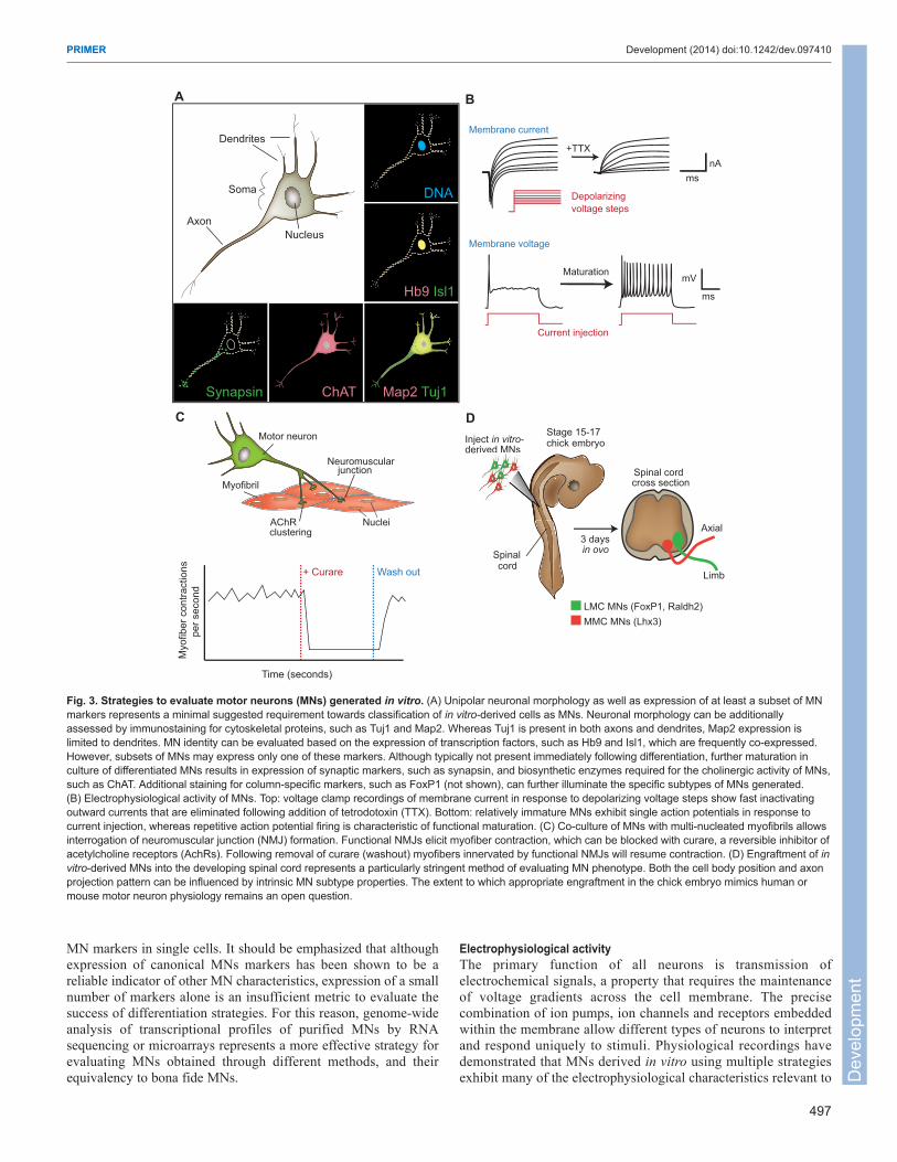
MN markers in single cells. It should be emphasized that althoughexpression of canonical MNs markers has been shown to be areliable indicator of other MN characteristics, expression of a smallnumber of markers alone is an insufficient metric to evaluate thesuccess of differentiation strategies. For this reason, genome-wideanalysis of transcriptional profiles of purified MNs by RNAsequencing or microarrays represents a more effective strategy forevaluating MNs obtained through different methods, and theirequivalency to bona fide MNs.
Electrophysiological activityThe primary function of all neurons is transmission ofelectrochemical signals, a property that requires the maintenanceof voltage gradients across the cell membrane. The precisecombination of ion pumps, ion channels and receptors embeddedwithin the membrane allow different types of neurons to interpretand respond uniquely to stimuli. Physiological recordings havedemonstrated that MNs derived in vitro using multiple strategiesexhibit many of the electrophysiological characteristics relevant to
497
PRIMER Development (2014) doi:10.1242/dev.097410
DNA
Hb9 Isl1
Map2 Tuj1ChATSynapsin
Axon
Soma
Dendrites
Nucleus
A
Membrane current
Depolarizing voltage steps
+TTX
msnA
Membrane voltage
Current injection
Maturation
ms
mV
B
Myofibril
Neuromuscular junction
AChR clustering
Nuclei
Motor neuron
Myo
fiber
con
tract
ions
per s
econ
d
Time (seconds)
+ Curare Wash out
C D
Inject in vitro- derived MNs
Stage 15-17chick embryo
Spinal cord
3 days in ovo
MMC MNs (Lhx3)LMC MNs (FoxP1, Raldh2)
Limb
Axial
Spinal cordcross section
Fig. 3. Strategies to evaluate motor neurons (MNs) generated in vitro. (A) Unipolar neuronal morphology as well as expression of at least a subset of MNmarkers represents a minimal suggested requirement towards classification of in vitro-derived cells as MNs. Neuronal morphology can be additionallyassessed by immunostaining for cytoskeletal proteins, such as Tuj1 and Map2. Whereas Tuj1 is present in both axons and dendrites, Map2 expression islimited to dendrites. MN identity can be evaluated based on the expression of transcription factors, such as Hb9 and Isl1, which are frequently co-expressed.However, subsets of MNs may express only one of these markers. Although typically not present immediately following differentiation, further maturation inculture of differentiated MNs results in expression of synaptic markers, such as synapsin, and biosynthetic enzymes required for the cholinergic activity of MNs,such as ChAT. Additional staining for column-specific markers, such as FoxP1 (not shown), can further illuminate the specific subtypes of MNs generated.(B) Electrophysiological activity of MNs. Top: voltage clamp recordings of membrane current in response to depolarizing voltage steps show fast inactivatingoutward currents that are eliminated following addition of tetrodotoxin (TTX). Bottom: relatively immature MNs exhibit single action potentials in response tocurrent injection, whereas repetitive action potential firing is characteristic of functional maturation. (C) Co-culture of MNs with multi-nucleated myofibrils allowsinterrogation of neuromuscular junction (NMJ) formation. Functional NMJs elicit myofiber contraction, which can be blocked with curare, a reversible inhibitor ofacetylcholine receptors (AchRs). Following removal of curare (washout) myofibers innervated by functional NMJs will resume contraction. (D) Engraftment of invitro-derived MNs into the developing spinal cord represents a particularly stringent method of evaluating MN phenotype. Both the cell body position and axonprojection pattern can be influenced by intrinsic MN subtype properties. The extent to which appropriate engraftment in the chick embryo mimics human ormouse motor neuron physiology remains an open question.
Dev
elop
men
t

498
motor neuron function and circuitry (Fig. 3B). Both stem cell-derived MNs as well as iMNs respond to applications of GABA,glutamate, and glycine with increased inward currents, indicatingthat they express the proper receptors and can elicit the correctresponse to these stimuli (Miles et al., 2004; Boulting et al., 2011;Son et al., 2011; Amoroso et al., 2013). MNs produced in vitroalso display spontaneous activity, express a range of voltage-activated channels and can fire action potentials in response toshort current injections, as well as generate calcium transients inresponse to kainate (Boulting et al., 2011; Son et al., 2011;Amoroso et al., 2013). Some electrophysiological phenotypes ofin vitro-derived MNs are dependent on the time spent in culture,including decrease in input resistance, spike frequency adaptationand rebound action potential firing (Miles et al., 2004; Takazawaet al., 2012).
As in the case of molecular markers of MNs, stereotypicalelectrophysiological response alone is insufficient to categorize cellsas MNs. It is also important to note that different types of MNsexhibit differences in excitability and firing patterns, and that theseresponses evolve as MNs mature both in vitro and in vivo (Kanninget al., 2010). For example, rat embryonic and postnatal MNs exhibitmarked differences in amplitude and rate of action potentials as wellas differing responsiveness to neurotransmitters (Gao and Ziskind-Conhaim, 1998). Because patch clamping individual cells representsa technically challenging and low throughput method, it isfrequently difficult to determine the extent to whichelectrophysiological characteristics vary between cells, even amongcells of the same culture. New advances such as multi-electrodearrays may allow more global evaluation of many cells in paralleland offer the exciting opportunity to screen multiple geneticbackgrounds or drug treatments for altered electrophysiologicalactivity.
Formation of NMJs and in vivo engraftmentThe main function of MNs in vivo is to innervate target muscles andintegrate signals from the CNS to allow coordinated musclecontraction and body movement. Thus, the ultimate evidence that invitro-derived MNs recapitulate their in vivo counterparts is theability to reproducibly engraft into the adult spinal cord, to projectto appropriate targets and to restore connectivity of a damaged CNSthrough formation of NMJs.
At the most basic level, it is clear that both mouse and humanPSC-derived MNs as well as iMNs can form functional NMJs invitro when co-cultured with muscle fibers (Miles et al., 2004; Sonet al., 2011) (Fig. 3C). Clustering of acetylcholine receptors onmyotubes adjacent to developing axons is observed within one dayof co-culture with MNs differentiated from mouse ESCs. As earlyas two days after the initiation of co-culture, small endplatepotentials can be detected by patch clamping innervated myotubes(Miles et al., 2004). In addition to the electrophysiologicalresponse to PSC-derived MNs or iMNs, innervated myotubesbegin to exhibit coordinated contractions, which can be abrogatedwith the reversible acetylcholine receptor inhibitor curare (Mileset al., 2004; Son et al., 2011) (Fig. 3C). Although clustering ofmyotube acetylcholine receptors is a hallmark of NMJ formation,it is worth noting that different substrates can promoteacetylcholine receptor clustering even in the absence of MNs(Peng and Cheng, 1982; Gingras et al., 2009). Thus, it is essentialto also evaluate the functionality of NMJs. Recent advances in theoptogenetics field have presented the possibility of engineeringMNs that express light-activated channel rhodopsins such thatneuronal activity, and as a consequence, myotube contraction, can
be induced simply by shining a specific wavelength light on co-cultures (Weick et al., 2010; Tye and Deisseroth, 2012). The abilityto tightly control MN activation should allow more in-depthstudies of the synaptic junction formed in vitro and could providea method to control MN activity when implanted in vivo.Moreover, as NMJs have been shown to undergo age-relateddegeneration both in wild-type mice and in transgenic models ofALS, it will be particularly interesting to determine if these eventscan be recapitulated and manipulated in vitro (Valdez et al., 2010;Valdez et al., 2012).
In addition to forming functional neuromuscular junctions withco-cultured myotubes, transplantation experiments have shown thatin vitro-derived MNs are capable of integrating into the developingspinal cord. Perhaps owing to the ease of manipulation, most studieshave been performed using chick embryos (Wichterle et al., 2002;Soundararajan et al., 2006; Son et al., 2011; Amoroso et al., 2013).For instance, transplantation of differentiated MNs and interneuronsfrom an Hb9::GFP reporter ESC line into the chick neural tubedemonstrated survival of many GFP+ MNs after transplantation.Importantly, although the dorso-ventral position of the graft was notcontrolled, GFP+ MNs settled in the ventral-lateral domain, whereasinterneurons (marked by a rodent-specific antibody to Lim2) wereobserved in the dorsoventral domain (Wichterle et al., 2002). Inaddition, grafted Hb9::GFP+ MNs differentiated from mouse ESCshave been shown to project axons into the periphery where theyreached muscle targets and displayed elaborated terminalsexpressing multiple synaptic markers (Wichterle et al., 2002;Soundararajan et al., 2006). Similar results have been obtained usinghuman ESC-derived MNs as well as mouse iMNs (Son et al., 2011;Amoroso et al., 2013).
Evidence that in vitro-derived MNs can correctly engraft intothe developing spinal cord is further supported by an elegant studyusing different small molecules to direct the differentiation ofmouse ESCs into two specific MN subtypes: cervical MMC MNsand brachial LMC MNs. When GFP-labeled LMC MNs weremixed with RFP-labeled MMC MNs and injected into the chickneural tube, the cell bodies of these neurons settled in theappropriate domains within the spinal cord and their axonsprojected to targets predicted by their transcription factorexpression (Peljto et al., 2010) (Fig. 3D). More recently, work byCorti and colleagues demonstrated that MNs differentiated fromgenetically corrected SMA iPSCs could survive transplantationand correctly engraft in the spinal cord of a one-day-old mousemodel of SMA (Corti et al., 2012). Furthermore, these cells couldameliorate disease phenotypes and extend the life span of SMNmutant mice, at least in part by providing neurotrophic support(Corti et al., 2012). It has also been shown that ESC-derived MNs,when transplanted into the tibial nerve of adult animals can formfunctional NMJs and ameliorate muscle atrophy caused by nervetransection (Yohn, et. al 2008).
In general, transplantation studies provide strong evidence thatPSC-derived MNs and iMNs bear strong resemblance to their invivo counterparts. However, these studies are technicallychallenging, largely qualitative and provide limited opportunities formechanistic study. As the ability to generate MNs in vitro throughdiverse methods continues to progress it will be crucial to reach aconsensus of best practices for evaluating MNs. Taken in isolation,gene expression, electrophysiological response and NMJ formationin vitro are, in our view, insufficient indicators of the success of MNdifferentiation strategies. Instead, we propose a multi-tieredapproach that combines each of these metrics to allow diverseaspects of MN biology to be evaluated.
PRIMER Development (2014) doi:10.1242/dev.097410
Dev
elop
men
t
Concluding remarks and perspectivesDecades of research into CNS development and spinal MNspecification have been synthesized to allow the in vitro generationof MNs through diverse methods. These cells can provide additionalmechanistic insights into developmental principles and MN biology,though perhaps their most exciting application is the prospect ofmodeling human MN diseases. Several studies have shown theusefulness of these cells in studying the mechanisms of neuraldegeneration. For example, human MNs have been shown to exhibitselective sensitivity to glia cells expressing a mutant gene linked toALS (Di Giorgio et al., 2008; Marchetto et al., 2008). Additionally,multiple groups have reported disease-specific phenotypes indifferentiated iPSC-derived MNs from patients with SMA (Ebert etal., 2009; Chang et al., 2011; Corti et al., 2012; Wang et al., 2013)or ALS (Bilican et al., 2012; Egawa et al., 2012; Donnelly et al.,2013; Sareen et al., 2013). Building on this, the in vitro generationof MNs via direct conversion of fibroblasts may additionallyaccelerate disease-modeling studies with patient cells, as it does notrequire the time-consuming step of iPSC generation andcharacterization. iMNs could be utilized to provide a snapshot ofdisease processes from a large cohort of patient fibroblasts, and celllines showing particularly interesting phenotypes could be thenreprogrammed into iPSCs to allow the production of differentiatedMNs in large quantities for further studies.
The progress in producing and understanding MNs has beenremarkable; however, substantial challenges still remain. Themolecular diversity of MN subtypes in vivo is only partiallyunderstood. Perhaps as a consequence of this, only a small numberof markers are currently used to evaluate MN subtypes generated invitro, and the extent to which altering methods of MN productionmay influence the resulting subtypes is largely unknown. Theapplication of RNA sequencing, single-cell qRT-PCR andproteomics approaches will allow comprehensive comparison ofMNs generated via different strategies. Moreover, as genome-wideinterrogation of bona fide human MNs is only possible throughlaser-capture of post-mortem samples (Ravits et al., 2005), high-throughput analysis of human PSC-derived MNs as well as humaniMNs may provide additional insights into the biology of humanspinal MNs.
Regardless of the strategy used to generate MNs, downstreamapplications warrant careful consideration of the experimentalconditions used for plating and culturing these cells. These includevariations in the methods of dissociation, cell density, substrate,media composition and cellular heterogeneity of the cultures(Sandoe and Eggan, 2013). Without controlling for andunderstanding these variables, we run into the risk of comparing theMN ‘apples’ generated and cultured by one protocol or in onelaboratory to the MN ‘oranges’ generated by another.
Spinal MNs were among the first specific neuronal cell types tobe derived from PSCs and the rapid progress made towards makingMNs in vitro might have provided inspiration to the locked-in Baubywho wondered, ‘Does the cosmos contain keys for opening mydiving bell? A subway line with no terminus? A currency strongenough to buy my freedom back?’ (Bauby, 1998). We are optimisticthat continued efforts to collaboratively establish best practices forMN production, culture and evaluation in vitro will provide the keysto unlock novel therapeutic strategies for devastating neurologicaldisorders, including ALS.
AcknowledgementsWe thank members of the Eggan Laboratory, in particular E. Kiskinis for insightfuldiscussions, and apologize to our colleagues whose work we could not cite owingto space limitations.
Competing interestsThe authors declare no competing financial interests.
FundingK.E. is an early career scientist with the Howard Hughes Medical Institute andB.N.D.-D is an ALS Association Milton Safenowitz post-doctoral fellow.
ReferencesAlaynick, W. A., Jessell, T. M. and Pfaff, S. L. (2011) SnapShot: spinal cord
development. Cell 146,178-178 e1. Ambasudhan, R., Talantova, M., Coleman, R., Yuan, X., Zhu, S., Lipton, S. A. and
Ding, S. (2011). Direct reprogramming of adult human fibroblasts to functionalneurons under defined conditions. Cell Stem Cell 9, 113-118.
Amoroso, M. W., Croft, G. F., Williams, D. J., O’Keeffe, S., Carrasco, M. A., Davis,A. R., Roybon, L., Oakley, D. H., Maniatis, T., Henderson, C. E. et al. (2013).Accelerated high-yield generation of limb-innervating motor neurons from humanstem cells. J. Neurosci. 33, 574-586.
Bain, G., Kitchens, D., Yao, M., Huettner, J. E. and Gottlieb, D. I. (1995). Embryonicstem cells express neuronal properties in vitro. Dev. Biol. 168, 342-357.
Bauby, J.-D. (1998). The Diving Bell and the Butterfly: A Memoir of Life in Death. NewYork, NY: Vintage Books.
Bilican, B., Serio, A., Barmada, S. J., Nishimura, A. L., Sullivan, G. J., Carrasco,M., Phatnani, H. P., Puddifoot, C. A., Story, D., Fletcher, J. et al. (2012). Mutantinduced pluripotent stem cell lines recapitulate aspects of TDP-43 proteinopathiesand reveal cell-specific vulnerability. Proc. Natl. Acad. Sci. USA 109, 5803-5808.
Boulting, G. L., Kiskinis, E., Croft, G. F., Amoroso, M. W., Oakley, D. H., Wainger,B. J., Williams, D. J., Kahler, D. J., Yamaki, M., Davidow, L. et al. (2011). Afunctionally characterized test set of human induced pluripotent stem cells. Nat.Biotechnol. 29, 279-286.
Briscoe, J., Pierani, A., Jessell, T. M. and Ericson, J. (2000). A homeodomainprotein code specifies progenitor cell identity and neuronal fate in the ventral neuraltube. Cell 101, 435-445.
Burghes, A. H. and Beattie, C. E. (2009). Spinal muscular atrophy: why do low levelsof survival motor neuron protein make motor neurons sick? Nat. Rev. Neurosci. 10,597-609.
Chambers, S. M., Fasano, C. A., Papapetrou, E. P., Tomishima, M., Sadelain, M.and Studer, L. (2009). Highly efficient neural conversion of human ES and iPS cellsby dual inhibition of SMAD signaling. Nat. Biotechnol. 27, 275-280.
Chambers, S. M., Qi, Y., Mica, Y., Lee, G., Zhang, X. J., Niu, L., Bilsland, J., Cao,L., Stevens, E., Whiting, P. et al. (2012). Combined small-molecule inhibitionaccelerates developmental timing and converts human pluripotent stem cells intonociceptors. Nat. Biotechnol. 30, 715-720.
Chang, T., Zheng, W., Tsark, W., Bates, S., Huang, H., Lin, R. J. and Yee, J. K.(2011). Brief report: phenotypic rescue of induced pluripotent stem cell-derived motorneurons of a spinal muscular atrophy patient. Stem Cells 29, 2090-2093.
Chen, J. A., Huang, Y. P., Mazzoni, E. O., Tan, G. C., Zavadil, J. and Wichterle, H.(2011). Mir-17-3p controls spinal neural progenitor patterning by regulating Olig2/Irx3cross-repressive loop. Neuron 69, 721-735.
Corti, S., Nizzardo, M., Simone, C., Falcone, M., Nardini, M., Ronchi, D., Donadoni,C., Salani, S., Riboldi, G., Magri, F. et al. (2012). Genetic correction of humaninduced pluripotent stem cells from patients with spinal muscular atrophy. Sci. Transl.Med. 4, 165ra162.
Dasen, J. S. and Jessell, T. M. (2009). Hox networks and the origins of motor neurondiversity. Curr. Top. Dev. Biol. 88, 169-200.
Dasen, J. S., Tice, B. C., Brenner-Morton, S. and Jessell, T. M. (2005). A Hoxregulatory network establishes motor neuron pool identity and target-muscleconnectivity. Cell 123, 477-491.
Dasen, J. S., De Camilli, A., Wang, B., Tucker, P. W. and Jessell, T. M. (2008). Hoxrepertoires for motor neuron diversity and connectivity gated by a single accessoryfactor, FoxP1. Cell 134, 304-316.
De Robertis, E. M. (2006). Spemann’s organizer and self-regulation in amphibianembryos. Nat. Rev. Mol. Cell Biol. 7, 296-302.
Di Giorgio, F. P., Carrasco, M. A., Siao, M. C., Maniatis, T. and Eggan, K. (2007).Non-cell autonomous effect of glia on motor neurons in an embryonic stem cell-based ALS model. Nat. Neurosci. 10, 608-614.
Di Giorgio, F. P., Boulting, G. L., Bobrowicz, S. and Eggan, K. C. (2008). Humanembryonic stem cell-derived motor neurons are sensitive to the toxic effect of glialcells carrying an ALS-causing mutation. Cell Stem Cell 3, 637-648.
Donnelly, C. J., Zhang, P. W., Pham, J. T., Heusler, A. R., Mistry, N. A., Vidensky,S., Daley, E. L., Poth, E. M., Hoover, B., Fines, D. M. et al. (2013). RNA toxicityfrom the ALS/FTD C9ORF72 expansion is mitigated by antisense intervention.Neuron 80, 415-428.
Ebert, A. D., Yu, J., Rose, F. F., Jr, Mattis, V. B., Lorson, C. L., Thomson, J. A. andSvendsen, C. N. (2009). Induced pluripotent stem cells from a spinal muscularatrophy patient. Nature 457, 277-280.
Egawa, N., Kitaoka, S., Tsukita, K., Naitoh, M., Takahashi, K., Yamamoto, T.,Adachi, F., Kondo, T., Okita, K., Asaka, I. et al. (2012). Drug screening for ALSusing patient-specific induced pluripotent stem cells. Sci. Transl. Med. 4, 145ra104.
Ericson, J., Rashbass, P., Schedl, A., Brenner-Morton, S., Kawakami, A., vanHeyningen, V., Jessell, T. M. and Briscoe, J. (1997). Pax6 controls progenitor cellidentity and neuronal fate in response to graded Shh signaling. Cell 90, 169-180.
Evans, M. (2011). Discovering pluripotency: 30 years of mouse embryonic stem cells.Nat. Rev. Mol. Cell Biol. 12, 680-686.
499
PRIMER Development (2014) doi:10.1242/dev.097410
Dev
elop
men
t

500
Evans, M. J. and Kaufman, M. H. (1981). Establishment in culture of pluripotentialcells from mouse embryos. Nature 292, 154-156.
Gao, B. X. and Ziskind-Conhaim, L. (1998). Development of ionic currents underlyingchanges in action potential waveforms in rat spinal motor neurons. J. Neurophysiol.80, 3047-3061.
Gingras, J., Rioux, R. M., Cuvelier, D., Geisse, N. A., Lichtman, J. W., Whitesides,G. M., Mahadevan, L. and Sanes, J. R. (2009). Controlling the orientation andsynaptic differentiation of myotubes with micropatterned substrates. Biophys. J. 97,2771-2779.
Graf, T. (2011). Historical origins of transdifferentiation and reprogramming. Cell StemCell 9, 504-516.
Greig, L. C., Woodworth, M. B., Galazo, M. J., Padmanabhan, H. and Macklis, J. D.(2013). Molecular logic of neocortical projection neuron specification, developmentand diversity. Nat. Rev. Neurosci. 14, 755-769.
Hamburger, V. (1988) The Heritage of Experimental Embryology: Hans Spemann andthe Organizer. New York, NY: Oxford University Press.
Hester, M. E., Murtha, M. J., Song, S., Rao, M., Miranda, C. J., Meyer, K., Tian, J.,Boulting, G., Schaffer, D. V., Zhu, M. X. et al. (2011). Rapid and efficientgeneration of functional motor neurons from human pluripotent stem cells usinggene delivered transcription factor codes. Mol. Ther. 19, 1905-1912.
Hu, B. Y. and Zhang, S. C. (2009). Differentiation of spinal motor neurons frompluripotent human stem cells. Nat. Protoc. 4, 1295-1304.
Jessell, T. M. (2000). Neuronal specification in the spinal cord: inductive signals andtranscriptional codes. Nat. Rev. Genet. 1, 20-29.
Joannides, A. J., Fiore-Hériché, C., Battersby, A. A., Athauda-Arachchi, P.,Bouhon, I. A., Williams, L., Westmore, K., Kemp, P. J., Compston, A., Allen, N.D. et al. (2007). A scaleable and defined system for generating neural stem cellsfrom human embryonic stem cells. Stem Cells 25, 731-737.
Jung, H., Lacombe, J., Mazzoni, E. O., Liem, K. F., Jr, Grinstein, J., Mahony, S.,Mukhopadhyay, D., Gifford, D. K., Young, R. A., Anderson, K. V. et al. (2010).Global control of motor neuron topography mediated by the repressive actions of asingle hox gene. Neuron 67, 781-796.
Kanning, K. C., Kaplan, A. and Henderson, C. E. (2010). Motor neuron diversity indevelopment and disease. Annu. Rev. Neurosci. 33, 409-440.
Karumbayaram, S., Novitch, B. G., Patterson, M., Umbach, J. A., Richter, L.,Lindgren, A., Conway, A. E., Clark, A. T., Goldman, S. A., Plath, K. et al. (2009).Directed differentiation of human-induced pluripotent stem cells generates activemotor neurons. Stem Cells 27, 806-811.
Kawasaki, H., Mizuseki, K., Nishikawa, S., Kaneko, S., Kuwana, Y., Nakanishi, S.,Nishikawa, S. I. and Sasai, Y. (2000). Induction of midbrain dopaminergic neuronsfrom ES cells by stromal cell-derived inducing activity. Neuron 28, 31-40.
Lacombe, J., Hanley, O., Jung, H., Philippidou, P., Surmeli, G., Grinstein, J. andDasen, J. S. (2013). Genetic and functional modularity of Hox activities in thespecification of limb-innervating motor neurons. PLoS Genet. 9, e1003184.
Lee, H., Shamy, G. A., Elkabetz, Y., Schofield, C. M., Harrsion, N. L.,Panagiotakos, G., Socci, N. D., Tabar, V. and Studer, L. (2007). Directeddifferentiation and transplantation of human embryonic stem cell-derived motorneurons. Stem Cells 25, 1931-1939.
Lee, S., Cuvillier, J. M., Lee, B., Shen, R., Lee, J. W. and Lee, S. K. (2012). Fusionprotein Isl1-Lhx3 specifies motor neuron fate by inducing motor neuron genes andconcomitantly suppressing the interneuron programs. Proc. Natl. Acad. Sci. USA109, 3383-3388.
Li, M., Pevny, L., Lovell-Badge, R. and Smith, A. (1998). Generation of purified neuralprecursors from embryonic stem cells by lineage selection. Curr. Biol. 8, 971-974.
Li, X. J., Du, Z. W., Zarnowska, E. D., Pankratz, M., Hansen, L. O., Pearce, R. A.and Zhang, S. C. (2005). Specification of motor neurons from human embryonicstem cells. Nat. Biotechnol. 23, 215-221.
Ling, S. C., Polymenidou, M. and Cleveland, D. W. (2013). Converging mechanismsin ALS and FTD: disrupted RNA and protein homeostasis. Neuron 79, 416-438.
Liu, J. P., Laufer, E. and Jessell, T. M. (2001). Assigning the positional identity ofspinal motor neurons: rostrocaudal patterning of Hox-c expression by FGFs, Gdf11,and retinoids. Neuron 32, 997-1012.
Maden, M. (2007). Retinoic acid in the development, regeneration and maintenance ofthe nervous system. Nat. Rev. Neurosci. 8, 755-765.
Marchetto, M. C., Muotri, A. R., Mu, Y., Smith, A. M., Cezar, G. G. and Gage, F. H.(2008). Non-cell-autonomous effect of human SOD1 G37R astrocytes on motorneurons derived from human embryonic stem cells. Cell Stem Cell 3, 649-657.
Marro, S., Pang, Z. P., Yang, N., Tsai, M. C., Qu, K., Chang, H. Y., Südhof, T. C. andWernig, M. (2011). Direct lineage conversion of terminally differentiated hepatocytesto functional neurons. Cell Stem Cell 9, 374-382.
Mazzoni, E. O., Mahony, S., Closser, M., Morrison, C. A., Nedelec, S., Williams, D.J., An, D., Gifford, D. K. and Wichterle, H. (2013a). Synergistic binding oftranscription factors to cell-specific enhancers programs motor neuron identity. Nat.Neurosci. 16, 1219-1227.
Mazzoni, E. O., Mahony, S., Peljto, M., Patel, T., Thornton, S. R., McCuine, S.,Reeder, C., Boyer, L. A., Young, R. A., Gifford, D. K. et al. (2013b). Saltatoryremodeling of Hox chromatin in response to rostrocaudal patterning signals. Nat.Neurosci. 16, 1191-1198.
Miles, G. B., Yohn, D. C., Wichterle, H., Jessell, T. M., Rafuse, V. F. andBrownstone, R. M. (2004). Functional properties of motor neurons derived frommouse embryonic stem cells. J. Neurosci. 24, 7848-7858.
Misra, M., Shah, V., Carpenter, E., McCaffery, P. and Lance-Jones, C. (2009).Restricted patterns of Hoxd10 and Hoxd11 set segmental differences in motorneuron subtype complement in the lumbosacral spinal cord. Dev. Biol. 330, 54-72.
Novitch, B. G., Chen, A. I. and Jessell, T. M. (2001). Coordinate regulation of motorneuron subtype identity and pan-neuronal properties by the bHLH repressor Olig2.Neuron 31, 773-789.
Odorico, J. S., Kaufman, D. S. and Thomson, J. A. (2001). Multilineagedifferentiation from human embryonic stem cell lines. Stem Cells 19, 193-204.
Okabe, S., Forsberg-Nilsson, K., Spiro, A. C., Segal, M. and McKay, R. D. (1996).Development of neuronal precursor cells and functional postmitotic neurons fromembryonic stem cells in vitro. Mech. Dev. 59, 89-102.
Pang, Z. P., Yang, N., Vierbuchen, T., Ostermeier, A., Fuentes, D. R., Yang, T. Q.,Citri, A., Sebastiano, V., Marro, S., Südhof, T. C. et al. (2011). Induction of humanneuronal cells by defined transcription factors. Nature 476, 220-223.
Passini, M. A. and Cheng, S. H. (2011). Prospects for the gene therapy of spinalmuscular atrophy. Trends Mol. Med. 17, 259-265.
Patani, R., Compston, A., Puddifoot, C. A., Wyllie, D. J., Hardingham, G. E., Allen,N. D. and Chandran, S. (2009). Activin/Nodal inhibition alone accelerates highlyefficient neural conversion from human embryonic stem cells and imposes a caudalpositional identity. PLoS ONE 4, e7327.
Patani, R., Hollins, A. J., Wishart, T. M., Puddifoot, C. A., Alvarez, S., de Lera, A.R., Wyllie, D. J., Compston, D. A., Pedersen, R. A., Gillingwater, T. H. et al.(2011). Retinoid-independent motor neurogenesis from human embryonic stem cellsreveals a medial columnar ground state. Nat. Commun. 2, 214.
Pearson, J. C., Lemons, D. and McGinnis, W. (2005). Modulating Hox gene functionsduring animal body patterning. Nat. Rev. Genet. 6, 893-904.
Peljto, M., Dasen, J. S., Mazzoni, E. O., Jessell, T. M. and Wichterle, H. (2010).Functional diversity of ESC-derived motor neuron subtypes revealed throughintraspinal transplantation. Cell Stem Cell 7, 355-366.
Peng, H. B. and Cheng, P. C. (1982). Formation of postsynaptic specializationsinduced by latex beads in cultured muscle cells. J. Neurosci. 2, 1760-1774.
Philippidou, P. and Dasen, J. S. (2013). Hox genes: choreographers in neuraldevelopment, architects of circuit organization. Neuron 80, 12-34.
Philippidou, P., Walsh, C. M., Aubin, J., Jeannotte, L. and Dasen, J. S. (2012).Sustained Hox5 gene activity is required for respiratory motor neuron development.Nat. Neurosci. 15, 1636-1644.
Ravits, J., Laurie, P. and Stone, B. (2005). Amyotrophic lateral sclerosismicrogenomics. Phys. Med. Rehabil. Clin. N. Am. 16, 909-924, viii.
Reubinoff, B. E., Itsykson, P., Turetsky, T., Pera, M. F., Reinhartz, E., Itzik, A. andBen-Hur, T. (2001). Neural progenitors from human embryonic stem cells. Nat.Biotechnol. 19, 1134-1140.
Rousso, D. L., Gaber, Z. B., Wellik, D., Morrisey, E. E. and Novitch, B. G. (2008).Coordinated actions of the forkhead protein Foxp1 and Hox proteins in the columnarorganization of spinal motor neurons. Neuron 59, 226-240.
Sandoe, J. and Eggan, K. (2013). Opportunities and challenges of pluripotent stemcell neurodegenerative disease models. Nat. Neurosci. 16, 780-789.
Sareen, D., O’Rourke, J. G., Meera, P., Muhammad, A. K., Grant, S., Simpkinson,M., Bell, S., Carmona, S., Ornelas, L., Sahabian, A. et al. (2013). Targeting RNAfoci in iPSC-derived motor neurons from ALS patients with a C9ORF72 repeatexpansion. Sci. Transl. Med. 5, 208ra149.
Shoemaker, L. D. and Arlotta, P. (2010). Untangling the cortex: Advances inunderstanding specification and differentiation of corticospinal motor neurons.Bioessays 32, 197-206.
Singh Roy, N., Nakano, T., Xuing, L., Kang, J., Nedergaard, M. and Goldman, S. A.(2005). Enhancer-specified GFP-based FACS purification of human spinal motorneurons from embryonic stem cells. Exp. Neurol. 196, 224-234.
Smith, J. R., Vallier, L., Lupo, G., Alexander, M., Harris, W. A. and Pedersen, R. A.(2008). Inhibition of Activin/Nodal signaling promotes specification of humanembryonic stem cells into neuroectoderm. Dev. Biol. 313, 107-117.
Son, E. Y., Ichida, J. K., Wainger, B. J., Toma, J. S., Rafuse, V. F., Woolf, C. J. andEggan, K. (2011). Conversion of mouse and human fibroblasts into functional spinalmotor neurons. Cell Stem Cell 9, 205-218.
Song, K., Nam, Y. J., Luo, X., Qi, X., Tan, W., Huang, G. N., Acharya, A., Smith, C.L., Tallquist, M. D., Neilson, E. G. et al. (2012). Heart repair by reprogrammingnon-myocytes with cardiac transcription factors. Nature 485, 599-604.
Soundararajan, P., Miles, G. B., Rubin, L. L., Brownstone, R. M. and Rafuse, V. F.(2006). Motor neurons derived from embryonic stem cells express transcriptionfactors and develop phenotypes characteristic of medial motor column neurons. J.Neurosci. 26, 3256-3268.
Stern, C. D. (2005). Neural induction: old problem, new findings, yet more questions.Development 132, 2007-2021.
Streit, A., Berliner, A. J., Papanayotou, C., Sirulnik, A. and Stern, C. D. (2000).Initiation of neural induction by FGF signalling before gastrulation. Nature 406, 74-78.
Sugimori, M., Nagao, M., Bertrand, N., Parras, C. M., Guillemot, F. and Nakafuku,M. (2007). Combinatorial actions of patterning and HLH transcription factors in thespatiotemporal control of neurogenesis and gliogenesis in the developing spinalcord. Development 134, 1617-1629.
Sun, Y., Dykes, I. M., Liang, X., Eng, S. R., Evans, S. M. and Turner, E. E. (2008). Acentral role for Islet1 in sensory neuron development linking sensory and spinal generegulatory programs. Nat. Neurosci. 11, 1283-1293.
Takahashi, K. and Yamanaka, S. (2006). Induction of pluripotent stem cells frommouse embryonic and adult fibroblast cultures by defined factors. Cell 126, 663-676.
Takahashi, K., Tanabe, K., Ohnuki, M., Narita, M., Ichisaka, T., Tomoda, K. andYamanaka, S. (2007). Induction of pluripotent stem cells from adult humanfibroblasts by defined factors. Cell 131, 861-872.
PRIMER Development (2014) doi:10.1242/dev.097410
Dev
elop
men
t

Takazawa, T., Croft, G. F., Amoroso, M. W., Studer, L., Wichterle, H. andMacdermott, A. B. (2012). Maturation of spinal motor neurons derived from humanembryonic stem cells. PLoS ONE 7, e40154.
Thaler, J. P., Lee, S. K., Jurata, L. W., Gill, G. N. and Pfaff, S. L. (2002). LIM factorLhx3 contributes to the specification of motor neuron and interneuron identitythrough cell-type-specific protein-protein interactions. Cell 110, 237-249.
Thomson, J. A., Itskovitz-Eldor, J., Shapiro, S. S., Waknitz, M. A., Swiergiel, J. J.,Marshall, V. S. and Jones, J. M. (1998). Embryonic stem cell lines derived fromhuman blastocysts. Science 282, 1145-1147.
Tiret, L., Le Mouellic, H., Maury, M. and Brûlet, P. (1998). Increased apoptosis ofmotor neurons and altered somatotopic maps in the brachial spinal cord of Hoxc-8-deficient mice. Development 125, 279-291.
Tye, K. M. and Deisseroth, K. (2012). Optogenetic investigation of neural circuitsunderlying brain disease in animal models. Nat. Rev. Neurosci. 13, 251-266.
Valdez, G., Tapia, J. C., Kang, H., Clemenson, G. D., Jr, Gage, F. H., Lichtman, J.W. and Sanes, J. R. (2010). Attenuation of age-related changes in mouseneuromuscular synapses by caloric restriction and exercise. Proc. Natl. Acad. Sci.USA 107, 14863-14868.
Valdez, G., Tapia, J. C., Lichtman, J. W., Fox, M. A. and Sanes, J. R. (2012). Sharedresistance to aging and ALS in neuromuscular junctions of specific muscles. PLoSONE 7, e34640.
Vermot, J., Schuhbaur, B., Le Mouellic, H., McCaffery, P., Garnier, J. M., Hentsch,D., Brûlet, P., Niederreither, K., Chambon, P., Dollé, P. et al. (2005).Retinaldehyde dehydrogenase 2 and Hoxc8 are required in the murine brachialspinal cord for the specification of Lim1+ motor neurons and the correct distributionof Islet1+ motor neurons. Development 132, 1611-1621.
Vierbuchen, T., Ostermeier, A., Pang, Z. P., Kokubu, Y., Südhof, T. C. and Wernig,M. (2010). Direct conversion of fibroblasts to functional neurons by defined factors.Nature 463, 1035-1041.
Vult von Steyern, F., Martinov, V., Rabben, I., Njå, A., de Lapeyrière, O. and Lømo,T. (1999). The homeodomain transcription factors Islet 1 and HB9 are expressed inadult alpha and gamma motor neurons identified by selective retrograde tracing. Eur.J. Neurosci. 11, 2093-2102.
Wahba, G. M., Hostikka, S. L. and Carpenter, E. M. (2001). The paralogous Hoxgenes Hoxa10 and Hoxd10 interact to pattern the mouse hindlimb peripheralnervous system and skeleton. Dev. Biol. 231, 87-102.
Wang, Z. B., Zhang, X. and Li, X. J. (2013). Recapitulation of spinal motor neuron-specific disease phenotypes in a human cell model of spinal muscular atrophy. CellRes. 23, 378-393.
Weick, J. P., Johnson, M. A., Skroch, S. P., Williams, J. C., Deisseroth, K. andZhang, S. C. (2010). Functional control of transplantable human ESC-derivedneurons via optogenetic targeting. Stem Cells 28, 2008-2016.
Wichterle, H., Lieberam, I., Porter, J. A. and Jessell, T. M. (2002). Directeddifferentiation of embryonic stem cells into motor neurons. Cell 110, 385-397.
Wilson, S. I., Graziano, E., Harland, R., Jessell, T. M. and Edlund, T. (2000). Anearly requirement for FGF signalling in the acquisition of neural cell fate in the chickembryo. Curr. Biol. 10, 421-429.
Woodworth, M. B., Custo Greig, L., Kriegstein, A. R. and Macklis, J. D. (2012).SnapShot: cortical development. Cell 151, 918-918.e1.
Wu, Y., Wang, G., Scott, S. A. and Capecchi, M. R. (2008). Hoxc10 and Hoxd10regulate mouse columnar, divisional and motor pool identity of lumbar motorneurons. Development 135, 171-182.
Yohn, D. C., Miles, G. B., Rafuse, V. F. and Brownstone, R. M. (2008). Transplantedmouse embryonic stem-cell-derived motoneurons form functional motor units andreduce muscle atrophy. J. Neurosci. 28, 12409-12418.
Yoo, A. S., Sun, A. X., Li, L., Shcheglovitov, A., Portmann, T., Li, Y., Lee-Messer,C., Dolmetsch, R. E., Tsien, R. W. and Crabtree, G. R. (2011). MicroRNA-mediated conversion of human fibroblasts to neurons. Nature 476, 228-231.
Zhang, S. C., Wernig, M., Duncan, I. D., Brüstle, O. and Thomson, J. A. (2001). Invitro differentiation of transplantable neural precursors from human embryonic stemcells. Nat. Biotechnol. 19, 1129-1133.
Zhou, J., Su, P., Li, D., Tsang, S., Duan, E. and Wang, F. (2010). High-efficiencyinduction of neural conversion in human ESCs and human induced pluripotent stemcells with a single chemical inhibitor of transforming growth factor beta superfamilyreceptors. Stem Cells 28, 1741-1750.
501
PRIMER Development (2014) doi:10.1242/dev.097410
Dev
elop
men
t