How the Brain Worksweb.eng.fiu.edu/mcabre05/DATA FOR PROJECTS/Anatomy... · How the Brain Works The...
101
1 How the Brain Works The brain is a soft mass of supportive tissues and nerves connected to the spinal cord and it is the center of the human nervous system. The brain controls your ability to think, talk, feel, see, hear, remember things, within other functions. Some of the nerves in the brain go right to the eyes, ears and other parts of the head. Other nerves connect the brain with other parts of the body through the spinal cord to control personality, senses and body functions from breathing to walking. Brain, Spinal Cord and Nerves form the central nervous system. 1
Transcript of How the Brain Worksweb.eng.fiu.edu/mcabre05/DATA FOR PROJECTS/Anatomy... · How the Brain Works The...

1
How the Brain Works
The brain is a soft mass of supportive tissues and nerves connected to the spinal cord and it is the center of the human nervous system.
The brain controls your ability to think, talk, feel,
see, hear, remember things, within other functions. Some of the nerves in the brain go right to the eyes,
ears and other parts of the head. Other nerves connect the brain with other parts of the body through the spinal cord to control personality, senses and body functions from breathing to walking.
Brain, Spinal Cord and Nerves form the central
nervous system.
1
2
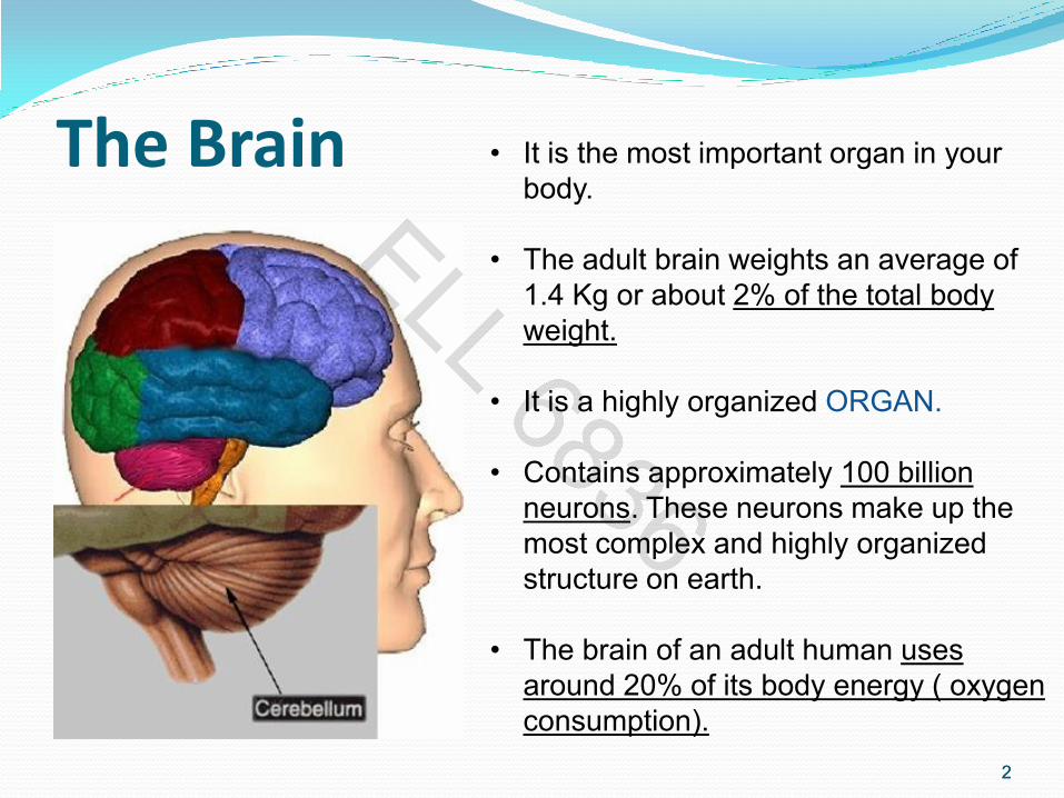
The Brain • It is the most important organ in your
body.
• The adult brain weights an average of
1.4 Kg or about 2% of the total body
weight.
• It is a highly organized ORGAN.
• Contains approximately 100 billion
neurons. These neurons make up the
most complex and highly organized
structure on earth.
• The brain of an adult human uses
around 20% of its body energy ( oxygen
consumption).
2

3
Nervous System: Anatomical Subdivisions Central nervous system
(CNS)
brain & spinal cord enclosed in bony coverings
Peripheral nervous system (PNS)
Sensory neurons
Motor neurons
3
• The Nervous system is the
control center for the entire body.
• The brain uses information it
receives from your nerves to
coordinate all of your actions
and reactions.

4
Central Nervous System
Human central nervous system (CNS):
• Constituents: Brain + Spinal Cord + Nerves going to muscles and sensory organs
• Cerebrospinal fluid: clear liquid surrounding brain & spinal cord and filling brain
cavities. (protects the brain from mechanical injury by acting as a shock absorber)
• Nerve cells (neurons): 1010 nerve cells with perhaps 1014 or more interconnections
4
5
CHARACTERISTICS OF NERVE CELLS / NEURONS
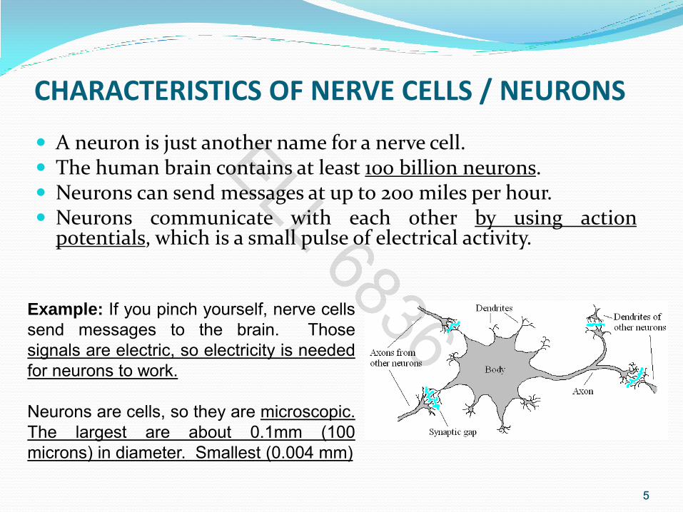
A neuron is just another name for a nerve cell. The human brain contains at least 100 billion neurons. Neurons can send messages at up to 200 miles per hour. Neurons communicate with each other by using action
potentials, which is a small pulse of electrical activity.
Example: If you pinch yourself, nerve cells
send messages to the brain. Those
signals are electric, so electricity is needed
for neurons to work.
Neurons are cells, so they are microscopic.
The largest are about 0.1mm (100
microns) in diameter. Smallest (0.004 mm)
5

6
Structure of a Neuron Body = membrane containing nucleus, also called
soma Axon
terminates on other nerve cells connecting to other neuron’s dendrites
transmits output to other dendrites of other neurons (rapid conduction)
Dendrites receive the signal from other neuron’s axons
Synapse Connection axon
Biochemical components: Sodium, Potassium, Chloride
Note: In the human brain each nerve is connected to approximately 10,000 other nerves, mostly to dendrite connections.
6

7
Synapses Between Two Neurons
First, neuron in path releases neurotransmitter onto second neuron that responds to it.
1st neuron is presynaptic neuron
2nd neuron is postsynaptic neuron
7

8
Postsynaptic Potentials Excitatory postsynaptic potentials (EPSP)
a positive voltage change causing postsynaptic cell to be more likely to fire result from Na+ flowing into the cell
Inhibitory postsynaptic potentials (IPSP) a negative voltage change causing postsynaptic cell to be less likely to fire
(hyperpolarize) result of K+ leaving the cell
8

9
Neuron Reproduction: Unlike most other cells, neurons cannot regrow after damage (except neurons
from the hippocampus).
*Fortunately, there are about 100 billion neurons in the brain.
9
Interesting Fact:
Old person has perhaps ≈ 1/3 of the
neurons at the time of birth => continuous
loss of neurons
• Most ancient part of the cortex
• LTP was first discovered to occur in
• this region

10
Brain Cells
There are different types of neurons. They all carry electro-chemical nerve signals, but differ in structure (the number of processes, or axons, emanating from the cell body) and are found in different parts of the body.
The brain and spinal cord are made up of many cells, including neurons and glial cells.
10

11
Brain Cells 1) Neurons Neurons typically consist of dendrites that
receive information, a cell body, and an axon used to transmit information throughout the nervous system.
Neurons send and receive electro-chemical signals to and from the brain and nervous system. There are about 100 billion neurons in the brain.
2) Glial cells There are many more glial cells than neurons;
they provide support functions for the neurons, and are far more numerous than neurons.
Glial cells have multiple functions, which include repairing the Central Nervous System, and regulating the biochemical balance of the brain.
11 Glial cells
Neuron

12
Cells of the cerebral cortex
1) Pyramidal cells
2) Stellate cells:
multiple dendrites
with short axons
12
Cerebral cortex:
• Thin layer of gray
matter
• 2-4 mm thick

13
Pyramidal cells Pyramidal cells are neurons found in the cerebral
cortex. These are responsible for the generation of EEG signals.
Pyramidal neurons (pyramidal cells) are a type of neuron found in areas of the brain including cerebral cortex, the hippocampus, and in the amygdala.
13

14
Pyramidal Cells
• Pyramidal neurons are the
primary excitation units of
the mammalian prefrontral
cortex and the corticospinal
tract.
• What makes a pyramidal
neuron unique is how the
dendrites are arranged and
the fact that both the axon
and dendrites undergo
extensive branching.
14
15
Pyramidal Cells
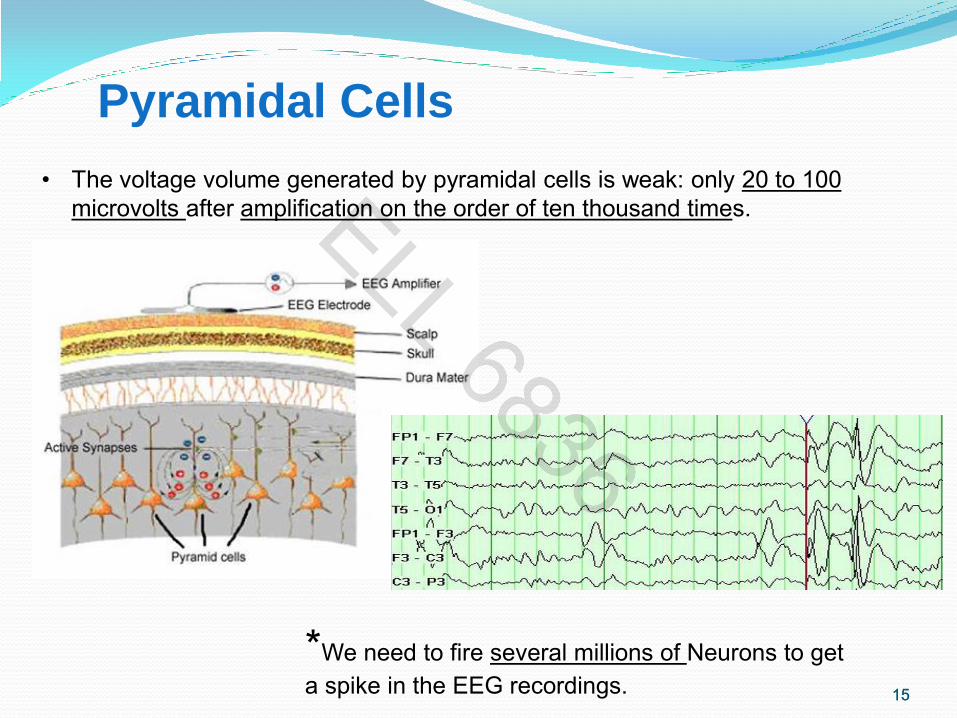
• The voltage volume generated by pyramidal cells is weak: only 20 to 100
microvolts after amplification on the order of ten thousand times.
15
*We need to fire several millions of Neurons to get
a spike in the EEG recordings.

Pyramidal Cells Properties
A single neural event is too small to be detected on the scalp.
Action potentials (APs) do not add up – too short.
EPSPs/IPSPs sum up in time through synchronization, and in space due to cortical architecture (open/closed electrical fields).
No EEG generated due to closed fields in glial cells and subcortical structures.
16
glial cells Pyramidal cells

17
Pyramidal Cells Properties (cont.)
Long apical dentrite perpendicular to the
surface of the cortex.
Axons run perpendicular to the cortical surface and travel to sites outside the cerebral cortex.
Aligned in parallel.
Synchronized in activity.
They received synchronous input from subcortical and cortical sources.
17
18
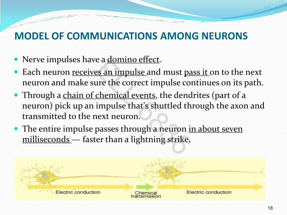
MODEL OF COMMUNICATIONS AMONG NEURONS
Nerve impulses have a domino effect.
Each neuron receives an impulse and must pass it on to the next neuron and make sure the correct impulse continues on its path.
Through a chain of chemical events, the dendrites (part of a neuron) pick up an impulse that's shuttled through the axon and transmitted to the next neuron.
The entire impulse passes through a neuron in about seven milliseconds — faster than a lightning strike.
18

19
Properties of the Neurons
Excitability (irritability)
ability to respond to changes in the body and external environment called stimuli
Conductivity
development of action potentials
produce traveling electrical signals
Synaptic linkage
when electrical signal reaches the end of a nerve fiber, a chemical neurotransmitter is released
19

20
Neurons properties (cont.)
Neurons are highly irritable (responsive to stimuli).
Action potentials, or nerve impulses, are:
--Electrical impulses conducted along the length of axons.
--Always the same regardless of stimulus.
--The underlying functional feature of the
nervous system.
20

21
Definitions
Voltage (V) – measure of potential energy between
two points generated by a charge separation.
(Voltage = Potential Difference = Potential)
Current (I) – Flow of electrical charge
Resistance (R) – Tendency to oppose the current
Conductor – Substance with low electrical resistance.
Units: V (volt), I (ampere), R (ohm)
21

Associated Electrical Concepts
Voltage:
the potential of current that flows from one point to another.
It is a relative measure.
Current :
number of charged particles (electrons, ions) that flow in a given time.
Resistance:
resistance to the movement of charges.
22

23
Ohm’s Law The relationship between voltage, current, and resistance is defined
by Ohm’s Law
Current (I) = Voltage (V)
Resistance (R)
• In the body, electrical current is the
flow of ions (rather than free
electrons) across membranes.
• A Potential Difference exists when
there is a difference in the numbers
of + and – ions on either side of
the membrane.
23

24
CHARACTERISTICS OF THE SIGNAL TRANSMISSION BETWEEN NEURONS
Binary output over time. Cell either transmits or not.
After a cell fires, it can not fire again for a short period of several ms, known as the refractory period.
Neuron activation = chain-like process = a neuron is activated by other activated neurons and, in turn, activates other neurons.
24

25
Voltage Generators • When gated ion channels open,
ions diffuse across the membrane
following their electrochemical
gradients.
• This movement of charge is an
electrical current and can create
voltage change across the
membrane.
• Ion movement (flow) along electrochemical gradients underlies all
the electrical phenomena in neurons.
• Ions move from a high concentration to a low concentration.
Voltage (V) Current (I) x Resistance (R) =
25

26
Polarization of the neuron's membrane
Sodium Na+ is on the outside, and potassium K+ is on the inside.
When a neuron is not stimulated; it's just sitting with no impulse to carry or transmit; its membrane is polarized.
Electrical charge on the outside of the membrane is positive while the electrical charge on the inside of the membrane is negative.
The outside of the cell contains excess sodium
ions (Na+); the inside of the cell contains excess potassium ions (K+).
26
- +
Note: (Ions are atoms of an element with a positive or negative charge.)

27
Question?
How can the charge inside the cell be negative if the cell contains positive ions (K+)?
Answer:
In addition to the K+, negatively charged protein and nucleic acid molecules also inhabit the cell; therefore, the inside is negative as compared to the outside.
There are Na+/K+ pumps on the membrane that pump the Na+ back outside and the K+ back inside.
27
- +

28
Resting potential It gives the neuron a break.
When the neuron is inactive and polarized, it's said to be at its resting potential. It remains this way until a stimulus comes along.
The resting membrane potential of a neuron is about -70 mV (mV=millivolt)
28
Na+ K+ Na+/K+

29
Action potential
Sodium ions Na+ move inside the membrane.
When a stimulus reaches a resting neuron, the gated ion channels on the resting neuron's membrane open suddenly and allow the Na+ that was on the outside of the membrane to go rushing into the cell.
Neuron goes from being polarized to being depolarized.
29

30
Action Potentials
Called a spike.
Characteristics of AP
follows an all-or-none law.
voltage gates either open or don’t
Non-decremental (do not get weaker with distance).
irreversible (once started goes to completion and can not be stopped).
30

31
Repolarization
Potassium ions move outside, and sodium ions stay inside the membrane.
After the inside of the cell becomes flooded with Na+, the gated ion channels on the inside of the membrane open to allow the K+ to move to the outside of the membrane.
With K+ moving to the outside, the membrane's repolarization restores electrical balance, although it's opposite of the initial polarized membrane that had Na+ on the outside and K+ on the inside. Just after the K+ gates open, the Na+ gates close; otherwise, the membrane couldn't repolarize.
31

32
Hyperpolarization
More potassium ions are on the outside than there are sodium ions on the inside.
When the K+ gates finally close, the
neuron has slightly more K+ on the outside than it has Na+ on the inside. This causes the membrane potential to drop slightly lower than the resting potential, and the membrane is said to be hyperpolarized because it has a greater potential.
This period doesn't last long. After the
impulse has traveled through the neuron, the action potential is over, and the cell membrane returns to normal (that is, the resting potential).
32

33
Refractory Period It puts everything back to normal:
Potassium returns inside, sodium returns outside.
The refractory period is when the Na+ and K+ are returned to their original sides: Na+ on the outside and K+ on the inside.
While the neuron is busy returning everything to normal, it doesn't respond to any incoming stimuli.
After the Na+/K+ pumps return the ions to their rightful side of the neuron's cell membrane, the neuron is back to its normal polarized state and stays in the resting potential until another impulse comes along.
33

34
Refractory Period (cont.)
Period of resistance to stimulation.
Absolute refractory period as long as Na+ gates are open
no stimulus will trigger AP
Relative refractory period as long as K+ gates are open
only especially strong stimulus will trigger new AP
34

35
During steady state
• Body membrane maintains balance K+,
Na+, Cl+ (Potassium, Sodium, Chloride).
• Sodium pump continuously passes Na+
out of the cell and K+ into the cell =>
dynamical chemical equilibrium
(resting/steady state of a neuron).
• Inside Neuron: High concentration of K+,
low concentration of Na+, Cl (in extra
cellular region: vice versa)
35

36
During stimulation
Occurs due to synaptic inputs through synaptic gaps (dendrites).
Soma: sums inputs received by the dendrites. If sufficient input received -> cell fires, i.e. transmits a signal over its axon to other cells.
Impulse travels via axon. When impulse arrives at the terminal of an
axon, Na channels open => neurotransmitter molecules enter the synaptic gap passing to the dendrites of other neurons
Impulse can be excitatory or inhibitory.
36

37
During Depolarization
37
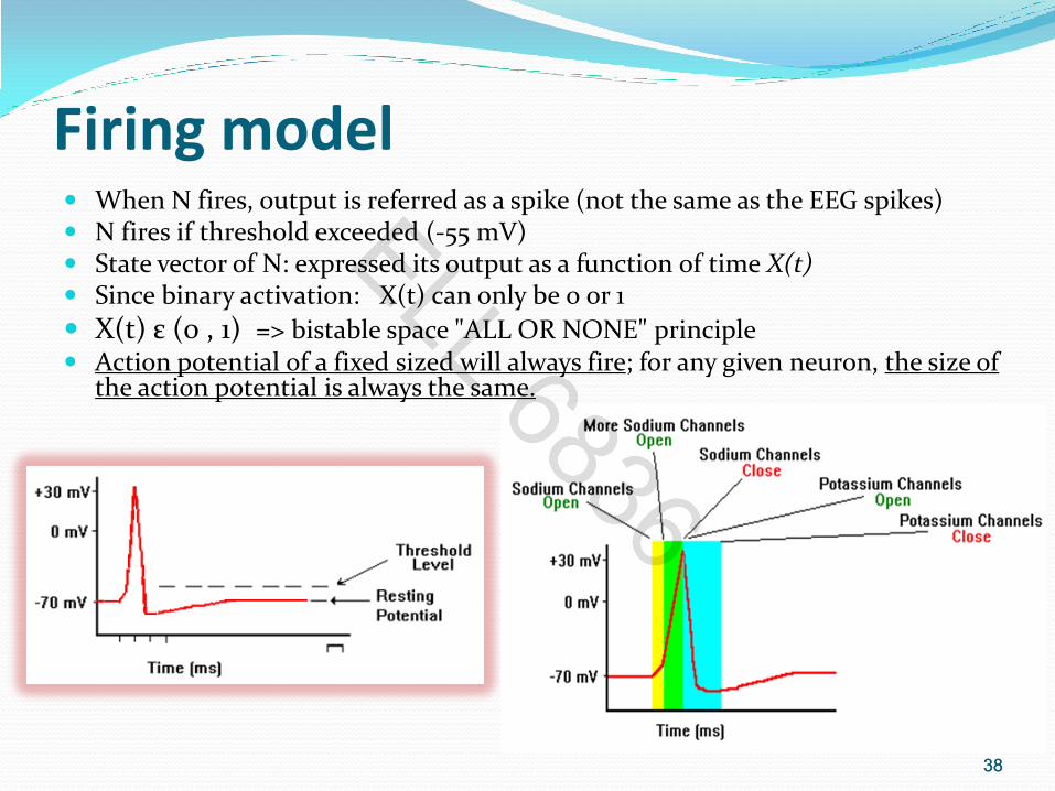
• Action potential is an explosion of electrical
activity that is created by a depolarizing
current.
• A stimulus causes the resting potential to
move toward 0 mV.
• When the depolarization reaches about
-55 mV a neuron will fire an action potential.
• This is the threshold. If the neuron does not
reach this critical threshold level, then no
action potential will fire.
38
Firing model When N fires, output is referred as a spike (not the same as the EEG spikes) N fires if threshold exceeded (-55 mV) State vector of N: expressed its output as a function of time X(t) Since binary activation: X(t) can only be 0 or 1
X(t) ε (0 , 1) => bistable space "ALL OR NONE" principle Action potential of a fixed sized will always fire; for any given neuron, the size of
the action potential is always the same.
38

39
Electrical Potentials & Currents
Neuron pathway is not a continuous “wire” but a series of separate cells.
Neuronal communication is based on mechanisms for producing electrical potentials & currents. electrical potential - difference in
concentration of charged particles between different parts of the cell.
electrical current - flow of charged particles from one point to another within the cell.
Living cells are polarized resting membrane potential is -70 mV
with a relatively negative charge on the inside of nerve cell membranes.
39

40
Neuron Pathway
40
• The Na+ channels have a
mechanism that avoids
"back propagation" of the
AP.
• This period prevents
bidirectional propagation
of the AP, constraining it
to go in only one
direction.
41
Diseases of the Nervous system
Alzheimer’s disease Parkinson’s disease Epilepsy
41

42
Parkinson Disease Progressive loss of motor function beginning
in 50’s or 60’s -- no recovery
degeneration of dopamine-releasing neurons in substantia nigra.
involuntary muscle contractions
facial rigidity, slurred speech, illegible handwriting, etc.
Treatment: drugs and physical therapy
At present, there is no cure for PD, but a variety of medications provide dramatic relief from the symptoms.
42
43
Parkinson Disease
43
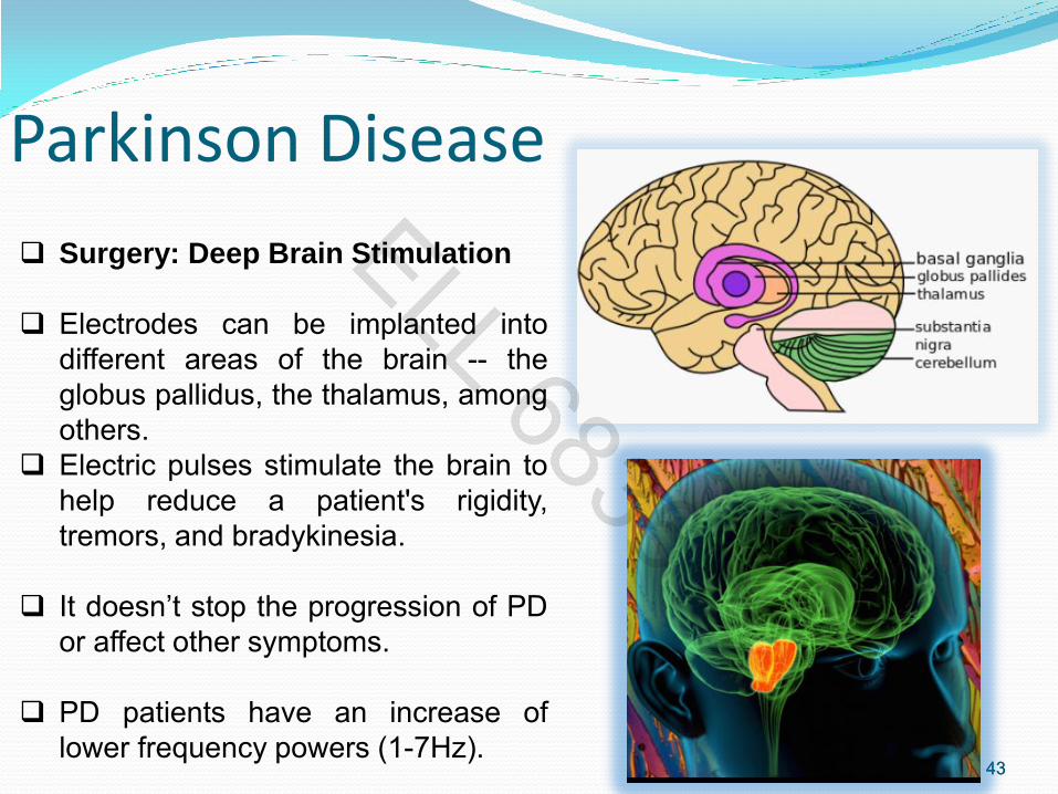
Surgery: Deep Brain Stimulation
Electrodes can be implanted into
different areas of the brain -- the
globus pallidus, the thalamus, among
others.
Electric pulses stimulate the brain to
help reduce a patient's rigidity,
tremors, and bradykinesia.
It doesn’t stop the progression of PD
or affect other symptoms.
PD patients have an increase of
lower frequency powers (1-7Hz).

44
Alzheimer’s Disease
The most common form of dementia among older people is Alzheimer’s
disease (AD), which initially involves the parts of the brain that control
thought, memory, and language.
Although scientists are learning more every day, right now they still do
not know what causes AD, and there is no cure.
The number of people with the disease doubles every 5 years beyond
age 65.
44
EEG of an Alzheimer’s
disease patient usually
shows a decrease in alpha
and beta (8-30Hz) waves
as well as an increase in
delta and theta waves.(1-7
Hz).

45
Alzheimer’s Disease
45
• An early, accurate diagnosis of AD helps
patients and their families plan for the
future.
• It gives them time to discuss care while the
patient can still take part in making
decisions.
• Early diagnosis will also offer the best
chance to treat the symptoms of the
disease.

46
The Brain…
46
• The brain is not a uniform material. It is a mass of
gray and white matter.
• The brain is made up of many different areas, each
having a particular structure and function.
• The brain is protected by several bones.

47
COMPONENTS OF THE BRAIN
Cerebrum: consists of the left and right cerebral hemispheres.
Cerebellum: structure located behind the brain stem.
Brain stem: lowest section of the brain and it is connected to the
spinal cord.
47
48
Cerebrum
48
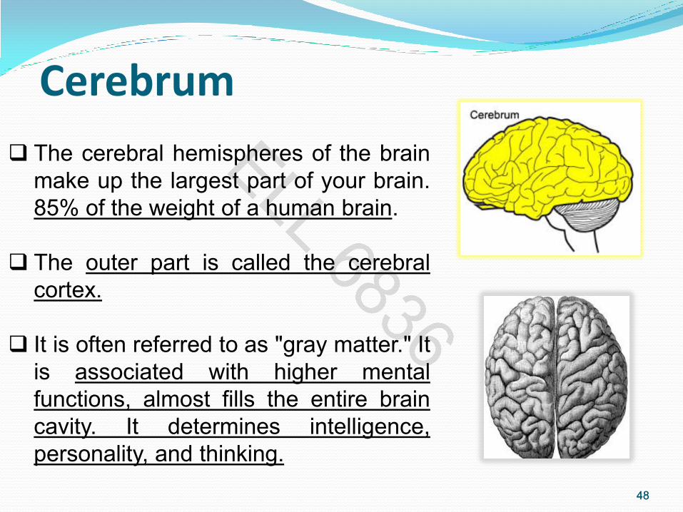
The cerebral hemispheres of the brain
make up the largest part of your brain.
85% of the weight of a human brain.
The outer part is called the cerebral
cortex.
It is often referred to as "gray matter." It
is associated with higher mental
functions, almost fills the entire brain
cavity. It determines intelligence,
personality, and thinking.

49
The Cerebellum
It is about the size of a pear.
The word "cerebellum" comes
from the Latin word for "little
brain.”
It coordinates and control
voluntary movements, balance and coordination.
49

50
Brain Stem
It controls breathing and circulation and connects the cerebrum and cerebellum to the spinal column.
Provides the main motor and sensory functions to the face and neck through the cranial nerves.
It is responsible for automatic survival
functions such as heartbeat, digestion, and keeping your body alive while you sleep.
50

51 51

52
Eloquent Cortex
52
Wernicke's area (central language area):
Difficulty speaking understandably and
comprehending speech; confusion between left
and right; difficulty reading, writing, naming
objects, and calculating.
Broca's area (speech): Difficulty speaking and
sometimes writing.
fMRI Language Areas

53 53
• The outermost layer of the cerebral
hemisphere which is composed of
gray matter. Both hemispheres are
able to analyze sensory data,
perform memory functions, learn
new information, form thoughts and
make decisions.
• The cortical thickness and
intelligence are associated.
• Somatosensory cortex is
thicker in migraine
sufferers.
• Average thickness of the
cerebral cortex:
approximately 2.5 mm
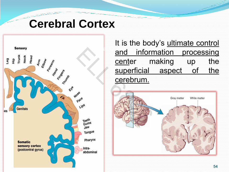
Cerebral Cortex
54 54
It is the body’s ultimate control
and information processing
center making up the
superficial aspect of the
cerebrum.
Cerebral Cortex
Cerebral Cortex

55 55
LEFT
Symbolic thinking
(Language)
Detail
Literal meaning
RIGHT
Spatial perception
Overall picture
Context, metaphor
Hemispheric Specialization
Right Temporal
Lobe - Mainly
involved in visual
memory
(i.e., memory for
pictures and faces).
Left temporal
Lobe - Mainly
involved in verbal
memory
(i.e., memory for
words and
names).
**90% of population is
Right-handed.

56
Hemispheric Specialization Left Hemisphere: Sequential Analysis: systematic, logical interpretation of information. Interpretation and production of symbolic information: language, mathematics, abstraction and reasoning. Memory stored in a language format. Most people are LH dominant for language.
Right Hemisphere: Holistic Functioning: processing multi-sensory input simultaneously to provide "holistic" picture of one's environment. Visual spatial skills. Holistic functions such as dancing and gymnastics are coordinated by the right hemisphere. Memory is stored in auditory, visual and spatial modalities.
56
LH
RH

57
Four Lobes of the Cerebral Cortex
57
Frontal (Forehead to top) Motor Cortex
Parietal (Top to rear) Sensory Cortex
Occipital (Back) Visual Cortex
Temporal (Above ears) Auditory Cortex

58
Brain Regions Functionality Frontal Lobe:
Cognition and memory. Prefrontal area: The ability to concentrate, attend and
elaboration of thoughts. Movement. Motor Cortex : voluntary motor activity. Premotor Cortex: storage of motor patterns and voluntary
activities. Language: motor speech.
Parietal Lobe: Processing of sensory input, sensory discrimination.
Occipital Lobe: Primary visual reception area. Primary visual
association area: Allows for visual interpretation. Temporal Lobe: Auditory receptive area and association
areas. Expressed behavior. Language: Receptive speech. Memory: Information retrieval.
58

59
Brain Functionality (cont.)
The overall goal of the neurosurgeon is to maintain blood flow and oxygen to all parts of the brain, thus minimizing the damage and increasing the prospect of survival and recovery.
If there is damage in any region, the functionality is gone.
59

60
Visual Cortex
Humans are primarily visual creatures.
Brain regions not adjacent to one another are connected by long tracts of cellular projections called axons.
60

61
Enigma of seeing
About 75% of the information
humans receive about our
environment comes from our
sight, making it the most
‘important’ of the five senses.
The vision depends, on the EYES to see and on the BRAIN to make sense of what we see.
The brain receives electrical impulses (stimuli) from our eyes which are interpreted as SIGHT, but the brain adds memory and interpretation. 61
• Sometimes the brain is deceived by
information received from the eyes.
• Visual illusions are caused when
differences occur between our
perceptions or expectations and the
image seen by the eye.

62
Physiological illusions Physiological illusions are the effects on the eyes
or brain of prolonged stimulation of a specific type: brightness, tilt, color, movement. It is a physiological imbalance that alters perception.
The eyes and the brain have an interesting relationship. What we see is transferred to the brain and then the brain tells us exactly what our eyes transmitted is not the real case.
One type of optical illusion is the physiological illusion. This type of illusion includes afterimages. Afterimages are really not "there," but the brain perceives them as being real because the brain was over stimulated.
62

63
Visual illusions
Stimuli have individual dedicated neural paths in the early stages of visual processing and that repetitive stimulation of one or a few channels causes a physiological imbalance that alters perception.
Optical illusions are characterized by visually
perceived images that differ from objective reality.
The information gathered by the eye is
processed in the brain to give a percept that differs from the object being imaged.
EEG responds of the optical illusion (Pz and
Poz) have negative peaks. Latency and amplitude are different from regular VEP.
63

64
Corpus Callosum A deep channel divides the cerebrum into two halves, known as the left and right hemispheres. The two hemispheres look mostly symmetrical yet it has been shown that each side functions slightly different than the other. The corpus callosum is a bundle of axons which connects these two hemispheres.
64
Millions of axons connecting the brain’s hemispheres.
Provides a pathway for communication.
Sometimes used to treat severed epilepsy.
65
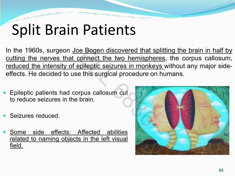
Split Brain Patients
Epileptic patients had corpus callosum cut to reduce seizures in the brain.
Seizures reduced.
Some side effects: Affected abilities related to naming objects in the left visual field.
65
In the 1960s, surgeon Joe Bogen discovered that splitting the brain in half by
cutting the nerves that connect the two hemispheres, the corpus callosum,
reduced the intensity of epileptic seizures in monkeys without any major side-
effects. He decided to use this surgical procedure on humans.

66
Corpus Callosum
66
The corpus callosum helps the hemispheres
share information, but it also contributes to the
spread of seizure impulses from one side of
the brain to the other. A corpus callosotomy is
an operation that cuts the corpus callosum,
interrupting the spread of seizures from
hemisphere to hemisphere.
Seizures generally do not completely stop after
this procedure (they continue on the side of
the brain in which they originate). However, the
seizures usually become less severe, as they
cannot spread to the opposite side of the
brain.

67
Brain Plasticity The ability of the brain to reorganize neural
pathways based on new experiences.
Persistent functional changes in the brain
represent new knowledge.
Age dependent component.
67
The brain’s ability to remain flexible, alert, responsive and
solution-oriented is due to its lifelong capacity for plasticity.
Before, it was thought that only infant brains were
plastic.
The brain is physically modified through strengthening, and
elimination of existing connections, and the growth of new
ones.
68
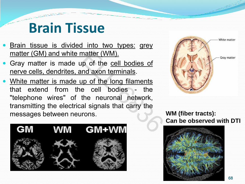
Brain Tissue Brain tissue is divided into two types: grey
matter (GM) and white matter (WM).
Gray matter is made up of the cell bodies of
nerve cells, dendrites, and axon terminals.
White matter is made up of the long filaments
that extend from the cell bodies - the
"telephone wires" of the neuronal network,
transmitting the electrical signals that carry the
messages between neurons.
68
WM (fiber tracts):
Can be observed with DTI

69
TYPES OF VIEWS of the MRI
What is an MRI?
Magnetic resonance imaging is an image of the structure of the brain. Intense magnetic fields and radio waves are used to make images of the inside of the head.
Produces no radiation.
Provides a detail view of the brain in different dimensions.
Safe, painless, and non-invasive.
No special preparation.
69

70
MRI
70
MRI machines look like a
large block with a tube
running through the middle
of the machine, called the
bore of the magnet.
The bore is where the patient is
located for the duration of the
scan.
MCH Philips 1.5 T

71
MRI Parameters
The relationship
between volume, slice
and a voxel in an image.
In general, voxels carry
the same color-intensity
properties as pixels.

72
TYPES OF VIEWS of the MRI T1-weighted: most useful
It provides grey/white/CSF delineation
Acquisition: 5-10 minutes Excellent for demonstrating anatomy
72
MRI Basics:
•Components: Magnet, antenna,
PC, software
•Magnet “ON” and “OFF” to change
directions in the magnetic field
•Water molecules spin around, they
give pulse in a form of Radio waves.
•Antenna will collect data and detect
radio signal when water molecules
spin.
•Pictures out of the radio signal.

73
MRI cont. The MRI machine picks points in the patients head, decides what
type of tissue the points define, then compiles the points into 2 dimensional and 3 dimensional images.
Once the 3 dimensional image is created, the MRI machine
creates a model of the tissue.
73

MRI cont.
74
The tissues with the help of the
magnetic field send a signal to
the computer.
The different signals are sent
and modified into images that the
clinicians can evaluate, and label
as normal or abnormal.

75
What does the image represent?
For every unit volume of tissue, there is a number of cells, these cells contain water molecules, each water molecule contain one oxygen and two hydrogen atoms.
Different tissues thus produce different images based on
the amount of their hydrogen atoms producing a signal
Moving proton induces a signal in the RF antenna(irritates and responds)
Signal is picked up and sent to the computer system
75

MRI Frequency MRI involves the absorption and emission of energy by nuclei(H+) at a
specific resonant (Larmor) frequency.
Larmor frequency scales directly with main magnetic field strength (Bo), and
for clinical MRI lies in the range of tens to hundreds of MHz.
These frequencies are part of the electromagnetic spectrum commonly used
for radio transmission.
1.5 T= 63.9 Mhz
3.0T=127.8 MHz
76

MRI
The MR signal in MRI is
produced by the process of
resonance, which is the
result of radiofrequency
coils.
77
• Two electromagnetic coils: transmitter and receiver coils generate and receive
electromagnetic fields.
• Atomic nuclei of interest in MRI studies have their own resonant frequencies, in
the radiofrequency portion of the electromagnetic spectrum.

78 78
MRI (cont.)
The RF pulses are applied through a coil that is specific to the part of the
body being scanned.
When Radio waves are turned off, protons realign and in doing so, send
out radio signals which are picked up by receivers.
Protons in different tissue types produce different signals.
Signals from millions of protons in the brain can create a detailed image
of the brain tissue.
79
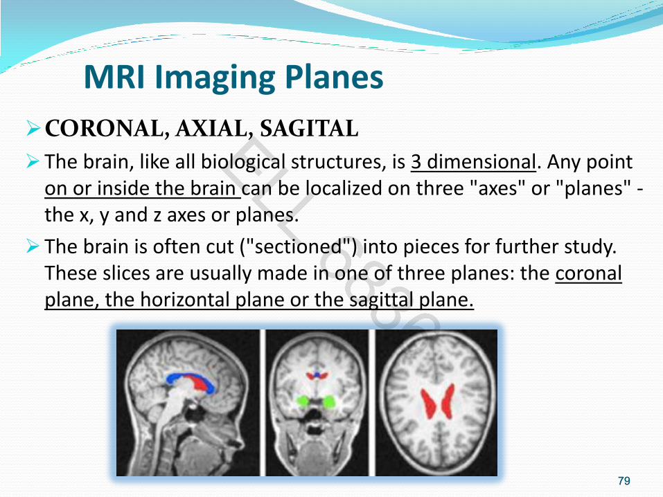
MRI Imaging Planes
CORONAL, AXIAL, SAGITAL
The brain, like all biological structures, is 3 dimensional. Any point on or inside the brain can be localized on three "axes" or "planes" - the x, y and z axes or planes.
The brain is often cut ("sectioned") into pieces for further study. These slices are usually made in one of three planes: the coronal plane, the horizontal plane or the sagittal plane.
79

80
MRI Imaging Planes
When analyzing medical images, please note that sometimes the left side of the picture is the right side of the patient. This does not always apply, but doctors need to be certain before making any assessment.
80

81
Surgical Candidates Selection Criteria
81
Seizures can be well controlled with appropriate medication. However, 20-30% of patients with epilepsy are refractory to all forms of medical therapy.
Another group of patients who might benefit from a surgery are those whose seizures may be relatively well controlled but who have certain characteristic presentations or lesions that strongly suggest surgical intervention might be curative.

82
Surgery Outcome
Overall, the single most important determinant of a successful surgical outcome is patient selection.
This requires detailed pre-surgical evaluation in order to select the most appropriate treatment from a variety of surgical options.
Evaluation includes: seizure type
frequency
site of onset
psychosocial functioning
degree of disability
82

83
Seizure There are many types of seizures and different forms of
epilepsy. A seizure is defined as a change in behavior associated with
excessive electrical discharge from the central nervous system.
83 Scalp EEG containing the transition from interictal to ictal state.

84
Epilepsy
Epilepsy is defined as a condition of recurrent seizures and medical intractability as recurrent seizures despite optimal treatment under the direction of an experienced neurologist over a 2-3 years period.
Epilepsy refers to a pattern of chronic seizures of any type over a long period. 30% of children diagnosed with epilepsy continue to have repeated seizures into adulthood, while others improve over time.
84
85
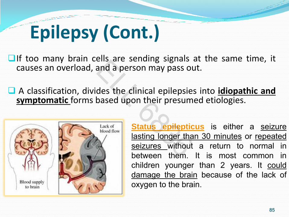
Epilepsy (Cont.) If too many brain cells are sending signals at the same time, it
causes an overload, and a person may pass out.
A classification, divides the clinical epilepsies into idiopathic and symptomatic forms based upon their presumed etiologies.
85
Status epilepticus is either a seizure
lasting longer than 30 minutes or repeated
seizures without a return to normal in
between them. It is most common in
children younger than 2 years. It could
damage the brain because of the lack of
oxygen to the brain.
86
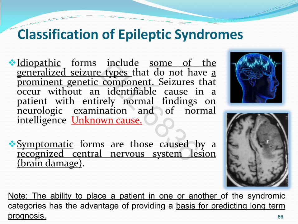
Classification of Epileptic Syndromes
Idiopathic forms include some of the generalized seizure types that do not have a prominent genetic component. Seizures that occur without an identifiable cause in a patient with entirely normal findings on neurologic examination and of normal intelligence Unknown cause.
Symptomatic forms are those caused by a recognized central nervous system lesion (brain damage).
86
Note: The ability to place a patient in one or another of the syndromic
categories has the advantage of providing a basis for predicting long term
prognosis.

87
FACTS For seizures without a known cause (idiopathic seizures) in a
child who is otherwise normal, there is only about a 30-40% chance that the child will ever have another seizure.
The chance for recurrence is higher if the child has an abnormal EEG, a family history of seizures, neurological problems, or other medical problems, such as meningitis.
87
Single seizures do not cause brain damage, so it
is usually safe to wait and see if the child is
going to have more seizures before starting
treatment.
Note: Meningitis is an inflammation of the membranes covering
the brain and spinal cord.
88
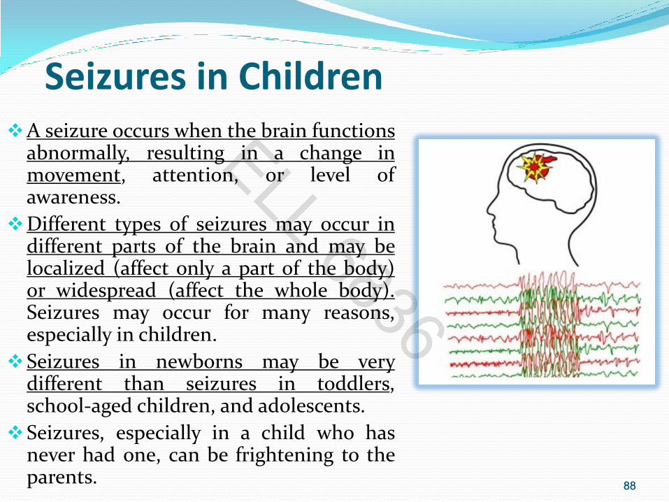
Seizures in Children A seizure occurs when the brain functions
abnormally, resulting in a change in movement, attention, or level of awareness.
Different types of seizures may occur in different parts of the brain and may be localized (affect only a part of the body) or widespread (affect the whole body). Seizures may occur for many reasons, especially in children.
Seizures in newborns may be very different than seizures in toddlers, school-aged children, and adolescents.
Seizures, especially in a child who has never had one, can be frightening to the parents.
88

89
Types of seizures
Neonatal seizures occur within 28 days of birth. Most occur soon after the child is born. They may be due to a large variety of conditions.
Note: It may be difficult to determine if a newborn is actually seizing, because they often do not have convulsions. Instead, their eyes appear to be looking in different directions. They may have lip smacking or periods of no breathing.
89

90
Partial Seizures
90
Partial seizures involve only a part of the
brain and therefore only a part of the body.
Simple partial seizures have a motor
(movement) component that is located in
one portion of the body. Children with
these seizures remain awake and alert.
Complex partial seizures are similar,
except that the child is not aware of what is
going on. Frequently, children with this type
of seizure repeat an activity, such as
clapping, throughout the seizure.
They have no memory of this activity. After
the seizure ends, the child is
often disoriented in a state known as the
postictal period.

91
Types of seizures (cont.)
91
Generalized seizures:
Involve a much larger portion of
the brain.
They are grouped into 2 types:
• convulsive (muscle jerking)
• Non-convulsive with several
subgroups.
Generalized seizure

92
Types of seizures (cont.)
92
Convulsive seizures are noted by
uncontrollable muscle jerking lasting for a
few minutes-usually less than 5 followed
by a period of drowsiness that is called
the post-ictal period.
-Tonic seizures result in continuous
muscle contraction in the arms and legs.
-Absence seizures are short episodes
during which the child stares or eye
blinks, with no apparent awareness of
their surroundings.
These episodes usually do not last longer
than a few seconds and start and stop
abruptly.

93
Types of Epilepsy Temporal lobe epilepsy, is the most common type of
epilepsy in teens and adults, the area where the seizures start (called the seizure focus) is located within the temporal lobe.
Extratemporal means the tissue is located in an area
of the brain other than the temporal lobe. The frontal lobe is the most common extratemporal site for seizures. In some cases, tissue may be removed from more than one area/lobe of the brain.
93
Cortical resection is an operation to resect, or
cut away, brain tissue that contains a seizure
focus.

94
Major surgical Questions The goal of epilepsy surgery is to identify an abnormal area of cortex from which the seizures originate and remove it without causing any significant functional impairment. The primary components of the pre-surgical evaluation includes: -detailed clinical history and physical examination, -advanced neuro-imaging, -video-EEG monitoring, -neuropsychological testing and assessment of psychosocial functioning.
The major surgical questions one hopes to answer with this evaluation are:
1) Are the seizures focal or generalized? 2) If focal, are they temporal or extra-temporal in origin? 3) Is there a lesion associated with the seizures? 4) If surgery is undertaken what functional deficits, if any, might be anticipated?
94

95
EEG and Epilepsy
Electroencephalographic (EEG) investigation remains the most important aspect of the pre-surgical evaluation.
Analysis of selected EEG activity between events (interictal) or of specific activity during events (ictal) can provide evidence of focal electrical dysfunction.
95

EEG and Epilepsy
Certain interictal EEG abnormalities (spike and slow wave complexes) can be of localizing value, it is considered extremely important to record the EEG with concomitant videotape during the spontaneous occurrence of the patient's events.
Video/EEG monitoring can continuously record the EEG over a 24 hour period which allows for careful inspection of the record during any event.
96

97
Structural MRI reveals
brain anatomy. Functional MRI (fMRI)
reveals brain function.
Structural MRI vs. Functional MRI
97

98
MRI vs. fMRI
MRI fMRI
Only one image collected
(one full head volume)
Series of several images
collected over time
(e.g., 1 full head volume
every 2 seconds over the
course of several minutes)
High Resolution
(<1 mm3)
Lower Resolution
(~3 mm3)
…
98

99 99

100 100

101
Seizure Threshold
101
Seizure threshold: Minimum stimulation required to trigger a seizure.
Stimulation could be:
• Electrical or magnetic stimulation
• Chemical compound that stimulates the brain
• External stimulus such as: noise and flashing lights
Drugs will raise the seizure threshold, so the brain will be less excitable.