How phloem-feeding insects face the challenge of phloem ...
12
REVIEW ARTICLE published: 29 August 2013 doi: 10.3389/fpls.2013.00336 How phloem-feeding insects face the challenge of phloem-located defenses Torsten Will 1 * † , Alexandra C. U. Furch 1† and Matthias R. Zimmermann 2 1 Institute of Phytopathology andApplied Zoology, Centre for BioSystems, Land Use and Nutrition, Justus-Liebig-University, Giessen, Germany 2 Institute of General Botany, Justus-Liebig-University, Giessen, Germany Edited by: Gary A. Thompson, Pennsylvania State University, USA Reviewed by: Sylvie Dinant, Institut National de la Recherche Agronomique, France Jyoti Shah, University of NorthTexas, USA Tom Wilkinson, University College Dublin, Ireland *Correspondence: Torsten Will, Institute of Phytopathology and Applied Zoology, Centre for BioSystems, Land Use and Nutrition, Justus-Liebig-University, Heinrich-Buff-Ring 26-32, D-35392 Giessen, Germany e-mail: torsten.will@agrar. uni-giessen.de † Torsten Will and Alexandra C. U. Furch have contributed equally to the article. Due to the high content of nutrient, sieve tubes are a primary target for pests, e.g., most phytophagous hemipteran. To protect the integrity of the sieve tubes as well as their content, plants possess diverse chemical and physical defense mechanisms. The latter mechanisms are important because they can potentially interfere with the food source accession of phloem-feeding insects. Physical defense mechanisms are based on callose as well as on proteins and often plug the sieve tube. Insects that feed from sieve tubes are potentially able to overwhelm these defense mechanisms using their saliva. Gel saliva forms a sheath in the apoplast around the stylet and is suggested to seal the stylet penetration site in the cell plasma membrane. In addition, watery saliva is secreted into penetrated cells including sieve elements; the presence of specific enzymes/effectors in this saliva is thought to interfere with plant defense responses. Here we detail several aspects of plant defense and discuss the interaction of plants and phloem-feeding insects. Recent agro-biotechnological phloem-located aphid control strategies are presented. Keywords: aphid-plant interactions, aphid saliva, P-proteins, plant defense, phloem, sieve element occlusion A key characteristic of higher-level plants (including monocots and dicots) is the existence of a vascular network that is composed of the phloem and xylem. The vascular system pervades the whole organism from root to shoot and distributes nutrients and water. The fact that sugars, amino acids, and other organic metabolites are available via phloem and xylem in significant amounts makes the vascular system a target for insect pests (Brodbeck et al., 1993; Gündüz and Douglas, 2009). Endogenous interference in the func- tioning of the vascular system may have disastrous consequences for a plant’s development. Using the interaction between plants and phloem-feeding insects as an example, offensive and defense strategies during the struggle for the valuable phloem content are explored. THE PHLOEM: A HIGH-DENSITY ENERGY PATHWAY In higher-level plants, a long-distance transport system has evolved to translocate photoassimilates from a source (e.g., mature leaves) to a sink (e.g., roots) – the phloem (Schulz, 1998). The angiosperm phloem is composed of sieve elements (SEs), companion cells (CCs), and phloem parenchyma cells (PPCs; van Bel, 1996; Hafke et al., 2005). Mature SEs are elongate cells lacking certain cellular components (nucleus, vacuoles, ribosomes, and dictyosomes) but lined parietally with a thin mictoplasmic layer consisting of an endoplasmic reticulum (ER), plastids, a few inactive mitochon- dria and phloem-specific proteins (P-proteins; van Bel, 2003). The CCs maintain SEs viability (van Bel, 2003). A high density of pore-plasmodesma units (PPUs) and tight ER coupling between SE and CC underline an intimate symplastic connection across this boundary; the entire connection constitutes the SE-CC complex (Kempers et al., 1998; Martens et al., 2006). The walls between the single SE-modules are transformed into sieve plates, perforated by plasmodesmata (PD) modified into sieve pores with a diameter up to 2.5 μm(Behnke and Sjolund, 1990; Schulz, 1998; van Bel, 2003; Mullendore et al., 2010). These adaptations provide the basis of formation of long sieve tubes based upon single SE-modules, forming a tube-like symplastic continuum that serves transloca- tion. The complex process of this translocation is regulated via highly active CCs (van Bel and Knoblauch, 2000). In general, the vascular system is a pressure system made up of two components (phloem and xylem) that effects the long- distance translocation of very heterogeneous constituents within higher-level plants. Xylem and phloem are parallel orientated vas- cular tissues in which pressure and tension gradients are built up in SEs and xylem vessels, respectively. In intact plants, the nega- tive hydrostatic potential in xylem vessels is in balance with that inside SEs (e.g., Zimmermann et al., 2013). The driving forces for translocation are, on the one hand, a longitudinal (axial) pressure gradient within phloem and xylem, and on the other, a lateral (radial) pressure gradient between the phloem and xylem (Will and van Bel, 2006). The balanced interaction between phloem and xylem is a basic requirement for long-distance transport (van Bel, 2003; Dinant and Lemoine, 2010). The longitudinal pres- sure gradient within the xylem is the result of water uptake in the root/rhizosphere and loss of water by transpiration. Accord- ing to the classic Münch concept (Münch, 1930) for phloem translocation, photoassimilates are amassed in the sieve tubes of www.frontiersin.org August 2013 | Volume 4 | Article 336 | 1
Transcript of How phloem-feeding insects face the challenge of phloem ...

“fpls-04-00336” — 2013/8/28 — 20:48 — page 1 — #1
REVIEW ARTICLEpublished: 29 August 2013
doi: 10.3389/fpls.2013.00336
How phloem-feeding insects face the challenge ofphloem-located defensesTorsten Will 1* †, Alexandra C. U. Furch 1† and Matthias R. Zimmermann 2
1 Institute of Phytopathology and Applied Zoology, Centre for BioSystems, Land Use and Nutrition, Justus-Liebig-University, Giessen, Germany2 Institute of General Botany, Justus-Liebig-University, Giessen, Germany
Edited by:
Gary A. Thompson, PennsylvaniaState University, USA
Reviewed by:
Sylvie Dinant, Institut Nationalde la Recherche Agronomique, FranceJyoti Shah, University of North Texas,USATom Wilkinson, University CollegeDublin, Ireland
*Correspondence:
Torsten Will, Institute ofPhytopathology and Applied Zoology,Centre for BioSystems, Land Use andNutrition, Justus-Liebig-University,Heinrich-Buff-Ring 26-32, D-35392Giessen, Germanye-mail: [email protected]
†Torsten Will and Alexandra C. U.Furch have contributed equally to thearticle.
Due to the high content of nutrient, sieve tubes are a primary target for pests, e.g., mostphytophagous hemipteran. To protect the integrity of the sieve tubes as well as theircontent, plants possess diverse chemical and physical defense mechanisms. The lattermechanisms are important because they can potentially interfere with the food sourceaccession of phloem-feeding insects. Physical defense mechanisms are based on calloseas well as on proteins and often plug the sieve tube. Insects that feed from sieve tubes arepotentially able to overwhelm these defense mechanisms using their saliva. Gel salivaforms a sheath in the apoplast around the stylet and is suggested to seal the styletpenetration site in the cell plasma membrane. In addition, watery saliva is secreted intopenetrated cells including sieve elements; the presence of specific enzymes/effectors inthis saliva is thought to interfere with plant defense responses. Here we detail severalaspects of plant defense and discuss the interaction of plants and phloem-feeding insects.Recent agro-biotechnological phloem-located aphid control strategies are presented.
Keywords: aphid-plant interactions, aphid saliva, P-proteins, plant defense, phloem, sieve element occlusion
A key characteristic of higher-level plants (including monocotsand dicots) is the existence of a vascular network that is composedof the phloem and xylem. The vascular system pervades the wholeorganism from root to shoot and distributes nutrients and water.The fact that sugars, amino acids, and other organic metabolitesare available via phloem and xylem in significant amounts makesthe vascular system a target for insect pests (Brodbeck et al., 1993;Gündüz and Douglas, 2009). Endogenous interference in the func-tioning of the vascular system may have disastrous consequencesfor a plant’s development. Using the interaction between plantsand phloem-feeding insects as an example, offensive and defensestrategies during the struggle for the valuable phloem content areexplored.
THE PHLOEM: A HIGH-DENSITY ENERGY PATHWAYIn higher-level plants, a long-distance transport system has evolvedto translocate photoassimilates from a source (e.g., mature leaves)to a sink (e.g., roots) – the phloem (Schulz, 1998). The angiospermphloem is composed of sieve elements (SEs), companion cells(CCs), and phloem parenchyma cells (PPCs; van Bel, 1996; Hafkeet al., 2005). Mature SEs are elongate cells lacking certain cellularcomponents (nucleus, vacuoles, ribosomes, and dictyosomes) butlined parietally with a thin mictoplasmic layer consisting of anendoplasmic reticulum (ER), plastids, a few inactive mitochon-dria and phloem-specific proteins (P-proteins; van Bel, 2003). TheCCs maintain SEs viability (van Bel, 2003). A high density ofpore-plasmodesma units (PPUs) and tight ER coupling betweenSE and CC underline an intimate symplastic connection across this
boundary; the entire connection constitutes the SE-CC complex(Kempers et al., 1998; Martens et al., 2006). The walls between thesingle SE-modules are transformed into sieve plates, perforated byplasmodesmata (PD) modified into sieve pores with a diameterup to 2.5 μm (Behnke and Sjolund, 1990; Schulz, 1998; van Bel,2003; Mullendore et al., 2010). These adaptations provide the basisof formation of long sieve tubes based upon single SE-modules,forming a tube-like symplastic continuum that serves transloca-tion. The complex process of this translocation is regulated viahighly active CCs (van Bel and Knoblauch, 2000).
In general, the vascular system is a pressure system made upof two components (phloem and xylem) that effects the long-distance translocation of very heterogeneous constituents withinhigher-level plants. Xylem and phloem are parallel orientated vas-cular tissues in which pressure and tension gradients are built upin SEs and xylem vessels, respectively. In intact plants, the nega-tive hydrostatic potential in xylem vessels is in balance with thatinside SEs (e.g., Zimmermann et al., 2013). The driving forces fortranslocation are, on the one hand, a longitudinal (axial) pressuregradient within phloem and xylem, and on the other, a lateral(radial) pressure gradient between the phloem and xylem (Willand van Bel, 2006). The balanced interaction between phloemand xylem is a basic requirement for long-distance transport (vanBel, 2003; Dinant and Lemoine, 2010). The longitudinal pres-sure gradient within the xylem is the result of water uptake inthe root/rhizosphere and loss of water by transpiration. Accord-ing to the classic Münch concept (Münch, 1930) for phloemtranslocation, photoassimilates are amassed in the sieve tubes of
www.frontiersin.org August 2013 | Volume 4 | Article 336 | 1
“fpls-04-00336” — 2013/8/28 — 20:48 — page 2 — #2
Will et al. Phloem-feeding insects face phloem-located defense
source areas and escape from the sieve tubes in sink areas. Theresulting turgor difference between source and sink drives the massflow. Hence, in contrast to the xylem, the phloem exhibits a bidi-rectional translocation, as sink regions are found in the root and inthe apex of the shoot (van Bel, 2003; Dinant and Lemoine, 2010).
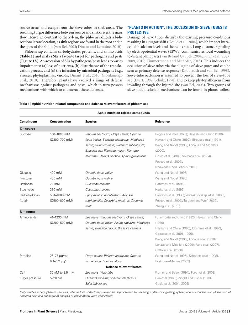
Phloem sap contains carbohydrates, proteins, and amino acids(Table 1) and makes SEs a favorite target for pathogens and pests(Figure 1A). An accession of SEs by pathogens/pests leads to variesimpairments: (a) loss of nutrients, (b) disturbance of the translo-cation process, and (c) the infection by microbial pathogens (e.g.,viruses, phytoplasmas, viroids; Dinant et al., 2010; Giordanengoet al., 2010). Therefore, plants have evolved a range of defensemechanisms against pathogens and pests, which in turn possessmechanisms with which to counteract these defenses.
“PLANTS IN ACTION”: THE OCCLUSION OF SIEVE TUBES ISPROTECTIVEDamage of sieve tubes disturbs the existing pressure conditionsresulting in a turgor shift (Gould et al., 2004), which impact intra-cellular calcium levels and the redox state. Long-distance signalingby electropotential waves (EPWs) communicates local woundingto distant plant parts (van Bel and Gaupels, 2004; Furch et al., 2007,2009, 2010; Zimmermann and Mithöfer, 2013). This induces theocclusion of sieve tubes via the plugging of sieve pores and can beseen as primary defense response (Knoblauch and van Bel, 1998).Sieve-tube occlusion is assumed to prevent the loss of sieve-tubesap (Evert, 1982; Schulz, 1998) and to keep phytopathogens frominvading through the injured site (van Bel, 2003). Two groups ofsieve-tube occlusion mechanisms can be found in plants: callose
Table 1 | Aphid nutrition-related compounds and defense relevant factors of phloem sap.
Aphid nutrition-related compounds
Constituent Concentration Species Reference
C – source
Sucrose 100–1800 mM
(Ø300–700 mM)
Triticum aestivum; Orzya sativa; Opuntia
ficus-indica; Sonchus oleraceus; Medicago
sativa; Salix viminalis; Solanum tuberosum;
Brassica sp.; Plantago major ; Plantago
maritime; Prunus persica; Apium graveolens
Rogers and Peel (1975); Hayashi and Chino (1986)
Hayashi and Chino (1990); Girousse et al. (1991),
Wang and Nobel (1995); Lohaus and Moellers
(2000),
Gould et al. (2004); Shimada et al. (2004),
Pescod et al. (2007),
Nadwodnik and Lohaus (2008)
Glucose 400 mM Opuntia ficus-indica Wang and Nobel (1995)
Fructose 400 mM Opuntia ficus-indica Wang and Nobel (1995)
Raffinose 70 mM Cucurbita maxima Haritatos et al. (1996)
Stachyose 330 mM Cucurbita maxima Haritatos et al. (1996)
Carbohydrates
(total)
534–1800 mM
(Ø500–800 mM)
Lycopersicon esculentum; Alonsoa
meridionalis; Cucurbita maxima; Cucumis
melo
Haritatos et al. (1996); Voitsekhovskaja et al. (2006),
Pescod et al. (2007); Turgeon and Wolf (2009),
Zhang et al. (2010)
N – source
Amino acids 41–1230 mM
(Ø200–500 mM)
Zea mays; Triticum aestivum; Orzya sativa;
Opuntia ficus-indica; Pisum sativum; Medicago
sativa; Brassica napus; Brassica carinata
Fukumorita and Chino (1982); Hayashi and Chino
(1986)
Hayashi and Chino (1990); Ohshima et al. (1990),
Girousse et al. (1991, 1996),
Wang and Nobel (1995); Lohaus et al. (1998),
Lohaus and Moellers (2000); Faria et al. (2007),
Gattolin et al. (2008)
Proteins 76–77 μg/ml;
0.1–0.2 μg/μl
Orzya sativa; Triticum aestivum; Opuntia
ficus-indica; Lupinus albus
Wang and Nobel (1995), Schobert et al. (1998),
Rodriguez-Medina (2009)
Defense relevant factors
Ca2+ 35 nM to 2.5 mM Zea mays; Vicia faba Fromm and Bauer (1994); Furch et al. (2009)
Turgor pressure 5–20 bar Quercus rubrum; Sonchus oleraceus;
Salix babylonica
Hammel (1968); Wright and Fisher (1980),
Gould et al. (2004, 2005)
Only studies where phloem sap was collected via stylectomy (sieve-tube sap obtained by severing stylets of ingesting aphids) and microdissection (dissection ofselected cells and subsequent analysis of cell content) were considered.
Frontiers in Plant Science | Plant Physiology August 2013 | Volume 4 | Article 336 | 2
“fpls-04-00336” — 2013/8/28 — 20:48 — page 3 — #3
Will et al. Phloem-feeding insects face phloem-located defense
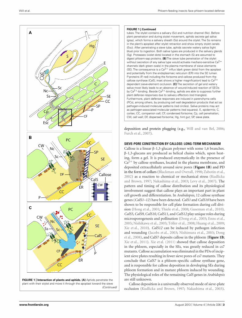
FIGURE 1 | Interaction of plants and aphids. (A) Aphids penetrate theplant with their stylet and move it through the apoplast toward the sieve
(Continued)
FIGURE 1 | Continued
tubes. The stylet contains a salivary (Sc) and nutrition channel (Nc). Beforeplant penetration and during stylet movement, aphids secrete gel saliva(gray), which forms a salivary sheath (Ss) around the stylet. The Ss remainsin the plant’s apoplast after stylet retraction and show empty stylet canals(Esc). After penetrating a sieve tube, aphids secrete watery saliva (lightblue) prior to ingestion. Both saliva types are produced in the salivary glands(Sg). Proteases (violet dots) located in the stomach (S) are assumed todigest phloem-sap proteins. (B) The sieve tube penetration of the styletwithout secretion of any saliva type would activate mechano-sensitive Ca2+channels (dark green ovals) in the plasma membrane of sieve elements(SE). The consequence is a Ca2+ influx (dark green dots) from the apoplastand potentially from the endoplasmaic reticulum (ER) into the SE lumen.P-proteins (P, red) including the forisome and callose produced from thecallose synthase (CalS; inset shows a higher magnification) lead to Ca2+dependent sieve-element occlusion. (C) The secretion of gel and waterysaliva most likely leads to an absence of wound-induced reaction of SEOsby Ca2+-binding. Beside Ca2+-binding, aphids are able to suppress furtherplant defense responses due to salivary effectors (red triangles).Furthermore, plant defense responses are induced in parenchyma cells(PCs), among others, by producing cell wall degradation products that act aspathogen-induced molecular patterns (red circles). Saliva proteins may actas pathogen-associated molecular patterns (red squares). E, epidermis; C,cortex; CC, companion cell; CF, condensed forisome; Cp, cell penetration;CW, cell wall; DF, dispersed forisome; Hg, hint gut; SP, sieve plate.
deposition and protein plugging (e.g., Will and van Bel, 2006;Furch et al., 2007).
SIEVE-PORE CONSTRICTION BY CALLOSE: LONG-TERM MECHANISMCallose is a linear β-1,3-glucan polymer with some 1,6 branches.β-1,3-glucans are produced as helical chains which, upon heat-ing, form a gel. It is produced enzymatically in the presence ofCa2+ by callose synthases, located in the plasma membrane, anddeposited extracellularly around sieve pores (Figure 1B) and PDin the form of collars (Blackman and Overall, 1998; Zabotin et al.,2002) as a reaction to chemical or mechanical stress (Kudlickaand Brown, 1997; Nakashima et al., 2003; Levy et al., 2007). Thepattern and timing of callose distribution and its physiologicalinvolvement suggest that callose plays an important part in plantcell growth and differentiation. In Arabidopsis, 12 callose synthasegenes (CalS1–12) have been detected. CalS1 and CalS10 have beenshown to be responsible for cell plate formation during cell divi-sion (Hong et al., 2001; Thiele et al., 2008; Guseman et al., 2010).CalS5, CalS9, CalS10, CalS11, and CalS12 play unique roles duringmicrosporogenesis and pollination (Dong et al., 2005; Enns et al.,2005; Nishikawa et al., 2005; Töller et al., 2008; Huang et al., 2009;Xie et al., 2010). CalS12 can be induced by pathogen infectionand wounding (Jacobs et al., 2003; Nishimura et al., 2003; Donget al., 2008), and CalS7 deposits callose in the phloem (Figure 1B;Xie et al., 2011). Xie et al. (2011) showed that callose depositionin the phloem, especially in the SEs, was greatly reduced in cs7mutants. Callose accumulation was eliminated in the PDs of incip-ient sieve plates resulting in fewer sieve pores of cs7 mutants. Theyconclude that CalS7 is a phloem-specific callose synthase gene,and is responsible for callose deposition in developing SEs duringphloem formation and in mature phloem induced by wounding.The physiological roles of the remaining CalS genes in Arabidopsisare still unknown.
Callose deposition is a universally observed mode of sieve-plateocclusion (Kudlicka and Brown, 1997; Nakashima et al., 2003).
www.frontiersin.org August 2013 | Volume 4 | Article 336 | 3

“fpls-04-00336” — 2013/8/28 — 20:48 — page 4 — #4
Will et al. Phloem-feeding insects face phloem-located defense
Reversible callose accumulation apparently plays a role inregulating cell-to-cell transport through sieve pores and PPUs(Furch et al., 2007). After a heat stimulus is applied to the leaftip of Vicia faba plants, callose gradually builds up at sieve platesand PD. After reaching a maximum, callose is degraded at a lowerrate than production. Callose appears to degrade more rapidly atPD (30–40 min) than at the sieve pores, where the level of callosedeposition reaches its original state after 1–2 h (Furch et al., 2007).β-1,3-glucan endo-hydrolases is the enzyme that catalyses callosedegradation (reviewed in Leubner-Metzger, 2003). It is present inlarge gene families in plants (e.g., 50 genes in Arabidopsis) andis located in the cell membrane and highly enriched at PD sites(Levy et al., 2007). The course of callose production/degradationis qualitatively similar between different plant species, but there arevariations in the time scale (Furch et al., 2007, 2008; Mullendoreet al., 2010).
SIEVE-TUBE OCCLUSION BY PROTEINS: FAST AND VERSATILEIn addition to callose, sieve pores can be blocked rapidly by pro-teins. In electron microscopic images, SEs show protein networksthat span the SE lumen and are attached to the cell periphery(Sjölund, 1997). Sieve tubes of grasses appear virtually emptybut may have an occlusion mechanism based on precipitationof soluble proteins (Will and van Bel, 2006). A specific groupof phloem proteins (P-proteins) enables rapid occlusion (withinsome seconds) to occur in sieve tubes of higher-level plants.Numerous aggregation forms (amorphous, granular, fibrillar, fil-amentous, tubular, or crystalline) of P-proteins that are thoughtto represent stages of P-protein differentiation (Cronshaw, 1981)denote an immense variation between plant species (Cronshawand Sabnis, 1990). The synthesis of P-proteins begins in immature,nucleate SEs, resulting in electron-dense proteinaceous structures(Ernst et al., 2012). In young SEs, subunits accumulate within thecytoplasm, forming large P-protein bodies (Steer and Newcomb,1969). As SEs mature the P-protein bodies disperse into smalleraggregates that move to the periphery of the cell (Knoblauch andvan Bel, 1998).
In cucurbits, phloem protein 1 (PP1) and phloem protein2 (PP2) produce insoluble aggregates in response to oxidation(Kleinig, 1975; Alosi et al., 1988) by cross-linking, forming high-molecular-weight polymers that plug the sieve pores of injuredsieve tubes (Read and Northcote, 1983). PP1 monomeric subunitshave a predicted molecular mass of 95.4 kDa, but the appar-ent molecular size is dependent on the pH and oxidation state,as conformational isoforms exist that appear to be related toeither the polymerized or unpolymerized, translocated forms ofthe protein (Clark et al., 1997; Leineweber et al., 2000). PP1 wasimmunolocalized in SE slime plugs and P-protein bodies, whereasthe corresponding mRNA was shown to accumulate in CCs (Clarket al., 1997). Due to an interaction of PP1 and PP2 in presenceof calcium and oxygen, sieve tubes and cut surfaces are rapidlyoccluded by gelling of the exudate (Kleinig, 1975; Clark et al.,1997; Furch et al., 2010). PP2-like proteins are lectins, sugar bind-ing proteins, which have been identified in many angiosperms andare specifically expressed in SE/CC complexes, suggesting that PP2may be a common component of P-proteins (Dinant et al., 2003).It has been shown to interact with phloem sap proteins, potentially
playing a role in the shuttling of glycoproteins between CC and SE(Beneteau et al., 2010). The phloem-specific PP2 homolog fromArabidopsis was shown to be anchored to P-proteins and otherphloem organelles rather than being a structural component ofP-proteins (Batailler et al., 2012). These findings indicate that PP2does not represent an essential part of the occlusion machinery. Aninsecticidal function for PP2 is described in vitro (Beneteau et al.,2010) as well as in vivo (Zhang et al., 2011). Hilder et al. (1995)observed that snowdrop lectin from Galanthus nivalis, artificiallyapplied or expressed in Nicotiana tabacum plants, reduced growth,decreased survival, and lowered reproduction in the aphid speciesMyzus persicae. Other lectins that showed insecticidal effectsagainst aphids are a mannose-binding lectin (Sauvion et al., 1996),a garlic lectin (Fitches et al., 2008), and concanavalin A (Sauvionet al., 2004). Similar effects were observed for protease inhibitors(PIs) applied to aphids via transgenic plants (Rahbé et al., 2003;Ribeiro et al., 2006; Carrillo et al., 2011). Erickson et al. (1985)observed that lectins negatively affect the activity of an aminopep-tidase in rats. The identified aminopeptidase inside the aphid gut,which represents 15.6% of total gut proteins, is suggested to be apotential binding site for lectins (Cristofoletti et al., 2006).
Sieve elements of Fabaceae contain elongate protein bodiescalled forisomes (Knoblauch et al., 2003). Forisomes consist offibrils (Tuteja et al., 2010) and were previously classified as “non-dispersive P-protein bodies” (Behnke, 1991). They were suspectedto undergo structural transformations, from a crystalloid statewith co-aligned fibrils to a “slime-body” with dispersed fibrils(Palevitz and Newcomb, 1971). The transition is a rapid andreversible conformational change in which forisomes shortenlongitudinally while expanding radially with a several-fold vol-ume increase (Knoblauch et al., 2001; Peters et al., 2007, 2008).Forisomes disperse upon wounding and occlude sieve tubes(Knoblauch and van Bel, 1998), leading to a stop of mass flowobserved in artificial sieve tubes (Knoblauch et al., 2012). Fur-thermore, Thorpe et al. (2010) showed a cooling rate dependenttransport interruption and parallel forisome dispersion in intactV. faba plants. Dispersion is triggered by an increase of free cal-cium (Ca2+) in sieve tubes (Knoblauch et al., 2001), although asof yet, no Ca2+-binding sites have been detected in forisomes.As observed in vitro (Knoblauch et al., 2005), high Ca2+ con-centration (>50 μM) is also needed to disperse forisome invivo (Furch et al., 2009). After burning the tip of a V. faba leaf,the elevation of Ca2+ concentration in most regions of sievetubes inside the respective leaf was demonstrated to be belowthe threshold that is necessary for forisome dispersion (Furchet al., 2009). Only in the close vicinity of the Ca2+-channelpore Ca2+-level goes beyond the threshold and increases up to100 μM (Figure 1B; Trewavas, 1999). Therefore, it was con-cluded that forisomes are directly associated with Ca2+ releasesites (Furch et al., 2009). An association between forisomes andthe ER (where the highest frequencies of Ca2+ channels wereobserved) was found. It was observed that the more intimatelyforisomes were associated with the ER or the plasma mem-brane of SEs, the greater was the probability of dispersion(Furch et al., 2009).
Scanning electron microscopic images show that forisomes arecomposed of largely identical subunits named forisomettes (Tuteja
Frontiers in Plant Science | Plant Physiology August 2013 | Volume 4 | Article 336 | 4

“fpls-04-00336” — 2013/8/28 — 20:48 — page 5 — #5
Will et al. Phloem-feeding insects face phloem-located defense
et al., 2010). Indicated by transmission electron microscopystudies (Ehlers et al., 2000), forisomettes consist of strictly orderedarrays of a number of forisome proteins. At least three proteins areinvolved in formation of forisomettes, called sieve element occlu-sion 1 (SEO1), SEO2, and SEO3, and were identified in Medicagotruncatula (Noll, 2005; Noll et al., 2007). SEOs are also presentin plant families that do not possess forisomes, e.g., Rosaceae,Solanaceae, and Brassicaceae (Rüping et al., 2010). Two Arabidop-sis thaliana genes (At3g01670 and At3g01680) encode SEO proteinsassigned AtSEOR1 and AtSEOR2 (Pélissier et al., 2008; Rüpinget al., 2010; Froelich et al., 2011). Both phloem filament pro-teins are required for formation of filaments that are arrangedas complex network inside SEs (Anstead et al., 2012). Whetherthe formation of dense SEO filament deposits at sieve plates stopsphloem mass flow (Figure 1B; Ernst et al., 2012) or mass flowremains intact (Froelich et al., 2011) is a matter of debate. Aphidsof the species Myzus persicae that feed on AtSEOR1 and AtSEOR2mutants without SEO filament formation show no benefit fromthe absence of filaments. Thus, Anstead et al. (2012) conclude thatSEOs are not involved in plant defense against phloem-feedinginsects. In fact, aphids perform worse when compared to aphids oncontrol plants, indicated by reduced reproduction and shortenedreproduction period. The authors suggest that reduced fitnesscould be associated with lower nitrogen supply due to reducedprotein content, but no data about amino acid concentration orprotein content in the phloem sap of these plants are available.A further explanation could be that the absence of SEO filamentsinfluences parameters in sieve tubes relevant for aphid feeding, e.g.,turgor pressure (Miles, 1999), reducing nutrition supply and lead-ing to the observed reduction of aphid reproduction. Although,Anstead et al. (2012) describe that the phenotype SEO mutantsdoes not differ to the wildtype this allows no conclusion about thestate of sieve tubes.
Callose deposition and protein plugging operate in parallel. Aburning stimulus elicits distant occlusion in V. faba with rapidforisome dispersion and a slower subsequent callose deposition(Furch et al., 2007, 2009). While forisomes reconstitute into thecondensed state, constriction of sieve pores by callose depositionreaches its maximum level (Furch et al., 2007). It is suggested,therefore, that plants possess a universal safety design for sieve-tube occlusion, one that proceeds rapidly and involves P-proteinand a slower and more long-lasting one based on callose (Furchet al., 2007).
The distant-induced occlusion was associated with the pas-sage of a damage-induced EPW. EPWs communicate sudden andprofound physiological changes over long-distances (Furch et al.,2007; Hafke et al., 2009). EPWs trigger a release of Ca2+ thatresults in callose deposition and protein plugging (Kauss, 1987;Colombani et al., 2004). Ca2+ originates from the apoplast viaopened plasma membrane channels or from the ER acting as anintracellular Ca2+ storage (Furch et al., 2009; Hafke et al., 2009;Zimmermann and Mithöfer, 2013).
“INSECTS IN ACTION”: HOW PHLOEM-FEEDING INSECTSOVERWHELM PLANT DEFENSESPhloem-feeding insects belong to the order Hemiptera. Of these,important pests are planthoppers (suborder Auchenorrhyncha)
and leafhoppers (suborder Clypeorrhyncha) as well as aphidsand whiteflies (suborder Sternorrhyncha). The most currentlyavailable information about interaction with plants is on aphids,which make them a model organism for phloem feeders. Phloem-feeding insects possess specialized mouthparts, so-called stylets,with which they are able to obtain nutrition from plant tissues thatare located deep inside the plant (Figure 1A). To access their foodsource, phloem-feeding insects secrete saliva that potentially inter-acts with defense mechanisms located in the sieve tube (Tjallingii,2006; Will and van Bel, 2006).
THE STYLET AND ITS PATHWAYThe thin stylets of phloem-feeding insects are formed of four sub-units, and their diameter and length are species dependent. Thetwo outer mandibular parts contain nerve canals; the inner maxil-lary parts form the nutrition channel and a saliva channel thatmerge to a common duct at the tip of the stylet (Uzest et al.,2010). An insect penetrates the plant with its stylet and movesthe stylet toward the sieve tubes (Figure 1A). The stylet movesthrough the apoplast without causing significant damage to plantcells (Tjallingii and Esch, 1993; Hewer et al., 2011). Plant cells ofdifferent cell types are regularly penetrated along the stylet track.Aphids take up small cell sap samples when penetrating; this sam-ple is most likely analyzed by chemosensilla in the precibarium(Wensler and Filshie, 1969) as observed for leafhoppers (Backusand McLean, 1985). Beside acceptance of host plants (Backus andMcLean, 1985) this behavior could allow aphids to orient them-selves inside the plant and helps them to detect sieve tubes, whereatparameters like sucrose and pH (Table 1) are suggested to be indi-cators for sieve tube penetration (Hewer et al., 2010, 2011). Aftera sieve tube is penetrated, ingestion starts.
SALIVA AND ITS RELEVANCE FOR APHID-PLANT INTERACTIONSBefore plant penetration with their stylets, during stylet move-ment through the apoplast, penetration of cells, and ingestion,saliva is secreted by planthoppers (Wang et al., 2008), leafhoppers(Günthardt and Wanner, 1981; Harris et al., 1981; DeLay et al.,2012), aphids (Prado and Tjallingii, 1994; Tjallingii, 2006) as wellas whiteflies (Morgan et al., 2013). Saliva has been suggested toplay a key role in the interaction of insect pests and their respectivehost plants (reviewed in Walling, 2008). Aphids in particular func-tion as model organisms for studying both phloem-feeding insectsand the role and functions of saliva inside the plant. Like otherhemiptera, aphids possess two types of saliva, one gel-like and onewatery (Miles, 1999), although recently the protein compositionof both types was shown to overlap partly (Will et al., 2012a).
Gel saliva forms a salivary flange on the plant surface prior toplant penetration (Figure 1A; Will et al., 2012b), which is sug-gested to stabilize the stylet before initiating stylet penetration ofthe plant (Pollard, 1973; Tjallingii, 2006). When the stylet moves,small amounts of gel saliva are secreted; these harden and are thenpenetrated by the stylet (McLean and Kinsey, 1965). This con-tinuous secretion of gel saliva leads to the formation of a solidsalivary sheath that envelops the stylet and is left in situ after it iswithdrawn from plant tissues (Will et al., 2012b). Sheath forma-tion was assumed to be associated with the oxidation of proteinsulphydryl groups, e.g., present in the amino acid cysteine (Miles,
www.frontiersin.org August 2013 | Volume 4 | Article 336 | 5

“fpls-04-00336” — 2013/8/28 — 20:48 — page 6 — #6
Will et al. Phloem-feeding insects face phloem-located defense
1965; Tjallingii, 2006). Will et al. (2012a) observed in this contextthat salivary sheath formation is disturbed under anoxic condi-tions. A protein that possesses a high content of cysteine wasidentified in saliva of the pea aphid Acyrthosiphon pisum by Car-olan et al. (2009) and termed “sheath protein” (SHP). It is assumedthat formation of disulfide bonds leads to SHP aggregation andformation of the solid sheath. The fact that most phytophagoushemiptera were observed to form a salivary sheath during the feed-ing process (Morgan et al., 2013), implies biological relevance butspecific functions are unknown.
It is suggested that gel saliva functions as a lubricant to facili-tate stylet movement and that the sheath protects the stylet againstmechanical forces and chemicals (Miles, 1999). Furthermore, Willand van Bel (2006) postulated that the salivary sheath preventsthe induction of defense responses in these conduits. In contrast,some enzyme components of the gel saliva are assumed to triggerplant defense responses by forming so-called pathogen-inducedmolecular patterns (PIMPs; Figure 1C). Potential candidates forPIMP production are cell-wall-degrading enzymes, such as cellu-lase and pectinase, which were detected in aphid saliva (Ma et al.,1990; Cherqui and Tjallingii, 2000). Whether the protein or pep-tide components of gel saliva act in a similar manner to, e.g., flg22from bacteria (Zipfel, 2008) as pathogen-associated molecular pat-terns (PAMPs; Figure 1C) that trigger plant defense responses in agene-to-gene interaction can be speculated (Will et al., 2012b). Itmay be that aphid gel saliva on one hand induces plant defense incortex cells along the stylet track and on the other hand suppressesdefense inside penetrated sieve tubes (Figure 1C; Will and van Bel,2008; Louis et al., 2012).
Aphid watery saliva is secreted intracellularly, either when thestylets briefly puncture cells during probing (Martin et al., 1997)or immediately before and during sap ingestion (Figure 1C; Pradoand Tjallingii, 1994), and recent studies on Aphis gossypii indicatethat watery saliva is secreted into the apoplast as well (Morenoet al., 2011). Gel saliva and watery saliva contain many differentproteins of a broad molecular weight range (e.g., Madhusudhanand Miles, 1998; Will et al., 2007, 2009; Harmel et al., 2008; Carolanet al., 2009; Cooper et al., 2011; Nicholson et al., 2012). The mainclasses of proteins that were identified with a proteomic approachin the species Acyrthosiphon pisum are proteases, detoxifyingenzymes and proteins that potentially interact with plant signalingcascades, so-called effectors (Carolan et al., 2011). Salivary pro-teins appear to move from the SE where they were secreted intoadjacent SEs (Madhusudhan and Miles, 1998), which suggests thatsaliva functions are not restricted to an aphid-penetrated SE. Thus,the activity of aphids in a population puncturing SEs downstreamfrom an SE already punctured by another aphid may be facili-tated by suppressed defense responses. The aphids may thereforebenefit from the saliva secretions of other individuals. The obser-vation that feeding is locally stimulated on potato for the aphidspecies Myzus persicae and Macrosiphon euphorbiae, respectively,96 h after first infestation (Dugravot et al., 2007) supports thishypothesis.
INTERACTION WITH OCCLUSION AND SIGNALINGSieve tubes lack most organelles and gene expression machinerybut possess a variety of defense components, both physical and
chemical mechanisms. Ca2+ represents a core of both groups(Figure 1B). The high concentration gradient of Ca2+ betweenapoplast and SE lumen leads to an influx of Ca2+ into the SElumen during penetration of the SE membrane by a thin glasscapillary, which induces occlusion (Figure 1B; Knoblauch andvan Bel, 1998). During SE penetration by an aphid stylet thistransient event is assumed to be suppressed initially by gel salivathat is secreted prior to penetration and seals the penetration site(Figure 1C; Will and van Bel, 2006). Walker and Medina-Ortega(2012) did not observe forisome dispersion in penetrated SEs priorto secretion of watery saliva and concluded that SE occlusion doesnot represent a defense mechanism against aphids, which is alsosuggested by Anstead et al. (2012). Nevertheless, findings of Walkerand Medina-Ortega (2012) support the hypothesis that sealing ofthe stylet penetration site by gel saliva already mediates suppres-sion of occlusion mechanisms (Figure 1C). The risk of triggeringSE occlusion also occurs when aphids start to remove solute fromthe SE lumen, as this too potentially decreases turgor in SEs(Table 1) and thus activates potential mechano-sensitive Ca2+-channels that results, e.g., in forisome dispersion (Knoblauch et al.,2001; Furch et al., 2009). For this reason, in a second step prior toingestion, aphids secrete watery saliva (Figure 1C) that containsproteins that bind Ca2+; these proteins were detected by functionalanalysis for the aphid species Megoura viciae and were shown tocounteract SE occlusion (Will et al., 2007). Because aphids of dif-ferent species change to watery saliva secretion if an occlusionevent is induced during ingestion, the phenomenon of counter-acting SE occlusion by secreting watery saliva is likely widespread(Will et al., 2009). In vitro experiments have demonstrated thatthis change of behavior is triggered by a decrease of turgor pres-sure inside the sieve tubes (Will et al., 2008) that is the consequenceof SE occlusion (Gould et al., 2004).
In the saliva of Acyrthosiphon pisum, a Ca2+-binding proteinwas detected by mass spectrometry and was identified as regucalcin(Carolan et al., 2009). The molecular mass of this protein, 43 kDa,is comparable to that of a previously detected Ca2+-binding pro-tein (Will et al., 2007). Regucalcin is a member of the senescencemarker protein-30 (SMP-30) family that helps sequester signal-ing molecules such as Ca2+ (Fujita et al., 1992; Shimokawa andYamaguchi, 1993). In addition, regucalcin maintains intracellu-lar Ca2+ homeostasis by activating Ca2+ pumps in the plasmamembrane, ER, and mitochondria of many animal cell types (Yam-aguchi, 2000). Moreover, in animals regucalcin has an inhibitoryeffect on the activation of Ca2+/calmodulin-dependent enzymesand protein kinase C (Yamaguchi, 2005). Thus, an inhibition ofsignaling cascades due to Ca2+-binding by saliva proteins appearslikely (Will and van Bel, 2006, 2008), although little informationabout the molecular level of defense signaling inside sieve tubesis available. A recent study by Rao et al. (2013) did not detect thepresence of regucalcin in the saliva of cereal aphids and the authorssuggest that different protein compositions of watery saliva of var-ious aphid species may illustrate the insects’ adaptation to varioushost plants.
Other aphid species than Megoura viciae and Acyrthosiphonpisum, were not screened for Ca2+-binding proteins in theirwatery saliva. A Ca2+-binding protein was also identified insaliva of the green rice leafhopper; that this insect’s saliva was
Frontiers in Plant Science | Plant Physiology August 2013 | Volume 4 | Article 336 | 6

“fpls-04-00336” — 2013/8/28 — 20:48 — page 7 — #7
Will et al. Phloem-feeding insects face phloem-located defense
secreted into sieve tubes may indicate the presence of comparablemechanisms in different groups of phloem-feeding insects (Hat-tori et al., 2012). Previously, Hao et al. (2008) demonstrated thatthe brown planthopper Nilaparvata lugens activates callose syn-thases during plant infestation but is able to unplug sieve poresby activating β-1,3-glucanases. Whether aphids also influence fila-ment formation of SEO proteins (Batailler et al., 2012) or influencecallose degradation is currently unknown.
INTERACTION WITH CHEMICAL DEFENSESeveral detoxifying proteins in the saliva of aphids were identi-fied by enzymatic essays and novel approaches, including massspectrometry. The detoxification of phenols by the secretion ofpolyphenoloxidase and peroxidase was reported for Sitobion ave-nae (Urbanska et al., 1998). The degradation of hydrogen peroxidecould most likely interfere with defense signaling because hydro-gen peroxide represents an activator of Ca2+ channels in theplasma membrane (Lecourieux et al., 2006).
Proteins that appear to interact directly with plant defense sig-naling are glucose dehydrogenase and glucose oxidase that weredetected in the aphid species Myzus persicae and Acyrthosiphonpisum (Harmel et al., 2008; Carolan et al., 2011). Both poten-tially interfere with jasmonic acid (JA)-regulated defense responsesthat were shown to be induced during infestation of Arabidop-sis by Brevicoryne brassicae (Kusnierczyk et al., 2011). Takemotoet al. (2013) noticed that endogenous JA production was less forAcyrthosiphon pisum infested broad bean plants. Furthermore,aphids appear to be able to modulate genes in the salicylic acid(SA) pathway (Zhu-Salzman et al., 2004). Cross-talk betweenJA and SA defense pathways (Pieterse et al., 2012) may allowaphids to suppress specific plant defense responses as has beenpreviously described for whiteflies by Zarate et al. (2007). Therole of SA and JA in plant-aphid interaction is reviewed byLouis and Shah (2013).
SALIVARY EFFECTORSEffectors are defined as proteins and/or small molecules that mod-ify cell structure and function inside the host of a pathogen(Hogenhout et al., 2009). Aphid species were shown to pur-sue similar strategies and secrete effectors as components oftheir saliva (Figure 1C). Effectors in saliva were first shownfor Megoura viciae by identifying proteins that bind Ca2+ (Willet al., 2007). Later it was shown that C002, a salivary proteinthat is secreted into the sieve tubes by Acyrthosiphon pisum,plays an important role in aphid feeding (Mutti et al., 2008). IfC002 is silenced by RNA interference (RNAi) in Acyrthosiphonpisum, aphids’ life spans are reduced because they have prob-lems reaching the sieve tubes and are thus unable to sustainingestion (Mutti et al., 2006, 2008). Silencing of C002 homologin Myzus persicae by feeding on transgenic plants showed loweraphid reproduction rates than usual but no overall change insurvival rates (Pitino et al., 2011). When Myzus persicae fed onMpC002-expressing plants, an enhanced fecundity was observed(Bos et al., 2010), while the reproduction rates of Myzus per-sicae feeding on plants that express C002 from Acyrthosiphonpisum are not influenced (Pitino and Hogenhout, 2013). Fur-ther effectors with beneficial effect on aphid reproduction and
thus on colonization are PIntO1 and PIntO2. Orthologs of C002,PIntO1and PIntO2 were detected in salivary gland transcriptomeof multiple aphid species and appear to be specific for the respec-tive aphid species (Pitino and Hogenhout, 2013). A recent studyof the effector Me23 shows that effectors are specific not onlyto aphid but also to plant by demonstrating that the fecun-dity of Macrosiphon euphorbiae was enhanced when aphids fedon Me23 expressing Nicotiana benthamiana and not on Me23expressing tomato (Atamian et al., 2013). Above described effec-tors may be able to facilitate ingestion by suppressing plantdefense responses, perhaps by interfering with signal cascades asdescribed for different fungi (reviewed in Stergiopoulos and deWit, 2009) and appear to contribute to aphid-plant compatibil-ity (Pitino and Hogenhout, 2013). In addition to effectors thatpromote aphid colonization, some effectors induce plant defenseresponses. MP10 and MP42 were shown to reduce fecunditywhen expressed in plants (Bos et al., 2010), possibly interact-ing with plant receptors of the NBS-LRR superfamily and thustriggering plant defense responses (Hogenhout and Bos, 2011).The identified aphid resistance genes Mi-1.2 in tomato (Mar-tinez de Ilarduya et al., 2003) and Vat in melon (Dogimontet al., 2008) belong to the NBS-LRR receptor family (reviewed inSmith and Clement, 2012).
PHLOEM-LOCATED APHID CONTROL STRATEGIESAs described, plant defense can be overwhelmed by aphids. Forthis reason, e.g., agro-biotechnological control strategies sup-port plant defense by inserting additional insecticidal compoundsinto the sieve tubes. Approaches are the expression of PIs andantimicrobial peptides (AMPs) that naturally do not belong tothe target plants defense system (reviewed in Will and Vilcin-skas, 2013). PIs can be used to target proteases detected in thewatery saliva (Carolan et al., 2009) and alimentary tract (Rahbéet al., 1995; Cristofoletti et al., 2003, 2006) of aphids; there the PIsmay prevent the digestion of proteins within the sieve-tube sap(Table 1). Although for a long time researchers did not believethat aphids were able to use proteins as a source of nutrition,new findings show that aphids can digest proteins in sieve-tubesap (Pyati et al., 2011). The use of AMPs represents an approachthat targets the endosymbiotic bacteria of aphids, assuming thatdisrupting these bacteria would negatively affect aphid fitness(Douglas, 2007). The primary (obligate) endosymbiotic bacteriaBuchnera aphidicola (Baumann et al., 1995) improves the qualityof aphid diet by supplying it with essential amino acids (IAGC,2010) that are absent in sieve-tube sap (Gündüz and Douglas,2009). Other endosymbiotic bacteria improve aphid fitness bygiving resistance to pathogenic fungi (e.g., Lukasik et al., 2012)or increasing thermo tolerance (e.g., Russell and Moran, 2006).Although described methods of controlling aphids address dif-ferent levels of interaction of pests and their respective hostplants, a common goal is disrupting plant accession and nutri-tion uptake. Expressing defense agents in the sieve tubes is aneffective way of accomplishing this disruption. Defense agentsnegatively affect fitness parameters (e.g., Le-Feuvre et al., 2007;Mutti et al., 2008; Pyati et al., 2011), such as growth, repro-duction, and survival, which may reduce infestations amongplants.
www.frontiersin.org August 2013 | Volume 4 | Article 336 | 7

“fpls-04-00336” — 2013/8/28 — 20:48 — page 8 — #8
Will et al. Phloem-feeding insects face phloem-located defense
ACKNOWLEDGMENTSTorsten Will acknowledges funding from the Hessian Ministryof Science and Art (HMWK) via the LOEWE research focus“Insect Biotechnology.” Alexandra C. U. Furch thanks funding
by the Justus-Liebig University in the frame of “Just’us postdocgrant” and “Margarete Bieber grant.” We thank the reviewersfor valuable comments and Emily Wheeler for editorial assis-tance.
REFERENCESAlosi, M. C., Melroy, D. L., and
Park, R. B. (1988). The regula-tion of gelation of phloem exudatesfrom Cucurbita fruit by dilution, glu-tathione, and glutathione reductase.Plant Physiol. 86, 1089–1094. doi:10.1104/pp.86.4.1089
Anstead, J. A., Froelich, D. R.,Knoblauch, M., and Thompson,G. A. (2012). Arabidopsis P-proteinfilament formation requires bothAtSEOR1 and AtSEOR2. PlantCell Physiol. 53, 1033–1042. doi:10.1093/pcp/pcs046
Atamian, H. S., Chaudhary, R., DalChin, V., Bao, E., Girke, T., andKaloshian, I. (2013). In plantaexpression or delivery of potatoaphid Macrosiphum euphorbiae effec-tors Me10 and Me23 enhances aphidfecundity. Mol. Plant Microbe Inter-act. 26, 67–74. doi: 10.1094/MPMI-06-12-0144-FI
Backus, E. A., and McLean, D. L.(1985). Behavioral evidence that theprecibarial sensilla of leafhoppers arechemosensory and function in hostdiscrimination. Entomol. Exp. Appl.37, 219–228. doi: 10.1111/j.1570-7458.1985.tb03478.x
Batailler, B., Lemaître, T., Vilaine,F., Sanchez, C., Renard, D., Cayla,T., et al. (2012). Soluble and fil-amentous proteins in Arabidopsissieve elements. Plant Cell Environ.35, 1258–1273. doi: 10.1111/j.1365-3040.2012.02487.x
Baumann, P., Baumann, L., Lai, C.-Y.,and Rouhbakhsh, D. (1995). Genet-ics, physiology, and evolutionaryrelationships of the genus Buchnera:intracellular symbionts of aphids.Annu. Rev. Microbiol. 49, 55–94.doi: 10.1146/annurev.mi.49.100195.000415
Behnke, H. D. (1991). Non dispersiveprotein bodies in sieve elements: asurvey and review of their origin,distribution, and taxonomic signifi-cance. IAWA Bull.12, 143–175.
Behnke, H. D., and Sjolund, R. D. (eds).(1990). Sieve Elements. ComparativeStructure, Induction, and Develop-ment. Berlin: Springer-Verlag. doi:10.1007/978-3-642-74445-7
Beneteau, J., Renard, D., Marche, L.,Douville, E., Lavenant, L., Rahbe,Y., et al. (2010). Binding prop-erties of the N-acetylglucosamineand high-mannose N-glycan PP2-A1 phloem lectin in Arabidopsis.
Plant Physiol. 153, 1345–1361. doi:10.1104/pp.110.153882
Blackman, L. M., and Overall,R. L. (1998). Immunolocalisationof the cytoskeleton to plasmodes-mata of Chara corallina. Plant J.14, 733–741. doi: 10.1046/j.1365-313x.1998.00161.x
Bos, J. I. B., Prince, D., Pitino,M., Maffei, M. E., Win, J., andHogenhout, S. A. (2010). A func-tional genomics approach identifiescandidate effectors from the aphidspecies Myzus persicae (green peachaphid). PLoS Genet. 6:e1001216. doi:10.1371/journal.pgen.1001216
Brodbeck, B. V., Mizell, R. F., andAndersen, P. C. (1993). Physiologicaland behavioral adaptations of threespecies of leafhoppers in responseto the dilute nutrient content ofxylem fluid. J. Insect Physiol. 39,73–81. doi: 10.1016/0022-1910(93)90020-R
Carolan, J. C., Caragea, D., Rear-don, K. T., Mutti, N. S., Dittmer,N., Pappan, K., et al. (2011). Pre-dicted effector molecules in the sali-vary secretome of the pea aphid(Acyrthosiphon pisum): a dual tran-scriptomic/proteomic approach. J.Proteome Res. 10, 1505–1518. doi:10.1021/pr100881q
Carolan, J. C., Fitzroy, C. I. J., Ashton, P.D., Douglas, A. E., and Wilkinson, T.L. (2009). The secreted salivary pro-teome of the pea aphid Acyrthosiphonpisum characterised by mass spec-trometry. Proteomics 9, 2457–2467.doi: 10.1002/pmic.200800692
Carrillo, L., Martinez, M., Álvarez-Alfageme, F., Castanera, P., Smagghe,G., Diaz, I., et al. (2011). A barleycysteine–proteinase inhibitor reducesthe performance of two aphid speciesin artificial diets and transgenic Ara-bidopsis plants. Transgenic Res. 20,305–319. doi: 10.1007/s11248-010-9417-2
Cherqui, A., and Tjallingii, W. F.(2000). Salivary proteins of aphids,a pilot study on identification, sep-aration, and immunolocalisation. J.Insect Physiol. 46, 1177–1186. doi:10.1016/S0022-1910(00)00037-8
Clark, A. M., Jacobsen, K. R., Bost-wick, D. E., Dannenhoffer, J. M.,Skaggs, M. I., and Thompson, G. A.(1997). Molecular characterizationof a phloem-specific gene encod-ing the filament protein, phloemprotein 1 (PP1), from Cucurbita
maxima. Plant J. 12, 49–61. doi:10.1046/j.1365-313X.1997.12010049.x
Colombani, A., Djerbi, S., Bessueille,L., Blomqvist, K., Ohlsson, A.,Berglund, T., et al. (2004). Invitro synthesis of (1→3)-β-D-glucan (callose) and cellulose bydetergent extracts of membranesfrom cell suspension cultures ofhybrid aspen. Cellulose 11, 313–327.doi: 10.1023/B:CELL.0000046404.25406.19
Cooper, W. R., Dillwith, J. W., andPuterka, G. J. (2011). Comparisonsof salivary proteins from five aphid(Hemiptera: Aphididae) species. Env-iron. Entomol. 40, 151–156. doi:10.1603/EN10153
Cristofoletti, P. T., Mendonca deSousa, F. A., Rahbé, Y., andTerra, W. R. (2006). Characterisationof a membrane-bound aminopep-tidase purified from Acyrthosiphonpisum midgut cells. FEBS J. 273,5574–5598. doi: 10.1111/j.1742-4658.2006.05547.x
Cristofoletti, P. T., Ribeiro, A. F., Derai-son, C., Rahbé, Y., and Terra, W.R. (2003). Midgut adaptation anddigestive enzyme distribution in aphloem feeding insect, the pea aphidAcyrthosiphon pisum. J. Insect Phys-iol. 49, 11–24. doi: 10.1016/S0022-1910(02)00222-6
Cronshaw, J. (1981). Phloem struc-ture and function. Annu. Rev.Plant Physiol. 32, 465–484. doi:10.1146/annurev.pp.32.060181.002341
Cronshaw, J., and Sabnis, D. D. (1990).“Phloem proteins,” in Sieve ele-ments. Comparative Structure, Induc-tion, and Development, eds H. D.Behnke and R. D. Sjolund (Berlin:Springer-Verlag), 257–283.
DeLay, B., Mamidala, P., Wijeratne,A., Wijeratne, S., Mittapalli, O.,Wang, J., et al. (2012). Transcrip-tome analysis of the salivary glandsof potato leafhopper, Empoasca fabae.J. Insect Physiol. 58, 1626–1634. doi:10.1016/j.jinsphys.2012.10.002
Dinant, S., Bonnemain, J.-L., Girousse,C., and Kehr, J. (2010). Phloem sapintricacy and interplay with aphidfeeding. C. R. Biol.333, 504–515. doi:10.1016/j.crvi.2010.03.008
Dinant, S., Clark, A. M., Zhu, Y., Vilaine,F., Palauqui, J. C., Kusiak, C., et al.(2003). Diversity of the superfamilyof phloem lectins (phloem protein2) in angiosperms. Plant Physiol.
131, 114–128. doi: 10.1104/pp.013086
Dinant, S., and Lemoine, R. (2010). Thephloem pathway: new issues and olddebates. C. R. Biol. 333, 307–319. doi:10.1016/j.crvi.2010.01.006
Dogimont, C., Chovelon, V., Tual, S.,Boissot, N., Rittener, V., Giovinazzo,N., et al. (2008). “Molecular diver-sity at the Vat/Pm-W resistance locusin melon. Cucurbitaceae 2008,” inProceedings of the IXth EUCARPIAmeeting on genetics and breeding ofCucurbitaceae, ed. Pitrat M (Avignon:INRA).
Dong, X., Hong, Z., Chatterjee, J.,Kim, S., and Verma, D. P. (2008).Expression of callose synthase genesand its connection with Npr1 sig-naling pathway during pathogeninfection. Planta 229, 87–98. doi:10.1007/s00425-008-0812-3
Dong, X., Hong, Z., Sivaramakr-ishnan, M., Mahfouz, M., andVerma, D. P. S. (2005). Callossynthase (CalS5) is required forexine formation during microgame-togenesis and for pollen viabilityin Arabidopsis. Plant J. 42, 315–328. doi: 10.1111/j.1365-313X.2005.02379.x
Douglas, A. E. (2007). Symbioticmicroorganisms: untapped resourcesfor insect pest control. TrendsBiotechnol. 25, 338–342. doi:10.1016/j.tibtech.2007.06.003
Dugravot, S., Brunissen, L., Letocart,E., Tjallingii, W. F., Vincent, C.,Giordanengo, P., et al. (2007). Localand systemic responses induced byaphids in Solanum tuberosum plants.Entomol. Exp. Appl. 123, 271–277. doi: 10.1111/j.1570-7458.2007.00542.x
Ehlers, K., Knoblauch, M., and van Bel,A. J. E. (2000). Ultrastructural fea-tures of well-preserved and injuredsieve elements: minute clamps keepthe phloem transport conduits freefor mass flow. Protoplasma 214, 80–92. doi: 10.1007/BF02524265
Enns, L. C., Kanaoka, M. M., Torii,K. U., Comai, L., Okada, K., andCleland, R. E. (2005). Two callosesynthases, GSL1 and GSL5, play anessential and redundant role in plantand pollen development and in fertil-ity. Plant Mol. Biol. 58, 333–349. doi:10.1007/s11103-005-4526-7
Erickson, R. H., Kim, J., Sleisenger, M.H., and Kim, Y. S. (1985). Effectof lectins on the activity of brush
Frontiers in Plant Science | Plant Physiology August 2013 | Volume 4 | Article 336 | 8

“fpls-04-00336” — 2013/8/28 — 20:48 — page 9 — #9
Will et al. Phloem-feeding insects face phloem-located defense
border membrane-bound enzymesof rat small intestine. J. Pediatr.Gastroenterol. Nutr. 4, 984–991. doi:10.1097/00005176-198512000-00022
Ernst, A. M., Jekat, S. B., Zielonka, S.,Müller, B., Neumann, U., Rüping,B., et al. (2012). Sieve element occlu-sion (SEO) genes encode structuralphloem proteins involved in woundsealing of the phloem. Proc. Natl.Acad. Sci. U.S.A. 109, 1980–1989. doi:10.1073/pnas.1202999109
Evert, R. F. (1982). Sieve-tube structurein relation to function. Bioscience 32,789–795. doi: 10.2307/1308972
Faria, C. A., Wackers, F. L., Pritchard,J., Barrett, D. A., and Turlings,T. C. (2007). High susceptibilityof Bt maize to aphids enhancesthe performance of parasitoidsof lepidopteran pests. PLoS ONE2:e600. doi: 10.1371/journal.pone.0000600
Fitches, E., Wiles, D., Douglas, E.E., Hinchcliffe, G., Audsley, N.,and Gatehouse, J. A. (2008). Theinsecticidal activity of recombinantgarlic lectins towards aphids. InsectBiochem. Mol. Biol. 38, 905–915. doi:10.1016/j.ibmb.2008.07.002
Froelich, D. R., Mullendore, D. L.,Jensen, K. H., Ross-Elliott, T. J.,Anstead, J. A., Thompson, G. A.,et al. (2011). Phloem ultrastructureand pressure flow: sieve-element-occlusion-related agglomerations donot affect translocation. Plant Cell23, 4428–4445. doi: 10.1105/tpc.111.093179
Fromm, J., and Bauer, T. (1994). Actionpotentials in maize sieve tubes changephloem translocation. J. Exp. Bot.45, 463–469. doi: 10.1093/jxb/45.4.463
Fujita, T., Uchida, K., and Maruyama,N. (1992). Purification of senes-cence marker protein-30 (SMP30)and its androgen-independentdecrease with age in the rat liver.Biochim. Biophys. Acta. 1116, 122–128. doi: 10.1016/0304-4165(92)90108-7
Fukumorita, T., and Chino, M. (1982).Sugar, amino acid, and inorganiccontents in rice phloem sap. PlantCell Physiol. 23, 273–283.
Furch, A. C. U., Hafke, J. B., Schulz, A.,and van Bel, A. J. E. (2007). Ca2+-mediated remote control of reversiblesieve tube occlusion in Vicia faba.J. Exp. Bot. 58, 2827–2838. doi:10.1093/jxb/erm143
Furch, A. C. U., Hafke, J. B., andvan Bel, A. J. E. (2008). Plant-and stimulus-specific variations inremote-controlled sieve-tube occlu-sion. Plant Signal. Behav. 3, 858–861.doi: 10.4161/psb.3.10.6040
Furch, A. C. U., van Bel, A. J. E.,Fricker, M. D., Felle, H. H., Fuchs,M., and Hafke, J. B. (2009). Sieveelement Ca2+ channels as relay sta-tions between remote stimuli andsieve tube occlusion in Vicia faba.Plant Cell 21, 2118–2132. doi:10.1105/tpc.108.063107
Furch, A. C. U., Zimmermann, M. R.,Will, T., Hafke, J. B., and van Bel,A. J. E. (2010). Remote-controlledstop of phloem mass flow by bipha-sic occlusion in Cucurbita maxima.J. Exp. Bot. 61, 3697–3708. doi:10.1093/jxb/erq181
Gattolin, S., Newbury, H. J., Bale,J. S., Tseng, H.-M., Barrett, D.A., and Pritchard, J. (2008). Adiurnal component to the vari-ation in sieve tube amino acidcontent in wheat. Plant Physiol.147, 912–921. doi: 10.1104/pp.108.116079
Giordanengo, P., Brunissen, L.,Rusterucci, C., Vincent, C., vanBel, A. J. E., Dinant, S., et al. (2010).Compatible plant-aphid interac-tions: how aphids manipulate plantresponses. C. R. Biol. 333, 516–523.doi: 10.1016/j.crvi.2010.03.007
Girousse, C., Bonnemain, J.-L., Del-rot, S., and Bournoville, R. (1991).Sugar and amino acid compositionof phloem sap of Medicago sativa: acomparative study of two collectingmethods. Plant Physiol. Biochem. 29,41–48.
Girousse, C., Bournoville, R., and Bon-nemain, J.-L. (1996). Water deficit-induced changes in concentrations inproline and some other amino acidsin the phloem sap of Alfalfa. PlantPhysiol. 111, 109–113.
Gould, N., Minchin, P. E. H., andThorpe, M. R. (2004). Directmeasurements of sieve elementhydrostatic pressure reveal strongregulation after pathway blockage.Funct. Plant Biol. 31, 987–993. doi:10.1071/FP04058
Gould, N., Thorpe, M. R., Koroleva,O., and Minchin, P. E. H. (2005).Phloem hydrostatic pressure relatesto solute loading rate: a direct test ofthe Münch hypothesis. Funct. PlantBiol. 32, 1019–1026. doi: 10.1071/FP05036
Gündüz, E. A., and Douglas, A. E.(2009). Symbiotic bacteria enableinsect to utilize a nutritionally-inadequate diet. Proc. R. Soc. Lond.B Biol. Sci. 276, 987–991. doi:10.1098/rspb.2008.1476
Günthardt, M. S., and Wanner, H.(1981). The feeding behaviour of twoleafhoppers on Vicia faba. Ecol. Ento-mol. 6, 17–22. doi: 10.1111/j.1365-2311.1981.tb00968.x
Guseman, J. M., Lee, J. S., Bogenschutz,N. L., Peterson, K. M., Virata, R. E.,Xie, B., et al. (2010). Dysregulation ofcell-to-cell connectivity and stomatalpatterning by loss-of-function muta-tion in Arabidopsis chorus (glucansynthase-like 8). Development 137,1731–1741. doi: 10.1242/dev.049197
Hafke, J. B., Furch, A. C. U., Fricker,M. D., and van Bel, A. J. E. (2009).Forisome dispersion in Vicia fabais triggered by Ca2+ hotspots cre-ated by concerted action of diverseCa2+ channels in sieve elements.Plant Signal. Behav. 4, 968–972. doi:10.4161/psb.4.10.9671
Hafke, J. B., van Amerongen, J.-K.,Kelling, F., Furch, A. C. U., Gau-pels, F., and van Bel, A. J. E. (2005).Thermodynamic battle for photo-synthate acquisition between sievetubes and adjoining parenchyma intransport phloem. Plant Physiol.138, 1527–1537. doi: 10.1104/pp.104.058511
Hammel, H. T. (1968). Measure-ment of turgor pressure and itsgradient in the phloem of oak.Plant Physiol. 43, 1042–1048. doi:10.1104/pp.43.7.1042
Hao, P., Liu, C., Wang, Y., Chen,R., Tang, M., Du, B., et al. (2008).Herbivore-induced callose deposi-tion on the sieve plates of rice: animportant mechanism for host resis-tance. Plant Physiol. 146, 1810–1820.doi: 10.1104/pp.107.111484
Haritatos, E., Keller, F., and Tur-geon, R. (1996). Raffinose oligosac-charide concentrations measured inindividual cell and tissue types inCucumis melo L. leaves: implicationsfor phloem loading. Planta 198, 614–622. doi: 10.1007/BF00262649
Harmel, N., Letocart, E., Cherqui,A., Giordanengo, P., Mazzuchelli,G., Guillonneau, P., et al. (2008).Identification of aphid salivary pro-teins: a proteomic investigation ofMyzus persicae. Insect Mol. Biol.17, 165–174. doi: 10.1111/j.1365-2583.2008.00790.x
Harris, K. F., Treur, B., Tsai, J., andToler, R. (1981). Observations onleafhopper ingestion-egestion behav-ior: its likely role in the transmissionof non-circulative viruses and otherplant pathogens. J. Econ. Entomol. 74,446–453.
Hattori, M., Nakamura, M., Komatsu,S., Tsuchihara, K., Tamura, Y.,and Hasegawa, T. (2012). Molec-ular cloning of a novel calcium-binding protein in the secretedsaliva of the green rice leafhop-per Nephotettix cincticeps. InsectBiochem. Mol. Biol. 42:1–9. doi:10.1016/j.ibmb.2011.10.001
Hayashi, H., and Chino, M. (1986).Collection of pure phloem sap fromwheat and its chemical composition.Plant Cell Physiol. 27, 1387–1393.
Hayashi, H., and Chino, M. (1990).Chemical composition of phloem sapfrom the uppermost internode of therice plant. Plant Cell Physiol. 31,247–251.
Hewer, A., Becker, A., and van Bel, A. J.E. (2011). An aphid’s Odyssey – thecortical quest for the vascular bun-dle. J. Exp. Biol. 214, 3868–3879. doi:10.1242/jeb.060913
Hewer, A., Will, T., and van Bel, A.J. E. (2010). Plant cues for aphidnavigation in vascular tissues. J.Exp. Biol. 213, 4030–4042. doi:10.1242/jeb.046326
Hilder, V. A., Powell, K. S., Gatehouse,A. M. R., Gatehouse, J. A., Gatehouse,L. N., Shi, Y., et al. (1995). Expres-sion of snowdrop lectin in transgenictobacco plants results in added pro-tection against aphids. Transgenic Res.4, 18–25. doi: 10.1007/BF01976497
Hogenhout, S. A., and Bos, J. I. B.(2011). Effector proteins that mod-ulate plant-insect interactions. Curr.Opin. Plant Biol. 14, 1–7. doi:10.1016/j.pbi.2011.05.003
Hogenhout, S. A., van der Hoorn, R.A. L., Terauchi, R., and Kamoun, S.(2009). Emerging concepts in effectorbiology of plant-associated organ-isms. Mol. Plant Microbe Interact. 22,115–122. doi: 10.1094/MPMI-22-2-0115
Hong, Z., Delauney, A. J., and Verma,D. P. S. (2001). A cell-plate specificcallose synthase and its interactionwith phragmoplastin. Plant Cell 13,755–768. doi: 10.1105/tpc.13.4.755
Huang, L., Chen, X.-Y., Rim, Y., Han, X.,Cho, W. K., Kim, S.-W., et al. (2009).Arabidopsis glucan synthase-like 10functions in male gametogenesis. J.Plant Physiol. 166, 344–352. doi:10.1016/j.jplph.2008.06.010
IAGC. (2010). Genome sequence of thepea aphid Acyrthosiphon pisum. PLoSBiol. 8:e1000313. doi: 10.1371/jour-nal.pbio.1000313
Jacobs, K., Lipka, V., Burton, R. A.,Panstruga, R., Strizhov, N., PaulSchulze-Lefert, P., et al. (2003). AnArabidopsis callose synthase, GSL5,is required for wound and papil-lary callose formation. Plant Cell 15,2503–2513. doi: 10.1105/tpc.016097
Kauss, H. (1987). Some aspectsof calcium-dependent regula-tion in plant metabolism. Annu.Rev. Plant Physiol. 38, 47–72.doi: 10.1146/annurev.pp.38.060187.000403
Kempers, R., Ammerlaan, A., and vanBel, A. J. E. (1998). Symplasmic
www.frontiersin.org August 2013 | Volume 4 | Article 336 | 9
“fpls-04-00336” — 2013/8/28 — 20:48 — page 10 — #10
Will et al. Phloem-feeding insects face phloem-located defense
constriction and ultrastructural fea-tures of the sieve element/companioncell complex in the transport phloemof apoplasmically and symplas-mically phloem-loading species.Plant Physiol. 116, 271–278. doi:10.1104/pp.116.1.271
Kleinig, H. (1975). Filament forma-tion in vitro of a sieve tube proteinfrom Cucurbita maxima and Cucur-bita pepo. Planta 127, 163–170. doi:10.1007/BF00388377
Knoblauch, M., Noll, G., Müller,T., Prüfer, D., Schneider-Hüther,I., Scharner, D., et al. (2003).ATP-independent contractile pro-teins from plants. Nat. Mater. 2,600–604. doi: 10.1038/nmat960
Knoblauch, M., Noll, G., Müller, T.,Prüfer, D., Schneider-Hüther, I.,Scharner, D., et al. (2005). Corrigen-dum: ATP-independent contractileproteins from plants. Nat. Mater. 4,353. doi: 10.1038/nmat1355a
Knoblauch, M., Peters, W. S.,Ehlers, K., and van Bel, A. J. E.(2001). Reversible calcium-regulatedstopcocks in legume sieve tubes.Plant Cell 13, 1221–1230. doi:10.1105/tpc.13.5.1221
Knoblauch, M., Stubenrauch, M., vanBel, A. J. E., and Peters, W. S. (2012).Forisome performance in artificialsieve tubes. Plant Cell Environ.35, 1419–1427. doi: 10.1111/j.1365-3040.2012.02499.x
Knoblauch, M., and van Bel, A.J. E. (1998). Sieve tubes inaction. Plant Cell 10, 35–50. doi:10.1105/tpc.10.1.35
Kudlicka, K., and Brown, R. M. (1997).Cellulose and callose biosynthesis inhigher plants. Plant Physiol. 115,643–656. doi: 10.1104/pp.115.2.643
Kusnierczyk, A., Tran, D. H. T., Winge,P., Jørstad, T. S., Reese, J. C., Troczyn-ska, J., et al. (2011). Testing theimportance of jasmonate signallingin induction of plant defences uponcabbage aphid (Brevicoryne brassicae)attack. BMC Genomics 12:423. doi:10.1186/1471-2164-12-423
Lecourieux, D., Ranjeva, R., andPugin, A. (2006). Calcium in plant-signalling pathways. New Phytol.171, 249–269. doi: 10.1111/j.1469-8137.2006.01777.x
Le-Feuvre, R. R., Ramirez, C. C., Olea,N., and Meza-Basso, L. (2007). Effectof the antimicrobial peptide indoli-cidin on the green peach aphid Myzuspersicae (Sulzer). J. Appl. Entomol.131, 71–75. doi: 10.1111/j.1439-0418.2006.01117.x
Leineweber, K., Schulz, A., and Thomp-son, G. A. (2000). Dynamic tran-sitions in the translocated phloemfilament protein. Aust. J. Plant
Physiol. 27, 733–741. doi: 10.1071/PP99161
Leubner-Metzger, G. (2003). Functionsand regulation of beta-1,3-glucanasesduring seed germination, dor-mancy release and after-ripening.Seed Sci. Res. 13, 17–34. doi:10.1079/SSR2002121
Levy, A., Erlanger, M., Rosen-thal, M., and Epel, B. L. (2007).A plasmodesmata-associated β-1,3-glucanase in Arabidopsis. Plant J.49, 669–682. doi: 10.1111/j.1365-313X.2006.02986.x
Lohaus, G., Büker, M., Hußmann,M., Soave, C., and Heldt, H.-W.(1998). Transport of amino acidswith special emphasis on the syn-thesis and transport of asparagine inthe Illinois low protein and Illinoishigh protein strains of maize. Planta205, 181–188. doi: 10.1007/s004250050310
Lohaus, G., and Moellers, C. (2000).Phloem transport of amino acidsin two Brassica napus L. genotypesand one B. carinata genotype inrelation to their seed protein con-tent. Planta 211, 833–840. doi:10.1007/s004250000349
Louis, J., and Shah J. (2013). Arabidop-sis thaliana–Myzus persicae interac-tion: shaping the understanding ofplant defense against phloem-feedingaphids. Front. Plant Sci. 4:213. doi:10.3389/fpls.2013.00213
Louis, J., Singh, V., and Shah, J. (2012).Arabidopsis thaliana—aphid interac-tion. Arabidopsis Book 10:e0159. doi:10.1199/tab.0159
Lukasik, P., van Asch, M., Guo, H.,Ferrari, J., Charles, H., and God-fray, J. (2012). Unrelated faculta-tive endosymbionts protect aphidsagainst a fungal pathogen. Ecol.Lett. 16, 214–218. doi: 10.1111/ele.12031
Ma, R., Reese, J. C., Black, W. C.IV, and Bramel-Cox, P. (1990).Detection of pectinesterase andpolygalacturonase from salivarysecretions of living greenbugs,Schizaphis graminum (Homoptera:Aphididae). J. Insect Physiol. 36, 507–512. doi: 10.1016/0022-1910(90)90102-L
Madhusudhan, V. V., and Miles, P. W.(1998). Mobility of salivary compo-nents as a possible reason for dif-ferences in the responses of alfalfato the spotted alfalfa aphid and peaaphid. Entomol. Exp. Appl. 86, 25–39. doi: 10.1046/j.1570-7458.1998.00262.x
Martens, H. J., Roberts, A. G.,Oparka, K. J., and Schulz, A.(2006). Quantification of plas-modesmatal endoplasmic reticulum
coupling between sieve elementsand companion cells using flu-orescence redistribution afterphotobleaching. Plant Physiol.142, 471–480. doi: 10.1104/pp.106.085803
Martin, B., Collar, J. L., Tjallingii, W.F., and Fereres, A. (1997). Intracellu-lar ingestion and salivation by aphidsmay cause the acquisition and inoc-ulation of non-persistently transmit-ted plant viruses. J. Gen. Virol. 78,2701–2705.
Martinez de Ilarduya, O., Xie, Q.-G.,and Kaloshian, I. (2003). Aphid-induced defense responses in Mi-1-mediated compatible and incom-patible tomato interactions. Mol.Plant Microbe Interact. 16, 699–708. doi: 10.1094/MPMI.2003.16.8.699
McLean, D. L., and Kinsey, M. G. (1965).Identification of electrically recordedcurve patterns associated with aphidsalivation and ingestion. Nature205, 1130–1131. doi: 10.1038/2051130a0
Miles, P. W. (1965). Studies on thesalivary physiology of plant-bugs:the salivary secretions of aphids. J.Insect Physiol. 11, 1261–1268. doi:10.1016/0022-1910(65)90119-8
Miles, P. W. (1999). Aphid saliva. Biol.Rev. Camb. Philos. Soc. 74, 41–85. doi:10.1017/S0006323198005271
Moreno, A., Garzo, E., Fernandez-Mata, G., Kassem, M., Aranda, M.A., and Fereres, A. (2011). Aphidssecrete watery saliva into plant tis-sues from the onset of stylet pen-etration. Entomol. Exp. Appl. 139,145–153. doi: 10.1111/j.1570-7458.2011.01117.x
Morgan, J. K., Luzio, G. A., Ammar,E.-D., Hunter, W. B., Hall, D. G.,and Shatters, R. G. Jr. (2013). For-mation of stylet sheaths in aere(in air) from eight species of phy-tophagous hemipterans from six fam-ilies (suborders: Auchenorrhynchaand Sternorrhyncha). PLoS ONE8:e62444. doi: 10.1371/journal.pone.0062444
Mullendore, D. L., Windt, C. W.,Henk Van, A., and Knoblauch, M.(2010). Sieve tube geometry in rela-tion to phloem flow. Plant Cell22, 579–593. doi: 10.1105/tpc.109.070094
Münch, E. (1930). Die Stoffbewegung inder Pflanze. Jena: Fischer.
Mutti, N. S., Louis, J., Pappan,L. K., Pappan, K., Begum, K.,Chen, M. S., et al. (2008). A pro-tein from the salivary glands ofthe pea aphid, Acyrthosiphon pisum,is essential in feeding on a hostplant. Proc. Natl. Acad. Sci. U.S A.
105, 9965–9969. doi: 10.1073/pnas.0708958105
Mutti, N. S., Park, Y., Reese, J. C., andReeck, G. R. (2006). RNAi knock-down of a salivary transcript lead-ing to lethality in the pea aphid,Acyrthosiphon pisum. J. Insect Sci. 6,38. doi: 10.1673/031.006.3801
Nadwodnik, J., and Lohaus, G.(2008). Subcellular concentrationsof sugar alcohols and sugars inrelation to phloem translocation inPlantago major, Plantago maritima,Prunus persica, and Apium grave-olens. Planta 227, 1079–1089. doi:10.1007/s00425-007-0682-0
Nakashima, J., Laosinchai, W., Cui,X., and Brown, R. M. Jr. (2003).New insight into the mecha-nism of cellulose and callosebiosynthesis: proteases may reg-ulate callose biosynthesis uponwounding. Cellulose 10, 369–389.doi: 10.1023/A:1027336605479
Nicholson, S. J., Hartson, S. D., andPuterka, G. J. (2012). Proteomicanalysis of secreted saliva from Rus-sian Wheat Aphid (Diuraphis noxiaKurd.) biotypes that differ in vir-ulence to wheat. J. Proteomics 75,2252–2268. doi: 10.1016/j.jprot.2012.01.031
Nishikawa, S., Zinkl, G. M., Swan-son, R. J., Maruyama, D., andPreuss, D. (2005). Callose (β-1,3glucan) is essential for Arabidopsispollen wall patterning, but not tubegrowth. BMC Plant Biol. 5:22. doi:10.1186/1471-2229-5-22
Nishimura, M. T., Stein, M., Hou,B. H., Vogel, J. P., Edwards, H.,and Somerville, S. C. (2003). Lossof a callose synthase results in sal-icylic acid-dependent disease resis-tance. Science 301, 969–972. doi:10.1126/science.1086716
Noll, G. A. (2005). MolekularbiologischeCharakterisierung der Forisome. Doc-toral thesis, Justus-Liebig Universität,Gießen.
Noll, G. A., Fontanellaz, M. E., Rüping,B., Ashoub, A., van Bel, A. J. E., Fis-cher, R., et al. (2007). Spatial andtemporal regulation of the forisomegene for1 in the phloem during plantdevelopment. Plant Mol. Biol. 65,285–294. doi: 10.1007/s11103-007-9217-0
Ohshima, T., Hayashi, H., and Chino,M. (1990). Collection and chemi-cal composition of pure phloem sapfrom Zea mays L. Plant Cell Physiol.31, 735–737.
Palevitz, B. A., and Newcomb, E.H. (1971). The ultrastructure anddevelopment of tubular and crys-talline P protein in the sieve ele-ments of certain papilionaceous
Frontiers in Plant Science | Plant Physiology August 2013 | Volume 4 | Article 336 | 10

“fpls-04-00336” — 2013/8/28 — 20:48 — page 11 — #11
Will et al. Phloem-feeding insects face phloem-located defense
legumes. Protoplasma 72, 399–426.doi: 10.1007/BF01289511
Pélissier, H. C., Peters, W. S., Collier, R.,van Bel, A. J. E., and Knoblauch, M.(2008). GFP tagging of sieve elementocclusion (SEO) proteins results ingreen fluorescent forisomes. PlantCell Physiol. 49, 1699–1710. doi:10.1093/pcp/pcn141
Pescod, K. V., Quick, W. P., andDouglas, A. E. (2007). Aphidresponses to plants with geneti-cally manipulated phloem nutri-ent levels. Physiol. Entomol. 32,253–258. doi: 10.1111/j.1365-3032.2007.00577.x
Peters, W. S., Knoblauch, M., Warmann,S. A., Pickard, W. F., and Shen, A.Q. (2008). Anisotropic contraction inforisomes: simple models won’t fit.Cell Motil. Cytoskeleton 65, 368–378.doi: 10.1002/cm.20266
Peters, W. S., Knoblauch, M., War-mann, S. A., Schnetter, R., Shen,A. Q., and Pickard, W. F. (2007).Tailed forisomes of Canavalia glad-iate: a new model to study Ca2+-driven protein contractility. Ann. Bot.100, 101–109. doi: 10.1093/aob/mcm080
Pieterse, C. M. J., van der Does, D.,Zamioudis, C., Leon-Reyes, A., andvan Wees, S. C. M. (2012). Hor-monal modulation of plant immu-nity. Annu. Rev. Cell Dev. Biol.28, 489–521. doi: 10.1146/annurev-cellbio-092910-154055
Pitino, M., Coleman, A. D., Maffei, M.E., Ridout, C. J., and Hogenhout, S.A. (2011). Silencing of aphid genesby dsRNA feeding from plants. PLoSONE 6:e25709. doi: 10.1371/jour-nal.pone.0025709
Pitino, M., and Hogenhout, S. A.(2013). Aphid protein effectorspromote aphid colonization in aplant species-specific manner. Mol.Plant Microbe Interact. 26, 130–139. doi: 10.1094/MPMI-07-12-0172-FI
Pollard, D. G. (1973). Plant penetrationby feeding aphids (Hemiptera: Aphi-doidea): a review. Bull. Entomol. Res.62, 631–714. doi: 10.1017/S0007485300005526
Prado, E., and Tjallingii, W. F. (1994).Aphid activities during sieve ele-ment punctures. Entomol. Exp. Appl.72, 157–165. doi: 10.1111/j.1570-7458.1994.tb01813.x
Pyati, P., Bandani, A. R., Fitches, E.,and Gatehouse, J. A. (2011). Pro-tein digestion in cereal aphids (Sito-bion avenae) as a target for plantdefence by endogenous proteinaseinhibitors. J. Insect Physiol. 57, 881–891. doi: 10.1016/j.jinsphys.2011.03.024
Rahbé, Y., Deraison, C., Bonadé-Bottino, M., Girard, C., Nardon, C.,and Jouanin, L. (2003). Effects of thecysteine protease inhibitor oryzacys-tatin (OC-I) of different aphids andreduced performance of Myzus per-sicae on OC-I expressing transgenicoilseed rape. Plant Sci. 164, 441–450. doi: 10.1016/S0168-9452(02)00402-8
Rahbé, Y., Sauvion, N., Febvay, G.,Peumans, W. J., and Gatehouse, A.M. R. (1995). Toxicity of lectinsand processing of ingested pro-teins in the pea aphid Acyrthosiphonpisum. Entomol. Exp. Appl. 76, 143–155. doi: 10.1111/j.1570-7458.1995.tb01956.x
Rao, S. A., Carolan, J. C., andWilkinson, T. L. (2013). Pro-teomic profiling of cereal aphidsaliva reveals both ubiquitous andadaptive secreted proteins. PLoSONE 8:e57413. doi: 10.1371/journal.pone.0057413
Read, S. M., and Northcote, D. H.(1983). Chemical and immunologi-cal similarities between the phloemproteins of three genera of the Cucur-bitaceae. Planta 158, 119–127. doi:10.1007/BF00397704
Ribeiro, A. P. O., Pereira, E. J. G.,Galvan, T. L., Picanco, M. C.,Picoli, E. A. T., da Silva, D. J.H., et al. (2006). Effect of eggplanttransformed with oryzacystatin geneon Myzus persicae and Macrosiphumeuphorbiae. J. Appl. Entomol. 130, 84–90. doi: 10.1111/j.1439-0418.2005.01021.x
Rodriguez-Medina, C. (2009). Study ofMacromolecules in Phloem Exudatesof Lupinus albus. Ph.D. dissertation,School of Plant Biology, University ofWestern Australia, Crawley.
Rogers, S., and Peel, A. J. (1975). Someevidence for the existence of turgorpressure gradients in the sieve tubesof willow. Planta 126, 259–267. doi:10.1007/BF00388967
Rüping, B., Ernst, A. M., Jekat, S.B., Nordzieke, S., Reineke, A. R.,Müller, B., et al. (2010). Molec-ular and phylogenetic characteriza-tion of the sieve element occlusiongene family in Fabaceae and non-Fabaceae plants. BMC Plant Biol.10:219. doi: 10.1186/1471-2229-10-219
Russell, J. A., and Moran, N. A. (2006).Costs and benefits of symbiont infec-tion in aphids: variation among sym-bionts and across temperatures. Proc.R. Soc. Lond. B Biol. Sci. 273, 603–610.doi: 10.1098/rspb.2005.3348
Sauvion, N., Nardon, C., Feb-vay, G., Gatehouse, A. M., andRahbé, Y. (2004). Binding of the
insecticidal lectin Concanavalin Ain pea aphid, Acyrthosiphon pisum(Harris) and induced effects onthe structure of midgut epithelialcells. J. Insect Physiol. 50, 1137–1150. doi: 10.1016/j.jinsphys.2004.10.006
Sauvion, N., Rahbé, Y., Peumans, W. J.,van Damme, E. J. M., Gatehouse, J.A., and Gatehouse, A. M. R. (1996).Effects of GNA and other mannosebinding lectins on development andfecundity of the peach-potato aphidMyzus persicae. Entomol. Exp. Appl.79, 285–293. doi: 10.1111/j.1570-7458.1996.tb00836.x
Schobert, C., Baker, L., Szederkényi, J.,Großmann, P., Komor, E., Hayashi,H., et al. (1998). Identification ofimmunologically related proteins insieve-tube exudate collected frommonocotyledonous and dicotyle-donous plants. Planta 206, 245–252.doi: 10.1007/s004250050396
Schulz, A. (1998). The phloem. Struc-ture related to function. Prog. Bot.59, 429–475. doi: 10.1007/978-3-642-80446-5_16
Shimada, H., Koishihara, H., Saito,Y., Arashima, Y., Furukawa, T., andHayashi, H. (2004). A rice anti-sense SPK transformant that lacks theaccumulation of seed storage sub-stances shows no correlation betweensucrose concentration in phloem sapand demand for carbon sources inthe sink organs. Plant Cell Physiol.45, 1105–1109. doi: 10.1093/pcp/pch122
Shimokawa, N., and Yamaguchi,M. (1993). Expression of hep-atic calcium-binding protein regu-calcin mRNA is mediated throughCa2+/calmodulin in rat liver. FEBSLett. 316, 79–84. doi: 10.1016/0014-5793(93)81740-Q
Sjölund, R. D. (1997). The phloemsieve element: a river runs throughit. Plant Cell 9, 1137–1146. doi:10.1105/tpc.9.7.1137
Smith, C. M., and Clement, S. L. (2012).Molecular bases of plant resistance toarthropods. Ann. Rev. Entomol. 57,309–328. doi: 10.1146/annurev-ento-120710-100642
Steer, M. W., and Newcomb, E. H.(1969). Development and dispersalof P-protein in the phloem of Coleusblumei Benth. J. Cell Sci. 4, 155–169.
Stergiopoulos, I., and de Wit, P. J. G.M. (2009). Fungal effector proteins.Annu. Rev. Phytopathol. 47, 233–263. doi: 10.1146/annurev.phyto.112408.132637
Takemoto, H., Uefune, M., Ozawa,R., Arimura, G. I., and Takabayashi,J. (2013). Previous infestation ofpea aphids Acyrthosiphon pisum on
broad bean plants resulted in theincreased performance of conspecificnymphs on the plants. J. Plant Inter-act. doi: 10.1080/17429145.2013.786792
Thiele, K., Wanner, G., Kindzierski, V.,Jürgens, G., Mayer, U., Pachl, F.,et al. (2008). The timely depositionof callose is essential for cytokine-sis in Arabidopsis. Plant J. 58, 13–26. doi: 10.1111/j.1365-313X.2008.03760.x
Thorpe, M. R., Furch, A. C. U., Minchin,P. E. H., Föller, J., van Bel, A. J.E., and Hafke, J. B. (2010). Rapidcooling triggers forisome dispersionjust before phloem transport stops.Plant Cell Environ. 33, 259–271. doi:10.1111/j.1365-3040.2009.02079.x
Tjallingii, W. F. (2006). Salivary secre-tions by aphids interacting with pro-teins of phloem wound responses.J. Exp. Bot. 57, 739–745. doi:10.1093/jxb/erj088
Tjallingii, W. F., and Esch, T. H.(1993). Fine structure of aphid styletroutes in plant tissues in correlationwith EPG signals. Physiol. Entomol.18, 317–328. doi: 10.1111/j.1365-3032.1993.tb00604.x
Töller, A., Brownfield, L., Neu, C., Twell,D., and Schulze-Lefert, P. (2008).Dual function of Arabidopsis glu-can synthase-like genes GSL8 andGSL10 in male gametophyte devel-opment and plant growth. Plant J.54, 911–923. doi: 10.1111/j.1365-313X.2008.03462.x
Trewavas, A. (1999). Le calcium, c’est lavie: calcium makes waves. Plant Phys-iol. 120, 1–6. doi: 10.1104/pp.120.1.1
Turgeon, R., and Wolf, S. (2009).Phloem transport: cellular path-ways and molecular trafficking.Annu. Rev. Plant Biol. 60, 207–221. doi: 10.1146/annurev.arplant.043008.092045
Tuteja, N., Umate, P., and vanBel, A. J. E. (2010). Forisomes:calcium-powered protein complexeswith potential as “smart” biomateri-als. Trends Biotech. 28, 102–110. doi:10.1016/j.tibtech.2009.11.005
Urbanska, A., Tjallingii, W. F., Dixon, A.F. G., and Leszczynski, B. (1998). Phe-nol oxidizing enzymes in the grainaphid’s saliva. Entomol. Exp. Appl.86, 197–203. doi: 10.1046/j.1570-7458.1998.00281.x
Uzest, M., Gargani, D., Dombrovsky,A., Cazevieille, C., Cot, D., andBlanc, S. (2010). The “acrostyle”:a newly described anatomical struc-ture in aphid stylets. ArthropodStruct. Dev. 39, 221–229. doi:10.1016/j.asd.2010.02.005
van Bel, A. J. E. (1996). Inter-action between sieve element and
www.frontiersin.org August 2013 | Volume 4 | Article 336 | 11

“fpls-04-00336” — 2013/8/28 — 20:48 — page 12 — #12
Will et al. Phloem-feeding insects face phloem-located defense
companion cell and the conse-quence for photoassimilate distribu-tion: two structural hardware frameswith associated physiological soft-ware packages in dicotyledons. J.Exp. Bot. 47, 1129–1140. doi:10.1093/jxb/47.Special_Issue.1129
van Bel, A. J. E. (2003). Thephloem, a miracle of ingenuity. PlantCell Environ. 26, 125–149. doi:10.1046/j.1365-3040.2003.00963.x
van Bel, A. J. E., and Gaupels,F. (2004). Pathogen-induced resis-tance and alarm signals in thephloem. Mol. Plant Pathol. 5, 465–504. doi: 10.1111/j.1364-3703.2004.00243.x
van Bel, A. J. E., and Knoblauch, M.(2000). Sieve element and compan-ion cell: the story of the comatosepatient and the hyperactive nurse.Aust. J. Plant Physiol. 27, 477–487.doi: 10.1071/PP99172
Voitsekhovskaja, O. V., Koroleva, O. A.,Batashev, D. R., Knop, C., Tomos,A. D., Gamalei, Y. V., et al. (2006).Phloem loading in two Scrophu-lariaceae species. What can drivesymplastic flow via plasmodesmata?Plant Physiol.140, 383–395. doi:10.1104/pp.105.068312
Walker, G. P., and Medina-Ortega, K.J. (2012). Penetration of faba beansieve elements by pea aphid doesnot trigger forisome dispersal. Ento-mol. Exp. Appl. 144, 326–335. doi:10.1111/j.1570-7458.2012.01297.x
Walling, L. L. (2008). Avoiding effec-tive defenses: strategies employedby phloem-feeding insects. PlantPhysiol. 146, 859–866. doi:10.1104/pp.107.113142
Wang, N., and Nobel, P. S. (1995).Phloem exudate collected via scaleinsect stylets for the CAM SpeciesOpuntia ficus-indica under currentand doubled CO2 concentrations.Ann. Bot. 75, 525–532. doi:10.1006/anbo.1995.1054
Wang, Y., Tang, M., Hao, P. Y., Yang,Z. F., Zhu, L. L., and He, G. C.(2008). Penetration into rice tissuesby brown planthopper and fine struc-ture of the salivary sheaths. Ento-mol. Exp. Appl. 129, 295–307. doi:10.1111/j.1570-7458.2008.00785.x
Wensler, R. J., and Filshie, B.K. (1969). Gustatory sense organs
in the food channel of aphids.J. Morphol. 129, 473–492. doi:10.1002/jmor.1051290406
Will, T., Steckbauer, K., Hardt, M.,and van Bel, A. J. E. (2012a). Aphidgel saliva: sheath structure, proteincomposition, and secretors depen-dence on stylet-tip milieu. PLoSONE 7:e46903. doi: 10.1371/jour-nal.pone.0046903
Will, T., Carolan, J. C., and Wilkinson, T.L. (2012b). “Breaching the sieve ele-ment: the role of saliva as the molec-ular interface between aphids and thephloem,” in Biochemistry of Phloem,eds G. A. Thompson and A. J. E.van Bel (Hoboken: Wiley-Blackwell),310–327.
Will, T., Hewer, A., and van Bel,A. J. E. (2008). A novel per-fusion system shows that aphidfeeding behaviour is altered bydecrease of sieve-tube pressure.Entomol. Exp. Appl. 127, 237–245. doi: 10.1111/j.1570-7458.2008.00687.x
Will, T., Kornemann, S. R., Furch, A.C. U., Tjallingii, W. F., and van Bel,A. J. E. (2009). Aphid watery salivacounteracts sieve-tube occlusion: auniversal phenomenon? J. Exp. Biol.212, 3305–3312. doi: 10.1242/jeb.028514
Will, T., Tjallingii, W. F., Thönnessen,A., and van Bel, A. J. E. (2007).Molecular sabotage of plant defenseby aphid saliva. Proc. Natl. Acad.Sci. U.S.A.104, 10536–10541. doi:10.1073/pnas.0703535104
Will, T., and van Bel, A. J. E.(2006). Physical and chemical inter-actions between aphids and plants.J. Exp. Bot. 57, 729–737. doi:10.1093/jxb/erj089
Will, T., and van Bel, A. J. E. (2008).Induction as well as suppression:how aphid saliva may exert oppo-site effects on plant defense. PlantSignal. Behav. 3, 427–430. doi:10.4161/psb.3.6.5473
Will, T., and Vilcinskas, A. (2013).“Aphid proofed plants: biotechnicalapproaches for aphid control,” inAdvances in Biochemical Engineer-ing/Biotechnology – Yellow Biotechnol-ogy, ed. A. Vilcinskas (Heidelberg:Springer). doi: 10.1007/10_2013_211[Epub ahead of print].
Wright, J. P., and Fisher, D. B. (1980).Direct measurement of sieve tubeturgor pressure using severed aphidstylets. Plant Physiol. 65, 1133–1135.doi: 10.1104/pp.65.6.1133
Xie, B., Wang, X., and Hong, Z.(2010). Precocious pollen germi-nation in Arabidopsis plants withaltered callose deposition duringmicrosporogenesis. Planta 231,809–823. doi: 10.1007/s00425-009-1091-3
Xie, B., Wang, X., Zhu, M., Zhang, Z.,and Hong, Z. (2011). CalS7 encodesa callose synthase responsible forcallose deposition in the phloem.Plant J. 65, 1–14. doi: 10.1111/j.1365-313X.2010.04399.x
Yamaguchi, M. (2000). Role of regu-calcin in calcium signaling. Life Sci.66, 1769–1780. doi: 10.1016/S0024-3205(99)00602-5
Yamaguchi, M. (2005). Role of regucal-cin in maintaining cell homeostasisand function. Int. J. Mol. Med. 15,371–389.
Zabotin, A. I., Barysheva, T. S., Trofi-mova, O. I., Lozovaya, V. V., andWidholm, J. (2002). Regulation ofcallose metabolism in higher plantcells in vitro. Russ. J. Plant Physiol. 49,792–798. doi: 10.1023/A:1020969730151
Zarate, S. I., Kempema, L. A., andWalling, L. L. (2007). Silverleafwhitefly salicylic acid defense andsuppresses effectual jasmonic aciddefense. Plant Physiol. 143, 866–875.doi: 10.1104/pp.106.090035
Zhang, B., Tolstikov, V., Turnbull,C., Hicks, L. M., and Fiehn, O.(2010). Divergent metabolome andproteome suggest functional inde-pendence of dual phloem transportsystems in cucurbits. Proc. Natl. Acad.Sci. U.S.A. 107, 13532–13537. doi:10.1073/pnas.0910558107
Zhang, C., Shi, H., Chen, L.,Wang, X., Lü, B., Zhang, S., et al.(2011). Harpin-induced expressionand transgenic over expression of thephloem protein gene AtPP2-A1 inArabidopsis repress phloem feedingof the green peach aphid Myzus per-sicae. BMC Plant Biol. 11:11. doi:10.1186/1471-2229-11-11
Zhu-Salzman, K., Salzman, R. A.,Ahn, J.-E., and Koiwa, H. (2004).
Transcriptional regulation ofSorghum defense determinantsagainst phloem-feeding aphid.Plant Physiol. 134, 420–431. doi:10.1104/pp.103.028324
Zimmermann, M. R., Hafke, J. B.,van Bel, A. J. E., and Furch, A.C. U. (2013). Interaction of xylemand phloem during exudation andwound occlusion in Cucurbita max-ima. Plant Cell Environ. 36, 237–247. doi: 10.1111/j.1365-3040.2012.02571.x
Zimmermann, M. R., and Mithöfer,A. (2013). “Electrical long dis-tance signaling in plants,” in Long-Distance Systemic Signaling andCommunication in Plants, ed. F.Baluska (Heidelberg: Springer), 291–308. doi: 10.1007/978-3-642-36470-9_15
Zipfel, C. (2008). Pattern-recognitionreceptors in plant innate immunity.Curr. Opin. Immunol. 20, 10–16. doi:10.1016/j.coi.2007.11.003
Conflict of Interest Statement: Theauthors declare that the research wasconducted in the absence of any com-mercial or financial relationships thatcould be construed as a potential con-flict of interest.
Received: 25 March 2013; accepted: 09August 2013; published online: 29 August2013.Citation: Will T, Furch ACU and Zim-mermann MR (2013) How phloem-feeding insects face the challenge ofphloem-located defenses. Front. Plant Sci.4:336. doi: 10.3389/fpls.2013.00336This article was submitted to Plant Phys-iology, a section of the journal Frontiersin Plant Science.Copyright © 2013 Will, Furch andZimmermann. This is an open-accessarticle distributed under the terms of theCreative Commons Attribution License(CC BY). The use, distribution or repro-duction in other forums is permitted,provided the original author(s) or licensorare credited and that the original publica-tion in this journal is cited, in accordancewith accepted academic practice. No use,distribution or reproduction is permit-ted which does not comply with theseterms.
Frontiers in Plant Science | Plant Physiology August 2013 | Volume 4 | Article 336 | 12