Formation Structure and Internal Functions of Microbial mats And Biofilms

fenvs-08-00082 July 1, 2020 Time: 18:41 # 1
REVIEWpublished: 03 July 2020
doi: 10.3389/fenvs.2020.00082
Edited by:Denise M. Mitrano,
Swiss Federal Institute of AquaticScience and Technology, Switzerland
Reviewed by:Ilaria Corsi,
University of Siena, ItalyElisa Bergami,
University of Siena, Italy
*Correspondence:Alexandre Gelabert
Specialty section:This article was submitted to
Biogeochemical Dynamics,a section of the journal
Frontiers in Environmental Science
Received: 11 April 2020Accepted: 25 May 2020Published: 03 July 2020
Citation:Desmau M, Carboni A,
Le Bars M, Doelsch E, Benedetti MF,Auffan M, Levard C and Gelabert A
(2020) How Microbial Biofilms Controlthe Environmental Fate of Engineered
Nanoparticles?Front. Environ. Sci. 8:82.
doi: 10.3389/fenvs.2020.00082
How Microbial Biofilms Control theEnvironmental Fate of EngineeredNanoparticles?Morgane Desmau1, Andrea Carboni2, Maureen Le Bars3, Emmanuel Doelsch4,5,Marc F. Benedetti6, Mélanie Auffan2, Clément Levard2 and Alexandre Gelabert6*
1 Department of Civil and Environmental Engineering, Northwestern University, Evanston, IL, United States, 2 Aix MarseilleUniv, Centre National de la Recherche Scientifique (CNRS), Institut de Recherche pour le Drveloppement (IRD), InstitutNational de Recherche pourl’agriculture, l’alimentation et l’environnement (INRAE), Coll France, Centre Européen deRecherche et d’Enseignement en Géosciences de l’Environnement (CEREGE), Aix-en-Provence, France, 3 Université de Pauet des Pays de l’Adour, E2S Université de Pau et des Pays de l’Adour (UPPA), Centre National de la Recherche Scientifique(CNRS), Institut des Sciences Analytiques et de Physico-Chimie pour l’Environnement et les Matériaux (IPREM), UMR 5254,Pau, France, 4 Centre de coopération International en Recherche Agronomique pourle Développement (CIRAD), UnitéPropre de Recherche (UPR) Recyclage et Risque, Montpellier, France, 5 Recyclage et Risque, University of Montpellier,Centre de coopération International en Recherche Agronomique pour le Développement (CIRAD), Montpellier, France,6 Université de Paris, Institut de Physique du Globe de Paris, UMR 7154, Centre National de la Recherche Scientifique(CNRS), Paris, France
Predicting the fate of engineered nanoparticles (ENPs) once they are released in theenvironment is essential to evaluate their impacts to ecosystems. Microbial biofilms,as highly reactive compartments in soils and sediments, have the potential to imposestrong controls on ENPs life cycle in natural settings. However, information regardingimpacts of biofilms toward ENPs environmental fate are not easily accessible, andsuch evidences are collected and discussed in this review, in order to identify commontrends and to better constrain the role played by these microbial structures. Biofilmsare reported to exhibit important ENPs accumulation capacities, and short to long-termENPs immobilization can thus be expected. Mechanisms that govern such accumulationand ENPs migration within biofilms depend strongly on electrostatic and hydrophobicinteractions, as well as biofilm structural properties, such as density and permeability.They are a combination of key parameters that include ENPs size and surface properties,mineral substrate reactivity, ability to develop organic corona around ENPs, or formationof aggregates within the biofilm thickness. In addition, these microbial structures exhibithighly reactive microenvironments, and are consequently able to impose major ENPstransformations such as dissolution, through ligand- or redox-mediated pathways, aswell as passivation or stabilization processes. Interestingly, exposure to toxic ENPscan even trigger a response from micro-organisms biofilms which has the potentialto strongly modify ENPs speciation. Promising approaches to investigate the role ofmicrobial biofilms for ENPs cycling in realistic systems are introduced through theuse of mesocosms, medium-size replicated ecosystems that allow to integrate thecomplexity of natural settings. Finally, biofilm-mediated nanoparticles synthesis in man-impacted systems is presented. This raises important questions regarding biofilms roleas secondary sources of nanoparticles.
Keywords: biofilm, engineered nanoparticles, mesocosm, microenvironment, ZnS, passivation, dissolution
Frontiers in Environmental Science | www.frontiersin.org 1 July 2020 | Volume 8 | Article 82

fenvs-08-00082 July 1, 2020 Time: 18:41 # 2
Desmau et al. Biofilms Impact on Nanoparticles Fate
INTRODUCTION
Since their generalized synthesis in the late 1980s, the useand production of engineered nanoparticles (ENPs) have grownsteadily (Giese et al., 2018). This specific class of material,defined as particles with at least one dimension inferior to100 nm, present an important variety of compositions (bothorganic and inorganic), shapes (spheres, rods, nanotubes), sizes,and functionalized capping agents at their surface (varioustypes of polymers or inorganic coatings). Attached to theirnanometric dimensions, the unique physico-chemical propertiesof ENPs have been widely exploited in numerous fields includingelectronics, optics, medicine, cosmetics, energy or informatics(Auffan et al., 2009; Piccinno et al., 2012). However, theincreasing number of products incorporating ENPs over thepast 20 years (Vance et al., 2015) combined to high productionvolumes of approximately 300,000 metric tons per year (Kelleret al., 2013), have raised important concerns regarding therelease of these highly reactive ENPs in the environment, andsubsequently their potential impacts on ecosystems. Keller et al.(2013) estimated that 63–91% of all produced ENPs wereentering landfills, soil, water, and air annually, either during theirutilization or at the end of life of ENPs-containing products(Mueller and Nowack, 2008).
Although some studies have recently been able to directlymeasure the concentration of ENPs in rivers (Peters et al., 2018;Wang et al., 2020), there are still major technical limitationsto quantify accurately ENPs presence in the environment.To circumvent these difficulties, material flow models havebeen developed in the last years (Mueller and Nowack, 2008;Gottschalk et al., 2009; Keller et al., 2013) in order to estimatethe flows and sinks of different ENPs types in the environment.These approaches based on production volumes and, morerecently, time-dependent ENPs release from nano-productsprovided estimations of ENPs release in natural systems (Sunet al., 2016; Wang and Nowack, 2018). Thanks to thosemodels, landfills, soils and sediments have been identified asthe main environmental compartments acting as sinks for ENPs(Sun et al., 2016; Wang and Nowack, 2018). However, theseapproaches are mostly designed to quantify fluxes enteringenvironmental and technical compartments and do not provideany information regarding ENPs fate and impact once inthe environment. The high degree of complexity inherentto soils and sediments significantly complexifies studies onENPs transformations and transport in such environmentalmatrices (Levard et al., 2012; Montano et al., 2014; Layet et al.,2017; Xu, 2018). Rodrigues et al. (2016) listed the principalfactors and processes that control the behavior of ENPs inthose matrices, which are homoaggregation, heteroaggregationwith organic matter (OM), pore straining, oxidation/dissolutionreactions, complexation with OM, and interaction with themineral surfaces. In particular, the latter is considered offirst importance when considering metal(loid)s, OM, colloidsand even ENPs cycling in the environment (Brown et al.,1999). Moreover, the development of a microorganisms-basedcoating at the mineral surfaces creates a highly reactiveinterface that strongly impacts the fate of toxic and essential
elements in the environment (Brown, 2001; Templeton et al.,2001, 2003a). In all subsurface environments where theycan develop, i.e., from soils to several kilometers depthbelow the surface, microorganisms tend to form biofilms,structures in which microorganisms are encased in highlyhydrated 3D-matrix composed of exopolymeric substances (EPS)(Costerton et al., 1995; Ménez et al., 2012; Flemming andWuertz, 2019). The transport limitations attached to thesestructures in addition with metabolic activity favor the creationof microenvironments within the biofilm thickness (Stewart,2003), which, in association to the high density of functionalgroups, confers a very high and specific reactivity at themineral/biofilm/solution interface.
While a large amount of studies exists on ENPs toxicitytoward microorganisms and microbial biofilms (Fulaz et al.,2019b), research focusing on biofilms impacts on ENPs fate in theenvironment are scarce and systematic information is not easilyaccessible. Herein, we report the current knowledge relative tocontrols exerted by microbial biofilms on ENPs transformationsin soils, sediments or sludge systems. A critical analysis of biofilmorganization and reactivity is introduced, as well as a descriptionof the physical interactions between microbial biofilms and ENPs.Mechanisms of ENPs dissolution, passivation, and stabilizationwhen in contact with microbial biofilms, or their EPS matrix,are thoroughly discussed. Promising strategies based on theuse of mesocosms to investigate biofilms impacts in realisticdynamic ecosystems are presented. Finally, the generation ofENPs by biofilms as a defense mechanism when associated withanthropogenic activities are also discussed.
MICROBIAL BIOFILMS IN NATURALSYSTEMS
Occurrence, Structure, and OverallReactivityBacteria and archaea constitute major phylogenetic groupswith reported concentrations around 107–1012 cells/g of soils(Watt et al., 2006), and a total biomass estimated at 77 GtC on Earth (Bar-On et al., 2018). They are ubiquitous in allenvironments, from subsurface aquatic systems to locationsseveral kilometers deep in the lithosphere. In most cases, soilmicroorganisms are organized as communities called biofilms,where microbial cells are encased in a 3-dimensional organicmatrix (Costerton et al., 1987; Flemming and Wuertz, 2019).However, despite their high occurrence in soils, biofilms aremostly concentrated in small scale microhabitats that encompassless than 1% of total soil volume (Young et al., 2008). There,biofilms are found as 2–10 µm thick structures, mainly locatedat the surfaces of soil pores (Young et al., 2008; Flemmingand Wuertz, 2019). The resulting microbial hotspots and highcell densities (Kuzyakov and Blagodatskaya, 2015; Nunan,2017) are correlated to specific reactive microenvironments(Figure 1), which are associated with either metabolicactivities or to the specific organization of these structures(Kuzyakov and Blagodatskaya, 2015; Nunan, 2017).
Frontiers in Environmental Science | www.frontiersin.org 2 July 2020 | Volume 8 | Article 82

fenvs-08-00082 July 1, 2020 Time: 18:41 # 3
Desmau et al. Biofilms Impact on Nanoparticles Fate
FIGURE 1 | Physico-chemical properties of microbial biofilms. From left to right, existence of gradients in pH and redox conditions with depth; diffusion limitedtransport of nutriments, molecules (such as siderophores, enzymes), and metabolites; existence of microenvironments within the biofilm thickness that exhibitspecific physico-chemical conditions (for instance low pH, high siderophore, superoxide, enzymes, or phosphate concentrations) at the local scale.
Indeed, microbial cell walls and membranes are known toexhibit very elevated specific surface areas, as well as high reactivesite densities (Fein et al., 1998; Borrok et al., 2005). For bacteria,the overall surface charge is usually reported to originate fromthe presence of carboxyl moieties (pKa in the range 3–4.5)and phosphoryl groups (pKa in the range 7–8), both functionsthat exhibit a negative charge when deprotonated, as well aspositively charged amino groups at neutral pH (pKa in the range8–11). In addition, some recent studies highlighted a strongaffinity of thiol groups toward soft metals (e.g., Hg, Cd), despitetheir lower density at the cell surfaces in comparison to otherfunctional sites (Yu et al., 2014; Mishra et al., 2017). In general,the external surface charge of microbes exposed to the solutionis dominated by negatively charged groups, with reportedelectrophoretic mobilities for bacteria suspensions generallynegative at pH higher than 3 (Ha et al., 2010). Moreover, to createand maintain biofilms, microbial cells secrete an exopolymericmatrix (EPS), which in turns provides additional functionalsites, mostly negatively charged (Tourney and Ngwenya, 2014),and confers specific density and permeability properties to thewhole structure. Thus, biofilms and their EPS matrix as well asthe substrate where they are attached to, can act as competingsorbents in all natural systems and are considered a major driverfor various environmental processes such as metal(loid)s cycling(Ha et al., 2010; Wang et al., 2016a,b). As such, biofilms play a
relevant role in metals and metalloids cycling, and are thereforeof first importance from an environmental perspective.
Microenvironments Within BiofilmThicknessBiofilms structure is intrinsically highly complex, and has beenthoroughly investigated, especially in the context of biomedicalapplications (Costerton et al., 1999; Donlan, 2002). Theirorganization is highly variable, depending on the strain, growthcondition, or even the substrate type on which these coloniesdevelop (Sutherland, 2001). Microbial biofilm matrices aredescribed as gel-like structures surrounding a high density ofcells, where the diffusion of chemical elements is limited bythe low porosity and permeability of the system (Warren andHaack, 2001). Consequently, because of their metabolic activities,microorganisms within these structures create and maintainmicro-environments that are intrinsically different from thesurrounding bulk solution (Figure 1). As a result, they imposeconcentration gradients for organic or inorganic compoundssuch as phosphate (Couasnon et al., 2019), enzymes or EPS(Flemming and Wingender, 2010), as well as physico-chemicalgradients (pH, Eh, pO2. . .) (Hunter and Beveridge, 2005;Hidalgo et al., 2009). For instance, Pseudomonas fluorescens WCS365 biofilms were reported to exhibit low pH regions (down to
Frontiers in Environmental Science | www.frontiersin.org 3 July 2020 | Volume 8 | Article 82

fenvs-08-00082 July 1, 2020 Time: 18:41 # 4
Desmau et al. Biofilms Impact on Nanoparticles Fate
pH 5.1) in cells clusters located at the inner-core of microcolonies,which gradually evolved to neutral pH in vicinity of the bulksolution at the biofilm surface (Fulaz et al., 2019a). These pHvariations within relatively short spatial scales could be explainedby the fast production of metabolic residues combined with alimited solute transfer through the EPS matrix, resulting in a localaccumulation of acidic by-products (Fulaz et al., 2019a). Multi-species biofilms, presenting an array of different metabolisms,could generate more acidic by-products under oxygen-limitingconditions compared to single-species biofilms, resulting in lowerminimal pH values, reported between 4 and 5 for multi-speciesoral biofilms (Schlafer et al., 2018). In addition, extracellularmedium within biofilms can be considered as an externaldigestive system, where extracellular enzymes are in close vicinitywith cells, and can thus metabolize dissolved, colloidal andsolid biopolymers (Flemming and Wingender, 2010). Thesephysico-chemical modifications at the local scale in biofilms’micro-environments constitute a key parameter regarding theirreactivity, and they are known to impact the metal(loid)s cyclingin environmental systems. For instance, numerous evidencespoint out the major control microbial communities exert onmineral and rock weathering. As such, a 20-fold increase indissolution rates is reported for soil bacteria (Kalinowski et al.,2000), and up to 2 orders of magnitude for groundwaterbacteria (Barker and Banfield, 1998). Local acidification as well assiderophore, organic acids, and metabolites produced in vicinityof microorganisms are usually invoked to explain this increasein weathering rates when microbial communities are present(Dehner et al., 2010; Gadd, 2010).
However, local physico-chemical conditions and the natureof gradients imposed, as well as metal(loid)s dynamic inbiofilms, remain poorly understood and certainly need furtherinvestigation. It can be expected that the improvement of existingtechniques to accurately probe specific patterns in biofilmmicroenvironments, such as confocal laser scanning microscopy(CLSM), will allow a better characterization and more precisedescription of the mechanisms of metal(loid)s dynamic inbiofilms. Understanding these processes is particularly relevantin the case of ENPs that are potentially very sensitive to physico-chemical gradients. For example, these microbial structures candrive ENPs dissolution under low pH or high complexing ligandsconcentrations (ligand-assisted dissolution) but can also stabilizeENPs surfaces. Conversely, biofilms are also able to impose localoversaturations relative to mineral phases (Finlay et al., 1999;Templeton et al., 2003b; Nancharaiah et al., 2010; Couasnonet al., 2019), that were kept undersaturated in the overlying bulksolution, resulting in the formation of incidental NPs. As a result,biofilms may constitute an important accumulator for ENPs ora source of incidental NPs that can strongly impact their fatethrough a number of bio-physico-chemical processes that will bediscussed in this review.
Biofilms: Important EnvironmentalAccumulatorsAs stated previously, biofilms have a 3-dimensional organizationwhich is close to a gel-like structure, and exhibit high reactive
site densities of cells and EPS. As a result, biofilms are frequentlyregarded as filters or even sinks for a variety of inorganic,organic and biological components (Ikuma et al., 2015). Actually,their sequestration capacity has been extensively studied formetal(loid)s by Templeton et al. (2003b) and Wang et al. (2016a;2016b), that identified biofilms as accumulative compartments inthe environment. Similarly, a large variety of ENPs are trappedin significant amounts by biofilms (Battin et al., 2009; Burnset al., 2013; Avellan et al., 2018). It was also observed in columntransport experiments where retention of ENPs increases inpresence of these microbial structures: latex NPs and CdSe/ZnSquantum dots (QDs) (Tripathi et al., 2012), zerovalent Fe-NPs(Lerner et al., 2012; Crampon et al., 2018), nano-ZnO (Jiang et al.,2013), biogenic nano-Se (Wang et al., 2019), nano-Ag (Xiao andWiesner, 2013). This increased retention in column experimentsis generally explained by changes in roughness, surface chargeand hydrophobicity at the surface of aquifer grains (Donlan,2002; Kurlanda-Witek et al., 2015) in addition to the intrinsicaccumulation properties of these microbial structures.
In addition, a mesocosm-based study showed that microbialbiofilms constituted the biggest NPs accumulation reservoir on aper mass basis for gold nanorods (Ferry et al., 2009). However,the reported degree of accumulation is variable, depending onthe biofilm and ENPs types as well as parameters such asENPs concentration, pH or presence of organic ligands. Nano-CeO2 accumulation in P. fluorescens biofilms and Mycobacteriumsmegmatis biofilms were estimated at approximately 20 and 50%respectively, for initial concentration ranging between 5 and30 mg.L−1 (Jing et al., 2014). Multi-species biofilms seem tobe more efficient nano-Ag accumulators compared to single-species biofilms, suggesting that higher structural heterogeneityand density in multi-species biofilm are important parametersfor ENPs trapping (Walden and Zhang, 2018). Shewanellaoneidensis MR1 biofilms exposed to 31.22 µg nano-Ag exhibitan accumulation extent of 1.7% at pH 7 (Desmau et al., 2018),while Pseudomonas putida exposed to 22.8 µg nano-Ag hasan accumulation level of 0.4% at pH 7.5, and 5.3% at pH 6.0(Fabrega et al., 2009). Interestingly, accumulation in P. putidabiofilms becomes largely predominant when going to low ENPsconcentrations, relevant to most expected concentrations inenvironmental systems (Gottschalk et al., 2009; Sun et al., 2016),reaching levels as high as 93% for an exposure to 20 µg.L−1 (total2.28 µg) nano-Ag (Fabrega et al., 2009).
Regarding ENPs accumulation kinetics, a pseudo-first orderwas used to model adsorption and desorption rate of nano-CeO2 onto bacteria biofilms (Jing et al., 2014). Usually,interactions between ENPs and biofilms are reported as beingfast, occurring in less than 30 min (Jing et al., 2014; Waldenand Zhang, 2018). However, in some studies, the evolutionat longer times varies among different experimental systems,either quickly reaching an apparent steady-state (Jing et al.,2014), or experiencing a continuous accumulation and ENPsmigration at the mineral/biofilm/solution interface over 24 h(Desmau et al., 2018). A decrease in accumulation rate wasalso reported for nano-Fe3O4, with a maximum accumulationof 17% reached after 5 h of exposure, dropping to 0.5% after24 h, potentially due to cells detachment (Herrling et al., 2016).
Frontiers in Environmental Science | www.frontiersin.org 4 July 2020 | Volume 8 | Article 82

fenvs-08-00082 July 1, 2020 Time: 18:41 # 5
Desmau et al. Biofilms Impact on Nanoparticles Fate
Nevertheless, accumulation trends over longer time periods, onthe order of months, remain mostly unexplored despite their highenvironmental importance.
The significant accumulation of ENPs in biofilms raises thequestion of considering biofilms as a potential secondary sourcefor ENPs in other environmental compartments (Walden andZhang, 2018). In soils, if biofilms are temporarily exposed tohigh ENPs concentrations on short time periods, they are likelyto accumulate these objects to a relatively high extent. Then,biofilms could gradually release ENPs, or ENPs products ofdegradation, in ENPs-free pore waters. This process, potentiallyimportant when considering ENPs spreading in the environmenton the longer term, specifically regarding their mobility andtheir associated toxicity to ecosystems, has remained largelyoverlooked up to now.
ENPs RETENTION AND MIGRATIONMECHANISMS WITHIN BIOFILMS
Retention properties imposed by biofilms are critical whenevaluating the environmental fate of ENPs. As a result, severalreviews specifically report the types of interactions existingbetween ENPs and these microbial structures (Ikuma et al.,2015; Joo and Aggarwal, 2018; Walden and Zhang, 2018).Ikuma et al. (2015) provide an extensive description of suchinteractions following a three-step process: (1) ENPs transportto the vicinity of the biofilm, (2) ENPs attachment to thebiofilm surface, (3) migration within the biofilm thickness. Forthe authors, if ENPs transport to the biofilm and its initialattachment are relatively well-documented, they point out thelack of comprehensive understanding regarding the processesthat govern ENPs migration within biofilms. However, thislast factor is absolutely essential since it determines to a largeextent the retention properties imposed by biofilms, and is thuspartly responsible for ENPs behavior in natural systems. As aresult, the identified key parameters that control the migrationand retention of ENPs within biofilm structures are reportedhere (Figure 2).
Biofilm Structural Properties and Effecton Size-Dependent ENPs TransportPorosity and permeability of biofilms constitute majorparameters for ENPs migration since they govern any solutetransport within the structure. As such, ENPs diffusioncoefficients are calculated to be much smaller (up to 50 times)than the values in aqueous solutions for a variety of modelNPs (Au-, Ti-, latex-, and silicone-based NPs) and biofilms(P. fluorescens, Lactococcus lactis, Stenotrophonas maltophilia)(Guiot et al., 2002; Golmohamadi et al., 2013). Porosity andpermeability are linked to the type, composition, thickness,age and roughness of the biofilm (Joo and Aggarwal, 2018).In more details, the presence of water channels, submitted toadvective transport and simple diffusion, as well as smallerpores and conduits (Costerton et al., 1995) constitute thetemplate that could allow the migration of ENPs at themineral/biofilm/solution interface. In that case, the overall
FIGURE 2 | Parameters controlling ENPs migration within biofilms. From leftto right, biofilm density and permeability that can limit access to the deepestlayers; aggregation state and size of ENPs; ENPs surface charge that controlsassociations with the biofilm matrix (EPS and cells) through electrostatic andhydrophobic interactions, with for instance positively charged ENPs that tendto interact strongly with the negatively charged biofilm matrix; complexation ofENPs with the mineral substrate surface that partly drives ENPs transportthrough the biofilm thickness.
density variations in the biofilm, channel sections, but also ENPssize constitute critical factors, associated to size exclusion anddiffusion limited processes (Golmohamadi et al., 2013).
For instance, when using silica NPs sensors as stains togenerate high-resolution maps of Escherichia coli biofilms, onlythe smallest 10 nm diameter particles allowed to access finedetails in biofilm structure, while 70 and 30 nm only displayeda limited access to this 3-dimensional organization (Hidalgoet al., 2009). Also, relative self-diffusion coefficients of 2 and10 nm nano-Ag was shown to decrease exponentially withthe square of the radius of the ENPs (Peulen and Wilkinson,2011). This size effect as a control of diffusion was criticalfor nano-Ag interacting with biofilm, when investigating theirmigration through a S. oneidensis MR1 biofilm (Desmau et al.,2018). Actually, it is recognized that ENPs penetration ismore efficient in less dense biofilms, or biofilm parts, sincethey exhibit higher pore space more readily accessible (Peulenand Wilkinson, 2011; Joo and Aggarwal, 2018). Similarly,more mature biofilms, usually displaying elevated structuraldensities, are expected to lower ENPs penetration (Mitzel andTufenkji, 2014). However, there are evidence that ENPs tend toaccumulate preferentially in dense rather than loosely attachedbiofilms (Peulen and Wilkinson, 2011), with higher structuralheterogeneity and density being critical parameters for ENPsaccumulation (Walden and Zhang, 2018). This may be explainedby the fact that denser biofilms exhibit a more developedextracellular matrix (EPS) that fills the spaces between cells,composed of polysaccharides, proteins and other molecules,either hydrophilic or hydrophobic. As a result, dense biofilmsare likely to display higher reactive site densities, enabling moreefficient interactions with ENPs through electrostatic or chemicalbonding in addition to steric immobilization, leading to strongENPs trapping. On the contrary, if ENPs transport is favored in
Frontiers in Environmental Science | www.frontiersin.org 5 July 2020 | Volume 8 | Article 82
fenvs-08-00082 July 1, 2020 Time: 18:41 # 6
Desmau et al. Biofilms Impact on Nanoparticles Fate
loose biofilms, a significant part of trapped ENPs is also submittedto advective transport out of the biofilm, thus disabling longterm sequestration.
Electrostatic and HydrophobicInteractionsEffect of Surface Properties of ENPs on ENPs-BiofilmInteractionsInteractions between ENPs and biofilms are usually describedas governed by electrostatic and hydrophobic factors (Fulazet al., 2019b). In this regard, a critical parameter to understandENPs-biofilm interactions is surface properties of ENPs, suchas hydrophobicity or type of coatings, and more specificallysurface charge. The importance of the ENPs surface propertieswas demonstrated for different types of ENPs, (un)coated orwith various capping agents. The uncoated ENPs showed amuch higher retention compared to the coated ones (Li et al.,2013). The significantly higher attachment to Pseudomonasaeruginosa biofilms, for sulfate-functionalized model NPscompared to their carboxylated counterparts, illustrates howsurface chemistry and NPs functionalization is an importantparameter (Tripathi et al., 2012).
As noted earlier (cf. section “Occurrence, Structure, andOverall Reactivity”), microbial biofilms exhibit an overallnegative charge (Ha et al., 2010; Tourney and Ngwenya, 2014).Based on electrostatic considerations, interactions with positivelycharged ENPs are thus expected to be favored, while ENPs ofnegative charge should only poorly interact with the biofilm. Thisgeneral trend is globally observed in many ENPs-biofilms systems(Peulen and Wilkinson, 2011; Lerner et al., 2012; Ikuma et al.,2014, 2015; Dzumedzey et al., 2017; Crampon et al., 2018; Wanget al., 2019). Interestingly, electrostatic repulsion energy is alsosize dependent and is reported to be roughly proportional to theparticle’s surface area. For instance, 70 nm particles are submittedto approximately a 50-fold increase in repulsion compared to10 nm particles (Hidalgo et al., 2009). Functionalized QDs(Cdse/ZnS) of cationic charge were shown to fully penetrate intoE. coli biofilms, while neutral and anionic QDs did not efficientlyaccumulate in the structure (Li et al., 2015). Similarly, a lowretention of negatively charged nano-Ag [polyvinylpyrrolidone(PVP) coating] onto P. aeruginosa PAO1 biofilms was explainedas the result of repulsive electrostatic forces (Mitzel and Tufenkji,2014). Noticeably, although electrostatic interactions constituteda critical parameter in numerous studies, they could reveal tobe inadequate to describe ENPs-biofilm interactions in somesystems (Peulen and Wilkinson, 2011; Golmohamadi et al., 2013).Indeed, this electrostatic attraction or repulsion rule is not alwaysaccurate, and negatively charged ENPs have been shown tointeract with biofilms despite their general negative charge (Tonget al., 2010; Tripathi et al., 2012; Desmau et al., 2018). Thiscould be related to local positive charges or domains, e.g., thepresence of positive functional moieties such as amino groups(Ha et al., 2010), or due to other attraction forces (attractionfrom the mineral surface, chemical binding and stabilization,hydrophobicity, advective transport).
In addition, ENPs hydrophobicity constitutes also animportant parameter for their sequestration and subsequent
migration within the biofilm thickness (Habimana et al.,2011; Xiao and Wiesner, 2013; Mitzel et al., 2016). Thisproperty is likely to be related to the existence of hydrophobicdomains in biofilms (Aldeek et al., 2011; Jian-Zhou et al.,2015; Flemming et al., 2016), such as protein-rich zones forsome microbial consortia (Xiao and Wiesner, 2013). Also,an increase in ENPs coating hydrophobicity could favorretention (Lerner et al., 2012). Li et al. (2015). reporteddistinct localization for hydrophobic and hydrophilic QDsthroughout E. coli biofilms, with hydrophobic particles beingmore homogeneously distributed than hydrophilic ones. Thisexample illustrates that dynamics of ENPs partitioning withindifferent domains of biofilms can be, at least partly, impacted byhydrophobic interactions.
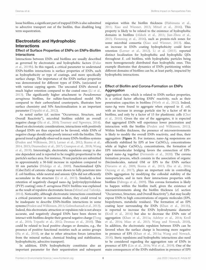
Effect of Biofilm and Corona-Formation on ENPsAggregationAggregation state, which is related to ENPs surface properties,is a critical factor affecting ENPs’ reactivity, such as theirpenetration capacities in biofilms (Wirth et al., 2012). Indeed,nano-Ag were found to aggregate when exposed to E. coli,with an increase in average particle size by a factor of 40 inbiofilms, and only by a factor of 15 for planktonic cells (Choiet al., 2010). Given the size of the aggregates, it is expectedthat aggregated ENPs will experience lower penetration andinteraction with the biofilm (Peulen and Wilkinson, 2011).Within biofilm thickness, the presence of microenvironmentsis likely to modify the overall ENPs reactivity, and thus, theiraggregation (Figure 3). For instance, nano-Ag suspensions areefficiently stabilized by EPS at low Ca(NO3)2 concentrationswhile at higher Ca(NO3)2 concentrations, the formation ofEPS intermolecular bridging favors the connection of EPS-caped nano-Ag together (Fernando et al., 2020). The coronaformation process, which consists in the association of organic(bio)molecules, natural OM or EPS to the ENPs surface(Navarro et al., 2009; Ikuma et al., 2015; Zhu et al., 2016;Ouyang et al., 2017), plays an important role in controllingENPs aggregation by modifying the colloidal stability of thenanoparticles, and in turn their interactions properties withbiofilms (Fabrega et al., 2009). This corona formation is likelyto happen within the biofilm itself, given the existence ofmicroenvironments along the biofilm thickness (cf. section“Occurrence, Structure, and Overall Reactivity”), that may locallyexpose ENPs to high concentrations of organic molecules (EPS,biopolymers, metabolic residues). The formation of an EPScoating layer surrounding the ENPs (Khan et al., 2011b),is reported to increase the ENPs hydrodynamic diameter(Kroll et al., 2014) but also to decrease the ENPs rate ofaggregation (Khan et al., 2011a; Adeleye et al., 2014; Krollet al., 2014; Miao et al., 2015; Wang and Nowack, 2018),In addition, the electrostatic repulsion between ENPs can befavored when the surface charge is becoming more negativein presence of EPS (Khan et al., 2011a; Wang and Nowack,2018). Steric repulsions and hydrophobic interactions also needto be considered regarding the aggregation rate of ENPs inpresence of EPS (Lin et al., 2016; Wei et al., 2019). One of themain consequences of the ENPs stabilization in presence of EPS
Frontiers in Environmental Science | www.frontiersin.org 6 July 2020 | Volume 8 | Article 82
fenvs-08-00082 July 1, 2020 Time: 18:41 # 7
Desmau et al. Biofilms Impact on Nanoparticles Fate
FIGURE 3 | Examples of processes associated to ENPs transformations and aggregation. From left to right, aggregation mediated by the formation, in the biofilmmatrix, of an organic corona, that modifies ENPs surface properties and promotes interactions between ENPs; ligand-mediated dissolution involving the presence ofhighly complexing ligands (such as siderophores) that complex free metals in solution and thus promotes ENPs dissolution; passivation of silver NPs in vicinity ofsulfato-reducing bacteria (SRB) that increase locally HS- concentrations, and favors sulfidation processes at the ENPs surface; ENPs stabilization through theformation of an organic corona that isolates ENPs from the surrounding solution thus limiting dissolution processes.
would be the favored transport of ENPs in aquatic environments(Wang and Nowack, 2018).
The presence of functional groups in EPS, their composition(protein, carboxylate, polysaccharide, and lipid contents) andconcentration will also strongly condition the interactions withENPs (Adeleye and Keller, 2016; Xu et al., 2016; Zhou et al.,2016; Morelli et al., 2018; Wang and Nowack, 2018; Fernandoet al., 2020). As such, investigations were conducted on thestabilization potential of three EPS types, soluble, loosely bound,and tightly bound EPS, characterized by different protein topolysaccharide ratio (Fernando et al., 2020). All three EPS-types were found to prevent nano-Ag aggregation, but looselybound EPS were the most efficient, due to its lower contentin hydrophilic dissolved OM. In addition, EPS hydrophobicproperties can be important when considering sorption at ENPssurfaces (Wei et al., 2019).
Noticeably, EPS composition is strongly determined by thetype of microorganisms considered (Sutherland, 2001; Flemmingand Wingender, 2010), and conditions in which they develop. Asa result, dynamic changes in ENPs reactivity when interacting
with biofilms can be expected for some specific conditions, butsuch mechanism remains today largely overlooked.
Mineral Substrate ReactivityMineral surfaces are known to exhibit strong complexingcapacities toward metal(loid)s and organic matter in mostecosystems (Brown et al., 1999). In soils, they can serve assubstrates on which biofilms develop, but doing so, they maintaintheir high complexing capacities despite the overlying microbialcommunity, as reported for different mineral/biofilm/solutioninterfaces (Templeton et al., 2001, 2003a,b; Wang et al., 2016a,b).In these studies, focused on the partitioning of metal(loid)s at themineral/biofilm/solution interface, mineral substrates constitutea strong complexing compartment able to drive the dynamicsof free metal transport within the whole interface, biofilmincluded. Regarding ENPs, a study by Desmau et al. (2018) onS. oneidensis MR1 biofilms exposed to nano-Ag shows that themineral surface remains highly reactive and tends to accumulatenegatively charged ENPs (PVP coated nano-Ag). As a result,the ENPs dynamics of accumulation and further transport at
Frontiers in Environmental Science | www.frontiersin.org 7 July 2020 | Volume 8 | Article 82

fenvs-08-00082 July 1, 2020 Time: 18:41 # 8
Desmau et al. Biofilms Impact on Nanoparticles Fate
the mineral/biofilm/solution interface are strongly driven bythe mineral surface. However, in most studies the reactivity ofthe mineral substrate was poorly or not considered and futureresearch will need to elucidate its role.
ENPs CHEMICAL TRANSFORMATIONSIN BIOFILMS
As previously discussed (cf. section “ENPs Retention andMigration Mechanisms Within Biofilms”), ENPs fate in naturalsystems is strongly controlled by retention mechanisms imposedby microbial biofilms. To this perspective, another critical factorthat needs to be considered is the transformation that ENPscan undergo when in contact with these biological structures(Holden et al., 2016). At a first approximation, ENPs potentialfor speciation changes could be considered similar to that of freemetals that are subjected to strong chemical modifications inbiofilms (Templeton et al., 2003a; Couasnon et al., 2019). It is thuslikely that ENPs experience similar transformations, driven bythe high reactivity of local microenvironments and the importantfunctional sites density spread within the biofilm thickness.
Dissolution and stabilization by surface passivation arereported here as the main processes able to drastically modifyENPs speciation (Figure 3). In addition, response to toxic stressfrom cells caused by ENPs exposure is discussed since the relatedprocesses are potentially impacting their environmental life cycle.
DissolutionToxicity of various ENPs comes from their dissolution and thesubsequent exposure of organisms to free metals. This toxicpathway is usually evoked for known soluble ENPs such as nano-Ag or nano-ZnO (Franklin et al., 2007; Xia et al., 2008; Auffanet al., 2009; Gelabert et al., 2015; Le Ouay and Stellacci, 2015).In that case, ENPs in close vicinity of bacteria cell walls becomea continuous source of novel metal species through dissolutionprocesses. This can result in high concentrations of toxic ionsand cause toxicity to the bacteria (Slavin et al., 2017). The extentof dissolution and the associated kinetics generally depend onthe size, shape, surface characteristics of the ENPs, as well asthe solvent properties (pH, ionic strength, presence of ligands),and is well-documented even in complex matrices (Gelabertet al., 2014; Sivry et al., 2014). In natural systems, dissolutionkinetics can be largely enhanced in contact with highly reactiveenvironmental compartments, such as biofilms. Even NPs knownas stable could be submitted to dissolution. As such, somestudies report a significant ENPs dissolution when in contactwith microbial biofilms (Wirth et al., 2012; Gil-Allué et al., 2015;Avellan et al., 2018; Wang and Nowack, 2018; Alizadeh et al.,2019; McGivney et al., 2019).
Even if the associated processes remain poorly documented,two main factors are likely to be responsible for ENPs dissolutionin microbial biofilms: (i) the high density of reactive ligands inthese bio-structures favoring ligand assisted dissolution, and (ii)the strong redox activities of microorganisms. These dissolutionprocesses are certainly favored in biofilm microenvironmentsthat exhibit specific physico-chemical properties at the local scale.
Ligand-Mediated DissolutionIn microbial biofilms, the high reactive site densities resultfrom the association of numerous functional sites present onthe cell walls of microorganisms (Ngwenya et al., 2003; Borroket al., 2004a,b, 2005), and on the EPS matrix (Ha et al., 2010;Tourney and Ngwenya, 2014). Regarding microbial surfaces,the compilation of several studies on Gram-positive and Gram-negative bacteria in planktonic suspension reported functionalsites densities in the range of 4.5 ± 0.8.10−3 mol.g−1 (dry weight)(Yee and Fein, 2001). On the other hand, EPS reactivity is afunction of the types of molecules included in their compositions,such as their polysaccharides/proteins ratio. Regarding ENPsforced dissolution, the high sites density in biofilms, cell wallsand EPS included, tends to complex metal(loid)s to high extent(Tourney and Ngwenya, 2014; Wang et al., 2016a,b), resulting ina decrease in local concentrations of free metal(loid)s in solutionwithin the biofilm matrix. In order to restore thermodynamicsequilibria, ENPs will in turn dissolve and release free metal(loid)s(Bian et al., 2011; Wirth et al., 2012). If the density of complexingsites is in high excess, ENPs total dissolution could thus beexpected. This process can be facilitated by formation of strongmetal-organic complexes at the ENPs surface that weakens metal-oxygen bonds.
Focusing specifically on EPS reactivity in absence of biofilm,Adeleye et al. (2014) noted a more important dissolution ofnano-CuO in presence of EPS after 90 days compared to EPS-free control. By extension, the EPS role is likely to be criticalwhen investigating biofilm impact reactivity. As a result, EPSare considered by many authors a reasonably good analog toevaluate the biofilm impact onto ENPs fate (Tong et al., 2010;Jiang et al., 2013). However, it must be noted that since cellsmetabolic activity constitutes also an important part of thebiofilms’ reactivity, which in association with their specific 3-dimensional structure that creates microenvironments, is likelyto strongly impact the ENPs fate in the environment.
The extracellular matrix is known to contain enzymesand other proteins secreted by living cells in the biofilm,leading to the notion of “external digestion system” that partlycontrols organic molecules and polymers degradation, as well ascertain extracellular redox process (Flemming and Wingender,2010, Flemming et al., 2016). As a result, degradation oforganic polymers that coat (and protect in a certain way)functionalized ENPs may arise from exposure to such enzymes(Ikuma et al., 2015). Interestingly, the type of capping agentsconstitutes an important factor regarding dissolution rate(Mitrano et al., 2014). Siderophores, metal-complexing moleculesof high affinity, are also produced by bacteria in biofilms(Visca et al., 2007; Saha et al., 2013). Siderophore productionby bacteria, including the ligands catecholate, hydroxamate,and carboxylate, is extensively reviewed (Springer and Butler,2016; Albelda-Berenguer et al., 2019), and present the centralrole of this class of molecules for metal scavenging. Otherhighly complexing ligands are also secreted by bacteria, suchas cysteine-containing molecules and glutathione that exhibitthiol groups of high affinity toward soft metals (Brown et al.,2006; Mugerfeld et al., 2009). Oxalic acid (Palmieri et al., 2019)produced by P. fluorescens (Hamel et al., 1999) or Burkholderia
Frontiers in Environmental Science | www.frontiersin.org 8 July 2020 | Volume 8 | Article 82
fenvs-08-00082 July 1, 2020 Time: 18:41 # 9
Desmau et al. Biofilms Impact on Nanoparticles Fate
glumae (Nakata and He, 2010) were also reported. Presence ofcomplexing ligands such as siderophores is frequently invokedwhen considering mineral dissolution mediated by bacteria, andcan be extended to ENPs, with ligand-promoted dissolutionkinetics being higher than proton-promoted dissolution abovepH 4 (Kraemer, 2004; Reichard et al., 2007; Wang et al., 2015).
The biofilm structure is known to induce microenvironmentsformation (cf. section “Microenvironments Within BiofilmThickness”), that are able to strongly impact localthermodynamics equilibria and favor both ligand-mediatedand redox-mediated dissolution. Existence of gradients in pH orredox state are both reported, and take place at very short spatialscales within the biofilm structure (Yu and Bishop, 2001; Billingset al., 2015; Flemming et al., 2016; Kataky and Knowles, 2018).However, despite their importance, the nature and propertiesof such microenvironments are still poorly known regardingligands or metal(loid)s concentrations, even if few studiessuggest strong and dynamic changes in local physico-chemicalconditions (Couasnon et al., 2019). Nonetheless, it can beanticipated that these “hot spots” of high reactivity exhibit lowpHs, important redox changes, and locally concentrate to highextent siderophores and other complexing ligands as well asenzymes. Since metal-based ENPs dissolution is usually favoredat low pH (Bian et al., 2011; Han et al., 2016) and in the presenceof high affinity complexing molecules (cf. section “Ligand-Mediated Dissolution”), it is likely that microenvironmentsstrongly impact ENPs dissolution.
Redox-Mediated DissolutionAs a second factor, many microorganisms are known toexhibit strong redox activities, thoroughly reported in numerousstudies, as for Acidothiobacillus ferrooxidans which is able tooxidize sulfur species by direct contact or indirect mechanisms(Monachon et al., 2019). Generally, microorganisms in biofilmssecrete a high variety of redox-active molecules associated to theEPS matrix (Tourney and Ngwenya, 2014), and some studiespropose that some types of EPS could be electron shuttles inbiofilms. Production of riboflavin or c-type cytochrome (Xiaoet al., 2017; Kataky and Knowles, 2018), and cyanide residues(Avellan et al., 2018) have been reported. Also, the productionof extracellular superoxide, a reactive oxygen species, has beenreported for an important variety of microorganisms such asFirmicutes, Actinobacteria, Bacteroidetes, Gammaproteobacteria,and Alphaproteobacteria (Diaz et al., 2013). As a result, thecollection of redox-active molecules in the extracellular matrixis also likely to play an important role for dissolution of redox-sensitive ENPs, such as nano-CeO2, nano-Ag or nano-Au, inmicrobial biofilms.
For instance, Nano-Au are thought to remain inert inabiotic conditions. However, presence of soil bacteriumChromobacterium violaceum, that produces cyanide residues,is able to provoke oxidative dissolution of nano-Au (McGivneyet al., 2019). Similarly, nano-Au was shown to dissolve in contactwith freshwater macrophyte biofilm through oxidation processesinvolving cyanide release (Avellan et al., 2018). Nano-CeO2exposed to wastewater biofilm experience higher dissolutionrates, following Ce(IV) reduction (Wang and Nowack, 2018),
with associated mechanism involving either direct contactbetween the NPs and bacteria surface or liberation of electronshuttle molecules in the extracellular medium (Thill et al., 2006).It is also proposed that light-induced production of reactiveoxygen species (ROS) at the EPS surface can also play a rolefor ENPs dissolution through oxidation of Ce(III) to Ce(IV)in solution, lowering the concentration of dissolved Ce(III)(Kroll et al., 2014).
ENPs Surface PassivationBiofilms can favor ENPs dissolution as discussed above butcan also induce surface passivation through either (i) chemicaltransformation of the surface to a more thermodynamically stablephase, or (ii) formation of a corona shell that will limit surfaceinteractions with the surrounding environment.
Passivation of ENPs surface through phase transformation,lowering their reactivity and solubility, is observed when incontact with biofilm and microorganisms. For instance, inanaerobic conditions, typically at the bottom of biofilms, sulfate-reducing bacteria can reduce sulfate to bisulfide (HS−) which isin turn able to promote nano-Ag sulfidation at the surface of theNPs (Levard et al., 2011, 2013). Such sulfidation is reported inwater resource recovery facilities biofilms (Alizadeh et al., 2019),with the creation of such an Ag2S passivation layer at the surfaceof nano-Ag that reduces the NP solubility, and consequently itstoxicity (Levard et al., 2013).
Organic polymers originally present as coatings on ENPs canalso be destabilized in the biofilm thickness by ligand exchangeprocesses involving EPS molecules, with strong consequences onENPs stability. Although the adsorption of EPS to the surfaceof ENPs can favor their dissolution or their aggregation, aspresented previously, they may also promote their passivation.This depends on the type of EPS and complexing ligandsconsidered, their charge and hydrophobic properties, as wellas the mode of interaction with ENPs. Strong metal-organiccomplexes at the ENPs surface are expected to lower the strengthof surrounding metal-oxygen bonds thus favoring dissolution,while weaker EPS interactions with ENPs can drastically modifyENPs surface properties, and potentially promote aggregationdepending on local physico-chemical conditions (throughintermolecular bridging for instance). Finally, ENPs enhancedpassivation takes place when the organic corona formed byEPS either isolates ENPs from the surrounding solution, thusdisabling their dissolution, or promotes repulsion among ENPs,lowering their aggregation.
Cellular Toxicity Response as a StrongModifier of ENPs SpeciationENPs toxicity to biofilm cells is clearly related to the penetrationof these particles and the effectiveness of interaction withmicrobial cells. As a result, several studies linked ENPs toxicityto biofilm physico-chemical properties such as thickness orroughness (Thuptimdang et al., 2017). Actually, bacteria encasedin biofilms structures are usually reported as more resistant totoxic substances than the corresponding planktonic analogs, anda significant amount of studies and reviews exist that discuss this
Frontiers in Environmental Science | www.frontiersin.org 9 July 2020 | Volume 8 | Article 82

fenvs-08-00082 July 1, 2020 Time: 18:41 # 10
Desmau et al. Biofilms Impact on Nanoparticles Fate
topic (Wirth et al., 2012; Flemming et al., 2016). However, whatis more important regarding the ENPs environmental fate is themicrobial cells response after exposure to NPs, since some ofthem are known toxic agents.
Multi-species communities exposed to ENPs may experienceimportant changes in microbial consortium structure withsignificant evolutions in microbial diversity (Binh et al., 2014,2016; Echavarri-Bravo et al., 2015; Jomini et al., 2015; Tanget al., 2017; Miao et al., 2018; Zhu et al., 2018a,b, 2019). Theseremodeling of microbial consortia can potentially modify thetypes of metabolisms expressed in these communities and impactthe environmental ENPs fate. Noticeably, mechanisms of celltoxicity resistance to ENPs will differ from one microorganismto the other, as is the case of E. coli and Pseudomonas aeruginosaexposed to nano-Ag (Saleh et al., 2015), or even among twodifferent strains of the same bacteria, such as E. coli MG1655 andW3110 exposed to ZnO-NPs (Gelabert et al., 2015). This suggeststhe existence of numerous metabolic pathways, potentiallygenerating the extracellular release of a variety of substances.
Nevertheless, whereas NPs impact on biofilms viability andtoxicity have been extensively studied, only a few articles focuson ENPs effects on microbial biofilms functioning. Exposureto photocatalytic ENPs (CdS, TiO2, Fe2O3) result in differentadaptations in peryphitic biofilms (Zhu et al., 2019). While nodecrease in biomass, chlorophyll content and ATPase activitywere detected, a significant increase in EPS production andsecretion of superoxide dismutase have been observed. Themicrobial community is also evolving due to an increase inthe proportion of phototrophic and high nutrient metabolicmicroorganisms. Such an evolution in microbial communitycomposition was also shown for periphytic biofilms exposed tonano-TiO2, with an increase in the abundance of denitrifyingbacteria (Zhu et al., 2018a) and a significant production ofEPS enriched in proteins. Exposure to nano-TiO2 and nano-CeO2 also lead to changes in bacteria consortium for surficialsediment microbial communities, with an observed inhibitionof microbial-mediated oxygen consumption, and strong changesin local redox state (Miao et al., 2018). Nano-CdS have beenshown to induce an increase in overall metabolic activity (ATPaseactivity), and to stimulate EPS production (Zhu et al., 2018b),while exposure to nano-CuO resulted in the production ofpolysaccharides-rich loosely bound-EPS (Hou et al., 2015).
Interestingly, the production or inhibition of key moleculesthat could modify ENPs speciation were also reported.S. oneidensis MR-1 biofilms exposed to nano-TiO2 showedan increased production of riboflavin (Maurer-Jones et al., 2013).This molecule is related to metal reduction capacity in bacteria,accounting for approximately 75% of extracellular electrontransfer to insoluble substrates by S. oneidensis (Kotloski andGralnick, 2013). Similarly, a riboflavin production was reportedfor planktonic S. oneidensis exposed to lithium nickel manganesecobalt oxide nanosheets (Mitchell et al., 2019), whereas nano-Feoxide did not induce any increase in riboflavin expression(Buchman et al., 2019). The production of pyoverdine, animportant siderophore exhibiting a high affinity for Fe, wasshown to be inhibited following exposure to nano-CuO onPseudomonas chlororaphis 06 (Dimkpa et al., 2012), or to
Te nanorods for P. aeruginosa (Mohanty et al., 2014, 2015).In bacterial communities, the activity of key extracellularenzymes such as alkaline phosphatase and ß-glucosidase wasalso restrained by the combined exposure to nano-TiO2 andCu (Fan et al., 2016) or polystyrene NPs (Awet et al., 2018).However, denitrification enzyme activity and respiration wereshown to increase after addition of nano-TiO2 (Ozaki et al.,2016). Importantly, a common metal-resistance mechanismby bacteria, such as S. oneidensis, to resist oxidative stressis the production of enzymes containing cysteine residuesand glutathione (Brown et al., 2006; Mugerfeld et al., 2009),molecules that usually exhibit high affinities toward soft metalsand could thus promote ligand-mediated dissolution for ENPscontaining such metals.
Regarding specific impacts on EPS matrix, several studiesreport the extracellular production of molecules by microbesexposed to ENPs (Maurer-Jones et al., 2013; Zhu et al., 2018a,b,2019). Particularly, the formation of more compacted biofilmsand higher active site densities was observed, following asignificant increase in EPS production after exposure to ENPs(Hou et al., 2015; Tang et al., 2017; Liu et al., 2018; Miaoet al., 2018; Zhu et al., 2018a,b, 2019). Interestingly, thisincreased EPS production is thought to reduce cells damage(Hessler et al., 2012), particularly when most metal-bearingENPs are known to produce ROS at their surface (Nel et al.,2006) which promotes oxidative stress at the cellular level.However, a denser and more functionalized EPS matrix providesmore attachment sites to ENPs, thus drastically limiting theirinteractions with cell membranes and thus lowering exposure toROS (Joshi et al., 2012; Zhou et al., 2016). Concurrently, evenROS generation at the ENPs surface is reported to be loweredin presence of an EPS coating, potentially due to a limitedcontact between NPs inorganic surface and solution (Hessleret al., 2012). However, if ENPs toxicity is too high, structuraldamages will occur and biofilms will, on the contrary, tend to getthinner and less dense.
All these studies show that ENPs can significantly impactmicrobial biofilms functioning by inducing changes in EPScomposition and by promoting EPS production. Such changescan result in more packed biofilms and a significant increase inactive site densities, simultaneously promoting ENPs trappingand an increased dissolution of metallic ENPs due to the presenceof higher local concentrations of complexing ligands. Also,changes in the enzymes production as well as the synthesis ofstrong metal complexants such as cysteine residues, or moleculesinvolved in metal reduction (e.g., flavins and riboflavins),can drastically govern changes in ENPs speciation throughdissolution processes.
LONG-TERM FATE OF ENPs INNATURAL ENVIRONMENT: THEMESOCOSM APPROACH
As stated previously, microbial biofilms control to high extentsthe environmental fate of ENPs. Most studies devoted to thesequestions of biofilm-ENPs interactions focus on model systems,
Frontiers in Environmental Science | www.frontiersin.org 10 July 2020 | Volume 8 | Article 82
fenvs-08-00082 July 1, 2020 Time: 18:41 # 11
Desmau et al. Biofilms Impact on Nanoparticles Fate
providing invaluable information regarding the associatedprocesses at the biofilm scale. However, these approaches lack thecomplexity of natural settings as well as the intricate interplaybetween the different environmental compartments. As such,in complement to these small-scale studies, more integrativeapproaches are required that evaluate the biofilms impact inglobal environmental systems.
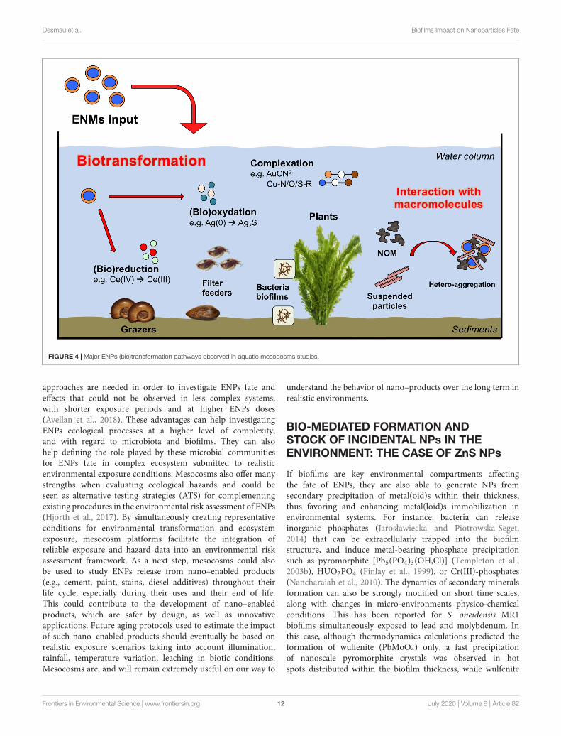
Understanding the long-term fate and biotransformation ofENPs in the environment is challenging due to the complexityof the bio-physical-chemical interactions within naturalsystems including aggregation and sorption of (in)organicsubstances, oxidation and reduction, as well as ecologicalfactors such as interacting organisms [e.g., microorganisms,(in)vertebrates, plants, fungi], trophic levels present (e.g.,primary producer, primary consumer, secondary consumer)and trophic and transgenerational transfer potentials (Auffanet al., 2009). Taking into account this complexity requires adiverse collection of expertise, including but not limited tophysical chemistry, (micro)biology, and ecology. Particularlywell-suited experimental units with which to engage suchmultidisciplinary teams are mesocosms, that consist in medium-size replicated ecosystems (Crossland and La Point, 1992;Shaw and Kennedy, 1996; Food and Agriculture Organization,2009). Mesocosms experiments can be designed to mimicany ecological scenarios of exposure (Auffan et al., 2019) andin the scope of ENP studies, have been defined as indooror outdoor experimental systems containing a portion ofthe natural environment which is (i) self–sustaining onceset up and acclimatized without any additional input ofnutrients or resources, and (ii) that allows monitoring ofall (or the maximum of) input and output parameters tomeasure changes in the environment, and in the concentrationand speciation of ENPs over time, space, and ecosystemcompartments (Figure 4).
This set of information is used to discern the kinetics of(bio)transformation, (bio)distribution and impacts of the ENPsto better inform studies on their fate and modeling of theirenvironmental risks. Biotransformation of the ENPs exertedby micro-organisms, as well as macro-organisms, is one ofthe main drivers of the ENPs fate and impacts on the long-term. For instance, sulfidation in the sediments is a majortransformation pathway for ENPs such as Ag and Cu, thatare enhanced by the activity of microbial consortia as themodulation of anoxic/anaerobic conditions in soils/sediments,or also the release of exudates and organic material duringmetabolism, respiration or death (e.g., Bone et al., 2012; Lowryet al., 2012). Such a release of exudates by microbial biofilmsand macro-organisms is important in inducing dissolution,homo- and hetero-aggregation, and by modulating their toxiceffects on the mid- to long-term, as presented previously inthis review. Although the formation of bio-corona shortlyafter introduction in mesocosms governed the persistency ofENPs in the water column (Espinasse et al., 2018), this willdepend on the size and surface properties of the pristine ENPs(Unrine et al., 2012; Geitner et al., 2018). For instance, PVPcoated Ag ENPs were colloidally stabilized by plant-derivedand biofilm-derived dissolved organic matter. On the contrary,
gum Arabic-coated Ag ENPs were rapidly removed from thewater column at the same conditions through deposition ordissolution of the particles and subsequent binding of ions toplant or biofilm on the sediment surfaces (Bone et al., 2012;Unrine et al., 2012).
Macrophytes, surficial sediments and associatedmicroorganisms were often reported as two major sinks forENPs. Regarding aquatic plants, studies reported extensivechange in the speciation of ENMs following their adsorption orinternationalization by aquatic plants. For instance, the majorityof nano-Au introduced in freshwater mesocosms (outdoor,300 L volume, 180 d exposure) were found associated to thebiofilm growing on Egeria densa, where the totality of Au wascomplexed with cyanide (43 ± 8%), hydroxide (38 ± 4%)and thiol (19 ± 4%) ligands (Avellan et al., 2018). The drivingbiodissolution mechanism was the Au oxidation and subsequentcomplexation with cyanide [Au(CN)2
−], produced as secondarymetabolites by the microbiome in the periphyton associatedwith leaves. The displacement of CN ligands by S-R groups,led to the formation of soluble thiol-Au species such as [R-S-Au-CN]− or [R-S2-Au]− complexes. These complexes werestable at the environmental physico-chemical conditions testedand likely represent major transformation pathways for otherredox active ENPs. Stegemeier et al. (2017) also investigatedthe speciation of nano-Ag and -Cu in wetland mesocosms(outdoor, 400 L volume, 270 d exposure). Ag(0) ENPs added tothe ecosystem were found in the plant tissues as Ag(0), Ag2S,and Ag bound to thiol whereas major transformation productsfor Cu ENPs were identified in Cu–O–R and Cu–S–R. Suchproducts differed from those measured in the surroundingsediments compartment and the authors suggested that plantsnatural defense, such as glutathione, may play an importantrole. Similarly, in freshwater pond mesocosms contaminatedwith Ce ENPs, metals speciation and redox reactions wereassociated with the benthic (micro)biota activity and were notmeasured in the surrounding natural matrices (Tella et al.,2014). A protein storage mechanism for Cu ENPs was alsoproposed to explain the transformation of Cu(OH)2 nano-pesticide in Cu-S, and Cu-O/N-R products in plant tissues(Avellan et al., 2020). Changes in the redox state followinginteractions with the micro-organisms/macrophytes systemswere also observed for CeO2 ENPs, where up to 57% of thepristine Ce(IV) atoms were reduced to Ce(III). In this case, thedegree of reduction was observed to increase at decreasing ENPssize, likely due to an intrinsic higher reactivity of the smallerENPs and their greater association with reducing microbes(Geitner et al., 2018).
These studies, conducted in complex realistic systems, confirmthe important impact exerted by microbial biofilms towardENPs fate, and allow investigating their role among thedifferent reactive environmental compartments. However, itmust be noted that although mesocosms experiments were ableto provide comprehensive data regarding mid-term/long-termENPs (bio)transformation in realistic environmental conditions,these strategies were seldom applied in environmental fatestudies, in comparison with standard methodologies (e.g., single-species ecotoxicity tests) (Bour et al., 2015). Nonetheless, similar
Frontiers in Environmental Science | www.frontiersin.org 11 July 2020 | Volume 8 | Article 82
fenvs-08-00082 July 1, 2020 Time: 18:41 # 12
Desmau et al. Biofilms Impact on Nanoparticles Fate
FIGURE 4 | Major ENPs (bio)transformation pathways observed in aquatic mesocosms studies.
approaches are needed in order to investigate ENPs fate andeffects that could not be observed in less complex systems,with shorter exposure periods and at higher ENPs doses(Avellan et al., 2018). These advantages can help investigatingENPs ecological processes at a higher level of complexity,and with regard to microbiota and biofilms. They can alsohelp defining the role played by these microbial communitiesfor ENPs fate in complex ecosystem submitted to realisticenvironmental exposure conditions. Mesocosms also offer manystrengths when evaluating ecological hazards and could beseen as alternative testing strategies (ATS) for complementingexisting procedures in the environmental risk assessment of ENPs(Hjorth et al., 2017). By simultaneously creating representativeconditions for environmental transformation and ecosystemexposure, mesocosm platforms facilitate the integration ofreliable exposure and hazard data into an environmental riskassessment framework. As a next step, mesocosms could alsobe used to study ENPs release from nano–enabled products(e.g., cement, paint, stains, diesel additives) throughout theirlife cycle, especially during their uses and their end of life.This could contribute to the development of nano–enabledproducts, which are safer by design, as well as innovativeapplications. Future aging protocols used to estimate the impactof such nano–enabled products should eventually be based onrealistic exposure scenarios taking into account illumination,rainfall, temperature variation, leaching in biotic conditions.Mesocosms are, and will remain extremely useful on our way to
understand the behavior of nano–products over the long term inrealistic environments.
BIO-MEDIATED FORMATION ANDSTOCK OF INCIDENTAL NPs IN THEENVIRONMENT: THE CASE OF ZnS NPs
If biofilms are key environmental compartments affectingthe fate of ENPs, they are also able to generate NPs fromsecondary precipitation of metal(oid)s within their thickness,thus favoring and enhancing metal(loid)s immobilization inenvironmental systems. For instance, bacteria can releaseinorganic phosphates (Jarosławiecka and Piotrowska-Seget,2014) that can be extracellularly trapped into the biofilmstructure, and induce metal-bearing phosphate precipitationsuch as pyromorphite [Pb5(PO4)3(OH,Cl)] (Templeton et al.,2003b), HUO2PO4 (Finlay et al., 1999), or Cr(III)-phosphates(Nancharaiah et al., 2010). The dynamics of secondary mineralsformation can also be strongly modified on short time scales,along with changes in micro-environments physico-chemicalconditions. This has been reported for S. oneidensis MR1biofilms simultaneously exposed to lead and molybdenum. Inthis case, although thermodynamics calculations predicted theformation of wulfenite (PbMoO4) only, a fast precipitationof nanoscale pyromorphite crystals was observed in hotspots distributed within the biofilm thickness, while wulfenite
Frontiers in Environmental Science | www.frontiersin.org 12 July 2020 | Volume 8 | Article 82

fenvs-08-00082 July 1, 2020 Time: 18:41 # 13
Desmau et al. Biofilms Impact on Nanoparticles Fate
production was observed only after 7 days of exposure tolead (Couasnon et al., 2019). Another example concerns Sulfate-Reducing bacteria (SRB), mentioned before, that are known toplay a major role in the precipitation of metal sulfide mineralsthrough the reduction of sulfate to sulfide. Sulfides (mostlyas HS- at neutral pH) readily precipitate with soft metalsto form metal sulfides. Among the variety of bio-mediatedNPs formed from anthropic metal(oid)s sources (also calledincidental NPs), the case of ZnS NPs is a good example, relativelywell-documented, and will be presented here as a case-study.As such, several examples are reported including ZnS NPsgenerated in abandoned mine sites, contaminated sedimentsand organic wastes.
Bio-Mediated Formation of ZnS-NPs inContaminated SitesBanfield’s group has been a pioneer in the discovery of ZnSNPs formed by SRB in abandoned tunnels and drainagesystem of the Piquette Pb-Zn mine in southwest Wisconsin(Labrenz et al., 2000; Druschel et al., 2002; Labrenz andBanfield, 2004; Moreau et al., 2004). ZnS NPs biofilmconcentration was found to be about 106 times that ofassociated groundwater (Labrenz et al., 2000). ZnS NPs ofthe order of 3–5 nm, mostly sphalerite, were observed withinmicron-sized spheroids. The authors proposed that the spheroidswere induced by microbially derived extracellular proteins,which potentially plays an important role in limiting NPdispersion in natural environments (Moreau et al., 2007).These proteins can interact with ZnS during and after theirformation. Indeed, thiol containing compounds play a key roleto limit ZnS NPs aggregation and was hypothesized to be akey factor contributing to their persistence in the environment(Lau and Hsu-Kim, 2008).
In another context, the formation of ZnS NPs in a varietyof organic waste (OW) has long been overlooked. The mainreason lies in the fact that extreme precaution should be given forsampling and storage of samples before their analyses to avoidchange in Zn speciation (Le Bars et al., 2018). OWs, used as afertilizer for crops in many countries, is a major source of inputof NPs in the environment. While several studies investigatedENPs accumulation and transformation in wastewater sludges,it has recently been shown that a considerable amount of ZnSNPs was non-intentionally produced in a variety of OWs. Amongthem, sewage sludge and livestock manures used as amendmenthave relatively high concentrations of Zn (40–4000 mg.kg−1)(Alburquerque et al., 2012; Romeo et al., 2014; Zirkler et al.,2014; Alvarenga et al., 2015). Zn in OW is mostly anthropogenic.For example, high Zn concentrations in livestock manures areexplained by important quantities of Zn added in animal feedcompared to their ability to assimilate it (Legros et al., 2010).Nanosized ZnS is a major Zn species in raw liquid OWs and inanaerobically digested OWs (Le Bars et al., 2018). The reductiveconditions combined with high sulfur content in these systemsare a favorable environment for SRB (Santegoeds et al., 1999;Tang et al., 2004; Chen et al., 2008). Therefore, it is likely thatthese ZnS NPs are formed because of sulfide release by SRB.
Crystallite size of ZnS formed in OW was estimated around 3 nmusing X-ray Absorption Spectroscopy (Formentini et al., 2017; LeBars et al., 2018). This is consistent with the identification of 2.5–7.5 nm ZnS in digested sewage sludge by Transmission ElectronMicroscopy (Kim et al., 2014). Interestingly, these particlestransformed within months during composting (Donner et al.,2011; Lombi et al., 2012; Le Bars et al., 2018) or after landapplication (Formentini et al., 2017). These results suggest a highreactivity of bio-mediated ZnS NPs compared to micrometerZnS analog which are stable over years in soils (Robson et al.,2014). The high reactivity of ZnS NPs formed in OW could berelated to their small size. Indeed, Gilbert et al. (2004) highlightedcontracted interatomic distances (1%) for ZnS NPs of this rangeof size (3.4 nm) compared to their bulk analog. Such structuraldisorder can increase NP surface reactivity (Yang et al., 2016)and enhance ZnS dissolution. A better understanding of potentialimpacts of ZnS NPs transformation on the ecosystem and inparticular on cultivated lands is needed.
Structural Properties of Biogenic ZnSNPsIt seems that SRB-induced ZnS NPs are bigger than ZnS-NPsformed in abiotic systems at similar conditions (Peltier et al.,2011; da Costa et al., 2012; Xu et al., 2016; Eskelsen et al.,2018). It could be explained by the progressive production ofsulfide by SRB leading to a lower saturation index, since alower saturation index favors particle growth (Mersmann, 1999).In the same way, ligands either initially present in the culturemedia, or produced metabolites by bacteria, could bind to Zn,thus lowering the saturation index. However, factors controllingthe size of biogenic ZnS NPs are not clear and would requirefurther investigation.
Biogenic ZnS NPs tend to exhibit structural defects. Indeed,Xu et al. (2016) showed stacking faults and twins on the (111) faceof biogenic ZnS NPs, the only polar face, i.e., prone to interactwith polar molecules such as water, phospholipids head or aminoacids. This suggests that surface interaction with surroundingcompounds influences ZnS NPs structure. Eskelsen et al. (2018)also observed structural defaults (twins) on the (111) face ofbiogenic ZnS NPs, to a lower extent because of the unfavorableparticles’s orientation. The authors detected carbon traces inbiogenic ZnS NPs aggregates and assumed a microbial metaboliteinteraction with ZnS NPs.
However, the effect of structural defects on their reactivityis unclear. For hematite NPs, Echigo et al. (2012) have shownthat surface defects enhance their dissolution. Regarding ZnSNPs, Xu et al. (2016) highlighted by electronic microscopy thatbiogenic ZnS NPs dissolution starts where the structural defectsare observed. Also, Eskelsen et al. (2018) have calculated a highersurface energy for biogenic ZnS NPs, compared with the abioticones, suggesting a higher reactivity. In contrast, smaller abioticZnS NPs have a higher available specific surface area for oxidativedissolution. Despite the gathered information regarding theirproperties, further investigations are needed to evaluate bioticinfluence on ZnS NPs reactivity, in order to predict their fatewhen released in the environment.
Frontiers in Environmental Science | www.frontiersin.org 13 July 2020 | Volume 8 | Article 82

fenvs-08-00082 July 1, 2020 Time: 18:41 # 14
Desmau et al. Biofilms Impact on Nanoparticles Fate
Biogenic ZnS NPs: An UnderestimatedStock in the Environment That NeedsFurther InvestigationAlthough biofilms have been identified as a key compartmentgoverning in part the environmental fate of ENPs, it alsohosts favorable conditions for the formation of biogenic NPsin particular from anthropic metal(oid)s inputs. The latteris clearly overlooked despite concentrations that can reachthe orders of magnitude of the expected ENPs environmentalrelease. As an example, the concentrations of ZnS NPsobserved in the studies described above are up to 4 ordersof magnitudes higher than the predicted concentrations ofZnO ENPs in soils, sediments and biosolids (about 0.1, 0.1,and 10–100 mg.kg−1, respectively) (Gottschalk et al., 2013).Indeed, although ZnO ENPs was shown to transform intoZnS NPs in wastewater treatment plants (Ma et al., 2013,2014), a large variety of other Zn sources can potentially betransformed into ZnS NPs within biofilms. In this regard, betterunderstanding of biomediated formation of NPs in biofilmsneeds further attention in particular when associated withanthropic activities that potentially affect biofilm and thereforeNPs stabilities. It includes the use of OW as amendments orsediment dredging for example that in both cases involve strongphysico-chemical changes (e.g., oxidation) that can potentiallyrelease significant amounts of free Zn with unknown effectsfor ecosystems. Thus, in addition to the control they exerton ENPs fate, biofilms can also become secondary sourcesof NPs release in the environment, showing the central rolethese microbial structures are likely to play in general NPscycling. However, numerous questions remain opened thatdeserve further investigation in order to better constrain theirenvironmental impact.
CONCLUDING REMARKS
This review highlights the central role played by biofilmsregarding ENPs fate in the environment. In details, thesemicrobial structures exert a control on ENPs cycling at multiplelevels, by exhibiting relatively high retention capacities and byenforcing ENPs transformations. Accumulation and migrationproperties within biofilms are governed by a combinationof key parameters, including biofilm density, ENPs size andsurface properties, creation of electrostatic and hydrophobicinteractions, mineral substrate reactivity, and formation ofaggregates within the biofilm thickness. Interestingly, theeffects of some of these key parameters remain poorlydefined despite evidence of their strong impact on ENPsaccumulation and transport within biofilms. This is for instancethe case of mineral substrates, on which biofilms develop,that are likely to exhibit strong complexation capacity towardENPs. In addition, given the relatively high accumulationcapacities of biofilms, these microbial structures could beconsidered as secondary sources of ENPs and products ofENPs transformations, able to release them gradually inENPs-free pore waters. This problematic remains an open
question of important environmental significance that needsfurther investigations.
ENPs are also submitted to speciation changes inbiofilms. They can experience dissolution, either mediatedby strong complexing ligands produced by microorganisms(siderophores, cysteine-containing molecules) or redoxconditions, as well as changes in local physico-chemicalproperties in microenvironments. Passivation mechanismscan also happen through the precipitation of an inertsecondary mineral layer, as well as through ENPs stabilizationfollowing the interactions with organic ligands fromthe EPS matrix. Finally, exposure to toxic ENPs cantrigger a response from micro-organisms in biofilms (forinstance riboflavin production), which in turn has thepotential to strongly impact the ENPs fate within thesemicrobial structures.
Mesocosms-based approaches allow to consider biofilmsactivity in complex ecosystems, in some ways similar to theenvironment, and submitted to realistic exposure conditions.As such, these approaches are extremely valuable to betterconstrain the role of microbial biofilms for ENPs fate,and certainly need to be developed for future researchdevoted to that topic.
Finally, the case of nano-ZnS shows that biofilms can alsosynthesize NPs in man-impacted systems. This raises importantquestions regarding the impact of such processes on a global NPscycling in natural systems. However, these questions have beenlargely overlooked and certainly require further investigation.
Some other important points deserve direct attention.First, the general lack of established standardized protocolsstrongly limits inter-comparison between studies whether atthe small-scale or in larger systems. Then, even if consideringsingle-species biofilm as a model system provides importantadvantages to investigate complex mechanisms of interactionbetween ENPs and biofilms, this approach presents alsosome limitations. Natural biofilms function as importantmicrobial consortia that usually include a high variety ofstrains. To that perspective, efforts should be conductedto investigate more realistic multi-species biofilms. Also,metagenomic approaches could help understanding the dynamicrole of the different microbial communities and metabolismsin biofilms, and the complex interplay among them, duringtheir interactions with ENPs. Finally, a vast majority ofthe studies compiled in this review were focused on theinteractions between ENPs and bacteria biofilms. Nevertheless,in the environment, fungi and archaea play also importantroles, and more studies should investigate their ability toimpact ENPs cycling.
AUTHOR CONTRIBUTIONS
MD, CL, and AG contributed to the conception of the study. MDand AG wrote the first draft of the manuscript. MA, AC, CL, andML wrote sections of the manuscript. MB, AC, and ED revised itcritically. All authors contributed to the article and approved thesubmitted version.
Frontiers in Environmental Science | www.frontiersin.org 14 July 2020 | Volume 8 | Article 82

fenvs-08-00082 July 1, 2020 Time: 18:41 # 15
Desmau et al. Biofilms Impact on Nanoparticles Fate
FUNDING
This work has received funding from Excellence Initiativeof Aix – Marseille University – A∗MIDEX, a French“Investissements d’Avenir” program, through its associated LabexSERENADE project, and from the MAMBA ANR project (ANR-18-CE01-0001-01). This work was also a contribution to the
OSU-Institut Pytheìas. The authors thank the PIREN-Seineprogram for the funding.
ACKNOWLEDGMENTS
We acknowledge the CNRS for the funding of the IRP iNOVE.
REFERENCESAdeleye, A. S., Conway, J. R., Perez, T., Rutten, P., and Keller, A. A.
(2014). Influence of extracellular polymeric substances on the long-termfate, dissolution, and speciation of copper-based nanoparticles. Environ. Sci.Technol. 48, 12561–12568. doi: 10.1021/es5033426
Adeleye, A. S., and Keller, A. A. (2016). Interactions between algal extracellularpolymeric substances and commercial TiO2 nanoparticles in aqueous media.Environ. Sci. Technol. 50, 12258–12265. doi: 10.1021/acs.est.6b03684
Albelda-Berenguer, M., Monachon, M., and Joseph, E. (2019). Siderophores: fromnatural roles to potential applications. Adv. Appl. Microbiol. 106, 193–225.doi: 10.1016/bs.aambs.2018.12.001
Alburquerque, J. A., de la Fuente, C., Ferrer-Costa, A., Carrasco, L., Cegarra,J., Abad, M., et al. (2012). Assessment of the fertiliser potential of digestatesfrom farm and agroindustrial residues. Biomass Bioenergy 40, 181–189. doi:10.1016/j.biombioe.2012.02.018
Aldeek, F., Mustin, C., Balan, L., Roques-Carmes, T., Fontaine-Aupart, M. P.,and Schneider, R. (2011). Surface-engineered quantum dots for the labeling ofhydrophobic microdomains in bacterial biofilms. Biomaterials 32, 5459–5470.doi: 10.1016/j.biomaterials.2011.04.019
Alizadeh, S., Ghoshal, S., and Comeau, Y. (2019). Fate and inhibitory effect of silvernanoparticles in high rate moving bed biofilm reactors. Sci. Total Environ. 647,1199–1210. doi: 10.1016/j.scitotenv.2018.08.073
Alvarenga, P., Mourinha, C., Farto, M., Santos, T., Palma, P., Sengo, J., et al. (2015).Sewage sludge, compost and other representative organic wastes as agriculturalsoil amendments: benefits versus limiting factors. Waste Manage. 40, 44–52.doi: 10.1016/j.wasman.2015.01.027
Auffan, M., Masion, A., Mouneyrac, C., de Garidel-Thorona, C., Hendren,C. O., Thiery, A., et al. (2019). Contribution of mesocosm testingto a single-step and exposure-driven environmental risk assessment ofengineered nanomaterials. Nanoimpact 13, 66–69. doi: 10.1016/j.impact.2018.12.005
Auffan, M., Rose, J., Bottero, J., Lowry, G. V., Jolivet, J., and Wiesner, M. R. (2009).Towards a definition of inorganic nanoparticles from an environmental, healthand safety perspective. Nat. Nanotechnol. 4, 634–641. doi: 10.1038/NNANO.2009.242
Avellan, A., Simonin, M., Anderson, S. M., Geitner, N. K., Bossa, N., Spielman-Sun,E., et al. (2020). Differential reactivity of copper- and gold-based nanomaterialscontrols their seasonal biogeochemical cycling and fate in a freshwater wetlandmesocosm. Environ. Sci. Technol. 54, 1533–1544. doi: 10.1021/acs.est.9b05097
Avellan, A., Simonin, M., McGivney, E., Bossa, N., Spielman-Sun, E., Rocca, J. D.,et al. (2018). Gold nanoparticle biodissolution by a freshwater macrophyteand its associated microbiome. Nat. Nanotechnol. 13, 1072–1077. doi: 10.1038/s41565-018-0231-y
Awet, T. T., Kohl, Y., Meier, F., Straskraba, S., Grün, A. L., Ruf, T., et al.(2018). Effects of polystyrene nanoparticles on the microbiota and functionaldiversity of enzymes in soil. Environ. Sci. Eur. 30:11. doi: 10.1186/s12302-018-0140-6
Barker, W., and Banfield, J. F. (1998). Zones of chemical and physicalinteraction at interfaces between microbial communities and minerals.A model. Geomicrobiol. J. 15, 223–244. doi: 10.1080/01490459809378078
Bar-On, Y. M., Phillips, R., and Milo, R. (2018). The biomass distribution on Earth.Proc. Natl. Acad. Sci. U.S.A. 115, 6506–6511. doi: 10.1073/pnas.1711842115
Battin, T. J., Kammer, F. V. D., Weilhartner, A., Ottofuelling, S., and Hofmann,T. (2009). Nanostructured TiO2: transport behavior and effects on aquaticmicrobial communities under environmental conditions. Environ. Sci. Technol.43, 8098–8104. doi: 10.1021/es9017046
Bian, S. W., Mudunkotuwa, I. A., Rupasinghe, T., and Grassian, V. H.(2011). Aggregation and dissolution of 4 nm ZnO nanoparticles in aqueousenvironments: influence of pH, Ionic Strength, Size, and Adsorption of HumicAcid. Langmuir 27, 6059–6068. doi: 10.1021/la200570n
Billings, N., Birjiniuk, A., Samad, T. S., Doyle, P. S., and Ribbeck, K. (2015).Material properties of biofilms - A review of methods for understandingpermeability and mechanics. Rep. Prog. Phys. 78:036601. doi: 10.1088/0034-4885/78/3/036601
Binh, C. T. T., Adams, E., Vigen, E., Tong, T., Alsina, M. A., Gaillard, J.-F., et al.(2016). Chronic addition of a common engineered nanomaterial alters bio-mass, activity and composition of stream biofilm communities. Environ. Sci.Nano 3, 619–630. doi: 10.1039/c5en00274e
Binh, C. T. T., Tong, T., Gaillard, J.-F., Gray, K. A., and Kelly, J. J. (2014). Acuteeffects of TiO2 nanomaterials on the viability and taxonomic compositionof aquatic bacterial communities assessed via high-throughput screening andnext generation sequencing. PLos One 9:e106280. doi: 10.1371/journal.pone.0106280
Bone, A. J., Colman, B. P., Gondikas, A. P., Newton, K. M., Harrold, K. H., Cory,R. M., et al. (2012). Biotic and abiotic interactions in aquatic microcosmsdetermine fate and toxicity of Ag nanoparticles: part 2-toxicity and Agspeciation. Environ. Sci. Technol. 46, 6925–6933. doi: 10.1021/es204683m
Borrok, D., Fein, J. B., and Kulpa, C. F. (2004a). Cd and proton adsorption ontobacterial consortia grown from industrial wastes and contaminated geologicsettings. Environ. Sci. Technol. 38, 5656–5664. doi: 10.1021/es049679n
Borrok, D., Fein, J. B., and Kulpa, C. F. (2004b). Proton and Cd adsorption ontonatural bacterial consortia: testing universal adsorption behavior. Geochim.Cosmochim. Acta 68, 3231–3238. doi: 10.1016/j.gca.2004.02.003
Borrok, D., Turner, B. F., and Fein, J. B. (2005). Universal surface complexationframework for modeling proton binding onto bacterial surfaces in geologicalsettings. Am. J. Sci. 305, 826–853. doi: 10.2475/ajs.305.6-8.826
Bour, A., Mouchet, F., Silvestre, J., Gauthier, L., and Pinelli, E. (2015).Environmentally relevant approaches to assess nanoparticles ecotoxicity: areview. J. Hazard. Mater. 283, 764–777. doi: 10.1016/j.jhazmat.2014.10.021
Brown, G. E. (2001). How minerals react with water. Science 294, 67–69. doi:10.1126/science.1063544
Brown, G. E., Henrich, V. E., Casey, W. H., Clark, D. L., Eggleston, C., Felmy, A.,et al. (1999). Metal oxide surfaces and their interactions with aqueous solutionsand microbial organisms. Chem. Rev. 99, 77–174. doi: 10.1021/cr980011z
Brown, S. D., Thompson, M. R., VerBerkmoes, N. C., Chourey, K., Shah, M., Zhou,J., et al. (2006). Molecular dynamics of the Shewanella oneidensis responseto chromate stress. Mol. Cell. Proteomics 5, 1054–1071. doi: 10.1074/mcp.M500394-MCP200
Buchman, J. T., Pho, T., Rodriguez, R. S., Feng, Z. V., and Haynes, C. L.(2019). Coating iron oxide nanoparticles with mesoporous silica reduces theirinteraction and impact on S. oneidensis MR-1. Chemosphere 237:124511. doi:10.1016/j.chemosphere.2019.124511
Burns, J. M., Pennington, P. L., Sisco, P. N., Frey, R., Kashiwada, S., Fulton, M. H.,et al. (2013). Surface charge controls the fate of Au nanorods in saline estuaries.Environ. Sci. Technol. 47, 12844–12851. doi: 10.1021/es402880u
Chen, Y., Cheng, J. J., and Creamer, K. S. (2008). Inhibition of anaerobic digestionprocess: a review. Bioresour. Technol. 99, 4044–4064. doi: 10.1016/j.biortech.2007.01.057
Choi, O., Yu, C.-P., Fernandez, G. E., and Hu, Z. (2010). Interactions of nanosilverwith Escherichia coli cells in planktonic and biofilm cultures. Water Res. 44,6095–6103. doi: 10.1016/j.watres.2010.06.069
Costerton, J. W., Cheng, K. J., Geesey, G. G., Ladd, T. I., Nickel, J. C., Dasgupta, M.,et al. (1987). Bacteria biofilms in nature and disease. Annu. Rev. Microbiol. 41,435–464. doi: 10.1146/annurev.mi.41.100187.002251
Frontiers in Environmental Science | www.frontiersin.org 15 July 2020 | Volume 8 | Article 82
fenvs-08-00082 July 1, 2020 Time: 18:41 # 16
Desmau et al. Biofilms Impact on Nanoparticles Fate
Costerton, J. W., Lewandowski, Z., Caldwell, D. E., Korber, D. R., and Lappin-Scott, H. M. (1995). Microbial biofilms. Annu. Rev. Microbiol. 49, 711–745.doi: 10.1146/annurev.mi.49.100195.003431
Costerton, J. W., Stewart, P., and Greenberg, E. P. (1999). Bacterial biofilms: acommon cause of persistent infections. Science 284, 1318–1322. doi: 10.1126/science.284.5418.1318
Couasnon, T., Gelabert, A., Menez, B., and Guyot, F. (2019). Experimentalassessment of occurrences and stability of lead-bearing minerals in bacterialbiofilms. Chem. Geol. 505, 23–35. doi: 10.1016/j.chemgeo.2018.11.023
Crampon, M., Hellal, J., Mouvet, C., Wille, G., Michel, C., Wiener, A., et al. (2018).Do natural biofilm impact nZVI mobility and interactions with porous media?A column study. Sci. Total Environ. 610, 709–719. doi: 10.1016/j.scitotenv.2017.08.106
Crossland, N. O., and La Point, T. W. (1992). The design of mesocosmsexperiments. Environ. Toxicol. Chem. 11, 1–4. doi: 10.1002/etc.5620110101
da Costa, J. P., Girao, A. V., Lourenco, J. P., Monteiro, O. C., Trindade, T.,and Costa, M. C. (2012). Synthesis of nanocrystalline ZnS using biologicallygenerated sulfide. Hydrometallurgy 117, 57–63. doi: 10.1016/j.hydromet.2012.02.005
Dehner, C. A., Awaya, J. D., Maurice, P. A., and DuBois, J. L. (2010). Roles ofsiderophores, oxalate, and ascorbate in mobilization of iron from hematite bythe aerobic bacterium Pseudomonas mendocina. Appl. Environ. Microbiol. 76,2041–2048. doi: 10.1128/AEM.02349-09
Desmau, M., Gélabert, A., Levard, C., Ona-Nguema, G., Vidal, V., Stubbs, J. E.,et al. (2018). Dynamics of silver nanoparticles at the solution/biofilm/mineralinterface. Environ. Sci. Nano 5, 2394–2405. doi: 10.1039/c8en00331a
Diaz, J. M., Hansel, C. M., Voelker, B. M., Mendes, C. M., Andeer, P. F., and Zhang,T. (2013). Widespread production of extracellular superoxide by HeterotrophicBacteria. Science 340, 1223–1226. doi: 10.1126/science.123
Dimkpa, C. O., McLean, J. E., Britt, D. W., Johnson, W. P., Arey, B., Lea, S. A.,et al. (2012). Nanospecific inhibition of pyoverdine siderophore production inPseudomonas chlororaphis O6 by CuO nanoparticles. Chem. Res. Toxicol. 25,1066–1074. doi: 10.1021/tx3000285
Donlan, R. M. (2002). Biofilms: microbial life on surfaces. Emerg. Infect. Dis. 8,881–890. doi: 10.3201/eid0809.020063
Donner, E., Howard, D. L., Jonge, M. D. D., Paterson, D., Cheah, M. H., Naidu,R., et al. (2011). X-ray absorption and micro X-ray fluorescence spectroscopyinvestigation of copper and zinc speciation in biosolids. Environ. Sci. Technol.45, 7249–7257. doi: 10.1021/es201710z
Druschel, G. K., Labrenz, M., Thomsen-Ebert, T., Fowle, D. A., and Banfield,J. F. (2002). Geochemical Modeling of ZnS in biofilms: an example of oredepositional processes. Econ. Geol. Bull. Soc. Econ. Geol. 97, 1319–1329. doi:10.2113/gsecongeo.97.6.1319
Dzumedzey, Y., Labille, J., Cathala, B., Moreau, C., and Santaella, C. (2017).Polysaccharide coating on environmental collectors affects the affinity anddeposition of nanoparticles. Nanoimpact 5, 83–91. doi: 10.1016/j.impact.2016.12.004
Echavarri-Bravo, V., Paterson, L., Aspray, T. J., Porter, J. S., Winson, M. K.,Thornton, B., et al. (2015). Shifts in the metabolic function of a benthic estuarinemicrobial community following a single pulse exposure to silver nanoparticles.Environ. Pollut. 201, 91–99. doi: 10.1016/j.envpol.2015.02.033
Echigo, T., Aruguete, D. M., Murayama, M., and Hochella, M. F. Jr.(2012). Influence of size, morphology, surface structure, and aggregationstate on reductive dissolution of hematite nanoparticles with ascorbicacid. Geochim. Cosmochim. Acta 90, 149–162. doi: 10.1016/j.gca.2012.05.008
Eskelsen, J. R., Xu, J., Chiu, M., Moon, J.-W., Wilkins, B., Graham, D. E., et al.(2018). Influence of structural defects on biomineralized ZnS nanoparticledissolution: an in-situ electron microscopy study. Environ. Sci. Technol. 52,1139–1149. doi: 10.1021/acs.est.7b04343
Espinasse, B. P., Geitner, N. K., Schierz, A., Therezien, M., Richardson, C. J., Lowry,G. V., et al. (2018). Comparative persistence of engineered nanoparticles in acomplex aquatic ecosystem. Environ. Sci. Technol. 52, 4072–4078. doi: 10.1021/acs.est.7b06142
Fabrega, J., Renshaw, J. C., and Lead, J. R. (2009). Interactions of silvernanoparticles with Pseudomonas putida biofilms. Environ. Sci. Technol. 43,9004–9009. doi: 10.1021/es901706j
Fan, W., Liu, T., Li, X., Peng, R., and Zhang, Y. (2016). Nano-TiO2 affectscu speciation, extracellular enzyme activity, and bacterial communitiesin sediments. Environ. Pollut. 218, 77–85. doi: 10.1016/j.envpol.2016.08.018
Fein, J. B., Daughney, C. J., Yee, N., and Davis, T. A. (1998). A chemical equilibriummodel for metal adsorption onto bacterial surfaces. Geochim. Cosmochim. Acta61, 3319–3328. doi: 10.1016/S0016-7037(97)00166-X
Fernando, I., Lu, D., and Zhou, Y. (2020). Interactive influence of extracellularpolymeric substances (EPS) and electrolytes on the colloidal stability of silvernanoparticles. Environ. Sci. Nano 7, 186–197. doi: 10.1039/c9en00861f
Ferry, J. L., Craig, P., Hexel, C., Sisco, P., Frey, R., Pennington, P. L., et al. (2009).Transfer of gold nanoparticles from the water column to the estuarine food web.Nat. Nanotechnol. 4, 441–444. doi: 10.1038/NNANO.2009.157
Finlay, J. A., Allan, V. J. M., Conner, A., Callow, M. E., Basnakova, G., andMacaskie, L. E. (1999). Phosphate release and heavy metal accumulation bybiofilm-immobilized and chemically-coupled cells of a citrobacter sp. pre-grown in continuous culture. Biotechnol. Bioeng. 63, 87–97. doi: 10.1002/(sici)1097-0290(19990405)63:1<87::aid-bit9>3.0.co;2-0
Flemming, H. C., and Wingender, J. (2010). The biofilm matrix. Nat. Rev. Microbiol.8, 623–633. doi: 10.1038/nrmicro2415
Flemming, H.-C., Wingender, J., Szewzyk, U., Steinberg, P., Rice, S. A., andKjelleberg, S. (2016). Biofilms: an emergent form of microbial life. Nat. Rev.Microbiol. 14, 563–575. doi: 10.1038/nrmicro.2016.94
Flemming, H. C., and Wuertz, S. (2019). Bacteria and archaea on Earth and theirabundance in biofilms. Nat. Rev. Microbiol. 17, 247–260. doi: 10.1038/s41579-019-0158-9
Food and Agriculture Organization [FAO] (2009). Biosafety of Genetically ModifiedOrganisms: Basic Concepts, Methods and Issues. Rome: FAO.
Formentini, T. A., Legros, S., Fernandes, C. V. S., Pinheiro, A., Le Bars, M., Levard,C., et al. (2017). Radical change of Zn speciation in pig slurry amended soil:Key role of nano-sized sulfide particles. Environ. Pollut. 222, 495–503. doi:10.1016/j.envpol.2016.11.056
Franklin, N. M., Rogers, N. J., Apte, S. C., Batley, G. E., Gadd, G. E., and Casey, P. S.(2007). Comparative toxicity of nanoparticulate ZnO, bulk ZnO, and ZnCl2to a freshwater microalga (Pseudokirchneriella subcapitata): the importance ofparticle solubility. Environ. Sci. Technol. 41, 8484–8490. doi: 10.1021/es071445r
Fulaz, S., Hiebner, D., Barros, C. H. N., Devlin, H., Vitale, S., et al. (2019a).Ratiometric imaging of the in situ pH distribution of biofilms by use offluorescent mesoporous silica nanosensors. ACS Appl. Mater. Interfaces 11,32679–32688. doi: 10.1021/acsami.9b09978
Fulaz, S., Vitale, S., Quinn, L., and Casey, E. (2019b). Nanoparticle–biofilminteractions: the role of the EPS matrix. Trends Microbiol. 27, 915–926. doi:10.1016/j.tim.2019.07.004
Gadd, G. M. (2010). Metals, minerals and microbes: geomicrobiology andbioremediation. Microbiology 156, 609–643. doi: 10.1099/mic.0.037143-0
Geitner, N. K., Cooper, J. L., Avellan, A., Castellon, B. T., Perrotta, B. G., Bossa,N., et al. (2018). Size-based differential transport, uptake, and mass distributionof ceria (CeO2) nanoparticles in wetland mesocosms. Environ. Sci. Technol. 52,9768–9776. doi: 10.1021/acs.est.8b02040
Gelabert, A., Sivry, Y., Ferrari, R., Akrout, A., Cordier, L., Nowak, S., et al.(2014). Uncoated and coated ZnO nanoparticles life cycle in synthetic seawater.Environ. Toxicol. Chem. 22, 341–349. doi: 10.1002/etc.2447
Gelabert, A., Sivry, Y., Gobbi, P., Mansour, N., Menguy, N., Brayner, R., et al.(2015). Testing nano effect onto model bacteria: impact of speciation andgenotypes. Nanotoxicology 10, 216–25. doi: 10.3109/17435390.2015.1048323
Giese, B., Klaessig, F., Park, B., Kaegi, R., Steinfeldt, M., Wigger, H., et al.(2018). Risks, release and concentrations of engineered nanomaterial in theenvironment. Sci. Rep. 8:1565. doi: 10.1038/s41598-018-19275-4
Gil-Allué, C., Schirmer, K., Tlili, A., Gessner, M. O., and Behra, R. (2015).Silver nanoparticle effects on stream periphyton during short-term exposures.Environ. Sci. Technol. 49, 1165–1172. doi: 10.1021/es5050166
Gilbert, B., Huang, F., Zhang, H., Waychunas, G. A., and Banfield, J. F. (2004).Nanoparticles: strained and stiff. Science 305, 651–654. doi: 10.1126/science.1098454
Golmohamadi, M., Clark, R. J., Veinot, J. G. C., and Wilkinson, K. J. (2013). Therole of charge on the diffusion of solutes and nanoparticles (silicon nanocrystals,nTiO2, nAu) in a biofilm. Environ. Chem. 10, 34–41. doi: 10.1071/EN12106
Frontiers in Environmental Science | www.frontiersin.org 16 July 2020 | Volume 8 | Article 82

fenvs-08-00082 July 1, 2020 Time: 18:41 # 17
Desmau et al. Biofilms Impact on Nanoparticles Fate
Gottschalk, F., Sonderer, T., Scholz, R. W., and Nowack, B. (2009). Modeledenvironmental concentrations of engineered nanomaterials (TiO2, ZnO, Ag,CNT, fullerenes) for Different Regions. Environ. Sci. Technol. 43, 9216–9222.doi: 10.1021/es9015553
Gottschalk, F., Sun, T., and Nowack, B. (2013). Environmental concentrations ofengineered nanomaterials: review of modeling and analytical studies. Environ.Pollut. 181, 287–300. doi: 10.1016/j.envpol.2013.06.003
Guiot, E., Georges, P., Brun, A., Fontaine-Aupart, M. P., Bellon-Fontaine,M. N., and Briandet, R. (2002). Heterogeneity of diffusion inside microbialbiofilms determined by fluorescence correlation spectroscopy under two-photon excitation. Photochem. Photobiol. 75, 570–578. doi: 10.1562/0031-8655(2002)0750570hodimb2.0.co2
Ha, J., Gelabert, A., Spormann, A., and Brown, G. E. Jr. (2010). Role ofextracellular polymeric substances in metal ion complexation on Shewanellaoneidensis: batch uptake, thermodynamic modeling, ATR-FTIR, and EXAFSstudy. Geochim. Cosmochim. Acta 74, 1–15. doi: 10.1016/j.gca.2009.06.031
Habimana, O., Steenkeste, K., Fontaine-Aupart, M. P., Bellon Fontaine, M. N.,Kulakauskas, S., and Briandet, R. (2011). Diffusion of nanoparticles in biofilmsis altered by bacterial cell wall hydrophobicity. Appl. Environ. Microbiol. 77,367–368. doi: 10.1128/AEM.02163-10
Hamel, R., Levasseur, R., and Apana, V. D. (1999). Oxalic acid production andaluminum tolerance in Pseudomonas fluorescens. J. Inorg. Biochem. 76, 99–104.doi: 10.1016/S0162-0134(99)00120-8
Han, Y., Hwang, G., Kim, D., Bradford, S. A., Lee, B., Eom, I., et al. (2016).Transport, retention, and long-term release behavior of ZnO nanoparticleaggregates in saturated quartz sand: role of solution pH and biofilm coating.Water Res. 90, 247–257. doi: 10.1016/j.watres.2015.12.009
Herrling, M. P., Lackner, S., Tatti, O., Guthausen, G., Delay, M., Franzreb, M.,et al. (2016). Short and long term biosorption of silica-coated iron oxidenanoparticles in heterotrophic biofilms. Sci. Total. Environ. 544, 722–729. doi:10.1016/j.scitotenv.2015.11.174
Hessler, C. M., Wu, M. Y., Xue, Z., Choi, H., and Seo, Y. (2012). The influenceof capsular extracellular polymeric substances on the interaction between TiO2nanoparticles and planktonic bacteria. Water Res. 46, 4687–4696. doi: 10.1016/j.watres.2012.06.009
Hidalgo, G., Burns, A., Herz, E., Hay, A. G., Houston, P. L., Wiesner,U., et al. (2009). Functional tomographic fluorescence imaging of pHmicroenvironments in microbial biofilms by use of silica nanoparticlesensors. Appl. Environ. Microbiol. 75, 7426–7435. doi: 10.1128/AEM.01220-09
Hjorth, R., Holden, P. A., Hansen, S. F., Colman, B. P., Grieger, and Hendren,C. O. (2017). The role of alternative testing strategies in environmental riskassessment of engineered nanomaterials. Environ. Sci. Nano 4, 292–301. doi:10.1039/c6en00443a
Holden, P. A., Gardea-Torresdey, J. L., Klaessig, F., Turco, R. F., Mortimer, M.,Hund-Rinke, K., et al. (2016). Considerations of Environmentally RelevantTest Conditions for Improved Evaluation of Ecological Hazards of EngineeredNanomaterials. Environ. Sci. Technol. 50, 6124–6145. doi: 10.1021/acs.est.6b00608
Hou, J., Miao, L. Z., Wang, C., Wang, P. F., Ao, Y. H., and Lv, B. W. (2015).Effect of CuO nanoparticles on the production and composition of extracellularpolymeric substances and physicochemical stability of activated sludge flocs.Bioresour. Technol. 176, 65–70. doi: 10.1016/j.biortech.2014.11.020
Hunter, R. C., and Beveridge, T. J. (2005). Application of a pH-sensitivefluoroprobe (C-SNARF-4) for pH microenvironment analysis in Pseudomonasaeruginosa biofilms. Appl. Environ. Microbiol. 71, 2501–2510. doi: 10.1128/AEM.71.5.2501-2510.2005
Ikuma, K., Decho, A. W., and Lau, B. L. T. (2015). When nanoparticles meetbiofilms-interactions guiding the environmental fate and accumulation ofnanoparticles. Front. Microbiol. 6:591. doi: 10.3389/fmicb.2015.00591
Ikuma, K., Madden, A. S., Decho, A. W., and Lau, B. L. T. (2014).Deposition of nanoparticles onto polysaccharide-coated surfaces: implicationsfor nanoparticle–biofilm interactions. Environ. Sci. Nano 1, 117–122. doi: 10.1039/c3en00075c
Jarosławiecka, A., and Piotrowska-Seget, S. (2014). Lead resistance in micro-organisms. Microbiology 160, 12–25. doi: 10.1099/mic.0.070284-0
Jiang, X., Wang, X., Tong, M., and Kim, H. (2013). Initial transport and retentionbehaviors of ZnO nanoparticles in quartz sand porous media coated with
Escherichia coli biofilm. Environ. Pollut. 174, 38–49. doi: 10.1016/j.envpol.2012.11.016
Jian-Zhou, H., Cheng-Cheng, L., Deng-Jun, W., and Zhou, D.-M. (2015). Biofilmsand extracellular polymeric substances mediate the transport of graphene oxidenanoparticles in saturated porous media. J. Hazard. Mater. 300, 467–474. doi:10.1016/j.jhazmat.2015.07.026
Jing, H., Mezgebe, B., Hassan, A. A., Sahle-Demessie, E., Sorial, G. A., and Bennett-Stamper, C. (2014). Experimental and modeling studies of sorption of ceriananoparticle on microbial biofilms. Bioresour. Technol. 161, 109–117. doi: 10.1016/j.biortech.2014.03.015
Jomini, S., Clivot, H., Bauda, P., and Pagnout, C. (2015). Impact of manufacturedTiO2 nanoparticles on planktonic and sessile bacterial communities. Environ.Pollut. 202, 196–204. doi: 10.1016/j.envpol.2015.03.022
Joo, S. H., and Aggarwal, S. (2018). Factors impacting the interactions of engineerednanoparticles with bacterial cells and biofilms: mechanistic insights and state ofknowledge. J. Environ. Manag. 225, 62–74. doi: 10.1016/j.jenvman.2018.07.084
Joshi, N., Ngwenya, B. T., and French, C. E. (2012). Enhanced resistance tonanoparticle toxicity is conferred by overproduction of extracellular polymericsubstances. J. Hazard. Mater. 241-242, 363–370. doi: 10.1016/j.jhazmat.2012.09.057
Kalinowski, B. E., Liermann, L. J., Givens, S., and Brantley, S. L. (2000). Rates ofbacteria-promoted solubilization of Fe from minerals: a review of problems andapproaches. Chem. Geol. 169, 357–370. doi: 10.1016/S0009-2541(00)00214-X
Kataky, R., and Knowles, E. (2018). Biofilm formation on abiotic surfaces and theirredox activity. Curr. Opin. Electrochem. 12, 121–128. doi: 10.1016/j.coelec.2018.07.007
Keller, A. A., McFerran, S., Lazareva, A., and Suh, S. (2013). Global life cycle releasesof engineered nanomaterials. J. Nanopart. Res. 15:1692. doi: 10.1007/s11051-013-1692-4
Khan, S. S., Mukherjee, A., and Chandrasekaran, N. (2011a). Impact ofexopolysaccharides on the stability of silver nanoparticles in water. Water Res.45, 5184–5190. doi: 10.1016/j.watres.2011.07.024
Khan, S. S., Srivatsan, P., Vaishnavi, N., Mukherjee, A., and Chandrasekaran, N.(2011b). Interaction of silver nanoparticles (SNPs) with bacterial extracellularproteins (ECPs) and its adsorption isotherms and kinetics. J. Hazard Mater. 192,299–306. doi: 10.1016/j.colsurfa.2011.03.032
Kim, B., Levard, C., Murayama, M., Brown, G. E., and Hochella, M. F.(2014). Integrated approaches of X-ray absorption spectroscopic and electronmicroscopic techniques on zinc speciation and characterization in a final sewagesludge product. J. Environ. Qual. 43, 908–916. doi: 10.2134/jeq2013.10.0418
Kotloski, N. J., and Gralnick, J. A. (2013). Flavin electron shuttles dominateextracellular electron transfer by Shewanella oneidensis. mBio 4:e00553-12. doi:10.1128/mBio.0055312
Kraemer, S. M. (2004). Iron oxide dissolution and solubility in the presence ofsiderophores. Aquat. Sci. 66, 3–18. doi: 10.1007/s00027-003-0690-5
Kroll, A., Behra, R., Kaegi, R., and Sigg, L. (2014). Extracellular polymericsubstances (EPS) of freshwater biofilms stabilize and modify CeO2 and Agnanoparticles. PLoS One 9:e110709. doi: 10.1371/journal.pone.0110709
Kurlanda-Witek, H., Ngwenya, B., and Butler, I. (2015). The influence of biofilmson the mobility of bare and capped zinc oxide nanoparticles in saturated sandand glass beads. J. Cont. Hydrol. 179, 160–170. doi: 10.1016/j.jconhyd.2015.06.009
Kuzyakov, Y., and Blagodatskaya, E. (2015). Microbial hotspots and hot momentsin soil: concept & review. Soil Biol. Biochem. 83, 184–199. doi: 10.1016/j.soilbio.2015.01.025
Labrenz, M., and Banfield, J. F. (2004). Sulfate-reducing bacteria-dominatedbiofilms that precipitate ZnS in a subsurface circumneutral-pH mine drainagesystem. Microb. Ecol. 47, 205–217. doi: 10.1007/s00248-003-1025-8
Labrenz, M., Druschel, G. K., Thomsen-Ebert, T., Gilbert, B., Welch, S. A., Kemner,K. M., et al. (2000). Formation of Sphalerite (ZnS) deposits in natural biofilmsof sulfate-reducing Bacteria. Science 290, 1744–1747. doi: 10.1126/science.290.5497.1744
Lau, B. L., and Hsu-Kim, H. (2008). Precipitation and growth of zincsulfide nanoparticles in the presence of thiol-containing naturalorganic ligands. Environ. Sci. Technol. 42, 7236–7241. doi: 10.1021/es801360b
Layet, C., Auffan, M., Santaella, C., Chevassus-Rosset, C., Montes, M., Ortet,P., et al. (2017). Evidence that soil properties and organic coating drive the
Frontiers in Environmental Science | www.frontiersin.org 17 July 2020 | Volume 8 | Article 82
fenvs-08-00082 July 1, 2020 Time: 18:41 # 18
Desmau et al. Biofilms Impact on Nanoparticles Fate
phytoavailability of cerium oxide nanoparticles. Environ. Sci. Technol. 51,9756–9764. doi: 10.1021/acs.est.7b02397
Le Bars, M., Legros, S., Levard, C., Chaurand, P., Tella, M., Rovezzi, M., et al. (2018).Drastic change in zinc speciation during anaerobic digestion and composting:instability of nanosized zinc sulfide. Environ. Sci. Technol. 52, 12987–12996.doi: 10.1021/acs.est.8b02697
Le Ouay, B., and Stellacci, F. (2015). Antibacterial activity of silver nanoparticles:a surface science insight. Nano Today 10, 339–354. doi: 10.1016/j.nantod.2015.04.002
Legros, S., Doelsch, E., Masion, A., Rose, J., Borshneck, D., Proux, O., et al.(2010). Combining size fractionation, scanning electron microscopy, and X-rayabsorption spectroscopy to probe zinc speciation in pig slurry. J. Environ. Qual.39, 531–540. doi: 10.2134/jeq2009.0096
Lerner, R. N., Lu, Q., Zeng, H., and Liu, Y. (2012). The effects of biofilm on thetransport of stabilized zerovalent iron nanoparticles in saturated porous media.Water Res. 46, 975–985. doi: 10.1016/j.watres.2011.11.070
Levard, C., Hotze, E. M., Colman, B. P., Dale, A. L., Truong, L., Yang, X. Y., et al.(2013). Sulfidation of silver nanoparticles: natural antidote to their toxicity.Environ. Sci. Technol. 47, 13440–13448. doi: 10.1021/es403527n
Levard, C., Hotze, E. M., Lowry, G. V., and Brown, G. E. (2012). EnvironmentalTransformations of Silver nanoparticles: impact on stability and toxicity.Environ. Sci. Technol. 46, 6900–6914. doi: 10.1021/es2037405
Levard, C., Reinsch, B. C., Michel, F. M., Oumahi, C., Lowry, G. V., and Brown,G. E. Jr. (2011). Sulfidation processes of PVP-coated silver nanoparticles inaqueous solution: impact on dissolution rate. Environ. Sci. Technol. 45, 5260–5266. doi: 10.1021/es2007758
Li, X., Yeh, Y. C., Giri, K., Mout, R., Landis, R. F., Prakash, Y. S., et al. (2015).Control of nanoparticle penetration into biofilms through surface design.Chem. Commun. 51, 282–285. doi: 10.1039/c4cc07737g
Li, Z., Aly Hassan, A., Sahle-Demessie, E., and Sorial, G. A. (2013). Transportof nanoparticles with dispersant through biofilm coated drinking water sandfilters. Water Res. 47, 6457–6466. doi: 10.1016/J.WATRES.2013.08.026
Lin, D., Story, S. D., Walker, S. L., Huang, Q., and Cai, P. (2016). Influenceof extracellular polymeric substances on the aggregation kinetics of TiO2nanoparticles. Water Res. 104, 381–388. doi: 10.1016/j.watres.2016.08.044
Liu, S., Cao, S., Guo, J., Luo, L., Zhou, Y., Lin, C., et al. (2018). Grapheneoxide–silver nanocomposites modulate biofilm formation and extracellularpolymeric substance (EPS) production. Nanoscale 10, 19603–19611. doi: 10.1039/c8nr04064h
Lombi, E., Donner, E., Tavakkoli, E., Turney, T. W., Naidu, R., Miller, B. W.,et al. (2012). Fate of zinc oxide nanoparticles during anaerobic digestionof wastewater and post-treatment processing of sewage sludge. Environ. Sci.Technol. 46, 9089–9096. doi: 10.1021/es301487s
Lowry, G. V., Espinasse, B. P., Badireddy, A. R., Richardson, C. J., Reinsch, B. C.,Lee, D. B., et al. (2012). Long-term transformation and fate of manufactured Agnanoparticles in a simulated large scale freshwater emergent wetland. Environ.Sci. Technol. 46, 7027–7036. doi: 10.1021/es204608d
Ma, R., Levard, C., Judy, J. D., Unrine, J. M., Durenkamp, M., Martin, B.,et al. (2014). Fate of zinc oxide and silver nanoparticles in a pilot wastewatertreatment plant and in processed biosolids. Environ. Sci. Technol. 48, 104–112.doi: 10.1021/es403646x
Ma, R., Levard, C., Michel, F. M., Brown, G. E. Jr., and Lowry, G. V. (2013).Sulfidation mechanism for zinc oxide nanoparticles and the effect of sulfidationon their solubility. Environ. Sci. Technol. 47, 2527–2534. doi: 10.1021/es3035347
Maurer-Jones, M. A., Gunsolus, I. L., Meyer, B. M., Christenson, C. J., and Haynes,C. L. (2013). Impact of TiO2 nanoparticles on growth, biofilm formation, andflavin secretion in Shewanella oneidensis. Anal. Chem. 85, 5810–5818. doi: 10.1021/ac400486u
McGivney, E., Gao, X., Liu, Y., Lowry, G. V., Casman, E. A., and Gregory, K. B.(2019). Biogenic cyanide production promotes dissolution of gold nanoparticlesin soil. Environ. Sci. Technol. 53, 1287–1295. doi: 10.1021/acs.est.8b05884
Ménez, B., Pasini, V., and Brunelli, D. (2012). Life in the hydrated suboceanicmantle. Nat. Geosci. 5, 133–137. doi: 10.1038/NGEO1359
Mersmann, A. (1999). Crystallization and precipitation. Chem. Eng. Process. 38,345–353. doi: 10.1016/S0255-2701(99)00025-2
Miao, L., Wang, C., Hou, J., Wang, P., Ao, Y., Li, Y., et al. (2015). Enhanced stabilityand dissolution of CuO nanoparticles by extracellular polymeric substances
in aqueous environment. J. Nanopart. Res. 17:404. doi: 10.1007/s11051-015-3208-x
Miao, L., Wang, P., Wang, C., Hou, J., Yao, Y., Liu, J., et al. (2018). Effect ofTiO2 and CeO2 nanoparticles on the metabolic activity of surficial sedimentmicrobial communities based on oxygen microelectrodes and high-throughputsequencing. Water Res. 129, 287–296. doi: 10.1016/j.watres.2017.11.014
Mishra, B., Shoenfelt, E., Yu, Q., Yee, N., Fein, J. B., and Myneni, S. C. B. (2017).Stoichiometry of mercury-thiol complexes on bacterial cell envelopes. Chem.Geol. 464, 137–146. doi: 10.1016/j.chemgeo.2017.02.015
Mitchell, S. L., Hudson-Smith, N. V., Cahill, M. S., Reynolds, B. N., Frand,S. D., Green, C. M., et al. (2019). Chronic exposure to complex metal oxidenanoparticles elicits rapid resistance in Shewanella oneidensis MR-1. Chem. Sci.10, 9768–9781. doi: 10.1039/c9sc01942a
Mitrano, D. M., Ranville, J., Bednar, A., Kazor, K., Hering, A. S., and Higgins, C.(2014). Tracking dissolution of silver nanoparticles at environmentally relevantconcentrations in laboratory, natural and processed waters using single particleICP-MS (spICP-MS). Environ. Sci. Nano 1, 248–259. doi: 10.1039/c3en00108c
Mitzel, M. R., Sand, S., Whalen, J. K., and Tufenkji, N. (2016). Hydrophobicity ofbiofilm coatings influences the transport dynamics of polystyrene nanoparticlesin biofilm-coated sand. Water Res. 92, 113–120. doi: 10.1016/j.watres.2016.01.026
Mitzel, M. R., and Tufenkji, N. (2014). Transport of Industrial PVP-StabilizedSilver Nanoparticles in Saturated Quartz Sand Coated with Pseudomonasaeruginosa PAO1 Biofilm of Variable Age. Environ. Sci. Technol. 48, 2715–2723.doi: 10.1021/es404598v
Mohanty, A., Kathawala, M. H., Zhang, J., Chen, W. N., Loo, J. S. C., Kjelleberg,S., et al. (2014). Biogenic tellurium nanorods as a novel antivirulence agentinhibiting pyoverdine production in Pseudomonas aeruginosa. Biotechnol.Bioeng. 111, 858–865. doi: 10.1002/bit.25147
Mohanty, A., Liu, Y., Yang, L., and Cao, B. (2015). Extracellular biogenicnanomaterials inhibit pyoverdine production in Pseudomonas aeruginosa: anovel insight into impacts of metal(loid)s on environmental bacteria. Appl.Microbiol. Biotechnol. 99, 1957–1966. doi: 10.1007/s00253-014-6097-5
Monachon, M., Albelda-Berenguer, M., and Joseph, E. (2019). Biological oxidationof iron sulfides. Adv. Appl. Microbiol. 107, 1–27. doi: 10.1016/bs.aambs.2018.12.002
Montano, M. D., Lowry, G. V., von der Kammer, F., Blue, J., and Ranville,J. F. (2014). Current status and future direction for examining engineerednanoparticles in natural systems. Environ. Chem. 11, 351–366. doi: 10.1071/EN14037
Moreau, J. W., Webb, R. I., and Banfield, J. F. (2004). Ultrastructure, aggregation-state, and crystal growth of biogenic nanocrystalline sphalerite and wurtzite.Am. Mineral. 89, 950–960. doi: 10.2138/am-2004-0704
Moreau, J. W., Weber, P. K., Martin, M. C., Gilbert, B., Hutcheon, I. D., andBanfield, J. F. (2007). Extracellular proteins limit the dispersal of biogenicnanoparticles. Science 316, 1600–1603. doi: 10.1126/science.1141064
Morelli, E., Gabellieri, E., Bonomini, A., Tognotti, D., Grassi, G., and Corsi, I.(2018). TiO2 nanoparticles in seawater: aggregation and interactions with thegreen alga Dunaliella tertiolecta. Ecotoxicol. Environ. Saf. 148, 184–193. doi:10.1016/j.ecoenv.2017.10.024
Mueller, N. C., and Nowack, B. (2008). Exposure modeling of engineerednanoparticles in the environment. Environ. Sci. Technol. 42, 4447–4453. doi:10.1021/es7029637
Mugerfeld, I., Law, B. A., Wickham, G. S., and Thompson, D. K. (2009). A putativeazoreductase gene is involved in the Shewanella oneidensis response to heavymetal stress. Appl. Microbiol. Biotechnol. 82, 1131–1141. doi: 10.1007/s00253-009-1911-1
Nakata, P. A., and He, C. (2010). Oxalic acid biosynthesis is encoded by an operonin Burkholderia glumae. FEMS Microbiol. Lett. 304, 177–182. doi: 10.1111/j.1574-6968.2010.01895.x
Nancharaiah, Y. V., Dodge, C., Venugopalan, V. P., Narasimhan, S. V., and Francis,A. J. (2010). Immobilization of Cr(VI) and its reduction to Cr(III) phosphate bygranular biofilms comprising a mixture of microbes. Appl. Environ. Microbiol.76, 2433–2438. doi: 10.1128/AEM.02792-09
Navarro, D. A. G., Watson, D. F., Aga, D. S., and Banerjee, S. (2009).Natural organic matter-mediated phase transfer of quantum dots in theaquatic environment. Environ. Sci. Technol. 43, 677–682. doi: 10.1021/es8017623
Frontiers in Environmental Science | www.frontiersin.org 18 July 2020 | Volume 8 | Article 82
fenvs-08-00082 July 1, 2020 Time: 18:41 # 19
Desmau et al. Biofilms Impact on Nanoparticles Fate
Nel, A., Xia, T., Mädler, L., and Li, N. (2006). Toxic potential of materials at thenanolevel. Science 311, 622–627. doi: 10.1126/science.1114397
Ngwenya, B. T., Sutherland, I. W., and Kennedy, L. (2003). Comparison of theacid–base behaviour and metal adsorption characteristics of a Gram-negativebacterium with other strains. Appl. Geochem. 18, 527–538. doi: 10.1016/S0883-2927(02)00118-X
Nunan, N. (2017). Game changer in soil science The microbial habitat in soil: scale,heterogeneity and functional consequences. J. Plant Nutr. Soil Sci. 180, 425–429.doi: 10.1002/jpln.201700184
Ouyang, K., Yu, X. Y., Zhu, Y., Gao, C., Huang, Q., and Cai, P. (2017). Effects ofhumic acid on the interactions between zinc oxide nanoparticles and bacterialbiofilms. Environ. Pollut. 231(Pt 1), 1104–1111. doi: 10.1016/j.envpol.2017.07.003
Ozaki, A., Adams, E., Binh, C., Tong, T. Z., Gaillard, J. F., Gray, K. A., et al.(2016). One-time addition of nano-TiO2 triggers short-term responses inbenthic bacterial communities in artificial streams. Microb. Ecol. 71, 266–275.doi: 10.1007/s00248-015-0646-z
Palmieri, F., Estoppey, A., House, G. L., Lohberger, A., Bindschedler, S., Chain,P. S. G., et al. (2019). Chapter Two - Oxalic acid, a molecule at the crossroads ofbacterial-fungal interactions. Adv. Appl. Microbiol. 106, 49–77. doi: 10.1016/bs.aambs.2018.10.001
Peltier, E., Ilipilla, P., and Fowle, D. (2011). Structure and reactivity of zinc sulfideprecipitates formed in the presence of sulfate-reducing bacteria. Appl. Geochem.26, 1673–1680. doi: 10.1016/j.apgeochem.2011.04.024
Peters, R. J. B., van Bemmel, G., Milani, N. B. L., den Hertog, G. C. T., Undas,A. K., van der Lee, M., et al. (2018). Detection of nanoparticles in Dutch surfacewaters. Sci. Total Environ. 621, 210–218. doi: 10.1016/j.scitotenv.2017.11.238
Peulen, T. O., and Wilkinson, K. J. (2011). Diffusion of Nanoparticles in a Biofilm.Environ. Sci. Technol. 45, 3367–3373. doi: 10.1021/es103450g
Piccinno, F., Gottschalk, F., Seeger, S., and Nowack, B. (2012). Industrialproduction quantities and uses of ten engineered nanomaterials in Europe andthe world. J. Nanopart. Res. 14:1109. doi: 10.1007/s11051-012-1109-9
Reichard, P., Kretzschmar, R., and Kraemer, S. (2007). Dissolution mechanismsof goethite in the presence of siderophores and organic acids. Geochim.Cosmochim. Acta 71, 5635–5650. doi: 10.1016/j.gca.2006.12.022
Robson, T. C., Braungardt, C. B., Rieuwerts, J., and Worsfold, P. (2014). Cadmiumcontamination of agricultural soils and crops resulting from sphaleriteweathering. Environ. Pollut. 184, 283–289. doi: 10.1016/j.envpol.2013.09.001
Rodrigues, S. M., Trindade, T., Duarte, A. C., Pereira, E., Koopmans, G. F., andRömkens, P. F. A. M. (2016). A framework to measure the availability ofengineered nanoparticles in soils: trends in soil tests and analytical tools. TrendsAnal. Chem. 75, 129–140. doi: 10.1016/j.trac.2015.07.003
Romeo, A., Vacchina, V., Legros, S., and Doelsch, E. (2014). Zinc fate in animalhusbandry systems. Metallomics 6, 1999–2009. doi: 10.1039/C4MT00062E
Saha, R., Saha, N., Donofrio, R. S., and Bestervelt, L. L. (2013). Microbialsiderophores: a mini review. J. Basic Microbiol. 53, 303–317. doi: 10.1002/jobm.201100552
Saleh, N. B., Chambers, B., Aich, N., Plazas-Tuttle, J., Phung-Ngoc, H. N., andKirisits, M. J. (2015). Mechanistic lessons learned from studies of planktonicbacteria with metallic nanomaterials: implications for interactions betweennanomaterials and biofilm bacteria. Front. Microbiol. 6:677. doi: 10.3389/fmicb.2015.00677
Santegoeds, C. M., Damgaard, L. R., Hesselink, G., Zopfi, J., Lens, P., Muyzer, G.,et al. (1999). Distribution of sulfate-reducing and methanogenic bacteria inanaerobic aggregates determined by microsensor and molecular analyses. Appl.Environ. Microbiol. 65, 4618–4629. doi: 10.1128/aem.65.10.4618-4629.1999
Schlafer, S., Baelum, V., and Dige, I. (2018). Improved pH-ratiometry for thethree-dimensional mapping of pH T microenvironments in biofilms underflow conditions. J. Microbiol. Methods 152, 194–200. doi: 10.1016/j.mimet.2018.08.007
Shaw, J. L., and Kennedy, J. H. (1996). The use of aquatic field mesocosm studiesin risk assessment. Environ. Toxicol. Chem. 15, 605–607. doi: 10.1002/etc.5620150501
Sivry, Y., Gelabert, A., Cordier, L., Ferrari, R., Lazar, H., Juillot, F., et al. (2014).Behavior and fate of industrial zinc oxide nanoparticles in a carbonate-rich river water. Chemosphere 95, 519–526. doi: 10.1016/j.chemosphere.2013.09.110
Slavin, Y. N., Asnis, J., Hafeli, U. O., and Bach, H. (2017). Metalnanoparticles: understanding the mechanisms behind antibacterial activity.J. Nanobiotechnology 15:65. doi: 10.1186/s12951-017-0308-z
Springer, S. D., and Butler, A. (2016). Microbial ligand coordination: considerationof biological significance. Coord. Chem. Rev. 306(Pt 2), 628–635. doi: 10.1016/j.ccr.2015.03.013
Stegemeier, J. P., Avellan, A., and Lowry, G. V. (2017). Effect of initialspeciation of copper- and silver-based nanoparticles on their long-term fate andphytoavailability in freshwater wetland mesocosms. Environ. Sci. Technol. 51,12114–12122. doi: 10.1021/acs.est.7b02972
Stewart, P. S. (2003). Diffusion in biofilms. J. Bacteriol. 185, 1485–1491. doi: 10.1128/JB.185.5.1485-1491.2003
Sun, T. Y., Bornhöft, N. A., Hungerbühler, K., and Nowack, B. (2016).Dynamic probabilistic modeling of environmental emissions of engineerednanomaterials. Environ. Sci. Technol. 50, 4701–4711. doi: 10.1021/acs.est.5b05828
Sutherland, I. W. (2001). The biofilm matrix – an immobilized but dynamicmicrobial environment. Trends Microbiol. 9, 222–227. doi: 10.1016/s0966-842x(01)02012-1
Tang, J., Zhu, N., Zhu, Y., Liu, J., Wu, C., Kerr, P., et al. (2017). Responses ofperiphyton to Fe2O3 nanoparticles: a physiological and ecological basis for de-fending nanotoxicity. Environ. Sci. Technol. 51, 10797–10805. doi: 10.1021/acs.est.7b02012
Tang, Y., Shigematsu, T., Morimura, S., and Kida, K. (2004). The effects of micro-aeration on the phylogenetic diversity of microorganisms in a thermophilicanaerobic municipal solid-waste digester. Water Res. 38, 2537–2550. doi: 10.1016/S0043-1354(04)00136-8
Tella, M., Auffan, M., Brousset, L., Issartel, J., Kieffer, I., Pailles, C., et al.(2014). Transfer, transformation, and impacts of ceria nanomaterials in aquaticmesocosms simulating a pond ecosystem. Environ. Sci. Technol. 48, 9004–9013.doi: 10.1021/es501641b
Templeton, A. S., Trainor, T. P., Spormann, A. M., and Brown, G. E. Jr. (2003a).Selenium speciation and partitioning within Burkholderia cepacia biofilmsformed on α-Al2O3 surfaces. Geochim. Cosmochim. Acta 67, 3547–3557. doi:10.1016/S0016-7037(03)00212-6
Templeton, A. S., Trainor, T. P., Spormann, A. M., Newville, M., Sutton, S. R.,Dohnalkova, A., et al. (2003b). Sorption versus biomineralization of Pb(II)within Burkholderia cepacia biofilms. Environ. Sci. Technol. 37, 300–307. doi:10.1021/es025972g
Templeton, A. S., Trainor, T. P., Traina, S. J., Spormann, A. M., and Brown, G. E.(2001). Pb(II) distributions at biofilm–metal oxide interfaces. Proc. Natl. Acad.Sci. U.S.A. 98, 11897–11902. doi: 10.1073/pnas.201150998
Thill, A., Zeyons, O., Spalla, O., Chauvat, F., Rose, J., Auffan, M., et al. (2006).Cytotoxicity of CeO2 nanoparticles for Escherichia coli: physico-chemicalinsight of the cytotoxicity mechanism. Environ. Sci. Technol. 40, 6151–6156.doi: 10.1021/es060999b
Thuptimdang, P., Limpiyakorn, T., and Khan, E. (2017). Dependence of toxicityof silver nanoparticles on Pseudomonas putida biofilm structure. Chemosphere188, 199–207. doi: 10.1016/j.chemosphere.2017.08.147
Tong, M., Ding, J., Shen, Y., and Zhu, P. (2010). Influence of biofilm on thetransport of fullerene (C60) nanoparticles in porous media. Water Res. 44,1094–1103. doi: 10.1016/j.watres.2009.09.040
Tourney, J., and Ngwenya, B. T. (2014). The role of bacterial extracellular polymericsubstances in geomicrobiology. Chem. Geol. 386, 115–132. doi: 10.1016/j.chemgeo.2014.08.011
Tripathi, S., Champagne, D., and Tufenkji, N. (2012). Transport behavior ofselected nanoparticles with different surface coatings in granular porous mediacoated with Pseudomonas aeruginosa biofilm. Environ. Sci. Technol. 46, 6942–6949. doi: 10.1021/es202833k
Unrine, J. M., Colman, B. P., Bone, A. J., Gondikas, A. P., and Matson, C. W.(2012). Biotic and abiotic interactions in aquatic microcosms determine fateand toxicity of Ag nanoparticles, Part 1. Aggregation and Dissolution. Environ.Sci. Technol. 46, 6915–6924. doi: 10.1021/es204682q
Vance, M. E., Kuiken, T., Vejerano, E. P., McGinnis, S. P., Hochella, M. F. Jr.,Rejeski, D., et al. (2015). Nanotechnology in the real world: redevelopingthe nanomaterial consumer products inventory. Beilstein J. Nanotechnol. 6,1769–1780. doi: 10.3762/bjnano.6.181
Frontiers in Environmental Science | www.frontiersin.org 19 July 2020 | Volume 8 | Article 82
fenvs-08-00082 July 1, 2020 Time: 18:41 # 20
Desmau et al. Biofilms Impact on Nanoparticles Fate
Visca, P., Imperi, F., and Lamont, I. L. (2007). Pyoverdine siderophores: frombiogenesis to biosignificance. Trends Microbiol. 15, 22–30. doi: 10.1016/j.tim.2006.11.004
Walden, C., and Zhang, W. (2018). Bioaccumulation of silver nanoparticles inmodel wastewater biofilms. Environ. Sci. Water Res. Technol. 4, 1163–1171.doi: 10.1039/C8EW00102B
Wang, J.-L., Alasonati, E., Tharaud, M., Gelabert, A., Fisicaro, P., and Benedetti,M. F. (2020). Flow and fate of silver nanoparticles in small French catchmentsunder different land-uses: the first one-year study. Water Res. 176, 115722.doi: 10.1016/j.watres.2020.115722
Wang, X., Liu, B., Pan, X., and Gadd, G. M. (2019). Transport and retention ofbiogenic selenium nanoparticles in biofilm-coated quartz sand porous mediaand consequence for elemental mercury immobilization. Sci. Total Environ.692, 1116–1124. doi: 10.1016/j.scitotenv.2019.07.309
Wang, Y., Gélabert, A., Michel, F. M., Choi, Y., Gescher, J., Ona-Nguema, G., et al.(2016a). Effect of biofilm coatings at metal-oxide/water interfaces I: Pb(II) andZn(II) partitioning and speciation at Shewanella oneidensis/metal-oxide/waterinterfaces. Geochim. Cosmochim. Acta 188, 368–392. doi: 10.1016/j.gca.2016.04.052
Wang, Y., Gélabert, A., Michel, F. M., Choi, Y., Peter, E., Spormann, A. M.,et al. (2016b). Effect of biofilm coatings at metal-oxide/water interfaces II:competitive sorption between Pb(II) and Zn(II) at Shewanella oneidensis/metal-oxide/water interfaces. Geochim. Cosmochim. Acta 188, 396–406. doi: 10.1016/j.gca.2016.04.054
Wang, Y., and Nowack, B. (2018). Dynamic probabilistic material flow analysis ofnano-SiO2, nano iron oxides, nano-CeO2, nano-Al2O3, and quantum dots inseven European regions. Environ. Pollut. 235, 589–601. doi: 10.1016/j.envpol.2018.01.004
Wang, Z., Schenkeveld, W. D., Kraemer, S. M., and Giammar, D. E. (2015).Synergistic effect of reductive and ligand-promoted dissolution of goethite.Environ. Sci. Technol. 49, 7236–7244. doi: 10.1021/acs.est.5b01191
Warren, L. A., and Haack, E. A. (2001). Biogeochemical controls on metalbehaviour in freshwater environments. Earth Sci. Rev. 54, 261–320. doi: 10.1016/S0012-8252(01)00032-0
Watt, M., Hugenholtz, P., White, R., and Vinall, K. (2006). Numbers and locationsof native bacteria on field-grown wheat roots quantified by fluorescence in situhybridization (FISH). Environ. Microbiol. 8, 871–884. doi: 10.1111/j.1462-2920.2005.00973.x
Wei, L., Ding, J., Xue, M., Qin, K., Wang, S., Xin, M., et al. (2019). Adsorptionmechanism of ZnO and CuO nanoparticles on two typical sludge EPS: effectof nanoparticle diameter and fractional EPS polarity on binding. Chemosphere214, 210–219. doi: 10.1016/j.chemosphere.2018.09.093
Wirth, S. M., Lowry, G. V., and Tilton, R. D. (2012). Natural organic matteralters biofilm tolerance to silver nanoparticles and dissolved silver. Environ. Sci.Technol. 46, 12687–12696. doi: 10.1021/es301521p
Xia, T., Kovochich, M., Liong, M., Madler, L., Gilbert, B., Shi, H., et al. (2008).Comparison of the mechanism of toxicity of zinc oxide and cerium oxidenanoparticles based on dissolution and oxidative stress properties. ACS Nano2, 2121–2134. doi: 10.1021/nn800511k
Xiao, Y., and Wiesner, M. R. (2013). Transport and retention of selected engineerednanoparticles by porous media in the presence of a biofilm. Environ. Sci.Technol. 47, 2246–2253. doi: 10.1021/es304501n
Xiao, Y., Zhang, E., Zhang, J., Dai, Y., Yang, Z., Christensen, H. E., et al.(2017). Extracellular polymeric substances are transient media for microbialextracellular electron transfer. Sci. Adv. 3:e1700623. doi: 10.1126/sciadv.1700623
Xu, F. (2018). Review of analytical studies on TiO2 nanoparticles andparticle aggregation, coagulation, flocculation, sedimentation, stabilization.Chemosphere 212, 662–677. doi: 10.1016/j.chemosphere.2018.08.108
Xu, J., Murayama, M., Roco, C. M., Veeramani, H., Michel, F. M., Rimstidt, J. D.,et al. (2016). Highly-defective nanocrystals of ZnS formed via dissimilatorybacterial sulfate reduction: a comparative study with their abiogenic analogues.Geochim. Cosmochim. Acta 180, 1–14. doi: 10.1016/j.gca.2016.02.007
Yang, S., Liu, F., Wu, C., and Yang, S. (2016). Tuning surface properties of lowdimensional materials via strain engineering. Small 12, 4028–4047. doi: 10.1002/smll.201601203
Yee, N., and Fein, J. (2001). Cd adsorption onto bacterial surfaces: a universaladsorption edge? Geochim. Cosmochim. Acta 65, 2037–2042. doi: 10.1016/S0016-7037(01)00587-7
Young, I. M., Crawford, J. W., Nunan, N., Otten, W., and Spiers, A. (2008). Chapter4 Microbial distribution in soils: physics and scaling. Adv. Agron. 100, 81–121.doi: 10.1016/S0065-2113(08)00604-4
Yu, Q., Szymanowski, J., Myneni, S. C. B., and Fein, J. B. (2014). Characterizationof sulfhydryl sites within bacterial cell envelopes using selective site-blockingand potentiometric titrations. Chem. Geol. 373, 50–58. doi: 10.1016/j.chemgeo.2014.02.027
Yu, T., and Bishop, P. L. (2001). Stratification and oxidation–reductionpotential change in an aerobic and sulfate-reducing biofilm studiedusing microelectrodes. Water Environ. Res. 73, 368–373. doi: 10.2175/106143001X139399
Zhou, K., Hu, Y., Zhang, L., Yang, K., and Lin, D. (2016). The role of exopolymericsubstances in the bioaccumulation and toxicity of Ag nanoparticles to algae. Sci.Rep. 6:32998. doi: 10.1038/srep32998
Zhu, N., Tang, J., Tang, C., Duan, P., Yao, L., Wu, Y., et al. (2018a). CombinedCdS nanoparticles-assisted photocatalysis and periphytic biological processesfor nitrate removal. Chem. Eng. J. 353, 237–245. doi: 10.1016/j.cej.2018.07.121
Zhu, N., Wang, S., Tang, C., Duan, P., Yao, L., Tang, J., et al. (2019). Protectionmechanisms of Periphytic biofilm to photocatalytic nanoparticle exposure.Environ. Sci. Technol. 53, 1585–1594. doi: 10.1021/acs.est.8b04923
Zhu, N., Wu, Y., Tang, J., Duan, P., Yao, L., Rene, E. R., et al. (2018b). A new conceptof promoting nitrate reduction in surface waters: simultaneous supplement ofdenitrifiers, electron donor pool and electron mediators. Environ. Sci. Technol.52, 8617–8626. doi: 10.1021/acs.est.8b01605
Zhu, T., Lawler, D. F., Chen, Y., and Lau, B. L. T. (2016). Effects of natural organicmatter and sulfidation on the flocculation and filtration of silver nanoparticles.Environ. Sci. Nano 3, 1436–1446. doi: 10.1039/C6EN00266H
Zirkler, D., Peters, A., and Kaupenjohann, M. (2014). Elemental compositionof biogas residues: variability and alteration during anaerobicdigestion. Biomass Bioenergy 67, 89–98. doi: 10.1016/j.biombioe.2014.04.021
Conflict of Interest: The authors declare that the research was conducted in theabsence of any commercial or financial relationships that could be construed as apotential conflict of interest.
Copyright © 2020 Desmau, Carboni, Le Bars, Doelsch, Benedetti, Auffan, Levard andGelabert. This is an open-access article distributed under the terms of the CreativeCommons Attribution License (CC BY). The use, distribution or reproduction inother forums is permitted, provided the original author(s) and the copyright owner(s)are credited and that the original publication in this journal is cited, in accordancewith accepted academic practice. No use, distribution or reproduction is permittedwhich does not comply with these terms.
Frontiers in Environmental Science | www.frontiersin.org 20 July 2020 | Volume 8 | Article 82