How Can Cranial Bones and Teeth in Children with ...
11
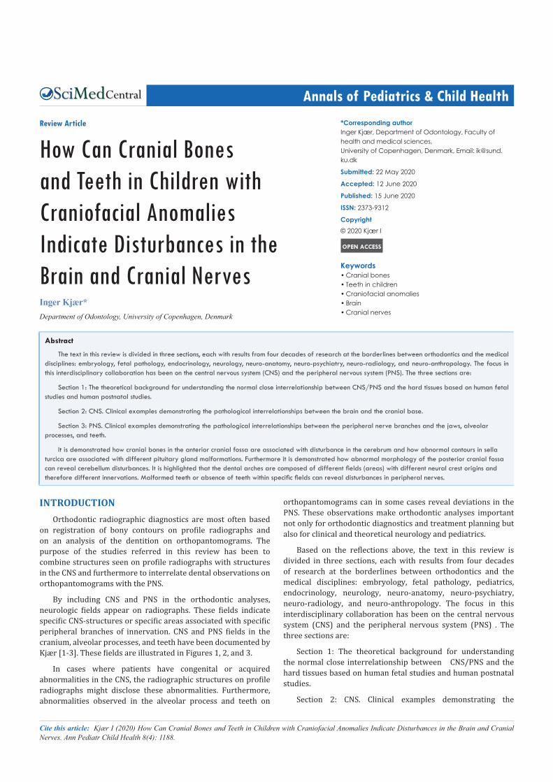
Annals of Pediatrics & Child Health Cite this article: Kjær I (2020) How Can Cranial Bones and Teeth in Children with Craniofacial Anomalies Indicate Disturbances in the Brain and Cranial Nerves. Ann Pediatr Child Health 8(4): 1188. Central *Corresponding author Inger Kjær, Department of Odontology, Faculty of health and medical sciences, University of Copenhagen, Denmark, Email: ik@sund. ku.dk Submitted: 22 May 2020 Accepted: 12 June 2020 Published: 15 June 2020 ISSN: 2373-9312 Copyright © 2020 Kjær I OPEN ACCESS Keywords • Cranial bones • Teeth in children • Craniofacial anomalies • Brain • Cranial nerves Abstract The text in this review is divided in three sections, each with results from four decades of research at the borderlines between orthodontics and the medical disciplines: embryology, fetal pathology, endocrinology, neurology, neuro-anatomy, neuro-psychiatry, neuro-radiology, and neuro-anthropology. The focus in this interdisciplinary collaboration has been on the central nervous system (CNS) and the peripheral nervous system (PNS). The three sections are: Section 1: The theoretical background for understanding the normal close interrelationship between CNS/PNS and the hard tissues based on human fetal studies and human postnatal studies. Section 2: CNS. Clinical examples demonstrating the pathological interrelationships between the brain and the cranial base. Section 3: PNS. Clinical examples demonstrating the pathological interrelationships between the peripheral nerve branches and the jaws, alveolar processes, and teeth. It is demonstrated how cranial bones in the anterior cranial fossa are associated with disturbance in the cerebrum and how abnormal contours in sella turcica are associated with different pituitary gland malformations. Furthermore it is demonstrated how abnormal morphology of the posterior cranial fossa can reveal cerebellum disturbances. It is highlighted that the dental arches are composed of different fields (areas) with different neural crest origins and therefore different innervations. Malformed teeth or absence of teeth within specific fields can reveal disturbances in peripheral nerves. INTRODUCTION Orthodontic radiographic diagnostics are most often based on registration of bony contours on profile radiographs and on an analysis of the dentition on orthopantomograms. The purpose of the studies referred in this review has been to combine structures seen on profile radiographs with structures in the CNS and furthermore to interrelate dental observations on orthopantomograms with the PNS. By including CNS and PNS in the orthodontic analyses, neurologic fields appear on radiographs. These fields indicate specific CNS-structures or specific areas associated with specific peripheral branches of innervation. CNS and PNS fields in the cranium, alveolar processes, and teeth have been documented by Kjær [1-3]. These fields are illustrated in Figures 1, 2, and 3. In cases where patients have congenital or acquired abnormalities in the CNS, the radiographic structures on profile radiographs might disclose these abnormalities. Furthermore, abnormalities observed in the alveolar process and teeth on orthopantomograms can in some cases reveal deviations in the PNS. These observations make orthodontic analyses important not only for orthodontic diagnostics and treatment planning but also for clinical and theoretical neurology and pediatrics. Based on the reflections above, the text in this review is divided in three sections, each with results from four decades of research at the borderlines between orthodontics and the medical disciplines: embryology, fetal pathology, pediatrics, endocrinology, neurology, neuro-anatomy, neuro-psychiatry, neuro-radiology, and neuro-anthropology. The focus in this interdisciplinary collaboration has been on the central nervous system (CNS) and the peripheral nervous system (PNS) . The three sections are: Section 1: The theoretical background for understanding the normal close interrelationship between CNS/PNS and the hard tissues based on human fetal studies and human postnatal studies. Section 2: CNS. Clinical examples demonstrating the Review Article How Can Cranial Bones and Teeth in Children with Craniofacial Anomalies Indicate Disturbances in the Brain and Cranial Nerves Inger Kjær* Department of Odontology, University of Copenhagen, Denmark
Transcript of How Can Cranial Bones and Teeth in Children with ...
Annals of Pediatrics & Child Health
Cite this article: Kjær I (2020) How Can Cranial Bones and Teeth in Children with Craniofacial Anomalies Indicate Disturbances in the Brain and Cranial Nerves. Ann Pediatr Child Health 8(4): 1188.
Central
*Corresponding authorInger Kjær, Department of Odontology, Faculty of health and medical sciences, University of Copenhagen, Denmark, Email: [email protected]
Submitted: 22 May 2020
Accepted: 12 June 2020
Published: 15 June 2020
ISSN: 2373-9312
Copyright© 2020 Kjær I
OPEN ACCESS
Keywords•Cranial bones•Teeth in children•Craniofacial anomalies•Brain•Cranial nerves
Abstract
The text in this review is divided in three sections, each with results from four decades of research at the borderlines between orthodontics and the medical disciplines: embryology, fetal pathology, endocrinology, neurology, neuro-anatomy, neuro-psychiatry, neuro-radiology, and neuro-anthropology. The focus in this interdisciplinary collaboration has been on the central nervous system (CNS) and the peripheral nervous system (PNS). The three sections are:
Section 1: The theoretical background for understanding the normal close interrelationship between CNS/PNS and the hard tissues based on human fetal studies and human postnatal studies.
Section 2: CNS. Clinical examples demonstrating the pathological interrelationships between the brain and the cranial base.
Section 3: PNS. Clinical examples demonstrating the pathological interrelationships between the peripheral nerve branches and the jaws, alveolar processes, and teeth.
It is demonstrated how cranial bones in the anterior cranial fossa are associated with disturbance in the cerebrum and how abnormal contours in sella turcica are associated with different pituitary gland malformations. Furthermore it is demonstrated how abnormal morphology of the posterior cranial fossa can reveal cerebellum disturbances. It is highlighted that the dental arches are composed of different fields (areas) with different neural crest origins and therefore different innervations. Malformed teeth or absence of teeth within specific fields can reveal disturbances in peripheral nerves.
INTRODUCTIONOrthodontic radiographic diagnostics are most often based
on registration of bony contours on profile radiographs and on an analysis of the dentition on orthopantomograms. The purpose of the studies referred in this review has been to combine structures seen on profile radiographs with structures in the CNS and furthermore to interrelate dental observations on orthopantomograms with the PNS.
By including CNS and PNS in the orthodontic analyses, neurologic fields appear on radiographs. These fields indicate specific CNS-structures or specific areas associated with specific peripheral branches of innervation. CNS and PNS fields in the cranium, alveolar processes, and teeth have been documented by Kjær [1-3]. These fields are illustrated in Figures 1, 2, and 3.
In cases where patients have congenital or acquired abnormalities in the CNS, the radiographic structures on profile radiographs might disclose these abnormalities. Furthermore, abnormalities observed in the alveolar process and teeth on
orthopantomograms can in some cases reveal deviations in the PNS. These observations make orthodontic analyses important not only for orthodontic diagnostics and treatment planning but also for clinical and theoretical neurology and pediatrics.
Based on the reflections above, the text in this review is divided in three sections, each with results from four decades of research at the borderlines between orthodontics and the medical disciplines: embryology, fetal pathology, pediatrics, endocrinology, neurology, neuro-anatomy, neuro-psychiatry, neuro-radiology, and neuro-anthropology. The focus in this interdisciplinary collaboration has been on the central nervous system (CNS) and the peripheral nervous system (PNS) . The three sections are:
Section 1: The theoretical background for understanding the normal close interrelationship between CNS/PNS and the hard tissues based on human fetal studies and human postnatal studies.
Section 2: CNS. Clinical examples demonstrating the
Review Article
How Can Cranial Bones and Teeth in Children with Craniofacial Anomalies Indicate Disturbances in the Brain and Cranial NervesInger Kjær*Department of Odontology, University of Copenhagen, Denmark
CentralKjær I (2020)
Ann Pediatr Child Health 8(5): 1188 (2020) 2/11
pathological interrelationships between the brain and the cranial base.
Section 3: PNS. Clinical examples demonstrating the pathological interrelationships between the peripheral nerve branches and the jaws, alveolar processes, and teeth.
SECTION 1
The theoretical background for understanding the normal close interrelationship between CNS/PNS and the hard tissues
CNS/Cranial Base: The central nervous system consists of the brainstem, the cerebrum, the cerebellum, and the spine. All structures are developed from the primitive neural tube. This development is controlled by the axially located cell chord, the notochord [3] (Figure 1). In this review, the brain part of the CNS is described. The brain has an axial posterior part, predominantly pons and cerebellum, and an anterior part where bilateral cerebrum structures develop (Figure 2). These bilateral structures are separated mid-axially by the falx cerebri from the dura mater, which is attached to the crista galli. Between the posterior and the anterior parts of the brain, the pituitary gland develops from the infundibulum cerebri (Figure 2).
When it comes to cranial development, exactly the same overall structures are seen, with a posterior cranial part, fossa cranii posterior, constituted of mainly clivus and the occipital squama, and an anterior part, mainly fossa cranii anterior, constituted of the body of the sphenoid bone, ethmoid bone, and nasal septum.
Pituitary gland and sella turcica: Between the posterior and anterior parts of the cranial base is where the sella turcica
Located, enclosing the pituitary gland centrally in the brain (Figure 2). The sella turcica represents the fossa cranii media on profile radiographs. As the sella turcica is a bordering structure between the anterior and posterior fossae, the anterior wall of the sella reflects the development of the fossa cranii anterior, while the posterior wall, including the cranial end of the notochord, reflects the posterior part of the cranial base [3].
While the sella turcica is an important bone structure postnatally, the sella and the pituitary gland have been ignored in prenatal autopsy descriptions, because the autopsy procedure does not allow ordinary histological preparation of the pituitary gland region [4].
Studies of sella turcica development have revealed how the posterior wall component develops first from the cartilage encircling the rostral end of the notochord5. At that stage, the anterior part of the pituitary gland, composed of adeno-pituitary tissue, attached to the infundibulum cerebri, is drawn cranially from the pharyngeal mucosa. Thereafter, the complete pituitary gland with the anterior lobe, the adeno-pituitary gland, an intermediate lobe and a posterior lobe, the neuro pituitary gland, is formed [5,6]. Sella turcica morphology and the pituitary gland have formerly been described based on histology and neuro-radiography7. Studies have demonstrated that the location of the anterior pituitary gland (adeno-pituitary gland) is interrelated with sella turcica morphology [8]. After this endocrinological developmental process, the anterior wall and lastly the floor of the sella turcica develop [3,9].
According to this developmental process, distinctions are made between bony abnormalities in the anterior wall of the sella turcica, in the posterior wall, and in the floor (see later). These distinctions are made prenatally as well as postnatally. Prenatal analyses the location of the pituitary gland tissues have been reported [3,9-15].
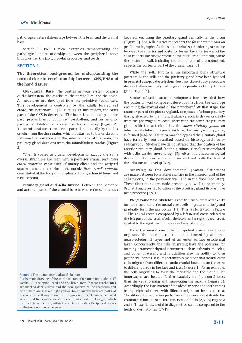
PNS/Craniofacial skeleton: From the rim or crest of the early formed neural tube, the neural crest cells migrate anteriorly and gradually form the jaw bones [1,3]. This is illustrated in Figure 1. The neural crest is composed by a left neural crest, related to the left part of the craniofacial skeleton, and a right neural crest, related to the right part of the craniofacial skeleton.
From the neural crest, the pluripotent neural crest cells originate. The neural crest is a crest formed by an inner neuro-ectodermal layer and of an outer surface ectodermal layer. Concurrently, the cells migrating have the potential for forming ectomesenchymal structures such as subcutis, muscles, and bones bilaterally and in addition also the ability to form peripheral nerves. It is important to remember that neural crest cells migrate from different caudo-cranial locations on the crest to different areas in the face and jaws (Figure 1). As an example, the cells migrating to form the mandible and the mandibular innervation are located further caudally on the neural crest than the cells forming and innervating the maxilla (Figure 1). Accordingly, the innervation of the alveolar bone and teeth comes from peripheral nerves with different origins on the neural crest. The different innervation paths from the neural crest divide the craniofacial hard tissues into innervation fields [2,3,16] Figure 2 and 3. These fields, useful in diagnostics, can be compared to the fields of dermatomes [17-19].
Figure 1 The human prenatal axial skeleton.A schematic drawing of the axial skeleton of a human fetus, about 17 weeks GA. The spinal cord and the brain stem (except cerebellum) are marked dark yellow, and the hemispheres of the cerebrum and cerebellum are marked light yellow. Green arrows indicate paths of neural crest cell migrations to the jaws and facial bones, coloured green. Red lines mark structures with an ectodermal origin, which includes the notochord, within the vertebral bodies. Peripheral nerves to the jaws are marked orange.

CentralKjær I (2020)
Ann Pediatr Child Health 8(5): 1188 (2020) 3/11
Figure 2 Registration on profile radiographs.a) A profile radiograph coloured according to craniofacial fields with different embryological origins. The fields in the maxilla are: frontal
nasal field (yellow), maxillary field (red), and palatal field (orange). In the mandible, the incisor field, canine/premolar field and molar field are all marked blue. The occipital field is marked green and the theca region purple. The interrelationship between the cranial base, jaw bone and teeth is exemplified in the maxillary field (red), where the canine and two premolars are drawn. Newer research [82], has suggested that the canine might have a separate field.
b) A profile radiograph coloured according to craniofacial fields with different embryological origins (same as 2a). Inserted on the figure are the cephalometric landmarks: n(nasion), s(sella), ba(basion), sp (anterior nasal spine), ss (subspinal), pm (pterygomaxillary), sm (supramental), pg(pogonion), gn(gnathion), tgo (gonion tangent point), ar (articular), o (opisthion), sqo (squamaoccipithal). Furthermore, the cranial base angle (n-s-ba) is marked, and the occipital angle (the angle between the lines s/ba and o/sqo). Also, the jaw prognathia angles, the palatal length and the mandibular angle are marked.This illustration indicates that the etiology behind deviations in cephalometric distances can be traced back to deviations in different fields. As an example, the etiology behind a short n-s distance is deviation in the fronto-nasal field and the etiology behind a short palate is specifically caused by deviations in the maxillary and/or palatine field. Also the size/shape of the occipital field (green) can be expressed cephalometrically by the occipital angle and s-sqo distance.
c) The same profile radiograph of the craniofacial fields, demonstrated in Figures 2a and 2b, now with insertion of the brain: H (cerebral hemispheres), D (diencephalon with an extension, infundibulum cerebri to the sella turcica, where it forms the neuro-pituitary gland), M (metencephalon), BS (brain stem, including the pons and cerebellum), S (upper and lower spine). Note that the fossa cranii anterior (s-n) (Figure 2c) supports the frontal hemispheres of the cerebrum. Also note that the fossa cranii posterior, expressed as the angle between s-ba and o-sqo (See Figure b), encircles mainly the cerebellum and pons located in the green craniofacial field (Figure 2a).
These diagrams a, b, and c can be used in the clinic for etiology based diagnostics.
PNS and maxilla formation: The early close spatial interrelationship between bone formation in the maxilla and innervation is documented prenatally [20,21].
PNS and mandible formation: The formation of the mental foramen demonstrated that the bone gradually encircles the mental nerve and thereby forms the mental foramen [22]. The formation of the mandibular canal was mapped anthropologically in fetal mandibles about 25-35 weeks of gestation [23]. This study revealed that stepwise outgrowing peripheral nerves were gradually enrolled in the formation of the mandibular canal. Thus the inferior mandibular nerve is a bundle of nerve fibers, developed at different periods, and innervating different teeth. Bone spiculae appear between the nerve fibers in the mandibular canal.
PNS/CNS and Vomero-nasal organs: In the anterior part of the craniofacial area, the bilateral vomero-nasal organs are developed. These organs, formed early in prenatal life close to the nasal septum, produce Luteinising Hormone Releasing Hormone (LHRH) [24,25]. The LHRH positive cells migrate from the vomero-nasal organ later in prenatal life cranially along the terminal nerves (PNS) to the olfactory bulbs, and further to the hypothalamus (CNS) from where the hormone, LHRH, postnatally influences the growth hormone production in the pituitary gland3.
PNS/Dentition: Systematic histological overviews of human tooth formation are always based on early fetal crown formation in the primary dentition before GA 21 weeks. This is due to limited access to human tissue from the periods where the permanent teeth develop pre- and postnatally.
Histochemical methods have demonstrated neuronal markings of human odontoblasts and of nerve fibers apically to the developing crown in a ligament-like structure, presumably the later Hammock ligament [26,27]. This ligament is in this early stage named the root membrane, even though the root has not formed. Further human studies have documented the spatio-temporal expression of innervation in the root membrane [28,29]. How the peripheral nerves find their way (path finding) to the developing tooth buds has been proven experimentally by Luuko et al. [30], and by Kettunen et al. [31].
Normal dental development, which cannot be studied prenatally
Root formation: Root formation in humans has been studied longitudinally by radiography. Histological studies on root formation have not been possible. For a better understanding of different root abnormalities in the human dentition, a study on extracted teeth with attached periodontal tissue was performed elucidating the cell-layers close to the root surface [32]. Closest

CentralKjær I (2020)
Ann Pediatr Child Health 8(5): 1188 (2020) 4/11
to the root surface is a close network of nerve fibers covered by an ectomesenchymal tissue sheet, and this is covered by an outermost layer of ectodermally derived Malassez epithelium, innervated by tiny nerve fibers. Together these three main tissue layers, illustrated in Figure 4, were named the peri-root sheet [32-34]. A similar root sheet with the same three tissue layers, though morphologically different, was also observed close to the roots of the primary teeth [35].
Normal eruption: Insight in the normal tooth eruption process is fundamental for understanding pathological tooth eruption. The eruption process is still not completely understood but different theories exist [36]. One of these will be highlighted in the following.
The normal tooth eruption is a physiological process which depends primarily on innervation of the root membrane but also on the penetration ability for resorptive cells in the crown follicle, and on the periodontic membrane’s ability to regenerate during the eruption process [3]. Histo-chemical studies have proven the existence of a peri-root sheet with three main cell layers covering the root surface [36]. The outer cell layer in this sheet is the mucosal layer of Malassez epithelium. The middle layer is the ecto-mesenchyme fiber layer, and the innermost layer, close to the root surface is rich in innervation [32,34]. Innervation also occurs in the Malassez epithelium [37]. The re-building ability of the peri-root sheet during tooth eruption, where the tooth moves and the bone stays stable, has been proven by marking of apoptotic reaction in cells close to the root [38]. Permeability of the crown follicle secures mono-nuclear resorptive cells to penetrate the membrane and resorb the overlying bone, which is important for tooth emergence [1,3]. The root membrane is highly innervated, similar to innervation observed in the end-pieces of glands. It is hypothesized that the innervation of the root membrane generates a pressure, comparable to the pressure at the highly innervated end-pieces in glands, creating flow of saliva. In the teeth, the pressure from the root membranes creates the force which is important for tooth movement [36]. Accordingly, the following three eruption factors, illustrated in Figure 4, are supposed to interact. The root membrane creates a pressure for eruption movement, the follicle makes a path in the bone for eruption, and the periodontal membrane secures the adaptation process under normal development. It is furthermore hypothesized that if one of these three factors does not function, then tooth eruption stops or deviates. The post-emergence eruption depends on the activity in the root membrane and in the periodontal membrane [36].
Conclusion: CNS/PNS and orthodontic analyses
The profile radiograph, the orthopantomogram, and the cone beam radiograph are usually the radiographs used in orthodontic diagnostics. On profile radiographs, the cephalometric landmarks and lines are used for expressing normal and abnormal craniofacial development, but the etiology is normally not expressed.
In this first section, the prenatal developmental fields with common origins are incorporated in the postnatal profiles and orthopantomograms (Figure 2a and 3).
If the fields are added to the cephalometric analyses on pro-
file radiographs (Figure 2b), then the abnormal cephalometric measurements can directly be interrelated to the fields. If fur-thermore, the brain is inserted in the “empty” cranium (Figure 2c), observed on the profile radiograph, then the cephalometric measurements can be interrelated also to the brain. Accordingly, the Figures. 2a, 2b, and 2c are used in the evaluation of cranio-facial etiology. If also the developmental fields are added to the orthopantomograms (Figure 3), then the evaluation of jaw bone and dental etiology can be expressed.
The background for this present short overview is mainly the author’s scientific observation of dental and craniofacial findings in human fetuses and individuals with normal development in the central nervous system (CNS), and normal development in the peripheral nervous system (PNS). These findings in large collections of materials form the basis for diagnostics of pathological cases [18,39], highlighted in the following two sections.
SECTION 2 CNS. Clinical examples demonstrating pathological interrelationships between the brain and the cranial base
Fossa cranii anterior/cerebrum: Associated with the observation of a single mid-axially located maxillary central incisor (SMMCI) in the fronto-nasal field is a significantly shorter fossa cranii anterior (sella –nasion distance) [40]. Also absence of the intermaxillary suture, short or absent nasal bone and abnormal crista galli are skeletal signs observed in SMMCI patients. According to Figure 2a and 2b, the fronto-nasal field is reduced or malformed.
The SMMCI condition has been observed in several cases with mental retardation. Neuro-radiographical observations have in these cases revealed non-separated bilateral cerebral hemispheres [41]. The involvement of the cerebral hemispheres is illustrated in Figure 2c. A severe variation of this condition called holoprosencephaly has been demonstrated neuro-embryologically with unseparated cerebral hemispheres and the presence of a single median centrally located eye (cyclopia) [42].
Fossa cranii posterior/cerebellum, pons: In patients with Down syndrome a reduced volume/size in the occipital region where the pons and cerebellum are located has been described by cephalometry43. The length of the clivus (sella-nasion distance) was reduced, compared to normal standard. The cranial base angle (s-n-a) was enlarged and the occipital area reduced compared to non-Down individuals. This morphological deviation was first described prenatally [44]. Interestingly, MR-studies of individuals with Down syndrome have demonstrated that the size of the cerebellum is less than 75% compared to the size in non-Down individuals [45].
Also complete cleft lip and palate patients can be diagnosed with cerebellum abnormalities [46]. In these cases, deviant shape abnormalities have been observed cephalometrically in the fossa cranii posterior [47].
Sella turcica/pituitary gland: The pituitary gland is normally located within the sella turcica, where the cephalometrically important sella point, S, is constructed for cephalometric
CentralKjær I (2020)
Ann Pediatr Child Health 8(5): 1188 (2020) 5/11
measurements of the cranial base angle (Figure 2b, 5b). In many orthodontic cases, abnormal morphology of the sella turcica makes the exact location of the s-point difficult. This influences the cephalometric analyses and conclusions. Normal postnatal size and morphology of the sella turcica have been elucidated, according to age and gender in an extensive study [48], while the location and morphology of the postnatal pituitary gland have only been sporadically described. The postnatal pituitary gland is not included in the present overview.
In profile radiographs of patients with severe craniofacial deviations, abnormal sella turcica morphology characterized by sella bridges has been described in 18.6% of a population of 177 individuals undergoing orthognathic surgery [49].
In the prenatal development of the sella turcica described in section 1, the description of pathological sellae focuses on
deviations in the anterior sella wall, the posterior sella wall, and deviations in the floor [3] (Figure 4).
Abnormal anterior wall of the sella turcica: Absence or low-slanting anterior wall has been reported in SMMCI, holoprosencephaly and combined cleft lip and palate [42,50] (Figure 5). Also in body axis abnormalities, such as in prenatal and postnatal encephaloceles and myelomeningoceles, oblique anterior wall deviations have been observed [51,52]. In pathological prenatal cases, such as anencephaly where the adeno-pituitary gland tissue is absent, also the anterior wall of sella turcica is absent53. In this case, the necessary early attachment between the pharyngeally located adeno-pituitary gland tissue and the infundibulum cerebri fails. When comparing the sella turcica morphology in fetuses with trisomy 21 and trisomy 18, there is a difference which seemingly is genetically determined13. When comparing the prenatal morphology of the
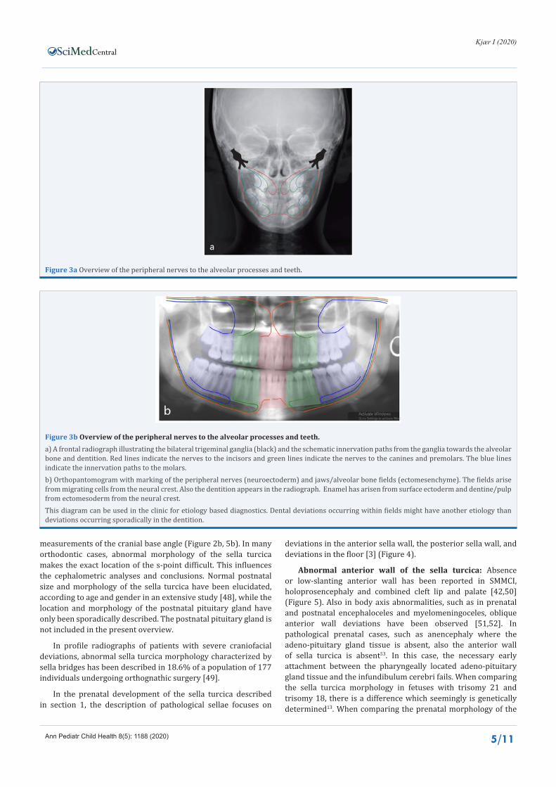
Figure 3b Overview of the peripheral nerves to the alveolar processes and teeth.a) A frontal radiograph illustrating the bilateral trigeminal ganglia (black) and the schematic innervation paths from the ganglia towards the alveolar bone and dentition. Red lines indicate the nerves to the incisors and green lines indicate the nerves to the canines and premolars. The blue lines indicate the innervation paths to the molars.b) Orthopantomogram with marking of the peripheral nerves (neuroectoderm) and jaws/alveolar bone fields (ectomesenchyme). The fields arise from migrating cells from the neural crest. Also the dentition appears in the radiograph. Enamel has arisen from surface ectoderm and dentine/pulp from ectomesoderm from the neural crest.This diagram can be used in the clinic for etiology based diagnostics. Dental deviations occurring within fields might have another etiology than deviations occurring sporadically in the dentition.
Figure 3a Overview of the peripheral nerves to the alveolar processes and teeth.
CentralKjær I (2020)
Ann Pediatr Child Health 8(5): 1188 (2020) 6/11
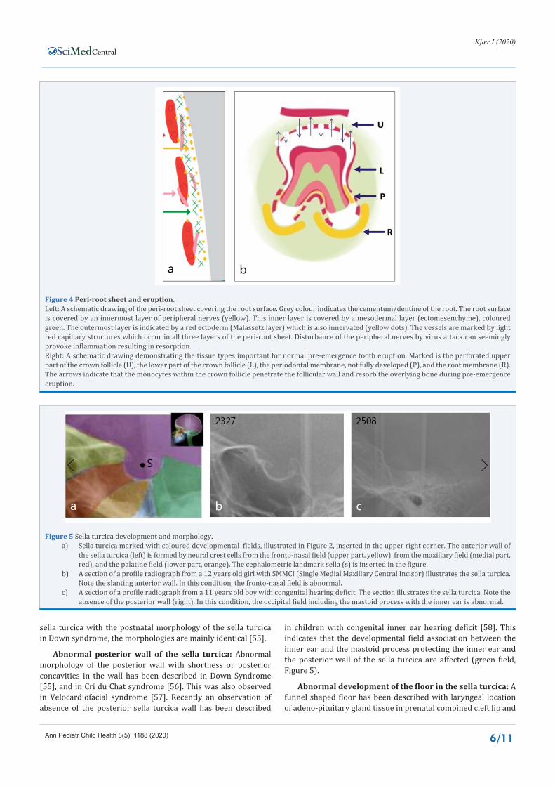
Figure 4 Peri-root sheet and eruption.Left: A schematic drawing of the peri-root sheet covering the root surface. Grey colour indicates the cementum/dentine of the root. The root surface is covered by an innermost layer of peripheral nerves (yellow). This inner layer is covered by a mesodermal layer (ectomesenchyme), coloured green. The outermost layer is indicated by a red ectoderm (Malassetz layer) which is also innervated (yellow dots). The vessels are marked by light red capillary structures which occur in all three layers of the peri-root sheet. Disturbance of the peripheral nerves by virus attack can seemingly provoke inflammation resulting in resorption.Right: A schematic drawing demonstrating the tissue types important for normal pre-emergence tooth eruption. Marked is the perforated upper part of the crown follicle (U), the lower part of the crown follicle (L), the periodontal membrane, not fully developed (P), and the root membrane (R). The arrows indicate that the monocytes within the crown follicle penetrate the follicular wall and resorb the overlying bone during pre-emergence eruption.
Figure 5 Sella turcica development and morphology.a) Sella turcica marked with coloured developmental fields, illustrated in Figure 2, inserted in the upper right corner. The anterior wall of
the sella turcica (left) is formed by neural crest cells from the fronto-nasal field (upper part, yellow), from the maxillary field (medial part, red), and the palatine field (lower part, orange). The cephalometric landmark sella (s) is inserted in the figure.
b) A section of a profile radiograph from a 12 years old girl with SMMCI (Single Medial Maxillary Central Incisor) illustrates the sella turcica. Note the slanting anterior wall. In this condition, the fronto-nasal field is abnormal.
c) A section of a profile radiograph from a 11 years old boy with congenital hearing deficit. The section illustrates the sella turcica. Note the absence of the posterior wall (right). In this condition, the occipital field including the mastoid process with the inner ear is abnormal.
sella turcica with the postnatal morphology of the sella turcica in Down syndrome, the morphologies are mainly identical [55].
Abnormal posterior wall of the sella turcica: Abnormal morphology of the posterior wall with shortness or posterior concavities in the wall has been described in Down Syndrome [55], and in Cri du Chat syndrome [56]. This was also observed in Velocardiofacial syndrome [57]. Recently an observation of absence of the posterior sella turcica wall has been described
in children with congenital inner ear hearing deficit [58]. This indicates that the developmental field association between the inner ear and the mastoid process protecting the inner ear and the posterior wall of the sella turcica are affected (green field, Figure 5).
Abnormal development of the floor in the sella turcica: A funnel shaped floor has been described with laryngeal location of adeno-pituitary gland tissue in prenatal combined cleft lip and
CentralKjær I (2020)
Ann Pediatr Child Health 8(5): 1188 (2020) 7/11
palate [59], and in other severe craniofacial malformations [59].
SECTION 3PNS. Clinical examples demonstrating the pathological interrelationships between the peripheral nerve branches and the jaws, alveolar processes, and teeth.
Jaws and alveolar bone/PNS: It has been hypothesized by neural cell markings of human osteoblasts with PGP9.5 that compensatory and dysplastic growth of the jaws could be under control by peripheral nerves [60]. A malformed human anthropological mandible confirmed the prenatally described formation of the mandibular canal. This case further demonstrated the dependence of innervation for tooth formation [61].
Postnatally, Arne Björk demonstrated in his lifelong cephalometric studies with metal markers inserted in the jaws that the mandibular canal stayed stable during growth, and was therefore usable for super-imposing of radiographs for growth analysis [62]. The shape of the mandibular canal observed on radiographs expresses the mandibular growth pattern [63]. Human palatal growth has been evaluated on medieval crania using nerve canal openings as references [64]. On a reduced number of human anthropological maxillae, the direction of the infraorbital canal expressed the maxillary growth pattern [65]. Postnatally, the direction of the orbital canal has been suggested as a stable structure in the maxilla important for revealing the
maxillary growth pattern. As the orbital canal is not visible on patient radiographs, this observation can only be used on anthropological material.
Dentition/PNS: In this short review, focus will be on selected examples of dental deviations, associated with abnormal jaw development and dental deviations in individuals with abnormal PNS development.
Seemingly, all pathological dental deviations can be innate and/or acquired.
Pathological tooth development can be caused by a disruptive factor, such as virus infection, or by an innate malformation with one or more tissue types involved in the tooth formation [3].
Tooth agenesis
Maxillary central incisor: SMMCI, Single Median Maxillary Central Incisor, is a specific type of incisor agenesis interrelated with brain malformation. This condition is also associated with bone malformations in the fronto-nasal field [66] (Figure 6a). In these cases only one mid-axially located central maxillary incisor exists.
Maxillary lateral incisor agenesis in Combined Cleft Lip and Palate (CLP): Maxillary lateral incisor agenesis is a common finding in combined CLP-patients [67], while not common in other cleft types [67]. In combined CLP, abnormal cerebellar fields occur. Also abnormal brain development has been reported
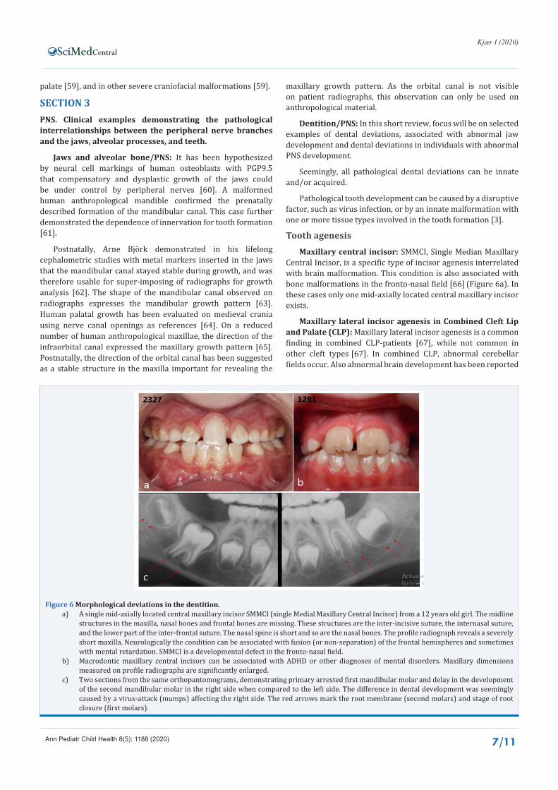
Figure 6 Morphological deviations in the dentition.a) A single mid-axially located central maxillary incisor SMMCI (single Medial Maxillary Central Incisor) from a 12 years old girl. The midline
structures in the maxilla, nasal bones and frontal bones are missing. These structures are the inter-incisive suture, the internasal suture, and the lower part of the inter-frontal suture. The nasal spine is short and so are the nasal bones. The profile radiograph reveals a severely short maxilla. Neurologically the condition can be associated with fusion (or non-separation) of the frontal hemispheres and sometimes with mental retardation. SMMCI is a developmental defect in the fronto-nasal field.
b) Macrodontic maxillary central incisors can be associated with ADHD or other diagnoses of mental disorders. Maxillary dimensions measured on profile radiographs are significantly enlarged.
c) Two sections from the same orthopantomograms, demonstrating primary arrested first mandibular molar and delay in the development of the second mandibular molar in the right side when compared to the left side. The difference in dental development was seemingly caused by a virus-attack (mumps) affecting the right side. The red arrows mark the root membrane (second molars) and stage of root closure (first molars).
CentralKjær I (2020)
Ann Pediatr Child Health 8(5): 1188 (2020) 8/11
[6,68]. This is a subject which needs to be sorted out, but the findings seemingly support the theory of different genotypic origins of the three cleft types in the maxilla.
Premolar agenesis
The most common types of agenesis (third molar, second premolar, and maxillary lateral incisor) seems to be agenesis at the site of nerve endings within a developmental field [1,70]. The etiology behind innate agenesis seems to be genetic, but the field patterns support that the innervation or the borderlines between fields are important factors in tooth development. Whether these borderlines between developmental fields influence the local growth of the alveolar bone is a question raised in juvenile periodontitis, where the initial areas of reduced bone levels appear at the borderline between fields [71]. If it is so, then the initial stages of juvenile periodontitis are local disturbances in alveolar bone growth71. Gender differences in the intra- and inter-jaw occurrences of agenesis of the second premolars have been studied [72]. This study seemingly indicated the intra- and inter-jaw influence of innervation on agenesis. Craniofacial development in multiple agenesis cases is different in patients with different numbers of congenitally missing permanent teeth [73].
Third molar agenesis
Third molar agenesis has been studied in CNS deviations such as in Down syndrome where the occurrence of agenesis was significantly higher compared to individuals without Down syndrome [74]. How the CNS condition is interrelated with third molar agenesis has not been determined.
Maxillary canine agenesis
Agenesis of the maxillary canines is a seldom phenomenon (prevalence 0.07-0.13%). Except for the canine agenesis, these dentitions appeared normal [75]. This could be the reason for defining a specific etiology behind maxillary canine agenesis and for presuming a specific canine PNS innervation field. Retrognathia of the maxilla appears in bilateral maxillary canine agenesis [75], which could indicate a close interrelationship between regional tooth and jawbone development.
Tooth malformations
Macrodontic incisors: Macrodontic incisors have been observed in patients with enlarged craniofacial dimensions [76] (Figure 6b). A later study on the same group of patients demonstrated that out of 22 cases with macrodontic central incisors 11 cases had neuro-psychiatric diagnoses [77].
En coup de sabre: Tooth malformation has been described in the condition “En coup de sabre”, characterized by areas with abnormal skin formation, specifically in the face. It has been demonstrated that the malformations occurs in the dental arch, exactly in the same region where the facial skin affection occurs.
In a recent study of six patients with “En coup de sabre” it was documented that the contours of the facial skin abnormalities were identical to the cranial regional fields and associated with tooth abnormalities within the affected fields [79]. In two cases with identical skin affections the pre-sphenoid bone area
demonstrated identical bony malformations [79]. Neurological complications, including epilepsy, have been reported in this condition [79]. The interrelationship between the skin, teeth and bone abnormalities, and the CNS and PNS abnormalities should be highlighted in future studies.
Arrested tooth formation in fields
Arrested tooth formation regionally was first documented in a case after a virus affecting the peripheral nervous system, seemingly by destruction of the Schwann cells. In this case, the virus infection also resulted in temporary hearing loss in the same side where the arrested tooth formation occurred [80]. After the Schwann cell had regenerated and more or less normalized the PNS function, the hearing improved, and the tooth formation caught up. The result of this temporary arrest in innervation was abnormal tooth morphologies (taurodontia) and, in some teeth, arrested root development [80].
ERUPTION DEVIATIONSArrested molar eruption
All 3 tissue types mentioned as important for normal tooth eruption (section 1) can be involved in arrestment in the eruption process. In this short overview, only the influence of innervation on tooth eruption will be highlighted. If the Schwann cells are destroyed by virus attack, e.g. mumps virus, then the periodontal membrane will be affected, resulting in eruption deviation, and sometimes in ankylosis in the innervation fields. It has been highlighted how arrestment of the first molar often is followed by arrestment of the second and third molars within the same innervation field [3] (Figure 6c). This can also appear in more than one field.
Transpositions
Transposition is a specific eruption deviation, often observed in a maxillary canine region when a maxillary canine erupts between premolars or between incisors. It has been hypothesized in a study of 63 dentitions with maxillary canine transpositions that a separate canine field during development overlaps either distally to the premolar field or mesially to the incisor field [81]. These processes can be compared to overlapping fields observed in dermatomes along the body axis [82]. A dermatome is defined as a specific skin region innervated by a specific nerve branch. The same definition is used for craniofacial fields where each field is innervated by a specific nerve fiber.
Resorption
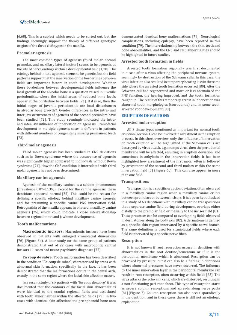
It is not known if root resorption occurs in dentition with abnormalities in the root dentine/cementum or if it is the periodontal membrane which is abnormal. Resorption can be provoked by pressure, but it can also be a finding in dentitions where abnormal pressures have never occurred. The influence by the inner innervation layer in the periodontal membrane can result in root resorption, often occurring within fields [83]. The virus attacks the Schwann cells, which are disturbed, resulting in a non-functioning peri-root sheet. This type of resorption starts as severe column resorptions and spreads along nerve paths [81] (Figure 7). Column resorption can also occur sporadically in the dentition, and in these cases there is still not an etiologic explanation.
CentralKjær I (2020)
Ann Pediatr Child Health 8(5): 1188 (2020) 9/11
Figure 7 Two dental films registered severe column resorption at the initial stages. The resorption processes are spread along the innervation fields in the maxilla until the midline and in the mandible restricted to the incisor field. The resorbed teeth were replaced by dental implants, which are observed in the orthopantomogram.
CONCLUSIONIn this present overview, examples from four decades of
human prenatal and postnatal studies on the interrelationship between hard tissue and nerve tissue have been presented with relevant references. This interrelationship, called neuro-osteology, crosses the borderline between odontology and neurology in medicine. All studies are performed in relation to diagnostic problems, with the purpose of improving diagnostics and treatment planning. The presentation focuses on how information from medical and odontological disciplines can add new insight to diagnostics of craniofacial anomalies in children.
ACKNOWLEDGEMENTSSincere thanks are extended to the many medical professionals
in different medical disciplines who have supported the studies behind this review. For preparation of the present manuscript, academic secretary Eva Reinwald is sincerely acknowledged.
REFERENCES1. Kjær I. Neuro-osteology. Crit Rev Oral Biol Med. 1998; 9: 224-244.
2. Kjær I. Orthodontics and foetal pathology: a personal view on craniofacial patterning. Eur J Orthod. 2010; 32: 140-147.
3. Kjær I. Etiology- Based Dental and Craniofacial Diagnostics. Wiley Blackwell, Oxford, UK. 2017.
4. Kjær I, Hansen BF. The prenatal pituitary gland - hidden and forgotten. Pediatric Neurology. 2000; 22: 155-156.
5. Kjær I, Hansen BF. The adenohypophysis and the cranial base in early human development. J Craniofac Genet Dev Biol. 1995; 15: 157-161.
6. Kjær I, Nolting D, Hansen BF. p75-NGFR Expression in the human prenatal pituitary gland. Pediatric neurology. 2004; 5: 345-348.
7. Kjær I. Sella turcica morphology and the pituitary gland - a new contribution to craniofacial diagnostics based on histology and neuroradiology. Online Eur J Orthod. 2012.
8. Kjær I. Location of anterior pituitary gland tissue is interrelated with sella turcica morphology in human fetuses -review and perspective. Int J of Clin Endocrin. 2017; 1: 59-62.
9. Kjær I, Hansen BF. Human fetal pituitary gland in holoprosencephaly and anencephaly. J Craniofac Genet Dev Biol. 1995; 15: 222-229.
10. Kjær I, Hansen BF, Keeling JW. Axial skeleton and pituitary gland in human fetuses with spina bifida and cranial encephalocele. Pediatr Pathol. 1996; 16: 909-926.
11. Kjær I, Becktor KB, Nolting D, Hansen BF. The association between prenatal sella turcica morphology and notochordal remnants in the dorsum sellae. J Craniofac Genet Dev Biol. 1997; 17: 105-111.
12. Kjær I, Keeling JW, Reintoft I, Hjalgrim H, Nolting D, Hansen BF. Pituitary gland and sella turcica in human trisomy 18 fetuses. Am J Med Genet. 1998; 76: 87-92.
13. Kjær I, Keeling JW, Reintoft I, Nolting D, Hansen BF. Pituitary gland and sella turcica in human trisomy 21 fetuses related to axial skeletal development. Am J Med Genet. 1998; 80: 494-500.
14. Kjær I, Hansen BF, Reintoft I, Keeling JW. Pituitary gland and axial skeleton malformations in human fetuses with spina bifida. Eur J Pediatr Surg. 1999; 9: 354-358.
15. Kjær KW, Hansen BF, Keeling JW, Nolting D, Kjær I. Malformation of cranial base structures and pituitary gland in prenatal Meckel syndrome. APMIS. 1999; 107: 937-944.
CentralKjær I (2020)
Ann Pediatr Child Health 8(5): 1188 (2020) 10/11
16. Kjær I. Prenatal traces of aberrant neurofacial growth. Acta Odontol Scand. 1998; 56: 326-330.
17. Kjær I. New diagnostics of the dentition on panoramic radiographs - focusing on the peripheral nervous system as an important aetiological factor behind dental anomalies. Orthodontic Waves. 2012; 71: 1-16.
18. Kjær I. Review: Dental approach to craniofacial syndromes: how can developmental fields show us a new way to understand pathogenesis? Int J Dent. 2012: 10: 145749.
19. Lauesen SR, Daugaard-Jensen J, Lauridsen E, Kjær. Localised scleroderma en coup de sabre affecting the skin, dentition and bone tissue within craniofacial neural crest fields. Clinical and radiographic study in six patients. Eur Arch Pediatr Dent. 2019; 20: 339-350.
20. Kjær I. Correlated appearance of ossification and nerve tissue in human fetal jaws. J Craniofac Genet Dev Biol 1990; 10: 329-336.
21. Takashi O, Hansen BF, Nolting D, Kjær I. Nerve Growth Factor Receptor Immunolocalization During Human Palate and Tongue Development. Cleft Palate-Craniofacial J. 2003; 40: 116-125.
22. Kjær I. Formation and early prenatal location of the human mental foramen. Scand J Dent Res. 1989; 97: 1-7.
23. Chavéz-Lomelí ME, Mansilla Lory J, Pompa JA, Kjær I. The human mandibular canal arises from three separate canals innervating different tooth groups. J Dent Res. 1996; 75: 1540-1544.
24. Kjær I, Hansen BF. The human vomeronasal organ: Prenatal developmental stages and distribution of luteinizing hormone-releasing hormone. Eur J Oral Sci. 1996; 104: 34-40.
25. Kjær I, Hansen BF. Luteinizing hormone-releasing hormone and innervation pathways in human prenatal nasal submucosa: Factors of importance in evaluating Kallmann’s syndrome. APMIS. 1996; 104: 680-688.
26. Christensen LR, Mølgård K, Kjær I, Janas MS. Immunocytochemical demonstration of nerve growth factor receptor (NGF-R) in developing human fetal teeth. Anat Embryol. 1993; 188: 247-255.
27. Christensen LR, Janas MS, Mølgård K, Kjær I. An immunocytochemical study of the innervation of developing human fetal teeth using protein gene product 9.5 (PGP 9.5). Arch Oral Biol. 1993; 38: 1113-1120.
28. Becktor KB, Hansen BF, Nolting D, Kjær I. Spatiotemporal expression of NGFR during pre-natal human tooth development. Orthod Craniofacial Res. 2002; 5: 85-89.
29. Kock M, Nolting D, Kjær KW, Hansen BF, Kjær I. Immunohistochemical expression of p63 in human prenatal tooth primordia. Acta Odont Scand. 2005; 63: 253-257.
30. Luukko K, Kvinnsland IH, Kettunen P. Tissue interactions in the regulation of axon pathfinding during tooth morphogenesis. Dev Dyn. 2005; 234: 482-488.
31. Kettunen P, Spencer-Dene B, Furmanek T, Kvinnsland IH, Dickson C, Thesleff I, Luukko K. Fgfr2b mediated epithelial-mesenchymal interactions coordinate tooth morphogenesis and dental trigeminal axon patterning. Mech Dev. 2007; 11: 868-883.
32. Kjær I, Nolting D. The human periodontal membrane - focusing on the spatial interrelation between the epithelial layer of Malassez, fibers, and innervation. Acta Odontol Scand. 2009; 67: 134-138.
33. Kjær I. External root resorption - different aetiologies explained from the composition of the human root-close periodontal membrane. Hypotheses 2013; 4: 75-79.
34. Hansen IV, Vedtofte H, Kjær I. Remember the periroot sheet in orthodontic treatment of ectodermal dysplasia patients. Dental Hypotheses. 2014; 5: 164-167.
35. Bille MLB, Nolting D, Kjær I. Immunohistochemical studies of the periodontal membrane in primary teeth. Acta Odontol Scand. 2009; 67: 382-387.
36. Kjær I. Mechanism of Human Tooth Eruption: Review Article Including a New Theory for Future Studies on the Eruption Process. Scientifica. 2014; 341905.
37. Bille MLB, Thomsen B, Kjær I. The inter-relation between epithelial cells of Malassez and vesels studied immunohistochemically in the periodontal membrane of human primary and permanent teeth. Early online 2011.Acta Odontol Scand. 2012; 69: 109-113.
38. Bille MLB, Thomsen B, Kjær I. Apoptosis in the human periodontal membrane evaluated in primary and permanent teeth. Acta Odontol Scand. 2011; 69: 385-388.
39. Kjær I. Neuroosteology - a discipline of importance for evaluation of human craniofacial development. EC Neurology. 2018; 10.
40. Tabatabaie F, Sonnesen L, Kjær I. The neurocranial and craniofacial morphology in children with solitary median maxillary central incisor (SMMCI). Orthod Craniofac Res. 2008; 11: 96-104.
41. Kjær I, Wagner Aa, Thomsen LL, Holm K. Brain malformation in Single Median Maxillary Central Incisor. Neuropediatrics. 2010; 40: 280-283.
42. Kjær I, Keeling JW, Hansen BF, Becktor KB. Midline skeletodental morphology in holoprosencephaly. Cleft Palate-Craniofacial J 2002; 39: 357-363.
43. Krebs BJ, Kjær l. Can cephalometry reveal abnormal cerebellum development in Down syndrome? Reviews Press 2018; 2: 73-76.
44. Lomholt JF, Keeling JW, Hansen BF, Ono T, Stoltze K, Kjær I. The pre-natal development of the human cerebellar field in Downs syndrome. Orthod Craniofacial Res. 2003; 6: 220-226.
45. Aylward EH, Habbak R, Warren AC, Pulsifer MB, Barta PE, Jerram M, Pearlson GD. Cerebellar volume in adults with Down syndrome. Arch Neurol. 1997: 54: 209-212.
46. DeVolder I, Richman L, Conrad AL, Magnotta V, Nopoulos P. Abnormal Cerebellar Structure Is Dependent on Phenotype of Isolated Cleft of the Lip and/or Palate Cerebellum. 2013; 12: 236-244.
47. Dahl E. Craniofacial morphology in congenital clefts of the lip and palate. An x-ray cephalometric study of young adult males. Acta Odontol Scand. 1970; 57: 11-167.
48. Axelsson S, Storhaug K, Kjær I. Post-natal size and morphology of the sella turcica. Longitudinal cephalometric standards for Norwegians between 6 and 21 years. Eur J Orthod. 2004; 26: 597-604.
49. Becktor JP, Einersen S, Kjær I. A sella turcica bridge in subjects with severe craniofacial deviations. Eur J Orthod. 2000; 22: 69-74.
50. Nielsen BW, Mølsted K, Kjær I. Maxillary and sella turcica morophology in newborns with cleft lip and palate. Cleft Palate-Craniofac J. 2005; 42: 610-617.
51. Kjær I, Hansen BF, Reintoft I, Keeling JW. Pituitary gland and axial skeleton malformations in human fetuses with spina bifida. Eur J Pediatr Surg. 1999; 9: 354-358.
52. Kjær I, Wagner Aa, Madsen P, Blichfeldt S, Rasmussen K, Russell B. The sella turcica in children with lumbosacral myelomeningocele. Eur J Orthod. 1998; 20: 443-448.
53. Kjær I, Keeling JW, Græm N. Midline maxillofacial skeleton in human anencephalic fetuses. Cleft Palate-Craniofac J. 1994; 31: 250-256.
54. Kjær I, Keeling JW, Reintoft I, Hjalgrim H, Nolting D, Hansen B. Pituitary gland and sella turcica in human trisomy 18 fetuses. Am J Med Genet. 1998; 76: 87-92.
CentralKjær I (2020)
Ann Pediatr Child Health 8(5): 1188 (2020) 11/11
Kjær I (2020) How Can Cranial Bones and Teeth in Children with Craniofacial Anomalies Indicate Disturbances in the Brain and Cranial Nerves. Ann Pediatr Child Health 8(4): 1188.
Cite this article
55. Russell BG, Kjær I. Postnatal structure of the sella turcica in Down syndrome. Am J Med Genet. 1999; 87: 183-188.
56. Kjær I, Niebuhr E. Studies of the cranial base in 23 patients with Cri-du-Chat syndrome suggest a developmental field involved in the condition. Am J Med Genet. 1999; 82: 6-14.
57. Mølsted K, Boers M, Kjær I. The morphology of the sella turcica in Velocardiofacial Syndrome suggests involvement of a neural crest developmental field. Am J Med Genet. 2010; Part A 152A: 1450-1457.
58. Kjær I. Abnormal sella turcica morphology in individuals with congenital inner-ear hearing deficits. Dent Craniofac Res. 2018; 1.
59. Kjær I. Orthodontics and foetal pathology: a personal view on craniofacial patterning. Eur J Orthod. 2010; 32: 140-147.
60. Kjær I, Nolting D. Immunohistochemical PGP 9.5 positivity in human osteoblasts may indicate that compensatory and dysplastic craniofacial growth are under control by peripheral nerves. Orthod Craniofac Res. 2008; 11: 196-200.
61. Jakobsen J, Jørgensen JB, Kjær I. Tooth and bone development in a Danish medieval mandible with unilateral absence of the mandibular canal. Am J Phys Anthropol. 1991; 85: 15-23.
62. Björk A, Skieller V. Normal and abnormal growth of the mandible. A synthesis of longitudinal cephalometric implant studies over a period of 25 years. Eur J Orthod. 1983; 5: 1-46.
63. Pálsson SR, Kjær I. Morphology of the mandibular canal and the angulation between the mandibular and mental canals in dry skulls. Eur J Orthod. 2008; 31: 59-63.
64. Sejrsen B, Kjær I, Jakobsen J. Human palatal growth evaluated on medieval crania using nerve canal openings as references. Am J Phys Anthropol. 1996; 99: 603-611.
65. Caspersen LM; Christensen IJ, Kjær I. Inclination of the infraorbital canal studied on dry skulls expresses the maxillary growth pattern: a new contribution to the understanding of change in inclination of ectopic canines during puberty. Acta Odontol Scand. 2009; 67: 341-345.
66. Kjær I, Keeling J, Russell B, Daugaard-Jensen J, Hansen BF. Palate structure in human holoprosencephaly correlates with the facial malformation and demonstrates a new palatal developmental field. Am J Med Genet. 1997; 73: 387-392.
67. Riis LC, Kjær I, Mølsted K. Dental anomalies in different cleft groups related to neural crest developmental fields contributes to the understanding of cleft aetiology. J Plast Surg Hand Surg. 2014; 48: 126-131.
68. Nopoulos P, Berg S, VanDemark D, Richman L Canady J, Andreasen NC. Increased incidence of a midline brain anomaly in patients with nonsyndromic clefts of the lip and/or palate. J Neuroimaging. 2001; 11: 418-424.
69. Nopoulos P, Langbehn DR, Canady J, Magnotta V, Richman L. Abnormal brain structure in children with isolated clefts of the lip or palate. Arch Pediatr Adolesc Med. 2007; 161: 753-758.
70. Kjær I. New diagnostics of the dentition on panoramic radiographs - focusing on the peripheral nervous system as an important aetiological factor behind dental anomalies. Orthodontic Waves. 2012; 71: 1-16.
71. Kjær I. Can the location of tooth agenesis and the location of initial bone loss seen in juvenile periodontitis be explained by neural developmental fields in the jaws? Acta Odontol Scand. 1997; 55: 70-72.
72. Kenrad Breum J, Jarle Christensen I, Kjær I. Gender difference in patterns of second premolar agenesis observed in 4,756 individuals. Eur Archs Paediatr Dent. 2013; 14: 397-403.
73. Nodal M, Kjær I, Solow B. Craniofacial morphology in patients with multiple congenitally missing permanent teeth. Eur J Orthod 1994; 16: 104-109.
74. Russell BG, Kjær I. Tooth agenesis in Down Syndrome. Am J Med Genet. 1995; 55: 466-471.
75. Arvedsen KP, Kjær I. Dental and craniofacial findings in 91 patients with agenesis of permanent maxillary canines. Eur Arch Pediatr Dent. 2017; 18: 243-250.
76. Kenrad A, Christensen IJ, Kjær I. Craniofacial morphology of the frontonasal segment in patients with one or two macrodontic maxillary central incisors. Eur J Orthod. 2013; 35: 329-334.
77. Kenrad AB, Kjær I. The interrelationship between permanent maxillary incisors and neuropsychiatric conditions. Neuropsychiatry. 2016; 6.
78. Hørberg M, Lauesen SR, Daugaard-Jensen J, Kjær I. Linear scleroderma en coup de sabre including abnormal dental development. Eur Arch Paediatr Dent. 2015; 16: 227-231.
79. Lauesen SR, Daugaard-Jensen J, Lauridsen E, Kjær l. Scleroderma en coup de sabre affecting skin, dentition and bone tissue within craniofacial neural crest fields. Eur Arch Pediatr Dent. 2019; 20: 339-350.
80. Bang E, Kjær I, Christensen LR. Etiologic aspects and orthodontic treatment of unilateral localized arrested tooth development combined with hearing loss. Am J Orthod Dentofac Orthop. 1995; 108: 154-161.
81. Danielsen JC, Karimian K, Ciarlantini R, Kjær I. Unilateral and bilateral dental transpositions in the maxilla—dental and skeletal findings in 63 individuals. Eur Arch Paediatr Dent. 2015; 16: 467-476.
82. Kjær I, Arvedsen K, Danielsen J. Can overlap of dermatome-like fields in the maxillary canine region explain canine transposition and canine agenesis? Dental Hypotheses. 2018; 9: 64-67.
83. Kjær I. External root resorption - different aetiologies explained from the composition of the human root-close periodontal membrane. Hypotheses. 2013; 4: 75-79.
84. Kjær I, Strøm C, Worsaae N. Regional aggressive root resorption caused by neuronal virus infection. Case Reports in Dentistry. 2012.