Hormonalcontrolofhuman carcinomacellgrowth serum- · PDF fileWithmanytypesofcancer,...
5
Proc. Nati. Acad. Sci. USA Vol. 77, No. 6, pp. 3464-3468, June 1980 Cell Biology Hormonal control of human colon carcinoma cell growth in serum-free medium (nude mice/primary culture/differentiation/collagen gel) HIROKI MURAKAMI* AND HIDEO MASUI Department of Biology, Q-058, University of California at San Diego, La Jolla, California 92093 Communicated by Nathan 0. Kaplan, March 6,1980 ABSTRACT A human colon carcinoma cell line, HC84S, was established in serum-supplemented medium from a colon tumor line T84 transplanted in nude mice. These cells also grew in a serum-free, synthetic medium supplemented with insulin, glucagon, epidermal growth factor, transferrin, hydrocortisone, triiodothyronine, selenium, and ascorbic acid. HC84S cells grew 3 times faster in this medium than in serum-containing medium and formed gland-like structures closely resembling the original tumor morphologically. In serum-containing medium, the cells grew as a monolayer and did not form such structures. Primary cultures from transplantable human colon tumor lines main- tained in nude mice and a primary tumor from a patient were established directly in this hormone-sup lemented medium in collagen-treated plastic dishes without Rbroblast overgrowth. The hormone-supplemented medium may be generally useful for the establishment of human colon carcinoma cell lines. With many types of cancer, the growth of cells is not totally unrestrained and may be influenced by the hormonal milieu of the host. Growth of some cancers of endocrine-responsive tissues-for example, mammary, prostate, endometrium, and thyroid-is dependent on the same hormones that control the growth and maintenance of function of the normal tissue of origin. The growth of nonendocrine normal tissue is rigidly controlled in animals, and growth of tumors in these nonen- docrine tissues probably is also affected by hormones. However, there is little information about which hormones control the growth of most nonendocrine tissues and far less knowledge of the control of growth of tumors of these tissues. Recently, Sato and his colleagues (1-7) have demonstrated hormone re- quirements for growth of more than 30 established cell lines by growing them in hormone-supplemented serum-free media. This approach provides a method for the study of the hormonal requirements for growth of tumor cells of nonendocrine origin on the assumption that many of the hormones that stimulate or inhibit the growth of cells of a particular tumor in culture will have the same effect on that tumor in the animal. To study the hormonal requirements for growth of a cancer, we have chosen human colon carcinoma, one of the major human cancers. These tumors can be transplanted and main- tained in nude mice with a high rate of success. This paper reports the establishment of a human colon carcinoma cell line (HC84S) and development of a hormone-supplemented, serum-free medium (medium HG) for this cell line that permits rapid cell growth and expression of morphological character- istics similar to those found in the original tumor line (T84) in nude mice. Other human colon carcinoma cell lines can be established in this defined medium in collagen-coated plastic culture dishes directly from colon tumor lines transplanted in nude mice or from primary tumors in patients. Under these conditions, carcinoma cells grow without fibroblast over- growth. MATERIALS AND METHODS Transplantable Human Colon Carcinomas in Nude Mice. A transplantable human colon carcinoma line (T84) was es- tablished in BALB/c nude mice. Tumor specimens were ob- tained from lung metastases in a patient with colon carcinoma and injected subcutaneously into nude mice. The tumors pro- duced were successfully transplanted. The original histological characteristics of these colon carcinomas were maintained throughout transplantation in nude mice. Their characteristics were described in detail by Reid et al. (8). The other trans- plantable human colon carcinoma lines referred to in this report were established from primary colon tumors by the same method. Cell Culture. T84 tumor tissue from nude mice was minced into small pieces in a 1:1 mixture of Dulbecco-Vogt modified Eagle's medium (DME medium) and Ham's F-12 medium supplemented with 15 mM Hepes buffer, 1.2 g of NaHCO3, 40 mg of penicillin, 8 mg of ampicillin, and 90 mg of streptomycin per liter, 2.5% fetal calf serum, and 5% horse serum (the mixed medium without serum is referred to as SFFD). Minced tissues from 1 g of tumor were diluted with 20 ml of culture medium and transferred to test tubes. When the large pieces of tissue, mainly connective tissue, had settled to the bottom of the tubes, the supernatant containing small aggregates of tumor cells was transferred to new tubes and the cells were collected by low- speed centrifugation. The cells were then plated in 15 culture flasks each containing 5 ml of the same medium and incubated in a humidified atmosphere of 5% C02/95% air at 370C. In a few weeks colonies of epithelial cells grew up. To kill fibroblasts, the cultures were then treated with antiserum prepared in rabbits against mouse melanoma cells. After 2-3 months of further culture, tumor cells grew to confluency. Thereafter, these tumor cells (HC84S) were cultivated in SFFD supple- mented with 2.5% fetal calf serum and 5% horse serum and subcultured every 14 days by trypsinization [0.1% trypsin and 0.9 mM EDTA in Ca2+- and Mg2+-free phosphate-buffered saline (Pi/NaCl)]. Cell Growth and Experimental Procedures. For growth assays, HC84S cells were washed with Pi/NaCl and trypsinized. When cells detached from the culture dishes, they were treated with an equal volume of 0.1% soybean trypsin inhibitor in PBS, centrifuged, washed twice with Pi/NaCl, and resuspended in serum-free SFFD. HC84S cells grew in islets and adhered tightly to culture dishes and to each other when grown more than a week. When the cells were separated into single cells by trypsinization or collagenase treatment, microscopic exami- Abbreviations: DME medium, Dulbecco-Vogt modification of Eagle's medium; SFFD, serum-free 1:1 mixture of DME and Ham's F-12 media; HC medium, SFFD supplemented with insulin, glucagon, transferrin, epidermal growth factor, hydrocortisone, triiodothyronine, sodium selenite, and ascorbic acid; Pi/NaCl, phosphate-buffered saline; T3, triiodothyronine; EGF, epidermal growth factor; VIP, vasoinhi- bitory peptide. * On leave of absence from Institute of Food Technology, Faculty of Agriculture, Kyushu University, Fukuoka 814, Japan. 3464 The publication costs of this article were defrayed in part by page charge payment. This article must therefore be hereby marked "ad- vertisement" in accordance with 18 U. S. C. §1734 solely to indicate this fact.
Transcript of Hormonalcontrolofhuman carcinomacellgrowth serum- · PDF fileWithmanytypesofcancer,...
Proc. Nati. Acad. Sci. USAVol. 77, No. 6, pp. 3464-3468, June 1980Cell Biology
Hormonal control of human colon carcinoma cell growth inserum-free medium
(nude mice/primary culture/differentiation/collagen gel)
HIROKI MURAKAMI* AND HIDEO MASUIDepartment of Biology, Q-058, University of California at San Diego, La Jolla, California 92093
Communicated by Nathan 0. Kaplan, March 6,1980
ABSTRACT A human colon carcinoma cell line, HC84S,was established in serum-supplemented medium from a colontumor line T84 transplanted in nude mice. These cells also grewin a serum-free, synthetic medium supplemented with insulin,glucagon, epidermal growth factor, transferrin, hydrocortisone,triiodothyronine, selenium, and ascorbic acid. HC84S cells grew3 times faster in this medium than in serum-containing mediumand formed gland-like structures closely resembling the originaltumor morphologically. In serum-containing medium, the cellsgrew as a monolayer and did not form such structures. Primarycultures from transplantable human colon tumor lines main-tained in nude mice and a primary tumor from a patient wereestablished directly in this hormone-sup lemented medium incollagen-treated plastic dishes without Rbroblast overgrowth.The hormone-supplemented medium may be generally usefulfor the establishment of human colon carcinoma cell lines.
With many types of cancer, the growth of cells is not totallyunrestrained and may be influenced by the hormonal milieuof the host. Growth of some cancers of endocrine-responsivetissues-for example, mammary, prostate, endometrium, andthyroid-is dependent on the same hormones that control thegrowth and maintenance of function of the normal tissue oforigin. The growth of nonendocrine normal tissue is rigidlycontrolled in animals, and growth of tumors in these nonen-docrine tissues probably is also affected by hormones. However,there is little information about which hormones control thegrowth of most nonendocrine tissues and far less knowledge ofthe control of growth of tumors of these tissues. Recently, Satoand his colleagues (1-7) have demonstrated hormone re-quirements for growth of more than 30 established cell lines bygrowing them in hormone-supplemented serum-free media.This approach provides a method for the study of the hormonalrequirements for growth of tumor cells of nonendocrine originon the assumption that many of the hormones that stimulateor inhibit the growth of cells of a particular tumor in culturewill have the same effect on that tumor in the animal.To study the hormonal requirements for growth of a cancer,
we have chosen human colon carcinoma, one of the majorhuman cancers. These tumors can be transplanted and main-tained in nude mice with a high rate of success. This paperreports the establishment of a human colon carcinoma cell line(HC84S) and development of a hormone-supplemented,serum-free medium (medium HG) for this cell line that permitsrapid cell growth and expression of morphological character-istics similar to those found in the original tumor line (T84) innude mice. Other human colon carcinoma cell lines can beestablished in this defined medium in collagen-coated plasticculture dishes directly from colon tumor lines transplanted innude mice or from primary tumors in patients. Under theseconditions, carcinoma cells grow without fibroblast over-growth.
MATERIALS AND METHODSTransplantable Human Colon Carcinomas in Nude Mice.
A transplantable human colon carcinoma line (T84) was es-tablished in BALB/c nude mice. Tumor specimens were ob-tained from lung metastases in a patient with colon carcinomaand injected subcutaneously into nude mice. The tumors pro-duced were successfully transplanted. The original histologicalcharacteristics of these colon carcinomas were maintainedthroughout transplantation in nude mice. Their characteristicswere described in detail by Reid et al. (8). The other trans-plantable human colon carcinoma lines referred to in this reportwere established from primary colon tumors by the samemethod.
Cell Culture. T84 tumor tissue from nude mice was mincedinto small pieces in a 1:1 mixture of Dulbecco-Vogt modifiedEagle's medium (DME medium) and Ham's F-12 mediumsupplemented with 15 mM Hepes buffer, 1.2 g of NaHCO3, 40mg of penicillin, 8 mg of ampicillin, and 90 mg of streptomycinper liter, 2.5% fetal calf serum, and 5% horse serum (the mixedmedium without serum is referred to as SFFD). Minced tissuesfrom 1 g of tumor were diluted with 20 ml of culture mediumand transferred to test tubes. When the large pieces of tissue,mainly connective tissue, had settled to the bottom of the tubes,the supernatant containing small aggregates of tumor cells wastransferred to new tubes and the cells were collected by low-speed centrifugation. The cells were then plated in 15 cultureflasks each containing 5 ml of the same medium and incubatedin a humidified atmosphere of 5% C02/95% air at 370C. In afew weeks colonies of epithelial cells grew up. To kill fibroblasts,the cultures were then treated with antiserum prepared inrabbits against mouse melanoma cells. After 2-3 months offurther culture, tumor cells grew to confluency. Thereafter,these tumor cells (HC84S) were cultivated in SFFD supple-mented with 2.5% fetal calf serum and 5% horse serum andsubcultured every 14 days by trypsinization [0.1% trypsin and0.9 mM EDTA in Ca2+- and Mg2+-free phosphate-bufferedsaline (Pi/NaCl)].
Cell Growth and Experimental Procedures. For growthassays, HC84S cells were washed with Pi/NaCl and trypsinized.When cells detached from the culture dishes, they were treatedwith an equal volume of 0.1% soybean trypsin inhibitor in PBS,centrifuged, washed twice with Pi/NaCl, and resuspended inserum-free SFFD. HC84S cells grew in islets and adheredtightly to culture dishes and to each other when grown morethan a week. When the cells were separated into single cells bytrypsinization or collagenase treatment, microscopic exami-
Abbreviations: DME medium, Dulbecco-Vogt modification of Eagle'smedium; SFFD, serum-free 1:1 mixture of DME and Ham's F-12media; HC medium, SFFD supplemented with insulin, glucagon,transferrin, epidermal growth factor, hydrocortisone, triiodothyronine,sodium selenite, and ascorbic acid; Pi/NaCl, phosphate-buffered saline;T3, triiodothyronine; EGF, epidermal growth factor; VIP, vasoinhi-bitory peptide.* On leave of absence from Institute of Food Technology, Faculty ofAgriculture, Kyushu University, Fukuoka 814, Japan.
3464
The publication costs of this article were defrayed in part by pagecharge payment. This article must therefore be hereby marked "ad-vertisement" in accordance with 18 U. S. C. §1734 solely to indicatethis fact.
Proc. Natl. Acad. Sci. USA 77 (1980) 3465
%, V4.1wi,:'w
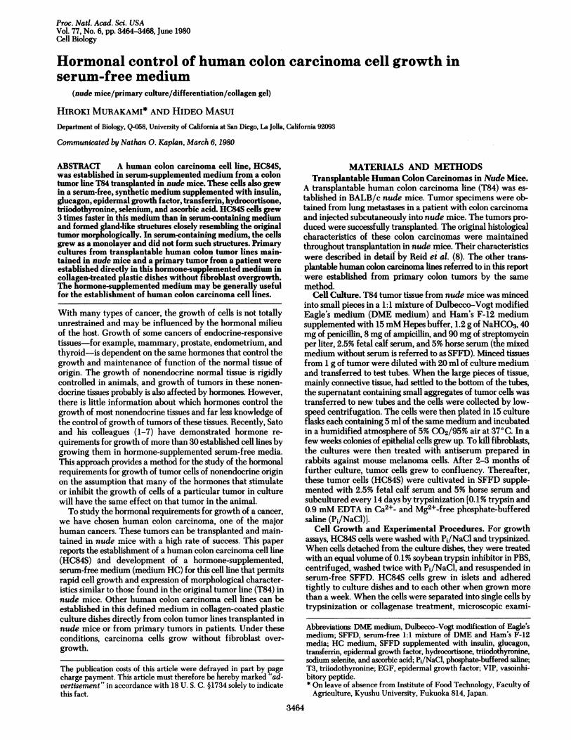
FIG. 1. Phase-contrast micrograph of HC84S cells in serum-supplemented medium. (X73.)
nation always revealed a substantial breakdown of cells.Comparisons of cell number and protein content were madefor HC84S cells grown in serum-supplemented and in serum-free SFFD supplemented with hormones (HC medium). Cellnumber and protein content varied linearly; the mean (+SD)was 1.5 + 0.3 ig/i103 cells for cells grown in serum-supple-mented medium and 1.2 + 0.3 jg/103 cells for those grown inHC medium. For this reason we estimated cell numbers byprotein content measured by Lowry's method (9) with bovineserum albumin (Sigma) as a standard.
Cells (45 jg of protein) were plated in 60-mm culture dishes(Falcon) containing 4 ml of SFFD. After overnight incubationto allow cell attachment, the medium was changed SFFDcontaining test supplements, and the cells were grown for 21days. Cell growth in response to the added factors was measuredby assaying protein content in the culture dishes. The disheswere washed twice with Pi/NaCl, cells were lysed in 4 ml of0.3% Na2COs/1 M NaOH, and protein was determined byLowry's method. Values reported are means of two separatedishes.
Preparation of Anti-Mouse Cell Antiserum. Mouse mela-noma cells (B16) were grown in culture and 107 cells were in-jected subcutaneously into a young adult rabbit. Three moreinjections were made at 1-week intervals. One week after thelast injection, the rabbit was bled and serum was isolated. Totest its titer against mouse cells, 3T3 and B16 cells were grownin culture dishes containing 5.0 ml of medium; then, 0.1 ml ofantiserum and 0.05 ml of unimmunized rabbit serum wereadded and incubated overnight. All mouse cells were lysed, buthuman cells (WI-38 and HeLa) were not affected when testedunder the same condition. Primary cultures were treated withantiserum in the same way to kill mouse fibroblasts.
Materials. Insulin, human transferrin, glucagon, hydro-cortisone, triiodothyronine (T3), gastrin, and ascorbic acid wereobtained from Sigma. Epidermal growth factor (EGF) wasobtained from Collaborative Research (Waltham, MA). Sodiumselenite was obtained from Difco. Purified bovine insulin wasa gift from Eli Lilly.
RESULTSGrowth of HC84S Cells. When grown in serum-supple-
mented medium, HC84S cells have the typical morphologicalcharacteristics of epithelial cells (Fig. 1). When HC84S cellswere injected subcutaneously into nude mice (106 cells peranimal) they produced tumors in all animals, and the histo-logical characteristics of these tumors were similar to those ofthe original T84 tumor line from which the HC84S line wasestablished.A typical growth curve of HC84S cells in serum-supple-
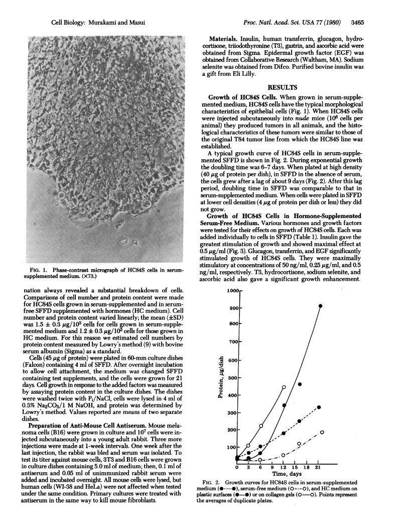
mented SFFD is shown in Fig. 2. During exponential growththe doubling time was 6-7 days. When plated at high density(40 ,g of protein per dish), in SFFD in the absence of serum,the cells grew after a lag of about 9 days (Fig. 2). After this lagperiod, doubling time in SFFD was comparable to that inserum-supplemented medium. When cells were plated in SFFDat lower cell densities (4 ,jg of protein per dish or less) they didnot grow.Growth of HC84S Cells in Hormone-Supplemented
Serum-Free Medium. Various hormones and growth factorswere tested for their effects on growth of HC84S cells. Each wasadded individually to cells in SFFD (Table 1). Insulin gave thegreatest stimulation of growth and showed maximal effect at0.5 ,g/ml (Fig. 3). Glucagon, transferrin, and EGF significantlystimulated growth of HC84S cells. They were maximallystimulatory at concentrations of 50 ng/ml, 0.25 ,jg/ml, and 0.5ng/ml, respectively. T3, hydrocortisone, sodium selenite, andascorbic acid also gave a significant growth enhancement.
. 600 /
500
500-
300
200
~0
100 °01
0 3 6 9 '12 15 18 21Time, daYS
FIG. 2. Growth curves for HC84S cells inserum-supplementedmedium (O .....0), serum-free medium (O- --0), and HC medium onplastic surfaces (O- *) or on collagen gels (O 0). Points representthe averages of duplicate plates.
Cell Biology: Murakami and Masui

3466 Cell Biology: Murakami and Masui
Table 1. Response of HC84S cells to hormones and factorsAddition Concentration % of growth
None (control) - 100Serum 7.5% 262Insulin 2,ug/ml 234Glucagon 200 ng/ml 157Transferrin 2,ug/ml 152EGF 1 ng/ml 138T3 0.5 nM 125Sodium selenite 25 nM 124Ascorbic acid 10,4g/ml 119Hydrocortisone 50 nM 118Gastrin 50 pM 114Vasoinhibitory peptide (VIP) 50 pM 104Secretin 50 pM 103Cholecystokinin (CCK) 50pM 102Tripeptide (Glu-His-Lys) 10 ng/ml 102Growth hormone 1,4g/ml 102Putrescine 0.1 mM 100Somatostatin 10 ng/ml 93Prostaglandin A1 25 ng/ml 77Prostaglandin B1 25 ng/ml 86Prostaglandin E1 25 ng/ml 107Prostaglandin E2 25 ng/ml 93Prostaglandin F2a 25 ng/ml 91Progesterone 50 nM 83Testosterone 50 nM 77Estradiol 50 nM 74
The numbers represent the averages of duplicate plates.
Among intestinal hormones tested, only gastrin gave somestimulation. With the exception of prostaglandin E1, all of theprostaglandins tested inhibited the growth of HC84S cells.Progesterone, testosterone, and estradiol also inhibited cellgrowth.
Because the insulin preparation used in these experimentscontained glucagon as an impurity, highly purified insulin wastested to exclude the possibility that the observed insulin effect
was due to glucagon. The stimulation of HC84S cell growth byhighly purified insulin was comparable to that of the previouslytested preparation. The glucagon preparation used containedsome insulin as an impurity. However, because glucagonstimulated the growth of HC84S cells at an order of magnitudelower concentration than did insulin, it is unlikely that the effectof glucagon was due to the contaminating insulin. Thus, bothinsulin and glucagon stimulate the growth of HC84S cells.HC84S cells were grown in SFFD medium supplemented
with insulin (2 ,ug/ml), glucagon (0.2,ug/ml), transferrin (2;g/ml), EGF (1 ng/ml), hydrocortisone (50 nM), T3 (0.5 nM),selenite (25 nM), and ascorbic acid (57 ,uM) (HC medium) andtheir growth was compared with that in serum-supplementedSFFD (Fig. 2). The cells began to grow exponentially after alag of 3-5 days in HC medium and thereafter grew with adoubling time of about 5 days, faster than in serum-containingmedium. When HC84S cells were plated on collagen gels in thehormone-supplemented, serum-free medium, they grewwithout a lag period. HC84S cells grown in this hormone-sup-plemented, serum-free medium piled up and producedgland-like structures (Figs. 4 and 5) morphologically closelyresembling those of T84 tumors in animals. This appearanceis strikingly different from the monolayers formed by HC84Scells in serum-supplemented medium (Fig. 1).
Establishment of Other Human Colon Cancer Cell Linesin Hormone-Supplemented Medium. We wished to determinewhether cells taken from T84 tumors maintained in nude micecould be established directly in the defined medium developedfor HC84S cells. Primary cultures were prepared from a T84tumor and placed in serum-supplemented SFFD overnight.The next day the medium was replaced with the serum-freeHC medium. The cells grew in this medium at a rate compa-rable to that in serum-containing medium. The substancesfound to be the major growth promotors for HC84S cells wereexamined individually for their effects on growth of primaryT84 cells (Fig. 6). Among the factors tested singly, transferrinshowed the most striking growth-promoting effect.
Fibroblast overgrowth is a problem often encountered in the
1**/0.
NI
300F
250
Insulin200
I I I I ..
0.5 1 1.5 2 5,g/ml
-( Traf*er --n
Transferrin
EGF
(- -
0.5 1 2 3 4 5ng/ml
3001
250I
200
0 *
Glucagon
I --I L50 200 400 600
ng/ml
FIG. 3. Dose-response curves of HC84S cell growth to insulin, transferrin, EGF, and glucagon. Points represent the averages of duplicateplates.
300
250 V
200 -s._
o
3001
250
200
0 1 2 3 4 5,ug/ml
Proc. Nati. Acad. Sci. USA 77 (1980)

Proc. Natl. Acad. Sci. USA 77 (1980) 3467
4v . .
FIG. 4. Phase-contrast micrograph ofHC84S cells in HC medium.(X73.)
establishment of primary cell cultures. Primary T84 cells weretested for growth in Falcon plastic culture dishes with or
without a collagen gel coating in serum-free or serum-con-
taining media to find optimal culture conditions. T84 cells at-
FIG. 5. Thin section of gland-like structures formed by HC84Scells in HC medium. Cells were harvested, fixed in 10% formalin, andembedded in 0.5% agar. The agar was dehydrated, embedded inparaffin, sectioned, and stained with hematoxylin and eosin.(X73.)
I,
FIG 6 Response of primary cultures from a T84 tumor to hormones and factors. After 21 days, the cultures were fixed with 10%formalin and stained with crystal violet. Bottles: 1, serum-supple-mented medium; 2, HC medium; 3, SFFD + transferrin; 4, SFFD +insulin; 5, SFFD + EGF; 6, SFFD + glucagon; 7, SFFD.tached more quickly to collagen-coated dishes than to uncoatedones irrespective of the growth medium. Overgrowth of T84cells by fibroblasts was a problem in cultures in the uncoatedplastic flasks where the T84 cells took longer to become estab-lished. Fibroblasts grew better in the cultures with serum sup-plementation than in HG medium. Thus, of the four growthconditions, the 'optimal one for establishment of primary cul-tures is hormone-supplemented serum-free medium and col-lagen-coated dishes.
These observations suggested that the use of collagen-coateddishes and the hormone-supplemented serum-free mediumdeveloped for HC84S cells might allow establishment of othercell lines from human colon carcinomas. As a test, primarycultures were prepared from two human colon carcinomas(T219 and T245) transplanted in nude mice and from a carci-noma taken directly from a patient and successfully culturedunder these conditions. The cells from T219 and T245 havebeen passaged eight times and the cells directly from a patienthave been passaged three times.
DISCUSSIONA human colon carcinoma cell line (HC84S) has been estab-lished in culture from a tumor line (T84) transplantable in nudemice. Insulin, glucagon, transferrin, EGF, hydrocortisone, T3,selenite, ascorbic acid, and gastrin were found to stimulategrowth of these cells in serum-free medium. When these sup-plements, with the exception of gastrin, were added to SFFD,to form HG medium, the HD84S cells grew 3 times faster thanin serum-containing medium and exhibited a gland-like ap-pearance similar to that of T84 tumors maintained in mice.[Gastrin was not tested for stimulation of HC84S cell growth(it was only mildly stimulatory) until after the experiments withHG medium had been completed.] Sato and coworkers havedetermined hormone requirements for a large number of cellslines (1-7). Each cell line requires a different set of hormones,with some components such as insulin and transferrin beingcommon to most but others being rather specific. Glucagon andgastrin, which promote growth of HC84S cells, have not beenidentified as requirements for growth of the other lines. Animportant unanswered question is whether the hormones thatstimulate the growth of human colon carcinoma cells in culturestimulate them in animals. This can now be tested with nudemice carrying human colon carcinomas.
Cell Biology: Murakami .and Masui

3468 Cell Biology: Murakami and Masui
The trace element selenium is routinely tested for effect oncell proliferation in our laboratory following the discovery byMcKeehan et al. (10) that selenium is essential for growth ofa human diploid fibroblast cell line (WI-38) and of a Chinesehamster ovary line in serum-depleted media. It has been foundto stimulate growth of a number of cell lines for which hor-mone-supplemented, serum-free media have been developed(7).
Glucagon has been reported to have various effects on thegastrointestinal tract including inhibition of acid secretion inthe stomach and inhibition of absorption of water and sodiumin the intestine (11). Enteroglucagon, a substance that cross-reacts immunologically with pancreatic glucagon, is known tobe secreted by the small intestine although its physiological roleis not known (12). The results reported here suggest that glu-cagon, or possibly enteroglucagon, may be involved in controlof growth of colon cell in vivo. Gastrin stimulates growth ofHC84 slightly. It has been reported (13) that gastrin stimulatesthe growth of intestinal cells in culture and that gastrinomapatients show hyperplasia of the intestinal mucosa. Recently,Laburthe et al. (14) reported that vasoinhibitory peptide (VIP),activates adenyl cyclase of a human colon carcinoma cell line(HT 29) but the effects of VIP on growth of these cells were notstudied. We found VIP not to affect growth of HC84S cells.These results raise the possibility that some hormones that donot affect cell growth nevertheless do affect cell function. Itremains to be elucidated whether any of the hormones foundto stimulate growth of HC84S cells affect their function, butthis is one possible interpretation of the result that HC84S cellsapparently differentiate morphologically when grown in hor-mone-supplemented, serum-free medium.
Somatostatin, prostaglandins (except prostaglandin E1),progesterone, testosterone, and estradiol appeared to inhibitgrowth of HC84S cells. This result and the demonstration thata hormone-supplemented, serum-free medium allows bettergrowth of HC84S cells than does serum-containing mediumraise the possibility that there are hormones present in serumthat inhibit the growth of human colon carcinoma cells. It maybe possible to develop combinations of hormones that effec-tively inhibit HC84S cell growth in just the way that the growthmedium was constructed.Some of the hormones present in serum may inhibit HC84S
cell differentiation of function because the cells grew in threedimension and formed gland-like structures similar to those ofT84 tumors in animals when put in hormone-supplementedmedium whereas they grew as a monolayer in serum-containingmedium. Recently, Taub et al. (6) reported that hemicyst for-mation by MDCK dog kidney cells was similarly dependent onthe hormone components of the medium.Human colon carcinoma cell lines could be established more
easily on collagen gels than on a plastic surface. Our observationthat HC84S cells started to grow in defined medium on collagengels without a lag period after plating suggests that the collagenmay facilitate formation of a cell-substrate adhesion that is anessential prerequisite for cell division. An advantage of thecollagen supports is that cell lines could be established from atumor before fibroblast overgrowth became a problem. Thisproblem has made it difficult to grow epithelial cells in culturein the past.The hormone-supplemented medium described here sup-
ported the growth of all the tumor cells tested, including thosetaken directly from a patient. The fact that primary cells froma T84 tumor gave the greatest growth response to transferrinrather than to insulin, which was the -single most effectivestimulatory of HC84S cells derived from another T84 tumor,
suggests that cells from different colon tumors will havesomewhat different hormonal requirements. Hence, the me-dium developed for HC84S cells may not be optimal for growthof all human colon carcinoma lines, but it appears to be gen-erally applicable.By using medium in which hormones and factors have re-
placed serum, the mechanisms by which human colon carci-nomas grow and differentiate become accessible to dissection.Carcinogenesis is a multistep process. Once cancers are estab-lished in animals, they undergo a series of changes in theirproperties known as tumor progression (14-16). We maintainseveral human colon carcinomas by transfer in nude mice.These originated in different patients with different extents ofmetastasis, and they differ in their histological characteristicsand growth rates in nude mice. Thus, they represent differentdegrees of malignancy. Because cell lines from these can beestablished in defined medium and their hormone requirementsdetermined, it may be possible to correlate hormone require-ments with various states of malignancy and to study the pro-cesses involved in establishing malignancy. HC medium wasdeveloped to support growth of a cell line derived from a lungmetastasis of a colon carcinoma, and it also supported growthof three other colon carcinomas. However, the colon primarytumor that gave rise to the lung metastasis was not available tous for a detailed comparison of hormonal requirements andcorrelation with tumor progression.We thank Terry White and Bonnie Wolfe for excellent technical
assistance, Drs. Nathan 0. Kaplan and Gordon Sato for advice andencouragement, and Patricia Romans and Dr. David Barnes for theircriticism of the manuscript. This investigation was supported by Na-tional Institutes of Health Grant CA-23052.
1. Hayashi, I. & Sato, G. (1976) Nature (London) 259, 132-134.2. Hutchings, S. E. & Sato, G. H. (1978) Proc. Natl. Acad. Sci. USA
75,901-904.3. Rizzino, A. & Sato, G. (1978) Proc. Natl. Acad. Sci. USA 75,
1844-1848.4. Bottenstein, J. & Sato, G. H. (1979) Proc. Nati. Acad. Sci. USA
76,514-517.5. Mather, J. P. & Sato, G. H. (1979) Exp. Cell Res. 120, 191-
200.6. Taub, M., Chuman, L., Saier, M. H., Jr. & Sato, G. (1979) Proc.
Natl. Acad. Sci. USA 76,3339-3342.7. Bottenstein, J., Hayashi, I., Hutchings, S., Masui, H., Mather, J.,
McClure, D., Ohasa, S., Rizzino, A., Sato, G., Serrero, G., Wolfe,R. & Wu, R. (1979) Methods Enzymol. 59,94-109.
8. Reid, L., Holland, J., Jones, C., Wolfe, B., Niwayama, G., Wil-liams, R., Kaplan, N. & Sato, G. (1978) in Proceedings of theSymposium on the Use of Athymic (Nude) Mice in CancerResearch, eds. Houchens, D. & Overjera, A. (Gustav, Fischer,NY), pp. 107-121.
9. Lowry, 0. H., Rosebrough, N. J., Farr, A. L. & Randall, P. J.(1951) J. Biol. Chem. 193,265-275.
10. McKeehan, W. L., Jones, W. G. & Ham, R. G. (1976) Proc. Natl.Acad. Sci. USA 73,2023-2027.
11. McGuigan, J. E. (1974) in Textbook of Endocrinology, ed. Wil-liams, R. H. (Saunders, Philadelphia), p. 850.
12. Moody, A. J., Jacobsen, H., Sundby, F., Frandsen, E. K., Baetens,D. & Orci, L. (1977) in Glucagon: Its Role in Physiology andClinical Medicine, eds. Foa, P. P., Bajaj, J. S. & Foa, N. L.(Springer, New York), pp. 129-136.
13. Lichtenberger, L., Miller, L. R., Erwin, D. N. & Johnson, L. R.(1973) Gastroenterology 65,242-251.
14. Laburthe, M., Rousset, M., Boissard, C., Chevalier, G., Zwebaum,A. & Rosselin, G. (1978) Proc. Natl. Acad. Sci. USA 75,2772-2775.
15. Foulds, L. (1964) in Cellular Control Mechanisms and Cancer,eds. Emmelot, P. & Muhlbock, 0. (Elsevier, Amsterdam), pp.242-258.
16. Medina, D. (1975) in Cancer, ed. Becker, F. F. (Plenum, NewYork), Vol. 3, pp. 99-119.
Proc. Natl. Acad. Sci. USA 77 (1980)