Homogeneity and Heterogeneity of Sizes of … · Republic of Germany ... following major types can...
13
J. Microscopie BioI. Cell. (1976), 25, 107 -116 Homogeneity and Heterogeneity of Sizes of Transcriptional Units and Spacer Regions in Nucleolar Genes of Acetabularia INTRODUCTION Herbert SPRING, Georg KROHNE, Werner W. FRANKE, Ulrich SCHEER and Michael F. TRENDELENBURG Division of Membrane Biology and Biochemistry, Institute of Experimental Pathology. German Cancer Research Center, 69-Heidelberg. Im Neuenheimer Feld 280, Federal Republic of Germany The arrangement of genes of precursor molecules for ribosomal RNA (pre-rRNA) in primary nuclei from two green algae species, Acetabularia mediterranea and A. major, has been analyzed in an electron microscope study. The pattern of transcriptional units in individual strands of nucleolar chromatin was investigated using spread and posi- tively stained preparations. The rDNA pattern is not uniform but differs in different strands. The predominant type of nucleolar chromatin exhibits a high degree of homogeneity in the sequence of matrix units (intercepts covered with fibrilst hat contain the pre-rRNA) and fibril-free spacer intercepts. Substantial differences, however, are observed between the patterns in different strands. In addition, there is evidence in some strands for intraaxial heterogeneity of both spacer and matrix units. The following major types can be distinguished: type la, ca. 2 micrometer long matrix units, extremely short spacer intercepts in A. mediterranea (ca. 1 micrometer long ones in A. major), completely homogeneous distribution; type Ib, as type la but with intercalated, isolated, significantly shorter and/or longer matrix units; type lIa, matrix unit sizes as in type la, but much longer spacer intercepts, high degree of homogeneity; type Ill, largely heterogeneous arrangements of matrix and spacer units of varying sizes. The matrix unit data are compared with the sizes of pre-rRNA as determined by polyacrylamide gelelectrophoresis under denaturing and non-denaturing conditions. The findings are discussed in relation to recent observations in amphibia and insects and with respect to current concepts of the species-specificity of rDNA arrangements. In eukaryotes the nuclear genes that code for the common precursor molecules of the large cytoplasmic ribosomal RNAs (pre-rRNA) are present in relatively large numbers and most, if not all, are either clustered in distinct regions (" nucleolar organizer regions ", NOR) of specific chromosomes, or, in some cell systems, as extrachromosomal amplified nucleolar units (for reviews see, e.g., Gall, 1969, 1974; Busch and Smetana, 1970; Birnstiel et al., 1971; Gall and Rochaix, 1974; Engberg et al., 1974; Tobler, 1975; Bohnert et al., 1975). It has been shown by various techniques that these genes are tandemly arranged in repeating units. Each repeating unit consists of one intercept coding for pre-rRNA and an adjacent intercept which is either not trans- cribed at all or is at least not transcribed into sequences that are present in the pre-rRNA (for details see Miller and Beatty, 1969a; Dawid et al., 1970; Wensink and Brown, 1971; Brown et al., 1972; Scheer et al., 1973 ; Forsheit et al., 1974; Wellauer and Reeder, 1975). Marked differences in the rDNA pattern have been demonstrated in comparisons of different organisms, in- cluding examples of different spacer lengths even among closely related species (Brown et al., 1972; Wellauer and Reeder, 1975) as well as demonstrations of diffe- rences in the sizes of both transcribed and untrans- cribed intercepts among taxonomically distant species (Trendelenburg et al., 1973, 1974a, b; Trendelenburg, 1974; Spring et al., 1974). On the other hand, most earlier studies have emphasized the homogeneity of rDNA arrangement, that is the uniformity of the lengths of the transcribed and untranscribed rDNA regions in a given organism, in particular in the well studied amphibian genus Xenopus. Several recent studies, however, have pointed out the existence of a significant degree of heterogeneity in the rDNA pattern of one and the same organism. For example, it has been demonstrated in electron micnscope studies of transcriptional complexes from various cell types that all three identified func- tional units, i.e. the intercepts associated with fibrils containing nascent pre-rRNA material ("matrix units "), the fibril-free sections (" spacers "), and the resulting repeat units, exhibit a distinct variation and hetero- geneity in the same cell (Scheer et al., 1973; Trende- lenburg et al., 1973; Spring et al., 1974). It has also been shown in some dasycladacean green algae (Tren- delenburg et al., 1974a; Spring et al., 1974; Berger and H. Spring et al. 107
Transcript of Homogeneity and Heterogeneity of Sizes of … · Republic of Germany ... following major types can...

J. Microscopie BioI. Cell. (1976), 25, 107 -116
Homogeneity and Heterogeneity of Sizes of Transcriptional Units and Spacer Regions in Nucleolar Genes of Acetabularia
INTRODUCTION
Herbert SPRING, Georg KROHNE, Werner W. FRANKE, Ulrich SCHEER and Michael F. TRENDELENBURG
Division of Membrane Biology and Biochemistry, Institute of Experimental Pathology. German Cancer Research Center, 69-Heidelberg. Im Neuenheimer Feld 280, Federal Republic of Germany
The arrangement of genes of precursor molecules for ribosomal RNA (pre-rRNA) in primary nuclei from two green algae species, Acetabularia mediterranea and A. major, has been analyzed in an electron microscope study. The pattern of transcriptional units in individual strands of nucleolar chromatin was investigated using spread and positively stained preparations. The rDNA pattern is not uniform but differs in different strands. The predominant type of nucleolar chromatin exhibits a high degree of homogeneity in the sequence of matrix units (intercepts covered with fibrilst hat contain the pre-rRNA) and fibril-free spacer intercepts. Substantial differences, however, are observed between the patterns in different strands. In addition, there is evidence in some strands for intraaxial heterogeneity of both spacer and matrix units. The following major types can be distinguished: type la, ca. 2 micrometer long matrix units, extremely short spacer intercepts in A. mediterranea (ca. 1 micrometer long ones in A. major), completely homogeneous distribution; type Ib, as type la but with intercalated, isolated, significantly shorter and/or longer matrix units; type lIa, matrix unit sizes as in type la, but much longer spacer intercepts, high degree of homogeneity; type Ill, largely heterogeneous arrangements of matrix and spacer units of varying sizes. The matrix unit data are compared with the sizes of pre-rRNA as determined by polyacrylamide gelelectrophoresis under denaturing and non-denaturing conditions. The findings are discussed in relation to recent observations in amphibia and insects and with respect to current concepts of the species-specificity of rDNA arrangements.
In eukaryotes the nuclear genes that code for the common precursor molecules of the large cytoplasmic ribosomal RNAs (pre-rRNA) are present in relatively large numbers and most, if not all, are either clustered in distinct regions (" nucleolar organizer regions ", NOR) of specific chromosomes, or, in some cell systems, as extrachromosomal amplified nucleolar units (for reviews see, e.g., Gall, 1969, 1974; Busch and Smetana, 1970; Birnstiel et al., 1971; Gall and Rochaix, 1974; Engberg et al., 1974; Tobler, 1975; Bohnert et al., 1975). It has been shown by various techniques that these genes are tandemly arranged in repeating units. Each repeating unit consists of one intercept coding for pre-rRNA and an adjacent intercept which is either not transcribed at all or is at least not transcribed into sequences that are present in the pre-rRNA (for details see Miller and Beatty, 1969a; Dawid et al., 1970; Wensink and Brown, 1971; Brown et al., 1972; Scheer et al., 1973 ; Forsheit et al., 1974; Wellauer and Reeder, 1975). Marked differences in the rDNA pattern have been demonstrated in comparisons of different organisms, including examples of different spacer lengths even among
closely related species (Brown et al., 1972; Wellauer and Reeder, 1975) as well as demonstrations of differences in the sizes of both transcribed and untranscribed intercepts among taxonomically distant species (Trendelenburg et al., 1973, 1974a, b; Trendelenburg, 1974; Spring et al., 1974). On the other hand, most earlier studies have emphasized the homogeneity of rDNA arrangement, that is the uniformity of the lengths of the transcribed and untranscribed rDNA regions in a given organism, in particular in the well studied amphibian genus Xenopus. Several recent studies, however, have pointed out the existence of a significant degree of heterogeneity in the rDNA pattern of one and the same organism. For example, it has been demonstrated in electron micnscope studies of transcriptional complexes from various cell types that all three identified functional units, i.e. the intercepts associated with fibrils containing nascent pre-rRNA material ("matrix units "), the fibril-free sections (" spacers "), and the resulting repeat units, exhibit a distinct variation and heterogeneity in the same cell (Scheer et al., 1973; Trendelenburg et al., 1973; Spring et al., 1974). It has also been shown in some dasycladacean green algae (Trendelenburg et al., 1974a; Spring et al., 1974; Berger and
H. Spring et al. 107

Journal de Microscopie et de Biologie Cellulaire, Volume 25,1976
Schweiger, 1975a) that different types of rDNA pattern may oeeur in different strands of nucleolar chromatin in the same cell and that differences in the lengths of the spacer intercepts are especially pronounced. Moreover, marked heterogeneity in the size of the spacer intercepts has been demonstrated in Xenopus laevis and X. muelleri by analyses of rDNA fragments obtained from cleavage by restriction endonucleases (Wellauer et al., 1974; Morrow et al., 1974; Wellauer and Reeder, 1975). In this article we present a detailed analysis of the different forms of heterogeneity in the arrangements of rDNA units in the primary nuclei from two green algae species, Acetabularia mediterranea and Acetabularia major, as revealed by electron microscopy of spread and positively stained preparations, together with an analysis of the pre-rRNA molecules formed in these nuclei as revealed by gel-electrophoresis under denaturing and non-denaturing conditions. The results indicate that in the nucleolar chromatin strands of one cell both inter- and intraaxial heterogeneity occurs in the rDNA pattern.
MATERIAL AND METHODS
Isolation of nuclei, electron microscopv and evaluation of spread nucleolar material
Acetabularia mediterranea and A. major were cultivated as described by Lateur and Bonotto (1973) and used before and during cap formation. After manual isolation of the nuclei in a medium consisting of 83 mM KCI and 17 mM NaCi, buffered to pH 7.2 with 10 mM Tris-HCl (c.t. Spring et aI., 1974), the nucleoli were spread according to the technique developed by Miller and coworkers (Miller and 8eatty, 1969a, b Miller and Bakken, 1972; for the specific modifications used see Scheer et aI., 1973; Trendelenburg et aI., 1974a; Spring et al., 1974). The spread preparations were positively stained with phosphotungstic acid (PTA) and observed in the Zeiss EM 10 electron microscope (Carl Zeiss, Oberkochen, Federal Republic of Germany). Some PTA-stained preparations were rotary shadowed with Aul Pd (75/25 %) at an angle of 7° for enhancing the contrast. For the length measurement (c.f. the refs quoted above) of repeating units and their two components, spacer and matrix intercepts, in individual strands micrographs were selected which showed well spread nucleolar chromatin with maximally extended axes and a high number of clearly traceable rDNA units in uninterrupted sequence.
Radioactive labelling of RNA Almost fully grown plants were incubated in culture medium supplemented with either :lH-uridine (lOO Il-Cilml; 45 CiI mM) alone or with all four tritiated ribonucleosides at concentrations of 180 Il-CiI ml with respect to :lH-uridine and 30 Il-CiI ml with respect to each of the other nucleosides eH-cytidine, 25 Ci/mM; :lH-guanosine, 10 Ci/mM; aHadenosine, 22 Ci/mM). All radiochemicals were from the Radiochemical Centre (Amersham, United Kingdom). The labelling periods were 6, 24 or 48 hours.
Preparation of ribosomal RNA
For each experiment about 200 plants of Acetabularia mediterranea (at the onset of cap formation) were blotted on filter paper and were then briefly immersed at 4 °C in a medium consisting of 0.1 M Tris-HCl (pH 7.3), 5 mM MgCl~, 0.4 M sucrose, and 0.05 % diethylpyrocarbonate (DEP). The cells were then cut into short pieces with fine scissors and were subsequently homogenized with a tight fitting, motor driven Potter-Elvejhem homogenizer. The homogenate was filtered through a nylon cloth (for details,
108
see Spring et al., 1974) and the filtrate was centrifuged in the cold at 3,000 X g for 10 minutes. The supernatant was collected and adjusted with sucrose to a final concentration of 1 M and centrifuged at 100,000 X g for 6 hours at 4°C. The pellet was resuspended with the homogenizer in 5 ml of Tris-HCl (0.05 M, pH 7.6) containing 1 % NaCl and 2 % (w/v) triisopropylnaphtalene sulphonate, and the suspension was thoroughly shaken with an equal volume of phenol! cresol! 8 - hydroxyquinoline (K i r by, 1965) as described by Loening (1967). The RNA was precipitated from the final aqueous phase by adding two volumes of absolute ethanol.
Extraction of RNA from isolated nuclei
Manually isolated nuclei (c.f. Trendelenburg et al., 1974a; Spring et aI., 1974) were collected in ice-cold 70 % ethanol containing 0.1 M NaCI. Portions of 10-35 nuclei were centrifuged at 3000 X g for 10 minutes. The pellets obtained were dried in vacuo at - 20 "C and digested in 0.5 ml of a solution consisting of 20 mM Tris-HCI, (pH 7.4), 0.5 X SDS and 0.5 mg pre-digested pronase (RNAse-free, Calbiochem, La Jolla, California, U.S.A.) for 20 minutes at room temperature. After adding of 20 Il-g of Escherichia coli rRNA the nucleic acids were precipitated by adding two volumes of absolute ethanol and stored at - 20°C.
Gel electrophoresis
a. Non-denaturing conditions
RNA was analyzed in cylindrical 2.4 % acrylamide gels (Loening, 1967, 1969) with the electrophoresis buffer (without Mg + +) recommended by Schuch and Loening (1975). RNA samples were suspended in 20 Il-I of diluted (I: I) electrophoresis buffer containing 10 % sucrose and were then directly applied to the top of the gels.
b. Partially denaturing conditions
The ethanol-precipitated RNA was freeze-dried and res uspended in 5 Il-I of diluted (I: 1) electrophoresis-buffer (see above). To this 25 Il-I of buffered formamide (see c) containing 10 % sucrose was added and the solution was heated at 60°C for 5 minutes. After rapidly cooling the samples were electrophoresed as described under (a).
c. Completely denaturing conditions
RNA was dissolved as described under (b). Buffered formamide was prepared as follows (see also Duesberg and Vogt, 1973; Pinder et al., 1974). Formamide (p.a., Merck, Darmstadt, Federal Republic of Germany) was purified by stirring for about 2 hours with 3.5 % Amberlite MB-l (Serva Feinbiochemica, Heidelberg, Germany) until the conductivity has dropped to 5 Il-Mho. After filtering through Watman GF/C glass fibre filter formamide stock solutions that contained 20 mM Na~HP04 and 20 mM NaH~PO.l X H 20, respectively, were prepared by dissolving the appropriate amounts of salt in the deionized formamide. By mixing of defined volumes of these stock solutions the apparent pH of the about 98 % formamide solution was adjusted to 7.0. Electrophoresis was carried out in 3,5 % or 3.0 % aCf;llamide gels containing formamide according to Duesberg and Vogt (1973), however, with the modification that after adding of the N, N, N', N'-tetramethylethylendiamine the final apparent pH was adjusted with 1 N HCI to 7.0. Separation was carried out at 11 V I cm at 40°C for 6 to 8 hours. Bromphenol blue was used as dye marker. After electrophoresis the formamide-containing gels were washed for 1 hour in 20 mM aqueous phosphate buffer (pH 7.2) and scanned at 260 nm in a Gilford Spectrophotometer 2400 S (Gilford, Oberlin, Ohio, USA) equipped with a linear gel transport device. The gels were
Patterns of nucleolar genes

then frozen on solid CO", cut into 1.1 mm thick slices with parallel razor blades, incubated overnight in a toluenebased scintillation mixture containing 4 % NCS (Nuclear Chicago Solubilizer, Amersham/ Searle, Illinois, USA) at 40 "C and were counted in a liquid scintillation spectrometer (Betaszint BF 5000, Berthold-Frieseke, Wildbad, Federal Republic of Germany). Molecular weights were calculated from the co-electrophoresed E. coli rRNAs (assuming weights 0.525 and 1.05 X 10⁶ D, respectively, Ebel et al .• 1974; Nomura and Tissieres, 1974) and a semilogarithmic inverse correlation between migration and molecular weight.
RESULTS
Morphology of spread nucleolar chromatin
The electron microscopic appearance of spread and positively stained nucleolar material isolated from transcriptionally active nucleoli and nuclei of Acetabularia mediterranea and A. major has been extensively described in previous articles (Trendelenburg et al., 1974a, b, 1975; Spring et al., 1974; Berger and Schweiger, 1975a; Woodcock et al., 1975). Survey micrographs of a typical strand of nucleolar chromatin from the primary nucleus of A cetabularia mediterranea are presented in Figs a and b, PI. 1. The regular pattern of the rDNA is recognized in the chromatin axes as a sequence of repeating units, each of which consists of a matrix unit covered with ribonucleoprotein (RNP) fibrils of increasing lengths and a spacer intercept that is either not transcribed at all or the transcription products of which are unstable (for detailed discussion see Franke et al, 1976; sce there also for special discussions of "spacer transcription complexes "). Only strands in which the sequence of transcriptional units was of uniform polarity have been considered in the present study. (Within a specific axis of nucleolar chromatin, the genes coding for pre-rRNA are generally arranged in the same polarity and transcribed from the same strand of the DNA double strand; it is not yet clear whether the few exceptions indicative of transcription of adjacent genes from different strands of the DNA described in preparations of nucleolar material are really showing rDNA transcription; c.f. Fig. 8 of Miller and Beatty, 1969h, Fig. 4 of Trendelenburg et al., 1974a; for discussion see also Spring et al., 1974, 1975; Angelier and Lacroix, 1975.)
Regarding the relative proportions of the matrix units and spacer intercepts, as well as the total length of the repeating unit, some heterogeneity was found; moreover, different types of arrangements of rDNA as revealed by the sequence of transcriptional units could be identified (c.f. Trendelenburg et al., 1974a; Spring et al., 1974). For analysis of the specific homogeneity and heterogeneity of the nucleolar DNA, the individual strands of nucleolar chromatin were treated as distinct and separate morphological units, although a considerable proportion of them were probably formed by a breakage from larger units. For the purpose of this analysis it was especially important to only evaluate those nucleolar chromatin strands in which the axis containing the rDNA was clearly traceable within both matrix units and spacer intercepts, in order to eliminate errors of length measurements due to insufficient spreading and staining. (Such measurement artifacts are more common in spacer regions than in matrix units and thus
H. Spring et al.
Memoires originaux
might result in underestimation of spacer lengths.) Most of the strands analyzed contained 10-25 uninterrupted and clearly identifiable transcriptional units, but very long segments containing up to 50 units were also occasionally encountered.
Some examples of the various types of heterogeneity in rDNA patterns of both Acetabularia mediterranea and A cetabularia major are presented in the electron micrographs shown in Plate I and II and in the histograms of Figure 1. With respect to the homogeneity of the specific pattern, three basic classes could be distinguished: (i) completely homogeneous distributions of the axial intercepts; (ii) widely homogeneous distributions with few and isolated but significant longer matrix and/or spacer units interspersed; (iii) markedly heterogeneous distributions of the lengths of all three units (matrix units, spacer units and repeating units). In particular, we noted marked differences of the rDNA pattern between different strands in that at least two types of homogeneous patterns, one with short and onc with long spacer intercepts, could be defined (PI. I; c.f. Trendelenburg et al., 1974a).
The variety of patterns observed were classified as follows:
(1) The most frequent rDNA pattern observed in Acetabularia mediterranea was obviously rather homogeneous and consisted of about 2.1 [.lm long matrix units and relatively short spacer intercepts, resulting in total repeat units of 2.5-2.7 [.lm (e.g. PI. I, figs. a, b, and t, and A. med. strand No. 725 in the upper panel of Figure 1). When these measurements are corrected with respect to the limits of resolution and detection of the technique used as well as for possible obliterations of nascent fibrils by cleavage and lack of detection due to incomplete association with protein (for details see Franke et al., 1976), one might even conclude that in this rDNA pattern only a minimal spacer, if any, exists (e.g., PI. I, fig. e; see also Trendelenburg et al., 1974a). The predominance of this pattern, which we designate "type la" in primary nuclei from Acetabularia mediterranea as well as from the related dasycladacean green alga Batophora oerstedii can also be seen in the previously published micrographs (e.g. Trendelenburg et al., 1974a; Spring et al., 1974; Berger and Schweiger, 1975b). It is obvious that this type of rDNA, with extremely small spacer portions, represents the most economical storage of information for the production of ribosomal RNA so far found in eukaryotes, since about 75 % of the rDNA sequences are conserved in the mature rRNAs (see below and Trendelenburg et al,. 1974a, b, 1975; Spring et al., 1974). There are three minor modifications of this type of pattern noticeable in Acetabularia mediterranea which, however, are all subsumed under the designation "la" in this study.
(2) One modification which occurs quite frequently is characterized by somewhat shorter matrix units (mean value ca. 1.85 [.lm) but correspondingly longer spacer intercepts so that the majority of the resulting total repeat units (2.5-2.8 [.lm) falls into the typical "la" size class (strand A. med. No. 35 in Figure I). It is possible that the appearance of relatively extended spacer intercepts in this pattern simply reflects the specific instability of pre-rRNP fibrils or is due to insufficient extension of the lateral fibrils during the spread preparation.
109

Journal de Microscopie et de Biologie Cellulaire, Volume 25, 1976
species and running nucleolar strand No.
A. med. 725
A. med. 733
A. med. 728
A. med. 799
A. med. 702
A. med. 35
A. med. 32
A. major 189
A. major 208
A. major 196
10
1.5 2 2.5 3 3.5 0.5 1 1.5 2 2.5 1.5 2 2.5 3 3.5 4 4.5)Am
FIGURE I. - Histograms presenting morphometric evaluations of rDNA patterns in some selected and representative nucleolar chromatin strands from primary nuclei of Acetabularia mediterranea (A. med.) and Acetabularia major (A. major) as revealed after spreading and positive staining in the electron microscope (for details see text). The numbers at the far left of each panel indicate the running registration number of the specific nucleolar chromatin strand in the course of this study. On the ordinate, the number of measurements (n) per strand is indicated. The left column shows the length distribution of matrix units, the central column the distribution of the corresponding spacer intercepts, and the right column the total repeat units. For the latter, both values per matrix unit are given, i.e., the length of the specific matrix unit plus that of the preceeding spacer intercept and the length of this matrix unit plus that of the subsequent spacer. A very frequently pattern found in A. mediterranea is presented by
(3) Another modification is illustrated in the example shown in the second panel on the top of Figure 1 (A. med. strand No. 733) and is characterized by lower figures for both matrix units and spacer regions and a corresponding reduction in the length of the total repeat units (2.1-2.5 !km). Here again, preparative difference might have caused this reduction in matrix unit length, but likewise it cannot be excluded that this pattern reflects a true and stable difference from the typical "la" pattern described above. If so, then this pattern would represent the most economical type of rDNA arrangement.
(4) A third, somewhat different modification is illustrated by A. med. strand No. 702 in Figure 1. This
110
strand No. 725 and is characterized by relatively short matrix units, short spacer intercepts, a total repeat unit of 2.5 - 2.7 /1, and an apparent homogeneity (type la). Similar to this pattern is that revealed by A. mediterranea strand No. 35 in that somewhat shorter matrix units but correspondingly longer spacer intercepts is observed such that their repeat unit distribution is almost identical to that shown in strand A. med. 725. (Such a pattern might well reflect specific differences in the spreading or the stability of association of the lateral fibrils and is not necessarily indicative of true differences in the lengths of the two functional regions). Slightly but significantly different from this type is the pattern recognized in A. med. strand No. 733; this type b~sically has type la character but shows shorter matrix units, shorter spacer intercepts and shorter repeat units (2.0 - 2.5 /1) as well as a generally broader distribution of all values. The pattern of strand A. med. 728 is similar to all of these type I patterns described but, it exhibits a significant heterogeneity in that some isolated short and long matrix units are interspersed in the sequence of the « normal» units. Therefore, this pattern is slightly heterogeneous and is designated «type Ib ». Another form of slightly heterogeneous distribution of matrix and spacer units is presented by strand A. med. No. 702. Here the specific units are found in rather broad distributions, but the individual values are not significantly outside of the normal distribution curve. Such cases may be different in character and type of heterogeneity from the patterns discussed above but shall be included here under the type la patterns. A clearly different pattern of Acetabularia mediterranea rDNA is that represented by strand No. 799. Such strands have matrix units of the normal size (mean lengths ca. 1.9 /1), but they contain very long spacer intercepts and, correspondingly, total repeat units of 4.3 - 4.5 /1. This pattern is designated type Ha; examples of a corresponding heterogeneous pattern (type lIb) have not yet been found. A third type of rDNA pattern is illustrated in strand A. med. No. 32 and has matrix units of the normal size range, though in a rather broad distribution, but clearly heterogeneous spacer intercepts and total repeat units ranging from 2.2 to 3.3 /1. This very heterogeneous pattern is designated type 111. In Acetabularia major, the most commonly found pattern was essentially of type la character (strands A. major 189 and 196). This pattern exhibited a particular similarity to that described above for Acetabularia mediterranea in nucleolar strand No. 35, that is relatively very short matrix units (mean lengths ca. 1.8 11) and relatively long spacer units, resulting in total repeat units with size distributions (between 2.5 and 3.0 11) similar to those found in the type la rDNA pattern of A. mediterranea. Note, however, that again minor differences of the repeat unit lengths among different strands can be recognized (compare, e.g., strand A. major No. 189 with strand No. 196). In addition, examples of gross heterogeneity were also found in A. major (strand No. 208 presents an example of a heterogeneous pattern of type 111 character).
pattern shows normal type 1 values for all three units (mean values: 2.1 flm matrix unit, 0.4 ~tm spacer, 2.58 flm repeat unit) but displays much broader distribution curves, indicative of a higher degree of heterogeneity and/0f variability.
(5) The most frequent pattern observed in the primary nuclei from the related species A cetabularia major seems to correspond to the type la pattern of A. mediterranea with the slight but significant difference of the occurrence of relatively longer spacer intercepts (mean value: 1.03 flm; compared to mean values of 1.82-1.87 flm for the matrix units in different nucleolar strands of the type represented by strand No. 189 in Figure 1). The length of the total repeat unit (mean values range from
Patterns of nucleolar genes

2.7 to 2.9 fAm in different strands) is close to that of type la transcriptional units of A. mediterranea. It is not clear whether minor differences among different nucleolar strands in this type of nucleolar chromatin (compare, e.g., A. major strands Nos. 189 and 196 in Figure I) are significant. The slightly longer spacer intercepts of the prevalent pattern of transcriptional units in A. major nucleoli is also notable in our earlier articles (Trendelenburg et al., 1974b, 1975; Spring et al., 1974; see also Berger and Schweiger 1975a).
(6) Essentially different from this type la pattern of transcriptional units within continuous nucleolar chromatin axes are cases of the type described in Figure 1 in the example of strand A. med. No. 728 and in PI. H. In this pattern, which is designated type Ib, repeating units of the la type are predominant but some intercalated short and/or long matrix units which are clearly out of the type la distribution curves are also observed. The individual intercalated matrix units in this case are as short as 1.3 fAm and up to 4 fAm long. The length of adjacent spacer regions, however, are not significantly different from those of the normal type Ta spacers. It is not clear whether these transcriptional complexes are involved in the formation of pre-rRNA or whether they represent genes of other nature which are simultaneously transcribed.
(7) A striking difference between the two modifications of the type I pattern, the homogeneous pattern of type Ta and the slightly heterogeneous pattern of type lb, is shown by those strands which have matrix units of about the same size as in type T strands but markedly longer spacer intercepts. The lengths of the spacers seem to be different among different strands. In some strands they are about as long as the matrix units (e.g. the example for A. major in fig. d, PI. I). or even exceed the matrix unit length considerably (e.g. fig. c in PI. I and strand A. med. No. 799 in Figure 1). Consequently, the total repeat units in these arrangements, which are classified here as type Ha (normal matrix units, long spacer units, homogeneous distribution within a given strand) are somewhat different in different strands, as well as between the two species studied, but are always distributed in a form indicative of a high degree of intra-axial homogeneity. This type resembles the spacer-containing type of transcriptional units recently described in the related species Batophora oerstedii by Berger and Schweiger (197 5 b). A corresponding slightly heterogeneous pattern (type lIb), however, has not been observed in the course of this study.
(8) A third type of arrangement of transcriptional units found in the nucleolar chromatin of A. major and A. mediterranea is characterized by a marked heterogeneity and a rather wide distribution of lengths of both matrix units and spacer intercepts (type HT). Examples of this type are given for both species in Figure 1 (A. med. strand No. 32 and A. major strand No. 208). In strands of this type, the heterogeneity again seems somewhat more pronounced in the spacer regions than in the matrix units. It should be emphasized, however, that at the present time we cannot decide whether all the matrix units of the type III nucleolar chromatin strand represent transcription of gene units for pre-rRNA, or whether a relatively high number of non-rONA genes is located in between the
H. Spring et al.
Memoires originaux
rONA units of these specific nucleolar strands and transcribed at the same time.
In most of the preparations three nuclei were used per grid. However, when contents of single nuclei were examined the same types of heterogeneity were noted. Therefore, we conclude that the various forms of heterogeneity described above represent true intranuclear heterogeneities.
Analyses of ribosomal RNAs and their nuclear precursor molecules
In the course of the present study, the molecular weights of the 26 Sand 18 S ribosomal RNAs isolated from microsomal fractions of Aeetabularia mediterranea (for purity of the fraction see Fig. 2a and Methods) were redetermined (c.f. Spring et al., 1974) including determinations in denaturing conditions as well as in improved and better defined gel systems. The molecular weights of these rRNAs as determined in non-denaturing conditions (see Fig. 2b) are 1.27 and 0.64 million daltons (means from three different preparations), and the corresponding figures obtained from separations under completely denaturing conditions are 1.20 and 0.63 X 1060, respectively. When the labelled RNA of isolated nuclei was extracted and analyzed by gel electrophoresis, two major components of high molecular weight (A and B, Figs. 2e-f) and two minor components (C, which appeared in somewhat variable proportions, and D) were always recognized. The two major components most likely represent the first stable pre-rRNA molecules (A) and the large rRNA or its immediate precursor (B). The smallest detectable component ( D) appears to be identical with either the 18 S rRNA or its only slightly larger precursor. As determined under non-denaturing conditions in 2.4 % acrylamide gels. the mean peak molecular weight of component A, the pre-rRNA, was 2.08 million daltons (2.05, 2.08, and 2.11 million daltons in three different experiments using different labelling conditions), but this component A appeared with much lower apparent molecular weight in gels which contained formamide (values varied from 1.60 to 1.78 million D). Component A did not have a hidden nick as was demonstrable in analyses of denatured molecules (c.f. Fig. 2d) which were shown in controls to completely separate the two halves of the 28 S rRNA of Chironomus tentans and Tetrahymena pyriformis (for refs. see Stevens and Pachler, 1972; Shine and Oalgarno, 1973; Ishikawa, 1973a; Ishikawa, 1973b; Lava-Sanchez and Puppo, 1975). Since the interpretation of gel electrophoretic separations in the presence of high concentrations of formamide is still somewhat problematic in regard to the determination of molecular weights higher than approximately 1.3 million daltons (for discussion see also Pinder et al., 1974; Ouesberg a~1d Vogt, 1973) we prefer at the moment 2.08 million daltons as the closest approximation of the size of the predominant and stable component of prerRNA which contains the sequences of both 26 and 18 S rRNA. (If the apparent molecular weights determined in high formamide concentrations were the true molecular sizes, one would have to conclude that either a significant portion of transcribed spacer does not exist in the pre-rRNA of Aeetabularia and processing occurs in a fashion like I. 7 8 X lOHO = 1.18 X
111

Journal de Microscopie et de Biologie Cellulaire, Volume 25, 1976
0.2 A260 [J26°1~~ 1 (a)
4
4
1~0.5 3
cpm x 10-3
cpm x 10-3
A
B
7cm
I c)
0.2
le)
8 cm
1
6
4
10
B
6
4
E coli 235 ( b)
E colilGS
5 cm
cpmxl0-2 B
FIGURE 2. - Gelelectrophoretic separations of unlabelled ribosomal RNA (a and b) and of radioactively labelled nuclear RNA from Acetabularia mediterranea (c - e). The molecular weights of cytoplasmic rRNAs extracted from the microsomal fraction were determined by coelectrophoresis with E. coli 23 Sand 16 S rRNA in 2.4 % acrylamide gels under nondenaturing conditions. The two arrows in Fig. b denote the position of cytoplasmic Acetabularia rRNA with apparent molecular weights of 1.28 x 10⁶ and 0.65 x 1060. The purity of this cytoplasmic rRNA fraction is demonstrated by the electropherogram shown in Fig. a (same conditions as in Fig. b but without added E. coli rRNA). The peaks denoted by the arrows represent the cytoplasmic rRNA, whereas only very little RNA with molecular weights charac-
10fJD + 0.60 X 10tiD, or that component A does not represent the rRNA precursor molecule common for both rRNAs.) As mentioned in our earlier analysis of nuclear RNA of A cetabularia mediterranea (Spring et al., 1974), we consistently observed some RNA with higher molecular weights than component A. Such heavy material could appear in variable proportions and also in variable forms of distributions. It was frequently recognized as a "heavy shoulder" on the peak of component A, indicative of some heterogeneity of pre-rRN A (Fig. 2c and f; as to problems for the significance of such "heavy shoulder" material in the pre-rRNA from other cell systerr;s see also Scheer et al., 1973; Loening et aI., 1972; Grierson and Loening, 1972; Slack et al., 1975) or in the form of a distinct minor peak with apparent molecular weights from 2.5 X 10⁶D (nondenaturing conditions; Fig. 2d) to 2.65 X lOHD (de-
112
teristic for plastidal and mitochondrial RNAs (position indicated by the bars) can be detected. By quantitative evaluation of such profiles, it can be calculated that the total contamination of the preparation of cytoplasmic rRNA by other rRNA species (mostly of plastidal origin) is less than 7 %. A typical gelelectrophoretic separation under non-denaturing conditions (2.4 % acrylamide) of RNA extracted from 30 manually isolated nuclei is shown in Fig. C. The Acetabularia mediterranea cells used here had been grown in a medium containing all four tritiated nucleosides (see Material and Methods) for 24 hours. The prominent peak (A) corresponds to a component with an apparent molecular weight of 2.05 - 2.1 x 1060, using E. coli rRNA as reference (arrows) and represents the first stable pre-rRNA species. When the cytoplasmic rRNA of Acetabularia mediterranea was run in the same gel system, the large rRNA (26S) nearly comigrated with the nuclear components of peak B, whereas the small rRNA (18S) was found in almost the same position as the nuclear component of peak O. A similar pattern was obtained when the nuclear RNA was dissolved in buffered formamide solution and heated to 60 QC for 5 minutes before application to a 2.4 % aqueous acrylamide gel (Fig. d; in this case the algae had been labelled for 48 hours with "H-uridine, and the RNA from 10 isolated nuclei was used for electrophoresis). Note the heterogeneity of components in the pre-rRNA region (A) with molecular weights ranging from 1.8 to 2.5 x 10⁶ 0 (mean peak value 2.05 x 1060). Under these conditions, the molecular weights of peak Band D components were virtually identical to those of the mature cytoplasmic rRNAs. Under fully denaturing conditions (Fig. e : 3.5 % acrylamide in formamide solution; Fig. f : 3 % acrylamide in formamide solution), the apparent molecular weight of the pre-rRNA (region A), relative to the E. coli rRNAs (indicated in Figs. C~J by the two arrows), is significantly lower (mean peak values between 1.62 and 1.75 x 10⁶D) as compared with that determined in aqueous polyacrylamide gels. In contrast, however, components Band D have the same molecular weights (component B : 1.21 - 1.24 x 1060; component D : 0.63 - 0.65 x 10⁶D) as determined under non-denaturing conditions. In some of the gels an additional component (peak C; about 0.85 x 1060) appeared between components Band D. Especially after prolonged labelling time (e.g. d and f : 48 h, 3H-uridine), the radioactivity of the pre-rRNA region displayed a more heterogeneous distribution and revealed a "heavy shoulder", indicative of the existence of molecules with molecular weights of up to 2.4 x 1060. In Fig. e (labelling with all 4 ribonucleosides for 24 h) a small but distinct component is located in a position corresponding to a molecular weight of 2.65 x 10⁶ 0 (11 mm from the start).
naturing conditions; Fig. 2e). If these components are precurwr molecules of rRNAs, they would represent transcripts from almost the entire repeating unit (for detailed discussion of transcription of spacer intercepts see Franke et al., 1976; Rungger and Crippa, 1976; Rungger et al., 1976). The apparent comigration of component B (molecular weight ranging from 1.22 to 1.29 X 10⁶D t.nder non-denaturing conditions, and from 1.20 to 1.22 X 10⁶D in denaturing conditions) with cytoplasmic 26 S rRNA is in a slight contrast to our previous findings based on agarose-polyacrylamide composite gels (Spring et al., 1974) but was repeatedly confirmed in the course of this study by co-electrophoresis of labelled nuclear RNA with unlabelled cytoplasmic rRNA. The finding of non-disproportionally low amounts of component D in the nucleus seems to correlate with various reports on the presence of re-
Patterns of nucleo/ar genes
latively low amounts of 18 S RNA in nuclei from various cell types (for discussion see Penman, 1966; Weinberg and Penman, 1970; Udem and Warner, 1973; Planta et al., 1972).
DISCUSSION
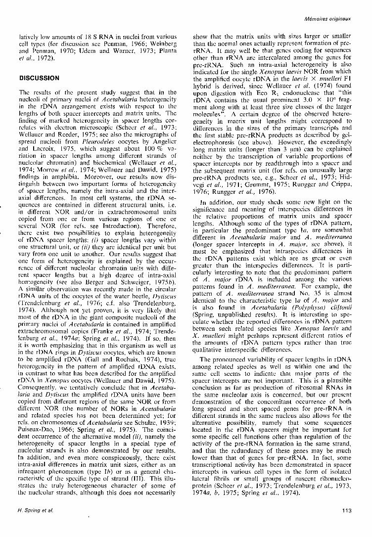
The results of the present study suggest that in the nucleoli of primary nuclei of A cetabularia heterogeneity in the rDNA arrangement exists with respect to the lengths of both spacer intercepts and matrix units. The finding of marked heterogeneity in spacer lengths correlates with electron microscopic (Scheer et al., 1973; Wellauer and Reeder, 1975; see also the micrographs of spread nucleoli from Pleurodeles oocytes by Angelier and Lacroix, 1975, which suggest about 100 % variation in spacer lengths among different strands of nucleolar chromatin) and biochemical (Wellauer et al., 1974; Morrow et al., 1974; Wellauer and Dawid, 1975) findings in amphibia. Moreover, our results now distingui~h between two important forms of heterogeneity of spacer lengths, namely the intra-axial and the interaxial differences. In most cell systems, the rDNA sequences are contained in different structural units, i.e. in different NOR and/or in extrachromosomal units copied from one or from various regions of one or several NOR (for refs. see Introduction). Therefore, there exist two possibilities to explain heterogeneity of rDNA spacer lengths: (i) spacer lengths vary within one structural unit, or (ii) they are identical per unit but vary from one unit to another. Our results suggest that one form of heterogeneity is explained by the occurrence of different nucleolar chromatin units with different spacer lengths but a high degree of intra-axial homogeneity (see also Berger and Schweiger, 1975b). A similar observation was recently made in the circular rDNA units of the oocytes of the water beetle, DytiscU5 (Trendelenburg et al., 1976; c.f. also Trendelenburg, 1974). Although not yet proven, it is very likely that most of the rDNA in the giant composite nucleoli of the primary nuclei of A cetabularia is contained in amplified extrachromosomal copies (Franke et al., 1974; Trendelenburg et al., 1974a; Spring et al., 1974). If so, then it is worth emphasizing that in this organism as well as in the rDNA rings in Dytiscus oocytes, which are known to be amplified rDNA (Gall and Rochaix, 1974), true heterogeneity in the pattern of amplified rDNA exists, in contrast to what has been described for the amplified rDNA in Xenopus oocytes (Wellauer and Dawid, 1975). Consequently, we tentatively conclude that in Acetabularia and Dytiscus the amplified rDNA units have been copied from different regions of the same NOR or from different NOR (the number of NORs in Acetabularia and related species has not been determined yet; for refs. on chromosomes of Acetabularia see Schulze, 1939; Puiseux-Dao, 1966; Spring et al., 1975). The coincident occurrence of the alternative model (U), namely the heterogeneity of spacer lengths in a special type of nucleolar strands is also demonstrated by our results. Tn addition, and even more conspicuously, there exist intra-axial differences in matrix unit sizes, either as an infrequent phenomenon (type Ib) or as a general characteristic of the specific type of strand (Ill). This illustrates the truly heterogeneous character of some of the nucleolar strands, although this does not necessarily
H. Spring et al.
Memoires originaux
show that the matrix units with sizes larger or smaller than the normal ones actually represent formation of prerRNA. It may well be that genes coding for sequences other than rRNA are intercalated among the genes for pre-rRNA. Such an intra-axial heterogeneity is also indicated for the single Xenopus laevis NOR from which the amplified oocyte rDNA in the laevis X muelleri Fl hybrid is derived, since Wellauer et al. (1974) found upon digestion with Eco Rl endonuclease that "this rDNA contains the usual prominent 3.0 X Ion fragment along with at least three size classes of the larger molecules". A certain degree of the observed heterogeneity in matrix unit lengths might correspond to differences in the sizes of the primary transcripts and the first stable pre-rRNA products as described by gelelectrophoresis (see above). However, the exceedingly long matrix units (longer than 3 fAm) can be explained neither by the transcription of variable proportions of spacer intercepts nor by read through into a spacer and the subsequent matrix unit (for refs. on unusually large pre-rRNA products see, e.g., Scheer et al., 1973; Hidvegi et al., 1971; Grummt, 1975; Runggcr and Crippa, 1976; Rungger et al., 1976).
In addition, our study sheds some new light on thc significance and meaning of interspecies differences in the relative proportions of matrix units and spacer lengths. Although some of the types of rDNA pattern, in particular the predominant type la, are somewhat different in Acetabularia major and A. mediterranea (longer spacer intercepts in A. major, see above), it must be emphasized that intraspecies differences in the rDNA patterns exist which are as great or even greater than the interspecies differences. It is particularly interesting to note that the predominant pattern of A. major rDNA is included among the various patterns found in A. mediterranea. For example, the pattern of A. mediterranea strand No. 35 is almost identical to the characteristic type la of A. major and is also found in A cetabularia (Polyphysa) cliftonii (Spring, unpublished results). It is interesting to speculate whether the reported differences in rDNA pattern between such related species like Xenopus laevis and X. muelleri might perhaps represent different ratios of the amounts of rDNA pattern types rather than true qualitative interspecific differences.
The pronounced variability of spacer lengths in rDNA among related species as well as within one and the same cell seems to indicate that major parts of the spacer intercepts are not important. This is a plausible conclusion as far as production of ribosomal RNAsin the same nucleolar axis is concerned, but our present demonstration of the concomitant occurrence of both long spaced and short spaced genes for pre-rRNA in different strands in the same nucleus also allows for the alternative possibility, namely that some sequences located in the rDNA spacers might be important for some specific cell functions other than regulation of the activity of the pre-rRNA formation in the same strand, and that the redundancy of these genes may be much lower than that of genes for pre-rRNA. In fact, some transcriptional activity has been demonstrated in spacer intercepts in various cell types in the form of isolated lateral fibrils or small groups of nascent ribonucleoprotein (Scheer et al., 1973; Trendelenburg et al., 1973, 1974a, b, 1975; Spring et al., 1974).
113

Journal de Microscopie et de Biologie Cellulaire, Volume 25, 1976
Acknowledgements
We gratefully acknowledge the generous supply with algal cultures by Dr. G. Werz (Free Vniversity of West Berlin). For composing the manuscript we thank Mme G. Lamprecht-Picot. The study was supported by the Deutsche Forschungsgemeinschaft (grant Fr 308/9).
BIBLIOGRAPHY
I. ANGELlER N. et LACROIX J.c., 1975. Complexes de transcription d 'origines nucleolaire et chromosomique d 'ovocytes de Pleurodeles waltlii et P. poireti (Amphibiens, Vrodeles). Chromosoma, 51, 323-335.
2. BERGER S. and SCHWEIGER H.G., 1975a. 80 S ribosomes in Acetabularia major. Redundancy of rRNA cistrons. Protoplasma, 83, 41-50.
3. BERGER S. and SCHWEIGER H.G., 1975b. An apparent lack of non-transcribed spacers in rDNA of a green alga. Molec. gen. Genet., 139, 269-275.
4. BIRNSTIEL M.L., CHIPCHASE M. and SPEIRS J., 1971. The ribosomal RNA cistrons. Progr. Nucleic Acid Res., 11, 351-389.
5. BOHNERT HJ., SCHILLER 8., BOHME R. and SAUER H.W., 1975. Circular DNA and rolling circles in nucleolar rDNA from mitotic nuclei of Physarum polycephalum. Eur. J. Biochem., 57, 361-369.
6. BROWN D.D., WENSINK P.C and JORDAN E., 1972. A comparison of the ribosomal DNAs of Xenopus laevis and Xenopus muelleri : the evolution of tandem genes. J. molec. Bioi., 63, 53-73.
7. BUSCH H. and SMETANA K., 1970. The Nucleolus, Academic Press, New York.
8. DAWID LB., BROWN D.D. and REEDER R.H., 1970. Composition and structure of chromosomal and amplified ribosomal DNAs of Xenopus laevis. J. molec. Bioi. 51, 341-369
9. DUESBERG P.H. and VOGT P.K., 1973. Gel electrophoresis of Avian leukosis and sarcoma viral RNA in formamide : Comparison with other viral and cellular species. J. Virol., 12, 594-599.
10. EBEL J.P., FELLNER P.C, EHREsMANN C, STIEGLER P., FISCHEL J.L., BRANLANT C, KROL A. and SRI WIDADA J., 1974. Studies on the primary structure and on the protein binding sites of ribosomal RNA's from E. coli and Proteus vulgaris. In: Proceedings of the 9th PEBS meeting, Hidvegi E.J., SUmegi J. and Venetianer P., eds., Akademiai Kiado, North Holland, Budapest, Amsterdam, 33, 109-123.
11. ENGBERG J., CHRISTIANSEN G. and LEICK V., 1974. Autonomous rDNA molecules containing single copies of the ribosomal RNA genes of the macronucleus of Tetrahymena pyriformis. Biochim. Biophys. Res. Commun., 59, 1356-1365.
12. FORSHEIT A.B., DAVIDSON N. and BROWN D.D., 1974. An electron microscope heteroduplex study of the ribosomal DNAs of Xenopus laevis and Xenopus muelleri. J. mol. BioI., 90, 301-315.
13. FRANKE W.W., BERGER S., FALK H., SPRING H., SCHEER V., HERTH W., TRENDELENBURG M.F. and SCHWEIGER H.G., 1974. Morphology of the nucleocytoplasmic interaction during the development of Acetabularia cells. I. The vegetative phase. Protoplasma, 82, 249-282.
14. FRANKE W.W., SCHEER V., SPRING H., TRENDELENBURG M.F. and KROHNE G. 1976. Morphology of transcriptional units of rDNA : Evidence for transcription in apparent spacer intercepts. Exp. Cell Res., in press.
15. GALL J.G., 1969. The genes for ribosomal RNA during oogenesis. Genetics (Suppl.), 61, 121-132.
114
16. GALL J.G., 1974. Free ribosomal RNA genes in the macronucleus of Tetrahymena. Proc. Natl. A cad. Sc. VSA, 71, 3078-3081.
17. GALL J.G. and ROCHAIX 1.D., 1974. The amplified ribosomal DNA of Dytiscid beetles. Proc. Natl. A cad. Sc. VSA, 71, 1819-1823.
18. GRIERSON D. and LOENING V.E., 1972. Distinct transcription products of ribosomal genes in two different tissues. Nature New Bioi., 235, 80-83.
19. GRUMMT I., 1975. Synthesis of RNA molecules larger than 45S by isolated rat-liver nucleoli. Eur. J. Biochem., 57, 159-167.
20. HIDVEGI E.J., PRESTAYKO A.W. and BUSCH H., 1971. 65S and 85S nucleolar RNA. Physiol. Chem. Phys., 3, 17-35.
21. ISHIKAWA H., 1973a. Comparative studies on the thermal stability of animal ribosomal RNA's. Comp. Biochem. Physiol., 46B, 217-227.
22. ISHIKAWA H., 1973b. Primary and secondary nicks in the ribosomal ribonucleic acid of insects. Biochem. Biophys. Res. Commun., 54, 301-307.
23. KIRBY K.S., 1965. Isolation and characterization of ribosomal ribonucleic acid. Biochem. J., 96, 266-269.
24. LATEUR L. and BONOTTO S., 1973. Culture of Acetabularia mediterranea in the laboratory. Bull. Soc. Roy. Bot. Belgique, 106, 17-38.
25. LAVA-SANCHEZ P.A. and PuPPo S., 1975. Occurrence in vivo of hidden breaks at specific sites of 26S ribosomal RNA of Musca carnaria. J. mol. Bioi. 95, 9-20.
26. LOENING V.E., 1967. The fractionation of high-molecularweight ribonucleic acid by polyacrylamide-gel electrophoresis. Biochem. J., 102, 251-257.
27. LOENING V.E., 1969. The determination of the molecular weight of ribonucleic acid by polyacrylamide-gel electrophoresis. Biochem. J., 113, 131-138.
28. LOENING V.E., GRIERSON D., ROGERS M.E. and SARTIRANA M.L., 1972. Properties of ribosomal RNA precursor. PEBS, 23, 395-405.
29. MILLER O.L. and BEATTY B.R., I 969a. Visualization of nucleolar genes. Science, 164, 955-957.
30. MILLER O.L. and BEATTY B.R., 1969b. Portrait of a gene. J. Cell Physiol., 74, 225-232.
31. MILLER O.L. and BAKKEN A.H., 1972. Morphological studies of transcription. Acta endocrinol. Suppl., 168, 155-177.
32. MORROW J.F., COHEN S.N., CHANG A.CY., Boy ER H.W., GOODMAN H.M. and HELLlNG R.B., 1974. Replication and transcription of eukaryotic DNA in Escherichia coli. Proc. Natl. A cad. Sc. VSA, 71, 1743-1747.
33. NOMuRA M., TISSIERES A and LENGYEL P., 1974. The Ribosomes, Cold Spring Harbour Lab., New York.
34. PENMAN S., 1966. RNA metabolism in the HeLa cell nucleus. J. mol. Bioi., 17, 117-130.
35. PINDER J.C., STAYNOV D.Z. and GRATZER W.B., 1974. Electrophoresis of RNA in formamide. Biochemistry, 13, 5373-5378.
36. PLANTA R.J., VAN DEN Bos R.C and KLOOTWIJK J., 1972. Biosynthesis of ribosomal RNA in eukaryotic cells. FEBS, 27, 183-1°1).
37. PUISEUX-DAO S., 1966. Siphonales and Siphonocladales. In : The chromosomes of the algae. Godward M.B.E., ed., Edward Arnold, London, 52-77.
38. RUNGGER D. and CRIPPA M., 1976. In vivo transcription of the spacer sequences in the rDNA in Xenopus. Submitted to publication.
39. RUNGGER D., CRIPPA M., TRENDELENBURG M.F., SCHEER V. and FRANKE W.W., 1976. Evidence for cleavage of the transcript of rDNA in statu nascendi in amphibian oocytes. Submitted to publication.
Patterns of nue/eo/ar genes

40. SCHEER U., TRENDELENBURG M.F. and FRANKE W.W., 1973. Transcription of ribosomal RNA cistrons. Correlation of morphological and biochemical data. Exp. Cell Res., 80, 175-190.
41. SCHUCH W. and LOENING U.E., 1975. The ribosomal ribonucleic acid of Agrobacterium tumefariens. Biochem. J., 149, 17-22.
42. SCHULZE K.L., 1939. Cytologische Vntersuchungen an Acetabularia mediterranea und Acetabularia wettsteinii. Arch. Protistenkunde, 92, 179-223.
43. SHINE J. and DALGARNO L., 1973. Occurrence of heatdissociable ribosomal RNA in insects : the presence of three polynucleotide chains in 26S RNA from cultured Aedes aegypti cells. J. mol. Bioi., 75, 57-72.
44. SLACK J.M.W., SARTIRANA M.L. and LOENING V.E., 1975. Multiple conformations of ribosomal precursor RNA. Nature, 253, 282-284.
45. SPRING H., TRENDELENBURG M.F., SCHEER V., FRANKE W.W. and HERTH W., 1974. Structural and biochemical studies of the primary nucleus of two green algal species, Acetabularia mediterranea and Acetabularia major. Cytobiologie, 10, 1-65.
46. SPRING H., SCHEER U., FRANKE W.W. and TRENDELENBURG M.F., 1975. Lampbrush-type chromosomes in the primary nucleus of green alga Acetabularia mediterrannea. Chromosoma, 50, 25-43.
47. STEVENS A.R. and PACHLER P.F., 1972. Discontinuity of 26S rRNA in Acanthamoeba castellani. J. mol. BioI., 66, 225-237.
48. TOBLER H., 1975. Occurrence and developmental significance of gene amplification. Biochemistry of Animal Development. Vo!. Molecular Aspects of Animal Development. Weber R., ed., Academic Press, New York, In, 91-143.
49. TRENDELENBURG M.F., 1974. Morphology of ribosomal RNA cistrons in oocytes of the water beetle, Dytiscus marginalis. Chromosoma, 48, 119-135.
50. TRENDELENBURG M.F., SCHEER V. and FRANKE W.W., 1973. Structural organization of the transcription of ribosomal DNA in oocytes of the house cricket. Nature New Bioi., 245, 167-170.
51. TRENDELENBURG M.F., SPRING H., SCHEER V. and FRANKE W.W., 1974a. Morphology of nucleolar cistrons
Manuscrit reru le 19 novembre 1975.
Note added in proof
Recently we have determined the molecular weights of the :lH-uridine labelled (24 hours labelling in vivo, concentration of radiolabelled nucleoside as described in the text) RNA from isolated primary nuclei of Acetabularia medJterranea in exponential formamide containing gels according to Spohr et al. (1976. Molecular weight determination of animal cell RNA by electrophoresis in formamide under fully denaturing conditions on exponential polyacrylamide gels, Europ. J. Biochem., in press), in comparison with 16 Sand 23 S rRNAs from E. coli and RNA from tobacco mosaic virus (TMV). The apparent molecular weights of the Acetabularia RNA components A. Band D under these conditions were 1.90 million, 1.28 million and 0.66
H. Spring et al.
Memoires originaux
in a plant cell, Acetabularia mediterranea. Proe. Natl. Aead. Se. VSA, 71, 3626-3630.
52. TRENDELENBURG M.F., FRANKE W.W., SPRING H. and SCHEER V., 1974b. Ultrastructure of transcription in the nucleoli of the green algae Acetabularia major and A. mediterranea. In: Proceeding of the 9th FEBS meeting, Hidvegi E.J., Slimegi J. and Venetianer P., eds., Akademiai Kiado, Nort6 Holland, Budapest, Amsterdam, 33, 159-168.
53. TRENDELENBURG M.F., SPRING H., SCHEER U. and FRANKE W.W., 1975. Demonstration of active nucleolar cistrons of the Acetabularia nucleus with the spreading technique. Protoplasma, 83, 180.
54. TRENDELENBURG M.F., FRANKE W.W. and SCHEER U., 1976. Frequency of circular copies of amplified nucleolar DNA in oocytes of two insects, the house cricket Aeheta domesticus and the water beetle Dytiseus marginalis. submitted.
55. UDEM S.A. and WARNER J.R., 1973. The cytoplasmic maturation of a ribosomal precursor RNA in yeast. J. Bioi. Chem., 25, 235-251.
56. WEINBERG R.A. and PENMAN S., 1970. Processing of 45S nucleolar RNA. J. mol. Bioi., 47, 169-178.
57. WELLAUER P.K., REEDER R.H., CARROL D., BROWN 0.0., DEuTcH A., HIGASHINAKAGAWA T. and DAVID l.B., 1974. Amplified ribosomal DNA from Xenopus laevis has heterogeneous spacer lengths. Proc. Natl. Aead. Se. USA, 71, 2823-2827.
58. WELLAUER P.K. and REED ER R.H., 1975. A comparison of the structural organization of amplified ribosomal DNA from Xenopus muelleri and Xenopus laevis. J. mol. BioI., 94, 151-161.
59. WELLAUER P.K. and DAVID LB., 1975. Studies on the structure of ribosomal RNA and DNA. Abstr. 10th FEBS meeting, Paris, July 20-25.
60. WENS INK P.e. and BROWN D.D., 1971. Denaturation map of the ribosomal DNA of Xenopus laevis. J. mol. Bioi., 60, 235-247.
61. WOODCOCK e.L.F., STANCHFIELD J.E. and GOULD R.R., 1975. Morphology and size of ribosomal cistrons in two plant species : Acetabularia mediterranea and Chlamydomonas reinhardii. Plant Sri. Left., 4, 17-23.
million daltons. The data show that the size of the component A, the putative pre-rRNA, is smaller than that of co-electrophoresed TMV-RNA (2.08 million daltons) and closely corresponds to the size of the most predominant type of transcriptional unit (matrix unit) of rDNA.
Heterogeneities in the rDNA pattern have also been demonstrated in an electron microscope study of nucleolar material from various other members of the Dasycladacea·;: (Acetabularia dentata, A. peniculus, A. ryukyuensis, Dasycladus clavae/ormis, Cymopolia van bosseae, Batophora oerstedii) including examples of intraaxial heterogeneity (S. Berger and H.G. Schweiger, 1975. Ribosomal DNA in different members of a family of green algae [Chlorophyta, Dasycladaceael: an electron microscopical study. Planta, 127, 49-62).
115

Journal de Microscopie et de Biologie Cellulaire, Volume 25, 1976
Plate I
Fig. a. - Electron micrograph of a spread and positively stained strand of transcriptionally active nucIeolar chromatin from primary nuclei of Acefabuiaria mediferranea. Note the regular alternation of fibril-covered regions (<< matrix units ») and fibrilfree intercepts (<< spacers »). The spacer intercepts are very short in this type of rDNA (type la; for details see text). x 5,600.
Fig. b. - Higher magnification of the nucIeolar strand shown in fig. a. Here the individual lateral fibrils are identified within the about 2 flm long matrix units and reveal their characteristic arrangement in distinct length gradients. Note also minor variations in the apparent lengths of the individual spacer intercepts; x 12,800.
Fig. c. - Spread and positively stained preparation of nucIeolar chromatin from Acetabularia mediferranea (here revealed after additional metal shadowing, see Material and Methods) which presents the type 11 rDNA pattern characterized by very long spacer intercepts. Note that the matrix units have about the same size as those shown in figs. a and b; x 12,800.
Fig. d. - Spread and positively stained transcriptional units of nucleolar chromatin from Acefabuiaria major as revealed at higher magnification. Note the relatively long spacer intercepts (begin and end of a spacer region is denoted by arrows) which in this type of pattern (not included in the histogram of fig. I) approach the length of the matrix units (mean matrix unit: 1.95 flm; mean spacer intercept: 1.9 Ilm; average repeat unit: 3.85 Ilm). This represents something like a type 11 pattern for A. major; x 38,000.
Fig. e. - Spread preparation of nucIeolar chromatin from Acetabularia mediferranea showing a representative repeating unit for type la rDNA pattern which is characterized by the existence of very short, if at all detectable, spacer intercepts (arrow); x 35,000.
Fig.,t: - A nucleolar chromatin strand from Acetabuiaria mediterranea showing a representative of type Ib rDNA pattern as characterized by the occurrence of individual longer matrix units (arrow) within a sequence of normal (i.e. ca. 2 Ilm long) matrix units and short spacer intercepts; x 23,800.
116 Patterns of nucleolar genes

1 "
JOURNAL DE MICROSCOPIE ET DE BIOLOGIE CELLULAIRE
H. Spring et al.
! JJ.m
~--tl'
Plate 1

Plate 11
Spread and positively stained preparation of transcribed nucleolar chromatin from Acetabularia mediterranea which displays a specific type of heterogeneity. (Ib). Here individual very short (arrow in the left) and very long (pair of arrows and insert) matrix units occur within otherwise normal type I arrangements; x 9,500; insert, x 23,500.

JOURNAL DE MICROSCOPIE ET DE BIOLOGIE CELLULAIRE
H. Spring et al.
5)J.m
Pla te n