HKSCCM interhospitalGR 21/5/2013 Dr Fong Man Chi, Natalie...
86
HKSCCM interhospital GR 21/5/2013 Dr Fong Man Chi, Natalie Dr Chan Yat Fat, Alfred
Transcript of HKSCCM interhospitalGR 21/5/2013 Dr Fong Man Chi, Natalie...

HKSCCM interhospital GR 21/5/2013
Dr Fong Man Chi, NatalieDr Chan Yat Fat, Alfred

40/F
Unremarkable past health
p/w fever/cough/sputum 3 days on 8/2/2013, admitted to medical ward
TOCC neg
BP 144/84 ST 149bpm Temp 38
WBC 411, Blast cell 369.9 (90%), Hb 8.2, Plt 71
INR 1.4 APTT 35.4
K 2.8 Cr 69 Bili/ALP normal ALT 67 CK 115 LDH 1931 Ca 2.14

Any comment?


WCC >= 100X10^9/L defines hyperleukocytosischaracterised by intravascular accumulation of blasts occupying the vascular lumen, without or without the presence of fibrinFunction of the blast cells less deformable than mature myeloid cells � intravascular pluggingHigh metabolic activity of blast and local production of various cytokines contribute to underlying cellular hypoxia

CNS, eyes and lungsOthers: extremities, kidneys, heart and penis
Dyspnea/stupor and WCC>100 in the absence of clear etiology � presumptive diagnosis of leukostasis
Fundoscopic exam is most helpful in establishing the diagnosis
Clinical picture can be less typical/ WCC <100 in rapidly increasing blast / monocytic variant


Chemotherapy with induction agents/ high dose hydroxyureaNo prospective randomised trial ofleukapheresis/ venesection for any outcome benefitCranial irradiation for neurologic symptomsDexamethasone recently proposed as it inhibit upregulation of adhesion molecules on leukemia and endothelial cellsAvoid PRBC transfusion

venesection done x 2 units
started on hydroxyurea and chemotherapy (cytarabine/daunorubicin) since 9/2 with allopurinol, rasburicase coverage

To TMH ICU for worsening resp failure, intubated
Refractory hypoxemia on FiO2 1.0


Transferred to PYNEH for VV-ECMO 10-15/2/13
Echo and USG :
good LV/RV systolic fxn, hyperdynamic with kissing wall movements, no RWMA, trace AR, mild TR, rim of pericardial effusion
hepatosplenomegaly, bil kidneys no hydronephrosis
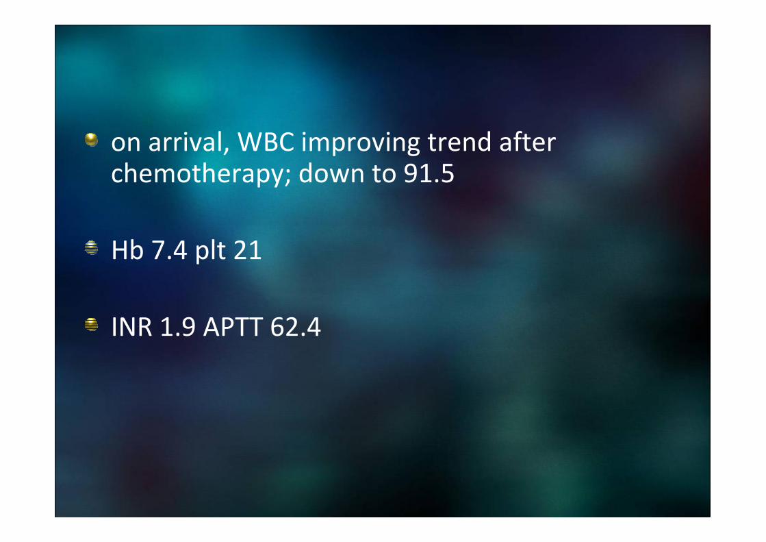
on arrival, WBC improving trend after chemotherapy; down to 91.5
Hb 7.4 plt 21
INR 1.9 APTT 62.4

TMH hematologist : not a case of M3 and unlikely differentiation syndrome
Developed pancytopenia afterward, requiring frequent transfusion of PC and plt conc

NPA 9/2
PCR for Respiratory syncytial virus +
Endotracheal aspirate 10/2
Respiratory syncytial virus viral antigen +ve
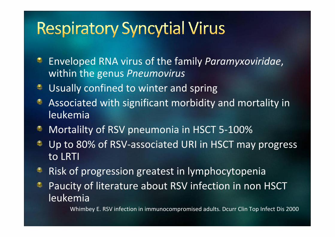
Enveloped RNA virus of the family Paramyxoviridae, within the genus Pneumovirus
Usually confined to winter and spring
Associated with significant morbidity and mortality in leukemia
Mortalilty of RSV pneumonia in HSCT 5-100%
Up to 80% of RSV-associated URI in HSCT may progress to LRTI
Risk of progression greatest in lymphocytopenia
Paucity of literature about RSV infection in non HSCT leukemia
Whimbey E. RSV infection in immunocompromised adults. Dcurr Clin Top Infect Dis 2000

Few studies reported to date suggest the epidemiology, clinical course and treatment response mirror findings in HSCT recipients
RSV seems to be frequent cause of life-threatening infection
Elderly, persistent myelosuppression, commorbidies, high APACHE II score and pneumonia


Considerations
Cost
Safety profile
Miscarriage, tetratogenicity
AE: Chronic eye/skin irritation, SOB
Discomfort of ribavirin administration
Conflicting results from studies
Many have good outcome without treatment
Small sample size < 20 patients


Daily dose of 6g at a concentration of 20mg/ml for 18 hrs/day
Median duration: 7 days, given at least 4 days
Small-particle aerosol generator unit via face mask inside a scavenging tent
FindingsFindings
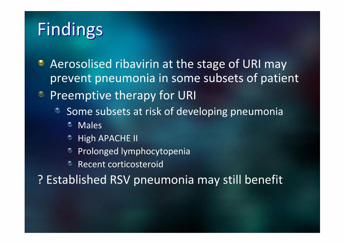
Aerosolised ribavirin at the stage of URI may prevent pneumonia in some subsets of patient
Preemptive therapy for URI
Some subsets at risk of developing pneumonia
Males
High APACHE II
Prolonged lymphocytopenia
Recent corticosteroid
? Established RSV pneumonia may still benefit

Neb Ribavirin given
ECMO decannulated on 15/2/2013
Bone marrow exam: aparticulate marrow aspirate. Cytochemical study on blast of peripheral blood showed AML. Favour monocytic lineage
Cytoreduction regime chemotherapy
Hydroxyurea and ‘3+7’ daunorubicin and cytarabine
Post D2 3+7 good cytoreduction: WCC 9.8
Mild tumor lysis syndrome given rasburicase/ allopurinol
Kick of fever despite tazocin, switched to meropenem on 18/2

Transferred back to TMH ICU on chemo D11 (19/2)
In Stable condition, transferred to haematologyward
Developed Neutropenic fever on D12 (20/2)
ALP 37 ALT 114 ALP 37
Cr 107�289 on D15 (23/2)
USG kidney
Left kidney 9.4cm Rt kidney 14.3cm
Trace perinephric collection, no hydronephrosis

Hb 7.8 WCC 0.2 plt 31 on D11
LDH 1916
Negative sputum / blood c/st
Antimicrobial therapy
Meropenem since D10
Vancomycin since D17
Micafungin since D18
Septrin since D19
Doxycycline since D20

D20 (28/2): Respiratory failure required intubation and shock on NA
E4V2M6
Fever+
Generalised petechaie
Transferred to TMH ICU again

CXR
Any comment?

Infection
Viral/ bacteria/Fungal…
MDR
Neoplastic
Pulmonary haemorrhage
Organising pneumonia
Toxic pneumonitis
Pulmonary leukemic infiltration
Acute lysis pneumopathy
Alveolar proteinosis
Others: PE, CHF, fluid overload, TRALI…

10 years observation in a resp ICU
42% infectious; 27% non-infectious
31% undetermined even after autopsy
23/37 (62%) died in non-transplanted patients
47/52 (90%) in died in bone marrow recipients
Mortality was 68/76 (90%) for ventilated cases compared to those not requiring (2/13; 15%)
Eur Respir J 1998; 12: 116–122

Consolidation mixed
with ground-glass
Thickened interstitium
at peripheries
Nodules
Thickening of
peribronchial tissue

Peripheral WCC 0.6 Hb 10.1 plt 27
BAL for PCP/ CMV/bacteria/proteinosisnegative
CMV PP65 antigenemia negative
Candida parapsilosis from serial lower respiratory cultures
TA 28/2, BAL 1/3

Does it mean Invasive Fungal Infection?
What else can be done to test?
When Should we start antifungal therapy
Prophylaxis
Empirical
Pre-emptive
What to be given?

EORTC/MSG criteria
Proven IFI: histology/culture from a normally sterile site which was clinically or radiologicallyinfected
Probable IFI : Requires host, clinical AND microbiological factors
Possible IFI: host, clinical or microbiological factors
• Prolonged Neutropenia >10 days and
fever
• Lower respiratory tract disease with lung
infiltrates on CT
• Candidial yeast grew on BAL not regarded
as microbiological factor

Is it useful in leukemic patients?Is it useful in leukemic patients?
Prompt and definitive diagnosis difficult
Low yield of microbiological cultures
Use of antifungal prophylaxis
Invasive procedures for histology rarely possible
Various causes for lung infiltrates
These criteria are made for clinical trials
High false negative rate at autopsy
75% died without diagnosed in life ( Chamilos et al 06)

What else can be done to test?What else can be done to test?
Serum galactomanan
Polysaccharide component of the cell wall of Aspergillus species, released during hyphal growth
Approved by US FDA for surveillance of invasive aspergillosis in immunocompromised patients
SE 0.71 ( 95% CI: 0.68-0.91)
SP 0.89 (95% CI: 0.88-0.90)
Variable cut–off 0.5-1.5
Pitfalls
False negative: fungal prophylaxis
False positive: piperacillin-tazobactam, cross-reacting antigens from other fungi, gut GVHD
Marr KA et al. Detection of galactomanan antigenemia by enzyme immunoassay for the diagnosis of IA.
J Infect Dis 2004

1,3-β-D-glucan
Panfungal antigen , not specific for any fungi
SE for candidiasis 78-81% depending on cut-off value
False-positive: HD with cellulose membrane, cephalosporin, carbapenems, ampicillin-sulbactam
Adjunctive test
Pickering JW. Evaluation of a (1�3)-β-D-glucanAssay for diagnosis of IFIs. J Clin Microbiol 2005
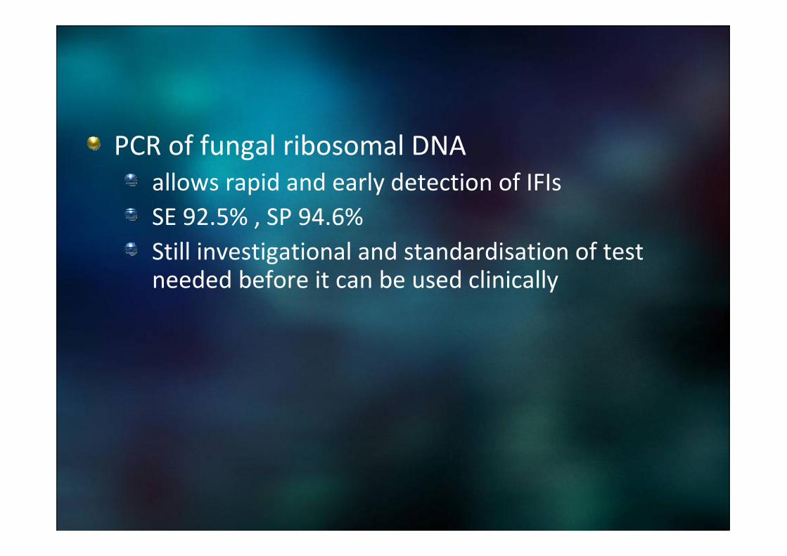
PCR of fungal ribosomal DNA
allows rapid and early detection of IFIs
SE 92.5% , SP 94.6%
Still investigational and standardisation of test needed before it can be used clinically

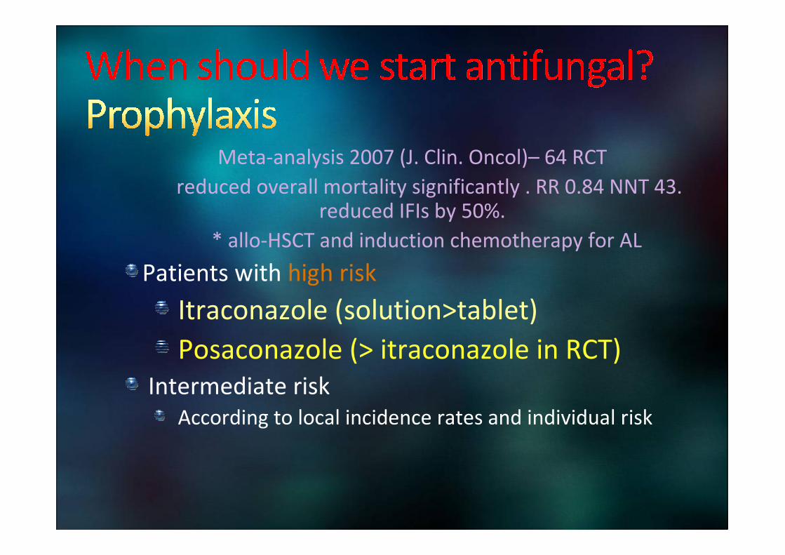
Meta-analysis 2007 (J. Clin. Oncol)– 64 RCT
reduced overall mortality significantly . RR 0.84 NNT 43. reduced IFIs by 50%.
* allo-HSCT and induction chemotherapy for AL
Patients with high risk
Itraconazole (solution>tablet)
Posaconazole (> itraconazole in RCT)
Intermediate risk
According to local incidence rates and individual risk

Fluconazole most widely studied, gold-standard in
Stem cell transplant ( Meta-analysis: Kanda et al., Cancer 2000)
Not effective after myelosuppressive treatment for acute leukemia
not active against molds and some candida species
Emergence of resistant candida strains and aspergillus

Prentice HG. Towards a targeted, riskPrentice HG. Towards a targeted, risk--based, antifungal strategy in based, antifungal strategy in neutropenicneutropenic patients. patients. Br. J. Br. J. HaematolHaematol. 2000. 2000

When should we start antiWhen should we start anti--Fungal Fungal
treatment?treatment?Under debate
Empirical approach
start antifungal treatment in patients with neutropenic fever not responding to antibiotics, irrespective of results of microbiological or radiological investigation
Pre-emptive
treat on the basis of clinical, imaging and/or laboratory findings indicative of IFIs in at-risk patients

Pros
Reported to have Antifungal use reduced by 78%
Limited studies
Depends heavily on SE and SP tests for IFI
Cons
mortality of untreated IFIs or delayed treatment reaches 100%
Incidence of IFIs signficantly higher in pre-emptive arm during induction chemotherapy. Non-inferior in lower risk group. Cordonnier C. Empirical versus preemptive antifungal therapy for high risk,
febrile, neutropenic patients: a randomised, controlled trial. Clin. Inf. Dis.
Whether to adopt depends on availability of intensive screening and rapid diagnostics

Still recommended in IDSA, NCCN and ECILAnd all panels restrict this strategy to expected duration of neutropenia of at least 10 days

What to be given?What to be given?
Empirical treatment
Amphotericin B, LAMB, itraconazole and caspofungin are approved for empirical treatment of IFIs in susceptible patients

Very ill patient
Poor immunity
Compatible radiology finding (At least partially)
No rapidly reversible cause being more likely

Persistent lung shadow for 2 weeks
Blood cell counts out of pancytopenia
On PS mode, FiO2 0.4
Still rather stiff lung and static imaging abnormality


strong association with AML M5

presumably pathologically related to the destruction of fibroblast cells in the pulmonary circulation
These cells release cytokines and cytotoxic enzymes, which promotes inflammation in the lungs, with subsequent airspace infiltration and hypoxia
Animal models have shown that monocytic cells have a predilection for the pulmonary circulation, probably as a result of monocyte-endothelium interaction.
all patients experienced acute worsening of respiratory status within hours of chemotherapy initiation

Supportive care
Ensuring adequate nutrition and maintaining electrolyte homeostasis
Prompt recognition and therapy directed toward the tumor lysis syndrome is crucial

Can absence of hyperleukocytosis exclude it?

Leonardo Potenza.Leukaemic pulmonary infiltrates in adult acute
myeloid leukaemia: a high-resolution computerized tomography
study. British Journal of Haematology 2003, 120.

Reported in patients without hyperleukocytosis
the type of blasts and their affinity for the pulmonary endothelium may be involved in the development of ARDS
AML M5 have predilection for pulmonary circulation
No established clinical criteria to firmly exclude or include diagnosis
No single low cutoff value for peripheral leukocyte or fibroblast counts that excludes this diagnosis.
The index of suspicion should be particularly high in patients with monocytic leukemia, especially when the peripheral blast cell count is high or increases rapidly

Most striking abnormality
interstitial thickening
Along lymphatics in the peribronchovascular, septal, and pleural interstitial tissue
Pulmonary nodules and focal homogeneous opacities

Diagnosis
based on histologic or cytologic studies and negative findings from a comprehensive investigation for more common causes
retrieval of leukemic cells by BAL
blast cell count above 40% in peripheral blood
Retrospective confirmation by effectiveness of chemotherapy in improving respiratory function

Azoulay, et al. Acute monocytic leukemia presenting as acute respiratory failure. American Journal of Respiratory and Critical Care Medicine167. 10 (May 15, 2003): 1329-33.

20 patients admitted to the intensive care unit (ICU) with threeremarkable features:
(1) rapidly progressive respiratory distress revealing acute leukemia
(2) monocytic leukemia
(3) respiratory status deterioration after chemotherapy initiation in all
median age was 50 years (17-72 years)
respiratory symptoms started 2 days (0-15 days) before ICU admission.
Chest radiographs revealed unilateral alveolar infiltrates (n = 1), bilateral alveolar infiltrates with (n = 3) or without (n = 11) pleural effusion, or diffuse interstitial infiltrates (n = 5)
Alveolar hemorrhage was the main bronchoalveolar lavagefinding, with monocytic cells retrieved from four patients

Leukemic pulmonary infiltration as the first manifestation of acute monocytic leukemia should be recognized
Index of suspicion should be particularly high in patients with monocytic leukemia
Especially when the peripheral blast cell count is high or increases rapidly
50% died in the group of Azoulay et al.

63 patients with hematologic malignancy to undergo 67 open lung biopsies for abnomality
Specific diagnosis was only found in 41 (62%) of the biopsies, with changes in therapy being made in 37 (57%)
Focal radiographic abnormality have higher yield than diffuse (79% versus 36%, p < 0.003)
Malignancy found in 18% of cases
Neutropenia or cases on ventilator lower yieldAM J RESPIR CRIT CARE MED 2000;161:723–729.

Repeat BM exam on 12/3
Marrow aspirate: regenerating marrow, blast accounts for 1% of total nucleated cells
Trephine: mild to moderately hypercellularmarrow,Full myeloid maturation still seen. Blasts are not obviously increased. Megakaryocytes are moderately increased. Dx: regenerating marrow.
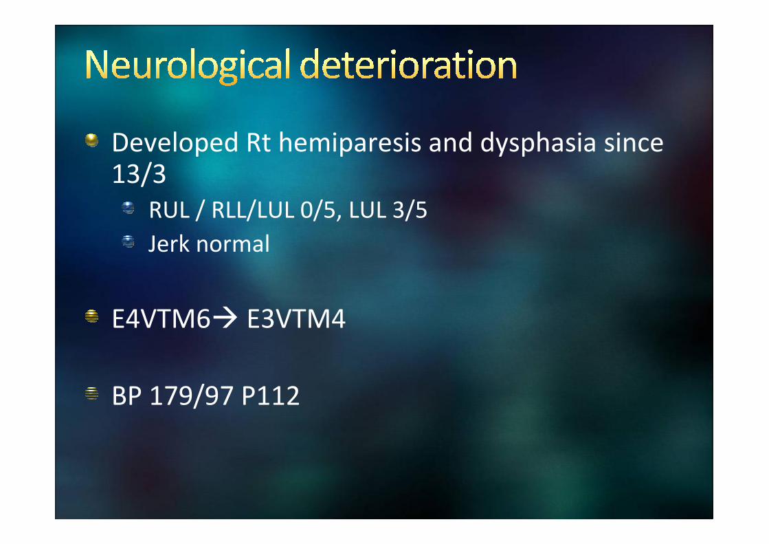
Developed Rt hemiparesis and dysphasia since 13/3
RUL / RLL/LUL 0/5, LUL 3/5
Jerk normal
E4VTM6� E3VTM4
BP 179/97 P112


Infection
Opportunistic CNS infection
cryptococcus, listeria monocytogenes, aspergillosis, nocardia, mucormycosis, toxoplasmosis, herpes simplex, varicella zoster, and JC virus Neoplastic
Neoplastiac
Direct effect
Meningeal, parenchymal, skull base metastasis
Indirect effect
Cerebrovascular complications from hyperviscosity
Drug toxicity
Demyelination ( necrotising leukoencephalopathy)


Incidence at diagnosis/relapse 3-7%
Pathogenesis unknown
Interruption of tight junctions of BBB through adhesion molecules on leukemic blasts or increased permeability from vascular endothelial growth factor
Increased risk in high WCC, high blast count, high LDH and younger age
monocytic lineage 5.7-fold increased risk

Can be subtle or present as increased ICP, CN palsy or motor deficit
Diagnosis based on CSF exam for cytology, immunophenotyping by flow cytometry and PCR ( for leukemia specific markers)
Gadolinum-enhanced MRI of brain and spine
MRI alone is neither sensitive nor specific

Lumbar PunctureLumbar Puncture
WCC trace RC +++
GS negative culture negative
Fungus negative
Protein 0.86 ( blood stained)
Glucose 3.9 ( Serum 6.8)
Negative malignant cells

Multifocal cortical and subcortical lesions with enhancement without underlying mass
lesion.
Abscess or cerebral metastases were excluded.
Absence of restriction of diffusion makes multiple infarcts not likely.
Features were likely due to severe atypical holohemispheric types of PRES
( related to hypertension and / or chemotherapy).
Differential diagnosis is encephalitis of usual presentation or types.


Hypertension
Eclampsia
Renal failure
Immunosuppressive therapy
Cytosine-arabinoside, cisplatin, methotrexate, gemcitabine
Rituximab, bevacizumab or growth factors ( erythropoietin, G-CSF)

Mechanism is still under clarified, is an acute central nervous system disorder characterized by reversible brain vasogenic edema
Mainly arising from failure of cerebrovascularautoregulation due to increased systemic BP and disruption of BBB, ? Direct endothelial dysfunction due to circulating toxin
Primarily implicates posterior white matter

Cardinal sign: acute rise in diastolic BP, can occur 24 hrs or more before other symptoms
Headache – constant, dull, non-localised
Mental state changes
GTC – more often multiple
Visual disturbance
Can be left with neurological sequalae

Diffusion-weighted imaging (DWI) and apparent diffusion coefficient (ADC) map in magnetic resonance imaging (MRI) are most sensitive and specific to differentiate vasogenic edema of PRES from cytotoxicedema
Commonly bilateral
White matter of Parietal-occipital 100% , frontal 68-82%
Less commonly postfrontal cortical/ subcortical ,brainstem, basal ganglia, cerebellum

Banu et al. MRI of non-neoplastic cranial complications of malignant
disorders.
Diagn Interv Radiol 2008

Mainstay of treatment
Control BP
Nicardapine, labetalol
Avoid clonidine and nitroglycerine
Hydration
Anti-convulsant
Withdrawal of offending medication and treatment of underlying systemic disease

BP gradually controlled with medication
Given Micafungin for 3 weeks , out of pancytopenia
Gradually improved and extubated on 19/3
Obey command, Rt power 0/5, static

New case of AML of monocytic lineage, with hyperleukocytosis and pulmonary leukostasis on presentation
requiring urgent induction chemotherapy and ECMO support
On presentation also had RSV chest infection treated with inhaled Ribavirin
Further complicated with
neutropenic sepsis/ ?fungal pneumonia
severe posterior reversible encephalopathy syndrome now with neurological deficits

House party protocol:
Numerous mobile suits in action
But where is the iron man?

We thought of many causes, were we correct?
Can we rule out leukemic infiltration
Should we believe only what we see – imaging/ single biopsy/cytology…
Should we shoot without knowing
Risk of further neurological damage and fatal neurological sequale
Any change in management if proceed to more invasive procedure or repeat test…
Or…..time will tell…

Acute bilateral loss of vision
Eye 31/3/2013: ? Leukaemic retinopathy with marked edema
MRI brain on 3/4
Multifocal T2W/FLAIR hyperintense lesions at bilateral cerebral/ cerebellar hemispheres, predominantly involving cortical/ subcorticalregions with leptomeningeal enhancement. Overall, they show interval improvement
FU MRI brain 8/5
Multiple wedge-shaped cortical/ subcortical lesions at bilateral cerebral hemispheres with evidence of early encephalomalacia

Rehab progress 5/2013
BI 13 �34
MMSE 18 (blind)�25
RUL power 0 , RLL 0�3/5 LUL/LL power 4/5
Near blindness →→ can only perceive light.
Haematologist:
Condition slowly improving, unlikely leukemic infiltration
Haematologically Not in relapse
Not for consolidative therapy meanwhile because of slow neurological recovery, ?Drug toxicity
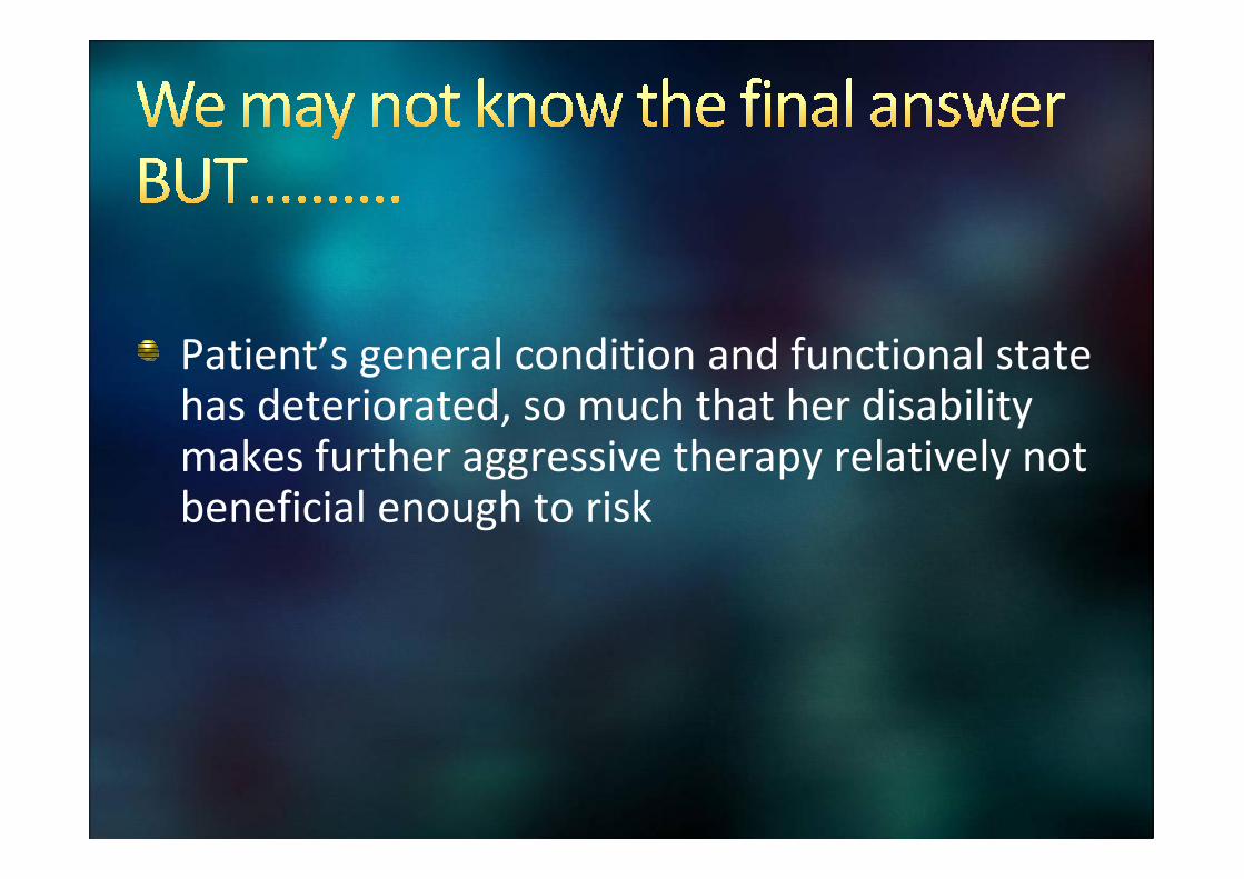
Patient’s general condition and functional state has deteriorated, so much that her disability makes further aggressive therapy relatively not beneficial enough to risk

Different complications of leukemia as a result of the disease and treatment
Leukemic infiltration
Infection
PRES
Emergencies in acute leukemia embrace multiple disciplines
Difficult dilemma and Controversy in management