History for the Discovery of DNA Chapter 16 The Molecular Basis of Inheritance.
68
History for the Discovery of DNA • Chapter 16 The Molecular Basis of Inheritance
-
Upload
corey-rogers -
Category
Documents
-
view
217 -
download
0
Transcript of History for the Discovery of DNA Chapter 16 The Molecular Basis of Inheritance.
History for the Discovery of DNA
• Chapter 16
The Molecular Basis of Inheritance
Next Unit:
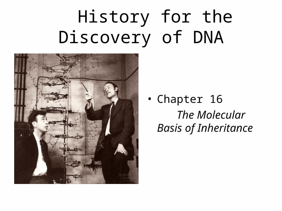
**Chapter 16: DNA: History, Structure & Replication**Chapter 17: Genetic Expression (protein synthesis)
Chapter 18: Viruses & Bacteria (selected parts) Chapter 19: Regulation (selected parts)**Chapter 20: Genetic Engineering & Biotechnology
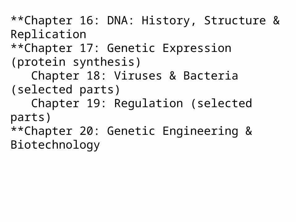
Overview of Chapter 16:TOPIC Pgs.
History & Discovery of DNA 293-296
as Genetic Material
Structure of DNA 296-298
DNA Replication 298-307
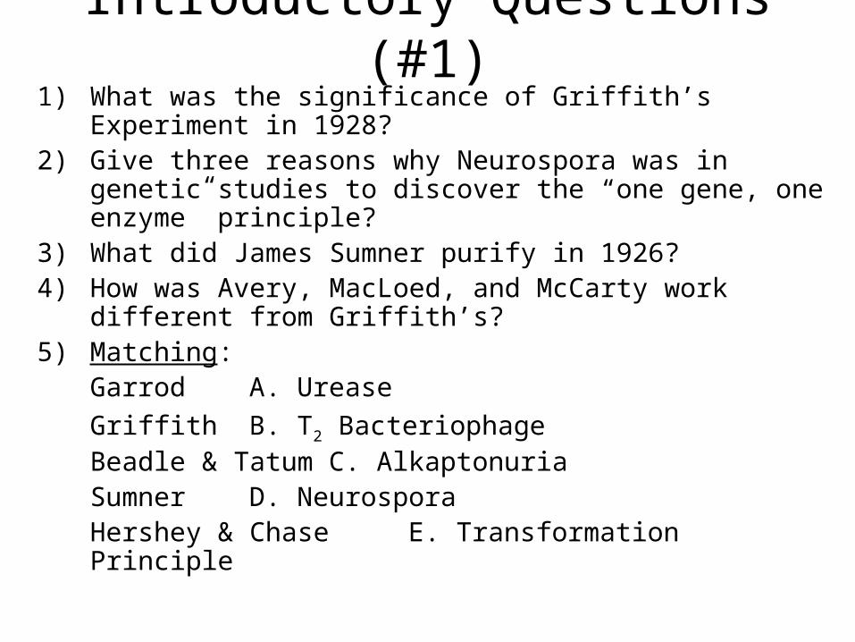
Introductory Questions (#1)1) What was the significance of Griffith’s Experiment in
1928?2) Give three reasons why Neurospora was in genetic
studies to discover the “one gene, one enzyme” principle?3) What did James Sumner purify in 1926? 4) How was Avery, MacLoed, and McCarty work different
from Griffith’s?5) Matching:
Garrod A. Urease
Griffith B. T2 BacteriophageBeadle & Tatum C. AlkaptonuriaSumner D. NeurosporaHershey & Chase E. Transformation Principle
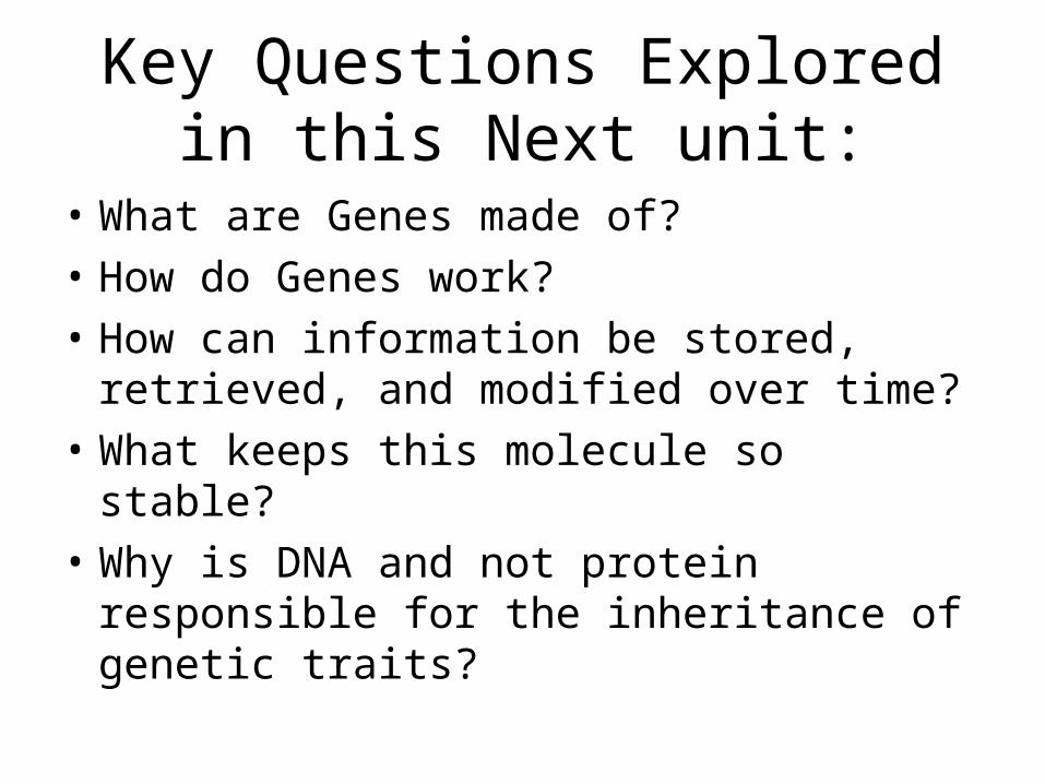
Key Questions Explored in this Next unit:
• What are Genes made of?
• How do Genes work?
• How can information be stored, retrieved, and modified over time?
• What keeps this molecule so stable?
• Why is DNA and not protein responsible for the inheritance of genetic traits?
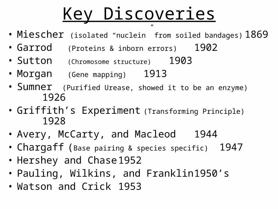
Key Discoveries• Miescher (isolated “nuclein” from soiled bandages) 1869• Garrod (Proteins & inborn errors) 1902• Sutton (Chromosome structure) 1903• Morgan (Gene mapping) 1913• Sumner (Purified Urease, showed it to be an enzyme) 1926• Griffith’s Experiment (Transforming Principle) 1928• Avery, McCarty, and Macleod 1944• Chargaff (Base pairing & species specific) 1947• Hershey and Chase 1952• Pauling, Wilkins, and Franklin
1950’s• Watson and Crick 1953
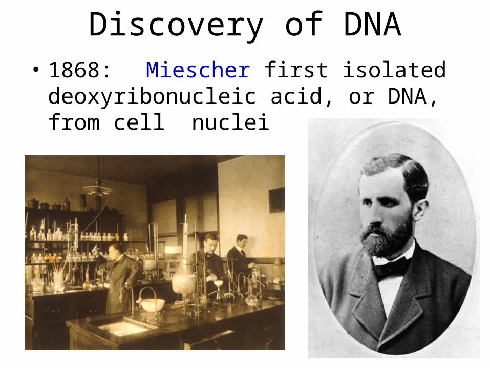
Discovery of DNA• 1868: Miescher first isolated
deoxyribonucleic acid, or DNA, from cell nuclei
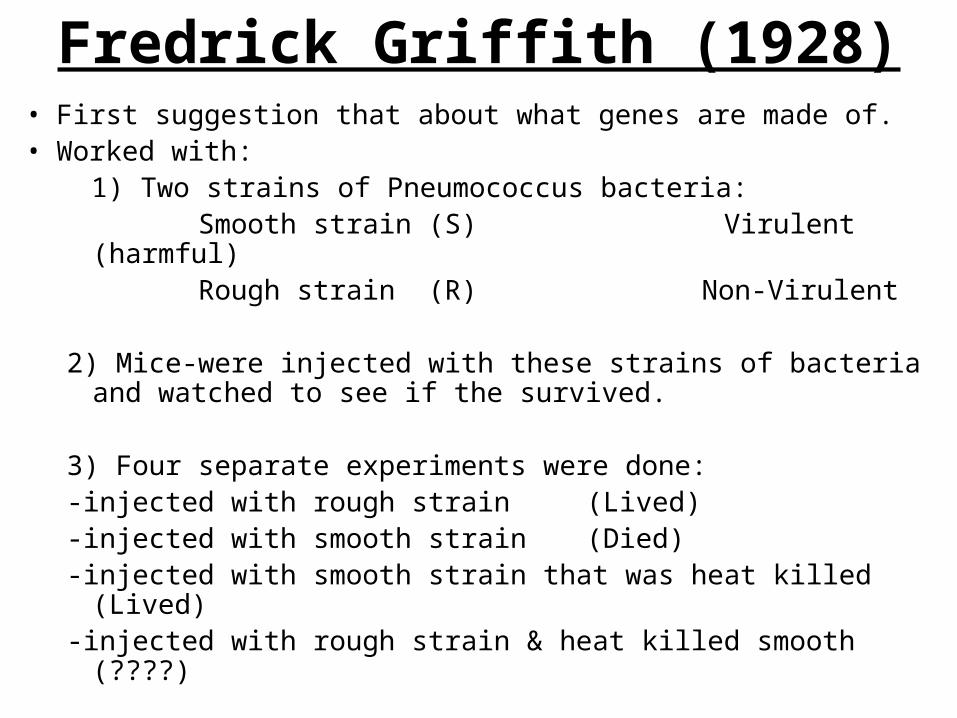
Fredrick Griffith (1928)• First suggestion that about what genes are made of. • Worked with: 1) Two strains of Pneumococcus bacteria:
Smooth strain (S) Virulent (harmful) Rough strain (R) Non-Virulent
2) Mice-were injected with these strains of bacteria and watched to see if the survived.
3) Four separate experiments were done:-injected with rough strain (Lived)-injected with smooth strain (Died)-injected with smooth strain that was heat killed (Lived)-injected with rough strain & heat killed smooth (????)
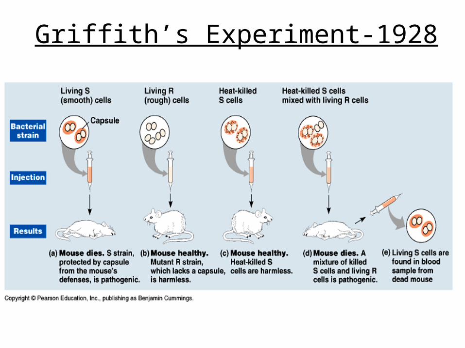
Griffith’s Experiment-1928
Conclusion of Griffith’s Experiment
• Somehow the heat killed smooth bacteria changed the rough cells to a virulent form.
• These genetically converted strains were called “Transformations”
• Something (a chemical) must have been transferred from the dead bacteria to the living cells which caused the transformation
• Griffith called this chemical a “Transformation Principle”
Avery, MacLeod, and McCarty (1944)
• Chemically identified Griffith’s transformation principle as DNA
• Separated internal contents of the S cells into these fractions:
(lipids, proteins, polysaccharides, and nucleic acids)
• They tested each fraction to see if it can cause transformation to occur in R cells to become S cells.
• Only the nucleic acids caused the transformation• This was the first concrete evidence that DNA is the
genetic material. • Some were not completely convinced because they
were not sure if this was true for eukaryotes.

Next Breakthrough came from the use of Viruses
• Viruses provided some of the earliest evidence that genes are made of DNA
• Molecular biology studies how DNA serves as the molecular basis of heredity
• Only composed of DNA and a protein shell
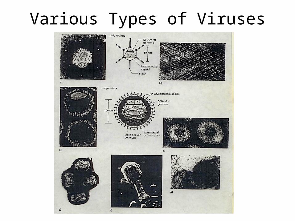
Various Types of Viruses
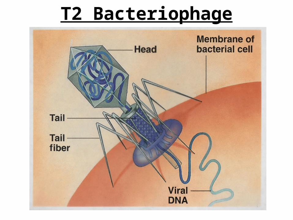
T2 Bacteriophage
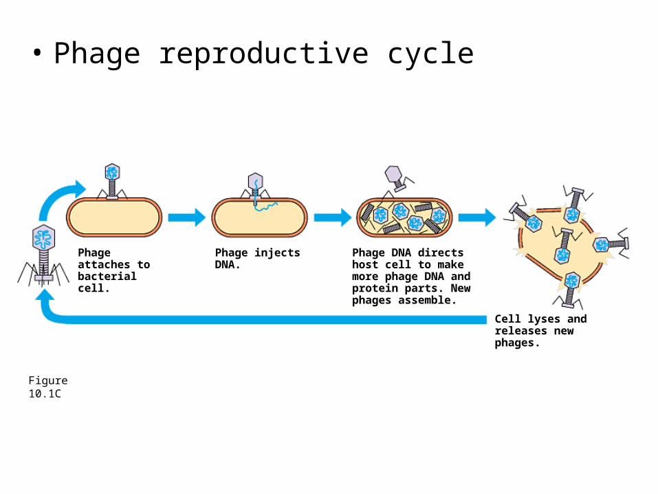
• Phage reproductive cycle
Figure 10.1C
Phage attaches to bacterial cell.
Phage injects DNA.
Phage DNA directs host cell to make more phage DNA and protein parts. New phages assemble.
Cell lyses and releases new phages.
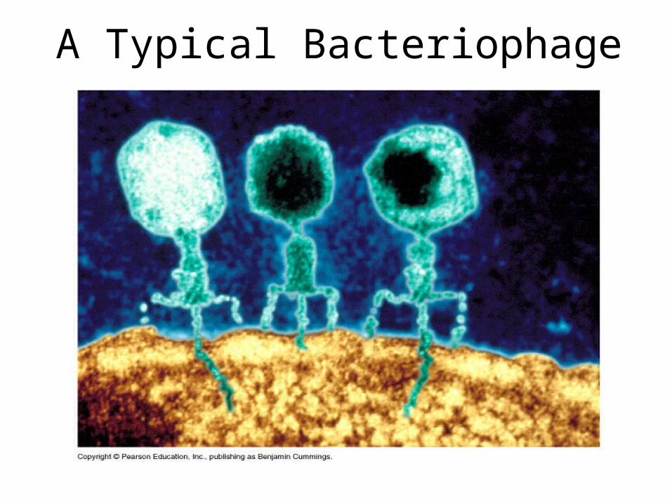
A Typical Bacteriophage
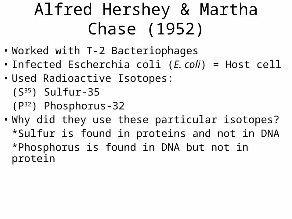
Alfred Hershey & Martha Chase (1952)
• Worked with T-2 Bacteriophages• Infected Escherchia coli (E. coli) = Host cell• Used Radioactive Isotopes:
(S35) Sulfur-35(P32) Phosphorus-32
• Why did they use these particular isotopes?*Sulfur is found in proteins and not in DNA*Phosphorus is found in DNA but not in protein
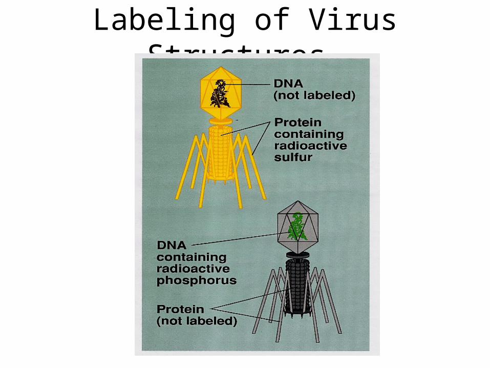
Labeling of Virus Structures
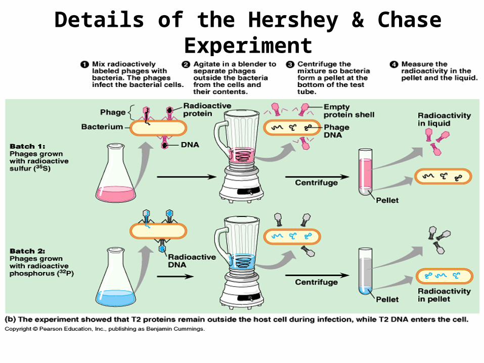
Details of the Hershey & Chase Experiment
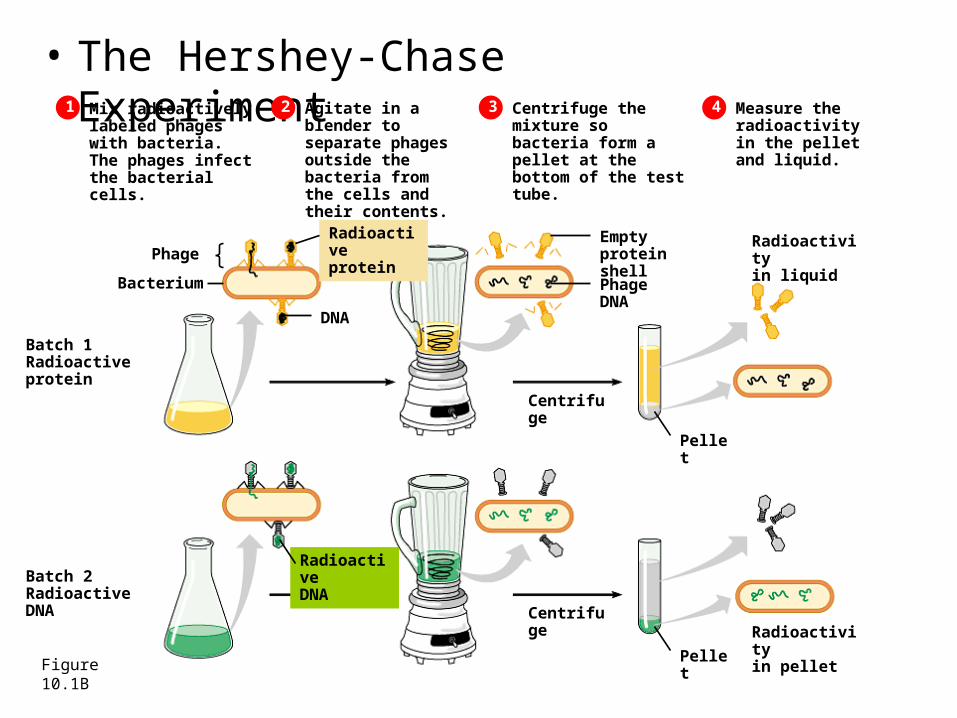
• The Hershey-Chase Experiment
Figure 10.1B
Mix radioactivelylabeled phages with bacteria. The phages infect the bacterial cells.
Phage
Bacterium
Radioactiveprotein
DNA
Emptyprotein shell
1 2 Agitate in a blender to separate phages outside the bacteria from the cells and their contents.
3 Centrifuge the mixture so bacteria form a pellet at the bottom of the test tube.
4 Measure the radioactivity in the pellet and liquid.
Batch 1Radioactiveprotein
Batch 2RadioactiveDNA
RadioactiveDNA
PhageDNA
Centrifuge
Pellet
Radioactivityin liquid
Radioactivityin pelletPellet
Centrifuge
Video clip of Hershey Chase Experiment
• http://highered.mcgraw-hill.com/sites/0072437316/student_view0/chapter14/animations.html#
• Key findings: the phage DNA entered in the host cell and when these cells were returned to the culture medium the infection ran its course producing E.coli and other bacteriophages with the radioactive phosphorus. (pg. 298)
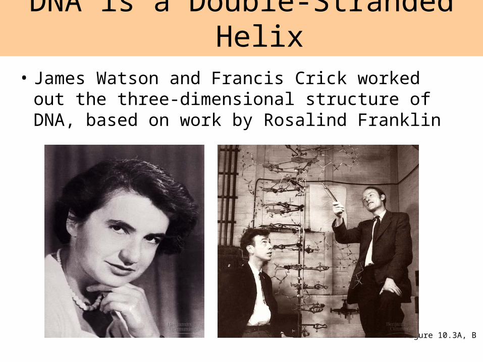
• James Watson and Francis Crick worked out the three-dimensional structure of DNA, based on work by Rosalind Franklin
DNA is a Double-Stranded Helix
Figure 10.3A, B
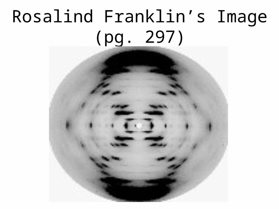
Rosalind Franklin’s Image (pg. 297)
• and Media
• DNA is a nucleic acid, made of long chains of nucleotides
DNA and RNA are polymers of Nucleotides
Figure 10.2A
Nucleotide
Phosphate group
Nitrogenous base
Sugar
Polynucleotide Sugar-phosphate backbone
DNA nucleotide
Phosphategroup
Nitrogenous base(A, G, C, or T)
Thymine (T)
Sugar(deoxyribose)
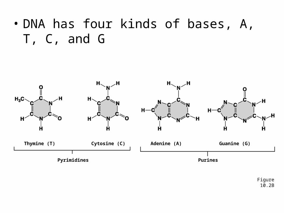
• DNA has four kinds of bases, A, T, C, and G
Figure 10.2B
Pyrimidines
Thymine (T) Cytosine (C)
Purines
Adenine (A) Guanine (G)
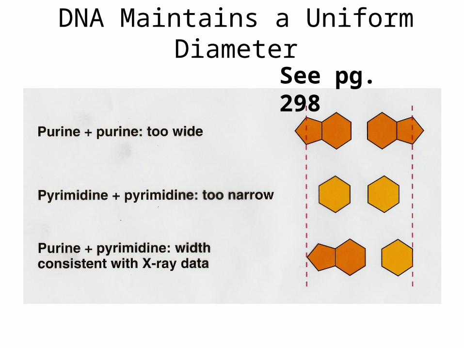
DNA Maintains a Uniform Diameter
See pg. 298
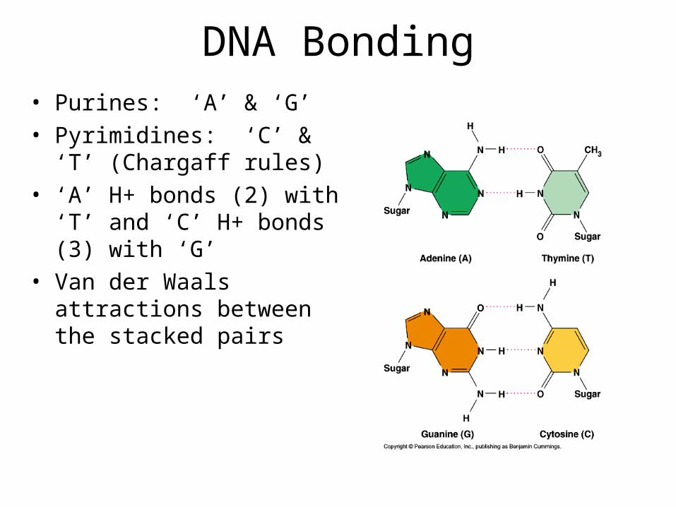
DNA Bonding• Purines: ‘A’ & ‘G’• Pyrimidines: ‘C’ & ‘T’
(Chargaff rules)• ‘A’ H+ bonds (2) with ‘T’ and
‘C’ H+ bonds (3) with ‘G’• Van der Waals attractions
between the stacked pairs
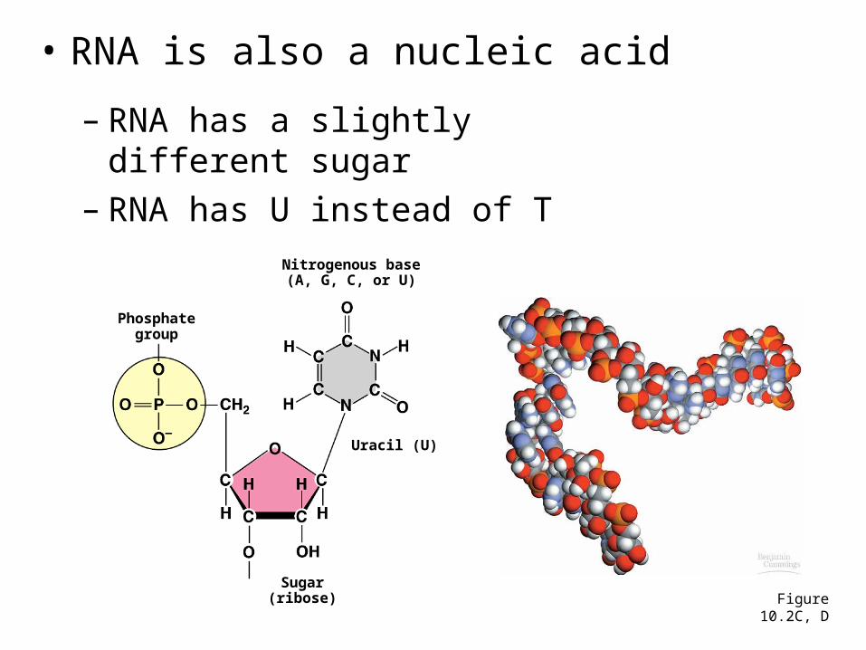
• RNA is also a nucleic acid
– RNA has a slightly different sugar– RNA has U instead of T
Figure 10.2C, D
Phosphategroup
Nitrogenous base(A, G, C, or U)
Uracil (U)
Sugar(ribose)
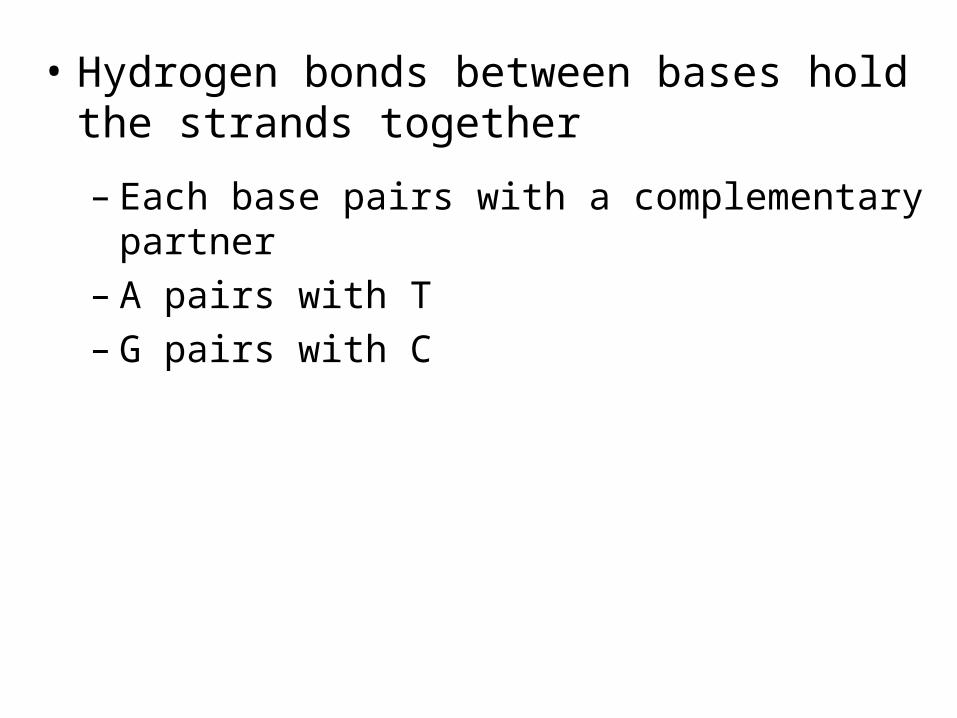
• Hydrogen bonds between bases hold the strands together
– Each base pairs with a complementary partner– A pairs with T– G pairs with C
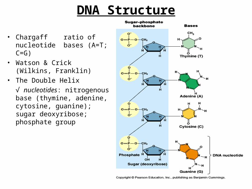
DNA Structure
• Chargaffratio of nucleotide bases (A=T; C=G)
• Watson & Crick (Wilkins, Franklin)
• The Double Helix
√ nucleotides: nitrogenous base (thymine, adenine, cytosine, guanine); sugar deoxyribose; phosphate group
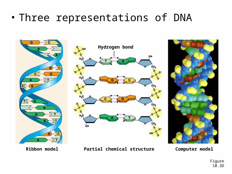
• Three representations of DNA
Figure 10.3D
Ribbon model Partial chemical structure Computer model
Hydrogen bond
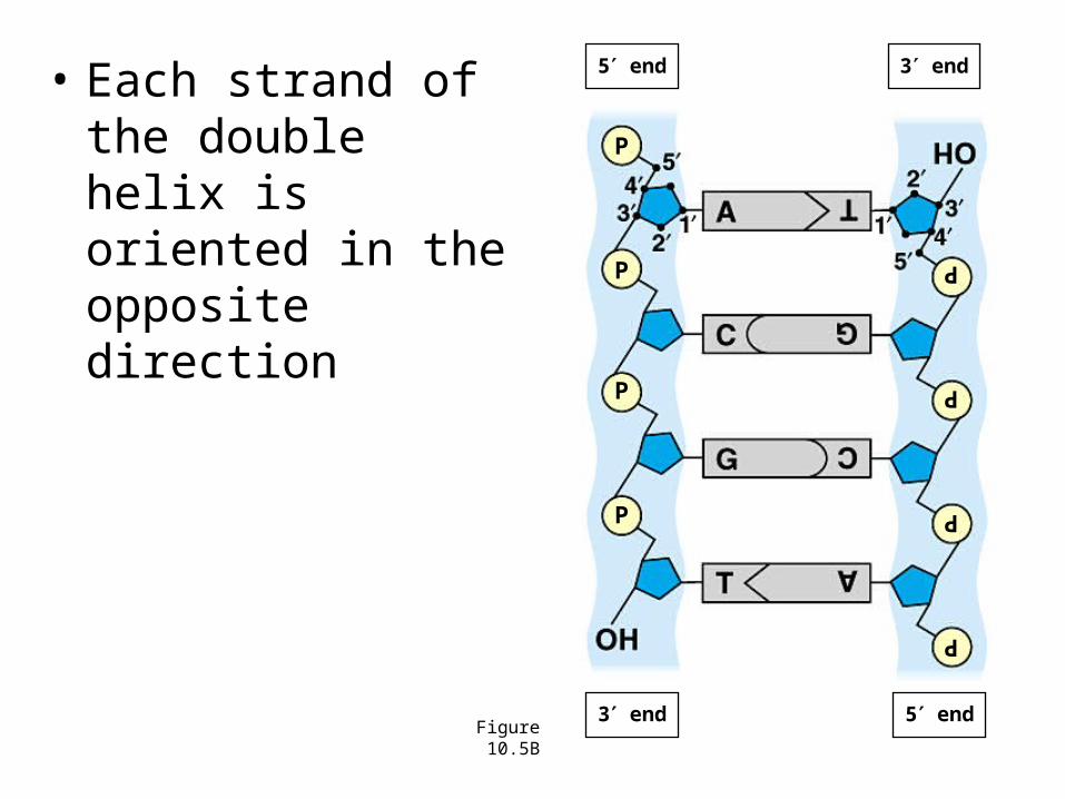
• Each strand of the double helix is oriented in the opposite direction
Figure 10.5B
5 end 3 end
3 end 5 end
P
P
P
PP
P
P
P
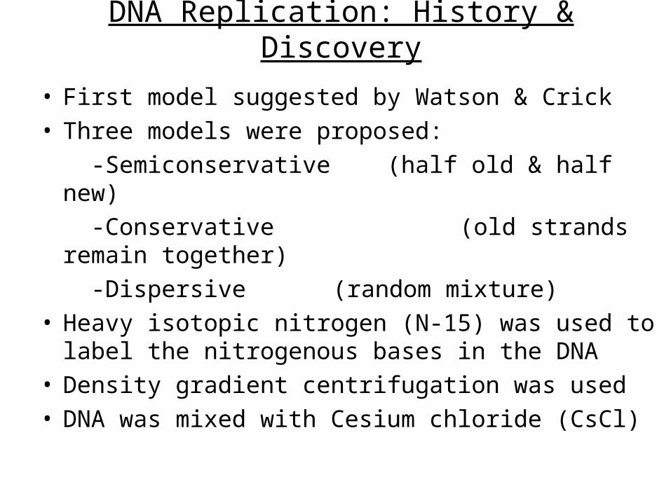
DNA Replication: History & Discovery
• First model suggested by Watson & Crick• Three models were proposed:
-Semiconservative (half old & half new)
-Conservative (old strands remain together)
-Dispersive (random mixture)• Heavy isotopic nitrogen (N-15) was used to label
the nitrogenous bases in the DNA• Density gradient centrifugation was used• DNA was mixed with Cesium chloride (CsCl)
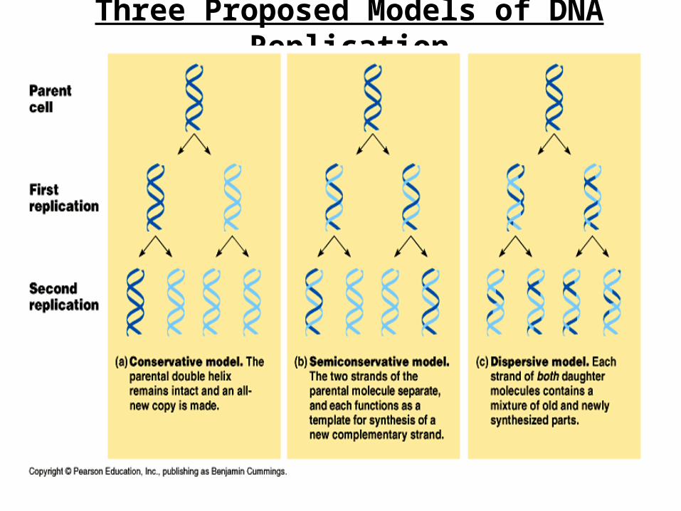
Three Proposed Models of DNA Replication
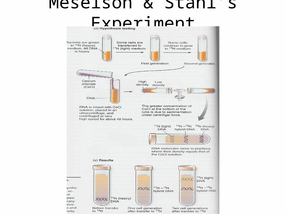
Meselson & Stahl’s Experiment
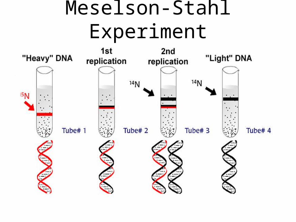
Meselson-Stahl Experiment
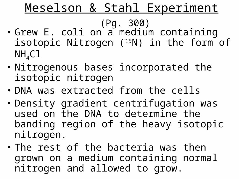
Meselson & Stahl Experiment (Pg. 300)
• Grew E. coli on a medium containing isotopic Nitrogen (15N) in the form of NH4Cl
• Nitrogenous bases incorporated the isotopic nitrogen
• DNA was extracted from the cells• Density gradient centrifugation was used on
the DNA to determine the banding region of the heavy isotopic nitrogen.
• The rest of the bacteria was then grown on a medium containing normal nitrogen and allowed to grow.
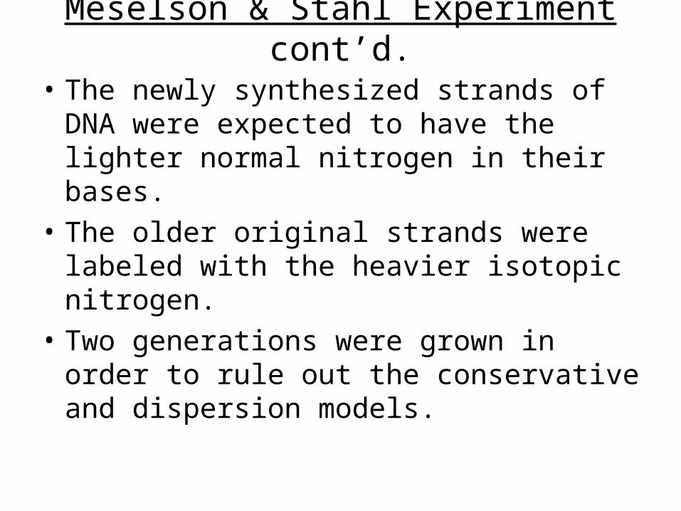
Meselson & Stahl Experiment cont’d.
• The newly synthesized strands of DNA were expected to have the lighter normal nitrogen in their bases.
• The older original strands were labeled with the heavier isotopic nitrogen.
• Two generations were grown in order to rule out the conservative and dispersion models.
Introductory Questions (#1)1) What was the significance of Griffith’s Experiment in 1928?
2) How was Avery, MacLoed, and McCarty work different from Griffith’s?
3) How was the dispersive model & conservative models ruled out as the way in which DNA replicates?
4) Matching:
Meselson & Stahl A. X-ray diffraction
Griffith B. T2 Bacteriophage
Franklin & Wilkins C. Semiconservative model
Chargaff D. Base pairing: C-G & T-A
Hershey & Chase E. Transformation Principle
Introductory Questions #21) Briefly explain what density gradient centrifugation
is and what it is used for.2) Name the organism used by Meselson & Stahl to
label the DNA.3) Name all of the enzymes required for DNA
replication to occur and what purpose they serve.4) In what direction is the newly synthesized strand
made? What end of the old strand do the nucleotides add to?
5) What direction is the new strand growing? (towards or away from the replication fork)
6) How long (# nucleotides) are the Okasaki fragments? How long are the RNA primers?
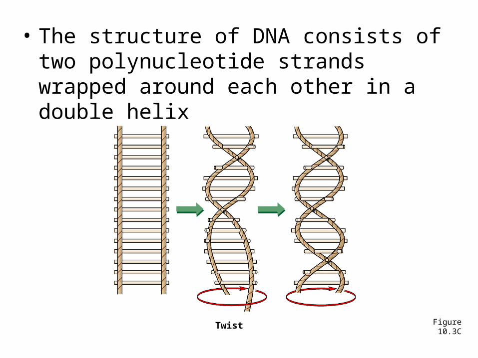
• The structure of DNA consists of two polynucleotide strands wrapped around each other in a double helix
Figure 10.3CTwist
1 chocolate coat,Blind (PRA)
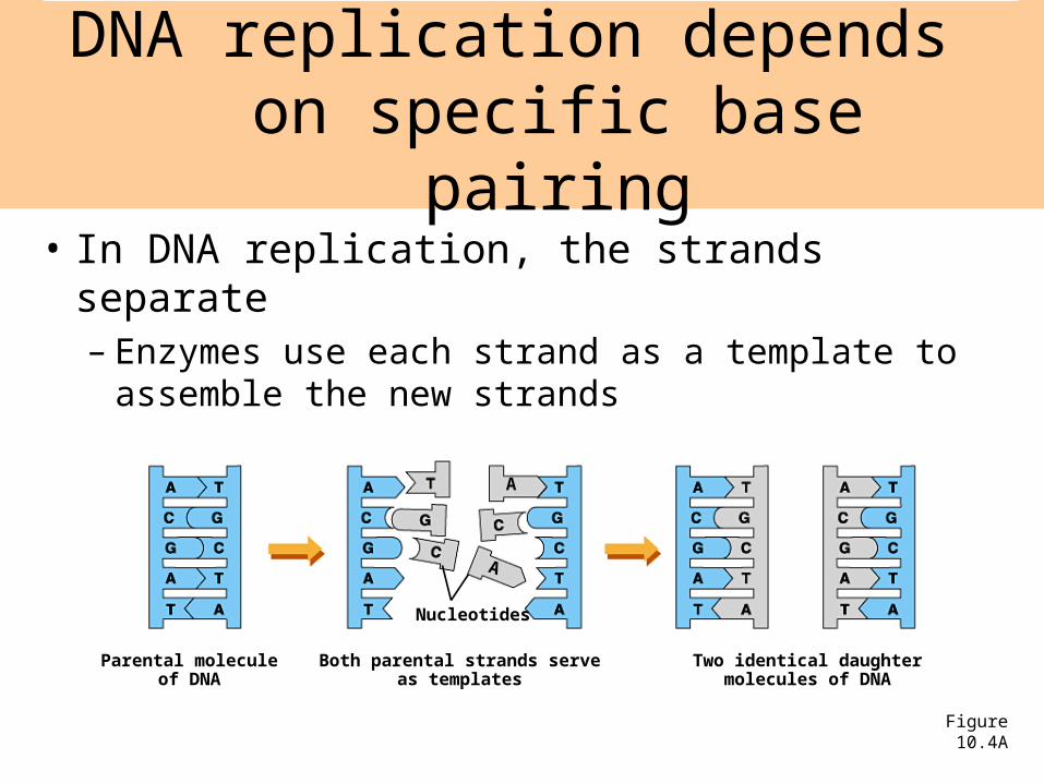
• In DNA replication, the strands separate– Enzymes use each strand as a template to
assemble the new strands
DNA replication depends on specific base pairing
Parental moleculeof DNA
Figure 10.4A
Both parental strands serveas templates
Two identical daughtermolecules of DNA
Nucleotides
A
A
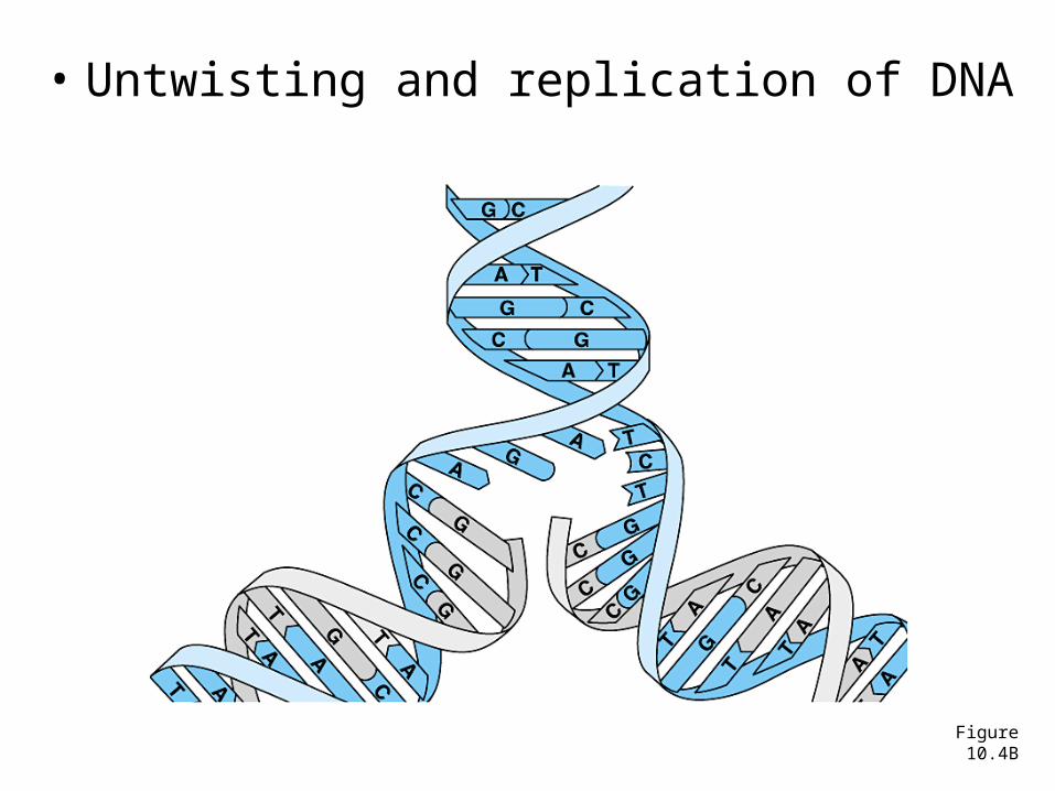
• Untwisting and replication of DNA
Figure 10.4B
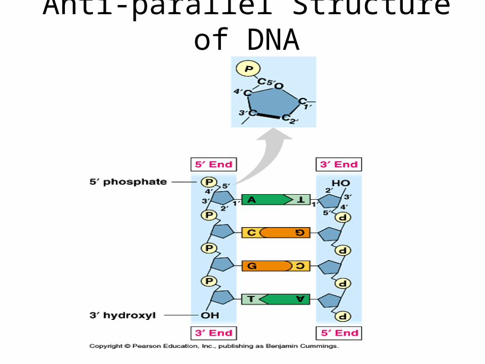
Anti-parallel Structure of DNA
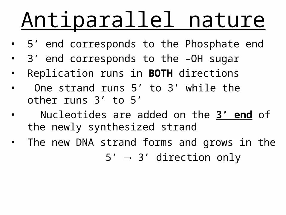
Antiparallel nature• 5’ end corresponds to the Phosphate end• 3’ end corresponds to the –OH sugar • Replication runs in BOTH directions
• One strand runs 5’ to 3’ while the other runs 3’ to 5’
• Nucleotides are added on the 3’ end of the newly synthesized strand
• The new DNA strand forms and grows in the
5’ 3’ direction only
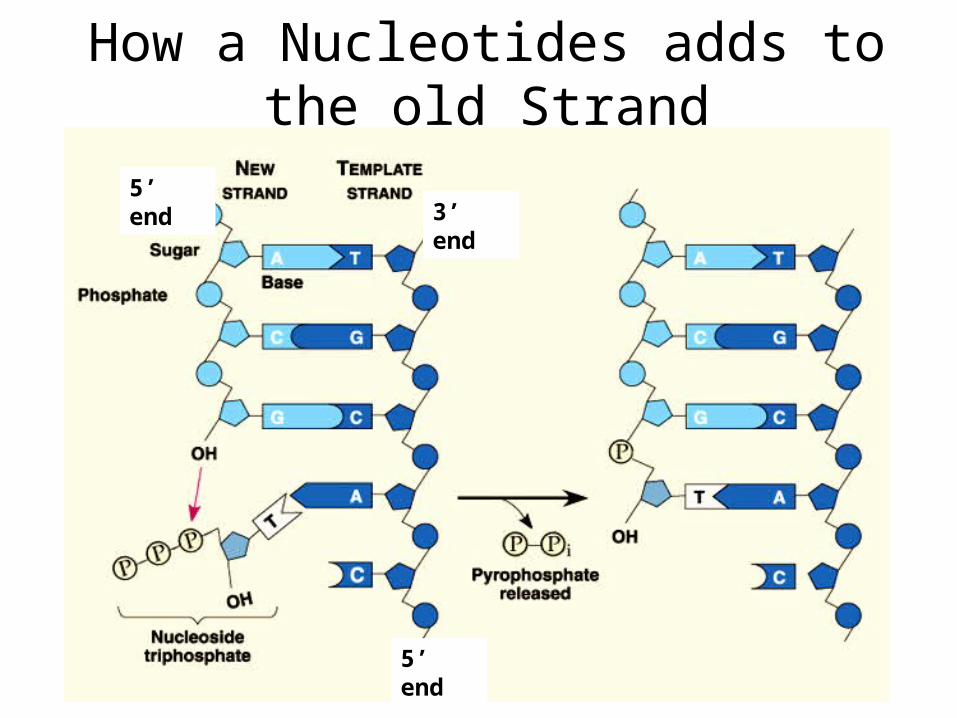
How a Nucleotides adds to the old Strand
5’ end
3’ end5’ end
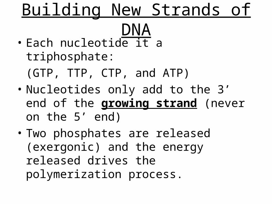
Building New Strands of DNA• Each nucleotide it a triphosphate:
(GTP, TTP, CTP, and ATP)
• Nucleotides only add to the 3’ end of the growing strand (never on the 5’ end)
• Two phosphates are released (exergonic) and the energy released drives the polymerization process.
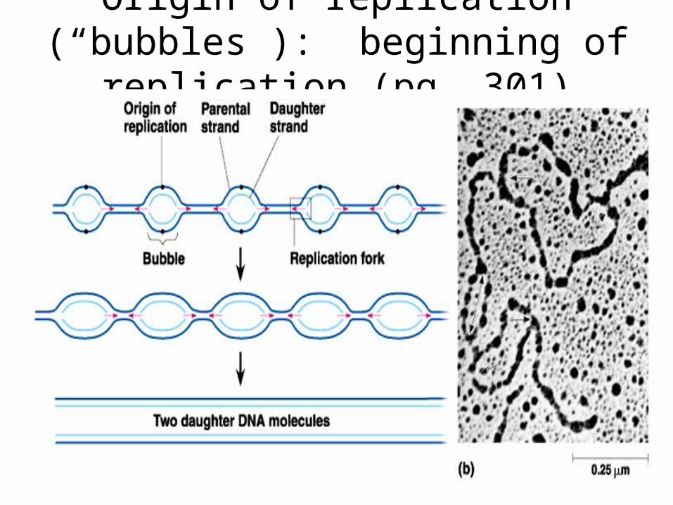
Origin of replication (“bubbles”): beginning of replication (pg. 301)
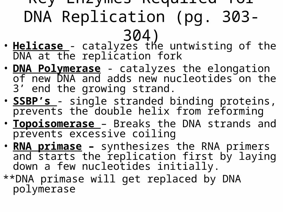
Key Enzymes Required for DNA Replication (pg. 303-304)
• Helicase - catalyzes the untwisting of the DNA at the replication fork
• DNA Polymerase - catalyzes the elongation of new DNA and adds new nucleotides on the 3’ end the growing strand.
• SSBP’s - single stranded binding proteins, prevents the double helix from reforming
• Topoisomerase – Breaks the DNA strands and prevents excessive coiling
• RNA primase – synthesizes the RNA primers and starts the replication first by laying down a few nucleotides initially.
**DNA primase will get replaced by DNA polymerase
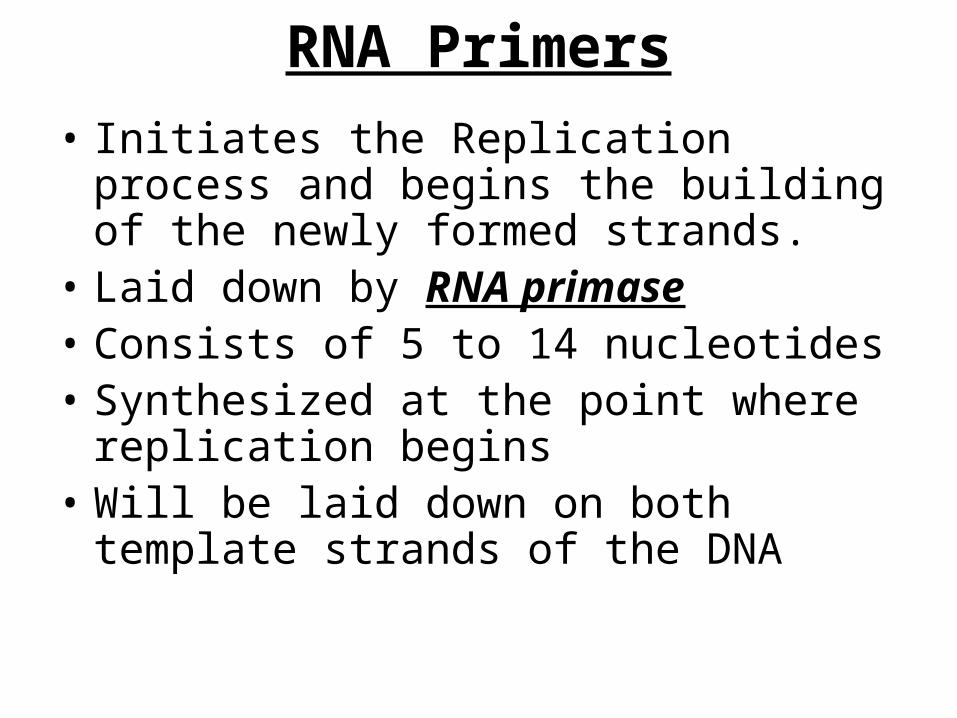
RNA Primers
• Initiates the Replication process and begins the building of the newly formed strands.
• Laid down by RNA primase• Consists of 5 to 14 nucleotides• Synthesized at the point where replication
begins• Will be laid down on both template strands
of the DNA
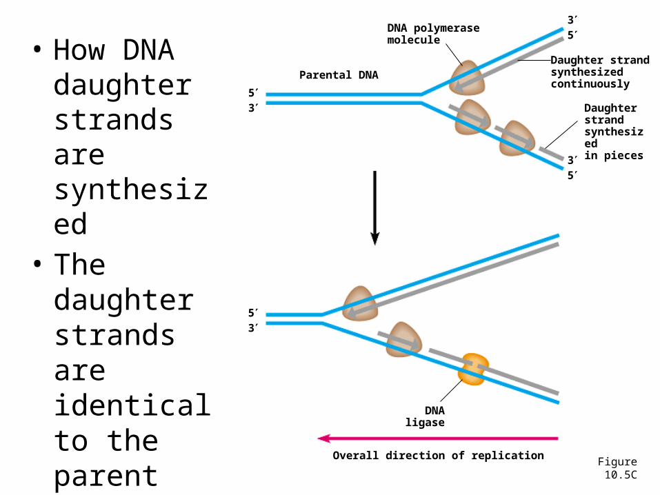
• How DNA daughter strands are synthesized
5 end
P
P
Parental DNA
Figure 10.5C
DNA polymerasemolecule
53
35
35
Daughter strandsynthesizedcontinuously
Daughter strandsynthesizedin pieces
DNA ligase
Overall direction of replication
53
• The daughter strands are identical to the parent molecule
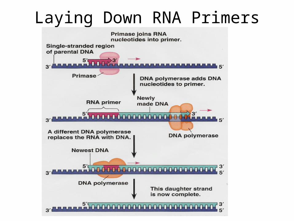
Laying Down RNA Primers
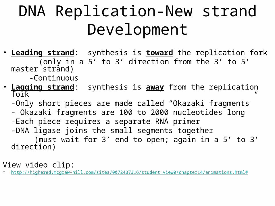
DNA Replication-New strand Development
• Leading strand: synthesis is toward the replication fork (only in a 5’ to 3’ direction from the 3’ to 5’ master strand)
-Continuous• Lagging strand: synthesis is away from the replication fork
-Only short pieces are made called “Okazaki fragments”- Okazaki fragments are 100 to 2000 nucleotides long-Each piece requires a separate RNA primer
-DNA ligase joins the small segments together (must wait for 3’ end to open; again in a 5’ to 3’ direction)
View video clip: • http://highered.mcgraw-hill.com/sites/0072437316/student_view0/chapter14/animations.html#

DNA Replication Fork
Video Clip of DNA Replication• http://highered.mcgraw-hill.com/sites/0072437316/student_view0/chapter14/animations.html#
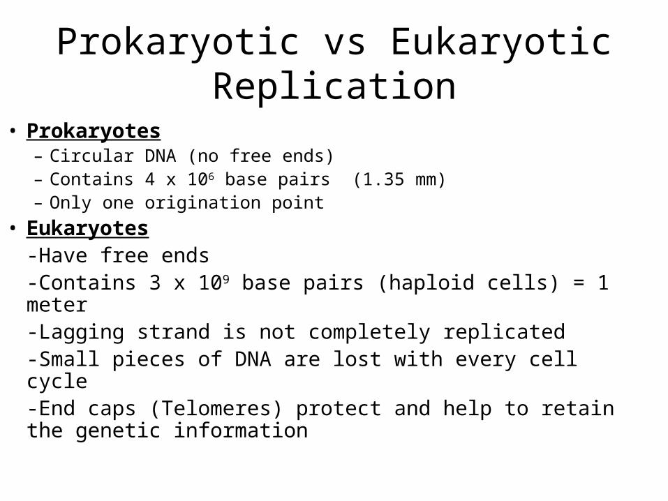
Prokaryotic vs Eukaryotic Replication
• Prokaryotes– Circular DNA (no free ends)– Contains 4 x 10⁶ base pairs (1.35 mm)– Only one origination point
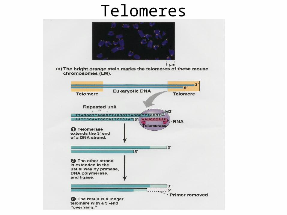
• Eukaryotes-Have free ends-Contains 3 x 10⁹ base pairs (haploid cells) = 1 meter-Lagging strand is not completely replicated-Small pieces of DNA are lost with every cell cycle-End caps (Telomeres) protect and help to retain the genetic information
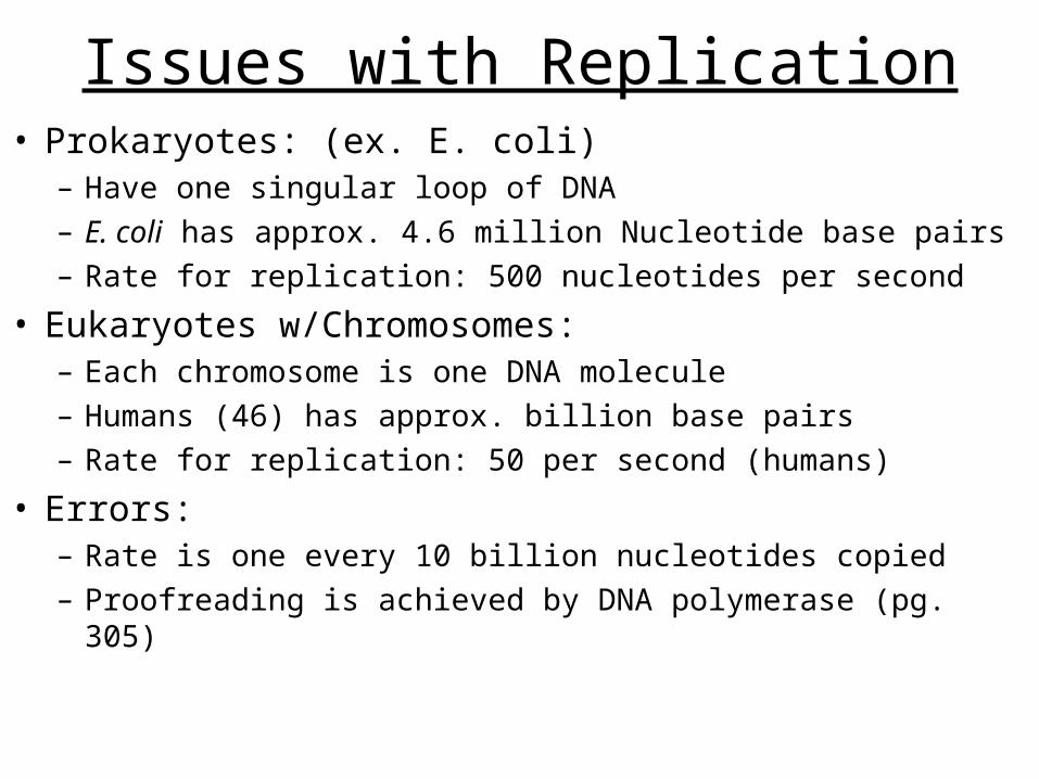
Issues with Replication• Prokaryotes: (ex. E. coli)
– Have one singular loop of DNA– E. coli has approx. 4.6 million Nucleotide base pairs– Rate for replication: 500 nucleotides per second
• Eukaryotes w/Chromosomes:– Each chromosome is one DNA molecule– Humans (46) has approx. billion base pairs– Rate for replication: 50 per second (humans)
• Errors: – Rate is one every 10 billion nucleotides copied– Proofreading is achieved by DNA polymerase (pg. 305)
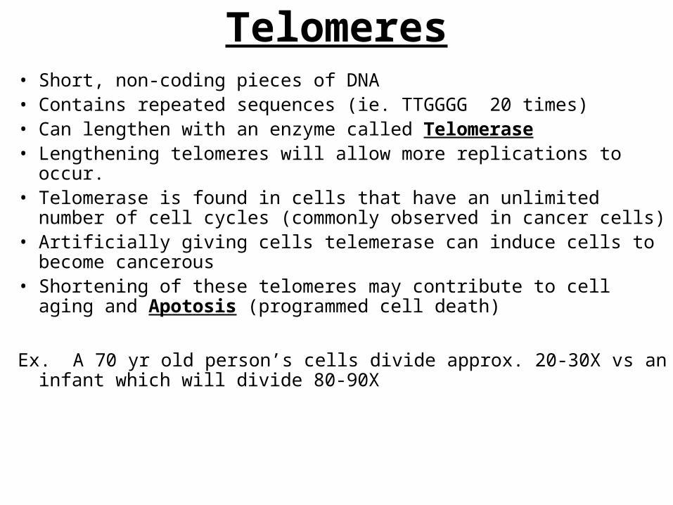
Telomeres• Short, non-coding pieces of DNA• Contains repeated sequences (ie. TTGGGG 20 times)• Can lengthen with an enzyme called Telomerase• Lengthening telomeres will allow more replications to occur.• Telomerase is found in cells that have an unlimited number of cell
cycles (commonly observed in cancer cells)• Artificially giving cells telemerase can induce cells to become
cancerous• Shortening of these telomeres may contribute to cell aging and
Apotosis (programmed cell death)
Ex. A 70 yr old person’s cells divide approx. 20-30X vs an infant which will divide 80-90X
Telomeres
Chapter 17
James Sumner (1926)
• Isolated the enzyme “Urease”
• First to identify an enzyme as a protein
• First to crystallize an enzyme
• Awarded the Nobel prize in 1946 in chemistry for his crystallization of an enzyme.
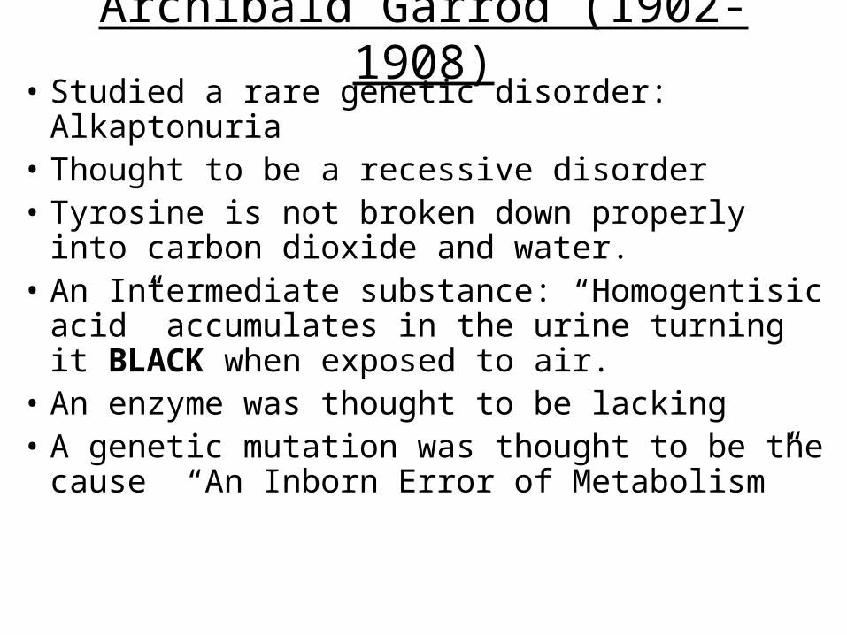
Archibald Garrod (1902-1908)• Studied a rare genetic disorder: Alkaptonuria• Thought to be a recessive disorder• Tyrosine is not broken down properly into
carbon dioxide and water.• An Intermediate substance: “Homogentisic
acid” accumulates in the urine turning it BLACK when exposed to air.
• An enzyme was thought to be lacking• A genetic mutation was thought to be the
cause “An Inborn Error of Metabolism”
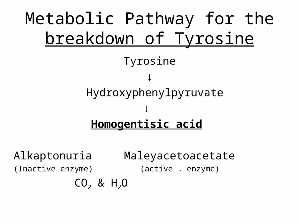
Metabolic Pathway for the breakdown of Tyrosine
Tyrosine
↓
Hydroxyphenylpyruvate
↓
Homogentisic acid
Alkaptonuria Maleyacetoacetate (Inactive enzyme) (active ↓ enzyme)
CO2 & H2O
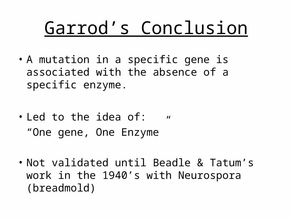
Garrod’s Conclusion
• A mutation in a specific gene is associated with the absence of a specific enzyme.
• Led to the idea of:
“One gene, One Enzyme”
• Not validated until Beadle & Tatum’s work in the 1940’s with Neurospora (breadmold)
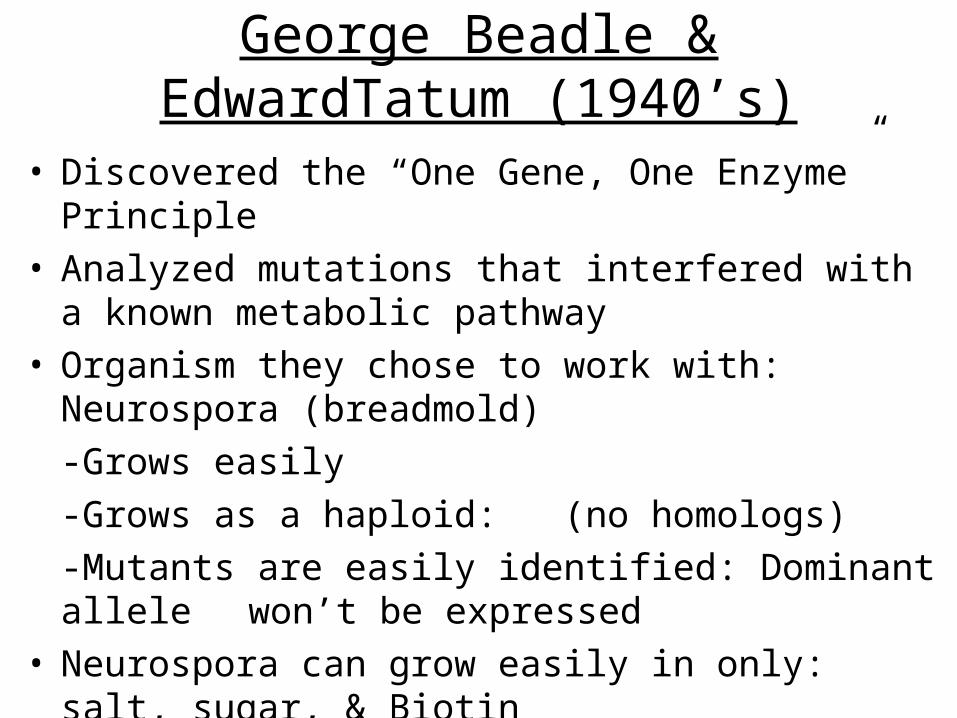
George Beadle & EdwardTatum (1940’s)
• Discovered the “One Gene, One Enzyme” Principle• Analyzed mutations that interfered with a known
metabolic pathway• Organism they chose to work with: Neurospora
(breadmold)
-Grows easily
-Grows as a haploid: (no homologs)
-Mutants are easily identified: Dominant allele won’t be expressed
• Neurospora can grow easily in only: salt, sugar, & Biotin
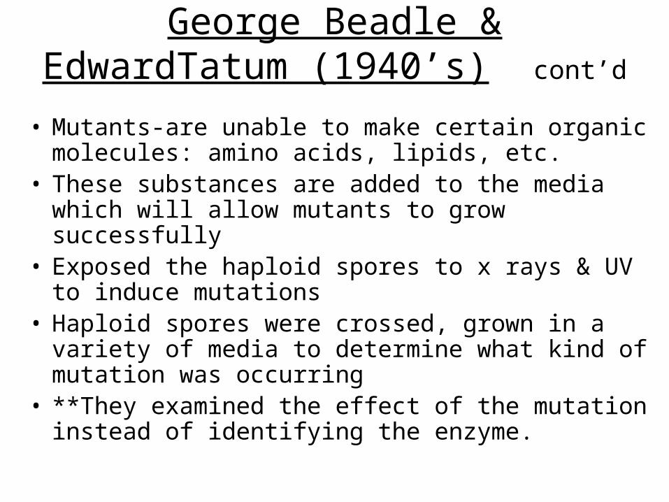
George Beadle & EdwardTatum (1940’s) cont’d
• Mutants-are unable to make certain organic molecules: amino acids, lipids, etc.
• These substances are added to the media which will allow mutants to grow successfully
• Exposed the haploid spores to x rays & UV to induce mutations
• Haploid spores were crossed, grown in a variety of media to determine what kind of mutation was occurring
• **They examined the effect of the mutation instead of identifying the enzyme.
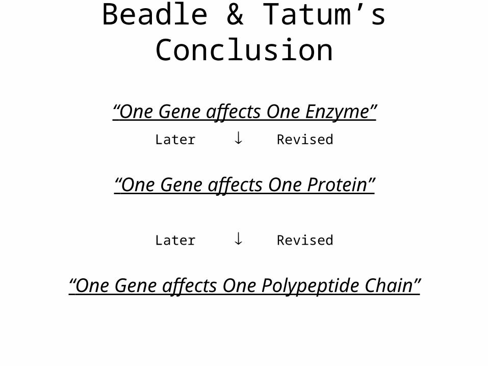
Beadle & Tatum’s Conclusion
“One Gene affects One Enzyme”Later Revised
“One Gene affects One Protein”
Later Revised
“One Gene affects One Polypeptide Chain”

Suggestions on how to Review• Make a List of all Bold Terms (See summaries)• Make a list of key people & generate a timeline• Answer all MC questions at end of each chapter• Review all your Quizzes from textbook website• Review all the MC Questions from your study guides• Look at all the key figures & diagrams discussed• Review all Tables from the four chapters• Re-Look at the Powerpoint Pres. From my website.
Think back to what was emphasized• Anticipate questions to be asked• Make an outline of all chapters & connect the
concepts discussed