REVIEW NLRP3 Inflammasome in Cardiovascular Disease: David ...

Instructions for use
Title Historical aspects of studies on roles of the inflammasome in the pathogenesis of periodontal diseases
Author(s) Shibata, Ken-ichiro
Citation Molecular Oral Microbiology, 33(3), 203-221https://doi.org/10.1111/omi.12217
Issue Date 2018-06
Doc URL http://hdl.handle.net/2115/74519
RightsThis is the peer reviewed version of the following article: Historical aspects of studies on roles of the inflammasome inthe pathogenesis of periodontal diseases, which has been published in final form at https://doi.org/10.1111/omi.12217.This article may be used for non-commercial purposes in accordance with Wiley Terms and Conditions for Self-Archiving
Type article (author version)
File Information Mol Oral Microbiol.pdf
Hokkaido University Collection of Scholarly and Academic Papers : HUSCAP
1
Historical aspects of studies on roles of the inflammasome in the pathogenesis of
periodontal diseases
Ken-ichiro Shibata
Department of Oral Molecular Microbiology, Faculty of Dental Medicine and Graduate
School of Dental Medicine, Hokkaido University

2
SUMMARY
The proinflammatory cytokine interleukin (IL)-1β is produced as inactive pro-IL-1β and
then processed by caspase-1 to become active. In 2002, it was demonstrated that the
intracellular multiprotein complex known as the inflammasome functions as a
molecular platform to trigger activation of caspase-1. Inflammasomes are known to
function as intracellular sensors for a broad spectrum of various pathogen- and
damage-associated molecular patterns.
In 1985, it was demonstrated that Porphyromonas gingivalis, a representative
bacterium causing chronic periodontitis, induces IL-1 production by murine peritoneal
macrophages. Since then, many studies have suggested that IL-1, particularly IL-1β,
plays key roles in the pathogenesis of periodontal diseases. However, the term
“inflammasome” was not used until Bostanci et al. suggested the involvement of
inflammasomes in periodontal disease in 2009. Several subsequent studies on the roles
of the inflammasome in the pathogenesis of periodontal diseases have been published.
Interestingly, two contradictory reports on the modulation of inflammasomes by P.
gingivalis have been published. Some papers have described that P. gingivalis activates
the inflammasome to produce IL-1β, whereas some stated that P. gingivalis inhibits
inflammasome activation to subvert immune responses. Several lines of evidence have
also been accumulated that the inflammasome activation is modulated by the
periodontopathic bacteria other than P. gingivalis.
Thus, studies on the roles of inflammasomes in the pathogenesis of periodontal
diseases began only 8 years ago and many pathological roles of inflammasomes remain
to be clarified.

3

4
INTRODUCTION
Interleukin-1 (IL-1), a proinflammatory cytokine, consists of IL-α and IL-1β. IL-1β
induces inflammatory mediator production, osteoclast formation, matrix
metalloproteinase expression, and matrix-producing cell death in periodontal tissues,
resulting in the destruction of alveolar bone and periodontal connective tissue 1. Thus,
IL-1β plays crucial roles in the onset and progression of periodontal disease. IL-1β is
produced extracellularly after pro-IL-1β is processed by caspase-1 to become activated
by the intracellular multiprotein complex known as the inflammasome. The word
“inflammasome” was first used to describe platforms for inflammatory caspase
activation 2. IL-18 is also produced as pro-IL-18 and processed by caspase-1 to become
activated by the inflammasome. The inflammasome is composed of the
“nucleotide-binding domain leucine-rich repeat-containing receptor” (NLR), adaptor
protein “apoptosis-associated speck-like protein containing a caspase-recruitment
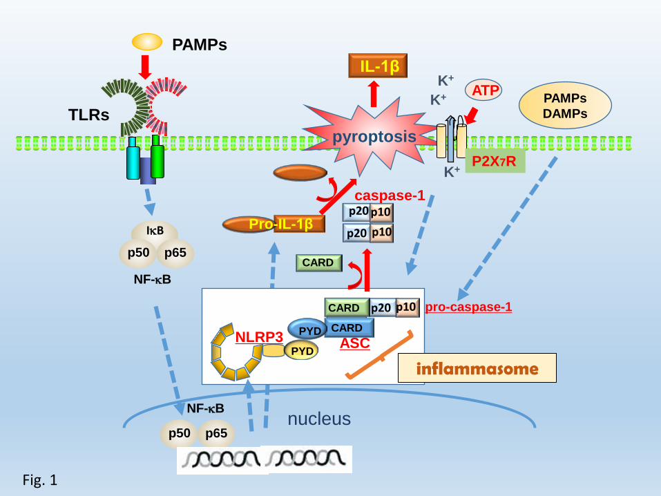
domain” (ASC), and procaspase-1 3-5 (Fig. 1). Several types of NLRs are involved in
inflammasome activation 6. Of these inflammasomes, the NLRP3 inflammasome is the
most well-studied (Fig. 1) and has been implicated in responses to a broad spectrum of
pathogen- and damage-associated molecular patterns 7-23 (Table 1). We previously
demonstrated that whole cells of two Mycoplasma species and Streptococcus sanguinis
activate the NLRP3 inflammasome to produce IL-1β 24,25.
Periodontitis, a chronic inflammation that occurs in many adults, is a major cause of
tooth loss and is characterized by chronic infection associated with Gram-negative
anaerobic bacteria in the subgingival biofilm, which leads to destruction of the tissue
supporting the teeth. Subgingival biofilms containing several gram-negative rods are
strongly involved in the onset and progression of periodontitis. Among gram-negative

5
rods, Porphyromonas gingivalis is a representative periodontal pathogen in chronic
periodontitis 26.
In this review, we focus on the historical aspects of the pathological roles of
inflammasomes in periodontal diseases and modulation of inflammasome activation by
P. gingivalis as a first step for developing a new therapeutic strategy.
INVOLVEMENT OF IL-1 IN PERIODONTAL DISEASES
The reported roles of IL-1 in the pathogenesis of periodontal diseases are summarized in
Fig. 2.
In 1985, the first paper regarding the functional role of IL-1 in periodontal disease
was published 27. However, this study only demonstrated that lipopolysaccharide (LPS)
from P. gingivalis induces IL-1 production by murine peritoneal macrophages, but did
not directly determine the roles of IL-1 in the pathogenesis of adult periodontal disease.
The first report on the role of IL-1, particularly IL-1β, in the pathogenesis of periodontal
diseases was published in 1991 28. The authors demonstrated that the number of cells
stained with anti-human IL-1β was nearly 3-fold higher in periodontally diseased tissue
compared to those in normal tissue, suggesting that IL-1β produced by cells in
periodontal tissues is related to the pathological processes associated with periodontal
disease. In 1992, the amount of IL-1 in crevicular fluid during human experimental
gingivitis was measured and shown to increase rapidly with plaque accumulation and in
advance of subsequent gingival inflammation, peaking within 7 days of the start of
gingivitis 29. Thus, the authors suggested that IL-1 is an early marker of gingival
inflammatory changes. In 1995, it was demonstrated that in vivo administration of
recombinant IL-1β accelerates silk ligature-induced alveolar bone resorption in rats 30.

6
In addition, several reports showed that an IL-1 genotype associated with increased IL-1
production is a strong indicator of the susceptibility to severe periodontitis in adults 31-33.
In 1997, Ishihara et al. demonstrated that the total amount of IL-1α, IL-1β, and the total
IL-1/IL-1 receptor antagonist ratio (IL-1AI), but not the total IL-1 receptor antagonist
(IL-1A), were correlated with alveolar bone loss score; a similar progressive decrease in
total IL-1AI was detected in gingival tissue with periodontitis 34. These results
suggested that the levels of both crevicular IL-1 and total IL-1AI are closely associated
with periodontal disease severity. In 1997, in a Macaca fascicularis primate model of
experimental periodontitis, IL-1 and tumor necrosis factor (TNF) antagonists, their
soluble receptors, inhibited the recruitment of inflammatory cells in close proximity to
bone by approximately 80% and reduced osteoclast by 67% at the experimental sites
compared to the levels at control sites; bone loss was reduced by 60% 35. Thus, IL-1 and
TNF, representative inflammatory cytokines, play important roles in the pathological
process of periodontitis. In 1998, in the same primate model, the same group
demonstrated that IL-1 and TNF antagonists inhibit the progression of inflammatory
cell infiltration toward alveolar bone 36. This research group also demonstrated that loss
of connective tissue attachment and progression of periodontal disease can be retarded
by antagonists to IL-1 and TNF in the same primate model 37.
Based on these reports, a previous review described the roles of IL-1 and TNF in
periodontal tissue destruction 1. These cytokines induce adhesion molecules and other
mediators that facilitate and amplify the inflammatory response, stimulation of matrix
metalloproteinase, and bone resorption.
INVOLVEMENT OF INFLAMMASOMES IN PERIODONTAL DISEASES

7
As described above, the inflammasome was first named in 2002 as a molecular platform
triggering activation of caspase-1 and processing of proIL-1β and proIL-18 2. Therefore,
we next focus on studies of the roles of the inflammasome in the pathogenesis of
periodontal diseases.
In 2009, 7 years after the discovery of the inflammasome, it was first demonstrated
that NLRP3 and NLRP2, but not ASC, are expressed at significantly higher levels in
gingival tissues of subjects with inflammatory periodontal disease than in healthy
subjects, and a positive correlation was found between NLRP3 and IL-1β or IL-18
expression levels in these tissues 38. In addition, in vitro data demonstrated that P.
gingivalis upregulates the expression of NLRP3, IL-1β, and IL-18 in a human
monocytic cell line (Mono-Mac-6), but downregulates NLRP2 and ASC. Thus, the
authors suggested that the NLRP3 inflammasome is involved in the pathogenesis of
periodontal diseases.
In 2011, Bostanci et al.39 demonstrated that supragingival and subgingival biofilms
differentially regulate the gene expressions of NLRP3 and AIM2 inflammasomes and
their down-stream IL-1 targets. Briefly, they found that the culture supernatant from
supragingival biofilm containing 6-spieces of supragingival Zurich biofilm model40
grown on the hydroxyapatite disc increased the expression of caspase-1, ASC, AIM2 as
well as IL-1β and IL-18, but did not upregulate NLRP3 expression. However, the
culture supernatant from subgingival biofilm containing 10-spieces of subgingival
Zurich biofilm model41 grown on the hydroxyapatite disc upregulated caspase-1, ASC,
AIM2, IL-1β and IL-18 gene expression at lower concentrations, followed by their
down-regulation at higher concentrations, which was also evident for NLRP3
expression. They suggest that upregulation of transcription of inflammasome

8
components by supragingival biofilms correlates with early inflammatory events in
periodontal disease, whereas the downregulation of the transcription by subgingival
biofilms is favorable for the survival and persistence of periodontopathic bacteria
including P. gingivalis.
In 2012, it was demonstrated that production of prostaglandin E2 does not require
the inflammasome adaptor function of ASC, but was dependent on mitogen-activated
protein kinase activation and that the mitogen-activated protein kinase kinase kinase
CARD domain-containing protein RIPK2 was induced by P. gingivalis in an
ASC-dependent manner 42. Thus, they found that the inflammasome adaptor
ASC-dependent RIP2 kinase regulates reduced prostaglandin E2 production in chronic
periodontitis, although they did not demonstrate the direct involvement of the
inflammasome in periodontal disease.
In 2014, novel findings suggested a strong correlation between the NLRP3
inflammasome and severe periodontitis 43. They demonstrated that blocking the
functions of CD24, a negative regulator of inflammation in periodontal tissues 44-47, by
its antibody-enhanced expression of NLRP3 together with the co-activators ASC and
caspase-1, resulting in burst release of activated IL-18, which plays important roles in
inflammation. Additionally, subjects with mild chronic periodontitis showed increased
titers of serum antibodies that were auto-reactive with CD24 compared with in subjects
with severe periodontitis. Thus, the negative regulator CD24 upregulates NLRP3 in
periodontal disease, strongly suggesting that the NLRP3 inflammasome plays
pathological roles in periodontal diseases.
In 2015, it was demonstrated that expression of NLRP3 and IL-1β in the oral
gingival epithelium of patients with chronic periodontitis and/or type 2 diabetes mellitus

9
was significantly upregulated compared to in control individuals, and simultaneous
stimulation with LPS and high glucose contributed to significant upregulation of
NLRP3 expression compared to stimulation with LPS or high glucose alone 48. These
results suggested that for patients with type 2 diabetes mellitus and chronic periodontitis,
a hyperglycemic status may exacerbate the inflammation state of gingival tissue by
activating the NLRP3 inflammasome pathway.
In 2015, it was demonstrated that the intensity of NLRP3 expression was
significantly higher in chronic periodontitis or generalized aggressive periodontitis than
in healthy tissue, and a more significant difference was observed in the periodontal
epithelium layer 49. In addition, they showed that NLRP1 was minimally expressed in
healthy and periodontitis gingival tissues, whereas absent in melanoma 2 (AIM2) was
expressed at a higher level in the chronic periodontitis group than in other subjects.
NLRP1 and AIM2 are also known to mediate inflammasome assembly against
microbial infections 9,10.
In 2017, the study was carried out to compare salivary levels of NLRP3, ASC,
caspase-1 and IL-1β from patients with aggressive periodontitis (AgP) or chronic
periodontitis (CP) and periodontally healthy controls (HC) as well as elucidate its
association with periodontal clinical status50. The study indicates the possibility that
salivary levels of NLRP3, ASC, and IL-1β, but not caspase-1, act as strong/independent
indicators of amount and extent of periodontal breakdown in both CP and AgP.
MODULATION OF THE INFLAMMASOME BY THE PERIDONTOPATHIC
BACTERIA P. gingivalis
There are several papers which describe modulation of inflammasomes by
10
periodontopathic bacteria such as Aggregatibacter actinomycetemcomitans51-54,
Fusobacterium nucleatum55 other than P. gingivalis. In this review, we focus on
involvement of the representative periodontopathic bacterium P. gingivalis in
inflammasome modulation. There have been two contradictory reports on modulation of
the inflammasomes by P. gingivalis, a representative periodontopathic bacteria. Some
papers demonstrated that P. gingivalis activates the inflammasome, which plays
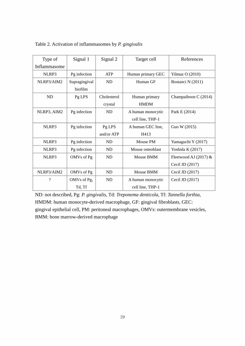
pathological roles in periodontal diseases 56-67 (Table 2). In contrast, some papers
demonstrated that P. gingivalis inhibits inflammasomes to subvert immune
responses 68-71 (Table 3). Olsen and Yilmaz72 have already published the review which
discusses in-depth on several molecular mechanisms by which P. gingivalis modulates
innate immunity by limiting the activation of the NLRP3 inflammasome. In addition,
Almeida-da-Silva et al.73 have published the review which mainly describes modulation
of the ability of P. gingivalis to evade the immune responses by purinerigic signaling
known as the second signal for the NLRP3 inflammasome activation. Therefore, this
review focuses on historical flow of studies on modulation of inflammasomes by P.
gingivalis.
1. ACTIVATION OF INFLAMMASOMES
As described above, in 1985, P. gingivalis LPS was shown to induce IL-1 production by
using murine peritoneal macrophages 27. At that time, however, it was not known that
the intracellular sensor inflammasome regulates the production of IL-1β. In addition, it
was demonstrated that ASC, one component of the inflammasome complex, is involved
in inducing cytokines such as IL-1β in response to P. gingivalis infection via a
caspase-1-dependent pathway 56, although the term “inflammasome” was not used.
11
Activation of the inflammasome by P. gingivalis was reported in a study showing
that treatment of gingival epithelial cells infected with P. gingivalis induced expression
of the IL-1β gene and intracellular accumulation of IL-1β protein, whereas IL-1β was
not secreted unless infected cells were subsequently stimulated with ATP, as well as
that knockdown of NLRP3 by siRNA attenuated the ability of ATP to induce
IL-1β secretion in infected cells 57 (Table 2). Similar findings have been reported in
another study 58. They also demonstrated that activation of the NLRP3 inflammasome
does not rely on P. gingivalis infection unless stimulated by P. gingivalis LPS and/or
extracellular ATP. Thus, these findings suggest that the NLRP3 inflammasome is an
important mediator of the inflammatory response in the gingival epithelium.
Several lines of evidence 59-61 support the finding that periodontitis is associated
pathologically with atherosclerotic vascular disease, although the details remain unclear.
It was recently found that macrophages primed with P. gingivalis LPS and then treated
with cholesterol crystals released IL-1β 62.
In 2014, it was demonstrated that expression levels of inflammasome components in
gingival tissues from patients with chronic periodontitis are much higher than in those
from healthy controls and that P. gingivalis induces the activation of NLRP3 and AIM2
inflammasome via TLR2 and TLR4 signaling, leading to IL-1β secretion and pyroptic
cell death 63. In addition, studies suggested that P. gingivalis-induced NLRP3
inflammasome activation depends on ATP release, K+ efflux, and cathepsin B.
In 2015, it was reported that P. gingivalis can activate the NLRP3 inflammasome, in
which the pathogenic factors gingipain and fimbriae play important roles, suggesting
that the NLRP3 inflammasome plays a critical role in periodontal disease and
atherosclerosis induced by P. gingivalis challenge through sustained inflammation 64. In

12
2017, the authors of the previous study also demonstrated that oral injection of P.
gingivalis to wild-type mice, but not to NLRP3-deficient mice, significantly increased
alveolar bone loss and gingival gene expression of pro-IL-1β and proIL-18 65. In
addition, they showed that P. gingivalis upregulated production of IL-1β and IL-18 by
peritoneal macrophages from wild-type mice, but not from NLRP3-deficient mice.
In 2017, Yoshida et al.74 have reported that P. gingivalis infection activated the
double-stranded RNA-dependent kinase (PKR) in osteoblasts, which in turn upregulated
NLRP3 expression through activation of NF-κB. Fleetwood et al.75 have reported that
treatment of murine bone marrow-derived macrophages (BMMs) with live P. gingivalis,
heat-killed P. gingivalis and outer membrane vesicles (OMVs) upregulate the
expression of NLRP3, procaspase-1 and pro-IL-1β and that treatment with OMVs, and
to a lesser extent heat-inactivated OMVs induces cleavage of procaspase-1 and
pro-IL-1β into their mature forms as well as inducing IL-1β and IL-18, whereas
treatment with live P. gingivalis and heat-killed P. gingivalis do not. That is, they
suggest that OMVs play more important roles in the activation of the NLRP3
inflammasome than P. gingivalis cells. In addition, Cecil et al.76 have also reported that
OMVs of P. gingivalis, Treponema denticola and Tannerella forthia activate
inflammasomes in a human monocytic THP-1 cells and its subsets to induce IL-1β
secretion and ASC speck formation and NLRP3 and/or AIM2 inflammasome(s) in
murine BMMs to induce IL-1β secretion.
There is growing evidence of a relationship between diabetes and inflammasome
activation 66. Recently, infection by P. gingivalis, a major bacterial species in
periodontal disease, was recognized as a common complication of diabetes. It has been
demonstrated that human gingival fibroblasts infected with P. gingivalis grown in BHI

13
medium containing high glucose showed increased expression of IL-1β and NLRP3 67.
Thus, these publications suggest that activation of the inflammasome by P.
ginigivalis leads to IL-1β production and plays important roles in the pathogenicity of
diabetes and atherosclerotic vascular disease as well as periodontal diseases.
2. INHIBITION OF INFLAMMASOME ACTIVATION
Several lines of evidence suggest that P. gingivalis inhibits inflammasome activation,
leading to evasion of the host immune response (Table 3).
In 2012, it was reported that P. gingivalis suppresses inflammasome activation by
another periodontal bacterium such as F. nucleatum, but not by ATP or nigericin 68.
Based on the finding that P. gingivalis limits both the number of cells taking up beads
and number of beads taken up for bead-positive cells, they speculated that the
mechanism by which P. gingivalis inhibits inflammasome activation contributes to
suppression of bacterial endocytosis.
In 2013, it was reported that 10 species from subgingival biofilms, including P.
gingivalis, reduce NLRP3 and IL-1β levels but do not affect AIM2 expression in human
gingival fibroblasts and that exclusion of P. gingivalis from the biofilm partially rescued
NLRP3 and IL-1β expression 69. These results suggest that subgingival biofilms
down-regulate NLRP3 and IL-1β expression, partly because of the existence of P.
gingivalis. Therefore, these dampened host innate immune responses may favor the
survival and persistence of associated biofilm species in periodontal tissues.
A recent study found that treatment of endothelial cells with live cells, but not with
heat-killed cells, of P. gingivalis induced proteolysis of NLRP3, but this proteolysis was
not observed after ATP pre-treatment and/or P. gingivalis-LPS stimulation 70.

14
Additionally, the levels of secreted IL-1β significantly increased after ATP
pre-treatment and/or P. gingivalis-LPS stimulation, but not after P. gingivalis infection.
These data demonstrate that P. gingivalis and its LPS differentially controlled the
NLRP3 inflammasome pathway in endothelial cells, suggesting a novel potential
mechanism developed by P. gingivalis to reduce IL-1β secretion and escape the host
immune response.
As described above, P. gingivalis stimulates pro-IL-1β synthesis but not mature
IL-1β secretion, unless the P2X7 receptor is activated by extracellular ATP 57. An
additional study demonstrated that fimbriae dampens P2X7-dependent IL-1β
secretion 71. Briefly, the authors showed that when bone marrow-derived macrophages
from wild-type or P2X7-deficient mice were infected with P. gingivalis 381 or the
isogenic fimbria-deficient (DPG3) strain with or without subsequent ATP stimulation,
DPG3 induced higher IL-1β secretion after ATP stimulation compared to 381 in
wild-type bone marrow-derived macrophages, but not in P2X7-deficient cells.
Recently, it was demonstrated that a nucleoside-diphosphate kinase of P. gingivalis
inhibits caspase-1 activation and IL-1β secretion in gingival epithelial cells by
downregulating the ATP-P2X7 signaling pathway and reducing the release of the high
mobility group box 1 protein, a pro-inflammatory danger signal 77.
Thus, these publications suggest that P. ginigivalis inhibits inflammasome activation
to subvert host immune responses.
SUMMARY AND CONCLUSIONS
IL-1β is produced as biologically inactive pro-IL-1β and then processed by caspase-1,

15
also known as IL-1β-converting enzyme, to active IL-1β 78. In 2002, it was first
demonstrated that the intracellular multiprotein complex known as the inflammasome
functions as a molecular platform that triggers activation of caspase-1 2. Inflammasomes
are intracellular sensors that drive host immune responses. However, excessive
inflammasome activity can be harmful because gain-of-function mutations in
inflammasome components are associated with autoimmune and autoinflammatory
disorders 3,4,79.
In 1985, LPS from P. gingivalis, a representative bacterium in chronic periodontitis,
was reported to induce IL-1 production by murine peritoneal macrophage 27. After this
report, many studies suggested that IL-1, particularly IL-1β, plays key roles in the
pathogenesis of periodontal diseases. Taxman et al. demonstrated that ASC, one
component of the inflammasome complex, is involved in inducing cytokines such as
IL-1β by P. gingivalis via a caspase-1-dependent pathway 56. However, they did not use
the term “inflammasome,” regardless of the discovery of inflammasome. In 2009, 7
years after the discovery of inflammasomes, Bostanci et al. first demonstrated that
inflammasome activation plays important roles in periodontal disease 38. Since then,
several studies on the roles of the inflammasome in the pathogenesis of periodontal
diseases have been published. Of these reports, there are two contradictory reports
regarding the modulation of inflammasomes by P. gingivalis, a representative bacterium
in chronic periodontitis. Some papers described that P. gingivalis activates the
inflammasome, whereas some papers described that P. gingivalis inhibits
inflammasome activation. The discrepancy between studies may be attributed to the
different cell types used in the studies or to differences between a live single-type
bacterial infection and biofilm infection. In addition, many pathogenic bacteria such as

16
Yersinia species 80, Legionella pneumophila 81, Pseudomonas aeruginosa 82, and
Mycobacterium tuberculosis 83 inhibit inflammasome activation 3,23, although the
mechanisms differ from each other.
Inflammasomes are intracellular sensors that drive host immune responses to
maintain homeostasis of host cells and tissues. Thus, the activity of P. gingivalis in
inhibiting inflammasome activation is more strongly associated with chronic
inflammation such as adult chronic periodontitis. Inhibition of inflammasome activation
by P. ginigivalis may allow other bacteria in the subgingival dental plaque as well as the
bacterium to survive for longer periods of time in the gingival tissue and contribute to
periodontal diseases.
As described above, studies on the roles of inflammasomes in the pathogenesis
of periodontal diseases began only 8 years ago. In the oral cavity, there are teeth
embedded in gingival tissue, which are mainly made of phosphate and calcium, and
sometimes several artificial dental prostheses to restore intraoral defects, including
resins, ceramics and metals such as titanium, cobalt and chromium, which is extremely
different from nasal, stomach and gut cavities. More Recently, interestingly, it was
found that titanium ions also stimulate inflammasome activation84. Thus, many
unknown pathological roles of inflammasomes in various oral diseases including
periodontal diseases remain to be clarified.
CONFLICT OF INTEREST
The author has no financial conflicts of interest.
17
REFERENCES
1. Graves DT, Cochran D. The contribution of interleukin-1 and tumor necrosis
factor to periodontal tissue destruction. J Periodontol. 2003;74(3):391-401.
2. Martinon F, Burns K, Tschopp J. The inflammasome: a molecular platform
triggering activation of inflammatory caspases and processing of proIL-beta.
Mol Cell. 2002;10(2):417-426.
3. Lamkanfi M, Dixit VM. Inflammasomes and their roles in health and disease.
Annu Rev Cell Dev Biol. 2012;28:137-161.
4. Mariathasan S, Monack DM. Inflammasome adaptors and sensors: intracellular
regulators of infection and inflammation. Nat Rev Immunol. 2007;7(1):31-40.
5. Martinon F, Mayor A, Tschopp J. The inflammasomes: guardians of the body.
Annu Rev Immunol. 2009;27:229-265.
6. Davis BK, Wen H, Ting JP. The inflammasome NLRs in immunity,
inflammation, and associated diseases. Annu Rev Immunol. 2011;29:707-735.
7. Duncan JA, Gao X, Huang MT, et al. . Neisseria gonorrhoeae activates the
proteinase cathepsin B to mediate the signaling activities of the NLRP3 and
ASC-containing inflammasome. J Immunol. 2009;182(10):6460-6469.
8. He X, Mekasha S, Mavrogiorgos N, Fitzgerald KA, Lien E, Ingalls RR.
Inflammation and fibrosis during Chlamydia pneumoniae infection is regulated
by IL-1 and the NLRP3/ASC inflammasome. J Immunol.
2010;184(10):5743-5754.
9. Vladimer GI, Marty-Roix R, Ghosh S, Weng D, Lien E. Inflammasomes and
host defenses against bacterial infections. Curr Opin Microbiol.

18
2013;16(1):23-31.
10. Franchi L, Munoz-Planillo R, Nunez G. Sensing and reacting to microbes
through the inflammasomes. Nat Immunol. 2012;13(4):325-332.
11. Duewell P, Kono H, Rayner KJ, et al. . NLRP3 inflammasomes are required for
atherogenesis and activated by cholesterol crystals. Nature.
2010;464(7293):1357-1361.
12. Franchi L, Kanneganti TD, Dubyak GR, Nunez G. Differential requirement of
P2X7 receptor and intracellular K+ for caspase-1 activation induced by
intracellular and extracellular bacteria. J Biol Chem. 2007;282(26):18810-18818.
13. Petrilli V, Papin S, Dostert C, Mayor A, Martinon F, Tschopp J. Activation of the
NALP3 inflammasome is triggered by low intracellular potassium concentration.
Cell Death Differ. 2007;14(9):1583-1589.
14. Shimada K, Crother TR, Karlin J, et al. . Oxidized mitochondrial DNA activates
the NLRP3 inflammasome during apoptosis. Immunity. 2012;36(3):401-414.
15. Zhou R, Yazdi AS, Menu P, Tschopp J. A role for mitochondria in NLRP3
inflammasome activation. Nature. 2011;469(7329):221-225.
16. Shenoy AR, Wellington DA, Kumar P, et al. . GBP5 promotes NLRP3
inflammasome assembly and immunity in mammals. Science.
2012;336(6080):481-485.
17. Lu B, Nakamura T, Inouye K, et al. . Novel role of PKR in inflammasome
activation and HMGB1 release. Nature. 2012;488(7413):670-674.
18. Sander LE, Davis MJ, Boekschoten MV, et al. . Detection of prokaryotic mRNA
signifies microbial viability and promotes immunity. Nature.
2011;474(7351):385-389.

19
19. Vyleta ML, Wong J, Magun BE. Suppression of ribosomal function triggers
innate immune signaling through activation of the NLRP3 inflammasome. PLoS
One. 2012;7(5):e36044.
20. Martinon F, Agostini L, Meylan E, Tschopp J. Identification of bacterial
muramyl dipeptide as activator of the NALP3/cryopyrin inflammasome. Curr
Biol. 2004;14(21):1929-1934.
21. Marina-Garcia N, Franchi L, Kim YG, et al. . Pannexin-1-mediated intracellular
delivery of muramyl dipeptide induces caspase-1 activation via
cryopyrin/NLRP3 independently of Nod2. J Immunol. 2008;180(6):4050-4057.
22. Kanneganti TD, Ozoren N, Body-Malapel M, et al. . Bacterial RNA and small
antiviral compounds activate caspase-1 through cryopyrin/Nalp3. Nature.
2006;440(7081):233-236.
23. von Moltke J, Ayres JS, Kofoed EM, Chavarria-Smith J, Vance RE. Recognition
of bacteria by inflammasomes. Annu Rev Immunol. 2013;31:73-106.
24. Sugiyama M, Saeki A, Hasebe A, et al. . Activation of inflammasomes in
dendritic cells and macrophages by Mycoplasma salivarium. Mol Oral
Microbiol. 2016;31(3):259-269.
25. Saeki A, Suzuki T, Hasebe A, et al. . Activation of nucleotide-binding
domain-like receptor containing protein 3 inflammasome in dendritic cells and
macrophages by Streptococcus sanguinis. Cell Microbiol. 2017;19(3).
26. Bostanci N, Belibasakis GN. Porphyromonas gingivalis: an invasive and evasive
opportunistic oral pathogen. FEMS Microbiol Lett. 2012;333(1):1-9.
27. Hanazawa S, Nakada K, Ohmori Y, Miyoshi T, Amano S, Kitano S. Functional
role of interleukin 1 in periodontal disease: induction of interleukin 1 production

20
by Bacteroides gingivalis lipopolysaccharide in peritoneal macrophages from
C3H/HeN and C3H/HeJ mice. Infect Immun. 1985;50(1):262-270.
28. Jandinski JJ, Stashenko P, Feder LS, et al. . Localization of interleukin-1 beta in
human periodontal tissue. J Periodontol. 1991;62(1):36-43.
29. Kinane DF, Winstanley FP, Adonogianaki E, Moughal NA. Bioassay of
interleukin 1 (IL-1) in human gingival crevicular fluid during experimental
gingivitis. Arch Oral Biol. 1992;37(2):153-156.
30. Koide M, Suda S, Saitoh S, et al. . In vivo administration of IL-1 beta
accelerates silk ligature-induced alveolar bone resorption in rats. J Oral Pathol
Med. 1995;24(9):420-434.
31. Thomson WM, Edwards SJ, Dobson-Le DP, et al. . IL-1 genotype and adult
periodontitis among young New Zealanders. J Dent Res. 2001;80(8):1700-1703.
32. Laine ML, Farre MA, Gonzalez G, et al. . Polymorphisms of the interleukin-1
gene family, oral microbial pathogens, and smoking in adult periodontitis. J
Dent Res. 2001;80(8):1695-1699.
33. Kornman KS, Crane A, Wang HY, et al. . The interleukin-1 genotype as a
severity factor in adult periodontal disease. J Clin Periodontol.
1997;24(1):72-77.
34. Ishihara Y, Nishihara T, Kuroyanagi T, et al. . Gingival crevicular interleukin-1
and interleukin-1 receptor antagonist levels in periodontally healthy and
diseased sites. J Periodontal Res. 1997;32(6):524-529.
35. Assuma R, Oates T, Cochran D, Amar S, Graves DT. IL-1 and TNF antagonists
inhibit the inflammatory response and bone loss in experimental periodontitis. J
Immunol. 1998;160(1):403-409.

21
36. Graves DT, Delima AJ, Assuma R, Amar S, Oates T, Cochran D. Interleukin-1
and tumor necrosis factor antagonists inhibit the progression of inflammatory
cell infiltration toward alveolar bone in experimental periodontitis. J
Periodontol. 1998;69(12):1419-1425.
37. Delima AJ, Oates T, Assuma R, et al. . Soluble antagonists to interleukin-1
(IL-1) and tumor necrosis factor (TNF) inhibits loss of tissue attachment in
experimental periodontitis. J Clin Periodontol. 2001;28(3):233-240.
38. Bostanci N, Emingil G, Saygan B, et al. . Expression and regulation of the
NALP3 inflammasome complex in periodontal diseases. Clin Exp Immunol.
2009;157(3):415-422.
39. Bostanci N, Meier A, Guggenheim B, Belibasakis GN. Regulation of NLRP3
and AIM2 inflammasome gene expression levels in gingival fibroblasts by oral
biofilms. Cell Immunol. 2011;270(1):88-93.
40. Shapiro S, Giertsen E, Guggenheim B. An in vitro oral biofilm model for
comparing the efficacy of antimicrobial mouthrinses. Caries Res.
2002;36(2):93-100.
41. Guggenheim B, Gmur R, Galicia JC, et al. . In vitro modeling of host-parasite
interactions: the 'subgingival' biofilm challenge of primary human epithelial
cells. BMC Microbiol. 2009;9:280.
42. Taxman DJ, Lei Y, Zhang S, Holley-Guthrie E, Offenbacher S, Ting JP.
ASC-dependent RIP2 kinase regulates reduced PGE2 production in chronic
periodontitis. J Dent Res. 2012;91(9):877-882.
43. Guo W, Ye P, Yu H, Liu Z, Yang P, Hunter N. CD24 activates the NLRP3
inflammasome through c-Src kinase activity in a model of the lining epithelium

22
of inflamed periodontal tissues. Immun Inflamm Dis. 2014;2(4):239-253.
44. Ye P, Simonian M, Nadkarni MA, Decarlo AA, Chapple CC, Hunter N.
Identification of epithelial auto-antigens associated with periodontal disease.
Clin Exp Immunol. 2005;139(2):328-337.
45. Chen GY, Tang J, Zheng P, Liu Y. CD24 and Siglec-10 selectively repress tissue
damage-induced immune responses. Science. 2009;323(5922):1722-1725.
46. Ye P, Nadkarni MA, Simonian M, Hunter N. CD24 regulated gene expression
and distribution of tight junction proteins is associated with altered barrier
function in oral epithelial monolayers. BMC Cell Biol. 2009;10:2.
47. Ye P, Yu H, Simonian M, Hunter N. Ligation of CD24 expressed by oral
epithelial cells induces kinase dependent decrease in paracellular permeability
mediated by tight junction proteins. Biochem Biophys Res Commun.
2011;412(1):165-169.
48. Huang X, Yang X, Ni J, et al. . Hyperglucose contributes to periodontitis:
involvement of the NLRP3 pathway by engaging the innate immunity of oral
gingival epithelium. J Periodontol. 2015;86(2):327-335.
49. Xue F, Shu R, Xie Y. The expression of NLRP3, NLRP1 and AIM2 in the
gingival tissue of periodontitis patients: RT-PCR study and
immunohistochemistry. Arch Oral Biol. 2015;60(6):948-958.
50. Isaza-Guzman DM, Medina-Piedrahita VM, Gutierrez-Henao C,
Tobon-Arroyave SI. Salivary Levels of NLRP3 Inflammasome-Related Proteins
as Potential Biomarkers of Periodontal Clinical Status. J Periodontol.
2017;88(12):1329-1338.
51. Kim S, Park MH, Song YR, Na HS, Chung J. Aggregatibacter
23
actinomycetemcomitans-Induced AIM2 Inflammasome Activation Is Suppressed
by Xylitol in Differentiated THP-1 Macrophages. J Periodontol.
2016;87(6):e116-126.
52. Zhao P, Liu J, Pan C, Pan Y. NLRP3 inflammasome is required for apoptosis of
Aggregatibacter actinomycetemcomitans-infected human osteoblastic MG63
cells. Acta Histochem. 2014;116(7):1119-1124.
53. Belibasakis GN, Johansson A. Aggregatibacter actinomycetemcomitans targets
NLRP3 and NLRP6 inflammasome expression in human mononuclear
leukocytes. Cytokine. 2012;59(1):124-130.
54. Shenker BJ, Ojcius DM, Walker LP, Zekavat A, Scuron MD, Boesze-Battaglia K.
Aggregatibacter actinomycetemcomitans cytolethal distending toxin activates
the NLRP3 inflammasome in human macrophages, leading to the release of
proinflammatory cytokines. Infect Immun. 2015;83(4):1487-1496.
55. Hung SC, Huang PR, Almeida-da-Silva CLC, Atanasova KR, Yilmaz O, Ojcius
DM. NLRX1 modulates differentially NLRP3 inflammasome activation and
NF-kappaB signaling during Fusobacterium nucleatum infection. Microbes
Infect. 2017.
56. Taxman DJ, Zhang J, Champagne C, et al. . Cutting edge: ASC mediates the
induction of multiple cytokines by Porphyromonas gingivalis via
caspase-1-dependent and -independent pathways. J Immunol.
2006;177(7):4252-4256.
57. Yilmaz O, Sater AA, Yao L, Koutouzis T, Pettengill M, Ojcius DM.
ATP-dependent activation of an inflammasome in primary gingival epithelial
cells infected by Porphyromonas gingivalis. Cell Microbiol.

24
2010;12(2):188-198.
58. Guo W, Wang P, Liu Z, Yang P, Ye P. The activation of pyrin
domain-containing-3 inflammasome depends on lipopolysaccharide from
Porphyromonas gingivalis and extracellular adenosine triphosphate in cultured
oral epithelial cells. BMC Oral Health. 2015;15(1):133.
59. Pussinen PJ, Tuomisto K, Jousilahti P, Havulinna AS, Sundvall J, Salomaa V.
Endotoxemia, immune response to periodontal pathogens, and systemic
inflammation associate with incident cardiovascular disease events. Arterioscler
Thromb Vasc Biol. 2007;27(6):1433-1439.
60. Lockhart PB, Bolger AF, Papapanou PN, et al. . Periodontal disease and
atherosclerotic vascular disease: does the evidence support an independent
association?: a scientific statement from the American Heart Association.
Circulation. 2012;125(20):2520-2544.
61. Haraszthy VI, Zambon JJ, Trevisan M, Zeid M, Genco RJ. Identification of
periodontal pathogens in atheromatous plaques. J Periodontol.
2000;71(10):1554-1560.
62. Champaiboon C, Poolgesorn M, Wisitrasameewong W, Sa-Ard-Iam N, Rerkyen
P, Mahanonda R. Differential inflammasome activation by Porphyromonas
gingivalis and cholesterol crystals in human macrophages and coronary artery
endothelial cells. Atherosclerosis. 2014;235(1):38-44.
63. Park E, Na HS, Song YR, Shin SY, Kim YM, Chung J. Activation of NLRP3
and AIM2 inflammasomes by Porphyromonas gingivalis infection. Infect Immun.
2014;82(1):112-123.
64. Yamaguchi Y, Kurita-Ochiai T, Kobayashi R, Suzuki T, Ando T. Activation of

25
the NLRP3 inflammasome in Porphyromonas gingivalis-accelerated
atherosclerosis. Pathog Dis. 2015;73(4).
65. Yamaguchi Y, Kurita-Ochiai T, Kobayashi R, Suzuki T, Ando T. Regulation of
the NLRP3 inflammasome in Porphyromonas gingivalis-accelerated periodontal
disease. Inflamm Res. 2017;66(1):59-65.
66. Lee HM, Kim JJ, Kim HJ, Shong M, Ku BJ, Jo EK. Upregulated NLRP3
inflammasome activation in patients with type 2 diabetes. Diabetes.
2013;62(1):194-204.
67. Kuo HC, Chang LC, Chen TC, et al. . Sterol Regulatory Element-Binding
Protein-1c Regulates Inflammasome Activation in Gingival Fibroblasts Infected
with High-Glucose-Treated Porphyromonas gingivalis. Front Cell Infect
Microbiol. 2016;6:195.
68. Taxman DJ, Swanson KV, Broglie PM, et al. . Porphyromonas gingivalis
mediates inflammasome repression in polymicrobial cultures through a novel
mechanism involving reduced endocytosis. J Biol Chem.
2012;287(39):32791-32799.
69. Belibasakis GN, Guggenheim B, Bostanci N. Down-regulation of NLRP3
inflammasome in gingival fibroblasts by subgingival biofilms: involvement of
Porphyromonas gingivalis. Innate Immun. 2013;19(1):3-9.
70. Huck O, Elkaim R, Davideau JL, Tenenbaum H. Porphyromonas
gingivalis-impaired innate immune response via NLRP3 proteolysis in
endothelial cells. Innate Immun. 2015;21(1):65-72.
71. Morandini AC, Ramos-Junior ES, Potempa J, et al. . Porphyromonas gingivalis
fimbriae dampen P2X7-dependent interleukin-1beta secretion. J Innate Immun.

26
2014;6(6):831-845.
72. Olsen I, Yilmaz O. Modulation of inflammasome activity by Porphyromonas
gingivalis in periodontitis and associated systemic diseases. J Oral Microbiol.
2016;8:30385.
73. Almeida-da-Silva CL, Morandini AC, Ulrich H, Ojcius DM, Coutinho-Silva R.
Purinergic signaling during Porphyromonas gingivalis infection. Biomed J.
2016;39(4):251-260.
74. Yoshida K, Okamura H, Hiroshima Y, et al. . PKR induces the expression of
NLRP3 by regulating the NF-kappaB pathway in Porphyromonas
gingivalis-infected osteoblasts. Exp Cell Res. 2017;354(1):57-64.
75. Fleetwood AJ, Lee MKS, Singleton W, et al. . Metabolic Remodeling,
Inflammasome Activation, and Pyroptosis in Macrophages Stimulated by
Porphyromonas gingivalis and Its Outer Membrane Vesicles. Front Cell Infect
Microbiol. 2017;7:351.
76. Cecil JD, O'Brien-Simpson NM, Lenzo JC, et al. . Outer Membrane Vesicles
Prime and Activate Macrophage Inflammasomes and Cytokine Secretion In
Vitro and In Vivo. Front Immunol. 2017;8:1017.
77. Johnson L, Atanasova KR, Bui PQ, et al. . Porphyromonas gingivalis attenuates
ATP-mediated inflammasome activation and HMGB1 release through
expression of a nucleoside-diphosphate kinase. Microbes Infect.
2015;17(5):369-377.
78. Thornberry NA, Bull HG, Calaycay JR, et al. . A novel heterodimeric cysteine
protease is required for interleukin-1 beta processing in monocytes. Nature.
1992;356(6372):768-774.

27
79. Strowig T, Henao-Mejia J, Elinav E, Flavell R. Inflammasomes in health and
disease. Nature. 2012;481(7381):278-286.
80. Lamkanfi M, Dixit VM. Modulation of inflammasome pathways by bacterial
and viral pathogens. J Immunol. 2011;187(2):597-602.
81. Abdelaziz DH, Gavrilin MA, Akhter A, et al. . Apoptosis-associated speck-like
protein (ASC) controls Legionella pneumophila infection in human monocytes.
J Biol Chem. 2011;286(5):3203-3208.
82. Sutterwala FS, Mijares LA, Li L, Ogura Y, Kazmierczak BI, Flavell RA.
Immune recognition of Pseudomonas aeruginosa mediated by the IPAF/NLRC4
inflammasome. J Exp Med. 2007;204(13):3235-3245.
83. Master SS, Rampini SK, Davis AS, et al. . Mycobacterium tuberculosis prevents
inflammasome activation. Cell Host Microbe. 2008;3(4):224-232.
84. Pettersson M, Kelk P, Belibasakis GN, Bylund D, Molin Thoren M, Johansson A.
Titanium ions form particles that activate and execute interleukin-1beta release
from lipopolysaccharide-primed macrophages. J Periodontal Res.
2017;52(1):21-32.

28
Table 1. Activators of the NLRP3 inflammasome
Activators References
Silica, asbestos Davis, BK (2011)
Cholesterol crystal Duewell, P (2010)
Various bacterial toxins Vladimer GI (2013); Franchi L (2012)
Bacterial ATP and K+ efflux Franchi L (2007); Petrilli V (2007)
Oxidized mitochondorial DNA Shimada K (2012)
ROS from mitochondorial Zhou R (2011)
Guanylate-binding protein 5 Shenoy AR (2012)
dsRNA-dependent kinase Lu B (2012)
Cathepsin B Davis, BK (2011)
Bacterial mRNA Sander LE (2011)
Blocking of ribosomal function Vyleta ML (2012)
Muramyl dipeptide Martinon F (2004); Marina-Garcia N (2008)
Bacterial RNA, PolyI:C,
LPS, lipopeptide, R848
Kanneganti TD (2006)
mycoplasmas Sugiyama M (2016)
29
Table 2. Activation of inflammasomes by P. gingivalis
Type of Inflammasome
Signal 1 Signal 2 Target cell References
NLRP3 Pg infection ATP Human primary GEC Yilmaz O (2010)
NLRP3/AIM2 Supragingival
biofilm
ND Human GF Bostanci N (2011)
ND Pg LPS Cholesterol
crystal
Human primary
HMDM
Champaiboon C (2014)
NLRP3, AIM2 Pg infection ND A human monocytic
cell line, THP-1
Park E (2014)
NLRP3 Pg infection Pg LPS
and/or ATP
A human GEC line,
H413
Guo W (2015)
NLRP3 Pg infection ND Mouse PM Yamaguchi Y (2017)
NLRP3 Pg infection ND Mouse osteoblast Yoshida K (2017)
NLRP3 OMVs of Pg ND Mouse BMM Fleetwood AJ (2017) &
Cecil JD (2017)
NLRP3/AIM2 OMVs of Pg ND Mouse BMM Cecil JD (2017)
? OMVs of Pg,
Td, Tf
ND A human monocytic
cell line, THP-1
Cecil JD (2017)
ND: not described, Pg: P. gingivalis, Td: Treponema denticola, Tf: Tannella forthia, HMDM: human monocyte-derived macrophage, GF: gingival fibroblasts, GEC: gingival epithelial cell, PM: peritoneal macrophages, OMVs: outermembrane vesicles, BMM: bone marrow-derived macrophage

30
Table 3. Inhibition of the inflammasome activation by P. gingivalis
Type of Inflammasome
Mechanism Active entity Target cell references
NLRP3/AIM2 Subgingival biofilm ND Human GF Bostanci N (2011)
NLRP3 Suppression of other
bacterial endocytosis ND Mouse BMDM Taxman DJ (2012)
NLRP3 ND ND Primary human GF Belibasakis GN (2013)
NLRP3 Proteolysis of NLRP3 ND Human UVEC Huck O (2015)
NLRP3 ND Fimbriae Mouse BMDM Morandini AC (2014)
ND Downregulation of
ATP/P2X7-signaling and
release of HMGB1
Nucleotide-
diphosphate
kinase
Human GEC Johnson L (2015)
ND: not described, BMDM: bone marrow-derived macrophage, GF: gingival fibroblasts, UVEC: umbilical vein endothelial cells, GEC: gingival epithelial cell, HMGB1: high mobility group box

31
FIGURE LEGENDS
Fig. 1. Production of biologically active IL-1β
IL-1β is produced as pro-IL-1β in the cytosol, which is biologically inactive,
through activation of nuclear factor-κB by Toll-like receptor-mediated signaling.
Biologically active IL-1β is produced after processing by caspase-1, which is also
processed from pro-caspase-1 by activation of the intracellular sensor inflammasome.
The inflammasome is an intracellular multiprotein complex comprising
“nucleotide-binding domain leucine-rich repeat-containing receptor,” the adaptor
protein “apoptosis-associated speck-like protein containing a caspase-recruitment
domain,” and procaspase-1, which is formed by signals mediated by extracellular ATP,
pathogen-associated molecular patterns, danger-associated molecular patterns or
K+ efflux.
PYD: pyrin domain, CARD: caspase recruitment domain
Fig. 2. Effects of IL-1 on periodontal tissues that have been published so far.
TLRs
inflammasome
IκB
p50 p65
NF-κB
PAMPs DAMPs
pro-caspase-1
NLRP3 ASC PYD
caspase-1
nucleus p50 p65
NF-κB
PAMPs
K+
K+
CARD
CARD PYD
CARD p10
p10 p10 p20
p20
p20
pyroptosis
K+
P2X7R
ATP
Pro-IL-1β
IL-1β
Fig. 1

IL-1
Enhancement of infiltration of inflammatory cell toward alveolar bone
Enhancement of connective tissue attachment loss and progression of periodontal disease
Inhibition of osteoclast formation
Correlation of IL-1 genotype with severity of periodontitis
Enhancement of alveolar bone loss
Higher number of IL-1β–producing cell in periodontally diseased tissue
Higher amount of IL-1 in crevicular fluid cell during experimental gingivitis
Close association of the amounts of both crevicular IL-1 and the total IL-1/ IL-1 receptor antagonist ratio with periodontal disease severity
Fig. 2
![NLRP3 inflammasome activation promotes inflammation ...DOI 10.1186/s13046-017-0589-y. products, environmental factors, and endogenous mole-cules [5]. The NLRP3 inflammasome, which](https://static.fdocuments.us/doc/165x107/60a525258e113a4b713113c4/nlrp3-inflammasome-activation-promotes-inflammation-doi-101186s13046-017-0589-y.jpg)