Histopathology Techniques - Postgraduate Books
28
Transcript of Histopathology Techniques - Postgraduate Books
Histopathology Techniquesand its Management
New Delhi | London | PanamaThe Health Sciences Publisher
Ramadas Nayak MBBS MD
Professor and Head Department of Pathology
Yenepoya Medical College Yenepoya University
Mangaluru, Karnataka, [email protected]
Formerly, Head Department of Pathology Kasturba Medical College
Manipal University Mangaluru, Karnataka, India
Jayp
ee B
rothe
rs
Jaypee Brothers Medical Publishers (P) Ltd
HeadquartersJaypee Brothers Medical Publishers (P) Ltd4838/24, Ansari Road, DaryaganjNew Delhi 110 002, IndiaPhone: +91-11-43574357Fax: +91-11-43574314Email: [email protected]
Overseas OfficesJ.P. Medical Ltd Jaypee-Highlights Medical Publishers Inc83 Victoria Street, London City of Knowledge, Bld. 235, 2nd Floor, ClaytonSW1H 0HW (UK) Panama City, PanamaPhone: +44 20 3170 8910 Phone: +1 507-301-0496Fax: +44 (0)20 3008 6180 Fax: +1 507-301-0499Email: [email protected] Email: [email protected]
Jaypee Brothers Medical Publishers (P) Ltd Jaypee Brothers Medical Publishers (P) Ltd17/1-B Babar Road, Block-B, Shaymali Bhotahity, Kathmandu, NepalMohammadpur, Dhaka-1207 Phone: +977-9741283608Bangladesh Email: [email protected]: +08801912003485Email: [email protected]
Website: www.jaypeebrothers.comWebsite: www.jaypeedigital.com
© 2018, Jaypee Brothers Medical Publishers
The views and opinions expressed in this book are solely those of the original contributor(s)/author(s) and do not necessarily represent those of editor(s) of the book.
All rights reserved. No part of this publication may be reproduced, stored or transmitted in any form or by any means, elec-tronic, mechanical, photocopying, recording or otherwise, without the prior permission in writing of the publishers.
All brand names and product names used in this book are trade names, service marks, trademarks or registered trademarks of their respective owners. The publisher is not associated with any product or vendor mentioned in this book.
Medical knowledge and practice change constantly. This book is designed to provide accurate, authoritative information about the subject matter in question. However, readers are advised to check the most current information available on procedures included and check information from the manufacturer of each product to be administered, to verify the recom-mended dose, formula, method and duration of administration, adverse effects and contraindications. It is the responsibility of the practitioner to take all appropriate safety precautions. Neither the publisher nor the author(s)/editor(s) assume any liability for any injury and/or damage to persons or property arising from or related to use of material in this book.
This book is sold on the understanding that the publisher is not engaged in providing professional medical services. If such advice or services are required, the services of a competent medical professional should be sought.
Every effort has been made where necessary to contact holders of copyright to obtain permission to reproduce copyright material. If any have been inadvertently overlooked, the publisher will be pleased to make the necessary arrangements at the first opportunity. The CD/DVD-ROM (if any) provided in the sealed envelope with this book is complimentary and free of cost. Not meant for sale.
Inquiries for bulk sales may be solicited at: [email protected]
Histopathology Techniques and its Management
First Edition: 2018
ISBN: 978-93-5270-234-3
Printed at
Jayp
ee B
rothe
rs

Dedicated toStudents who inspired me,
Patients who provided the knowledge,My parents and family members, who encouraged and supported me.
Jayp
ee B
rothe
rs
Preface
It is very important for trainees in histopathology technology as well as trainees in pathology to be familiar with various histological techniques. Presently, histotechniques have become increasingly sophisticated and it is essential to know the basic knowledge by these trainees. There is only few voluminous books available on histotechnique in the market with very few emphasize on practical problems encountered. As a postgraduate examiner in pathology for the last 30 years, I found postgraduates in pathology find it difficult to answer the basic questions in histopathology techniques. This encouraged me to write a comprehensive book which provides essential knowledge and practical solutions to problems encountered in histopathology techniques. Hence, the book is titled as Histopathology Techniques and its Management. This textbook deals with basic histopathology techniques: Introduction and examination of tissue, fixation and fixatives, tissue processing and embedding, decalcification, microtomy and section cutting, frozen section and cryostat, theoretical aspects of staining, hematoxylins and eosin, staining procedure and mounting, demonstration of carbohydrates, pigments and minerals, staining for amyloid, connective tissues stains, stains for lipids, stains for microorganisms, immunohistochemistry, automation in histopathology, museum technique, microscope, photography, biomedical waste & its management and total quality management. This also deals with the management of various problems encountered in histopathology techniques. It is textbook meant for learning histopathology technique. Hence numerous illustrations, photographs, tables, text boxes and figures with different steps in some of the techniques have been incorporated for easy understanding of the subject.
Ramadas NayakJayp
ee B
rothe
rs
Acknowledgments
My sincere thanks to all my family members, especially my wife Smt Rekha Nayak, my daughter Ms Rashmitha Nayak and my son-in-law Mr Ramnath Kini, who have patiently accepted my long preoccupation with this work. A special thanks to my grandson master Rishab Kini. I wish my gratitude to Mr Yenepoya Abdulla Kunhi, Honorable Chancellor, and Mr Farhaad Yenepoya, Director, Department of Finance, Yenepoya University (Accredited by NAAC with “A” grade), Mangaluru, Karnataka, India, for their inspiration and encouragement. I am grateful to all those who provided support, talked things over, read, offered comments and assisted in the editing, proofreading and designing. My special thanks to the following:• Dr Rakshatha Nayak (Tutor), Dr Krishnaraj Upadhyaya (Professor), Dr Premal Saldhana
(Professor), Dr Anuradha Rao CK (Professor), Dr Krishnaprasad HV (Assistant Professor), Dr Renuka Patil (Tutor), Ms Biji Francis (Laboratory Technician), Ms Pavithra K (Laboratory Technician) and Mr Vasantha M (Laboratory Technician), Department of Pathology, Yenepoya Medical College, a constituent of Yenepoya University (Accredited by NAAC with “A” grade), Mangaluru.
• Ms Anu Catherine (Lecturer), Department of Pathology, Amrita Institute of Medical Sciences, Kochi, Kerala, India.
• Dr RGW Pinto (Professor and Head) and Dr Francisco Couto (Associate Professor), Department of Pathology, Goa Medical College, Bambolim, Goa, India.
• Shri Jitendar P Vij (Group Chairman), Mr Ankit Vij (Group President), Ms Ritu Sharma (Director–Content Strategy), and Ms Chetna Malhotra Vohra (Associate Director–Content Strategy) of M/s Jaypee Brothers Medical Publishers (P) Ltd, New Delhi, India, for publishing the book in the same format as wanted, well in time.
• Ms Sunita Katla (PA to Group Chairman and Publishing Manager), Ms Samina Khan (Executive Assistant to Director–Content Strategy), Ms Seema Dogra (Cover Visualizer), Mr Rajesh Sharma (Production Coordinator), Ms Geeta Rani Barik (Proofreader), Mr Rajesh Ghurkundi (Graphic Designer) and Mr Raj Kumar (DTP Operator) of M/s Jaypee Brothers Medical Publishers (P) Ltd, New Delhi, India.
• Mr Venugopal V (Bengaluru) and Mr Vasudev H (Mangaluru) of M/s Jaypee Brothers Medical Publishers (P) Ltd, Bengaluru Branch, Karnataka, for taking this book to every corner of Karnataka.
• I would like to express my gratitude to all my friends, technical staff and colleagues, who helped, inspired and supported me in the different stages of preparing this book.
Jayp
ee B
rothe
rs
x Histopathology Techniques and its Management
• Last but definitely not least, a thank you to our undergraduate and postgraduate students. Without you, I would not write. You make all my books possible.
There are many more people I could thank, but space, and modesty compel us to stop here.
Images ContributionI am extremely grateful to all my friends who willing provided required images for this book.• Dr Veena Shenoy, MD, Chief, Department of Pathology and Laboratory Medical Services,
VA Medical Center, Jackson, Mississippi, USA. • Dr Krishnaraj Upadhyaya (Professor), Dr Krishnaprasad HV (Assistant Professor),
Dr Prema Saldhana (Professor), Dr Anuradha CK (Professor), and Dr Rakshatha (Tutor), Department of Pathology, Yenepoya Medical College, A constituent of Yenepoya University, Mangaluru, Karnataka, India.
• Dr Ajit Nambiar (Professor and Head), Ms Anu Catherine (Lecturer), Dr Seethalekshmy NV (Professor), Dr Annie Jojo (Professor), Department of Pathology, Amrita Institute of Medical Sciences, Kochi, Kerala, India.
• Dr Raja Parthiban SR (Professor), Mr Muni Kumar Babu PV (Chief Laboratory Technician), Dr Shruthi (Assistant Professor) Dr Rekha TP (Tutor), Dr Sumaiya (Tutor), Department of Pathology, MVJ Medical College and Research Hospital, Hosokote, Bengaluru, Karnataka, India.
• Dr Hemalatha M (Associate Professor) and Dr Suguna BV (Professor and Head), Department of Pathology, Kempegowda Institute of Medical Sciences, Bengaluru, Karnataka, India.
• Dr Leena Dennis (Professor, Department of Pathology), Sri Ramachandra Medical College, Chennai, Tamil Nadu, India.
• Dr Sharada Rai (Associate Professor), Dr Saraswathy Sreeram (Assistant Professor), Department of Pathology. Kasturba Medical College, A constituent of Manipal University, Mangaluru, Karnataka, India.
Jayp
ee B
rothe
rs
Contents
1. Introduction and Examination of Tissues 1Examination of tissues 3
2. Fixation and Fixatives 12Chemical fixation 18Classification of fixatives 24Secondary fixation 32Physical methods of fixation 33Choice of a fixative 35Special fixation of organs and tissues 36
3. Tissue Processing and Embedding 41Labeling of specimens 41Selection of tissue (bit taking) 42Transference of labeled tissues 43Tissue processing 43Stages of tissue processing 45Types of tissue processing 58Embedding (casting or ‘blocking’) 60Tissue microarray 68
4. Decalcification 71Bone specimens 71Techniques for demonstration of bone 73Decalcification 74Processing decalcified bone 82
Jayp
ee B
rothe
rs
xii Histopathology Techniques and its Management
Undecalcified bone section 83Softening of hard tissues 86
5. Microtomy and Section Cutting 87Microtomy 87Microtome knives 90Trimming the block 101Cutting/sectioning technique 102
6. Frozen Section and Cryostat 115Frozen section 115Cryostat 118
7. Theoretical Aspects of Staining 123Dyes 125Theory of staining 127Terminology in staining 136
8. Hematoxylins and Eosin 141Hematoxylin 141Alum hematoxylins 142Iron hematoxylins 147Tungsten hematoxylins 150Other hematoxylins 152Cytoplasmic stains 152Hematoxylin and eosin stain (H & E) 152Quality control in routine H & E staining 153
9. Staining Procedure and Mounting 155Staining procedure 156Mountants 158Mounting of paraffin section 163Routine staining procedures 166Technical problems and solutions in H & E staining 169Filing of tissue blocks and slides 173
Jayp
ee B
rothe
rs
xiiiContents
Destaining and restaining 174Errors in histopathology 175
10. Demonstration of Carbohydrates 176Demonstration of carbohydrates 182Alcian blue technique 190Methods of demonstration of mucin 195Lectins and immunohistochemistry 198
11. Pigments and Minerals 200Hematogenous endogenous pigments 201Non-hematogenous endogenous pigments 205Artefact pigments 211Exogenous pigments and minerals 213
12. Staining for Amyloid 215Amyloid 215Demonstration of amyloid 215
13. Connective Tissues Stains 219Extracellular matrices 219Cells of the connective tissue 223Stains for collagen 223Stains for reticular fibers 230Stain's for elastic tissue fibers 237Fibrin and fibrinoid 245
14. Stains for Lipids 246Fat stains and the Sudan dyes 247
15. Stains for Microorganisms 250Detection and identification of bacteria 251Techniques for mycobacteria 253Helicobacter 258Spirochete 259Identification of fungi 260Practical application of histochemical stains 265
Jayp
ee B
rothe
rs
xiv Histopathology Techniques and its Management
16. Immunohistochemistry 267Immunohistochemical methods 269Unmasking of antigen sites 274Immunohistochemistry in practice 277Quality control in immunohistochemistry 279Practical aspects of immunohistochemical staining 280
17. Automation in Histopathology 291Automation in histopathology 291
18. Museum Technique 299Museum technique 299Mounting of museum specimens 303
19. Microscope 306Compound microscope 307Magnification and illumination 311Phase contrast microscopy 313Polarized light microscopy 314Immunofluorescence microscopy 316Measuring with the microscope 317Setting up the microscope illumination 320Use, care and maintenance of microscope 321Electron microscope 323Confocal microscopy 324
20. Photography 325Photomicroscopy 325Photography of gross specimens 326Digital pathology and telepathology 327
21. Biological Waste and its Management 330Biomedical waste 330Biomedical waste management 333Occupational safety and health of healthcare workers 342
Jayp
ee B
rothe
rs
xvContents
22. Total Quality Management 346Total quality management framework 346Quality laboratory processes 346Quality assurance 347Quality assessment 348Quality control 349Accreditation and certification 357
Bibliography 359
Appendices 361
Index 373
Jayp
ee B
rothe
rs
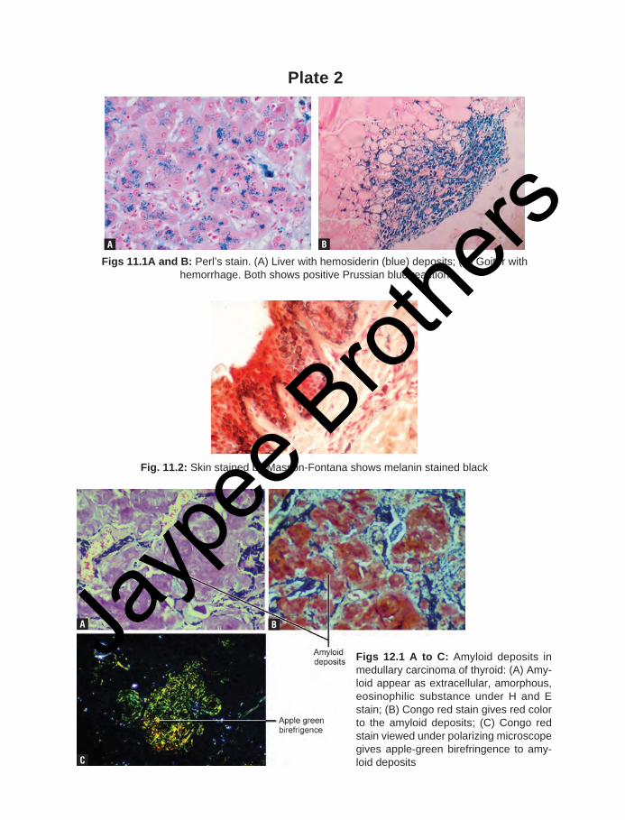
Plate 2
Fig. 11.2: Skin stained by Masson-Fontana shows melanin stained black
Figs 11.1A and B: Perl’s stain. (A) Liver with hemosiderin (blue) deposits; (B) Goiter with hemorrhage. Both shows positive Prussian blue reaction
A B
A
C
B
Figs 12.1 A to C: Amyloid deposits in medullary carcinoma of thyroid: (A) Amy-loid appear as extracellular, amorphous, eosinophilic substance under H and E stain; (B) Congo red stain gives red color to the amyloid deposits; (C) Congo red stain viewed under polarizing microscope gives apple-green birefringence to amy-loid deposits
Jayp
ee B
rothe
rs
Introduction Hematogenousendogenous pigments Non-hematogenous endogenous
pigments Artefact pigments exogenous pigments and minerals
C h a p t e r O u t l i n e
INTRODUCTIONThe pigments are colored substances in living matter that absorb visible light (electromag-netic energy within a narrow band that lies approximately between 400 and 800 nm). They are encountered in both normal and patho-logical conditions.
Classification (Box 11.1)The various pigments differ in origin, chemical constitution and biological significance.
Endogenous PigmentsThese pigments are produced either within tissues and serve a physiological function, or are by-products of normal metabolic processes. They can be further sub-classified as:• Hematogenous (blood-derived)
pigments
• Non-hematogenous pigments• Endogenous minerals.
Exogenous PigmentsThese pigments have no physiological function. They enter the body accidentally through a variety of methods. Entry into the body may occur either by inhalation into the lungs (e.g. carbon anthracotic pigment in the
C h a p t e r
11
Box 11.1: Classification of pigments
◆ Endogenous pigments Hematogenous
− Hemosiderins − Hemoglobin − Bile pigments and hematoidin − Porphyrins
Non-hematogenous − Melanins − Lipofuscins − Chromaffin − Ceroid-type lipofuscins − Pseudomelanosis (melanosis coli) − Dubin-Johnson pigment − Hamazaki-Wesenberg bodies
◆ Exogenous pigments: Carbon, silica, asbes-tos, lead, silver, tattoo pigments
◆ Artefact pigments − Formalin − Malaria (hemozoin) − Schistosome − Mercury − Chromic oxide − StarchJa
ypee
Brot
hers
201pigments and minerals
environment) or by implantation (e.g. tattoos) into the skin. Others examples include silica, asbestos, lead and silver.
Artefact PigmentsThese are artifactually produced pigments and are caused by the interactions between certain tissue components and some chemical substances (e.g. formalin fixative). Common artifactual pigments are formalin, mercuric and chrome pigments. Formalin and malaria pigments are sometimes classified as a subdi-vision of endogenous pigments.
HEMATOGENOUS ENDOGENOUS PIGMENTS
This group includes the following blood-derived pigments: Hemosiderins, hemoglobin, bile pigments and porphyrins (Box 11.1).
HemosiderinsThese endogenous pigments seen as yellow to brown granules (when unstained) and nor-mally present within the cells (intracellular). Iron is an important component of the human body because it is an essential constituent of the oxygen-carrying hemoglobin found in the red blood cells. Hemosiderin is a breakdown product of hemoglobin and contains iron in the form of ferric hydroxide that is bound to a protein. Iron is also present in myoglobin and certain enzymes, such as cytochrome oxidase and the peroxidases.
Precaution: Tissues which are to be examined for iron-containing pigments must be fixed in non-metallic containers and iron-free distilled water to be used for the reaction.
Fixative: Buffered neutral formalin is the ideal fixative for the demonstration of this pigment.
Demonstration: Hemosiderin is demonstrated by Perls’ Prussian blue reaction.
Significance: Large amount of hemosiderin are found only in pathologic conditions. If the
production and destruction of RBC are not balanced, there may be increased deposition of hemosiderin in tissue.• Reduced hemosiderin: In iron deficiency,
the iron stores in the bone marrow become depleted and there is reduced production of hemoglobin. The iron deficiency is characteristically demonstrated by the absence of stainable iron in the bone marrow.
• Excess iron: − Hemosiderosis− Hemochromatosis: It is a disease
caused by excessive absorption of dietary iron and excessive hemosiderin deposits.
Demonstration of hemosiderin and iron• In unfixed tissue, hemosiderin is insol-
uble in alkalis but soluble in strong acid solutions. After fixation in formalin, it is slowly soluble in dilute acids especially oxalic acid. Fixatives that contain acids but no formalin can remove hemosiderin or alter it in such a way that reactions for iron are negative.
• Certain types of iron found in tissues can-not be demonstrated using traditional techniques, because the iron is tightly bound within a protein complex (exam-ples include hemoglobin and myoglobin protein complexes). If they are treated with hydrogen peroxide (100 vol), the iron is released and it can then be demonstrat-ed using Perls’ Prussian blue reaction. It can also be demonstrated if the acid fer-rocyanide solution is heated to 60°C in a water bath, oven, or microwave oven. However, heat can sometimes produce a fine, diffuse, blue precipitate on both the tissue section and slide. This precipi-tate will not develop when the slides are stained at room temperature.
• Metallic iron deposits or inert iron oxide may be seen in tissues due to industrial
Jayp
ee B
rothe
rs
202 Histopathology Techniques and its management
exposure. They do not give positive reac-tion when treated with acid ferrocyanide solutions.
Perls’ Prussian Blue Reaction for Ferric IronIt is the first classical histochemical reaction. Purpose: It is used for demonstration of hemosiderin. Hemosiderin is complex of ferric ion (hydroxides), polysaccharides, and proteins. Perls’ Prussian blue stain detects ferric (Fe3+) iron in tissues. Ferric iron is normally found in small amounts in the bone marrow and the spleen. Abnormally large deposits may be seen in hemochromatosis and hemosiderosis.
Principle: Hemosiderin is soluble in acid. Hydrochloric acid (HCl) liberates the ferric (inorganic iron in the form of the hydroxide, Fe(OH)3) ions from the protein fraction of the hemosiderin molecule. Potassium ferrocyanide in acid solution combines with ferric ions to produce an insoluble blue compound ferric ferrocyanide (Prussian blue). Thus it gives blue color to iron.
Protein with ferric iron + HCl → Ferric iron + potassium ferrocyanide → Potassium ferric ferrocyanide.Mechanism of staining: Histochemical
Fixative: 10% buffered formalin. Avoid acid fixatives. Also ensure the formalin is buffered and not acidic.Quality control: A tissue section containing ferric iron as a positive control (spleen, bone marrow) must be used.
Ferrocyanide solution (Table 11.1)
Table 11.1: Composition of ferrocyanide solutionConstituent Quantity
1% aqueous potassium ferrocyanide 20 mL
2% aqueous hydrochloric acid 20 mL
Note: Prepare freshly just before use
Method of staining ◆ Take a test and positive control* section. Bring
both sections to water. ◆ Treat sections with the freshly prepared acid fer-
rocyanide solution (equal parts of 2% aqueous potassium ferrocyanide and 2% aqueous hydro-chloric acid) for 10–30 minutes.
◆ Wash thoroughly in several change of distilled water.
◆ Counter stain the nuclei with 0.5% aqueous neu-tral red or 0.1% nuclear fast red or safranin for 3–5 minutes.
◆ Rinse in distilled water. ◆ Dehydrate, clear, and mount in synthetic resin.
Handle specimens gently
*Note: It is necessary to use a positive control with all test sections. The choice of suitable control is important. A use-ful control is postmortem lung tissue containing a reasonable number of iron-positive macrophages (heart failure cells). Other controls containing hemosiderin laden macrophages include chocolate cysts (endometriosis) of ovary, multinodular goiter etc.
Results (Table 11.2 and Fig. 11.1)
Table 11.2: Results of Perls’ Prussian blue reactionComponent Color
Ferric iron-containing pigments (hemosiderin)
Blue
Nuclei Red
Disadvantage: Perls’ Prussian blue reaction cannot demonstrate ferrous iron. Both ferric and ferrous form can be demonstrated by the following stains:• Lillie’s method for ferric and ferrous
iron.• Hukill and Putt’s method for ferrous and
ferric iron.
Technical problems and solutionsUnexpectedly weak staining1. If there is surprised weak staining, or a
failure to stain, check whether correct procedure or the reagents are used. If they are correct then check at the positive control.
Jayp
ee B
rothe
rs
203pigments and minerals
2. If there are low concentrations of iron containing pigment in the tissue, it may give pale staining. Try by extending the staining time.
3. Acidic fixation: Fixation in acidic media may cause loss of iron. Try other fixatives in future work.
4. Loss during pre-treatment: Failure to demonstrate can be due to the loss of the highly water-soluble hydrated ferric iron from the specimen during the pre- treatments necessary to release the cation from its complexed state. Explore shorter pre-treatment times.
5. The ferrocyanide ions diffuse slower than hydrogen ions. Hence, acid-induced losses of iron can be reduced by pre-treating the section with potas-sium ferrocyanide solution prior to staining with the acidified ferrocyanide reagent.
Unexpected structures stain1. Iron contamination: It is one of the
possible sources of false-positives. The contamination source may be the re-agents, glassware or water (tap water). Check glass washing procedures and the water source.
2. Temperature: Finely granular blue deposit throughout the section can occur due to staining at elevated temperatures. Use a low temperature method, if necessary with an extended staining time.
3. Contamination of test section: Do not use a strongly staining control section, because some leaching of the blue reaction product may occur and contaminate the test section, giving rise to false-positives reaction.
Over staining• If the tissue contains abundant iron, the
usual procedure of staining may result in overstaining and obscure the surrounding tissue elements. Should this occur, reduce staining times.
• Sources of error and effects in Perl’s Prussian blue staining are listed in Table 11.3.
HemoglobinHemoglobin is a conjugated protein which transports oxygen and carbon dioxide within the bloodstream. It is composed of a color-less protein, globin, and a red pigmented component, heme. Heme is composed of protoporphyrin combined with ferrous iron.
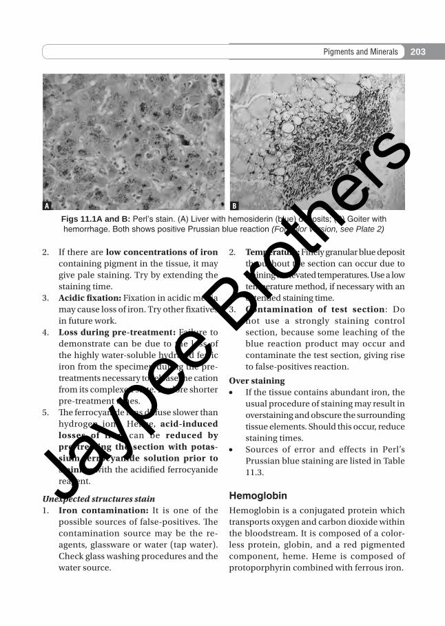
Figs 11.1A and B: Perl’s stain. (A) Liver with hemosiderin (blue) deposits; (B) Goiter with hemorrhage. Both shows positive Prussian blue reaction (For color version, see Plate 2)
a B
Jayp
ee B
rothe
rs
204 Histopathology Techniques and its management
Demonstration of hemoglobin: Histochemical demonstration of the ferrous iron is possible only if the close binding in the heme molecules is cleaved. This can be done by treating it with hydrogen peroxide. Because hemoglobin appears normally within red blood cells its histochemical demonstration is not usually needed. However, it may be necessary in certain pathological conditions such as casts (renal/RBC casts) in the lumen of renal tubules in cases of hemoglobinuria or active glomerulonephritis. The hemoglobin appear as yellow-brown granules within the casts. Distinction between the various types of hemoglobin (e.g. methemoglobin) is possible only by spectroscopy. There are two methods of demonstration of hemoglobin in tissue sections.• Peroxidase method: This method dem-
onstrates the enzyme namely hemoglobin peroxidase. This peroxidase activity was demonstrated by the benzidine nitro-prusside methods. However, because benzidine is carcinogenic these methods are no longer used.
• Other methods: These include Leuco patent blue V method, tinctorial methods, amido black technique and the kiton red-almond green technique.
Bile PigmentTwo most important bile pigments are bili-rubin (orange or yellow) and its oxidized form biliverdin (green). Bile pigment is formed by breakdown of RBCs when they
have reached the end of their life, i.e. after 120 days. Histologically, bile is most frequently en-countered in sections of liver. In H & E stained sections bile appears as yellow brown globules or masses. Bile stains a characteristic green color with van Gieson’s stain. Increased levels of bilirubin is found in blood in patients with jaundice. In jaundice caused by obstruction of bile flow, bile may accumulate in liver. Stain for demonstration of bile is Hall’s stain (bile or bilirubin gives emerald green to olive drab color).
Demonstration of Bile PigmentsIdentification of bile pigments and its distinction from lipofuscin may be necessary in the histological examination of the liver. In H&E-stained paraffin sections, both appear yellow-brown, and the green color of biliverdin is often masked by eosin. In such cases, unstained paraffin or frozen sections, lightly counterstained with a suitable hematoxylin (e.g. Mayer), will help in their differentiation. Bile pigments are not autofluorescent and fail to rotate the plane of polarized light (monorefringent), whereas lipofuscin is autofluorescent.
Methods• Modified Fouchet’s method: It is the
most commonly used routine method for the demonstration of bile pigments. In this technique, the bile pigment is con-verted to the green color of biliverdin and blue cholecyanin by the oxidative action
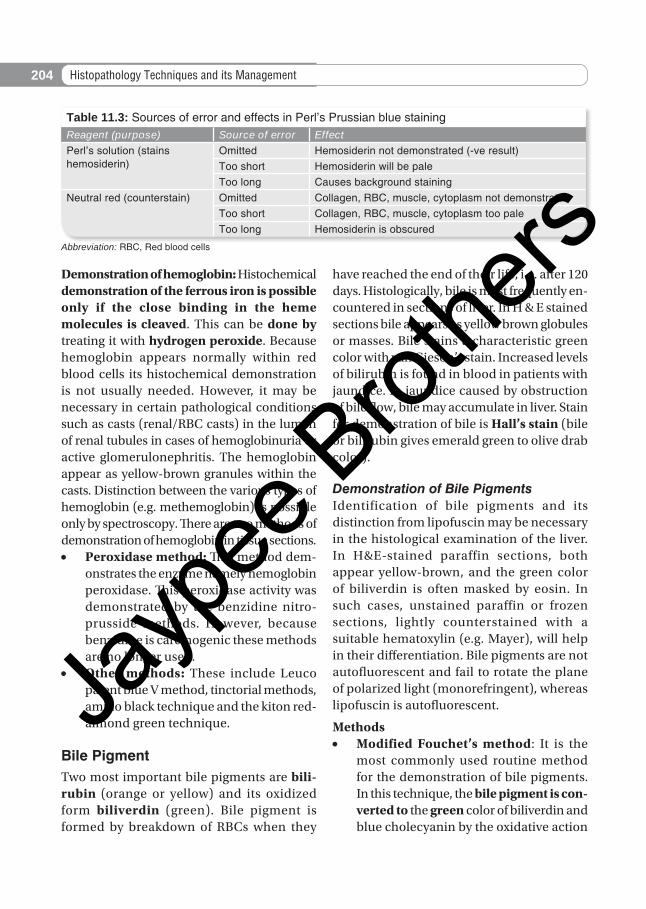
Table 11.3: Sources of error and effects in Perl’s Prussian blue stainingReagent (purpose) Source of error EffectPerl’s solution (stains hemosiderin)
Omitted Hemosiderin not demonstrated (-ve result)Too short Hemosiderin will be paleToo long Causes background staining
Neutral red (counterstain) Omitted Collagen, RBC, muscle, cytoplasm not demonstratedToo short Collagen, RBC, muscle, cytoplasm too paleToo long Hemosiderin is obscured
Abbreviation: RBC, Red blood cells
Jayp
ee B
rothe
rs
205pigments and minerals
of the ferric chloride in the presence of tri-chloroacetic acid. It is a unique quick and simple method and when counterstained with van Gieson’s solution the green color is accentuated.
• Gmelin’s method: This technique shows an identical result with liver bile, gallblad-der bile, and hematoidin. This method is messy, capricious, and gives imperma-nent results.
HematoidinVirchow (1847) first described extracellular yellow brown crystals and amorphous masses within old hemorrhagic areas, which he called hematoidin. Microscopically, hematoidin ap-pears as a bright yellow pigment in old splenic infarcts, where it contrasts well against the pale gray of the infarcted tissue. Hematoidin can also be found in old hemorrhagic areas in the brain. Hematoidin is related to both biliru-bin and biliverdin, even though it differs from them both morphologically and chemically.
Porphyrin Pigments• These pigments normally occur in tissues
in only small amounts and are considered to be precursors of the heme portion of hemoglobin.
• The porphyrias are rare pathological conditions that are disorders of the biosynthesis of porphyrins and heme. In erythropoietic protoporphyria (a type of porphyria), porphyrin pigment can be seen as focal deposits in liver sections. The pigment appears dense dark brown and in fresh frozen sections show a brilliant red fluorescence that rapidly fades with exposure to ultraviolet light. The pigment, when seen in paraffin sections and viewed under polarized light, appears bright red in color with a centrally located, dark Maltese cross.
NON-HEMATOGENOUS ENDOGENOUS PIGMENTS
This group consists of melanins, lipofuscins, chromaffin, ceroid-type lipofuscins, pseu-domelanosis (melanosis coli), Dubin-Johnson pigment etc. (Table 11.1).
MelaninsMelanin is light brown to black pigment normally found in the skin, eye, substantia nigra of the brain, and hair. Under patho-logical conditions, it is found in benign nevus cell tumors and malignant melanomas. Melanin is produced from tyrosine by the action of an enzyme tyrosinase (also called DOPA oxidase). This enzyme acts on the tyrosine slowly to produce DOPA (dihydroxyphenylalanine) which is subsequently rapidly acted upon by the same enzyme to produce an intermediate pigment which then polymerizes to produce melanin. Melanin is tightly bound to protein within the melanosome and completely insoluble in most organic solvents.
Common Sites of Melanin• Skin: It is produced melanocytes present
within the basal layer of the epidermis. In certain inflammatory conditions of skin, melanin may also be found in phagocytic cells (‘melanophages’) in the upper dermis. Melanin is also found in the hair follicles.− Pathological deposition of melanin
is found in a benign lesion called a nevus or ‘mole’ and malignant tumor namely malignant melanoma. The histological demonstration of melanin is important in malignant melanoma and its metastases.
• Eye: It is found normally in the choroid, ciliary body, and iris. Rarely melanomas can occur in the eye.
• Brain: It is found in the substantia nigra.
Jayp
ee B
rothe
rs
206 Histopathology Techniques and its management
Methods of identification of melanin (Box 11.2)
◆ Reducing methods: Masson-Fontana silver technique, Schmorl’s ferric ferricyanide reduc-tion test.
◆ Enzyme methods: DOPA reaction. ◆ Solubility and bleaching method. ◆ Fluorescent methods. ◆ Immunohistochemistry: Melanin-activation
antigens.
Box 11.2: Methods for the identification of melanin and melanin-producing cells
Reducing Methods for MelaninMelanin is a powerful reducing agent and has the capacity to reduce both silver and acid ferricyanide solutions. This property is used for its demonstration and the methods include:• Argentaffin reaction: Melanin can
reduce ammoniacal silver solutions to metallic silver without the use of an extraneous reducer and this is termed as the argentaffin reaction. Masson’s method (using Fontana’s silver solution) and its modifications depend on melanin’s argentaffin properties and are now used for routine purposes. Melanins are blackened by acid silver nitrate solutions. Melanin is also argyrophilic. The term argyrophilic means that it can be colored black by silver impregnation methods using an extraneous reducer. However, argyrophilia is not considered to be of diagnostic value.
• Schmorl’s reaction: Melanin can reduce ferricyanide to ferrocyanide with the production of Prussian blue in the presence of ferric salts (the Schmorl’s reaction). This type of reaction is also observed with other pigments such as lipofuscins, bile, and neuroendocrine cell granules.
• Other methods for demonstrating melanin: Lillie’s ferrous ion uptake
Masson-Fontana method for melanin
Purpose: To identify melanin. Melanin is a non-lipid, non-hematogenous brown-black pigment.
Fixation: 10% formalin fixation is best. Avoid chromate and mercuric chloride fixatives.
Principle: Melanin has the ability to reduce solutions of ammoniacal silver nitrate to metallic silver without the use of an external reducing agent.
Control: A nevi.
Solutions required for Masson-Fontana stain (Table 11.4)
Table 11.4: Solutions required for Masson-Fontana stain and their compositionComposition Quantity
Ammoniacal Silver Stock Solution
• 10% silver nitrate 25.0 mL
Take 20 mL of a 10% silver nitrate solution in a glass flask and add concentrated ammonia drop by drop using a fine-pointed dropper pipette. Agitate the flask constantly until the formed precipitate almost dissolves. At the end point of the titration, a faint opalescence is seen. To this add 20 mL triple distilled water and filter. Store in a dark bottle within the refrigerator. The solution should be used within 4 weeks.Note: Ammoniacal silver solutions are potentially explosive if incorrectly stored. Avoid contact and inhalation
10% Silver Nitrate
• Silver nitrate 20.0 gm
• Distilled water 200.0 mL
Mix well, store in acid cleaned brown bottle, in the refrigerator. Remains good for 6 months
Ammoniacal Silver Working Solution
• Ammoniacal silver stock solution 12.5 mL
• Distilled water 37.5 mL
Pipette the ammoniacal silver off the top, leaving the precipitate on the bottom. Filter discard after use.
5% sodium thiosufate (also known as hypo)
• Sodium thiosulfate 5 g
• Distilled water 100 mL
Mix until dissolved and store at room temperature
Jayp
ee B
rothe
rs
207pigments and minerals
Table 11.6: Sources of error and effects in Masson-Fontana stainingReagent (purpose)
Source of error
Effect
Ammoniacal silver (impregnation)
Omitted Melanin will not demonstrated (-ve result)
Too short Trace amount of melanin will not be detected
Too long Non-specific staining may occur
5% Sodium thiosulfate (Bleaching)
Omitted Discoloration in counterstain. Tissue sections appear discolored
Too short Sections may be slightly discolored
Too long Will not affect staining
Neutral red or Light green (counterstain)
Omitted Background will not be demonstrated
Too short Background difficult to see
Too long May obscure melanin staining
Table 11.5: Results of Masson-Fontana stain for melaninComponent Color
Melanin Black
Argentaffin, chromaffin and some lipofuscins
Black
Nuclei Red
Note: Friable material may require coating with celloidin as the ammonia in the silver solution could lead to sections lifting off the slide
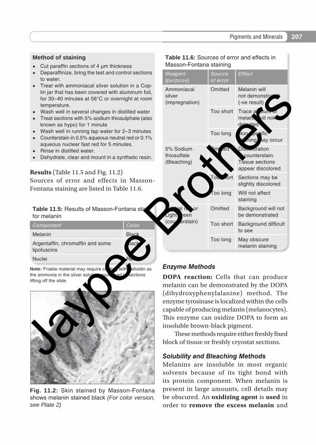
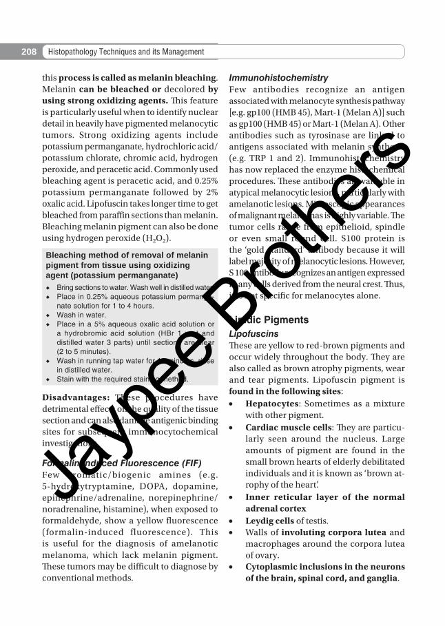
Fig. 11.2: Skin stained by Masson-Fontana shows melanin stained black (For color version, see Plate 2)
Method of staining ◆ Cut paraffin sections of 4 µm thickness ◆ Deparaffinize, bring the test and control sections
to water. ◆ Treat with ammoniacal silver solution in a Cop-
lin jar that has been covered with aluminum foil, for 30–40 minutes at 56°C or overnight at room temperature.
◆ Wash well in several changes in distilled water ◆ Treat sections with 5% sodium thiosulphate (also
known as hypo) for 1 minute ◆ Wash well in running tap water for 2–3 minutes ◆ Counterstain in 0.5% aqueous neutral red or 0.1%
aqueous nuclear fast red for 5 minutes. ◆ Rinse in distilled water. ◆ Dehydrate, clear and mount in a synthetic resin.
Results (Table 11.5 and Fig. 11.2)Sources of error and effects in Masson-Fontana staining are listed in Table 11.6.
Enzyme Methods
DOPA reaction: Cells that can produce melanin can be demonstrated by the DOPA (dihydroxyphenylalanine) method. The enzyme tyrosinase is localized within the cells capable of producing melanin (melanocytes). This enzyme can oxidize DOPA to form an insoluble brown-black pigment. These methods require either freshly fixed block of tissue or freshly cryostat sections.
Solubility and Bleaching MethodsMelanins are insoluble in most organic solvents because of its tight bond with its protein component. When melanin is present in large amounts, cell details may be obscured. An oxidizing agent is used in order to remove the excess melanin and
Jayp
ee B
rothe
rs
208 Histopathology Techniques and its management
this process is called as melanin bleaching. Melanin can be bleached or decolored by using strong oxidizing agents. This feature is particularly useful when to identify nuclear detail in heavily have pigmented melanocytic tumors. Strong oxidizing agents include potassium permanganate, hydrochloric acid/ potassium chlorate, chromic acid, hydrogen peroxide, and peracetic acid. Commonly used bleaching agent is peracetic acid, and 0.25% potassium permanganate followed by 2% oxalic acid. Lipofuscin takes longer time to get bleached from paraffin sections than melanin. Bleaching melanin pigment can also be done using hydrogen peroxide (H2O2).
Bleaching method of removal of melanin pigment from tissue using oxidizing agent (potassium permanganate) ◆ Bring sections to water. Wash well in distilled water. ◆ Place in 0.25% aqueous potassium permanga-
nate solution for 1 to 4 hours. ◆ Wash in water. ◆ Place in a 5% aqueous oxalic acid solution or
a hydrobromic acid solution (HBr 1 part and distilled water 3 parts) until sections are clear (2 to 5 minutes).
◆ Wash in running tap water for 10 minutes, rinse in distilled water.
◆ Stain with the required staining method.
Disadvantages: These procedures have detrimental effects on the quality of the tissue section and can also damage antigenic binding sites for subsequent immunocytochemical investigations.
Formalin-induced Fluorescence (FIF)Few aromatic/biogenic amines (e.g. 5-hydroxytryptamine, DOPA, dopamine, epinephrine/adrenaline, norepinephrine/noradrenaline, histamine), when exposed to formaldehyde, show a yellow fluorescence (formalin-induced fluorescence). This is useful for the diagnosis of amelanotic melanoma, which lack melanin pigment. These tumors may be difficult to diagnose by conventional methods.
ImmunohistochemistryFew antibodies recognize an antigen associated with melanocyte synthesis pathway [e.g. gp100 (HMB 45), Mart-1 (Melan A)] such as gp100 (HMB 45) or Mart-1 (Melan A). Other antibodies such as tyrosinase are linked to antigens associated with melanin synthesis (e.g. TRP 1 and 2). Immunohistochemistry has now replaced the enzyme histochemical procedures. These antibodies are valuable in atypical melanocytic lesions, particularly with amelanotic lesions. Microscopic appearances of malignant melanomas is highly variable. The tumor cells range from epithelioid, spindle or even small round cell. S100 protein is the ‘gold standard’ antibody because it will label majority of melanocytic lesions. However, S 100 antibody recognizes an antigen expressed in any cells derived from the neural crest. Thus, it is not specific for melanocytes alone.
Lipidic PigmentsLipofuscinsThese are yellow to red-brown pigments and occur widely throughout the body. They are also called as brown atrophy pigments, wear and tear pigments. Lipofuscin pigment is found in the following sites:• Hepatocytes: Sometimes as a mixture
with other pigment.• Cardiac muscle cells: They are particu-
larly seen around the nucleus. Large amounts of pigment are found in the small brown hearts of elderly debilitated individuals and it is known as ‘brown at-rophy of the heart’.
• Inner reticular layer of the normal adrenal cortex
• Leydig cells of testis.• Walls of involuting corpora lutea and
macrophages around the corpora lutea of ovary.
• Cytoplasmic inclusions in the neurons of the brain, spinal cord, and ganglia.
Jayp
ee B
rothe
rs
209pigments and minerals
• Edge of a cerebral hemorrhage or infarct. • Some lipid storage disorders• Other tissues: Bone marrow, involuntary
muscle, cervix, and kidney.
Demonstration of lipofuscinsLipofuscins are probably produced by slow progressive oxidation of lipids and lipopro-teins. The oxidation process occurs slowly and progressively. Hence, these pigments show variable staining reactions, different colors, and variation in shape and size. Their histo-chemical reactions will vary according to the degree of oxidation. Therefore, it is advisable to carry out a variety of techniques to be sure whether the pigment is lipofuscin. Commonly used methods are:• Periodic acid-Schiff (PAS) method• Schmorl’s ferric-ferricyanide reduction
test (same as for melanin mentioned above)
• Long Ziehl-Neelsen method• Sudan black B method• Gomori’s aldehyde fuchsin technique • Masson-Fontana silver method (see
above page 206)• Basophilia, using methyl green • Oil red O.
Ceroid This is not a single substance but is a mixture of lipofuscins like pigments and probably represents early stage of lipofuscins. Ceroid exhibit auto fluorescence and appear greenish yellow in frozen and brownish-yellow in paraffin section. Ceroid are rarely seen in humans. Stains used for its demonstration include Oil Red O and Sudan Black B.
ChromaffinChromaffin pigment is dark brown, granular material. It is normally found in the cells of the adrenal medulla after chrome fixation. It
is derived from adrenaline and noradrenaline. This reaction is termed as chromaffin reaction.
Demonstration of chromaffin reaction • For demonstration of chromaffin reaction
fresh tissue should be fixed in Regaud’s fluid, Orth’s or other dichromate-con-taining fixatives. Formalin fixation is not recommended, and fixatives con-taining alcohol, mercury bichloride, or acetic acid should be avoided. Staining of the section with Giemsa will produce a characteristic yellow-green staining of the chromaffin cells. It may be also demonstrated in tumors of the adrenal medulla (pheochromocytomas).− Treatment of fresh tissue with po-
tassium iodate produces a brown pigment with noradrenaline within a few minutes whereas adrenaline needs up to 24 hours treatment to produce brown coloration.
• Chromaffin granules can also be demon-strated by Schmorl’s reaction (positive), Lillie’s Nile blue A, the Masson-Fontana, and the periodic acid-Schiff (PAS-appears grey red) technique.
Endogenous MineralsIron is discussed under hematogenous pig-ments on pages 201-03.
CalciumInsoluble inorganic calcium salts are normally present in bones and teeth. Free ionic form of calcium is found in the blood and it cannot be demonstrated by histochemical stains. Abnormal depositions of calcium can occur in necrotic tissue such as tuberculosis, infarction (Gandy-Gamma bodies), atheroma in blood vessels, and malakoplakia of the bladder (Michaelis-Gutman bodies). The most common forms of calcium salts found in these
Jayp
ee B
rothe
rs
210 Histopathology Techniques and its management
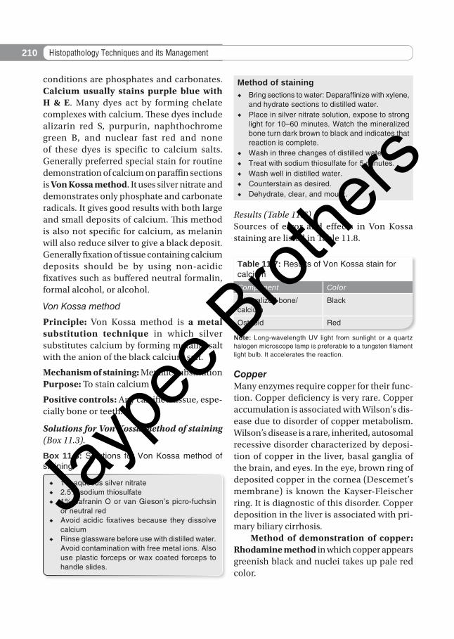
conditions are phosphates and carbonates. Calcium usually stains purple blue with H & E. Many dyes act by forming chelate complexes with calcium. These dyes include alizarin red S, purpurin, naphthochrome green B, and nuclear fast red and none of these dyes is specific to calcium salts. Generally preferred special stain for routine demonstration of calcium on paraffin sections is Von Kossa method. It uses silver nitrate and demonstrates only phosphate and carbonate radicals. It gives good results with both large and small deposits of calcium. This method is also not specific for calcium, as melanin will also reduce silver to give a black deposit. Generally fixation of tissue containing calcium deposits should be by using non-acidic fixatives such as buffered neutral formalin, formal alcohol, or alcohol.
Von Kossa method
Principle: Von Kossa method is a metal substitution technique in which silver substitutes calcium by forming metallic salt with the anion of the black calcium salt.
Mechanism of staining: Metallic substitution Purpose: To stain calcium
Positive controls: Any calcified tissue, espe-cially bone or teeth.
Solutions for Von Kossa method of staining (Box 11.3).
Box 11.3: Solutions for Von Kossa method of staining
◆ 1% aqueous silver nitrate ◆ 2.5% sodium thiosulfate ◆ 1% safranin O or van Gieson’s picro-fuchsin
or neutral red ◆ Avoid acidic fixatives because they dissolve
calcium ◆ Rinse glassware before use with distilled water.
Avoid contamination with free metal ions. Also use plastic forceps or wax coated forceps to handle slides.
Method of staining ◆ Bring sections to water: Deparaffinize with xylene,
and hydrate sections to distilled water. ◆ Place in silver nitrate solution, expose to strong
light for 10–60 minutes. Watch the mineralized bone turn dark brown to black and indicates that reaction is complete.
◆ Wash in three changes of distilled water. ◆ Treat with sodium thiosulfate for 5 minutes. ◆ Wash well in distilled water. ◆ Counterstain as desired. ◆ Dehydrate, clear, and mount.
Results (Table 11.7)Sources of error and effects in Von Kossa staining are listed in Table 11.8.
Table 11.7: Results of Von Kossa stain for calciumComponent Color
Mineralized bone/calcium
Black
Osteoid Red
Note: Long-wavelength UV light from sunlight or a quartz halogen microscope lamp is preferable to a tungsten filament light bulb. It accelerates the reaction.
CopperMany enzymes require copper for their func-tion. Copper deficiency is very rare. Copper accumulation is associated with Wilson’s dis-ease due to disorder of copper metabolism. Wilson’s disease is a rare, inherited, autosomal recessive disorder characterized by deposi-tion of copper in the liver, basal ganglia of the brain, and eyes. In the eye, brown ring of deposited copper in the cornea (Descemet’s membrane) is known the Kayser-Fleischer ring. It is diagnostic of this disorder. Copper deposition in the liver is associated with pri-mary biliary cirrhosis. Method of demonstration of copper: Rhodamine method in which copper appears greenish black and nuclei takes up pale red color.
Jayp
ee B
rothe
rs
211pigments and minerals
Uric Acid and UratesUric acid is a breakdown product of the purine (nucleic acid) metabolism. Most of uric acid is excreted by the kidneys. The uric acid circulating in the blood is in the form of monosodium urate. In gout its level may be high and may form a supersaturated solution. These high levels may result in deposition of water soluble urate in tissues, causing:• Subcutaneous nodular deposits of urate
crystals termed ‘tophi’• Synovitis and arthritis• Renal disease and calculi. Condition that occasionally mimic gout is termed as pseudogout or chondrocalcinosis. It is a pyrophosphate arthropathy and results in deposition of calcium pyrophosphate crystals in joint cartilage. It is important to differentiate gout and pseudogout. Under polarizing microscope pyrophosphate crystals show a positive birefringence and urate crystals show a negative birefringence with needle-shaped crystals.
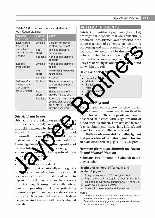
ARTEFACT PIGMENTSArtefact (or artifact) pigments (Box 11.4) are pigment deposits that are artifactually produced. These pigments are deposited in the tissue as a result of a chemical action during processing and most commonly result from fixation. They are caused by the interactions between certain tissue components and some chemical substances (components of fixative). They are normally lie on top of the tissue and not within the cell.
◆ Formalin ◆ Malaria ◆ Schistosome ◆ Mercury ◆ Chromic oxide ◆ Starch
Box 11.4: Artefact pigments
Formalin PigmentFormalin pigment is a brown or brown-black deposit seen in tissues which are fixed in acidic formalin. These deposits are usually observed in tissues with large amount of blood such as spleen, hemorrhagic lesions (e.g. cerebral hemorrhage, lung infarct), and large blood vessels filled with blood. Methods of removal of formalin pigment and prevention of formalin pigment forma-tion are discussed on pages 19-20 Chapter 2.
Removal (Extraction Method) for Forma-lin and Malarial Pigment
Solutions: 10% ammonium hydroxide in 70% ethyl alcohol.
Method of removal of formalin and malarial pigment1. Bring the sections to 70% ethyl alcohol.2. Place sections in a Coplin jar containing 10%
ammonium hydroxide alcohol for 5–15 minutes.3. Wash well in distilled water.4. Stain with the required staining method.
Notesa. The time required for the removal of formalin pigment de-
pends on the amount of pigment present.b. Removal of malarial pigment usually requires treatment
for at least 15 minutes or longer.
Table 11.8: Sources of error and effects in Von Kossa stainingReagent (purpose)
Source of error
Effect
Silver nitrate (reacts with phosphate and carbonate groups)
Omitted Calcium not demon-strated (-ve result)
Too short
Minimal calcium or hard to see
Too long Non-specific staining possible
Sodium thiosulphate
Omitted Non-specific staining occurs
Too short
Few black precipitates might occur
Too long No effectSafranin O or neutral red or van Gieson (counterstain)
Omitted Tissue not containing calcium not demon-strated
Too short
Tissue component may be hard to see
Too long Poor contrast with primary stain and small amounts of calcium may be obscured
Jayp
ee B
rothe
rs
212 Histopathology Techniques and its management
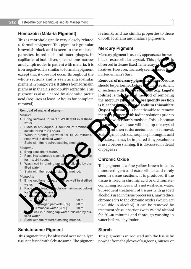
Hemozoin (Malaria Pigment)This is morphologically very closely related to formalin pigment. This pigment is granular brownish black and is seen in the malarial parasites, in red cells and macrophages, in capillaries of brain, liver, spleen, bone marrow and lymph nodes in patient with malaria. It is iron negative. It is similar to formalin pigment except that it does not occur throughout the whole sections and is seen as intracellular pigment in phagocytes. It differs from formalin pigment in that it is not doubly refractile. This pigment is also cleared by alcoholic picric acid (requires at least 12 hours for complete removal).
Removal of malarial pigmentMethod I1. Bring sections to water. Wash well in distilled
water.2. Place in 5% aqueous solution of ammonium
sulfide for 20 to 24 hours.3. Wash in running tap water for 15–20 minutes,
rinse well in distilled water.4. Stain with the required staining method.
Method II1. Bring sections to water.2. Place in a saturated alcoholic picric acid solution
for 1 to 24 hours.3. Wash well in running tap water followed by dis-
tilled water4. Stain with the required staining method.
Method III1. Bring sections to water. Rinse well in distilled
water.2. Place in the bleaching solution (mentioned below)
for 5 minutes or less. ◆ Bleaching solution
– Acetone 50 mL– Hydrogen peroxide (3%) 50 mL– Ammonia water (28%) 10 mL
3. Wash well in running tap water followed by dis-tilled water.
4. Stain with the required staining method.
Schistosome PigmentThis pigment may be observed occasionally in tissue infested with Schistosoma. The pigment
is chunky and has similar properties to those of both formalin and malaria pigments.
Mercury PigmentMercury pigment is usually appears as a brown-black, extracellular crystal. This pigment is observed in tissues fixed in mercury-containing fixatives. However, it is rarely seen in tissue fixed in Heidenhain’s Susa.
Removal of mercury pigment: This procedure should be performed before staining. Treatment of sections with iodine solutions (e.g. Lugol’s iodine) is the classical method of removing the mercury pigment. Subsequently section is bleached with a weak sodium thiosulfate (hypo) solution. It is not advisable to remove mercury pigment with iodine solutions prior to staining with Gram’s method. This is because the connective tissue will take up the crystal violet and then resist acetone color removal. Staining methods such as phosphotungstic acid hematoxylin may be impaired if ‘hypo’solution is used before staining. It is discussed in detail on pages 22.
Chromic OxideThis pigment is a fine yellow-brown in color, monorefringent and extracellular and rarely seen in tissue sections. It is produced if the tissue is fixed in chromic acid or dichromate-containing fixatives and is not washed in water. Subsequent treatment of tissues with graded alcohols used in tissue processors, may reduce chrome salts to the chromic oxides (which are insoluble in alcohol). It can be removed by treatment of tissue sections with 1% acid alcohol for 20–30 minutes and thorough washing in water before dehydration.
StarchThis pigment is introduced into the tissue by powder from the gloves of surgeons, nurses, or
Jayp
ee B
rothe
rs
213pigments and minerals
pathologists. It shows positivity with PAS and Gomori methenamine silver (GMS).
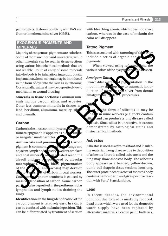
EXOGENOUS PIGMENTS AND MINERALS
Majority of exogenous pigments are colorless. Some of them are inert and unreactive, while other materials can be seen in tissue sections using various histochemical methods that are not reliable. Route of entry of some minerals into the body is by inhalation, ingestion, or skin implantation. Some minerals may be introduced in the form of dye into the skin as in tattooing. Occasionally, mineral may be deposited due to medication or wound dressing.
Minerals in tissue sections: Common min-erals include carbon, silica, and asbestos. Other less common minerals in tissues are lead, beryllium, aluminum, mercury, silver, and bismuth.
CarbonCarbon is the most commonly seen exogenous mineral/pigment. It appears as black clumps or irregular small particles.
Anthracosis and pneumoconiosis: Carbon pigment is commonly found in the lungs and adjacent lymph nodes in city dwellers, smokers and coal miners. Carbon inhaled reach the alveoli and are phagocytosed by alveolar macrophages. Heavy black pigmentation of the lung (i.e. anthracosis) may develop due to deposition of carbon in coal workers. Coal workers’ pneumoconiosis is caused by massive deposition of carbon. Some carbon particles also deposited in the peribronchiolar lymphatics and lymph nodes draining the lungs.
Identification: In the lung identification of the carbon pigment is relatively easy. In skin, it can be confused with melanin deposition. This can be differentiated by treatment of section
with bleaching agents which does not affect carbon, whereas in the case of melanin the color will disappear.
Tattoo PigmentThis is associated with tattooing of skin. They include a series of organic and inorganic pigments. When viewed using reflected light, the various colors of the dye pigments can be seen.
Amalgam TattooBrown-black areas of pigmentation in the mouth may develop due to traumatic intro-duction of mercury and silver from dental amalgam during dental procedures.
SilicaSilica in the form of silicates is may be inhaled in mine workers (e.g. rocks contain silica) and can produce a lung disease called silicosis. Since silica is unreactive, it cannot demonstrated by histological stains and histochemical methods.
AsbestosAsbestos is used as a fire-resistant and insulat-ing material. Lung disease due to deposition of asbestos fibers is called asbestosis and the lung may show asbestos body. The asbestos body appears as a beaded, yellow-brown, dumb-bell shape in tissue sections from lung. The outer proteinaceous coat of asbestos body contains hemosiderin and gives positive reac-tion with Perls’ Prussian blue.
LeadIn recent decades, the environmental pollution due to lead is markedly reduced. Lead pipes which were used for the domestic water supply have been replaced by alternative materials. Lead in paint, batteries,
Jayp
ee B
rothe
rs
214 Histopathology Techniques and its management
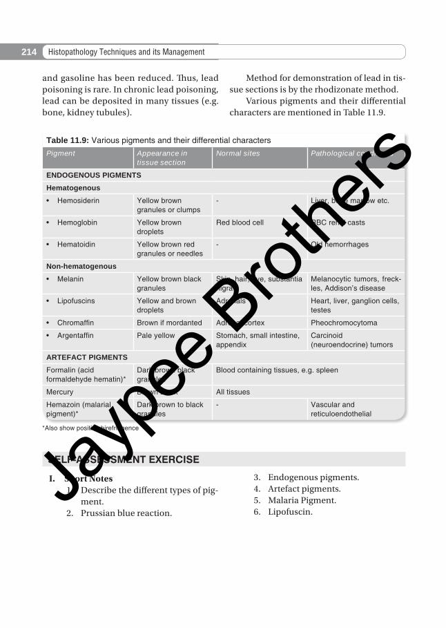
Table 11.9: Various pigments and their differential charactersPigment Appearance in
tissue sectionNormal sites Pathological conditions
ENDOGENOUS PIGMENTS
Hematogenous
• Hemosiderin Yellow brown granules or clumps
- Liver, bone marrow etc.
• Hemoglobin Yellow brown droplets
Red blood cell RBC renal casts
• Hematoidin Yellow brown red granules or needles
- Old hemorrhages
Non-hematogenous
• Melanin Yellow brown black granules
Skin, hair, eye, substantia nigra
Melanocytic tumors, freck-les, Addison’s disease
• Lipofuscins Yellow and brown droplets
Adrenals Heart, liver, ganglion cells, testes
• Chromaffin Brown if mordanted Adrenal cortex Pheochromocytoma
• Argentaffin Pale yellow Stomach, small intestine, appendix
Carcinoid (neuroendocrine) tumors
ARTEFACT PIGMENTS
Formalin (acid formaldehyde hematin)*
Dark brown black granules
Blood containing tissues, e.g. spleen
Mercury Brown black All tissues
Hemazoin (malarial pigment)*
Dark brown to black granules
- Vascular and reticuloendothelial
*Also show positive birefringence
and gasoline has been reduced. Thus, lead poisoning is rare. In chronic lead poisoning, lead can be deposited in many tissues (e.g. bone, kidney tubules).
Method for demonstration of lead in tis-sue sections is by the rhodizonate method. Various pigments and their differential characters are mentioned in Table 11.9.
SELF-ASSESSMENT EXERCISE
I. Short Notes 1. Describe the different types of pig-
ment. 2. Prussian blue reaction.
3. Endogenous pigments. 4. Artefact pigments. 5. Malaria Pigment. 6. Lipofuscin.
Jayp
ee B
rothe
rs