Histopathology Confirms White-Nose Syndrome in Bats in Europe
5
Journal of Wildlife Diseases, 48(1), 2012, pp. 207–211 # Wildlife Disease Association 2012 Histopathology Confirms White-Nose Syndrome in Bats in Europe Jiri Pikula, 1 Hana Bandouchova, 1 Ladislav Novotny ´, 1 Carol U. Meteyer, 2 Jan Zukal, 3,4 Nancy R. Irwin, 5 Jan Zima, 3 and Nata ´ lia Martı ´nkova ´ 3,6,7 1 University of Veterinary and Pharmaceutical Sciences, Palackeho 1/3, 612 42 Brno, Czech Republic; 2 US Geological Survey, National Wildlife Health Center, 6006 Schroeder Road, Madison, Wisconsin 53711-6223, USA; 3 Institute of Vertebrate Biology, Academy of Sciences of the Czech Republic, v.v.i., Kve ˇtna ´ 8, Brno, 603 65 Czech Republic; 4 Department of Botany and Zoology, Masaryk University, Kotla ´r ˇska ´ 2, 611 37 Brno, Czech Republic; 5 Department of Biology, York University, York YO10 5DD, UK; 6 Institute of Biostatistics and Analyses, Masaryk University, Kamenice 3, 625 00 Brno, Czech Republic; 7 Corresponding author (email: [email protected]) ABSTRACT: White-nose syndrome, associated with the fungal skin infection geomycosis, caused regional population collapse in bats in North America. Our results, based on histopa- thology, show the presence of white-nose syndrome in Europe. Dermatohistopathology on two bats (Myotis myotis) found dead in March 2010 with geomycosis in the Czech Republic had characteristics resembling Geo- myces destructans infection in bats confirmed with white-nose syndrome in US hibernacula. In addition, a live M. myotis, biopsied for histopathology during hibernation in April 2011, had typical fungal infection with cupping erosion and invasion of muzzle skin diagnostic for white-nose syndrome and conidiospores identical to G. destructans that were genetically confirmed as G. destructans. Key words: Geomyces destructans, geo- mycosis, histopathology, Myotis myotis, white- nose syndrome. White-nose syndrome (WNS) is an emerging disease, associated with the fungus Geomyces destructans, that is spreading among hibernating bats in North America (Blehert et al., 2009). Dermato- histopathology confirms WNS character- ized by fungal invasion of living tissue and epidermal cupping erosions (Meteyer et al., 2009). Field clinical signs of WNS in bats include a suite of descriptors including white fungal growth on hairless parts of wings, muzzle, and ears, aberrant behavior in winter, loss of fat reserves, and increased mortality at affected sites (Foley et al., 2011). Mass mortality of bats can lead to regional population collapse and extinction (Frick et al., 2010). In Europe, G. destructans was first identified morphologically from culture and sequencing of ITS and SSU rRNA regions from affected bats in 2008 (Wibbelt et al., 2010). Photographs of bats with white muzzles have been taken sporadically over several decades, but lately there has been an increase in these observations (Martı´nkova ´ et al., 2010). Recent genetic evidence confirms that G. destructans is widespread in Europe, but mass mortality has not been observed (Puechmaille et al., 2011b). There has been no histologic study of bats with geomycosis in Europe because bats may not be euthanatized without a permit, and detection of the fungus was therefore restricted to live bats without histopatho- logic examination. We present the histo- pathologic confirmation of WNS in bats from Europe. A carcass of a greater mouse-eared bat (Myotis myotis) with white fungal patches on its muzzle was found in Stara ´ Dra ´te- nicka ´ cave (Moravian Karst, Czech Re- public) on 6 March 2010, during a winter survey. Conidiospores and hyphae mor- phologically identical to G. destructans were found on an adhesive tape sample taken from the bat in the cave (Fig. 1a), but cultures and PCR on this sample were negative (sample 5 t , Martı´nkova ´ et al., 2010). No other bats in the hibernaculum (29 bats of seven species) had signs of fungal infection. Five greater mouse-eared adult bat car- casses were found 12 days after the survey on the floor of Byc ˇı ´ ska ´la cave (Moravian Karst, Czech Republic; ,3 km from Stara ´ Dra ´ tenicka ´ cave) approximately 40 m from the entrance. The carcasses were collected and frozen at 220 C for later examination. In 2010, 1,192 greater mouse-eared bats were counted in Byc ˇı ´ ska ´ la cave, hibernating alone or in clusters of around 30 individuals 207
Transcript of Histopathology Confirms White-Nose Syndrome in Bats in Europe
Journal of Wildlife Diseases, 48(1), 2012, pp. 207–211# Wildlife Disease Association 2012
Histopathology Confirms White-Nose Syndrome in Bats in Europe
Jiri Pikula,1 Hana Bandouchova,1 Ladislav Novotny,1 Carol U. Meteyer,2 Jan Zukal,3,4 Nancy R. Irwin,5
Jan Zima,3 and Natalia Martınkova3,6,7 1 University of Veterinary and Pharmaceutical Sciences, Palackeho 1/3,612 42 Brno, Czech Republic; 2 US Geological Survey, National Wildlife Health Center, 6006 Schroeder Road,Madison, Wisconsin 53711-6223, USA; 3 Institute of Vertebrate Biology, Academy of Sciences of the CzechRepublic, v.v.i., Kvetna 8, Brno, 603 65 Czech Republic; 4 Department of Botany and Zoology, MasarykUniversity, Kotlarska 2, 611 37 Brno, Czech Republic; 5 Department of Biology, York University, York YO10 5DD,UK; 6 Institute of Biostatistics and Analyses, Masaryk University, Kamenice 3, 625 00 Brno, Czech Republic;7 Corresponding author (email: [email protected])
ABSTRACT: White-nose syndrome, associatedwith the fungal skin infection geomycosis,caused regional population collapse in bats inNorth America. Our results, based on histopa-thology, show the presence of white-nosesyndrome in Europe. Dermatohistopathologyon two bats (Myotis myotis) found dead inMarch 2010 with geomycosis in the CzechRepublic had characteristics resembling Geo-myces destructans infection in bats confirmedwith white-nose syndrome in US hibernacula.In addition, a live M. myotis, biopsied forhistopathology during hibernation in April2011, had typical fungal infection with cuppingerosion and invasion of muzzle skin diagnosticfor white-nose syndrome and conidiosporesidentical to G. destructans that were geneticallyconfirmed as G. destructans.
Key words: Geomyces destructans, geo-mycosis, histopathology, Myotis myotis, white-nose syndrome.
White-nose syndrome (WNS) is anemerging disease, associated with thefungus Geomyces destructans, that isspreading among hibernating bats in NorthAmerica (Blehert et al., 2009). Dermato-histopathology confirms WNS character-ized by fungal invasion of living tissue andepidermal cupping erosions (Meteyer et al.,2009). Field clinical signs of WNS in batsinclude a suite of descriptors includingwhite fungal growth on hairless parts ofwings, muzzle, and ears, aberrant behaviorin winter, loss of fat reserves, and increasedmortality at affected sites (Foley et al.,2011). Mass mortality of bats can lead toregional population collapse and extinction(Frick et al., 2010).
In Europe, G. destructans was firstidentified morphologically from cultureand sequencing of ITS and SSU rRNAregions from affected bats in 2008 (Wibbelt
et al., 2010). Photographs of bats with whitemuzzles have been taken sporadically overseveral decades, but lately there has been anincrease in these observations (Martınkovaet al., 2010). Recent genetic evidenceconfirms that G. destructans is widespreadin Europe, but mass mortality has not beenobserved (Puechmaille et al., 2011b).There has been no histologic study of batswith geomycosis in Europe because batsmay not be euthanatized without a permit,and detection of the fungus was thereforerestricted to live bats without histopatho-logic examination. We present the histo-pathologic confirmation of WNS in batsfrom Europe.
A carcass of a greater mouse-eared bat(Myotis myotis) with white fungal patcheson its muzzle was found in Stara Drate-nicka cave (Moravian Karst, Czech Re-public) on 6 March 2010, during a wintersurvey. Conidiospores and hyphae mor-phologically identical to G. destructanswere found on an adhesive tape sampletaken from the bat in the cave (Fig. 1a),but cultures and PCR on this sample werenegative (sample 5t, Martınkova et al.,2010). No other bats in the hibernaculum(29 bats of seven species) had signs offungal infection.
Five greater mouse-eared adult bat car-casses were found 12 days after the surveyon the floor of Bycı skala cave (MoravianKarst, Czech Republic; ,3 km from StaraDratenicka cave) approximately 40 m fromthe entrance. The carcasses were collectedand frozen at 220 C for later examination.In 2010, 1,192 greater mouse-eared batswere counted in Bycı skala cave, hibernatingalone or in clusters of around 30 individuals
207

in crevices in high domes. In a previousstudy, G. destructans was genetically con-firmed in two live bats found deeper in thecave than the carcasses discussed in thispaper (Martınkova et al., 2010).
Gross visual inspection of the six deadbats (one from Stara Dratenicka cave andfive from Bycı skala cave) in the laboratoryshowed no visible fungal growth or skinlesions, although fungal growth was pres-ent while the bat from Stara Dratenickacave remained on the cave wall. The bodycondition was poor to moderate with lowadipose tissue stores. The mean bodyweight of the six bat cadavers was 18 g,which contrasts with live bats duringhibernation that weigh about 26 g (Krapp,2001). The upper jaw together with theskin, wing membranes (plagiopatagium),and lungs were dissected for histopatho-logic examination. Formalin-fixed samplescontaining bones of the upper jaw weredecalcified using formic acid to facilitatepreparation of sections. Wing membraneswere rolled for paraffin embedding. Serialtissue sections of 5 mm were prepared andstained with periodic acid–Schiff stain.Internal organs were stained with hema-toxylin and eosin.
No fungal infection was seen in themicroscopic sections of wing skin fromfour bats, but fungal colonization of theskin of two bats from Stara Dratenicka andBycı skala caves was heavy. Althoughautolysis was present, organized hyphaewere seen in all layers of epidermis of themuzzle and deep in the hair follicles(Fig. 1b). The hyphae penetrated thebasement membrane of the epidermiswith focal pigmentary incontinence andinvaded the superficial and deep dermisand underlying connective tissue (Fig. 1c).Fungal hyphae and detritus were seen inthe nasal cavity that may have beenpostmortem invasion. Hyphae were alsopresent in the nasal cartilage perichondri-um and endomysium of nasal muscles. Noreactive inflammatory response was ob-served, except for sporadic small pustulescontaining hyphae in the dermal tissue.Lungs were congested and alveoli filledwith slightly eosinophilic liquid, probablyas a result of postmortem changes.
An 24-g, adult, male greater mouse-eared bat, hibernating in Bycı skala cave
FIGURE 1. Geomyces destructans infection inbats (Myotis myotis) found dead in hibernacula intwo caves in the Czech Republic. (a) Hyphae andspores morphologically identical to G. destructans,collected using adhesive tape from the muzzle of abat from Stara Dratenicka cave. (b) Fungal growthon the muzzle of a bat from Bycı skala cave resultingin colonization of the hair follicle (arrow). Periodicacid–Schiff (PAS) stain. (c) Fungal growth overepidermis of a bat from Bycı skala cave with hyphaeobscuring the basement membrane and invading thesuperficial dermis (arrow). Fungal growth in thedermal tissue consisted of dichotomic branchinghyphae, ,2 im in diameter, with occasional septa.PAS stain.
208 JOURNAL OF WILDLIFE DISEASES, VOL. 48, NO. 1, JANUARY 2012
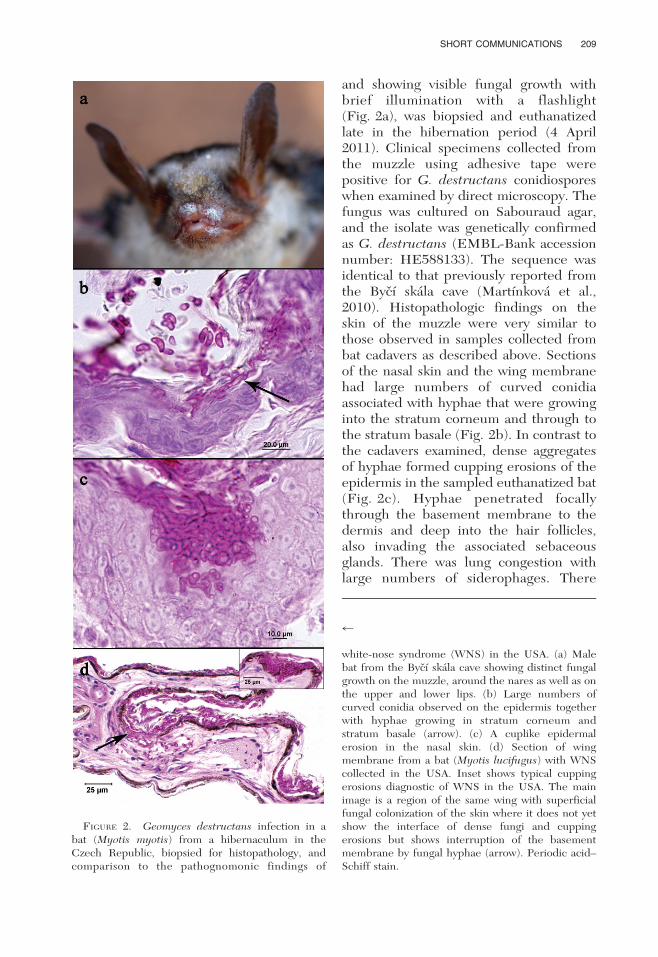
and showing visible fungal growth withbrief illumination with a flashlight(Fig. 2a), was biopsied and euthanatizedlate in the hibernation period (4 April2011). Clinical specimens collected fromthe muzzle using adhesive tape werepositive for G. destructans conidiosporeswhen examined by direct microscopy. Thefungus was cultured on Sabouraud agar,and the isolate was genetically confirmedas G. destructans (EMBL-Bank accessionnumber: HE588133). The sequence wasidentical to that previously reported fromthe Bycı skala cave (Martınkova et al.,2010). Histopathologic findings on theskin of the muzzle were very similar tothose observed in samples collected frombat cadavers as described above. Sectionsof the nasal skin and the wing membranehad large numbers of curved conidiaassociated with hyphae that were growinginto the stratum corneum and through tothe stratum basale (Fig. 2b). In contrast tothe cadavers examined, dense aggregatesof hyphae formed cupping erosions of theepidermis in the sampled euthanatized bat(Fig. 2c). Hyphae penetrated focallythrough the basement membrane to thedermis and deep into the hair follicles,also invading the associated sebaceousglands. There was lung congestion withlarge numbers of siderophages. There
FIGURE 2. Geomyces destructans infection in abat (Myotis myotis) from a hibernaculum in theCzech Republic, biopsied for histopathology, andcomparison to the pathognomonic findings of
r
white-nose syndrome (WNS) in the USA. (a) Malebat from the Bycı skala cave showing distinct fungalgrowth on the muzzle, around the nares as well as onthe upper and lower lips. (b) Large numbers ofcurved conidia observed on the epidermis togetherwith hyphae growing in stratum corneum andstratum basale (arrow). (c) A cuplike epidermalerosion in the nasal skin. (d) Section of wingmembrane from a bat (Myotis lucifugus) with WNScollected in the USA. Inset shows typical cuppingerosions diagnostic of WNS in the USA. The mainimage is a region of the same wing with superficialfungal colonization of the skin where it does not yetshow the interface of dense fungi and cuppingerosions but shows interruption of the basementmembrane by fungal hyphae (arrow). Periodic acid–Schiff stain.
SHORT COMMUNICATIONS 209

were no pathologic findings in spleen,kidneys, heart, or liver.
The histopathology in the skin of thisgreater mouse-eared bat from Bycı skalacave (Fig. 2c) fulfilled the criteria cur-rently used to confirm WNS in NorthAmerica (Meteyer et al., 2009). Thelocalized areas of dense hyphae with adiscrete interface where the fungus formscup-shaped erosions was present in thebat from the Czech Republic collected in2011, and these findings are the currentgold standard for diagnosing WNS. Be-tween these localized areas of erosion,colonization of the superficial keratin bythe fungal hyphae is also present in wingmembranes from bats with white-nosesyndrome in the USA (Fig. 2d).
Our results confirm the presence ofWNS in Europe and demonstrate that G.destructans infection in hibernating batsin Europe can be associated with sporadicdeaths that may remain unrecognized inthe hibernaculum. Mortality rates are lowand, to date, have not affected the long-term population size of greater mouse-eared bats (Martınkova et al., 2010). Withthe increase and extent of G. destructansfound in hibernacula in the Czech Re-public and Slovakia in 2010, monitoringbat populations in subsequent years iseven more critical. Histopathology of batswith geomycosis was identified as one ofthe key research priorities in the field inEurope (Puechmaille et al., 2011a). Fol-lowing confirmation of WNS in Europe,further comparisons between NorthAmerican and European G. destructansinfections can enhance the understandingof the pathogenesis of geomycosis in bats(Cryan et al., 2010).
Material from the Bycı skala cave wasprovided by Vlastislav Kana in 2010. Wethank Paul A. Racey, Maja Rutten, andfour anonymous reviewers for helpfulcomments on the manuscript. This studywas supported by the Ministry of Educa-tion, Youth and Sports of the Czech Re-public (Projects MSM 6215712402, MSM0021622416, LC06073). The research was
conducted in accordance with follow-ing permits: 35335/ENV/06-2793/620/06,00163/MK/2009, S01389/MK/2008,142/2010,169/2011.
LITERATURE CITED
BLEHERT, D. S., A. C. HICKS, M. J. BEHR, C. U.METEYER, B. M. BERLOWSKI-ZIER, E. L. BUCKELS,J. T. H. COLEMAN, S. R. DARLING, A. GARGAS, R.NIVER, J. C. OKONIEWSKI, R. J. RUDD, AND W. B.STONE. 2009. Bat white-nose syndrome: Anemerging fungal pathogen? Science 323: 227.
CRYAN, P. M., C. U. METEYER, J. G. BOYLES, AND D. S.BLEHERT. 2010. Wing pathology of white-nosesyndrome in bats suggests life-threatening dis-ruption of physiology. BMC Biology 8: 135.
FOLEY, J., D. CLIFFORD, K. CASTLE, P. CRYAN, AND
R. S. OSTFELD. 2011. Investigating and managingthe rapid emergence of white-nose syndrome, anovel, fatal, infectious disease of hibernatingbats. Conservation Biology 25: 223–231.
FRICK, W. F., J. F. POLLOCK, A. C. HICKS, K. E.LANGWIG, D. S. REYNOLDS, G. G. TURNER, C. M.BUTCHKOSKI, AND T. H. KUNZ. 2010. An emergingdisease causes regional population collapse of acommon North American bat species. Science329: 679–682.
KRAPP, F. 2001. Handbuch der Saugetiere Europas,Teil 1, Chiroptera 1. Aula Verlag, Wiebelsheim,Germany, 602 pp.
MARTINKOVA, N., P. BACKOR, T. BARTONICKA, P. BLAZKOVA,J. CERVENY, L. FALTEISEK, J. GAISLER, V. HANZAL, D.HORACEK, Z. HUBALEK, H. JAHELKOVA, M. KOLARIK,KORYTAR, A. KUBATOVA, B. LEHOTSKA, R. LEHOTSKY,R. K. LUCAN, O. MAJEK, J. MATEJU, Z. REHAK, J.SAFAR, P. TAJEK, E. TKADLEC, M. UHRIN, J. WAGNER,D. WEINFURTOVA, J. ZIMA, J. ZUKAL, AND I. HORACEK.2010. Increasing incidence of Geomyces destructansfungus in bats from the Czech Republic andSlovakia. PLoS ONE 5: e13853.
METEYER, C. U., E. L. BUCKLES, D. S. BLEHERT, A. C.HICKS, D. E. GREEN, V. SHEARN-BOCHSLER, N. J.THOMAS, A. GARGAS, AND M. J. BEHR. 2009.Histopathologic criteria to confirm white-nosesyndrome in bats. Journal of Veterinary Diag-nostics and Investigation 21: 411–414.
PUECHMAILLE, S. J., W. F. FRICK, T. H. KUNZ, P. A.RACEY, C. C. VOIGT, G. WIBBELT, AND E. C.TEELING. 2011a. White-nose syndrome: Is thisemerging disease a threat to European bats?Trends in Ecology and Evolution, 26: 570–576.
———, G. WIBBELT, V. KORN, H. FULLER, F. FORGET,K. MUHLDORFER, A. KURTH, W. BOGDANOWICZ, C.BOREL, T. BOSCH, T. CHEREZY, M. DREBET, T.GORFOL, A.-J. HAARSMA, F. HERHAUS, G. HAL-
LART, M. HAMMER, C. JUNGMANN, Y. LE BRIS, L.LUTSAR, M. MASING, B. MULKENS, K. PASSIOR, M.STARRACH, A. WOJTASZEWSKI, U. ZOPHEL, AND E.
210 JOURNAL OF WILDLIFE DISEASES, VOL. 48, NO. 1, JANUARY 2012

C. TEELING. 2011b. Pan-European distributionof white-nose syndrome fungus (Geomycesdestructans) not associated with mass mortality.PLoS ONE 6: e19167.
WIBBELT, G., A. KURTH, D. HELLMANN, M. WEISHAAR,A. BARLOW, M. VEITH, J. PRUGER, T. GORFOL, L.GROSCHE, F. BONTADINA, U. ZOPHEL, H.-P. SEIDL,
P. M. CRYAN, AND D. S. BLEHERT. 2010. White-nose syndrome fungus (Geomyces destructans) inbats, Europe. Emerging Infectious Diseases 16:1237–1243.
Submitted for publication 31 December 2010.Accepted 12 September 2011.
SHORT COMMUNICATIONS 211