HistoneAcetyltransferase1PromotesHomologous ... RecombinationinDNARepairbyFacilitatingHistone...
13
Histone Acetyltransferase 1 Promotes Homologous Recombination in DNA Repair by Facilitating Histone Turnover * □ S Received for publication, March 29, 2013, and in revised form, May 7, 2013 Published, JBC Papers in Press, May 7, 2013, DOI 10.1074/jbc.M113.473199 Xiaohan Yang ‡ , Lei Li ‡ , Jing Liang ‡ , Lei Shi § , Jianguo Yang ‡ , Xia Yi ‡ , Di Zhang ‡ , Xiao Han ‡ , Na Yu ‡ , and Yongfeng Shang ‡§1 From the ‡ Key Laboratory of Carcinogenesis and Translational Research (Ministry of Education), Department of Biochemistry and Molecular Biology, Peking University Health Science Center, Beijing 100191, China and § Tianjin Key Laboratory of Medical Epigenetics, Department of Biochemistry and Molecular Biology, Tianjin Medical University, Tianjin 300070, China Background: HAT1 is involved in homologous recombination repair. Results: HAT1 facilitates the incorporation of H4K5/K12-acetylated H3.3 at double-strand break sites through its HIRA-de- pendent histone turnover activity, thereby promoting efficient homologous recombination process. Conclusion: HAT1 is a key regulator of homologous recombination repair. Significance: This work provides a mechanistic insight into the regulation of histone dynamics by HAT1. Faithful repair of DNA double-strand breaks is vital to the maintenance of genome integrity and proper cell functions. His- tone modifications, such as reversible acetylation, phosphory- lation, methylation, and ubiquitination, which collectively con- tribute to the establishment of distinct chromatin states, play important roles in the recruitment of repair factors to the sites of double-strand breaks. Here we report that histone acetyltrans- ferase 1 (HAT1), a classical B type histone acetyltransferase responsible for acetylating the N-terminal tail of newly synthe- sized histone H4 in the cytoplasm, is a key regulator of DNA repair by homologous recombination in the nucleus. We found that HAT1 is required for the incorporation of H4K5/K12- acetylated H3.3 at sites of double-strand breaks through its HIRA-dependent histone turnover activity. Incorporated his- tones with specific chemical modifications facilitate subsequent recruitment of RAD51, a key repair factor in mammalian cells, to promote efficient homologous recombination. Significantly, depletion of HAT1 sensitized cells to DNA damage compro- mised the global chromatin structure, inhibited cell prolifera- tion, and induced cell apoptosis. Our experiments uncovered a role for HAT1 in DNA repair in higher eukaryotic organisms and provide a mechanistic insight into the regulation of histone dynamics by HAT1. The maintenance of genome integrity is vital to the proper cell functions. Faithful repair of DNA damage is thus essential for the suppression of genetic instability and tumorigenesis. Eukaryotic cells have evolved two conserved mechanisms to detect and repair DNA double-strand breaks (DSBs) 2 : homol- ogous recombination (HR) repairs the break using genetic information that is retrieved from an undamaged sister chro- matid or chromosomal homologue, whereas non-homologous end joining (NHEJ) involves direct ligations of DNA ends. Characterization of the factors and pathways governing DSB repair is important to the understanding of the mutagenesis processes during oncogenic events and to the development of proper cancer therapies. Histone modifications, such as reversible acetylation, phos- phorylation, methylation, ubiquitination, and ADP-ribosyla- tion, play important roles in the promotion of DSB repairs through creating specific interaction platforms for regulatory proteins and complexes (1–5). Additionally, incorporation of variant histones could affect DSB repairs by influencing chro- matin configurations; thus, the recognition by downstream effectors. Histone turnover, referring to the replacement of “old” his- tones by “new” histones in a replication-independent manner, does not involve obvious changes in nucleosome occupancy; instead, it affects the composition of histone marks and thereby the epigenetic mechanisms of chromatin-based events such as gene regulation and DNA repair (6 – 8). A well known example of histone turnover activity is the yeast Swr1p ATPase complex, which catalyzes the replacement of conventional histone H2A with histone H2A.Z variant in nucleosome arrays (9, 10). Cou- pling of histone acetylation with histone exchange activity at the site of damaged DNA has been observed with the Drosoph- ila Tip60 complex (11), suggesting that histone exchange fac- tors can play an important role during DNA repair process. HAT1 is the first histone acetyltransferase identified and is highly conserved from yeast to human (12, 13, 64). HAT1 has * This work was supported by National Natural Science Foundation of China Grants 91219201 and 81130048 (to Y. S.) and by Ministry of Science and Technology of China grants (973 Program: 2011CB504204; to Y. S.). □ S This article contains supplemental File 1. 1 To whom correspondence should be addressed: Dept. of Biochemistry and Molecular Biology, Peking University Health Science Center, 38 Xue Yuan Rd., Beijing 100191, China. Tel.: 86-10-82805118; Fax: 86-10-82801355; E-mail: [email protected] or [email protected]. 2 The abbreviations used are: DSB, DNA double-strand break; HR, homolo- gous recombination; NHEJ, non-homologous end joining; HAT1, his- tone acetyltransferase 1; TRITC, tetramethylrhodamine isothiocyanate; CPT, camptothecin; reHAT1, recombinant HAT1; reRbAp46, recombi- nant RbAp46. THE JOURNAL OF BIOLOGICAL CHEMISTRY VOL. 288, NO. 25, pp. 18271–18282, June 21, 2013 © 2013 by The American Society for Biochemistry and Molecular Biology, Inc. Published in the U.S.A. JUNE 21, 2013 • VOLUME 288 • NUMBER 25 JOURNAL OF BIOLOGICAL CHEMISTRY 18271 by guest on June 7, 2018 http://www.jbc.org/ Downloaded from
Transcript of HistoneAcetyltransferase1PromotesHomologous ... RecombinationinDNARepairbyFacilitatingHistone...
Histone Acetyltransferase 1 Promotes HomologousRecombination in DNA Repair by Facilitating HistoneTurnover*□S
Received for publication, March 29, 2013, and in revised form, May 7, 2013 Published, JBC Papers in Press, May 7, 2013, DOI 10.1074/jbc.M113.473199
Xiaohan Yang‡, Lei Li‡, Jing Liang‡, Lei Shi§, Jianguo Yang‡, Xia Yi‡, Di Zhang‡, Xiao Han‡, Na Yu‡,and Yongfeng Shang‡§1
From the ‡Key Laboratory of Carcinogenesis and Translational Research (Ministry of Education), Department of Biochemistry andMolecular Biology, Peking University Health Science Center, Beijing 100191, China and §Tianjin Key Laboratory of MedicalEpigenetics, Department of Biochemistry and Molecular Biology, Tianjin Medical University, Tianjin 300070, China
Background: HAT1 is involved in homologous recombination repair.Results: HAT1 facilitates the incorporation of H4K5/K12-acetylated H3.3 at double-strand break sites through its HIRA-de-pendent histone turnover activity, thereby promoting efficient homologous recombination process.Conclusion: HAT1 is a key regulator of homologous recombination repair.Significance: This work provides a mechanistic insight into the regulation of histone dynamics by HAT1.
Faithful repair of DNA double-strand breaks is vital to themaintenance of genome integrity andproper cell functions.His-tone modifications, such as reversible acetylation, phosphory-lation, methylation, and ubiquitination, which collectively con-tribute to the establishment of distinct chromatin states, playimportant roles in the recruitment of repair factors to the sites ofdouble-strand breaks. Here we report that histone acetyltrans-ferase 1 (HAT1), a classical B type histone acetyltransferaseresponsible for acetylating the N-terminal tail of newly synthe-sized histone H4 in the cytoplasm, is a key regulator of DNArepair by homologous recombination in the nucleus. We foundthat HAT1 is required for the incorporation of H4K5/K12-acetylated H3.3 at sites of double-strand breaks through itsHIRA-dependent histone turnover activity. Incorporated his-tones with specific chemicalmodifications facilitate subsequentrecruitment of RAD51, a key repair factor in mammalian cells,to promote efficient homologous recombination. Significantly,depletion of HAT1 sensitized cells to DNA damage compro-mised the global chromatin structure, inhibited cell prolifera-tion, and induced cell apoptosis. Our experiments uncovered arole for HAT1 in DNA repair in higher eukaryotic organismsand provide a mechanistic insight into the regulation of histonedynamics by HAT1.
The maintenance of genome integrity is vital to the propercell functions. Faithful repair of DNA damage is thus essentialfor the suppression of genetic instability and tumorigenesis.Eukaryotic cells have evolved two conserved mechanisms to
detect and repair DNA double-strand breaks (DSBs)2: homol-ogous recombination (HR) repairs the break using geneticinformation that is retrieved from an undamaged sister chro-matid or chromosomal homologue, whereas non-homologousend joining (NHEJ) involves direct ligations of DNA ends.Characterization of the factors and pathways governing DSBrepair is important to the understanding of the mutagenesisprocesses during oncogenic events and to the development ofproper cancer therapies.Histone modifications, such as reversible acetylation, phos-
phorylation, methylation, ubiquitination, and ADP-ribosyla-tion, play important roles in the promotion of DSB repairsthrough creating specific interaction platforms for regulatoryproteins and complexes (1–5). Additionally, incorporation ofvariant histones could affect DSB repairs by influencing chro-matin configurations; thus, the recognition by downstreameffectors.Histone turnover, referring to the replacement of “old” his-
tones by “new” histones in a replication-independent manner,does not involve obvious changes in nucleosome occupancy;instead, it affects the composition of histonemarks and therebythe epigenetic mechanisms of chromatin-based events such asgene regulation and DNA repair (6–8). A well known exampleof histone turnover activity is the yeast Swr1pATPase complex,which catalyzes the replacement of conventional histone H2Awith histone H2A.Z variant in nucleosome arrays (9, 10). Cou-pling of histone acetylation with histone exchange activity atthe site of damaged DNA has been observed with theDrosoph-ila Tip60 complex (11), suggesting that histone exchange fac-tors can play an important role during DNA repair process.HAT1 is the first histone acetyltransferase identified and is
highly conserved from yeast to human (12, 13, 64). HAT1 has* This work was supported by National Natural Science Foundation of ChinaGrants 91219201 and 81130048 (to Y. S.) and by Ministry of Science andTechnology of China grants (973 Program: 2011CB504204; to Y. S.).
□S This article contains supplemental File 1.1 To whom correspondence should be addressed: Dept. of Biochemistry and
Molecular Biology, Peking University Health Science Center, 38 Xue YuanRd., Beijing 100191, China. Tel.: 86-10-82805118; Fax: 86-10-82801355;E-mail: [email protected] or [email protected].
2 The abbreviations used are: DSB, DNA double-strand break; HR, homolo-gous recombination; NHEJ, non-homologous end joining; HAT1, his-tone acetyltransferase 1; TRITC, tetramethylrhodamine isothiocyanate;CPT, camptothecin; reHAT1, recombinant HAT1; reRbAp46, recombi-nant RbAp46.
THE JOURNAL OF BIOLOGICAL CHEMISTRY VOL. 288, NO. 25, pp. 18271–18282, June 21, 2013© 2013 by The American Society for Biochemistry and Molecular Biology, Inc. Published in the U.S.A.
JUNE 21, 2013 • VOLUME 288 • NUMBER 25 JOURNAL OF BIOLOGICAL CHEMISTRY 18271
by guest on June 7, 2018http://w
ww
.jbc.org/D
ownloaded from
been described as a classical B type HAT, which can only acet-ylate the N-terminal tail of newly synthesized histone H4 butnot nucleosomal histones. On the contrary, type A histoneacetyltransferases are nuclear enzymes capable of acetylatinghistones that have already been packaged into the chromatin.Evidence from yeast model show that HAT1 forms the NuB4complexwithHAT2 (humanRbAp46homolog),Hif1p (HAT1-interacting factor 1, a histone chaperone that selectively inter-acts with histonesH3), and histone tetramer in nuclei and playsan important role in de novo chromatin assembly during repli-cation. The NuB4 complex has also been shown to regulate HRrepair of DSBs by participating in repair-linked chromatin reas-sembly through its B-HAT activity (14–16). Although HAT1has long been thought to play a role in DNA repair, it has beenmostly studied in yeast cells; whether and how HAT1 partici-pates in the DNA repair process in mammalian cells are largelyunknown.In current study we found that, in addition to a role of HAT1
in post-repair chromatin reassembly, HAT1 has a direct role inHR repair in human cells. HAT1 facilitates the enrichment ofH4K5/K12-acetylated H3.3 (H3.3-H4K5/12ac) to the DSB sitesthrough its HIRA-dependent histone turnover activity, therebymarking the damaged area for subsequent recruitment of keyrepair factor RAD51 to promote efficient HR process.
EXPERIMENTAL PROCEDURES
Cells, Plasmids, Antibodies, and Reagents—DR-GFP-U2OSand EJ5-GFP-HEK293 stable cell lines were from Dr. XingzhiXu (TheCapital NormalUniversity, Beijing, China). The cDNAfor wild-type HAT1 was amplified by PCR and ligated intoEcoRI/BamHI sites of pcDNA3.1 plasmid containing a FLAGtag. siRNA-resistant pcDNA3.1(�)-FLAG-HAT1 with threesynonymousmutations (C510A,T762C, andC1161T)was con-structed by site-directed mutagenesis of wild-type plasmid. Allclones were confirmed by DNA sequencing. Recombinantbaculoviruses containing the coding region of HAT1 andRbap46 were generated, and the proteins were purified frominfected Sf9 cells. Full-length Xenopus histone expression plas-mids pET-H2A, pET-H2B, pET-H3, and pET-H4 were fromDr. Karolin Luger (Colorado State University). pBlueScriptSK(�) plasmid, Nap1p-plasmid, and Isw1–3�FLAG yeaststrain were kindly provided by Dr. Toshio Tsukiyama (Univer-sity of Washington). Recombinant DnaK ATPase domain(1–384) was from Prospec. Antibodies used were: �H2A,�H2AK5ac, �H3, �H3K14ac, �H3K23ac, �H4, �H4K5ac,�H4K8ac, �H4K12ac, �H4K91ac, and �Ku80 from Abcam;�H3.3 fromMillipore;��-actin,�HIRA,�HIRIP3, and�FLAGfrom Sigma; goat �HAT1 from Santa Cruz Biotechnology andrabbit�HAT1 fromSigma; rabbit�H2AXp (Ser-139) fromCellSignaling; mouse �H2AXp (Ser-139) from Millipore; �ATR,�phospho-ATR (Ser-428) from Cell Signaling; �p53 fromMBL. Control siRNA was synthesized by Shanghai GeneChemInc (Shanghai, China). The sequence for control siRNA was5�-UUCUCCGAACGUGUCACGU-3�. The siRNAs targetinghumanHAT1were purchased from Santa Cruz Biotechnology.Fluorescence Confocal Microscopy—HeLa, MDA-MB-231,
MCF-7, and U2OS cells growing on six-well chamber slideswere washed with phosphate-buffered saline (PBS), fixed in 4%
paraformaldehyde, permeabilized with 0.1% Triton X-100 inPBS, blocked with 0.8% BSA, and incubated with appropriateprimary antibodies followed by staining with TRITC-conju-gated secondary antibodies. Cells were washed 4 times. and afinal concentration of 0.1 �g/ml 4,6-diamidino-2-phenylindoledihydrochloride (DAPI) (Sigma) was included in the last wash-ing to stain nuclei. Images were visualized and recordedwith anOlympus FV1000S confocal microscope.Cell Cycle Synchronization and Flow Cytometry Analysis—
MCF-7 cells were synchronized in the G0/G1 phase by serumstarvation, in the G1/S phase by double thymidine block, and inthe G2/M phase by thymidine/nocodazole block, and then theywere released for various periods of time. In all cases cells weretrypsinized, washed with PBS, and fixed in 70% ethanol at 4 °Covernight. After washing with PBS, cells were incubated withRNase A (Sigma) in PBS for 30 min at 37 °C and then stainingwith 50mg/ml propidium iodide. Cell cycle data were collectedusing a FACSCalibur flow cytometer (BDBiosciences) and ana-lyzed with ModFit LT 3.0 (Verity Software House Inc., Top-sham, ME).Cell Viability/Proliferation Assay—For cell proliferation
assays, HCT116 or U2OS cells were transfected with HAT1siRNA and seeded into 96-well plates. Cells were treated oruntreated with 0.0001 or 0.0005% methyl methanesulfonate, 2or 10 mM hydroxyurea, or 100 or 500 nM camptothecin (CPT)for 12 h. On the day of harvesting, the medium was replacedwith an equal volume of fresh medium containing 10% Alamarblue. Plateswere incubated at 37 °C for 6 h, and cell viabilitywasdetermined by measuring the absorbance of converted dye atwavelengths 570 and 630 nm (17).RNA Interference and Western Blotting—Synthesized siRNAs
were transfected into cells with the final concentration of 50nM. For Western blotting, 72 h after transfection total cellularproteins were extracted and resolved on 8 or 15% SDS-PAGEgels and transferred toNCmembranes (Millipore).Membraneswere incubated with appropriate antibodies for 1 h at roomtemperature or overnight at 4 °C followed by incubation with asecondary antibody. Immunoreactive bands were visualizedusing Western blotting luminol reagent (Santa Cruz Biotech-nology) according to the manufacturer’s recommendation.ChIP Assay—ChIPs were performed in DR-U2OS cells as
described previously (3, 18–21). Briefly, 5 � 107 cells werecross-linkedwith 1% formaldehyde, sonicated, pre-cleared, andincubated with 5–10 �g of antibody per reaction. Complexeswere washed with low and high salt buffers, and the DNA wasextracted and precipitated. The enrichment of the DNA tem-plate was analyzed by quantitative PCR using primers 500bp from I-SceI cutting: forward, 5�-GATGGCACAGTGGTC-AAGAGC-3�, and reverse, 5�-GAAGGATGGAAGGGTCA-GGAG-3�.Micrococcal Nuclease Sensitivity Assay—siRNA-treated
U2OS cells werewashedwith cold PBS, resuspended in ice-coldNonidet P-40 cell lysis buffer (10 mM Tris-HCl, pH 7.4, 10 mM
NaCl, 3 mMMgCl2, 0.5% Nonidet P-40, 0.15 mM spermine, and0.5 mM spermidine) and incubated on ice for 5 min. Permeabi-lized nuclei were washed with micrococcal nuclease digestionbuffer (10 mM Tris-HCl, pH 7.4, 15 mM NaCl, 60 mM KCl, 0.15mM spermine, and 0.5mM spermidine) and digested at 37 °C for
HAT1 Facilitates H4K5/K12-acetylated H3.3 Enrichment at DSB
18272 JOURNAL OF BIOLOGICAL CHEMISTRY VOLUME 288 • NUMBER 25 • JUNE 21, 2013
by guest on June 7, 2018http://w
ww
.jbc.org/D
ownloaded from
10minwith 2.5 or 5 units/mlmicrococcal nuclease in the diges-tion buffer containing 1 mM CaCl2. The reaction was stoppedby adding 10 mM EDTA and 0.5% SDS, and DNA was thenpurified after incubation with proteinase K and RNase A andwas electrophoresed on 2% agarose gels.Assembly of Nucleosome Arrays—Assembly of nucleosomal
arrays on an immobilized DNA was performed as previouslydescribed (22, 23). pBlueScript SK(�) plasmid was used as atemplate. Dynabeads M-280 (Dynal) were used for couplingwith the plasmid DNA linearized by EcoRI and ClaI asinstructed. In a nucleosome assembly reaction,Dynabeads cou-pled with 1.5 �g of DNA were incubated with 1.5 �g of recom-binantXenopushistone octamer, 0.91 pmol of ISW1 complex, 6�g of purified recombinant Nap1p, and 3 mM ATP in 10 mM
HEPES-KOH, pH 7.6, 0.5 mM EGTA, 5 mM MgCl2, 10% glyc-erol, 1 mM DTT, 0.1 mg/ml BSA, 70 mM KCl in a reactionvolume of 180 �l for 4 h at room temperature with constantmixing. The resulting nucleosomal arrays were washed 4 timeswith 1 ml of buffer H (25 mM HEPES-KOH, pH 7.6, 0.5 mM
EGTA, 0.1 mM EDTA, 5 mM MgCl2, 0.02% Nonidet P-40, 10%glycerol, 1 mM DTT, 0.1 mg/ml BSA) containing 0.6 M KCl toremove ATP, Nap1p, ISW1 complex, and non-nucleosomalhistones before starting the transfer reaction.Histone Transfer Assay—In a standard histone transfer assay,
immobilized nucleosomal arrays (150 ng of DNA equivalents)were incubated in a 100-�l reaction volume with 6 pmol of freecalf thymus histone, titrated FLAG-HAT1 complex, or recom-binant HAT1 (reHAT1)/reRbAp46 with or without 1 mM ATPin exchange buffer (25 mM HEPES-KOH, pH 7.6, 0.37 mM
EDTA, 0.35mM EGTA, 5mMMgCl2, 10% glycerol, 0.02%Non-idet P-40, 1mMDTT, 0.1mg/ml BSA, 70mMKCl) for 60min atroom temperature with constant mixing. Beads were concen-trated on a magnetic particle concentrator (Dynal), and thesupernatants were saved. Beads were thenwashed 3 times, eachtime with 100 �l of buffer H containing 70 mM KCl, and thebound proteins were eluted with SDS-PAGE sample buffer.The wash fractions, and the supernatants were combined, pre-cipitated with TCA, and dissolved in SDS-PAGE sample bufferfor analysis of total free (unbound) proteins. Bound and freeproteins were analyzed by Western blotting.
RESULTS
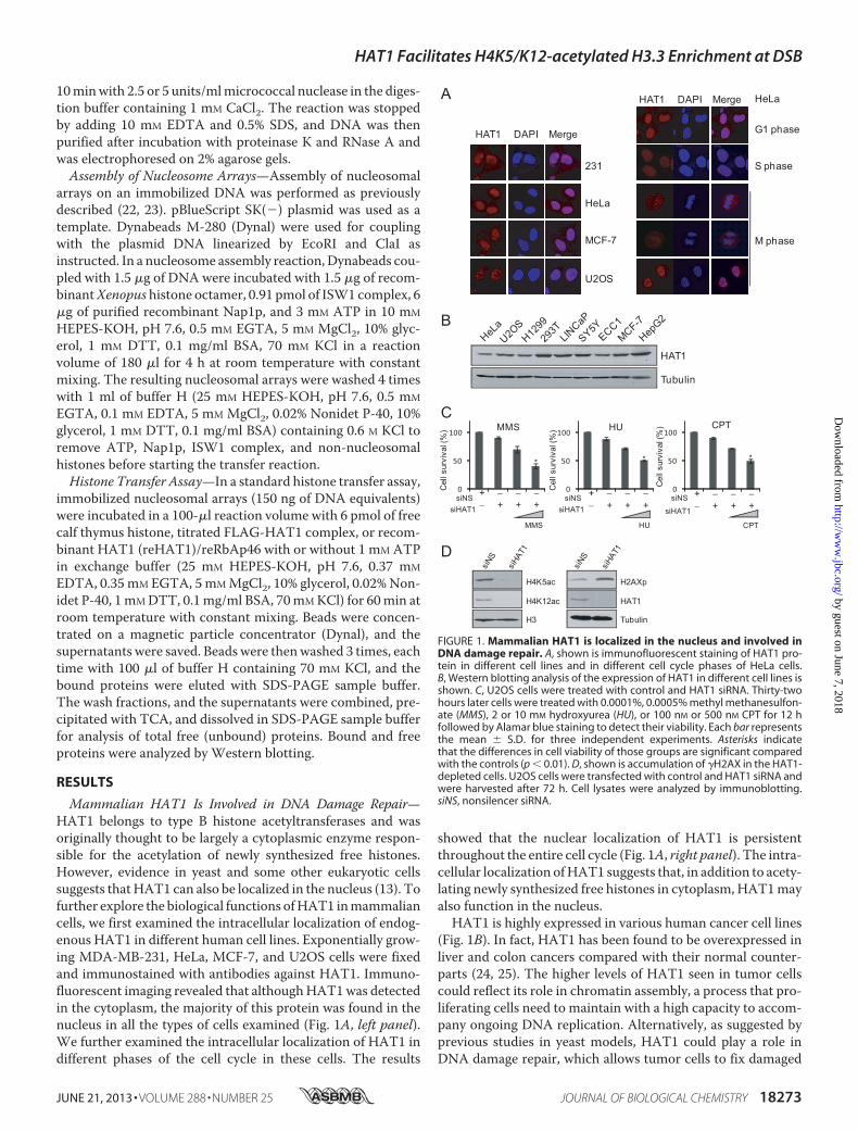
Mammalian HAT1 Is Involved in DNA Damage Repair—HAT1 belongs to type B histone acetyltransferases and wasoriginally thought to be largely a cytoplasmic enzyme respon-sible for the acetylation of newly synthesized free histones.However, evidence in yeast and some other eukaryotic cellssuggests thatHAT1 can also be localized in the nucleus (13). Tofurther explore the biological functions ofHAT1 inmammaliancells, we first examined the intracellular localization of endog-enous HAT1 in different human cell lines. Exponentially grow-ing MDA-MB-231, HeLa, MCF-7, and U2OS cells were fixedand immunostained with antibodies against HAT1. Immuno-fluorescent imaging revealed that althoughHAT1was detectedin the cytoplasm, the majority of this protein was found in thenucleus in all the types of cells examined (Fig. 1A, left panel).We further examined the intracellular localization of HAT1 indifferent phases of the cell cycle in these cells. The results
showed that the nuclear localization of HAT1 is persistentthroughout the entire cell cycle (Fig. 1A, right panel). The intra-cellular localization ofHAT1 suggests that, in addition to acety-lating newly synthesized free histones in cytoplasm, HAT1mayalso function in the nucleus.HAT1 is highly expressed in various human cancer cell lines
(Fig. 1B). In fact, HAT1 has been found to be overexpressed inliver and colon cancers compared with their normal counter-parts (24, 25). The higher levels of HAT1 seen in tumor cellscould reflect its role in chromatin assembly, a process that pro-liferating cells need to maintain with a high capacity to accom-pany ongoing DNA replication. Alternatively, as suggested byprevious studies in yeast models, HAT1 could play a role inDNA damage repair, which allows tumor cells to fix damaged
0
50
100
0
50
100
+ − − −− + + +
231
HeLa
MCF-7
U2OS
HAT1 DAPI Merge G1 phase
S phase
M phase
HAT1 DAPI Merge HeLa
HAT1
Tubulin
A
B
0
50
100HUMMS
Cel
l sur
viva
l (%
)
Cel
l sur
viva
l (%
)
siNSsiHAT1
siNSsiHAT1
MMS HU
CPT
Cel
l sur
viva
l (%
)
siNS
siHAT1
CPT
+ − − −− + + +
+ − − −− + + +
C
HAT1
H4K5ac
H4K12ac
H2AXp
H3 Tubulin
D
***
FIGURE 1. Mammalian HAT1 is localized in the nucleus and involved inDNA damage repair. A, shown is immunofluorescent staining of HAT1 pro-tein in different cell lines and in different cell cycle phases of HeLa cells.B, Western blotting analysis of the expression of HAT1 in different cell lines isshown. C, U2OS cells were treated with control and HAT1 siRNA. Thirty-twohours later cells were treated with 0.0001%, 0.0005% methyl methanesulfon-ate (MMS), 2 or 10 mM hydroxyurea (HU), or 100 nM or 500 nM CPT for 12 hfollowed by Alamar blue staining to detect their viability. Each bar representsthe mean � S.D. for three independent experiments. Asterisks indicatethat the differences in cell viability of those groups are significant comparedwith the controls (p � 0.01). D, shown is accumulation of �H2AX in the HAT1-depleted cells. U2OS cells were transfected with control and HAT1 siRNA andwere harvested after 72 h. Cell lysates were analyzed by immunoblotting.siNS, nonsilencer siRNA.
HAT1 Facilitates H4K5/K12-acetylated H3.3 Enrichment at DSB
JUNE 21, 2013 • VOLUME 288 • NUMBER 25 JOURNAL OF BIOLOGICAL CHEMISTRY 18273
by guest on June 7, 2018http://w
ww
.jbc.org/D
ownloaded from
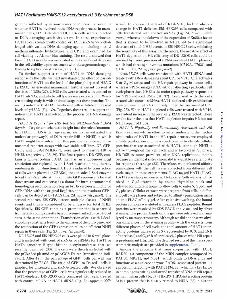
genome inflicted by various stress conditions. To examinewhether HAT1 is involved in the DNA repair process in mam-malian cells, HAT1-depleted HCT116 cells were subjectedto DNA-damaging sensitivity assays. In these experiments,HCT116 cells treated with control or HAT1 siRNAs were chal-lenged with various DNA-damaging agents including methylmethanesulfonate, hydroxyurea, and CPT and examined forcell viability by Alamar blue staining. The results showed thatloss of HAT1 in cells was associated with a significant decreasein the cell viability upon treatment with these genotoxic agentsleading to replication stress (26) (Fig. 1C).To further support a role of HAT1 in DNA-damaging
response by the cells, we next investigated the effect of loss-of-function of HAT1 on the level of the phosphorylated H2A.X(�H2AX), an essential mammalian histone variant present atthe sites of DSBs (27). U2OS cells were treated with control orHAT1 siRNAs, and whole cell lysates were extracted for West-ern blotting analysis with antibodies against these proteins. Theresults indicated that HAT1-deficient cells exhibited increasedlevels of �H2AX (Fig. 1D). Together, these results support thenotion that HAT1 is involved in the process of DNA damagerepair.HAT1 Is Required for HR- but Not NHEJ-mediated DNA
Repair—To gain amechanistic insight into the role ofmamma-lian HAT1 in DNA damage repair, we first investigated themolecular pathway(s) of DNA repair (HR or NHEJ) in whichHAT1 might be involved. For this purpose, GFP-based chro-mosomal reporter assays with two stable cell lines, DR-GFP-U2OS and EJ5-GFP-HEK293, were used to measure HR orNHEJ, respectively (28, 29). The first reporter, DR-GFP, con-tains a GFP-encoding cDNA that has an endogenous BcgIrestriction site replaced by an I-SceI restriction site, therebyrendering its non-functional. A DSB is induced by transfectionof cells with a plasmid (pCBASce) that encodes I-SceI enzymeto cut the I-SceI site. An incomplete GFP sequence is locateddownstream and can serve as a donor for intra-chromosomalhomologous recombination. Repair byHR restores a functionalGFP cDNA with the original BcgI site, and the resultant GFP�
cells can be detected by FACS (Fig. 2A, upper left panel). Thesecond reporter, EJ5-GFP, detects multiple classes of NHEJevents and thus is considered to be an assay for total NHEJ.Specifically, EJ5-GFP contains a promoter that is separatedfromaGFP coding cassette by a puro gene flanked by two I-SceIsites in the same orientation. Transfection of cells with I-SceI-encoding constructs leads to the excision of the puro gene, andthe restoration of the GFP expression relies on efficient NHEJrepair in these cells (Fig. 2A, lower left panel).
DR-U2OS and EJ5-HEK293 cells were plated in 6-well platesand transfected with control siRNAs or siRNAs for HAT1 orHAT4 (another B-type histone acetyltransferase that werecently identified) (30). The cells were then transfected withthe pCBASce plasmid or pCAGGS-Ds-red (transfection indi-cator). After 48 h, the percentage of GFP� cells per well wasdetermined by FACS. The ratio of GFP� to Ds-red� cells isgraphed for untreated and siRNA-treated cells. We observedthat the percentage of GFP� cells was significantly reduced inHAT1-depleted DR-U2OS cells compared with cells treatedwith control siRNA or HAT4 siRNA (Fig. 2A, upper middle
panel). In contrast, the level of total-NHEJ had no obviouschange in HAT1-deficient EJ5-HEK293 cells compared withcells transfected with control siRNAs (Fig. 2A, lower middlepanel), whereas knockdown of the expression of Ku80, a factorthat is known to be involved in NHEJ, led to a significantdecrease of total-NHEJ events in EJ5-HEK293 cells, validatingthe sensitivity of this assay. Furthermore, the negative effect ofHAT1 depletion on HR efficiency of DR-U2OS cells could berescued by overexpression of siRNA-resistant HAT1 plasmid,which had three synonymous mutations (C510A, T762C, andC1161T) (Fig. 2A, upper right panel).Next, U2OS cells were transfected with HAT1 siRNAs and
treated with DNAdamaging agent CPT or VP16. CPT activatesS or G2-M arrest and the HR repair pathway in tumor cells,whereas VP16 damages DNAwithout affecting a particular cellcycle phase; thus, NHEJ is themajor repair pathway responsiblefor VP16 induced DSBs (31, 32). Compared with the cellstreated with control siRNAs, HAT1-depleted cells exhibited anelevated level of �H2AX but only under the treatment of CPT(Fig. 2B). When HAT1-depleted cells were treated with VP16,no evident increase in the level of �H2AX was detected. Theseresults favor the idea that HAT1 depletion impairs HR but notNHEJ repair of DSBs.HAT1 Is Physically and Functionally Associated with HR
Repair Proteins—In an effort to better understand the mecha-nistic roles of HAT1 in the HR repair process, we employedaffinity purification and mass spectrometry to identify cellularproteins that are associated with HAT1. Although NHEJ isactive throughout the cell cycle and is favored in G1 phase,HR-DSB is more prevalent after DNA replication, mainlybecause an identical sister chromatid is available as a templatefor repair at this stage (33). Therefore, we performed affinitypurification with the cell lysates collected from different cellcycle stages. In these experiments, FLAG-tagged HAT1 (FLAG-HAT1) was stably expressed in HeLa cells. Cells were synchro-nized in G1/S transition by double thymidine block andreleased for different hours to allow cells to enter S, G2/M, andG1 phases. Cellular extracts were prepared from cells in differ-ent cell cycle phases and subjected to affinity purification usingan anti-FLAG affinity gel. After extensive washing, the boundprotein complex was eluted with excess FLAG peptides. Boundproteins were resolved by SDS-PAGE and visualized by silverstaining. The protein bands on the gel were retrieved and ana-lyzed bymass spectrometry. Althoughwe did not observe obvi-ous differences in the staining profiles with the extracts fromdifferent phases of cell cycle, the total amount of HAT1 inter-acting proteins increased in S (represented by 0, 3, and 16 hafter release) andG2 (6 h after release), 2 phaseswhenHR repairis predominant (Fig. 3A). The detailed results of the mass spec-trometric analysis are provided in supplemental File 1.Among the proteins that were co-purified with HAT1,
RAD50 is a component of the MRN complex (composed byRAD50, MRE11, and NBS1), which binds to DNA ends andfunctions as a nuclease enzyme. RAD51-associated protein 1 isa protein interacting with RAD51 (34, 35), which is a key factorin homologous pairing and strand transfer of DNA inHR repairinmammalian cells (36, 37). HIRIP3 (HIRA-interacting protein3) is a protein that is closely related to HIRA (38), a histone
HAT1 Facilitates H4K5/K12-acetylated H3.3 Enrichment at DSB
18274 JOURNAL OF BIOLOGICAL CHEMISTRY VOLUME 288 • NUMBER 25 • JUNE 21, 2013
by guest on June 7, 2018http://w
ww
.jbc.org/D
ownloaded from

chaperone that preferentially places the variant histone H3.3 innucleosomes (39). As expected, various histone species werealso detected in HAT1 co-eluted complexes. Based on the rolesof these proteins in DNA repair, their association with HAT1further supports a role for HAT1 in HR of DNA repair.We confirmed the interaction of HAT1 with RAD50,
RAD51, HIRA, HIRIP3, and histones by co-immunoprecipita-tion assays with specific antibodies against these proteins,whereas no interaction was detected between HAT1 and otherrepair proteins such as Ku80, which is responsible for NHEJ
repair. Notably, HAT1 interactedwith these proteinsmore effi-ciently in S phase of the cell cycle (Fig. 3B), suggesting thatinteraction of HAT1/Rbap46 with these proteins is cell cycle-regulated, a notion that is consistent with their roles in HRrepair and chromatin assembly.Based on previous reports that RAD50 and RAD51 exhibit
ATPase activity (40, 41), we next examinedwhether theHAT1-purified complex was indeed associated with an ATPase activ-ity. To this end, BioAssay Systems QuantiChromTM ATPase/GTPase assay kit was used in amicroplate format.We set up the
FIGURE 2. HAT1 is required for HR but not for NHEJ. A, left panel, shown is a schematic illustration of I-SceI-mediated DSB induction and repair usingthe DR-GFP and EJ5-GFP transgenes. Right panels, the efficiency of HR in DR-GFP transgenic U2OS cells and of NHEJ in EJ5-GFP transgenic 293 cells wereanalyzed after HAT1 knockdown. DR-U2OS cells were treated with 50 �M Nonsilencer, HAT1 siRNA, or HAT4 siRNA 6 h before transfection with pCBASceor pCAGGS-Ds-red (transfection indicator), whereas EJ5-HEK293 cells were treated with comparable amounts of Nonsilencer, HAT1 siRNA, HAT4 siRNA,or Ku80 siRNA. HR or NHEJ repair efficiency was measured 48 h later by measuring the fraction of GFP� cells. Ds-red was used to determine transfectionefficiency. The ratio of GFP� to Ds-red� cells is graphed for untreated and siRNA-treated cells. Each bar represents the mean � S.D. for three independ-ent experiments. Asterisks denote the significance of the difference in the repair efficiency between HAT1- or Ku80-depleted cells and control (orHAT4-depleted) cells (p � 0.01). RNAi-resistant FLAG-HAT1 construct could rescue the HR-deficient effect of HAT1 knockdown. siRNA-treated DR-U2OScells were transfected with I-SceI plus empty vector, I-SceI plus RNAi resistant pcDNA3.1(�)-FLAG-HAT1, or pCAGGS-Ds-red. 48 h later the ratio of GFP�
to Ds-red� cells was graphed (upper right panel). Each bar represents the mean � S.D. for three independent experiments. Western analysis ofFLAG-HAT1 resisting HAT1 RNAi is shown (lower right panel). B, accumulation of �H2AX in the HAT1-depleted cells after CTP stimulation is shown. U2OScells were transfected with control and HAT1 siRNA and then treated with 100 nM CPT or 40 �M VP16 for 8 h before harvesting. Cell lysates were analyzedby immunoblotting. siNS, nonsilencer siRNA.
HAT1 Facilitates H4K5/K12-acetylated H3.3 Enrichment at DSB
JUNE 21, 2013 • VOLUME 288 • NUMBER 25 JOURNAL OF BIOLOGICAL CHEMISTRY 18275
by guest on June 7, 2018http://w
ww
.jbc.org/D
ownloaded from
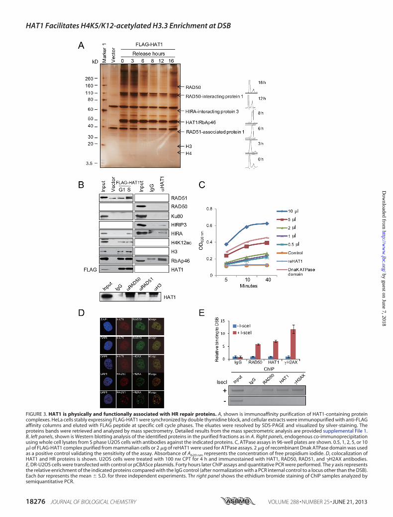
FIGURE 3. HAT1 is physically and functionally associated with HR repair proteins. A, shown is immunoaffinity purification of HAT1-containing proteincomplexes. HeLa cells stably expressing FLAG-HAT1 were synchronized by double thymidine block, and cellular extracts were immunopurified with anti-FLAGaffinity columns and eluted with FLAG peptide at specific cell cycle phases. The eluates were resolved by SDS-PAGE and visualized by silver-staining. Theproteins bands were retrieved and analyzed by mass spectrometry. Detailed results from the mass spectrometric analysis are provided supplemental File 1.B, left panels, shown is Western blotting analysis of the identified proteins in the purified fractions as in A. Right panels, endogenous co-immunoprecipitationusing whole cell lysates from S phase U2OS cells with antibodies against the indicated proteins. C, ATPase assays in 96-well plates are shown. 0.5, 1, 2, 5, or 10�l of FLAG-HAT1 complex purified from mammalian cells or 2 �g of reHAT1 were used for ATPase assays. 2 �g of recombinant Dnak ATPase domain was usedas a positive control validating the sensitivity of the assay. Absorbance of A620 nm represents the concentration of free propidium iodide. D, colocalization ofHAT1 and HR proteins is shown. U2OS cells were treated with 100 nM CPT for 4 h and immunostained with HAT1, RAD50, RAD51, and �H2AX antibodies.E, DR-U2OS cells were transfected with control or pCBASce plasmids. Forty hours later ChIP assays and quantitative PCR were performed. The y axis representsthe relative enrichment of the indicated proteins compared with the IgG control (after normalization with a PCR internal control to a locus other than the DSB).Each bar represents the mean � S.D. for three independent experiments. Thr right panel shows the ethidium bromide staining of ChIP samples analyzed bysemiquantitative PCR.
HAT1 Facilitates H4K5/K12-acetylated H3.3 Enrichment at DSB
18276 JOURNAL OF BIOLOGICAL CHEMISTRY VOLUME 288 • NUMBER 25 • JUNE 21, 2013
by guest on June 7, 2018http://w
ww
.jbc.org/D
ownloaded from
experiments with a series dilution of FLAG-HAT1 complexpurified fromHeLa cells or in vitro purified reHAT1 as well as anegative control with no enzyme and a positive control withrecombinant DnakATPase domain in separate wells. The reac-tions were incubated for 30 min in room temperature beforethe addition of themalachite green reagent, which forms a darkgreen color with liberated phosphate after desired incubationtime. The color change was measured at 620 nm on a platereader. As expected, we observed that purified FLAG-HAT1complex, but not reHAT1, possessed a strong and dose-depen-dent ATPase activity (Fig. 3C), supporting our observation thatRAD50 and RAD51 are associated with HAT1 in vivo.The interaction of HAT1 with HR proteins was further
demonstrated by immunofluorescent analysis. The colocaliza-tion of HAT1 and RAD50/RAD51 were shown at the damagedDNA, which was represented by the accumulation of �H2AX(Fig. 3D).Because it is well established that RAD50 and RAD51 are
recruited to DSB lesions (37, 42), the interaction of HAT1 withRAD50 and RAD51 could mean that HAT1 is also present atthe sites of DSBs. To verify this, chromatin immunoprecipita-tion (ChIP) assays were carried out inDR-U2OS cells with anti-bodies against human HAT1. PCR primers that amplify aregion surrounding the I-SceI restriction site were used, andsamples were taken at 14 h after I-SceI cutting. The resultsindicated that HAT1 was clearly recruited to the site of DNAdamage (Fig. 3E).HAT1 Complex Possesses an ATP-independent and Histone
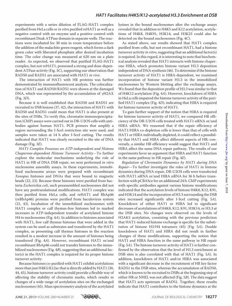
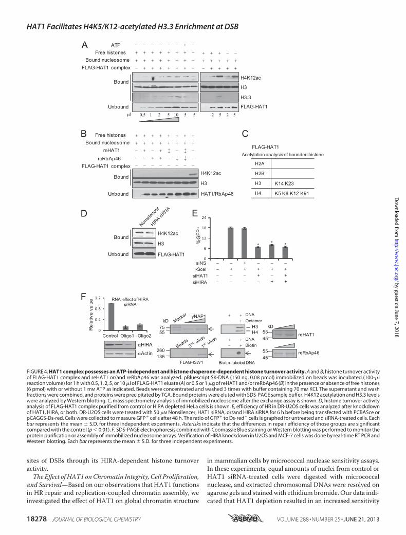
Chaperone-dependent Histone Turnover Activity—To furtherexplore the molecular mechanisms underlying the role ofHAT1 in HR of DNA DSB repair, we next performed in vitronucleosome assembly assays. In these experiments, immobi-lized nucleosome arrays were prepared with recombinantXenopus histones and DNAs that were bound to magneticbeads (22, 23). Because these histones were purified from bac-teria Escherichia coli, such preassembled nucleosomes did nothave any posttranslational modifications. HAT1 complex wasthen purified from HeLa cells, and reHAT1 and RbAp46(reRbAp46) proteins were purified from baculovirus system(21, 43). Incubation of the immobilized nucleosomes withHAT1 complex or calf thymus-free histones led to markedincreases in ATP-independent transfer of acetylated histoneH4 to nucleosomes (Fig. 4A). In addition to histones associatedwith HAT1, free calf thymus histones that were added in thesystem can be used as substrates and transferred by the HAT1complex, as presenting calf thymus histones in the reactionresulted in a modest increase in the amount of histones beingtransferred (Fig. 4A). However, recombinant HAT1 or/andrecombinant RbAp46 could not transfer histones to the immo-bilized nucleosomes (Fig. 4B), suggesting that an additional fac-tor(s) in the HAT1 complex is required for its proper histoneturnover activity.Because histones co-purified withHAT1 exhibit acetylations
more than justH4K5/K12ac that is directly added byHAT1 (30,44, 45), histone turnover activity could provide a flexible way ofaffecting the stability of chromatin marks, which results inchanges of a wide range of acetylation sites on the exchangednucleosomes (45).Mass spectrometry analysis of the acetylated
lysines in the bound nucleosomes after the exchange assaysrevealed that in addition to H4K5 and K12 acetylation, acetyla-tions of H4K8, H4K91, H3K14, and H3K23 could also bedetected on the bound nucleosomes (Fig. 4C).As stated above, our results showed that HAT1 complex
purified from cells, but not recombinant HAT1, had a histoneturnover activity in vitro, suggesting that an additional factor(s)is required. In this regard, it is interesting to note that biochem-ical analysis revealed that HAT1 interacts with histone chaper-one HIRA, which promotes histone variant H3.3 depositionindependent ofDNAsynthesis (46). To determine if the histoneturnover activity of HAT1 is HIRA-dependent, we examinedincorporation of histone variant H3.3 in the immobilizednucleosomes by Western blotting after the exchange assays.We found that the deposition profile of H3.3 was similar to thatof H4K12 acetylation (Fig. 4A). However, knockdown of HIRAinHeLa cells impaired the histone turnover activity of the puri-fied HAT1 complex (Fig. 4D), indicating that HIRA is requiredfor histone turnover activity of HAT1.To gain further support of the notion that HIRA is required
for histone turnover activity of HAT1, we compared HR effi-ciency of the DR-U2OS cells treated with HAT1 siRNA or/andHIRA siRNA. We reasoned that if the repair efficiency ofHAT1/HIRA co-depletion cells is lower than that of cells withHAT1 orHIRA individually depleted, it could reflect a possibil-ity that HAT1 and HIRA affect different aspects of HR; con-versely, a similar HR efficiency would suggest that HAT1 andHIRA affect the same DNA repair pathway. The results of ourexperiments favor an argument that HIRA and HAT1 functionin the same pathway in HR repair (Fig. 4E).Regulation of Chromatin Dynamics by HAT1 during DNA
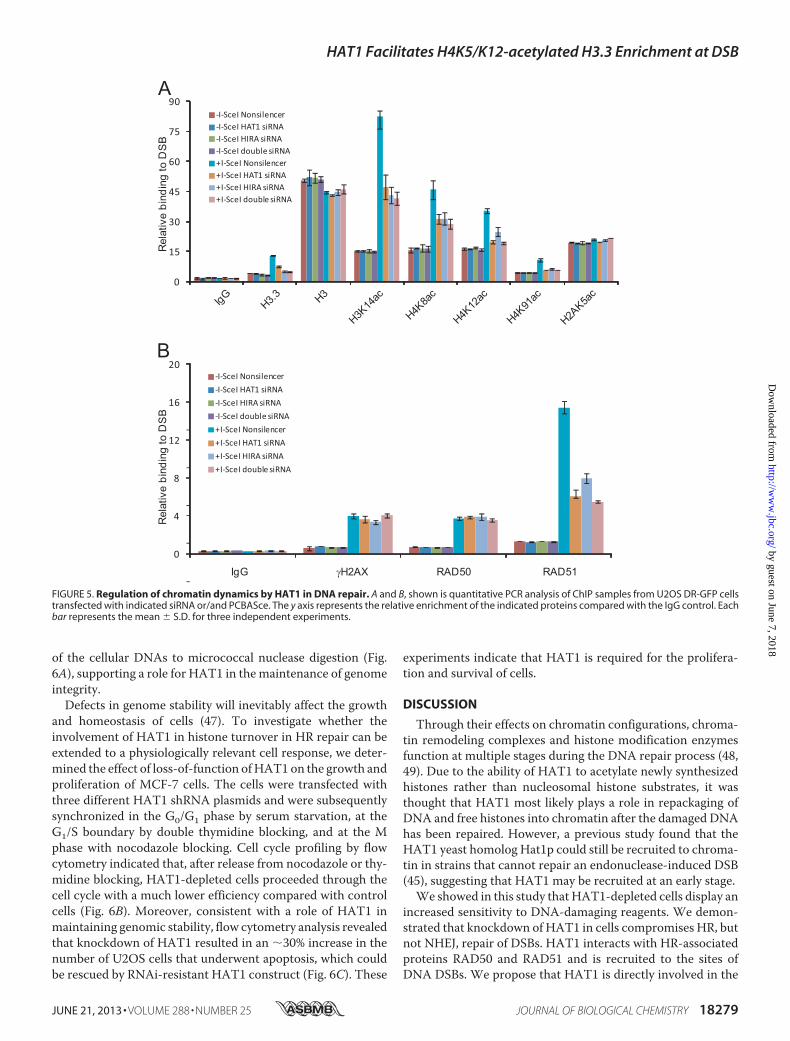
Repair—To further investigate the roles of HAT1 in histonedynamics during DNA repair, DR-U2OS cells were transfectedwith HAT1 siRNA or/and HIRA siRNA for 36 h before trans-fection with pCBASce for an additional 24 h. ChIP experimentswith specific antibodies against various histone modificationsindicated that the acetylation levels of histone H4K8, K12, K91,andH3K14 and the incorporation ofH3.3 surrounding theDSBsites increased significantly after I-SceI cutting (Fig. 5A).Knockdown of either HAT1 or HIRA led to significantdecreases of acetylations of H4K8, K12, K91, H3K14, or H3.3 atthe DSB sites. No changes were observed on the levels ofH2AK5 acetylation, consisting with the previous predictionthat HAT1-induced histone exchange is specific for the substi-tution of histone H3/H4 tetramers (45) (Fig. 5A). Doubleknockdown of HAT1 and HIRA did not result in furtherchanges of these modifications, supporting the notion thatHAT1 and HIRA function in the same pathway in HR repair(Fig. 5A). The histone turnover activity of HAT1 is further con-firmed by the observation that the level of H3.3 enrichment atDSB sites is also correlated with that of HAT1 (Fig. 5A). Inaddition, knockdown of HAT1 and/or HIRA was associatedwith a significant decrease in the recruitment of HR key factorRAD51 to the DSB sites, whereas the accumulation of RAD50,which is known to be recruited to DSBs at the beginning step ofeither HR or NHEJ, was not affected (Fig. 5B). The data suggestthat HAT1 acts upstream of RAD51. Together, these resultsindicate that HAT1 contributes to the histone dynamics at the
HAT1 Facilitates H4K5/K12-acetylated H3.3 Enrichment at DSB
JUNE 21, 2013 • VOLUME 288 • NUMBER 25 JOURNAL OF BIOLOGICAL CHEMISTRY 18277
by guest on June 7, 2018http://w
ww
.jbc.org/D
ownloaded from
sites of DSBs through its HIRA-dependent histone turnoveractivity.The Effect of HAT1 onChromatin Integrity, Cell Proliferation,
and Survival—Based on our observations that HAT1 functionsin HR repair and replication-coupled chromatin assembly, weinvestigated the effect of HAT1 on global chromatin structure
in mammalian cells by micrococcal nuclease sensitivity assays.In these experiments, equal amounts of nuclei from control orHAT1 siRNA-treated cells were digested with micrococcalnuclease, and extracted chromosomal DNAs were resolved onagarose gels and stained with ethidium bromide. Our data indi-cated that HAT1 depletion resulted in an increased sensitivity
0
6
12
18
24
A − − − − − − + −
+ + + + + + + −
+ + + + + + + +
− + + + + + + +
FLAG - ISW1
kD 75 55
260 135
yNAP1 + + − +
+ + − +
H3 H4
DNA Octamer
DNA Biotin
Biotin - labeled DNA
+ + + − −
+ + + + +
− + + + +
reHAT1
reRbAp46
55 45
55 45
kD
+ + + + + + + +
+ + + + + + + +
− + − + + + − +
+ −
− − + + − + +
+ + −
− − − − − − − +
D
H2A
H2B
H3 K14 K23
H4 K5 K8 K12 K91
Acetylation analysis of bounded histone
− − + − − − − + + + + + − − − + − + − − − − + +
E
siNS
siHAT1 siHIRA
I - SceI
%G
FP +
0
0.4
0.8
1.2
Rel
ativ
e va
lue RNAi effect of HIRA
siRNA
Control Oligo1 Oligo2
α HIRA
α Actin
B C
FLAG - HAT1 complex
Free histones Bound nucleosome
reHAT1
reRbAp46
HAT1/RbAp46
H3
H4K12ac Bound
Unbound
F
FLAG - HAT1
H4K12ac
H3 Bound
Unbound
FLAG - HAT1 complex
FLAG - HAT1
Bound
Unbound
H3
H4K12ac
ATP Free histones
Bound nucleosome
µ l 1 2 10 5 5 0.5 5 2 5 2 5
H3.3
FLAG - HAT1
* * *
FIGURE 4. HAT1 complex possesses an ATP-independent and histone chaperone-dependent histone turnover activity. A and B, histone turnover activityof FLAG-HAT1 complex and reHAT1 or/and reRbAp46 was analyzed. pBluescript SK-DNA (150 ng; 0.08 pmol) immobilized on beads was incubated (100-�lreaction volume) for 1 h with 0.5, 1, 2, 5, or 10 �l of FLAG-HAT1 eluate (A) or 0.5 or 1 �g of reHAT1 and/or reRbAp46 (B) in the presence or absence of free histones(6 pmol) with or without 1 mM ATP as indicated. Beads were concentrated and washed 3 times with buffer containing 70 mM KCl. The supernatant and washfractions were combined, and proteins were precipitated by TCA. Bound proteins were eluted with SDS-PAGE sample buffer. H4K12 acetylation and H3.3 levelswere analyzed by Western blotting. C, mass spectrometry analysis of immobilized nucleosome after the exchange assay is shown. D, histone turnover activityanalysis of FLAG-HAT1 complex purified from control or HIRA depleted HeLa cells is shown. E, efficiency of HR in DR-U2OS cells was analyzed after knockdownof HAT1, HIRA, or both. DR-U2OS cells were treated with 50 �M Nonsilencer, HAT1 siRNA, or/and HIRA siRNA for 6 h before being transfected with PCBASce orpCAGGS-Ds-red. Cells were collected to measure GFP� cells after 48 h. The ratio of GFP� to Ds-red� cells is graphed for untreated and siRNA-treated cells. Eachbar represents the mean � S.D. for three independent experiments. Asterisks indicate that the differences in repair efficiency of those groups are significantcompared with the control (p � 0.01). F, SDS-PAGE electrophoresis combined with Coomassie Blue staining or Western blotting was performed to monitor theprotein purification or assembly of immobilized nucleosome arrays. Verification of HIRA knockdown in U2OS and MCF-7 cells was done by real-time RT PCR andWestern blotting. Each bar represents the mean � S.D. for three independent experiments.
HAT1 Facilitates H4K5/K12-acetylated H3.3 Enrichment at DSB
18278 JOURNAL OF BIOLOGICAL CHEMISTRY VOLUME 288 • NUMBER 25 • JUNE 21, 2013
by guest on June 7, 2018http://w
ww
.jbc.org/D
ownloaded from
of the cellular DNAs to micrococcal nuclease digestion (Fig.6A), supporting a role for HAT1 in themaintenance of genomeintegrity.Defects in genome stability will inevitably affect the growth
and homeostasis of cells (47). To investigate whether theinvolvement of HAT1 in histone turnover in HR repair can beextended to a physiologically relevant cell response, we deter-mined the effect of loss-of-function ofHAT1on the growth andproliferation of MCF-7 cells. The cells were transfected withthree different HAT1 shRNA plasmids and were subsequentlysynchronized in the G0/G1 phase by serum starvation, at theG1/S boundary by double thymidine blocking, and at the Mphase with nocodazole blocking. Cell cycle profiling by flowcytometry indicated that, after release from nocodazole or thy-midine blocking, HAT1-depleted cells proceeded through thecell cycle with a much lower efficiency compared with controlcells (Fig. 6B). Moreover, consistent with a role of HAT1 inmaintaining genomic stability, flow cytometry analysis revealedthat knockdown of HAT1 resulted in an �30% increase in thenumber of U2OS cells that underwent apoptosis, which couldbe rescued by RNAi-resistant HAT1 construct (Fig. 6C). These
experiments indicate that HAT1 is required for the prolifera-tion and survival of cells.
DISCUSSION
Through their effects on chromatin configurations, chroma-tin remodeling complexes and histone modification enzymesfunction at multiple stages during the DNA repair process (48,49). Due to the ability of HAT1 to acetylate newly synthesizedhistones rather than nucleosomal histone substrates, it wasthought that HAT1 most likely plays a role in repackaging ofDNA and free histones into chromatin after the damaged DNAhas been repaired. However, a previous study found that theHAT1 yeast homolog Hat1p could still be recruited to chroma-tin in strains that cannot repair an endonuclease-induced DSB(45), suggesting that HAT1 may be recruited at an early stage.We showed in this study that HAT1-depleted cells display an
increased sensitivity to DNA-damaging reagents. We demon-strated that knockdown of HAT1 in cells compromises HR, butnot NHEJ, repair of DSBs. HAT1 interacts with HR-associatedproteins RAD50 and RAD51 and is recruited to the sites ofDNA DSBs. We propose that HAT1 is directly involved in the
A
B
IgG RAD50γH2AX RAD51
Rel
ativ
e bi
ndin
g to
DS
B
0
15
30
45
60
75
90-I-SceI Nonsilencer-I-SceI HAT1 siRNA-I-SceI HIRA siRNA-I-SceI double siRNA+I-SceI Nonsilencer+I-SceI HAT1 siRNA+I-SceI HIRA siRNA+I-SceI double siRNA
Rel
ativ
e bi
ndin
g to
DS
B
0
4
8
12
16
20-I-SceI Nonsilencer
-I-SceI HAT1 siRNA
-I-SceI HIRA siRNA
-I-SceI double siRNA
+I-SceI Nonsilencer
+I-SceI HAT1 siRNA
+I-SceI HIRA siRNA
+I-SceI double siRNA
FIGURE 5. Regulation of chromatin dynamics by HAT1 in DNA repair. A and B, shown is quantitative PCR analysis of ChIP samples from U2OS DR-GFP cellstransfected with indicated siRNA or/and PCBASce. The y axis represents the relative enrichment of the indicated proteins compared with the IgG control. Eachbar represents the mean � S.D. for three independent experiments.
HAT1 Facilitates H4K5/K12-acetylated H3.3 Enrichment at DSB
JUNE 21, 2013 • VOLUME 288 • NUMBER 25 JOURNAL OF BIOLOGICAL CHEMISTRY 18279
by guest on June 7, 2018http://w
ww
.jbc.org/D
ownloaded from

histone turnover during HR repair, analogous to the histoneexchange activity seen in the replication-independent histonevariant assembly in transcriptionally active chromatin (6,50–54).
Coupling of histone acetylation with histone exchange at thesites of DNA damage has been observed with the DrosophilaTip60 complex (11), indicating that histone exchange factorsplay important roles during DNA repair. In addition, increaseof the levels of H4K12 acetylation, which is mediated by HAT1,is thought to be an early event at DSB sites (55–57). It is alsointeresting to note that HAT1 was found in both replication-dependent histone H3.1 and replication-independent histoneH3.3 nucleosome assembly complexes (46). In our system,although knockdown of either HAT1 or HIRA leads to changesof acetylation at multiple sites of H3 and H4, H2AK5 acetyla-tion and H2A phosphorylation levels remained unaltered.HAT1-induced histone exchange is likely to be specific for thesubstitution of histone H3/H4 tetramers, as also suggested by aprevious report (45).We demonstrated that HAT1 complex exhibited an ATP-
independent histone turnover activity in vitro. Because histoneturnover activity was not detected with recombinant HAT1or/and recombinant RbAp46, it is reasonable to believe that anadditional factor(s) in theHAT1 complex is needed. Among theproteins that are associated with HAT1, we propose that HIRAis the chaperone that facilitates HAT1 to fulfill its histone turn-over activity at DSB sites. HIRA is a histone chaperone impli-cated in H3.3-specific deposition and DNA synthesis-indepen-dent nucleosome assembly (39, 58–60). Moreover, HIRA hasbeen shown to localize to lasermicro-irradiation-inducedDNAlesions (61). These observations are consistent with the role ofHIRA in DNA repair and support our argument. Interestingly,knockdown of both HAT1 and HIRA in DR-U2OS cells hadsimilar defects in HR repair compared with individual deple-tion of either one of the factors, supporting that HAT1 andHIRA function in the same pathway of HR repair.In a previous report, HAT1 was shown to be recruited to
DNA lesions in HR deficient RAD52� yeast strains, indicatingthe recruitment ofHAT1 is upstream to that ofHR key factor atDSB sites (45).We demonstrated that the recruitment ofHAT1is required for the subsequent deposition of histone variantH3.3 and altered histone modifications. The change of the his-tone dynamics at DSBs may function to define a chromatinstate for the subsequent recruitment of mammalian HR keyfactor RAD51 to the sites for successful HR repair.HAT1 is highly conserved from yeast to human. Depletion of
HAT1 in yeast and chicken cells does not significantly changethe overall cell growth or viability (62, 63). In S. cerevisiae,HAT1 is only required for HR repair in strains with H3K14Rand K23Rmutation, suggesting that HAT1may be functionallyredundant in these species (21).Our results showed that knock-down of HAT1 causes significant HR repair defects, cell cycledelay, and growth retardation even in the absence ofDNAdam-aging agents, indicating that mammalian cells are more sensi-tive to HAT1 deficiency due to the absence of a compensatoryactivity.HR repair of DSBs in mammalian cells is a complex, multi-
step process. Although considerable progress has beenmade inunderstanding the role of histone dynamics in HR repair,important questions remain to be answered. For instance, it isnot clear how chromatin disassembly/assembly and sequentialrecruitment of effectors toDSBs are affected by histone dynam-
Double thymidine block
Con
Sh-1
Sh-2
Sh-3
Per
cent
age
of c
ells
Serum starvationC
on Sh-
1
Sh-
2
Sh-
3
Con
Con
HAT1
Tubulin
Con
Sh-1
Sh-2
Sh-3
Per
cent
age
of c
ells
Con
Sh-1
Sh-2
Sh-3
Per
cent
age
of c
ells
Nocodazole block
A
B%
of a
popt
otic
cel
ls
siHAT1siNS
0
10
20
30
40
VectorFLAG-HAT1
+ − + −− + − +− − + −− − − +
750500
250100
MNase 0.5 u 2.5 u 5 u
C
0
20
40
60
80
100G1 phase S phase G2 phase
0
20
40
60
80
100G1 phase S phase G2 phase
0
20
40
60
80
100G1 phase S phase G2 phase
siNS siHAT1
siNS+Vector siHAT1+FLAG-HAT1
Mono-
Tri -Di-
FIGURE 6. The effect of HAT1 on chromatin integrity, cell proliferation,and survival. A, U2OS cells were treated with control or HAT1 siRNA. Nucleiwere prepared and treated with 2.5 or 5 Worthington units of micrococcalnuclease (MNase)/ml as indicated. Genomic DNA was purified and fraction-ated by agarose gel electrophoresis. Mononucleosome, dinucleosome, andtrinucleosome are indicated. u, units. B, MCF-7 cells were transfected withpGCsi-Nonsilencer, pGCsi-HAT1 shRNA1#, pGCSi-HAT1 shRNA2#, and pGCSi-HAT1 shRNA3# and then synchronized by serum starvation, double thymi-dine block, or thymidine nocodazole block at G0/G1, G1/S, or the G2/M bound-ary, respectively. Cell cycle progression after releasing was analyzed by flowcytometry. RNAi efficiency of HAT1 by these three HAT1 shRNA plasmids wasmonitored by Western blotting. C, HAT1 knockdown promotes cell apoptosis.siRNA-treated or siRNA and vector/FLAG-HAT1(RNAi-resistant) co-trans-fected U2OS cells were double-stained with annexin V and propidium iodide.Cell apoptosis was determined by flow cytometry. Each bar represents themean � S.D. for three independent experiments. siNS, nonsilencer siRNA.
HAT1 Facilitates H4K5/K12-acetylated H3.3 Enrichment at DSB
18280 JOURNAL OF BIOLOGICAL CHEMISTRY VOLUME 288 • NUMBER 25 • JUNE 21, 2013
by guest on June 7, 2018http://w
ww
.jbc.org/D
ownloaded from

ics; that is, whether and how histone dynamics-flanking DSBsare regulated during cell cycle. We found that HAT1 promotesacetylation of histone H4 as well as incorporation of histonevariant H3.3 at DSBs with its HIRA-dependent histone turn-over activity. It is likely that HAT1-induced histone dynamicsdistinguishes damaged chromatin from the surrounding nor-mal area, facilitating subsequent recruitment of key HR proteinRAD51, for the ultimate HR repair. Although our present studycannot exclude the possibility that HAT1 participates in multi-ple events of HR repair of DSBs, it is clear that HAT1 plays animportant role in the maintenance of genomic stability inhigher eukaryotic cells.
REFERENCES1. Tsukuda, T., Fleming, A. B., Nickoloff, J. A., and Osley, M. A. (2005)
Chromatin remodelling at a DNA double-strand break site in Saccharo-myces cerevisiae. Nature 438, 379–383
2. Downs, J. A., Lowndes, N. F., and Jackson, S. P. (2000) A role for Saccha-romyces cerevisiae histone H2A in DNA repair. Nature 408, 1001–1004
3. Pei, H., Zhang, L., Luo, K., Qin, Y., Chesi,M., Fei, F., Bergsagel, P. L.,Wang,L., You, Z., and Lou, Z. (2011) MMSET regulates histone H4K20 methyl-ation and 53BP1 accumulation at DNA damage sites. Nature 470,124–128
4. Wang, H., Zhai, L., Xu, J., Joo, H. Y., Jackson, S., Erdjument-Bromage, H.,Tempst, P., Xiong, Y., and Zhang, Y. (2006) Histone H3 and H4 ubiquity-lation by the CUL4-DDB-ROC1 ubiquitin ligase facilitates cellular re-sponse to DNA damage.Mol. Cell 22, 383–394
5. Althaus, F. R. (1992) Poly ADP-ribosylation. A histone shuttle mechanismin DNA excision repair. J. Cell Sci. 102, 663–670
6. Verzijlbergen, K. F., van Welsem, T., Sie, D., Lenstra, T. L., Turner, D. J.,Holstege, F. C., Kerkhoven, R. M., and van Leeuwen, F. (2011) A barcodescreen for epigenetic regulators reveals a role for the NuB4/HAT-B his-tone acetyltransferase complex in histone turnover. PLoS Genet. 7,e1002284
7. Chen, D., Dundr, M., Wang, C., Leung, A., Lamond, A., Misteli, T., andHuang, S. (2005) Condensed mitotic chromatin is accessible to transcrip-tion factors and chromatin structural proteins. J. Cell Biol. 168, 41–54
8. Kimura, H., and Cook, P. R. (2001) Kinetics of core histones in livinghuman cells. Little exchange of H3 and H4 and some rapid exchange ofH2B. J. Cell Biol. 153, 1341–1353
9. Mizuguchi, G., Shen, X., Landry, J., Wu,W. H., Sen, S., andWu, C. (2004)ATP-driven exchange of histone H2AZ variant catalyzed by SWR1 chro-matin remodeling complex. Science 303, 343–348
10. Luk, E., Ranjan, A., Fitzgerald, P. C., Mizuguchi, G., Huang, Y., Wei, D.,and Wu, C. (2010) Stepwise histone replacement by SWR1 requires dualactivation with histone H2A.Z and canonical nucleosome. Cell 143,725–736
11. Kusch, T., Florens, L., Macdonald, W. H., Swanson, S. K., Glaser, R. L.,Yates, J. R., 3rd, Abmayr, S. M., Washburn, M. P., and Workman, J. L.(2004) Acetylation by Tip60 is required for selective histone variant ex-change at DNA lesions. Science 306, 2084–2087
12. Parthun, M. R., Widom, J., and Gottschling, D. E. (1996) The major cyto-plasmic histone acetyltransferase in yeast. Links to chromatin replicationand histone metabolism. Cell 87, 85–94
13. Ai, X., and Parthun, M. R. (2004) The nuclear Hat1p/Hat2p complex. Amolecular link between type B histone acetyltransferases and chromatinassembly.Mol. Cell 14, 195–205
14. Ge, Z., Wang, H., and Parthun, M. R. (2011) Nuclear Hat1p complex(NuB4) components participate in DNA repair-linked chromatin reas-sembly. J. Biol. Chem. 286, 16790–16799
15. Verreault, A., Kaufman, P. D., Kobayashi, R., and Stillman, B. (1998)Nucleosomal DNA regulates the core-histone-binding subunit of the hu-man Hat1 acetyltransferase. Curr. Biol. 8, 96–108
16. Dutnall, R. N., Tafrov, S. T., Sternglanz, R., and Ramakrishnan, V. (1998)Structure of the histone acetyltransferase Hat1. A paradigm for theGCN5-related N-acetyltransferase superfamily. Cell 94, 427–438
17. Nociari, M. M., Shalev, A., Benias, P., and Russo, C. (1998) A novel one-step, highly sensitive fluorometric assay to evaluate cell-mediated cytotox-icity. J. Immunol. Methods 213, 157–167
18. Zhang, H., Yi, X., Sun, X., Yin, N., Shi, B., Wu, H., Wang, D., Wu, G., andShang, Y. (2004) Differential gene regulation by the SRC family of coacti-vators. Genes Dev. 18, 1753–1765
19. Wu, H., Chen, Y., Liang, J., Shi, B., Wu, G., Zhang, Y., Wang, D., Li, R., Yi,X., Zhang, H., Sun, L., and Shang, Y. (2005) Hypomethylation-linked ac-tivation of PAX2mediates tamoxifen-stimulated endometrial carcinogen-esis. Nature 438, 981–987
20. Li, Q., Shi, L., Gui, B., Yu, W., Wang, J., Zhang, D., Han, X., Yao, Z., andShang, Y. (2011) Binding of the JmjC demethylase JARID1B to LSD1/NuRD suppresses angiogenesis and metastasis in breast cancer cells byrepressing chemokine CCL14. Cancer Res. 71, 6899–6908
21. Wang, Y., Zhang, H., Chen, Y., Sun, Y., Yang, F., Yu, W., Liang, J., Sun, L.,Yang, X., Shi, L., Li, R., Li, Y., Zhang, Y., Li, Q., Yi, X., and Shang, Y. (2009)LSD1 is a subunit of the NuRD complex and targets the metastasis pro-grams in breast cancer. Cell 138, 660–672
22. Sandaltzopoulos, R., and Becker, P. B. (1994) Solid phase DNase I foot-printing. Quick and versatile. Nucleic Acids Res. 22, 1511–1512
23. Gelbart, M. E., Rechsteiner, T., Richmond, T. J., and Tsukiyama, T. (2001)Interactions of Isw2 chromatin remodeling complex with nucleosomalarrays. Analyses using recombinant yeast histones and immobilized tem-plates.Mol. Cell. Biol. 21, 2098–2106
24. Seiden-Long, I. M., Brown, K. R., Shih, W., Wigle, D. A., Radulovich, N.,Jurisica, I., and Tsao, M. S. (2006) Transcriptional targets of hepatocytegrowth factor signaling and Ki-ras oncogene activation in colorectal can-cer. Oncogene 25, 91–102
25. Parthun,M. R. (2007)Hat1. The emerging cellular roles of a type B histoneacetyltransferase. Oncogene 26, 5319–5328
26. Kleiman, N. J., Wang, R. R., and Spector, A. (1990) Ultraviolet light in-duced DNA damage and repair in bovine lens epithelial cells. Curr. EyeRes. 9, 1185–1193
27. Morrison, A. J., Highland, J., Krogan,N. J., Arbel-Eden,A., Greenblatt, J. F.,Haber, J. E., and Shen, X. (2004) INO80 and gamma-H2AX interactionlinks ATP-dependent chromatin remodeling to DNA damage repair. Cell119, 767–775
28. Bennardo, N., Cheng, A., Huang, N., and Stark, J. M. (2008) Alternative-NHEJ is a mechanistically distinct pathway of mammalian chromosomebreak repair. PLoS Genet. 4, e1000110
29. Oberdoerffer, P., Michan, S., McVay, M., Mostoslavsky, R., Vann, J., Park,S. K., Hartlerode, A., Stegmuller, J., Hafner, A., Loerch, P., Wright, S. M.,Mills, K. D., Bonni, A., Yankner, B. A., Scully, R., Prolla, T. A., Alt, F. W.,and Sinclair, D. A. (2008) SIRT1 redistribution on chromatin promotesgenomic stability but alters gene expression during aging. Cell 135,907–918
30. Yang, X., Yu,W., Shi, L., Sun, L., Liang, J., Yi, X., Li, Q., Zhang, Y., Yang, F.,Han, X., Zhang, D., Yang, J., Yao, Z., and Shang, Y. (2011) HAT4, a Golgiapparatus-anchored B-type histone acetyltransferase, acetylates free his-tone H4 and facilitates chromatin assembly.Mol. Cell 44, 39–50
31. Adachi, N., Suzuki, H., Iiizumi, S., andKoyama,H. (2003)Hypersensitivityof nonhomologous DNA end-joining mutants to VP-16 and ICRF-193.Implications for the repair of topoisomerase II-mediated DNA damage.J. Biol. Chem. 278, 35897–35902
32. Horwitz, S. B., and Horwitz, M. S. (1973) Effects of camptothecin on thebreakage and repair of DNA during the cell cycle. Cancer Res. 33,2834–2836
33. Chapman, J. R., Taylor, M. R., and Boulton, S. J. (2012) Playing the endgame. DNA double-strand break repair pathway choice. Mol. Cell 47,497–510
34. Dunlop, M. H., Dray, E., Zhao, W., Tsai, M. S., Wiese, C., Schild, D., andSung, P. (2011) RAD51-associated protein 1 (RAD51AP1) interacts withthemeiotic recombinase DMC1 through a conservedmotif. J. Biol. Chem.286, 37328–37334
35. Xiao, J., Liu, C. C., Chen, P. L., and Lee, W. H. (2001) RINT-1, a novelRad50-interacting protein, participates in radiation-inducedG2/Mcheck-point control. J. Biol. Chem. 276, 6105–6111
36. Bai, Y., and Symington, L. S. (1996) A Rad52 homolog is required for
HAT1 Facilitates H4K5/K12-acetylated H3.3 Enrichment at DSB
JUNE 21, 2013 • VOLUME 288 • NUMBER 25 JOURNAL OF BIOLOGICAL CHEMISTRY 18281
by guest on June 7, 2018http://w
ww
.jbc.org/D
ownloaded from
RAD51-independent mitotic recombination in Saccharomyces cerevisiae.Genes Dev. 10, 2025–2037
37. Sugawara, N., Wang, X., and Haber, J. E. (2003) In vivo roles of Rad52,Rad54, and Rad55 proteins in Rad51-mediated recombination. Mol. Cell12, 209–219
38. Lorain, S., Quivy, J. P., Monier-Gavelle, F., Scamps, C., Lécluse, Y., Al-mouzni, G., and Lipinski, M. (1998) Core histones and HIRIP3, a novelhistone-binding protein, directly interact with WD repeat protein HIRA.Mol. Cell. Biol. 18, 5546–5556
39. Ray-Gallet, D., Quivy, J. P., Scamps, C., Martini, E. M., Lipinski, M., andAlmouzni, G. (2002) HIRA is critical for a nucleosome assembly pathwayindependent of DNA synthesis.Mol. Cell 9, 1091–1100
40. Hopfner, K. P., Karcher, A., Shin, D. S., Craig, L., Arthur, L. M., Carney,J. P., and Tainer, J. A. (2000) Structural biology of Rad50 ATPase. ATP-driven conformational control inDNAdouble-strand break repair and theABC-ATPase superfamily. Cell 101, 789–800
41. Sung, P. (1994) Catalysis of ATP-dependent homologous DNA pairingand strand exchange by yeast RAD51 protein. Science 265, 1241–1243
42. He, J., Shi, L. Z., Truong, L. N., Lu, C. S., Razavian, N., Li, Y., Negrete, A.,Shiloach, J., Berns,M.W., andWu,X. (2012) Rad50 zinc hook is importantfor the Mre11 complex to bind chromosomal DNA double-strandedbreaks and initiate various DNA damage responses. J. Biol. Chem. 287,31747–31756
43. Zhang, Y., Yang, X., Gui, B., Xie, G., Zhang, D., Shang, Y., and Liang, J.(2011) Corepressor protein CDYL functions as a molecular bridge be-tween polycomb repressor complex 2 and repressive chromatin marktrimethylated histone lysine 27. J. Biol. Chem. 286, 42414–42425
44. Verreault, A., Kaufman, P. D., Kobayashi, R., and Stillman, B. (1996)Nucleosome assembly by a complex of CAF-1 and acetylated histonesH3/H4. Cell 87, 95–104
45. Qin, S., and Parthun, M. R. (2006) Recruitment of the type B histoneacetyltransferase Hat1p to chromatin is linked to DNA double-strandbreaks.Mol. Cell. Biol. 26, 3649–3658
46. Tagami, H., Ray-Gallet, D., Almouzni, G., andNakatani, Y. (2004) HistoneH3.1 and H3.3 complexes mediate nucleosome assembly pathways de-pendent or independent of DNA synthesis. Cell 116, 51–61
47. Li, Q., Zhou, H., Wurtele, H., Davies, B., Horazdovsky, B., Verreault, A.,and Zhang, Z. (2008) Acetylation of histone H3 lysine 56 regulates repli-cation-coupled nucleosome assembly. Cell 134, 244–255
48. Vidanes,G.M., Bonilla, C. Y., andToczyski, D. P. (2005)Complicated tails.Histone modifications and the DNA damage response.Cell 121, 973–976
49. Thiriet, C., and Hayes, J. J. (2005) Chromatin in need of a fix. Phosphoryl-ation of H2AX connects chromatin to DNA repair.Mol. Cell 18, 617–622
50. Ahmad, K., and Henikoff, S. (2002) Histone H3 variants specify modes ofchromatin assembly. Proc. Natl. Acad. Sci. U.S.A. 99, 16477–16484
51. Ahmad, K., and Henikoff, S. (2002) The histone variant H3.3 marks activechromatin by replication-independent nucleosome assembly.Mol. Cell 9,1191–1200
52. McKittrick, E., Gafken, P. R., Ahmad, K., and Henikoff, S. (2004) Histone
H3.3 is enriched in covalent modifications associated with active chroma-tin. Proc. Natl. Acad. Sci. U.S.A. 101, 1525–1530
53. Schwartz, B. E., and Ahmad, K. (2005) Transcriptional activation triggersdeposition and removal of the histone variant H3.3. Genes Dev. 19,804–814
54. Thiriet, C., and Hayes, J. J. (2005) Replication-independent core histonedynamics at transcriptionally active loci in vivo. Genes Dev. 19, 677–682
55. Unnikrishnan, A., Gafken, P. R., and Tsukiyama, T. (2010) Dynamicchanges in histone acetylation regulate origins of DNA replication. Nat.Struct. Mol. Biol. 17, 430–437
56. Tamburini, B. A., and Tyler, J. K. (2005) Localized histone acetylation anddeacetylation triggered by the homologous recombination pathway ofdouble-strand DNA repair.Mol. Cell. Biol. 25, 4903–4913
57. Qin, S., and Parthun,M. R. (2002) HistoneH3 and the histone acetyltrans-ferase Hat1p contribute to DNA double-strand break repair. Mol. Cell.Biol. 22, 8353–8365
58. Lamour, V., Lécluse, Y., Desmaze, C., Spector, M., Bodescot, M., Aurias,A., Osley, M. A., and Lipinski, M. (1995) A human homolog of theS. cerevisiae HIR1 and HIR2 transcriptional repressors cloned from theDiGeorge syndrome critical region. Hum. Mol. Genet. 4, 791–799
59. Ray-Gallet, D., Woolfe, A., Vassias, I., Pellentz, C., Lacoste, N., Puri, A.,Schultz, D. C., Pchelintsev, N. A., Adams, P. D., Jansen, L. E., and Al-mouzni, G. (2011) Dynamics of histone H3 deposition in vivo reveal anucleosome gap-fillingmechanism for H3.3 tomaintain chromatin integ-rity.Mol. Cell 44, 928–941
60. Goldberg, A. D., Banaszynski, L. A., Noh, K. M., Lewis, P. W., Elsaesser,S. J., Stadler, S., Dewell, S., Law, M., Guo, X., Li, X., Wen, D., Chapgier, A.,DeKelver, R. C., Miller, J. C., Lee, Y. L., Boydston, E. A., Holmes, M. C.,Gregory, P. D., Greally, J. M., Rafii, S., Yang, C., Scambler, P. J., Garrick, D.,Gibbons, R. J., Higgs, D. R., Cristea, I.M., Urnov, F. D., Zheng,D., andAllis,C. D. (2010) Distinct factors control histone variant H3.3 localization atspecific genomic regions. Cell 140, 678–691
61. Adamson, B., Smogorzewska, A., Sigoillot, F. D., King, R. W., and Elledge,S. J. (2012) A genome-wide homologous recombination screen identifiesthe RNA-binding protein RBMX as a component of the DNA-damageresponse. Nat. Cell Biol. 14, 318–328
62. Barman, H. K., Takami, Y., Ono, T., Nishijima, H., Sanematsu, F., Shiba-hara, K., and Nakayama, T. (2006) Histone acetyltransferase 1 is dispen-sable for replication-coupled chromatin assembly but contributes to re-cover DNA damages created following replication blockage in vertebratecells. Biochem. Biophys. Res. Commun. 345, 1547–1557
63. Benson, L. J., Phillips, J. A., Gu, Y., Parthun, M. R., Hoffman, C. S., andAnnunziato, A. T. (2007) Properties of the type B histone acetyltransferaseHat1. H4 tail interaction, site preference, and involvement in DNA repair.J. Biol. Chem. 282, 836–842
64. Kleff, S., Andrulis, E. D., Anderson, C.W., and Sternglanz, R. (1995) Iden-tification of a gene encoding a yeast histone H4 acetyltransferase. J. Biol.Chem. 270, 24674–24677
HAT1 Facilitates H4K5/K12-acetylated H3.3 Enrichment at DSB
18282 JOURNAL OF BIOLOGICAL CHEMISTRY VOLUME 288 • NUMBER 25 • JUNE 21, 2013
by guest on June 7, 2018http://w
ww
.jbc.org/D
ownloaded from
Na Yu and Yongfeng ShangXiaohan Yang, Lei Li, Jing Liang, Lei Shi, Jianguo Yang, Xia Yi, Di Zhang, Xiao Han,
by Facilitating Histone TurnoverHistone Acetyltransferase 1 Promotes Homologous Recombination in DNA Repair
doi: 10.1074/jbc.M113.473199 originally published online May 7, 20132013, 288:18271-18282.J. Biol. Chem.
10.1074/jbc.M113.473199Access the most updated version of this article at doi:
Alerts:
When a correction for this article is posted•
When this article is cited•
to choose from all of JBC's e-mail alertsClick here
Supplemental material:
http://www.jbc.org/content/suppl/2013/05/07/M113.473199.DC1
http://www.jbc.org/content/288/25/18271.full.html#ref-list-1
This article cites 64 references, 27 of which can be accessed free at
by guest on June 7, 2018http://w
ww
.jbc.org/D
ownloaded from