Histone Deacetylation of RB-Responsive Promoters: Requisite for ...
13
MOLECULAR AND CELLULAR BIOLOGY, Nov. 2003, p. 7719–7731 Vol. 23, No. 21 0270-7306/03/$08.000 DOI: 10.1128/MCB.23.21.7719–7731.2003 Copyright © 2003, American Society for Microbiology. All Rights Reserved. Histone Deacetylation of RB-Responsive Promoters: Requisite for Specific Gene Repression but Dispensable for Cell Cycle Inhibition Hasan Siddiqui, David A. Solomon, Ranjaka W. Gunawardena, Ying Wang, and Erik S. Knudsen* Department of Cell Biology, Vontz Center for Molecular Studies, University of Cincinnati College of Medicine, Cincinnati, Ohio 45267-0521 Received 4 March 2003/Returned for modification 31 March 2003/Accepted 24 July 2003 The retinoblastoma tumor suppressor protein (RB) is targeted for inactivation in the majority of human tumors, underscoring its critical role in attenuating cellular proliferation. RB inhibits proliferation by re- pressing the transcription of genes that are essential for cell cycle progression. To repress transcription, RB assembles multiprotein complexes containing chromatin-modifying enzymes, including histone deacetylases (HDACs). However, the extent to which HDACs participate in transcriptional repression and are required for RB-mediated repression has not been established. Here, we investigated the role of HDACs in RB-dependent cell cycle inhibition and transcriptional repression. We find that active RB mediates histone deacetylation on cyclin A, Cdc2, topoisomerase II, and thymidylate synthase promoters. We also demonstrate that this deacetylation is HDAC dependent, since the HDAC inhibitor trichostatin A (TSA) prevented histone deacety- lation at each promoter. However, TSA treatment blocked RB repression of only a specific subset of genes, thereby demonstrating that the requirement of HDACs for RB-mediated transcriptional repression is pro- moter specific. The HDAC-independent repression was not associated with DNA methylation or gene silencing but was readily reversible. We show that this form of repression resulted in altered chromatin structure and was dependent on SWI/SNF chromatin remodeling activity. Importantly, we find that cell cycle inhibitory action of RB is not intrinsically dependent on the ability to recruit HDAC activity. Thus, while HDACs do play a major role in RB-mediated repression, they are dispensable for the repression of critical targets leading to cell cycle arrest. The retinoblastoma tumor suppressor, RB, functions as a negative regulator of cell cycle progression that is frequently inactivated in human cancers (10, 22, 75). In G 0 and early G 1 cells, RB is hypophosphorylated and inhibits the transition into the S phase of the cell cycle. Mitogenic signaling cascades activate CDK4/cyclin D1 complexes that initiate the phosphor- ylation of RB on a subset of serine and threonine residues (65). Subsequent phosphorylation catalyzed by CDK2/cyclin E leads to RB hyperphosphorylation (23). These combined events serve to functionally inactivate RB and thereby facilitate pro- gression through the S phase (2, 23). In contrast with mitogenic signaling pathways, antimitogens (e.g., transforming growth factor or DNA damage) serve to inhibit RB phosphorylation and prevent progression through the cell cycle (28). Thus, RB integrates multiple signaling cascades to modify proliferation. In cancer, RB is inactivated through the activity of several disparate mechanisms. These modes of inactivation include the biallelic inactivation of the RB gene, binding by oncoproteins of DNA tumor viruses, and aberrant phosphorylation (2, 3, 30, 59, 62, 76). Through these distinct mechanisms of RB inacti- vation, tumors are able to evade cell cycle regulation and proliferate uncontrollably. RB inhibits cellular proliferation by assembling complexes involved in transcriptional repression. Biochemical analyses have shown that RB interacts with a plethora (100) of dif- ferent cellular proteins (46). The significance of most of these interactions remains elusive. However, the E2F family of tran- scription factors represents critical targets of RB (8, 16, 49). E2F complexes exist in vivo as heterodimers composed of subunits from E2F (E2F1 to E2F6) and DP (DP1 and DP2) gene families. E2F-DP heterodimers bind to specific DNA sequences and function as transcriptional activators. E2F-re- sponsive genes include cell cycle regulators, such as cyclin E, cyclin A, cdc2, and cdk2 (5, 12, 18, 20, 29, 54, 61, 73), as well as factors important for DNA synthesis, including DNA poly- merase , thymidine kinase, and dihydrofolate reductase (31, 53, 57, 69). Recently, E2F proteins have been shown to directly interact with the promoters of many of these genes (72, 78). Genetic and biochemical analyses have shown RB to func- tionally antagonize E2F activity (27, 64). In addition, we have recently shown that RB potently represses a significant number of E2F-regulated genes that are requisite for cell cycle pro- gression (42). Currently, there are two models which describe how RB impinges upon E2F-directed transcription: (i) RB binds to the E2F family of transcription factors, thus blocking their transactivation capacity (17, 27), and (ii) RB assembles large multiprotein complexes at E2F-regulated promoters that actively repress transcription (7, 63, 77). A number of func- tional studies demonstrate that E2F-dependent repression is required for RB to inhibit proliferation. For example, E2F alleles which displace E2F/RB complexes from DNA inhibit RB-dependent cell cycle control (82). * Corresponding author. Mailing address: Department of Cell Biol- ogy, Vontz Center for Molecular Studies, University of Cincinnati College of Medicine, Cincinnati, OH 45267-0521. Phone: (513) 558- 8885. Fax: (513) 558-4454. E-mail: [email protected]. 7719 on February 11, 2018 by guest http://mcb.asm.org/ Downloaded from
Transcript of Histone Deacetylation of RB-Responsive Promoters: Requisite for ...
MOLECULAR AND CELLULAR BIOLOGY, Nov. 2003, p. 7719–7731 Vol. 23, No. 210270-7306/03/$08.00�0 DOI: 10.1128/MCB.23.21.7719–7731.2003Copyright © 2003, American Society for Microbiology. All Rights Reserved.
Histone Deacetylation of RB-Responsive Promoters: Requisite forSpecific Gene Repression but Dispensable for
Cell Cycle InhibitionHasan Siddiqui, David A. Solomon, Ranjaka W. Gunawardena, Ying Wang, and Erik S. Knudsen*
Department of Cell Biology, Vontz Center for Molecular Studies, University of CincinnatiCollege of Medicine, Cincinnati, Ohio 45267-0521
Received 4 March 2003/Returned for modification 31 March 2003/Accepted 24 July 2003
The retinoblastoma tumor suppressor protein (RB) is targeted for inactivation in the majority of humantumors, underscoring its critical role in attenuating cellular proliferation. RB inhibits proliferation by re-pressing the transcription of genes that are essential for cell cycle progression. To repress transcription, RBassembles multiprotein complexes containing chromatin-modifying enzymes, including histone deacetylases(HDACs). However, the extent to which HDACs participate in transcriptional repression and are required forRB-mediated repression has not been established. Here, we investigated the role of HDACs in RB-dependentcell cycle inhibition and transcriptional repression. We find that active RB mediates histone deacetylation oncyclin A, Cdc2, topoisomerase II�, and thymidylate synthase promoters. We also demonstrate that thisdeacetylation is HDAC dependent, since the HDAC inhibitor trichostatin A (TSA) prevented histone deacety-lation at each promoter. However, TSA treatment blocked RB repression of only a specific subset of genes,thereby demonstrating that the requirement of HDACs for RB-mediated transcriptional repression is pro-moter specific. The HDAC-independent repression was not associated with DNA methylation or gene silencingbut was readily reversible. We show that this form of repression resulted in altered chromatin structure andwas dependent on SWI/SNF chromatin remodeling activity. Importantly, we find that cell cycle inhibitoryaction of RB is not intrinsically dependent on the ability to recruit HDAC activity. Thus, while HDACs do playa major role in RB-mediated repression, they are dispensable for the repression of critical targets leading tocell cycle arrest.
The retinoblastoma tumor suppressor, RB, functions as anegative regulator of cell cycle progression that is frequentlyinactivated in human cancers (10, 22, 75). In G0 and early G1
cells, RB is hypophosphorylated and inhibits the transition intothe S phase of the cell cycle. Mitogenic signaling cascadesactivate CDK4/cyclin D1 complexes that initiate the phosphor-ylation of RB on a subset of serine and threonine residues (65).Subsequent phosphorylation catalyzed by CDK2/cyclin E leadsto RB hyperphosphorylation (23). These combined eventsserve to functionally inactivate RB and thereby facilitate pro-gression through the S phase (2, 23). In contrast with mitogenicsignaling pathways, antimitogens (e.g., transforming growthfactor � or DNA damage) serve to inhibit RB phosphorylationand prevent progression through the cell cycle (28). Thus, RBintegrates multiple signaling cascades to modify proliferation.In cancer, RB is inactivated through the activity of severaldisparate mechanisms. These modes of inactivation include thebiallelic inactivation of the RB gene, binding by oncoproteinsof DNA tumor viruses, and aberrant phosphorylation (2, 3, 30,59, 62, 76). Through these distinct mechanisms of RB inacti-vation, tumors are able to evade cell cycle regulation andproliferate uncontrollably.
RB inhibits cellular proliferation by assembling complexesinvolved in transcriptional repression. Biochemical analyses
have shown that RB interacts with a plethora (�100) of dif-ferent cellular proteins (46). The significance of most of theseinteractions remains elusive. However, the E2F family of tran-scription factors represents critical targets of RB (8, 16, 49).E2F complexes exist in vivo as heterodimers composed ofsubunits from E2F (E2F1 to E2F6) and DP (DP1 and DP2)gene families. E2F-DP heterodimers bind to specific DNAsequences and function as transcriptional activators. E2F-re-sponsive genes include cell cycle regulators, such as cyclin E,cyclin A, cdc2, and cdk2 (5, 12, 18, 20, 29, 54, 61, 73), as wellas factors important for DNA synthesis, including DNA poly-merase �, thymidine kinase, and dihydrofolate reductase (31,53, 57, 69). Recently, E2F proteins have been shown to directlyinteract with the promoters of many of these genes (72, 78).
Genetic and biochemical analyses have shown RB to func-tionally antagonize E2F activity (27, 64). In addition, we haverecently shown that RB potently represses a significant numberof E2F-regulated genes that are requisite for cell cycle pro-gression (42). Currently, there are two models which describehow RB impinges upon E2F-directed transcription: (i) RBbinds to the E2F family of transcription factors, thus blockingtheir transactivation capacity (17, 27), and (ii) RB assembleslarge multiprotein complexes at E2F-regulated promoters thatactively repress transcription (7, 63, 77). A number of func-tional studies demonstrate that E2F-dependent repression isrequired for RB to inhibit proliferation. For example, E2Falleles which displace E2F/RB complexes from DNA inhibitRB-dependent cell cycle control (82).
* Corresponding author. Mailing address: Department of Cell Biol-ogy, Vontz Center for Molecular Studies, University of CincinnatiCollege of Medicine, Cincinnati, OH 45267-0521. Phone: (513) 558-8885. Fax: (513) 558-4454. E-mail: [email protected].
7719
on February 11, 2018 by guest
http://mcb.asm
.org/D
ownloaded from

To facilitate transcriptional repression, RB interacts notonly with E2F but also with multiple corepressor molecules.Many of these interactions are mediated by LXCXE motifsthat are present in the corepressor and which interact with theA/B pocket domain of RB (6, 15, 68). For example, RB inter-acts with histone deacetylases (HDAC) (6, 11, 36, 39, 40),ATP-dependent chromatin-modifying enzymes BRG-1 andBRM (15, 68), and histone methyltransferases (52) which con-tain LXCXE motifs. It is believed that these enzymes playcritical roles in RB-mediated transcriptional repression. Suchan idea is supported by the observation that the combined lossof BRG-1 and BRM compromise RB-mediated transcriptionalrepression and cell cycle inhibition (71). In contrast, the dis-crete role of HDACs in cell cycle control is less clear.
Acetylation and deacetylation of core histones is a key mech-anism by which transcription can be altered (21). In general,transcriptional activators recruit histone acetyltransferases(HATs), which add acetyl groups to the lysine residues ofhistone tails. This acetylation neutralizes the positive charge onlysine, resulting in loosening of the chromatin structure. Suchdisruption is believed to unfold DNA, allowing basal transcrip-tion machinery to gain access to the promoter regions of genesthat are to be activated. In contrast, HDACs remove acetylgroups from lysine residues of core histones, thereby prevent-ing basal transcription machinery access to the promoter. In-deed, transcriptional repressors recruit HDACs and make useof their transcription suppression function (21, 24–26, 35, 51).HDACs belong to a family of enzymes which is divided intothree different classes, encompassing �10 distinct proteins(43). Three of these HDACs (HDAC1 to HDAC3) have beenshown to interact with RB (6, 9, 11, 36, 37, 39, 40). It wasinitially demonstrated that RB could recruit HDAC1 andmediate histone deacetylation at a synthetic promoter (39).Further studies have shown that several endogenous RB/E2F-regulated promoters exhibit changes in promoter histone acet-ylation as a function of cell cycle position (47, 58). However, nodirect evidence has elucidated a role for HDACs in cell cycleinhibition and transcriptional repression mediated by RB.
Here we focused on delineating the role of HDACs in RBfunction. We found that RB-mediated repression occurs inconcert with the deacetylation of histones. These deacetylationevents are catalyzed through the action of HDACs, as additionof the HDAC inhibitor trichostatin A (TSA) prevents RB-mediated histone deacetylation. The functional requirement ofHDACs exhibited promoter specificity, as only a subset of RBtargets remained repressed when HDAC activity was inhibited.The HDAC-independent repression was shown to be readilyreversible and independent of DNA methylation, and suchrepression required functional SWI/SNF activity. Importantly,inhibition of HDAC activity with TSA was not sufficient toovercome RB-mediated cell cycle arrest. Together, these dataprovide critical insights into the action of RB in transcriptionalrepression and the relative role of HDACs in cell cycle control.
MATERIALS AND METHODS
Cell culture. The A5-1 cell line, harboring conditional expression of a phos-phorylation site mutant RB (PSM-RB) (1), was cultured in Dulbecco’s modifiedEagle’s medium supplemented with 10% heat-denatured fetal bovine serum,glutamine, penicillin-streptomycin, G418 (400 �g/ml), hygromycin B (200 �g/ml), and doxycycline (Dox; 1 �g/ml). To induce expression of PSM-RB, cells
were washed with phosphate-buffered saline (PBS) and maintained in medialacking Dox for times as indicated. TSA (Sigma, St. Louis, Mo.) was added to theculture media in concentrations as described. Freshly prepared medium contain-ing 5 �M 5-aza-2-deoxycytidine (Sigma) was added every 24 h for 96 h, and cellswere subsequently maintained in the presence or absence of Dox for another24 h.
Western blotting. Immunoblotting was performed by following standard bio-chemical techniques. To detect histone H4 and acetylated histone H4, proteinswere isolated by acid extraction. Antibodies against the following proteins wereused: RB 851 (gift from Jean Wang), cyclin A (sc-751; Santa Cruz), Cdc2 (sc-747;Santa Cruz), topoisomerase II� (topoII�; TopoGen, Inc.), thymidylate synthase(TS; gift from Masakazu Fukushima), RNRII (sc-10848; Santa Cruz), BRG-1(sc-17796; Santa Cruz), �-tubulin (sc-5274; Santa Cruz), FLAG M2 (F-3165;Sigma), histone H4 (07-108; Upstate Biotechnology), acetylated histone H4(06-866; Upstate Biotechnology), HDAC1 (sc-7872; Santa Cruz), and HDAC3(sc-11417; Santa Cruz).
Plasmids. Primers were used to amplify from rat genomic DNA the followinggene promoter regions: topoII�, bases �217 to �19 (79); TS, bases �124 to �5(38); Cdc2, bases �193 to �1 (66); cyclin A, bases �135 to �33 (67); andribonucleotide reductase II, bases �310 to �26. These promoter regions werecloned into the firefly luciferase expression vector pGL2-Basic (Promega, Mad-ison, Wis.) and then confirmed by DNA sequencing. The pTS-dnBRG-1 con-struct has been described previously (13, 14).
Generation of stable cell lines and transcriptional reporter assays. The re-porter constructs along with the empty vector pGL2-B were integrated into theA5-1 cell line, and three clones were isolated for each construct. Each clone wascultured in the presence or absence of Dox for 18 h. Cells were then harvested,and luciferase activity was quantified by using the Promega luciferase assay kit.Luciferase activity was normalized to total protein concentration by using the DC
protein assay (Bio-Rad, Hercules, Calif.). Reporter assays were also performedon cells treated with a 100 nM concentration of the HDAC inhibitor TSA(Sigma) for 18 h in the presence or absence of Dox. The pTS-dnBRG-1 plasmidwas transfected into A5-1 cells and selected for the stable inducible expression ofdnBRG-1 by FLAG immunoblot.
RT-PCR. A5-1 cells were maintained in the presence or absence of Dox for18 h prior to RNA extraction by using TRIzol (Invitrogen, Carlsbad, Calif.).Reverse transcription (RT) of purified RNA was performed using oligo(dT)priming and Superscript II RT (Invitrogen). cDNA was then amplified for 24 to28 cycles by using the following primer pairs: for topoII�, 5�-TGCCCAGTTAGCTGGGTCAGTG-3� and 5�-TGAGCATTGTAAAGATGTACCT-3� (200 bp);for TS, 5�-TTTTATGTGGTGAATGGGGAGC-3� and 5�-TGGGAAAGGTCTTGGTTCTCGC-3� (231 bp); for Cdc2, 5�-GGATTGTGTTTTGTCACTCCCG-3� and 5�-CCTATGCTCCAGATGTCAACCG-3� (229 bp); for cyclin A, 5�-GAGAATGTCAACCCCGAAAAAG-3� and 5�-TGGTGAAGGCAGGCTGTTTAC-3� (205 bp); for RNRII, 5�-CTTCAACGCCATTGAGACAA-3� and 5�-TCACAGTGCAGACCCTCATC-3� (234 bp); for �-actin, 5�-ATGGATGACGATATCGCTGC-3� and 5�-CTTCTGACCCATACCCACCA-3� (150 bp); andfor BRG1-FLAG, 5�-GCCCGTGGACTTCAAG-3� and 5�-CGTCGTCCTTGTAGTCG-3� (450 bp). PCR products were resolved by agarose gel electro-phoresis and visualized by ethidium bromide staining. RT-PCR was also per-formed on RNA extracted from A5-1 cells that had been treated with 100 nMTSA for 18 h in the presence or absence of Dox.
BrdU incorporation and flow cytometry. A5-1 cells were cultured in the pres-ence or absence of Dox and 100 nM TSA for 16 h. To detect progression throughS phase, cells were pulse-labeled with bromodeoxyuridine (BrdU; Amersham).Following 8 h of labeling, cells were fixed and BrdU incorporation was detectedby indirect immunofluorescence (anti-BrdU; Accurate Scientific). Results arerepresentative of three independent experiments. Flow cytometry was performedas previously described (32).
Chromatin immunoprecipitation assays. Chromatin immunoprecipitation(ChIP) assays were performed as previously described (78) with a few modifi-cations. A5-1 cells were cultured in 15-cm culture plates with or without Dox andTSA for 24 h. Formaldehyde (Fisher Scientific) was added directly into theculture medium to a final concentration of 1% and fixed for 15 min at roomtemperature with mild shaking. To stop the fixation reaction, glycine was addedto a final concentration of 0.125 M. Cells were then washed with ice-cold PBSand harvested by trypsinization (20% in PBS). Cells were lysed in cell lysis buffer(5 mM PIPES [pH 8.0], 85 mM KCl, 0.5% NP-40, 0.5 mM phenylmethylsulfonylfluoride, 100 ng of leupeptin and aprotinin per ml) and then incubated on ice for10 min. Nuclei were collected by microcentrifugation (3,000 � g for 5 min) andresuspended in nucleus lysis buffer (50 mM Tris-Cl [pH 8.1], 10 mM EDTA, 1%sodium dodecyl sulfate [SDS], 0.5 mM phenylmethylsulfonyl fluoride, 100 ng ofleupeptin and aprotinin per ml). After incubation on ice for 10 min, nuclei were
7720 SIDDIQUI ET AL. MOL. CELL. BIOL.
on February 11, 2018 by guest
http://mcb.asm
.org/D
ownloaded from
sonicated seven times with 10-s pulses and then centrifuged. Chromatin solutionwas precleared with Staphylococcus aureus protein A-positive cells for 15 min at4°C. Prior to use, these cells were blocked with sheared herring sperm DNA andbovine serum albumin for at least 3 h at 4°C. Precleared chromatin from ap-proximately 107 cells was incubated with 1 �g of the indicated antibodies andwithout an antibody for at least 3 h at 4°C. Mock IP buffer contained nuclear lysisbuffer only. Antibodies against the following proteins were used: acetylatedhistone H4 (06-866; Upstate Biotechnology), dimethyl-K9 histone H3 (07-212;Upstate Biotechnology), E2F4 (sc-1082X; Santa Cruz), Dbf-4 (sc-11354; SantaCruz), and HDAC1 (sc-7872; Santa Cruz). Staph A cells were then added andincubated at room temperature for 15 min. Staph A-immune complexes werewashed twice in dialysis buffer (2 mM EDTA, 50 mM Tris-Cl [pH 8.0], 0.2% N-lauryl sarcosine), five times in IP wash buffer (100 mM Tris-Cl [pH 9.0], 500 mMLiCl, 1% NP-40, 1% deoxycholic acid) and twice in Tris-EDTA (10 mM Tris [pH8.0], 1 mM EDTA) buffer. After washing, immune complexes were eluted byadding elution buffer (50 mM NaHCO3, 1% SDS). Inputs were processed from1% of the total chromatin used in IPs. Cross-links were reversed by the additionof NaCl to a final concentration of 300 mM, and RNA was removed by theaddition of 10 �g of RNase per sample followed by incubation at 65°C for 6 h.DNA was purified using the QIAquick PCR Purification Kit by following themanufacturer’s protocol (QIAGEN, Valencia, Calif.). Promoter regions for thegenes encoding the indicated proteins were then amplified with the followingprimer pairs: for cyclin A, 5�-CGACCGGCGCTCCTGGTGACGTC-3� and 5�-TGGCGGCCGACGCACGGAGCA-3�; for Cdc2, 5�-TGAGCTCAAGAGTCAGTTGGCGCC-3� and 5�-CGGCACAGCAGTTTCAAACTCAC-3�; fortopoII�, 5�-GACCGTCTGCGATTGATTGC-3� and 5�-TGACCGTCCTGAAGGGGCTC-3�; for TS, 5�-GGGTCTGTCAATTTCGG-3� and 5�-GAGCAGTCTGGTGGCAGTGTAGTC-3�; for myogenin, 5�-AGAGGGAAGGGGAATCACAT-3� and 5�-TCCATCAGGTCGGAAAAGAC-3�; and for hypoxan-thine-guanine phosphoribosyltransferase, 5�-CAGGCCCAACTTGTCAGAAC-3� and 5�-TGCACAACACCTCAGAGACG-3�. PCR was performed in a50-�l volume containing 3 �l of purified DNA, 50 ng of each primer set, 0.25 Uof Taq DNA polymerase (Promega), and 5 �Ci of [�-32 P]dCTP. PCR parameters were 94°C for 4 min; 27 cycles of 94°C for 30 s, annealing for 30 s, 72°C for30 s, and a final extension at 72°C for 10 min. PCR products were separated ona 6% polyacrylamide gel and visualized with a phosphorimager (MolecularDynamics, Sunnyvale, Calif.).
Restriction enzyme accessibility assay. The restriction enzyme accessibilityassay was used to investigate nucleosome position at the cyclin A promoter. Thisassay was performed as previously described (4, 56). Nuclei were harvested fromA5-1 cells that were grown in the presence and absence of Dox for 18 h. Nucleipermeabilized in 0.5% NP-40 were digested with EagI for 2 h at 37°C followedby digestion in 1 mg of proteinase K (Sigma) per ml overnight. Protein-freegenomic DNA was extracted and subsequently digested overnight with KpnI andEcoRI. Digested DNA was then resolved by agarose gel electrophoresis andtransferred onto an Immobilon-Ny nylon membrane (Millipore). Enzyme acces-sibility was visualized by radioactive Southern blot with a probe generated byPCR against bases �135 to �33 of the cyclin A gene promoter.
RESULTS
Active RB represses cell cycle genes. Studying the functionof RB in mediating transcriptional repression is hampered, aswild-type RB is readily phosphorylated and inactivated byCDK/cyclin complexes in most cell types. To circumvent this
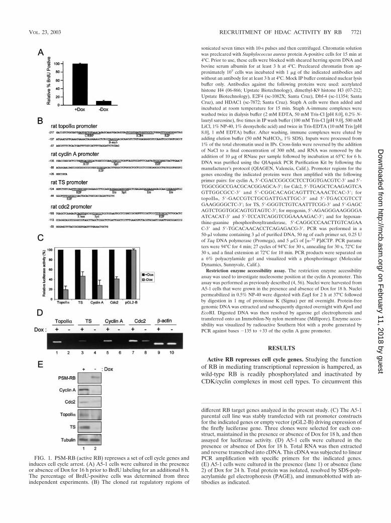
FIG. 1. PSM-RB (active RB) represses a set of cell cycle genes andinduces cell cycle arrest. (A) A5-1 cells were cultured in the presenceor absence of Dox for 16 h prior to BrdU labeling for an additional 8 h.The percentage of BrdU-positive cells was determined from threeindependent experiments. (B) The cloned rat regulatory regions of
different RB target genes analyzed in the present study. (C) The A5-1parental cell line was stably transfected with rat promoter constructsfor the indicated genes or empty vector (pGL2-B) driving expression ofthe firefly luciferase gene. Three clones were selected for each con-struct, maintained in the presence or absence of Dox for 18 h, and thenassayed for luciferase activity. (D) A5-1 cells were cultured in thepresence or absence of Dox for 18 h. Total RNA was then extractedand reverse transcribed into cDNA. This cDNA was subjected to linearPCR amplification with specific primers for the indicated genes.(E) A5-1 cells were cultured in the presence (lane 1) or absence (lane2) of Dox for 24 h. Total protein was isolated, resolved by SDS-poly-acrylamide gel electrophoresis (PAGE), and immunoblotted with an-tibodies as indicated.
VOL. 23, 2003 RECRUITMENT OF HDAC ACTIVITY BY RB 7721
on February 11, 2018 by guest
http://mcb.asm
.org/D
ownloaded from

problem, we utilized a PSM-RB that is refractory to phosphor-ylation and is therefore constitutively active. In this study, aRat-1-derived cell line with tetracycline-regulated PSM-RBexpression (A5-1) was utilized to specifically analyze the down-stream action of RB. In order to confirm the cell cycle inhib-itory potential of PSM-RB in this setting, we examined theBrdU incorporation of A5-1 cells cultured in the presence orabsence of the tetracycline analogue Dox. Consistent with pre-viously reported data, we found that the induction of PSM-RBupon removal of Dox dramatically reduced the number ofBrdU-positive cells (Fig. 1A). Therefore, A5-1 cells representan effective model for the study of RB activity.
We have recently identified and validated a large number ofRB-repressed target genes by using microarray analysis (42).To initially investigate the promoter activity of representativegenes, we cloned regions of the genes encoding the cyclin A,Cdc2, topoII�, and TS promoters into the luciferase reportervector pGL2-B (Fig. 1B). These cloned promoter fragmentsrepresent less than two nucleosomes of DNA and are thususeful for investigating the regulatory action of RB on a rela-tively small region of chromatin. The reporter constructs andthe vector (pGL2-B) were then integrated into A5-1 cells toprovide an effective means for studying promoter activity in thecontext of chromatin. Three independent clones for each con-struct were selected and maintained in medium with or withoutDox for 18 h. We found that the activity of cyclin A, Cdc2,topoII�, and TS promoters was reduced three- to fivefold uponinduction of PSM-RB expression (Fig. 1C, compare �Dox to�Dox). In contrast, PSM-RB had no effect on the basal tran-scription of pGL2-B. These data demonstrate that active RBrepresses the promoter activity of these genes in chromatin.
Next we investigated the correlation between promoter ac-tivity and endogenous RNA levels (Fig. 1D). We utilized RT-PCR to determine the levels of endogenous RNA in A5-1 cellsthat were cultured in the presence or absence of Dox. Induc-tion of active RB attenuated the expression of cyclin A, Cdc2,topoII�, and TS RNA (Fig. 1D, compare �Dox to �Dox). Incontrast, induction of PSM-RB had no effect on the expressionof the �-actin gene. Consistent with these observations, endog-enous protein levels of cyclin A, Cdc2, topoII�, and TS werealso attenuated when Dox was removed from the medium (Fig.1E, compare �Dox to �Dox). The attenuations are due to thepresence of PSM-RB and not merely to differences in loading,as revealed by immunoblotting for �-tubulin. Immunoblottingwith a PSM-RB-specific antibody clearly demonstrated thatPSM-RB is expressed only in the absence of Dox (Fig. 1E).Taken together, these results confirm that active RB inhibitsthe expression of the genes encoding cyclin A, Cdc2, topoII�,and TS by repressing promoter activity.
Active RB induces histone deacetylation at promoters ofspecific cell cycle genes. RB has been shown to recruit HDACsto the promoters of target genes (81). This event is believed tobe responsible for transcriptional repression and the subse-quent antiproliferative action of RB. However, the extent towhich HDACs are required for RB to act as a transcriptionalrepressor and tumor suppressor is unclear. To assess the actionof HDAC enzymatic activity in RB-mediated transcriptionalrepression, we utilized in vivo formaldehyde cross-linking ofDNA-protein complexes followed by ChIP assay. Initially, wevalidated the linearity of PCR amplification over a wide range
of template concentrations. Increasing amounts of input chro-matin were amplified with primers specific for the Cdc2 pro-moter gene in the presence of [�-32P]dCTP for quantitation.As shown in Fig. 2A, the PCR was linear throughout thetitration, indicating that changes in chromatin levels could beaccurately observed by radioactive PCR. Using this approach,we then investigated the promoters of the cyclin A, Cdc2,topoII�, and TS genes for changes in histone H4 acetylation, asthese targets were repressed by PSM-RB (Fig. 2B). Specifi-cally, A5-1 cells were cultured in the presence or absence ofDox for 24 h and were formaldehyde cross-linked, and ChIPassays were performed by utilizing antibody to acetylated his-tone H4. For each target promoter, input lanes (nonimmuno-precipitated DNA) confirmed that equal amounts of chroma-tin were used in all ChIP assays (Fig. 2B, lanes 1 and 2). Theantibody to Dbf-4 was utilized as a negative control, since thereplication factor Dbf-4 is not expected to occupy these pro-moters (Fig. 2A, lanes 5 and 6). We found that, in the presenceof PSM-RB, acetylated histone H4 association with the pro-moters of cyclin A, Cdc2, topoII�, and TS was nearly abolished(Fig. 2B, compare lanes 3 and 4). This effect is specific toRB-repressed genes, as no change in histone acetylation wasobserved on the hypoxanthine-guanine phosphoribosyltrans-ferase promoter (Fig. 2B). In contrast with acetylated histoneH4 occupancy, which was diminished in the presence of PSM-RB, we found that E2F4 occupancy was unaffected by theinduction of active RB (Fig. 2C, compare lanes 3 and 4).Moreover, we detected HDAC1 preferentially bound to Cdc2promoter in the presence of PSM-RB (Fig. 2C, compare lanes5 and 6). Therefore, the decrease in acetylated histone H4 onthese promoters is a consequence of PSM-RB induction, andnot all protein-promoter interactions were similarly affected.The reduction in acetylated histone H4 at these promotersstrongly suggests that these promoters are actively deacety-lated. This observation is consistent with RB/HDAC com-plexes acting on these promoters.
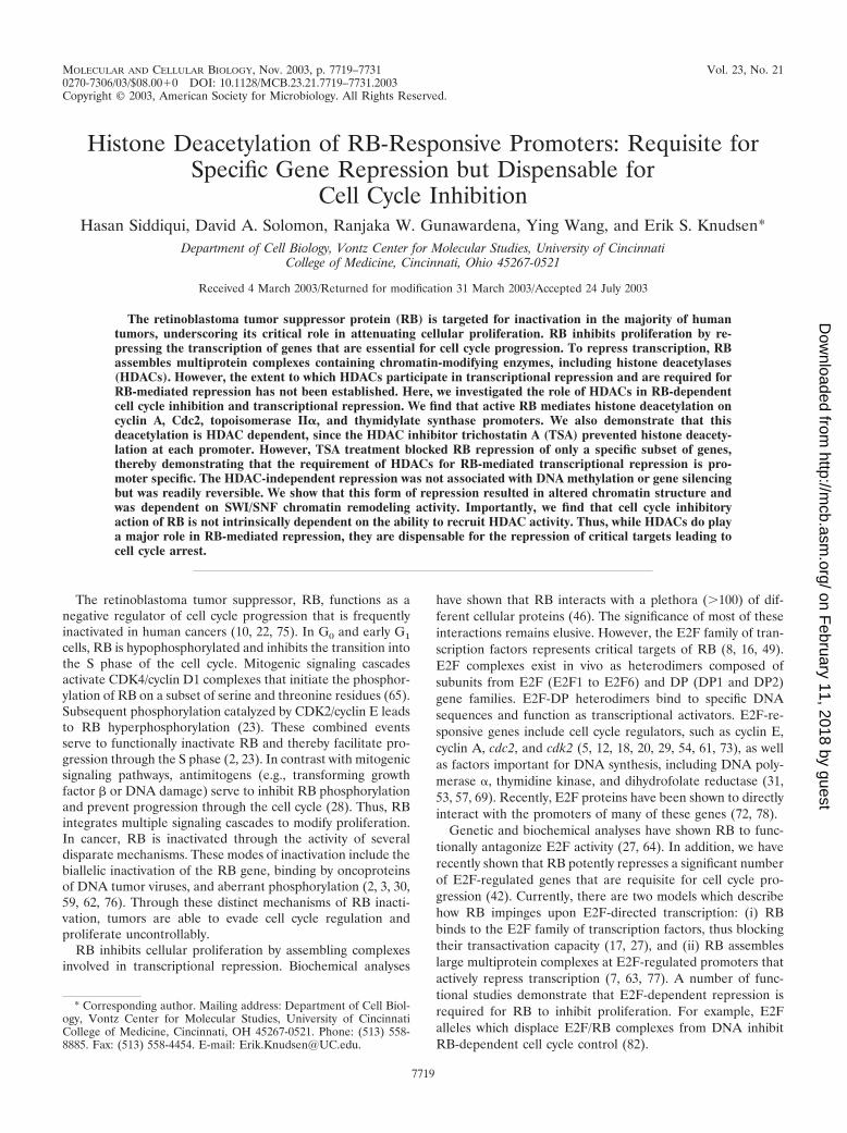
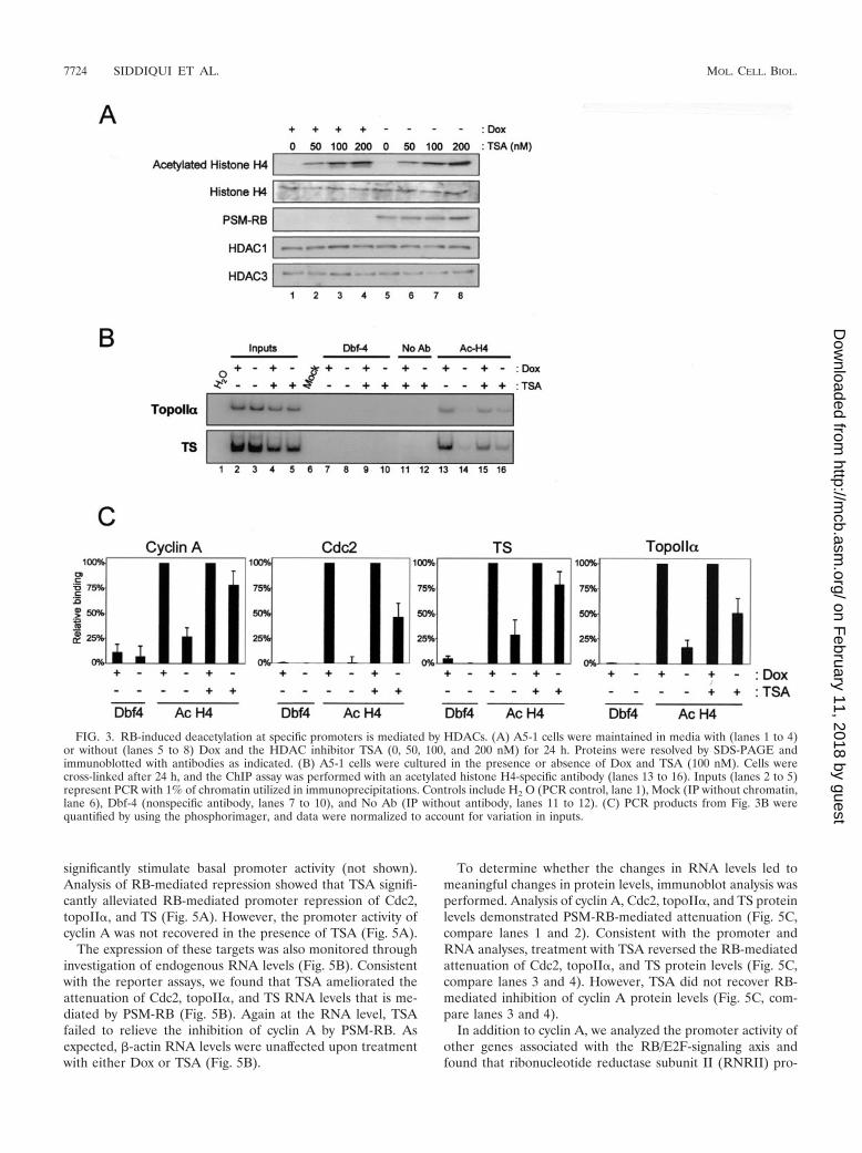
RB-induced histone deacetylation at specific promoters ismediated by HDACs. Histone deacetylation at the promotersof cyclin A, Cdc2, topoII�, and TS in the presence of active RB(shown in Fig. 2) supports a critical role for HDAC in tran-scriptional repression. To determine the explicit requirementof HDACs for this transcriptional repression, we used TSA, apharmacological inhibitor of HDAC activity. Initially, we de-termined the effect of TSA on bulk histone acetylation (Fig.3A). A5-1 cells were cultured in the presence and absence ofDox and TSA for 24 h. Cells were then harvested, and thelevels of acetylated histone H4 and total histone H4 wereevaluated by immunoblotting (Fig. 3A). We found that TSAtreatment resulted in the marked accumulation of acetylatedhistone H4 in a dose-dependent manner (Fig. 3A, top panel,compare lanes 1, 2, 3, and 4). Importantly, analysis of totalhistone H4 levels showed that TSA inhibited deacetylation anddid not merely augment the expression of histone H4. Further-more, TSA did not affect the expression of PSM-RB, HDAC1,or HDAC3. While performing these studies, we found that 100nM TSA had a minimal effect on cell viability (data notshown). Therefore, we employed 100 nM TSA to study therequirement of HDAC activity for RB-mediated transcrip-tional repression.
To investigate the requirement of HDAC activity for
7722 SIDDIQUI ET AL. MOL. CELL. BIOL.
on February 11, 2018 by guest
http://mcb.asm
.org/D
ownloaded from

RB-mediated histone deacetylation, we used TSA to inhibitHDAC activity and then monitored promoter histone acetyla-tion. For this analysis, A5-1 cells were treated with or withoutDox and TSA for 24 h. These cells were then used as substratesfor ChIP assays with antibodies specific for acetylated histoneH4 (Fig. 3B). Nonimmunoprecipitated chromatin (Inputs)showed the relative amounts of chromatin used in each IP (Fig.3B, lanes 2 to 5). IP with nonspecific antibody to Dbf4 (Fig. 3B,lanes 7 to 10), without an antibody (Fig. 3B, lanes 11 to 12),and without chromatin (Fig. 3B, lane 6) did not result inappreciable PCR product and thus provided evidence of spec-ificity for the ChIP assay. Consistent with data describedabove, we observed histone H4 deacetylation on the promotersanalyzed (Fig. 3B, compare lanes 13 and 14). Surprisingly, TSAby itself did not augment the histone H4 acetylation observedon any promoter in the presence of Dox (Fig. 3B, comparelanes 13 and 15), indicating that HDAC activity was likelyabsent from the promoter in normal asynchronously prolifer-ating cells. However, inhibition of HDAC activity by TSAresulted in marked acetylation of histone H4 on the promotersof topoII� and TS in the presence of PSM-RB (Fig. 3B, com-pare lane 14 to lane 16). Similar results were obtained forcyclin A and Cdc2 promoters. The use of [�-32P]dCTP in PCRenabled the quantitative analysis of product signal intensity.PCR product bands from acetylated histone H4 IPs were nor-malized to their corresponding inputs to account for variationsin chromatin used in the ChIP assay. PCR-amplified productsthat were quantified by using phosphorimaging clearly showedthat TSA blocks deacetylation of these promoters (Fig. 3C).Collectively, these data demonstrate that RB-induced deacety-lation of cyclin A, Cdc2, topoII�, and TS promoters requiresHDAC activity. Since histone deacetylation is a critical mech-anism of transcriptional repression, these results suggest thatTSA may reverse RB-mediated cell cycle inhibition.
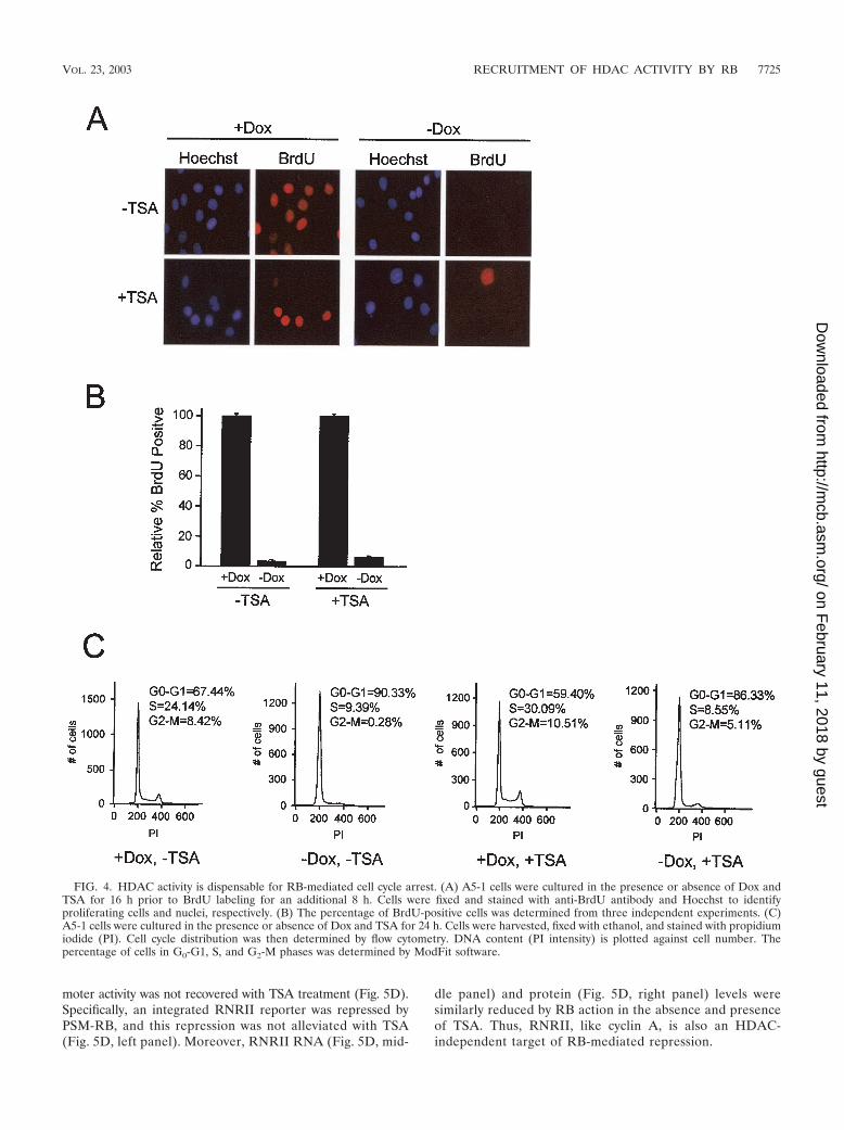
HDAC activity is dispensable for RB-mediated cell cyclearrest. To monitor the influence of HDAC inhibition on cellcycle progression, A5-1 cells were cultured in the presence orabsence of Dox and TSA for 16 h before being pulsed withBrdU for 8 h. Surprisingly, cells cultured in the absence of Doxremained inhibited for BrdU incorporation in the presence ofTSA (Fig. 4A and B). To examine cell cycle distribution, weperformed flow cytometric analysis of cells treated with orwithout Dox and TSA. As shown in Fig. 4C, we found thattreatment with or without TSA, concurrent with PSM-RB(�Dox) expression, did not significantly alter cell cycle posi-tion. Taken together, these data demonstrate that RB is able tomaintain cell cycle arrest even in the absence of HDAC activ-ity.
HDAC requirement for repression is promoter specific. Thefailure of TSA to rescue cells from RB-mediated cycle arrestsuggested that inhibition of HDAC activity was not sufficient toalleviate transcriptional repression. Having established that his-tone deacetylation at cyclin A, Cdc2, topoII�, and TS promot-ers by RB is the result of HDAC activity (Fig. 3B), we soughtto elucidate the functional significance of this event (Fig. 5).A5-1 cells with integrated luciferase reporters of cyclin A,Cdc2, topoII�, TS, and vector pGL2-B were cultured in thepresence or absence of Dox and TSA for 18 h. Consistent withthe failure of TSA treatment to augment promoter histoneacetylation in the absence of PSM-RB (�Dox), TSA did not
FIG. 2. Active RB induces histone deacetylation at promoters of spe-cific cell cycle genes. (A) Total chromatin was isolated from A5-1 cellscultured in the presence of Dox, and increasing amounts of chromatin (0to 4 �l) were subjected to PCR in the presence of [�-32P]dCTP andprimers specific for the cdc2 promoter. Production of PCR product wasquantified by using a phosphorimager. (B) A5-1 cells were cultured in thepresence (lanes 1, 3, and 5) or absence (lanes 2, 4, and 6) of Dox for 24 hand cross-linked with formaldehyde, and ChIP assays were performed asdescribed in Materials and Methods. Residency of acetylated histone H4at the indicated gene promoters was determined by carrying out the ChIPassay with antibodies specific to acetylated histone H4 (lanes 3 and 4).Input (lanes 1 and 2) refers to PCR containing 1% of the total chromatinused in IP. IP with Dbf-4 (lanes 5 and 6) is a negative control. PCRproducts were detected by autoradiography. HPRT, hypoxanthine-gua-nine phosphoribosyltransferase. (C) Cells were cultured as described forpanel B, except that immunoprecipitation was performed with antibodiesspecific for Dbf-4 (lanes 1 and 2), E2F4 (lanes 3 and 4), and HDAC1(lanes 5 and 6). The mock represents a ChIP assay that was performedwithout the inclusion of chromatin substrate. PCR products were detectedby autoradiography.
VOL. 23, 2003 RECRUITMENT OF HDAC ACTIVITY BY RB 7723
on February 11, 2018 by guest
http://mcb.asm
.org/D
ownloaded from
significantly stimulate basal promoter activity (not shown).Analysis of RB-mediated repression showed that TSA signifi-cantly alleviated RB-mediated promoter repression of Cdc2,topoII�, and TS (Fig. 5A). However, the promoter activity ofcyclin A was not recovered in the presence of TSA (Fig. 5A).
The expression of these targets was also monitored throughinvestigation of endogenous RNA levels (Fig. 5B). Consistentwith the reporter assays, we found that TSA ameliorated theattenuation of Cdc2, topoII�, and TS RNA levels that is me-diated by PSM-RB (Fig. 5B). Again at the RNA level, TSAfailed to relieve the inhibition of cyclin A by PSM-RB. Asexpected, �-actin RNA levels were unaffected upon treatmentwith either Dox or TSA (Fig. 5B).
To determine whether the changes in RNA levels led tomeaningful changes in protein levels, immunoblot analysis wasperformed. Analysis of cyclin A, Cdc2, topoII�, and TS proteinlevels demonstrated PSM-RB-mediated attenuation (Fig. 5C,compare lanes 1 and 2). Consistent with the promoter andRNA analyses, treatment with TSA reversed the RB-mediatedattenuation of Cdc2, topoII�, and TS protein levels (Fig. 5C,compare lanes 3 and 4). However, TSA did not recover RB-mediated inhibition of cyclin A protein levels (Fig. 5C, com-pare lanes 3 and 4).
In addition to cyclin A, we analyzed the promoter activity ofother genes associated with the RB/E2F-signaling axis andfound that ribonucleotide reductase subunit II (RNRII) pro-
FIG. 3. RB-induced deacetylation at specific promoters is mediated by HDACs. (A) A5-1 cells were maintained in media with (lanes 1 to 4)or without (lanes 5 to 8) Dox and the HDAC inhibitor TSA (0, 50, 100, and 200 nM) for 24 h. Proteins were resolved by SDS-PAGE andimmunoblotted with antibodies as indicated. (B) A5-1 cells were cultured in the presence or absence of Dox and TSA (100 nM). Cells werecross-linked after 24 h, and the ChIP assay was performed with an acetylated histone H4-specific antibody (lanes 13 to 16). Inputs (lanes 2 to 5)represent PCR with 1% of chromatin utilized in immunoprecipitations. Controls include H2 O (PCR control, lane 1), Mock (IP without chromatin,lane 6), Dbf-4 (nonspecific antibody, lanes 7 to 10), and No Ab (IP without antibody, lanes 11 to 12). (C) PCR products from Fig. 3B werequantified by using the phosphorimager, and data were normalized to account for variation in inputs.
7724 SIDDIQUI ET AL. MOL. CELL. BIOL.
on February 11, 2018 by guest
http://mcb.asm
.org/D
ownloaded from
moter activity was not recovered with TSA treatment (Fig. 5D).Specifically, an integrated RNRII reporter was repressed byPSM-RB, and this repression was not alleviated with TSA(Fig. 5D, left panel). Moreover, RNRII RNA (Fig. 5D, mid-
dle panel) and protein (Fig. 5D, right panel) levels weresimilarly reduced by RB action in the absence and presenceof TSA. Thus, RNRII, like cyclin A, is also an HDAC-independent target of RB-mediated repression.
FIG. 4. HDAC activity is dispensable for RB-mediated cell cycle arrest. (A) A5-1 cells were cultured in the presence or absence of Dox andTSA for 16 h prior to BrdU labeling for an additional 8 h. Cells were fixed and stained with anti-BrdU antibody and Hoechst to identifyproliferating cells and nuclei, respectively. (B) The percentage of BrdU-positive cells was determined from three independent experiments. (C)A5-1 cells were cultured in the presence or absence of Dox and TSA for 24 h. Cells were harvested, fixed with ethanol, and stained with propidiumiodide (PI). Cell cycle distribution was then determined by flow cytometry. DNA content (PI intensity) is plotted against cell number. Thepercentage of cells in G0-G1, S, and G2-M phases was determined by ModFit software.
VOL. 23, 2003 RECRUITMENT OF HDAC ACTIVITY BY RB 7725
on February 11, 2018 by guest
http://mcb.asm
.org/D
ownloaded from

FIG. 5. HDAC-dependent deacetylation is required by RB to repress specific promoters. (A) A5-1-integrated reporter cell lines described inthe legend of Fig. 1C were cultured in the presence or absence of Dox and 100 nM TSA for 18 h. Cells were harvested and assayed for luciferaseactivity. (B) A5-1 cells were cultured in 100 nM TSA in the presence or absence of Dox for 18 h. Total RNA was then extracted and reversetranscribed into cDNA. This cDNA was subjected to linear PCR amplification with specific primers for the indicated genes. (C) A5-1 cells werecultured in the presence (lanes 1 and 3) or absence (lanes 2 and 4) of Dox in the absence (lanes 1 and 2) or presence (lanes 3 and 4) of 100 nMTSA for 24 h. Total protein was isolated, resolved by SDS-PAGE, and immunoblotted with antibodies as indicated. (D) Top panel: The clonedregulatory region of the rat RNRII promoter. Left panel: The A5-1 parental cell line was stably transfected with the rat RNRII promoter drivingexpression of the firefly luciferase gene. Cells were cultured in the presence or absence of Dox with or without 100 nM TSA as indicated, andrelative luciferase activity was determined. Center panel: A5-1 cells were cultured in the presence (lanes 1 and 3) or absence (lanes 2 and 4) ofDox and in the absence (lanes 1 and 2) or presence (lanes 3 and 4) of TSA for 18 h. Cells were harvested, and RNRII RNA levels were determinedby RT-PCR. Right panel: A5-1 cells were cultured in the presence (lanes 1 and 2) or absence (lanes 3 and 4) of Dox and the absence (lanes 1 and3) or presence (lanes 2 and 4) of TSA for 24 h. Cells were harvested, and RNRII protein levels were determined by immunoblotting.
7726
on February 11, 2018 by guest
http://mcb.asm
.org/D
ownloaded from

HDAC-independent mechanism of cyclin A repression. It isknown that transcriptional repression elicited by RB can occurthrough the action of corepressors in addition to HDACs.Specifically, it has recently been demonstrated that the cyclin Apromoter is irreversibly silenced during induced senescence(48). This pathway likely involves histone H3 lysine 9 methyl-ation, heterochromatin protein 1 (HP1) recruitment, and DNAmethylation of the promoter (34, 48, 52). Therefore, we ini-tially analyzed the histone H3 lysine 9 methylation of the cyclinA promoter during RB-mediated arrest (Fig. 6A). Cells werecultured in the presence or absence of Dox to induce PSM-RB,and isolated chromatin was subjected to ChIP analysis usingantibodies specific for histone H3 methylated on lysine 9. Weobserved histone H3 lysine 9 methylation on the myogeninpromoter that is silenced in fibroblastic cells (Fig. 6A). In
contrast, we failed to detect histone H3 lysine 9 methylation onthe cyclin A promoter above background, suggesting that thissilencing mechanism is not responsible for cyclin A repression.Consistent with this observation, culture in 5-aza-2-deoxcyti-dine, which blocks DNA methylation and reverses silencing(41, 45, 50, 80), failed to augment cyclin A promoter activityor protein levels in the presence of PSM-RB (Fig. 6B).Finally, we determined whether the RB-mediated repressionof the cyclin A promoter was reversible. To do so, cells werecultured in the absence of Dox to induce PSM-RB, and cyclinA promoter activity was repressed (Fig. 6C, left panel). Read-dition of Dox to the media resulted in the restoration of cyclinA promoter activity. Additionally, cyclin A protein levels wererestored following the readdition of Dox (Fig. 6C, right panel).Together, these results indicate that stable epigenetic silencing
FIG. 6. The cyclin A promoter is not subjected to stable gene silencing. (A) A5-1 cells were cultured in the presence or absence of Dox asindicated. Chromatin was isolated and utilized in ChIP assays with antibodies specific for dimethylated K9 histone H3 (lanes 5 and 6). Input (lanes1 and 2) and Dbf-4 (lanes 3 and 4) controls are shown. Chromatin was amplified with primers specific for the cyclin A and myogenin promoters,and products were detected by autoradiography. (B) A5-1 cells harboring the integrated cyclin A reporter were cultured in the presence of5-aza-2-dC as described in Materials and Methods and then cultured in the absence of Dox for 24 h. Relative luciferase activity was determinedby reporter assay (left panel), and endogenous protein levels were determined by immunoblotting (right panel). (C) A5-1 cells harboring theintegrated cyclin A reporter were cultured in the presence or absence of Dox for 24 h. To attenuate PSM-RB, Dox was readministered to theindicated cultures. Relative luciferase activity was determined by reporter assay (left panel), and endogenous protein levels were determined byimmunoblotting (right panel).
VOL. 23, 2003 RECRUITMENT OF HDAC ACTIVITY BY RB 7727
on February 11, 2018 by guest
http://mcb.asm
.org/D
ownloaded from

mechanisms are not responsible for the observed RB-mediatedrepression of the cyclin A promoter.
In addition to HDACs and silencing mechanisms, we andothers have demonstrated that SWI/SNF activity plays a req-uisite role in the repression of the cyclin A promoter (70, 81).Consistent with these studies, we observed that, unlike 5-aza-2-deoxcytidine or TSA, ectopic expression of dominant nega-tive BRG-1 (dnBRG-1) that inhibits SWI/SNF activity aug-ments the expression of cyclin A RNA (Fig. 7A, left panel) andprotein (Fig. 7A, right panel) levels in the presence of PSM-RB (Fig. 7A). These results indicate that chromatin remod-eling represents a critical means through which cyclin A re-pression occurs. One possible explanation is that SWI/SNFfunctions in concert with RB to position nucleosomes near thetranscription start site to inhibit transcription of the cyclin Apromoter. Analysis of promoter structure was carried out inthe presence or absence of PSM-RB by using the restrictionenzyme accessibility assay in proximity to the transcriptionstart site. We observed that chromatin from cells cultured inthe absence of PSM-RB (�Dox) was readily digestible (Fig.7B, lanes 3), whereas chromatin from cells expressing PSM-RB(�Dox) was resistant to enzyme cleavage (Fig. 7B, lanes 4).This change in chromatin structure was largely dependentupon SWI/SNF, as dnBRG-1 retarded the formation of thenuclease-resistant chromatin structure (Fig. 7B, lanes 6).These results indicate that chromatin remodeling occurs on thecyclin A promoter to mediate transcriptional repression.
DISCUSSION
RB represses the expression of multiple genes involved incell cycle transitions. This transcriptional control has been at-
tributed to multiple corepressors recruited by RB. Here wespecifically focused on elucidating the role of HDACs in RB-mediated transcriptional repression of four critical targets(cyclinA, Cdc2, topoII�, and TS). We demonstrate that activeRB leads to histone deacetylation on the promoters of thesefour genes, and the observed deacetylation is dependent onHDAC activity. This action of HDACs was required for thetranscriptional repression of Cdc2, topoII�, and TS genes, asTSA reversed RB-mediated repression. However, this actionwas promoter specific, as TSA failed to recover cyclin A orRNRII levels. Analysis of the HDAC-independent repressionof cyclin A indicated that it was not due to epigenetic silencingmechanisms but was reversible and involved chromatin remod-eling. Importantly, we demonstrate that the cell cycle-inhibi-tory action of RB is independent of HDAC enzymatic activity.Together, these results demonstrate the intricate interplay be-tween RB and HDACs in transcriptional regulation and cellcycle control.
Role of HDACs in RB-mediated transcriptional repression.HDACs represent corepressors that are identified as interact-ing with RB (6, 39, 40). However, relatively few studies haveanalyzed their role in RB-mediated transcriptional repressionand cell cycle control. Originally, it was demonstrated thatHDAC activity is associated with RB, but only in the context ofa synthetic promoter was histone deacetylation observed (39).Subsequent studies have shown that specific RB/E2F targetgenes in fact undergo changes in promoter histone acetylationduring the cell cycle (47, 58). Our results shown here demon-strate that RB-mediated repression leads to histone deacety-lation at all promoters analyzed. In principle, such an effectcould be due to either recruitment of HDAC or the inhibitionof HAT activity. Inhibition of HAT as a mechanism for pro-
FIG. 7. RB-mediated repression of the cyclin A promoter involves chromatin remodeling. (A) Parental A5-1 cells (lanes 1 and 2) or A5-1 cellsengineered to inducibly coexpress dnBRG-1 and PSM-RB (lanes 3 and 4) were cultured in the presence (lanes 1 and 3) or absence (lanes 2 and4) of Dox for 24 h. Cells were harvested, and protein and RNA levels were determined by RT-PCR (left panel) or immunoblotting (right panel)as indicated. (B) Left panel: genomic structure of the rat cyclin A locus. Right panel: Southern blot analysis of genomic DNA isolated from A5-1cells (lanes 1 and 2). Parental A5-1 cells (lanes 3 and 4) or A5-1 cells engineered to inducibly express dnBRG-1 (lanes 5 and 6) were cultured inthe presence or absence of Dox as indicated. Permeabilized nuclei were subjected to digestion with EagI, and then isolated genomic DNA wassubjected to cleavage with KpnI and EcoRI. Restriction fragments were detected by radioactive Southern blotting.
7728 SIDDIQUI ET AL. MOL. CELL. BIOL.
on February 11, 2018 by guest
http://mcb.asm
.org/D
ownloaded from

moter deacetylation is not without merit, as E2F proteins likelyemploy HAT-dependent mechanisms for gene activation (74).By using the HDAC inhibitor TSA, we could specifically dem-onstrate that the deacetylation of these promoters is depen-dent on the enzymatic activity associated with class I HDACmolecules (43). The effect of RB on histone acetylation is notdue to a bulk-deacetylation phenomenon, as total cellular lev-els of acetylated histone are not changed by the expression ofPSM-RB. Interestingly, while TSA does lead to bulk histonehyperacetylation, it does not lead intrinsically to the hyper-acetylation of histones at the E2F/RB-regulated promotersstudied here. This could be because the histones at these pro-moters are already hyperacetylated or because HDAC is notassociated with the promoter in asynchronously proliferatingcells. Such a hypothesis is supported by the finding that weobserved HDAC1 specifically associated with the Cdc2 pro-moter when PSM-RB was expressed. Clearly, in the context ofRB-mediated repression, the mechanism of histone deacetyla-tion is HDAC dependent and can be reversed by TSA on allpromoters analyzed. The inhibition of histone deacetylation byTSA had functional consequences in the context of Cdc2,topoII�, and TS gene expression. Interestingly, even in cases inwhich TSA only partially reversed RB-mediated histone de-acetylation, TSA was capable of fully restoring promoter ac-tivity (i.e., the cdc2 promoter). Such a finding, for which thereis precedent in the literature (60, 83), suggests that only amoderate level of histone acetylation is required for transcrip-tional activity on specific promoters. In fact, TSA fully restoredthe promoter activity, endogenous RNA, and protein levels ofCdc2, topoII�, and TS in the presence of PSM-RB. Such afinding is critical, as it demonstrates that HDAC activity rep-resents the sole means through which RB mediates repressionof these targets. In contrast, we failed to observe recovery ofcyclin A expression when HDAC activity was inhibited in pres-ence of RB, even though promoter histones were acetylatedfollowing TSA treatment.
HDAC-independent mechanisms of transcriptional repres-sion. The findings that we observe with cyclin A gene regula-tion indicate that RB utilizes mechanisms in addition to his-tone deacetylation to mediate repression. Such a conclusion isnot without precedent (44, 81). However, to definitively makesuch a conclusion, it is critical to determine that inhibition ofHDAC activity actually reversed the histone deacetylation onthe promoter. We clearly observed that TSA was sufficient toefficiently reverse RB-mediated histone deacetylation on thecyclin A promoter. Thus, the failure of TSA to reverse RB-mediated repression of the cyclin A promoter is due to amechanism that is clearly distinct from HDAC activity. Addi-tionally, while we did not explicitly evaluate the RNRII pro-moter, it behaved in a manner similar to that of cyclin A in thatits repression was independent of HDAC activity. Therefore, itseems likely that repression of cyclin A and RNRII by RB isdependent on other chromatin-modifying factors.
RB has been shown to associate with other chromatin-mod-ifying enzymes to mediate transcriptional repression. Specifi-cally, recent studies indicate that RB target genes (includingcyclin A) are silenced through a mechanism involving histonemethylation and HP1 chromatin association (48, 52). Suchsilencing, which is observed in senescent cells, is irreversible(48, 52). Here we find that the repression of cyclin A by PSM-
RB does not involve irreversible silencing mechanisms, as wefailed to detect any influence of histone or DNA methylationon RB-mediated repression of the cyclin A promoter. Addi-tionally, the repression of the cyclin A promoter was readilyreversible. Such a result is consistent with the ability of quies-cent cells with the cyclin A promoter repressed to reenter thecell cycle (48) and with the failure to detect histone methyl-ation or HP1 chromatin association with the cyclin A promoterin quiescent cells (48).
In addition to silencing mechanisms and HDACs, RB isknown to associate with components of the SWI/SNF chroma-tin remodeling complex, and the activity of SWI/SNF is re-quired for the repression of cyclin A. For example, PSM-RB orexpression of p16ink4a to activate endogenous RB in BRG-1-or BRM-deficient cell lines does not attenuate cyclin A andRNRII levels or lead to cell cycle arrest (references 70 and 81and our unpublished data). One means through which SWI/SNF acts is by facilitating histone acetylation and deacetyla-tion. This action of SWI/SNF is not sufficient for cyclin Arepression, as the promoter remained repressed even with his-tones acetylated. Rather, our results suggest that SWI/SNF isfunctioning to modify chromatin structure to inhibit transcrip-tion. Consistent with this model, we find that the nucleaseaccessibility in the region of the cyclin A transcription start siteis inhibited during RB-mediated repression. Thus, chromatintopology, not histone modification, is likely sufficient for RB-mediated repression events on the cyclin A promoter.
HDAC activity is not required for RB-mediated cell cycleinhibition. Another finding from our studies is that inhibitionof HDAC activity by TSA does not rescue cells from arrestimposed by RB. This result is quite surprising, as TSA exertspronounced effects on gene expression but has no detectableeffect on RB-mediated cell cycle inhibition. Most likely, failureof TSA to block RB-mediated repression of cyclin A and sim-ilarly regulated genes (e.g., RNRII) is responsible for thisphenomenon. Cyclin A is required for traversing the cell cycle(19, 55), and thus lack of cyclin A expression alone might wellexplain the failure of cells to proliferate when HDAC activityis inhibited. An important extension of our work is based onthe current use of HDAC inhibitors in clinical trials for treat-ment of several forms of cancer (33). In this context, HDACinhibitors are believed to reactivate genes that have been in-appropriately silenced during tumor progression and thus in-hibit tumor growth. Based on our findings that TSA does notreverse RB-mediated cell cycle arrest, we conclude that thetreatment of tumors with HDAC inhibitors will not have theundesired effect of inactivating the RB pathway of cell cycleinhibition.
In summary, transcriptional repression of cell cycle genes byRB is a complex process involving multiple chromatin-modi-fying factors. Here we find that one class of factors, HDACs,play a critical role in the transcriptional repression programselicited by RB. This action of HDACs is promoter specific andas such is not required for RB-mediated cell cycle inhibition.
ACKNOWLEDGMENTS
We are grateful to Karen Knudsen, Christopher Mayhew, StevenAngus, Craig Burd, Michael Markey, and Christin Petre for criticalreading of the manuscript. We thank Shelly Barton, Anthony Imbal-zano, Bernard Weissman, and members of the Knudsens’ laboratories
VOL. 23, 2003 RECRUITMENT OF HDAC ACTIVITY BY RB 7729
on February 11, 2018 by guest
http://mcb.asm
.org/D
ownloaded from

for helpful discussions. Sandy Schwemberger provided technical assis-tance with flow cytometry analysis.
This work was supported by grants to E.S.K. from the NationalCancer Institute (no. CA82525).
H.S. and D.A.S. contributed equally to this work.
REFERENCES
1. Angus, S. P., A. F. Fribourg, M. P. Markey, S. L. Williams, H. F. Horn, J.DeGregori, T. F. Kowalik, K. Fukasawa, and E. S. Knudsen. 2002. Active RBelicits late G1/S inhibition. Exp. Cell Res. 276:201–213.
2. Bartek, J., J. Bartkova, and J. Lukas. 1997. The retinoblastoma proteinpathway in cell cycle control and cancer. Exp. Cell Res. 237:1–6.
3. Bartkova, J., J. Lukas, and J. Bartek. 1997. Aberrations of the G1- and G1/S-regulating genes in human cancer. Prog. Cell Cycle Res. 3:211–220.
4. Boeger, H., J. Griesenbeck, J. S. Strattan, and R. D. Kornberg. 2003. Nu-cleosomes unfold completely at a transcriptionally active promoter. Mol.Cell 11:1587–1598.
5. Botz, J., K. Zerfass-Thome, D. Spitkovsky, H. Delius, B. Vogt, M. Eilers, A.Hatzigeorgiou, and P. Jansen-Durr. 1996. Cell cycle regulation of the murinecyclin E gene depends on an E2F binding site in the promoter. Mol. Cell.Biol. 16:3401–3409.
6. Brehm, A., E. A. Miska, D. J. McCance, J. L. Reid, A. J. Bannister, and T.Kouzarides. 1998. Retinoblastoma protein recruits histone deacetylase torepress transcription. Nature 391:597–601.
7. Bremner, R., B. L. Cohen, M. Sopta, P. A. Hamel, C. J. Ingles, B. L. Gallie,and R. A. Phillips. 1995. Direct transcriptional repression by pRB and itsreversal by specific cyclins. Mol. Cell. Biol. 15:3256–3265.
8. Chellappan, S. P., S. Hiebert, M. Mudryj, J. M. Horowitz, and J. R. Nevins.1991. The E2F transcription factor is a cellular target for the RB protein.Cell 65:1053–1061.
9. Chen, T. T., and J. Y. Wang. 2000. Establishment of irreversible growtharrest in myogenic differentiation requires the RB LXCXE-binding function.Mol. Cell. Biol. 20:5571–5580.
10. Classon, M., and E. Harlow. 2002. The retinoblastoma tumour suppressor indevelopment and cancer. Nat. Rev. Cancer 2:910–917.
11. Dahiya, A., M. R. Gavin, R. X. Luo, and D. C. Dean. 2000. Role of theLXCXE binding site in Rb function. Mol. Cell. Biol. 20:6799–6805.
12. Dalton, S. 1992. Cell cycle regulation of the human cdc2 gene. EMBO J.11:1797–1804.
13. de la Serna, I. L., K. A. Carlson, D. A. Hill, C. J. Guidi, R. O. Stephenson,S. Sif, R. E. Kingston, and A. N. Imbalzano. 2000. Mammalian SWI-SNFcomplexes contribute to activation of the hsp70 gene. Mol. Cell. Biol. 20:2839–2851.
14. de la Serna, I. L., K. A. Carlson, and A. N. Imbalzano. 2001. MammalianSWI/SNF complexes promote MyoD-mediated muscle differentiation. Nat.Genet. 27:187–190.
15. Dunaief, J. L., B. E. Strober, S. Guha, P. A. Khavari, K. Alin, J. Luban, M.Begemann, G. R. Crabtree, and S. P. Goff. 1994. The retinoblastoma proteinand BRG1 form a complex and cooperate to induce cell cycle arrest. Cell79:119–130.
16. Dyson, N. 1998. The regulation of E2F by pRB-family proteins. Genes Dev.12:2245–2262.
17. Flemington, E. K., S. H. Speck, and W. G. Kaelin, Jr. 1993. E2F-1-mediatedtransactivation is inhibited by complex formation with the retinoblastomasusceptibility gene product. Proc. Natl. Acad. Sci. USA 90:6914–6918.
18. Furukawa, Y., Y. Terui, K. Sakoe, M. Ohta, and M. Saito. 1994. The role ofcellular transcription factor E2F in the regulation of cdc2 mRNA expressionand cell cycle control of human hematopoietic cells. J. Biol. Chem. 269:26249–26258.
19. Furuno, N., N. den Elzen, and J. Pines. 1999. Human cyclin A is required formitosis until mid prophase. J. Cell Biol. 147:295–306.
20. Geng, Y., E. N. Eaton, M. Picon, J. M. Roberts, A. S. Lundberg, A. Gifford,C. Sardet, and R. A. Weinberg. 1996. Regulation of cyclin E transcription byE2Fs and retinoblastoma protein. Oncogene 12:1173–1180.
21. Grunstein, M. 1997. Histone acetylation in chromatin structure and tran-scription. Nature 389:349–352.
22. Harbour, J. W., and D. C. Dean. 2000. The Rb/E2F pathway: expanding rolesand emerging paradigms. Genes Dev. 14:2393–2409.
23. Harbour, J. W., R. X. Luo, S. A. Dei, A. A. Postigo, and D. C. Dean. 1999. Cdkphosphorylation triggers sequential intramolecular interactions that progres-sively block Rb functions as cells move through G1. Cell 98:859–869.
24. Hassig, C. A., T. C. Fleischer, A. N. Billin, S. L. Schreiber, and D. E. Ayer.1997. Histone deacetylase activity is required for full transcriptional repres-sion by mSin3A. Cell 89:341–347.
25. Hassig, C. A., and S. L. Schreiber. 1997. Nuclear histone acetylases anddeacetylases and transcriptional regulation: HATs off to HDACs. Curr.Opin. Chem. Biol. 1:300–308.
26. Heinzel, T., R. M. Lavinsky, T. M. Mullen, M. Soderstrom, C. D. Laherty,J. Torchia, W. M. Yang, G. Brard, S. D. Ngo, J. R. Davie, E. Seto, R. N.Eisenman, D. W. Rose, C. K. Glass, and M. G. Rosenfeld. 1997. A complex
containing N-CoR, mSin3 and histone deacetylase mediates transcriptionalrepression. Nature 387:43–48.
27. Helin, K., C. L. Wu, A. R. Fattaey, J. A. Lees, B. D. Dynlacht, C. Ngwu, andE. Harlow. 1993. Heterodimerization of the transcription factors E2F-1 andDP-1 leads to cooperative trans-activation. Genes Dev. 7:1850–1861.
28. Herrera, R. E., T. P. Makela, and R. A. Weinberg. 1996. TGF-�-inducedgrowth inhibition in primary fibroblasts requires the retinoblastoma protein.Mol. Biol. Cell 7:1335–1342.
29. Huet, X., J. Rech, A. Plet, A. Vie, and J. M. Blanchard. 1996. Cyclin Aexpression is under negative transcriptional control during the cell cycle.Mol. Cell. Biol. 16:3789–3798.
30. Kaelin, W. G., Jr. 1997. Recent insights into the functions of the retinoblas-toma susceptibility gene product. Cancer Investig. 15:243–254.
31. Karlseder, J., H. Rotheneder, and E. Wintersberger. 1996. Interaction of Sp1with the growth- and cell cycle-regulated transcription factor E2F. Mol. Cell.Biol. 16:1659–1667.
32. Knudsen, E. S., C. Buckmaster, T. T. Chen, J. R. Feramisco, and J. Y. Wang.1998. Inhibition of DNA synthesis by RB: effects on G1/S transition andS-phase progression. Genes Dev. 12:2278–2292.
33. Kramer, O. H., M. Gottlicher, and T. Heinzel. 2001. Histone deacetylase asa therapeutic target. Trends Endocrinol. Metab. 12:294–300.
34. Lachner, M., D. O’Carroll, S. Rea, K. Mechtler, and T. Jenuwein. 2001.Methylation of histone H3 lysine 9 creates a binding site for HP1 proteins.Nature 410:116–120.
35. Laherty, C. D., W. M. Yang, J. M. Sun, J. R. Davie, E. Seto, and R. N.Eisenman. 1997. Histone deacetylases associated with the mSin3 corepressormediate Mad transcriptional repression. Cell 89:349–356.
36. Lai, A., J. M. Lee, W. M. Yang, J. A. DeCaprio, W. G. Kaelin, Jr., E. Seto, andP. E. Branton. 1999. RBP1 recruits both histone deacetylase-dependent and-independent repression activities to retinoblastoma family proteins. Mol.Cell. Biol. 19:6632–6641.
37. Lai, A., R. C. Marcellus, H. B. Corbeil, and P. E. Branton. 1999. RBP1induces growth arrest by repression of E2F-dependent transcription. Onco-gene 18:2091–2100.
38. Lee, Y., and L. F. Johnson. 2000. Transcriptional control elements of the ratthymidylate synthase promoter: evolutionary conservation of regulatory fea-tures. Exp. Cell Res. 258:53–64.
39. Luo, R. X., A. A. Postigo, and D. C. Dean. 1998. Rb interacts with histonedeacetylase to repress transcription. Cell 92:463–473.
40. Magnaghi-Jaulin, L., R. Groisman, I. Naguibneva, P. Robin, S. Lorain, J. P.Le Villain, F. Troalen, D. Trouche, and A. Harel-Bellan. 1998. Retinoblas-toma protein represses transcription by recruiting a histone deacetylase.Nature 391:601–605.
41. Mal, A., and M. L. Harter. 2003. MyoD is functionally linked to the silencingof a muscle-specific regulatory gene prior to skeletal myogenesis. Proc. Natl.Acad. Sci. USA 100:1735–1739.
42. Markey, M. P., S. P. Angus, M. W. Strobeck, S. L. Williams, R. W. Gunawar-dena, B. J. Aronow, and E. S. Knudsen. 2002. Unbiased analysis of RB-mediated transcriptional repression identifies novel targets and distinctionsfrom E2F action. Cancer Res. 62:6587–6597.
43. Marks, P., R. A. Rifkind, V. M. Richon, R. Breslow, T. Miller, and W. K.Kelly. 2001. Histone deacetylases and cancer: causes and therapies. Nat.Rev. Cancer 1:194–202.
44. Meloni, A. R., E. J. Smith, and J. R. Nevins. 1999. A mechanism for Rb/p130-mediated transcription repression involving recruitment of the CtBPcorepressor. Proc. Natl. Acad. Sci. USA 96:9574–9579.
45. Merlo, A., J. G. Herman, L. Mao, D. J. Lee, E. Gabrielson, P. C. Burger, S. B.Baylin, and D. Sidransky. 1995. 5� CpG island methylation is associated withtranscriptional silencing of the tumour suppressor p16/CDKN2/MTS1 inhuman cancers. Nat. Med. 1:686–692.
46. Morris, E. J., and N. J. Dyson. 2001. Retinoblastoma protein partners. Adv.Cancer Res. 82:1–54.
47. Morrison, A. J., C. Sardet, and R. E. Herrera. 2002. Retinoblastoma proteintranscriptional repression through histone deacetylation of a single nucleo-some. Mol. Cell. Biol. 22:856–865.
48. Narita, M., S. Nunez, E. Heard, M. Narita, A. W. Lin, S. A. Hearn, D. L.Spector, G. J. Hannon, and S. W. Lowe. 2003. Rb-mediated heterochromatinformation and silencing of E2F target genes during cellular senescence. Cell113:703–716.
49. Nevins, J. R. 1998. Toward an understanding of the functional complexity ofthe E2F and retinoblastoma families. Cell Growth Differ. 9:585–593.
50. Nguyen, C. T., D. J. Weisenberger, M. Velicescu, F. A. Gonzales, J. C. Lin,G. Liang, and P. A. Jones. 2002. Histone H3-lysine 9 methylation is associ-ated with aberrant gene silencing in cancer cells and is rapidly reversed by5-aza-2�-deoxycytidine. Cancer Res. 62:6456–6461.
51. Nicolas, E., V. Morales, L. Magnaghi-Jaulin, A. Harel-Bellan, H. Richard-Foy, and D. Trouche. 2000. RbAp48 belongs to the histone deacetylasecomplex that associates with the retinoblastoma protein. J. Biol. Chem. 275:9797–9804.
52. Nielsen, S. J., R. Schneider, U. M. Bauer, A. J. Bannister, A. Morrison, D.O’Carroll, R. Firestein, M. Cleary, T. Jenuwein, R. E. Herrera, and T.
7730 SIDDIQUI ET AL. MOL. CELL. BIOL.
on February 11, 2018 by guest
http://mcb.asm
.org/D
ownloaded from

Kouzarides. 2001. Rb targets histone H3 methylation and HP1 to promoters.Nature 412:561–565.
53. Ogris, E., I. Mudrak, and E. Wintersberger. 1993. Distinct amounts ofpolyomavirus large T antigen are required for different functions of theprotein. Oncogene 8:1277–1283.
54. Ohtani, K., J. DeGregori, and J. R. Nevins. 1995. Regulation of the cyclin Egene by transcription factor E2F1. Proc. Natl. Acad. Sci. USA 92:12146–12150.
55. Pagano, M., R. Pepperkok, F. Verde, W. Ansorge, and G. Draetta. 1992.Cyclin A is required at two points in the human cell cycle. EMBO J. 11:961–971.
56. Pattenden, S. G., R. Klose, E. Karaskov, and R. Bremner. 2002. Interferon--induced chromatin remodeling at the CIITA locus is BRG1 dependent.EMBO J. 21:1978–1986.
57. Pearson, B. E., H. P. Nasheuer, and T. S. Wang. 1991. Human DNA poly-merase � gene: sequences controlling expression in cycling and serum-stim-ulated cells. Mol. Cell. Biol. 11:2081–2095.
58. Rayman, J. B., Y. Takahashi, V. B. Indjeian, J. H. Dannenberg, S. Catchpole,R. J. Watson, H. te Riele, and B. D. Dynlacht. 2002. E2F mediates cellcycle-dependent transcriptional repression in vivo by recruitment of anHDAC1/mSin3B corepressor complex. Genes Dev. 16:933–947.
59. Reed, S. I. 1997. Control of the G1/S transition. Cancer Surv. 29:7–23.60. Rietveld, L. E., E. Caldenhoven, and H. G. Stunnenberg. 2002. In vivo
repression of an erythroid-specific gene by distinct corepressor complexes.EMBO J. 21:1389–1397.
61. Schulze, A., K. Zerfass, D. Spitkovsky, S. Middendorp, J. Berges, K. Helin,P. Jansen-Durr, and B. Henglein. 1995. Cell cycle regulation of the cyclin Agene promoter is mediated by a variant E2F site. Proc. Natl. Acad. Sci. USA92:11264–11268.
62. Sellers, W. R., and W. G. Kaelin, Jr. 1997. Role of the retinoblastomaprotein in the pathogenesis of human cancer. J. Clin. Oncol. 15:3301–3312.
63. Sellers, W. R., J. W. Rodgers, and W. G. Kaelin, Jr. 1995. A potent transre-pression domain in the retinoblastoma protein induces a cell cycle arrestwhen bound to E2F sites. Proc. Natl. Acad. Sci. USA 92:11544–11548.
64. Shan, B., T. Durfee, and W. H. Lee. 1996. Disruption of RB/E2F-1 interac-tion by single point mutations in E2F-1 enhances S-phase entry and apopto-sis. Proc. Natl. Acad. Sci. USA 93:679–684.
65. Sherr, C. J. 1996. Cancer cell cycles. Science 274:1672–1677.66. Shimizu, M., E. Ichikawa, U. Inoue, T. Nakamura, T. Nakajima, H. Nojima,
H. Okayama, and K. Oda. 1995. The G1/S boundary-specific enhancer of therat cdc2 promoter. Mol. Cell. Biol. 15:2882–2892.
67. Shimizu, M., Y. Nomura, H. Suzuki, E. Ichikawa, A. Takeuchi, M. Suzuki,T. Nakamura, T. Nakajima, and K. Oda. 1998. Activation of the rat cyclin Apromoter by ATF2 and Jun family members and its suppression by ATF4.Exp. Cell Res. 239:93–103.
68. Singh, P., J. Coe, and W. Hong. 1995. A role for retinoblastoma protein inpotentiating transcriptional activation by the glucocorticoid receptor. Nature374:562–565.
69. Slansky, J. E., and P. J. Farnham. 1996. Transcriptional regulation of thedihydrofolate reductase gene. Bioessays 18:55–62.
70. Strobeck, M. W., K. E. Knudsen, A. F. Fribourg, M. F. DeCristofaro, B. E.Weissman, A. N. Imbalzano, and E. S. Knudsen. 2000. BRG-1 is required forRB-mediated cell cycle arrest. Proc. Natl. Acad. Sci. USA 97:7748–7753.
71. Strobeck, M. W., D. N. Reisman, R. W. Gunawardena, B. L. Betz, S. P.Angus, K. E. Knudsen, T. F. Kowalik, B. E. Weissman, and E. S. Knudsen.2002. Compensation of BRG-1 function by Brm: insight into the role of thecore SWI-SNF subunits in retinoblastoma tumor suppressor signaling.J. Biol. Chem. 277:4782–4789.
72. Takahashi, Y., J. B. Rayman, and B. D. Dynlacht. 2000. Analysis of promoterbinding by the E2F and pRB families in vivo: distinct E2F proteins mediateactivation and repression. Genes Dev. 14:804–816.
73. Tommasi, S., and G. P. Pfeifer. 1995. In vivo structure of the human cdc2promoter: release of a p130-E2F-4 complex from sequences immediatelyupstream of the transcription initiation site coincides with induction of cdc2expression. Mol. Cell. Biol. 15:6901–6913.
74. Trouche, D., A. Cook, and T. Kouzarides. 1996. The CBP co-activator stim-ulates E2F1/DP1 activity. Nucleic Acids Res. 24:4139–4145.
75. Wang, J. Y., E. S. Knudsen, and P. J. Welch. 1994. The retinoblastoma tumorsuppressor protein. Adv. Cancer Res. 64:25–85.
76. Weinberg, R. A. 1995. The retinoblastoma protein and cell cycle control. Cell81:323–330.
77. Weintraub, S. J., K. N. Chow, R. X. Luo, S. H. Zhang, S. He, and D. C. Dean.1995. Mechanism of active transcriptional repression by the retinoblastomaprotein. Nature 375:812–815.
78. Wells, J., K. E. Boyd, C. J. Fry, S. M. Bartley, and P. J. Farnham. 2000.Target gene specificity of E2F and pocket protein family members in livingcells. Mol. Cell. Biol. 20:5797–5807.
79. Yoon, J. H., J. K. Kim, G. B. Rha, M. Oh, S. H. Park, R. H. Seong, S. H.Hong, and S. D. Park. 1999. Sp1 mediates cell proliferation-dependentregulation of rat DNA topoisomerase II� gene promoter. Biochem. J. 344:367–374.
80. Zhang, C. L., T. A. McKinsey, and E. N. Olson. 2002. Association of class IIhistone deacetylases with heterochromatin protein 1: potential role for his-tone methylation in control of muscle differentiation. Mol. Cell. Biol. 22:7302–7312.
81. Zhang, H. S., M. Gavin, A. Dahiya, A. A. Postigo, D. Ma, R. X. Luo, J. W.Harbour, and D. C. Dean. 2000. Exit from G1 and S phase of the cell cycleis regulated by repressor complexes containing HDAC-Rb-hSWI/SNF andRb-hSWI/SNF. Cell 101:79–89.
82. Zhang, H. S., A. A. Postigo, and D. C. Dean. 1999. Active transcriptionalrepression by the Rb-E2F complex mediates G1 arrest triggered by p16INK4a,TGF�, and contact inhibition. Cell 97:53–61.
83. Zhang, Y., and M. L. Dufau. 2002. Silencing of transcription of the humanluteinizing hormone receptor gene by histone deacetylase-mSin3A complex.J. Biol. Chem. 277:33431–33438.
VOL. 23, 2003 RECRUITMENT OF HDAC ACTIVITY BY RB 7731
on February 11, 2018 by guest
http://mcb.asm
.org/D
ownloaded from