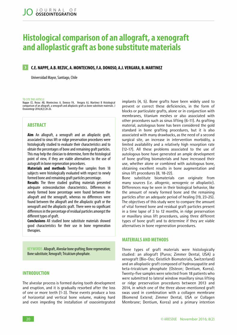
Histological comparison of an allograft, a xenograft and ...
7
20 © ARIESDUE November 2016; 8(2) ABSTRACT Aim An allograft, a xenograft and an alloplastic graft, associated to sinus lift or ridge preservation procedures were histologically studied to evaluate their characteristics and to obtain the percentages of bone and remaining graft particles. This may help the clinician to determine, form the histological point of view, if they are viable alternatives to the use of autograft in bone regeneration procedures. Materials and methods Twenty-five samples from 18 subjects were histologically evaluated with respect to newly formed bone and remaining graft particles percentage. Results The three studied grafting materials presented adequate osteoconduction characteristics. Differences in newly formed bone percentage were found between the allograft and the xenograft, whereas no differences were found between the allograft and the alloplastic graft or the xenograft and the alloplastic graft. There were no significant differences in the percentage of residual particles amongst the different types of graft. Conclusions All studied bone substitute materials showed good characteristics for their use in bone regeneration therapies. Histological comparison of an allograft, a xenograft and alloplastic graft as bone substitute materials C.E. NAPPE, A.B. REZUC, A. MONTECINOS, F.A. DONOSO, A.J. VERGARA, B. MARTINEZ INTRODUCTION The alveolar process is formed during tooth development and eruption, and it is gradually resorbed after the loss of one or more teeth (1-3). These events produce a loss of horizontal and vertical bone volume, making hard and even impeding the installation of osseointegrated implants (4, 5). Bone grafts have been widely used to prevent or correct these deficiencies, in the form of blocks or particulate grafts, alone or in conjunction with membranes, titanium meshes or also associated with other procedures such as sinus lifting (6-11). As grafting material, autologous bone has been considered the gold standard in bone grafting procedures, but it is also associated with many drawbacks, as the need of a second surgical site, an increase in intervention morbidity, a limited availability and a relatively high resorption rate (12-17). All these problems associated to the use of autologous bone have generated an ample development of bone grafting biomaterials and have increased their use, whether alone or combined with autologous bone, obtaining excellent results in bone augmentation and sinus lift procedures (8, 18-22). Bone substitute biomaterials can originate from many sources (i.e. allogenic, xenogenic or alloplastic). Differences may be seen in their biological behavior, like the amount of newly formed bone and the remaining particles after an adequate period of healing (19, 23-25). The objectives of this study were to compare the amount of vital formed bone and residual graft particles present in a time lapse of 3 to 12 months, in ridge preservation or maxillary sinus lift procedures, using three different types of bone graft and to determine if they are viable alternatives in bone regeneration procedures. MATERIALS AND METHODS Three types of graft materials were histologically studied: an allograft (Puros; Zimmer Dental, USA) a xenograft (Bio-Oss; Geistlich Biomaterials, Switzerland) and an alloplastic graft composed of hydroxyapatite and beta-tricalcium phosphate (Osteon; Dentium, Korea). Twenty-five samples were selected from 18 patients who were submitted to lateral window maxillary sinus lifting or ridge preservation procedures between 2013 and 2014, in which one of the three above-mentioned graft swas used in combination with a collagen membrane (Biomend Extend; Zimmer Dental, USA or Collagen Membrane; Dentium, Korea) and a primary intention Universidad Mayor, Santiago, Chile TO CITE THIS ARTICLE Nappe CE, Rezuc AB, Montecinos A, Donoso FA, Vergara AJ, Martinez B Histological comparison of an allograft, a xenograft and alloplastic graft as bone substitute materials. J Osseointegr 2016;8(2):20-26. KEYWORDS Allograft, Alveolar bone grafting; Bone regeneration; Bone substitute; Xenograft; Tricalcium phosphate.
Transcript of Histological comparison of an allograft, a xenograft and ...
20 © ariesdue November 2016; 8(2)
ABSTRACT
Aim An allograft, a xenograft and an alloplastic graft, associated to sinus lift or ridge preservation procedures were histologically studied to evaluate their characteristics and to obtain the percentages of bone and remaining graft particles. This may help the clinician to determine, form the histological point of view, if they are viable alternatives to the use of autograft in bone regeneration procedures. Materials and methods Twenty-five samples from 18 subjects were histologically evaluated with respect to newly formed bone and remaining graft particles percentage. Results The three studied grafting materials presented adequate osteoconduction characteristics. Differences in newly formed bone percentage were found between the allograft and the xenograft, whereas no differences were found between the allograft and the alloplastic graft or the xenograft and the alloplastic graft. There were no significant differences in the percentage of residual particles amongst the different types of graft. Conclusions All studied bone substitute materials showed good characteristics for their use in bone regeneration therapies.
Histological comparison of an allograft, a xenograft and alloplastic graft as bone substitute materials
C.E. NAppE, A.B. REzuC, A. MoNTECiNoS, F.A. DoNoSo, A.J. VERgARA, B. MARTiNEz
iNTRoDuCTioN
The alveolar process is formed during tooth development and eruption, and it is gradually resorbed after the loss of one or more teeth (1-3). These events produce a loss of horizontal and vertical bone volume, making hard and even impeding the installation of osseointegrated
implants (4, 5). Bone grafts have been widely used to prevent or correct these deficiencies, in the form of blocks or particulate grafts, alone or in conjunction with membranes, titanium meshes or also associated with other procedures such as sinus lifting (6-11). As grafting material, autologous bone has been considered the gold standard in bone grafting procedures, but it is also associated with many drawbacks, as the need of a second surgical site, an increase in intervention morbidity, a limited availability and a relatively high resorption rate (12-17). All these problems associated to the use of autologous bone have generated an ample development of bone grafting biomaterials and have increased their use, whether alone or combined with autologous bone, obtaining excellent results in bone augmentation and sinus lift procedures (8, 18-22).Bone substitute biomaterials can originate from many sources (i.e. allogenic, xenogenic or alloplastic). Differences may be seen in their biological behavior, like the amount of newly formed bone and the remaining particles after an adequate period of healing (19, 23-25).The objectives of this study were to compare the amount of vital formed bone and residual graft particles present in a time lapse of 3 to 12 months, in ridge preservation or maxillary sinus lift procedures, using three different types of bone graft and to determine if they are viable alternatives in bone regeneration procedures.
MATERiALS AND METHoDS
Three types of graft materials were histologically studied: an allograft (Puros; Zimmer Dental, USA) a xenograft (Bio-Oss; Geistlich Biomaterials, Switzerland) and an alloplastic graft composed of hydroxyapatite and beta-tricalcium phosphate (Osteon; Dentium, Korea). Twenty-five samples were selected from 18 patients who were submitted to lateral window maxillary sinus lifting or ridge preservation procedures between 2013 and 2014, in which one of the three above-mentioned graft swas used in combination with a collagen membrane (Biomend Extend; Zimmer Dental, USA or Collagen Membrane; Dentium, Korea) and a primary intention
Universidad Mayor, Santiago, Chile
To CiTe ThiS ArTiCleNappe Ce, rezuc AB, Montecinos A, Donoso FA, Vergara AJ, Martinez B histological comparison of an allograft, a xenograft and alloplastic graft as bone substitute materials. J osseointegr 2016;8(2):20-26.
KeyworDS Allograft, Alveolar bone grafting; Bone regeneration; Bone substitute; Xenograft; Tricalcium phosphate.
21
Histological evaluation of three grafting materials for sinus lift and ridge preservation
November 2016; 8(2) © ariesdue
healing was obtained. All patients where non-smokers and systemically healthy, with age ranging from 24 to 67 years. The study was approved by the Mayor University ethic committee (Santiago, Chile, 2014).Every patient signed a written consent regarding his or her acknowledgment and participation on the study.Inclusion criteria were based on patients, on whom allograft, xenograft or alloplastic graft was used, along with ridge preservation or maxillary sinus lift procedures to allow a posterior installation of osseointegrated implants with a healing period between 3 to 12 months.Patients who received grafting materials in combination with autologous bone, other types of grafts or other bioactive substances were excluded from the study.A mucoperiosteal flap was performed in the previously regenerated site and the samples were retrieved using a 2 mm interior diameter and 3 mm external diameter trephine (Dentium, Korea), under abundant irrigation with 0.9% saline solution and at room temperature, to avoid any alteration of the samples due to overheating of the tissue. The depth of the trephine drilling was determined by the amount of previously regenerated bone, measured with a previously taken cone-beam computer tomography, and the length of the planned implant, obtaining a minimum drilling depth of 8 mm. Then, the samples were immediately fixed in 10% buffered formalin and were divided according to each patient, graft material used and type of grafting procedure previously performed. The histological processing of the samples was performed at the department of pathological anatomy (Mayor University Dentitry School, Santiago, Chile). Six longitudinal cuts, 8 x 2.5 mm, for each sample were made. They were subjected to decalcification and staining with haematoxylin–eosin. The vital bone was stained with a characteristic eosinophilic color highlighting concentric apposition of bone and basophilic dots corresponding to osteocytes. On the other side, the remaining graft particles were completely stained with a basophilic color.A descriptive analysis of the characteristics of the bone/graft particle interface was made for all samples, using an optical microscope (DM 750 Leica, Germany) at a 40 times magnification. Four times magnification microphotographs were obtained for the different samples using a 1920 x 1080 pixel camera (ICC50HD, Leica. Germany) installed in the same optical microscope. All photographs were saved in the same resolution (1980x1020) and size, using an image processing software, provided by the camera manufacturer (Leica Suite EZ, Leica, Germany). Another computer software (Image J; NIH, Bethesda, MD, USA) was then used to calculate the percentage of newly formed bone and remaining graft particles in each sample by selecting the image color threshold, using the hue – brightness and saturation filter according to the sample staining. To isolate the newly formed bone, eosinophilic color
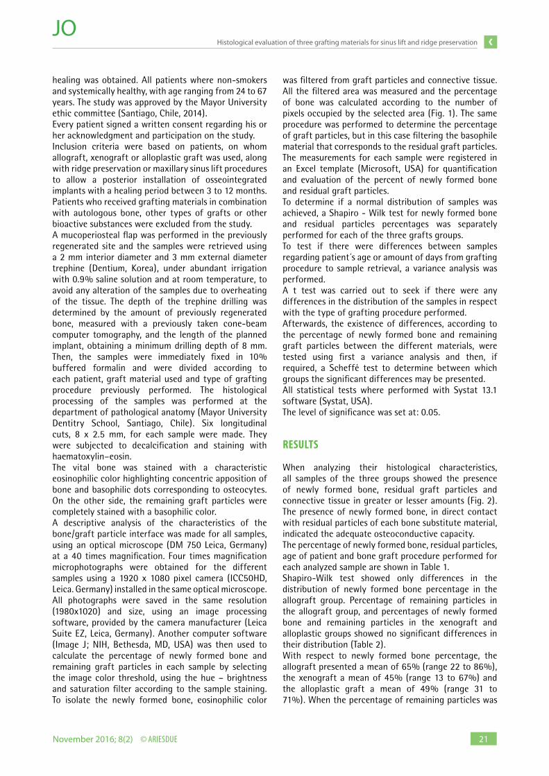
was filtered from graft particles and connective tissue. All the filtered area was measured and the percentage of bone was calculated according to the number of pixels occupied by the selected area (Fig. 1). The same procedure was performed to determine the percentage of graft particles, but in this case filtering the basophile material that corresponds to the residual graft particles. The measurements for each sample were registered in an Excel template (Microsoft, USA) for quantification and evaluation of the percent of newly formed bone and residual graft particles. To determine if a normal distribution of samples was achieved, a Shapiro - Wilk test for newly formed bone and residual particles percentages was separately performed for each of the three grafts groups.To test if there were differences between samples regarding patient´s age or amount of days from grafting procedure to sample retrieval, a variance analysis was performed. A t test was carried out to seek if there were any differences in the distribution of the samples in respect with the type of grafting procedure performed.Afterwards, the existence of differences, according to the percentage of newly formed bone and remaining graft particles between the different materials, were tested using first a variance analysis and then, if required, a Scheffé test to determine between which groups the significant differences may be presented. All statistical tests where performed with Systat 13.1 software (Systat, USA).The level of significance was set at: 0.05.
RESuLTS
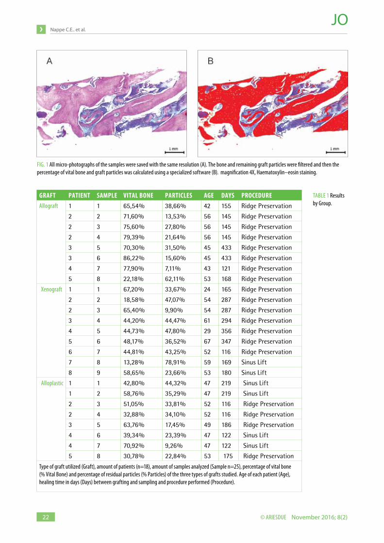
When analyzing their histological characteristics, all samples of the three groups showed the presence of newly formed bone, residual graft particles and connective tissue in greater or lesser amounts (Fig. 2). The presence of newly formed bone, in direct contact with residual particles of each bone substitute material, indicated the adequate osteoconductive capacity. The percentage of newly formed bone, residual particles, age of patient and bone graft procedure performed for each analyzed sample are shown in Table 1. Shapiro-Wilk test showed only differences in the distribution of newly formed bone percentage in the allograft group. Percentage of remaining particles in the allograft group, and percentages of newly formed bone and remaining particles in the xenograft and alloplastic groups showed no significant differences in their distribution (Table 2).With respect to newly formed bone percentage, the allograft presented a mean of 65% (range 22 to 86%), the xenograft a mean of 45% (range 13 to 67%) and the alloplastic graft a mean of 49% (range 31 to 71%). When the percentage of remaining particles was
22
Nappe C.E.. et al.
© ariesdue November 2016; 8(2)
gRAFT pATiENT SAMpLE ViTAL BoNE pARTiCLES AgE DAyS pRoCEDuREAllograft
1 1 65,54% 38,66% 42 155 Ridge Preservation
2 2 71,60% 13,53% 56 145 Ridge Preservation
2 3 75,60% 27,80% 56 145 Ridge Preservation
2 4 79,39% 21,64% 56 145 Ridge Preservation
3 5 70,30% 31,50% 45 433 Ridge Preservation
3 6 86,22% 15,60% 45 433 Ridge Preservation
4 7 77,90% 7,11% 43 121 Ridge Preservation
5 8 22,18% 62,11% 53 168 Ridge Preservation
Xenograft
1 1 67,20% 33,67% 24 165 Ridge Preservation
2 2 18,58% 47,07% 54 287 Ridge Preservation
2 3 65,40% 9,90% 54 287 Ridge Preservation
3 4 44,20% 44,47% 61 294 Ridge Preservation
4 5 44,73% 47,80% 29 356 Ridge Preservation
5 6 48,17% 36,52% 67 347 Ridge Preservation
6 7 44,81% 43,25% 52 116 Ridge Preservation
7 8 13,28% 78,91% 59 169 Sinus Lift
8 9 58,65% 23,66% 53 180 Sinus Lift
Alloplastic
1 1 42,80% 44,32% 47 219 Sinus Lift
1 2 58,76% 35,29% 47 219 Sinus Lift
2 3 51,05% 33,81% 52 116 Ridge Preservation
2 4 32,88% 34,10% 52 116 Ridge Preservation
3 5 63,76% 17,45% 49 186 Ridge Preservation
4 6 39,34% 23,39% 47 122 Sinus Lift
4 7 70,92% 9,26% 47 122 Sinus Lift
5 8 30,78% 22,84% 53 175 Ridge Preservation
Type of graft utilized (Graft), amount of patients (n=18), amount of samples analyzed (Sample n=25), percentage of vital bone (% Vital Bone) and percentage of residual particles (% Particles) of the three types of grafts studied. Age of each patient (Age), healing time in days (Days) between grafting and sampling and procedure performed (Procedure).
TABle 1 results by Group.
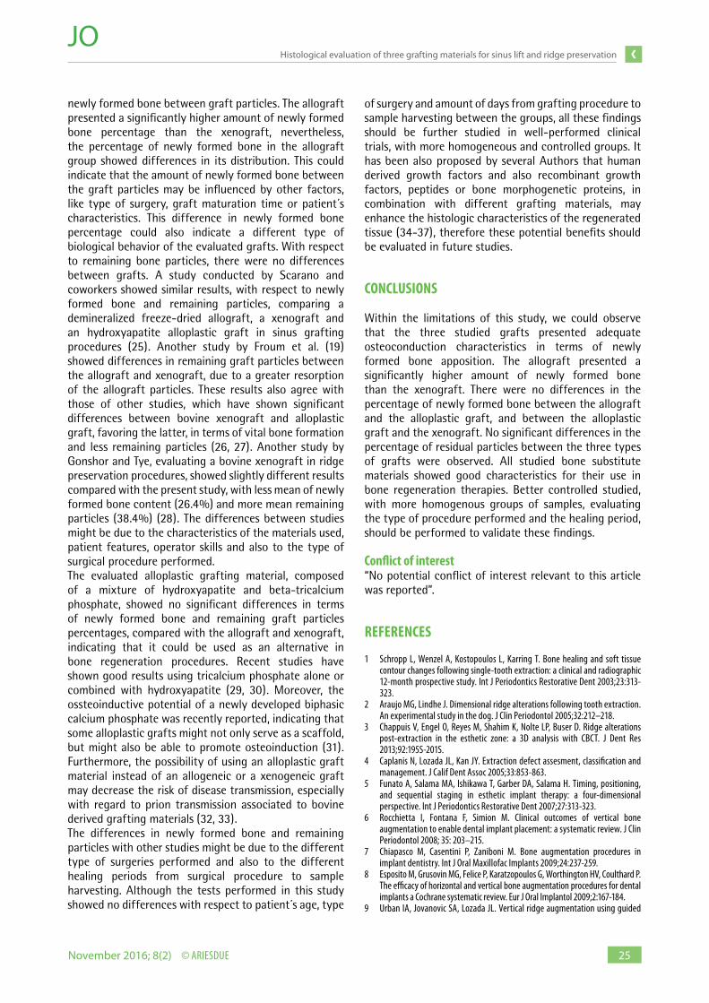
FiG. 1 All micro-photographs of the samples were saved with the same resolution (A). The bone and remaining graft particles were filtered and then the percentage of vital bone and graft particles was calculated using a specialized software (B). magnification 4X, haematoxylin–eosin staining.
23
Histological evaluation of three grafting materials for sinus lift and ridge preservation
November 2016; 8(2) © ariesdue
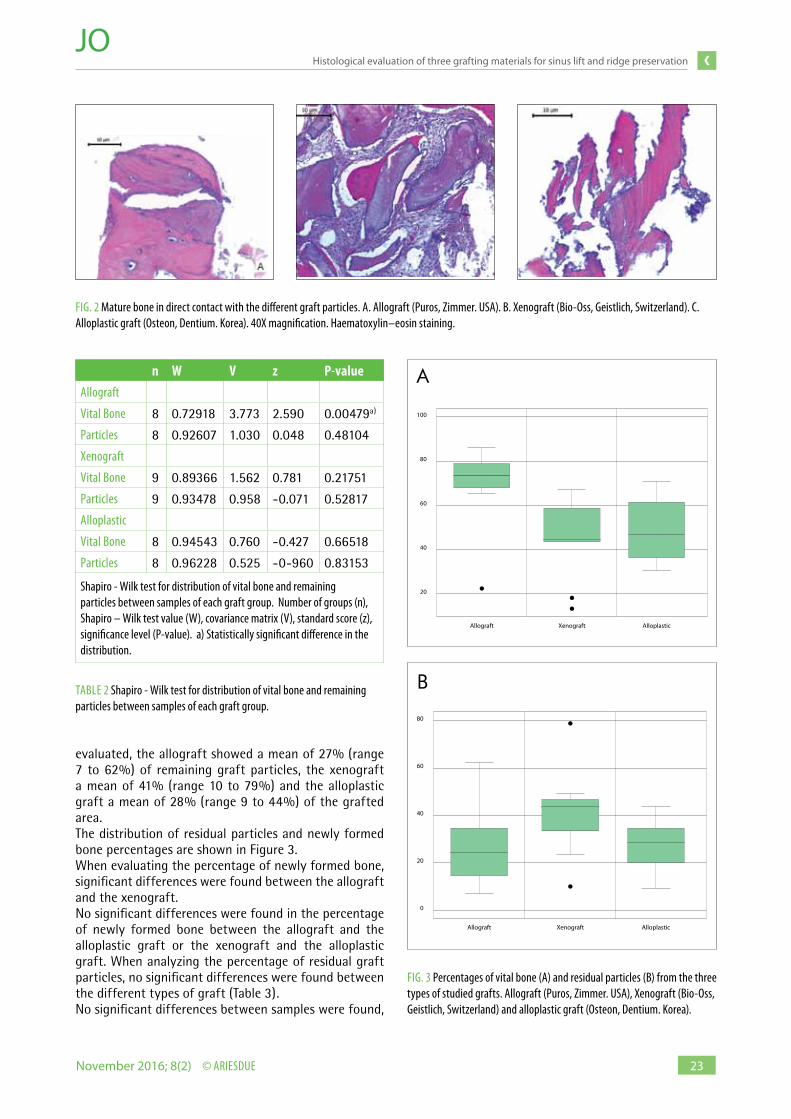
evaluated, the allograft showed a mean of 27% (range 7 to 62%) of remaining graft particles, the xenograft a mean of 41% (range 10 to 79%) and the alloplastic graft a mean of 28% (range 9 to 44%) of the grafted area.The distribution of residual particles and newly formed bone percentages are shown in Figure 3.When evaluating the percentage of newly formed bone, significant differences were found between the allograft and the xenograft. No significant differences were found in the percentage of newly formed bone between the allograft and the alloplastic graft or the xenograft and the alloplastic graft. When analyzing the percentage of residual graft particles, no significant differences were found between the different types of graft (Table 3).No significant differences between samples were found,
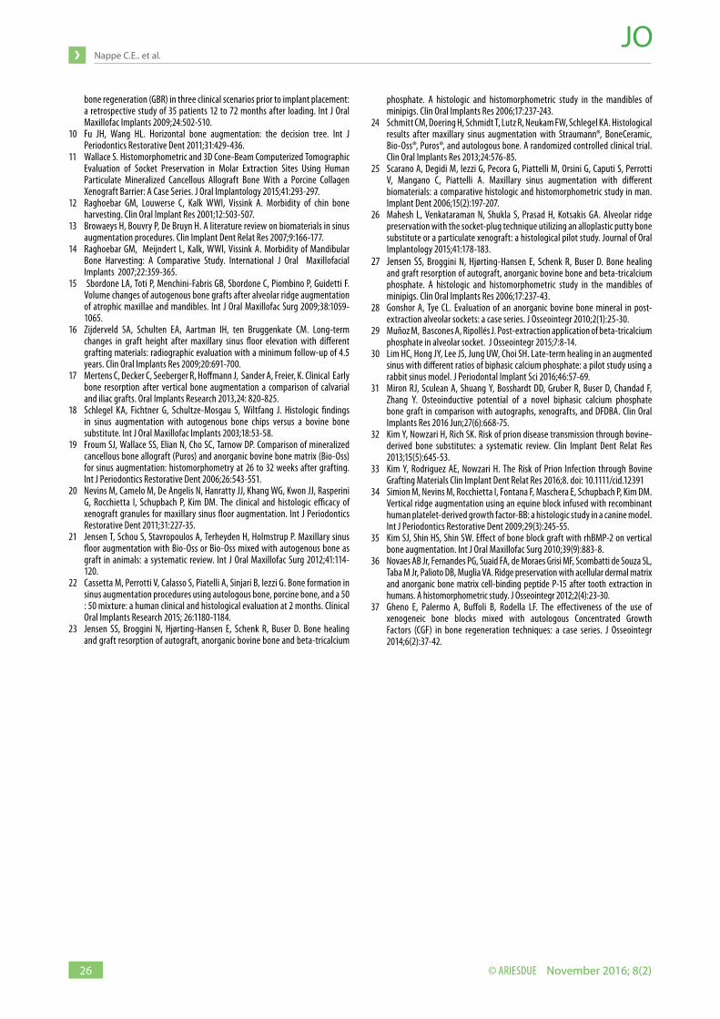
FiG. 2 Mature bone in direct contact with the different graft particles. A. Allograft (Puros, Zimmer. USA). B. Xenograft (Bio-oss, Geistlich, Switzerland). C. Alloplastic graft (osteon, Dentium. Korea). 40X magnification. haematoxylin–eosin staining.
FiG. 3 Percentages of vital bone (A) and residual particles (B) from the three types of studied grafts. Allograft (Puros, Zimmer. USA), Xenograft (Bio-oss, Geistlich, Switzerland) and alloplastic graft (osteon, Dentium. Korea).
Allograft
20
40
60
80
100
Xenograft Alloplastic
Allograft
0
40
20
60
80
Xenograft Alloplastic
A
B
n W V z p-valueAllograft
Vital Bone 8 0.72918 3.773 2.590 0.00479a)
Particles 8 0.92607 1.030 0.048 0.48104
Xenograft
Vital Bone 9 0.89366 1.562 0.781 0.21751
Particles 9 0.93478 0.958 -0.071 0.52817
Alloplastic
Vital Bone 8 0.94543 0.760 -0.427 0.66518
Particles 8 0.96228 0.525 -0-960 0.83153
Shapiro - wilk test for distribution of vital bone and remaining particles between samples of each graft group. Number of groups (n), Shapiro – wilk test value (w), covariance matrix (V), standard score (z), significance level (P-value). a) Statistically significant difference in the distribution.
TABle 2 Shapiro - wilk test for distribution of vital bone and remaining particles between samples of each graft group.
24
Nappe C.E.. et al.
© ariesdue November 2016; 8(2)
in respect with patient´s age and amount of days from grafting procedure to sample retrieval between the three groups (Table 4). There were also no differences, regarding newly formed bone and remaining particles, with respect to the type of procedure, in the xenograft and alloplastic graft groups (Table 5).
DiSCuSSioN
According to the results found in the present study, it was possible to microscopically observe that the different bone grafting materials presented osteoconductive characteristics that allowed apposition of mature vital bone in direct contact; the newly formed bone was characterized by the presence of osteocytes in the inner part, as well as osteoblastic and osteoclastic activity in the outer part. These findings indicate that the three materials may be suitable for bone augmentation procedures due to their osteoconduction characteristics. There were, however, differences in the percentage of
n ± SDViTAl BoNe
Allograft 8 69 ± 20
Xenograft 9 45 ± 19
Alloplastic 8 49 ± 15
ANoVA F= 4,12; df = 22; p = 0.030 a)
Sheffé Allograft vs. Xenograft
T= 2.7081; p= 0.042 b)
Allograft vs. Alloplastic
T= 2.2096; p= 0.110
Xenograft vs. Alloplastic
T= 0.4344; p= 0.910
PArTiCleS
Allograft 8 27 ± 17
Xenograft 9 41 ± 19
Alloplastic 8 28 ± 11
ANoVA F= 1,87; df = 22; p = 0.178
Number of samples (n), mean ( ), standard deviation (SD), ANoVA critical value (F), Sheffé test critical value (T), degrees of freedom (df) and significance level (p).a) Statistically significant difference between groups. b) Statistically significant difference between these two groups
TABle 3 Variance analysis for vital bone percentage and remaining particles percentage.
TABle 4 Variance analysis for patient s age and amount of days from grafting procedure to sample obtaining.
n ± SDAge
Allograft 8 49.500 ± 6,302
Xenograft 9 50,333 ± 14,370
Alloplastic 8 49,250 ± 2,659
ANoVA F= 0,031; df = 22; p = 0.970
Days Grafted
Allograft 8 218,125 ± 133,264
Xenograft 9 244,556 ± 87,817
Alloplastic 8 159,375 ± 45,701
ANoVA F= 1,741; df = 22; p = 0.199
Number of samples (n), mean ( ), standard deviation (SD), ANoVA critical value (F), degrees of freedom (df) and significance level (p).
TABle 5 The differences between xenograft and alloplastic graft groups for vital bone and remaining particles with respect to the type of procedure..
n ± SDXenograft - Vital Bone
ridge Preservation
7 47.584 ± 16.182
Sinus lift 2 35.965 ± 32.081
T test t= 0,752; df = 7; c = 2.365; p > 0.05
Xenograft - Particles
ridge Preservation
7 37.526 ± 13.271
Sinus lift 2 51.285 ± 39.068
T test t= -0,893; df = 7; c = 2.365; p > 0.05
Alloplastic Graft - Vital Bone
ridge Preservation
4 44.598 ± 15.694
Sinus lift 4 52.955 ± 14.662
T test t= -0,778; df = 6; c = 2.447; p > 0.05
Alloplastic Graft - Particles
ridge Preservation
4 27.050 ± 8.272
Sinus lift 4 28.065 ± 15.187
T test t= -0,117; df = 6; c = 2.447; p > 0.05
Number of samples (n), mean ( ), standard deviation (SD), t value (t), degrees of freedom (df), critical value (c) and significance level (p)
25
Histological evaluation of three grafting materials for sinus lift and ridge preservation
November 2016; 8(2) © ariesdue
newly formed bone between graft particles. The allograft presented a significantly higher amount of newly formed bone percentage than the xenograft, nevertheless, the percentage of newly formed bone in the allograft group showed differences in its distribution. This could indicate that the amount of newly formed bone between the graft particles may be influenced by other factors, like type of surgery, graft maturation time or patient´s characteristics. This difference in newly formed bone percentage could also indicate a different type of biological behavior of the evaluated grafts. With respect to remaining bone particles, there were no differences between grafts. A study conducted by Scarano and coworkers showed similar results, with respect to newly formed bone and remaining particles, comparing a demineralized freeze-dried allograft, a xenograft and an hydroxyapatite alloplastic graft in sinus grafting procedures (25). Another study by Froum et al. (19) showed differences in remaining graft particles between the allograft and xenograft, due to a greater resorption of the allograft particles. These results also agree with those of other studies, which have shown significant differences between bovine xenograft and alloplastic graft, favoring the latter, in terms of vital bone formation and less remaining particles (26, 27). Another study by Gonshor and Tye, evaluating a bovine xenograft in ridge preservation procedures, showed slightly different results compared with the present study, with less mean of newly formed bone content (26.4%) and more mean remaining particles (38.4%) (28). The differences between studies might be due to the characteristics of the materials used, patient features, operator skills and also to the type of surgical procedure performed. The evaluated alloplastic grafting material, composed of a mixture of hydroxyapatite and beta-tricalcium phosphate, showed no significant differences in terms of newly formed bone and remaining graft particles percentages, compared with the allograft and xenograft, indicating that it could be used as an alternative in bone regeneration procedures. Recent studies have shown good results using tricalcium phosphate alone or combined with hydroxyapatite (29, 30). Moreover, the ossteoinductive potential of a newly developed biphasic calcium phosphate was recently reported, indicating that some alloplastic grafts might not only serve as a scaffold, but might also be able to promote osteoinduction (31). Furthermore, the possibility of using an alloplastic graft material instead of an allogeneic or a xenogeneic graft may decrease the risk of disease transmission, especially with regard to prion transmission associated to bovine derived grafting materials (32, 33).The differences in newly formed bone and remaining particles with other studies might be due to the different type of surgeries performed and also to the different healing periods from surgical procedure to sample harvesting. Although the tests performed in this study showed no differences with respect to patient´s age, type
of surgery and amount of days from grafting procedure to sample harvesting between the groups, all these findings should be further studied in well-performed clinical trials, with more homogeneous and controlled groups. It has been also proposed by several Authors that human derived growth factors and also recombinant growth factors, peptides or bone morphogenetic proteins, in combination with different grafting materials, may enhance the histologic characteristics of the regenerated tissue (34-37), therefore these potential benefits should be evaluated in future studies.
CoNCLuSioNS
Within the limitations of this study, we could observe that the three studied grafts presented adequate osteoconduction characteristics in terms of newly formed bone apposition. The allograft presented a significantly higher amount of newly formed bone than the xenograft. There were no differences in the percentage of newly formed bone between the allograft and the alloplastic graft, and between the alloplastic graft and the xenograft. No significant differences in the percentage of residual particles between the three types of grafts were observed. All studied bone substitute materials showed good characteristics for their use in bone regeneration therapies. Better controlled studied, with more homogenous groups of samples, evaluating the type of procedure performed and the healing period, should be performed to validate these findings.
Conflict of interest“No potential conflict of interest relevant to this article was reported”.
REFERENCES
1 Schropp l, wenzel A, Kostopoulos l, Karring T. Bone healing and soft tissue contour changes following single-tooth extraction: a clinical and radiographic 12-month prospective study. int J Periodontics restorative Dent 2003;23:313-323.
2 Araujo MG, lindhe J. Dimensional ridge alterations following tooth extraction. An experimental study in the dog. J Clin Periodontol 2005;32:212–218.
3 Chappuis V, engel o, reyes M, Shahim K, Nolte lP, Buser D. ridge alterations post-extraction in the esthetic zone: a 3D analysis with CBCT. J Dent res 2013;92:195S-201S.
4 Caplanis N, lozada Jl, Kan Jy. extraction defect assesment, classification and management. J Calif Dent Assoc 2005;33:853-863.
5 Funato A, Salama MA, ishikawa T, Garber DA, Salama h. Timing, positioning, and sequential staging in esthetic implant therapy: a four-dimensional perspective. int J Periodontics restorative Dent 2007;27:313-323.
6 rocchietta i, Fontana F, Simion M. Clinical outcomes of vertical bone augmentation to enable dental implant placement: a systematic review. J Clin Periodontol 2008; 35: 203–215.
7 Chiapasco M, Casentini P, Zaniboni M. Bone augmentation procedures in implant dentistry. int J oral Maxillofac implants 2009;24:237-259.
8 esposito M, Grusovin MG, Felice P, Karatzopoulos G, worthington hV, Coulthard P. The efficacy of horizontal and vertical bone augmentation procedures for dental implants a Cochrane systematic review. eur J oral implantol 2009;2:167-184.
9 Urban iA, Jovanovic SA, lozada Jl. Vertical ridge augmentation using guided
26
Nappe C.E.. et al.
© ariesdue November 2016; 8(2)
bone regeneration (GBr) in three clinical scenarios prior to implant placement: a retrospective study of 35 patients 12 to 72 months after loading. int J oral Maxillofac implants 2009;24:502-510.
10 Fu Jh, wang hl. horizontal bone augmentation: the decision tree. int J Periodontics restorative Dent 2011;31:429-436.
11 wallace S. histomorphometric and 3D Cone-Beam Computerized Tomographic evaluation of Socket Preservation in Molar extraction Sites Using human Particulate Mineralized Cancellous Allograft Bone with a Porcine Collagen Xenograft Barrier: A Case Series. J oral implantology 2015;41:293-297.
12 raghoebar GM, louwerse C, Kalk wwi, Vissink A. Morbidity of chin bone harvesting. Clin oral implant res 2001;12:503-507.
13 Browaeys h, Bouvry P, De Bruyn h. A literature review on biomaterials in sinus augmentation procedures. Clin implant Dent relat res 2007;9:166-177.
14 raghoebar GM, Meijndert l, Kalk, wwi, Vissink A. Morbidity of Mandibular Bone harvesting: A Comparative Study. international J oral Maxillofacial implants 2007;22:359-365.
15 Sbordone lA, Toti P, Menchini-Fabris GB, Sbordone C, Piombino P, Guidetti F. Volume changes of autogenous bone grafts after alveolar ridge augmentation of atrophic maxillae and mandibles. int J oral Maxillofac Surg 2009;38:1059-1065.
16 Zijderveld SA, Schulten eA, Aartman ih, ten Bruggenkate CM. long-term changes in graft height after maxillary sinus floor elevation with different grafting materials: radiographic evaluation with a minimum follow-up of 4.5 years. Clin oral implants res 2009;20:691-700.
17 Mertens C, Decker C, Seeberger r, hoffmann J, Sander A, Freier, K. Clinical early bone resorption after vertical bone augmentation a comparison of calvarial and iliac grafts. oral implants research 2013,24: 820-825.
18 Schlegel KA, Fichtner G, Schultze-Mosgau S, wiltfang J. histologic findings in sinus augmentation with autogenous bone chips versus a bovine bone substitute. int J oral Maxillofac implants 2003;18:53-58.
19 Froum SJ, wallace SS, elian N, Cho SC, Tarnow DP. Comparison of mineralized cancellous bone allograft (Puros) and anorganic bovine bone matrix (Bio-oss) for sinus augmentation: histomorphometry at 26 to 32 weeks after grafting. int J Periodontics restorative Dent 2006;26:543-551.
20 Nevins M, Camelo M, De Angelis N, hanratty JJ, Khang wG, Kwon JJ, rasperini G, rocchietta i, Schupbach P, Kim DM. The clinical and histologic efficacy of xenograft granules for maxillary sinus floor augmentation. int J Periodontics restorative Dent 2011;31:227-35.
21 Jensen T, Schou S, Stavropoulos A, Terheyden h, holmstrup P. Maxillary sinus floor augmentation with Bio-oss or Bio-oss mixed with autogenous bone as graft in animals: a systematic review. int J oral Maxillofac Surg 2012;41:114-120.
22 Cassetta M, Perrotti V, Calasso S, Piatelli A, Sinjari B, iezzi G. Bone formation in sinus augmentation procedures using autologous bone, porcine bone, and a 50 : 50 mixture: a human clinical and histological evaluation at 2 months. Clinical oral implants research 2015; 26:1180-1184.
23 Jensen SS, Broggini N, hjørting-hansen e, Schenk r, Buser D. Bone healing and graft resorption of autograft, anorganic bovine bone and beta-tricalcium
phosphate. A histologic and histomorphometric study in the mandibles of minipigs. Clin oral implants res 2006;17:237-243.
24 Schmitt CM, Doering h, Schmidt T, lutz r, Neukam Fw, Schlegel KA. histological results after maxillary sinus augmentation with Straumann®, BoneCeramic, Bio-oss®, Puros®, and autologous bone. A randomized controlled clinical trial. Clin oral implants res 2013;24:576-85.
25 Scarano A, Degidi M, iezzi G, Pecora G, Piattelli M, orsini G, Caputi S, Perrotti V, Mangano C, Piattelli A. Maxillary sinus augmentation with different biomaterials: a comparative histologic and histomorphometric study in man. implant Dent 2006;15(2):197-207.
26 Mahesh l, Venkataraman N, Shukla S, Prasad h, Kotsakis GA. Alveolar ridge preservation with the socket-plug technique utilizing an alloplastic putty bone substitute or a particulate xenograft: a histological pilot study. Journal of oral implantology 2015;41:178-183.
27 Jensen SS, Broggini N, hjørting-hansen e, Schenk r, Buser D. Bone healing and graft resorption of autograft, anorganic bovine bone and beta-tricalcium phosphate. A histologic and histomorphometric study in the mandibles of minipigs. Clin oral implants res 2006;17:237-43.
28 Gonshor A, Tye Cl. evaluation of an anorganic bovine bone mineral in post-extraction alveolar sockets: a case series. J osseointegr 2010;2(1):25-30.
29 Muñoz M, Bascones A, ripollés J. Post-extraction application of beta-tricalcium phosphate in alveolar socket. J osseointegr 2015;7:8-14.
30 lim hC, hong Jy, lee JS, Jung Uw, Choi Sh. late-term healing in an augmented sinus with different ratios of biphasic calcium phosphate: a pilot study using a rabbit sinus model. J Periodontal implant Sci 2016;46:57-69.
31 Miron rJ, Sculean A, Shuang y, Bosshardt DD, Gruber r, Buser D, Chandad F, Zhang y. osteoinductive potential of a novel biphasic calcium phosphate bone graft in comparison with autographs, xenografts, and DFDBA. Clin oral implants res 2016 Jun;27(6):668-75.
32 Kim y, Nowzari h, rich SK. risk of prion disease transmission through bovine-derived bone substitutes: a systematic review. Clin implant Dent relat res 2013;15(5):645-53.
33 Kim y, rodriguez Ae, Nowzari h. The risk of Prion infection through Bovine Grafting Materials Clin implant Dent relat res 2016;8. doi: 10.1111/cid.12391
34 Simion M, Nevins M, rocchietta i, Fontana F, Maschera e, Schupbach P, Kim DM. Vertical ridge augmentation using an equine block infused with recombinant human platelet-derived growth factor-BB: a histologic study in a canine model. int J Periodontics restorative Dent 2009;29(3):245-55.
35 Kim SJ, Shin hS, Shin Sw. effect of bone block graft with rhBMP-2 on vertical bone augmentation. int J oral Maxillofac Surg 2010;39(9):883-8.
36 Novaes AB Jr, Fernandes PG, Suaid FA, de Moraes Grisi MF, Scombatti de Souza Sl, Taba M Jr, Palioto DB, Muglia VA. ridge preservation with acellular dermal matrix and anorganic bone matrix cell-binding peptide P-15 after tooth extraction in humans. A histomorphometric study. J osseointegr 2012;2(4):23-30.
37 Gheno e, Palermo A, Buffoli B, rodella lF. The effectiveness of the use of xenogeneic bone blocks mixed with autologous Concentrated Growth Factors (CGF) in bone regeneration techniques: a case series. J osseointegr 2014;6(2):37-42.