His to Tech Staining (UPDATED)
51
Staining Ma. Minda Luz Meneses-Manuguid, M.D.
-
Upload
kevz-nicdao -
Category
Documents
-
view
663 -
download
1
Transcript of His to Tech Staining (UPDATED)
Staining
Ma. Minda Luz Meneses-Manuguid, M.D.

Staining
• the process of applying stains/dyes to tissue sections
• purpose: to make tissues/cells/structures visible & appreciable; enables one to study morphology & placement of cells & tissues
• stains/dyes : colored substances: natural/synthetic;
• different tissues have different affinities for different dyes
• acidic tissues (e.g. nucleoplasm) have an affinity for basic dyes (e.g. Hematoxylin); basic tissues (e.g. cytoplasmic proteins) have a greater affinity for acidic stains (e.g. Eosin)
• processes involved: Osmosis, Solvation, Adsorption, Absorption
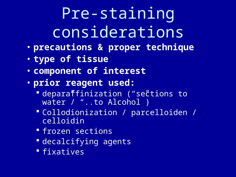
Pre-staining considerations
• precautions & proper technique• type of tissue• component of interest• prior reagent used:
deparaffinization (“sections to water”/ “..to Alcohol”) Collodionization / parcelloiden / celloidin frozen sections decalcifying agents fixatives

deparaffination
• Paraffin wax is almost impermeable to most staining solutions & must therefore be removed before staining the tissue sections
• Sections to Water - the reverse of Impregnation, the removal of paraffin from a tissue section by successive immersions in a solvent (e.g. Xylene), decreasing grades of Alcohol, & finally Water
• Sections to Alcohol – same as above up to Alcohol immersions only – if an alcoholic stain is to be used
collodionization• Collodionization – coating a slide with a
dilute solution of thin Celloidin for better attachment of the paraffin ribbon, especially those that are torn, inadequately infiltrated, or contain air bubbles; those that contain Glycogen; & those that will be subjected to strong Alkaline or strong Acid solutions.
• Procedure: immerse in Xylene (to remove Paraffin), immerse in absolute Alcohol, in dilute Ether Alcohol solution, then in dilute Ether solution of Celloidin; hold slide at one end to drain(1 min); wipe off back of slide & place in 80% Alcohol 3-5 mins. to harden the Celloidin
Fixatives
• Mercuric chloride & formalin–fixed tissues favor staining with basic dyes
• Trichloroacetic acid, picric acid, & Chromium –fixed tissues favor staining with acidic dyes
• Ethanol or Acetic acid –fixed tissues readily take up both acidic & basic dyes

Types of Dye• Natural dyes – obtained from plants or
animals – cochineal dyes & derivatives, Hematoxylin Saffron vegetable extracts e.g. Orcein
• Synthetic dyes – aniline / coal tar dyes – originally manufactured from substances taken from coal tar, derived
from Benzene (C6H6) Chromophores – substances with definite atomic groupings capable
of producing visible colors Chromogen – simple Benzene compound with chromophore – by
itself, imparts color that is easily removed, so must be combined with an auxiliary salt-forming substance (Auxochome) for color retention (dyeing)

Stains: Hematoxylin • extracted from the heartwood of Hematoxylin
campechianum • active coloring agent : Hematin (from oxidizing of
Hematoxylin by exposure to air & sunlight (“ripening”) or by the addition of strong oxidizing agents like H2O2, mercuric oxide, KMnO4, sodium perborate or sodium Iodate
• usually combined with Alum, Iron, Chromium, Copper salts (mordants) to improve tissue affinity
• Alum Hematoxylin – for progressive staining – usually counterstained with Eosin, Congo Red, & Safranin; in Harris’ solution (ripened with mercuric chloride), in Ehrlich’s reagent (ripened with sodium Iodate)
• Iron Hematoxylin – for differential or regressive staining – usually with Acid Alcohol as differentiating agent; in Weigert’s stain with Ferric Chloride
• Copper Hematoxylin – for spermatogenesis studies
Stains • Cochineal dye – derived from an extract
from the female cochineal bug (Coccus cacti), treated with Alum to produce Carmine – powerful chromatin/nuclear stain for fresh specimens & smears Picrocarmine – with Picric acid – in neuropathological studies Best’s Carmine – with Aluminun chloride – to demonstrate
Glycogen • Orcein – extract from certain
Lichens(normally colorless) treated with Ammonia & exposed to air to produce blue or violet colors weak acid, soluble in Alkali mainly used for staining Elastic fibers
Synthetic Dyes• Acid dyes – the chromophore is found in the acid
component; base is usually Sodium Picric acid – can fix, differentiate, stain tissues by itself; forms salt
with an alkali; may be used as a fixative, decalcifying agent, or tissue softener; useful as counterstain to acid fuchsin in van Gieson’s stain, & to Crystal violet in examining fungi
• Basic dyes – coloring substance is found in the basic component; acid is usually acetic, sulfuric or hydrochloric Methylene blue – may be used as indicator or as a dye; widely
used in Microbiology, evaluation of Milk, diagnosis of diphtheria• Neutral dyes – combination of aqueous solutions
of acid & basic dyes, capable of staining nucleus & cytoplasm simultaneously & differentially; usually soluble in Alcohol, insoluble in water Giemsa, Leishman’s stain – for peripheral blood smears
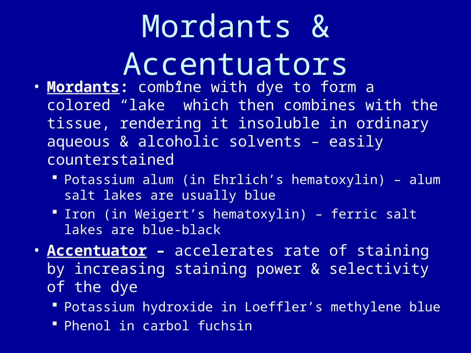
Mordants & Accentuators• Mordants: combine with dye to form a
colored “lake” which then combines with the tissue, rendering it insoluble in ordinary aqueous & alcoholic solvents – easily counterstained Potassium alum (in Ehrlich’s hematoxylin) – alum salt lakes are usually
blue Iron (in Weigert’s hematoxylin) – ferric salt lakes are blue-black
• Accentuator – accelerates rate of staining by increasing staining power & selectivity of the dye Potassium hydroxide in Loeffler’s methylene blue Phenol in carbol fuchsin
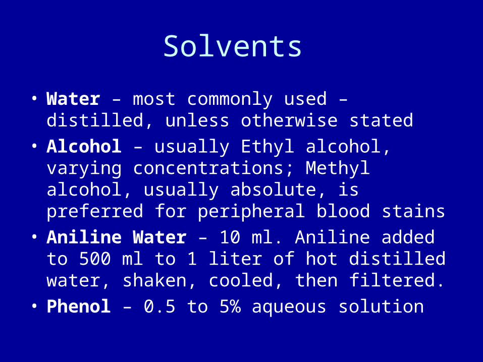
Solvents
• Water – most commonly used – distilled, unless otherwise stated
• Alcohol – usually Ethyl alcohol, varying concentrations; Methyl alcohol, usually absolute, is preferred for peripheral blood stains
• Aniline Water – 10 ml. Aniline added to 500 ml to 1 liter of hot distilled water, shaken, cooled, then filtered.
• Phenol – 0.5 to 5% aqueous solution

Staining

Staining Methods
• Direct• Indirect• Progressive• Regressive• Metachromatic• Differential /• Counterstainin
g
• Microanatomical /
• Histological• Histochemical• Vital • Intravital• Supravital • Metallic
impregnation
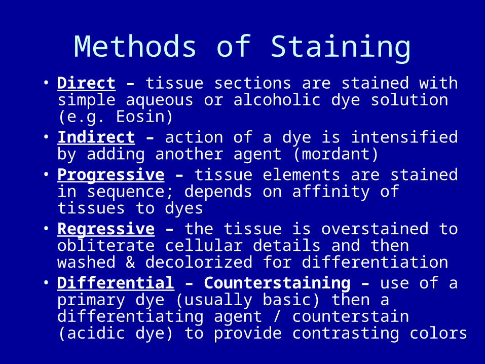
Methods of Staining• Direct – tissue sections are stained with
simple aqueous or alcoholic dye solution (e.g. Eosin)
• Indirect – action of a dye is intensified by adding another agent (mordant)
• Progressive – tissue elements are stained in sequence; depends on affinity of tissues to dyes
• Regressive – the tissue is overstained to obliterate cellular details and then washed & decolorized for differentiation
• Differential – Counterstaining – use of a primary dye (usually basic) then a differentiating agent / counterstain (acidic dye) to provide contrasting colors

Staining Methods • Metachromatic – use of specific dyes
(cations) which differentiate particular substances by staining them with a color different from that of the stain itself. Used to stain cartilage, mucin, mast cell granules, amyloid, connective tissues: basic dyes of the thizine & triphenylmethane groups Methyl violet/crystal violet ▪ Methylene blue Cresyl blue (for retic) ▪ Toluidine blue Safranin ▪ Thionine Bismarck brown ▪ Azure A, B, C Basic fuchsin

Staining Methods• Histological / Microanatomical staining:
tissue constituents interact directly with staining solutions; to demonstrate general relationship of tissues & cells and differentiate nucleus from cytoplasm Cytoplasmic staining – demonstrates minute specific
structures found in cytoplasm & nucleus Negative staining – demonstrates bacterial morphology –
substance & organisms are mixed on the slide (unstained organism in a black background)
• Histochemical staining: dyes react chemically with specific parts of the cell
• Immunohistochemical staining: use of antibodies to induce a reaction with specific antigenic components of cells/tissues
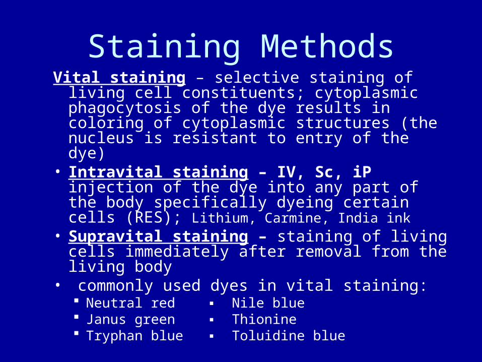
Staining MethodsVital staining – selective staining of living
cell constituents; cytoplasmic phagocytosis of the dye results in coloring of cytoplasmic structures (the nucleus is resistant to entry of the dye)
• Intravital staining – IV, Sc, iP injection of the dye into any part of the body specifically dyeing certain cells (RES); Lithium, Carmine, India ink
• Supravital staining – staining of living cells immediately after removal from the living body
• commonly used dyes in vital staining: Neutral red ▪ Nile blue Janus green ▪ Thionine Tryphan blue ▪ Toluidine blue

Staining Methods• Counterstaining – application of a
different color or stain to provide contrast/background to the structure being demonstrated Cytoplasmic stains: red – EosinY, Eosin B, Phloxine B; yellow –
Picric acid, OrangeG, rose Bengal; green – light green SF, Lissamine green
Nuclear stains: red – neutral Red, Safranin O, Carmine, Hematoxylin; blue – Methylene blue, Toluidine blue, Celestine blue
• Metallic impregnation – certain tissue elements reduce colorless metallic salt solutions to produce a black (usually) precipitate on the surface of the tissue Silver nitrate ▪ Gold chloride
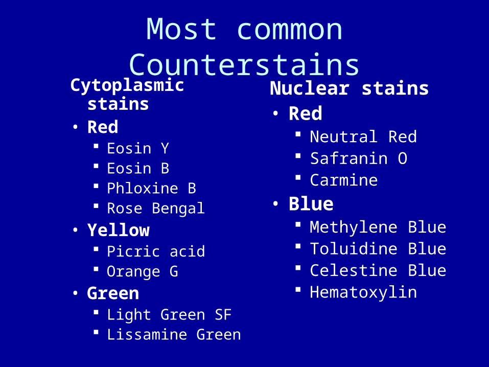
Most common CounterstainsCytoplasmic
stains• Red
Eosin Y Eosin B Phloxine B Rose Bengal
• Yellow Picric acid Orange G
• Green Light Green SF Lissamine Green
Nuclear stains• Red
Neutral Red Safranin O Carmine
• Blue Methylene Blue Toluidine Blue Celestine Blue Hematoxylin

Staining of Frozen Sections
Eye-dropper Method• Place 1 – 2 drops of a metachromatic
or polychrome stain directly on a thin frozen section on a clean glass slide. Leave on for at least 10 sec
• Rinse the slide gently on water to remove excess stain
• Drain the slide• Mount on Glycerin /gum syrup / Brun’s
fluid

Restaining Old Sections
• immerse in Xylene for 24 hrs or heat gently until mounting medium begins to bubble
• remove coverslip, re-immerse in Xylene for 30 mins, then bring down to water
• place in a 0.5% KMnO4 solution for 5–10 mins
• rinse in tap water• immerse in 5% Oxalic acid for 5 mins or
until decolorized• wash in running tap water for 5 mins• re – stain section
Commonly used Staining Solutions
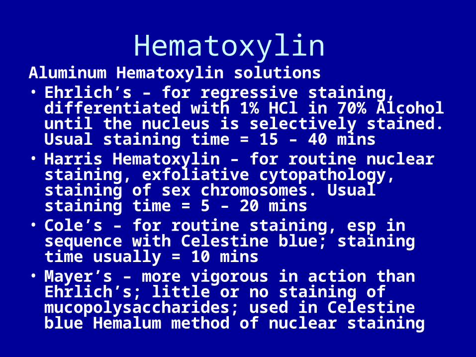
Hematoxylin Aluminum Hematoxylin solutions• Ehrlich’s – for regressive staining,
differentiated with 1% HCl in 70% Alcohol until the nucleus is selectively stained. Usual staining time = 15 – 40 mins
• Harris Hematoxylin – for routine nuclear staining, exfoliative cytopathology, staining of sex chromosomes. Usual staining time = 5 – 20 mins
• Cole’s – for routine staining, esp in sequence with Celestine blue; staining time usually = 10 mins
• Mayer’s – more vigorous in action than Ehrlich’s; little or no staining of mucopolysaccharides; used in Celestine blue Hemalum method of nuclear staining

Iron Hematoxylin solutions• Weigert’s – standard Iron Htx in the lab, useful in
demonstrating muscle fibers & connective tissues, esp. when preceding stains contain acid e.g. van Gieson w/ picric acid, which decolorizes nuclei stained w/ Alum Htx
• Heidenhain’s – cytological stain recommended for regressive staining of thin sections; demonstration of chromatin, chromosomes, nucleoli, centrosomes, mitochondria, skeletal muscle striations, & myelin
• PTAH – phosphotungstic acid hematoxylin – progressive staining of (blue)nuclei, fibrin, muscle striations, myofibrils, fibroglia, & (orange-red or brown-red) collagen, bone, cartilage; staining time = 12 – 24 hrs

Eosin • Eosin – red acid dye for routine differential
staining of cytoplasm & connective tissues, as a counterstain after Htx or before Methylene blue; Eosin B (bluish, deeper red) & Eosin Y (yellowish, readily soluble in water, less in alcohol)
• 5% aqueous Eosin Y Eosin Y 5 gm. dissolved in distilled Water 100 ml by gentle heating,
cooled & filtered. Thymol crystals may be added to prevent formation of molds
• Stock Eosin alcoholic solution Eosin Y 1 gm dissolved in 20 ml distilled Water by gentle heating,
cooled before adding 95% Alcohol 80 ml. For use: One part stock solution is added to 3 parts 80% Alcohol.
Addition of 0.5 ml glacial Acetic acid imparts a deeper red stain to tissues

Methylene Blue• Methylene blue (Polychrome) – basic
nuclear stain; for plasma cells, examination of fresh sputum for malignant cells, bacterial stain for evaluation & grading of milk, for diagnosing diphtheria, vital staining of nervous tissue, as indicator for differentiation of Colon & Aerogenes bacteria Methylene blue 1 gm Potassium carbonate 1 gm Distilled Water 100 ml
• Mix in a flask that has been lightly plugged with cotton or gauze. Let stand at 37ºC for weeks to oxidize. For use, dilute with distilled water (1 : 5 or 1 : 6)
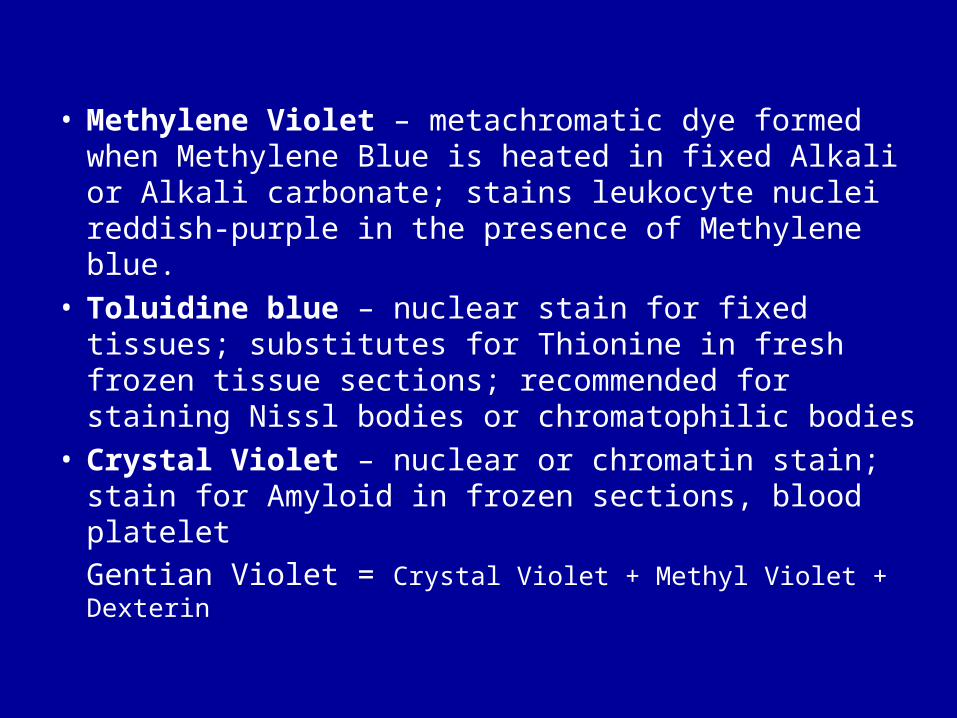
• Methylene Violet – metachromatic dye formed when Methylene Blue is heated in fixed Alkali or Alkali carbonate; stains leukocyte nuclei reddish-purple in the presence of Methylene blue.
• Toluidine blue – nuclear stain for fixed tissues; substitutes for Thionine in fresh frozen tissue sections; recommended for staining Nissl bodies or chromatophilic bodies
• Crystal Violet – nuclear or chromatin stain; stain for Amyloid in frozen sections, blood plateletGentian Violet = Crystal Violet + Methyl Violet + Dexterin
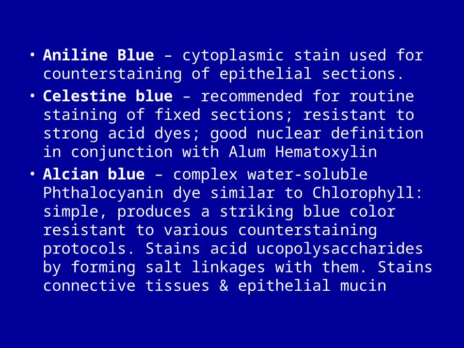
• Aniline Blue – cytoplasmic stain used for counterstaining of epithelial sections.
• Celestine blue – recommended for routine staining of fixed sections; resistant to strong acid dyes; good nuclear definition in conjunction with Alum Hematoxylin
• Alcian blue – complex water-soluble Phthalocyanin dye similar to Chlorophyll: simple, produces a striking blue color resistant to various counterstaining protocols. Stains acid ucopolysaccharides by forming salt linkages with them. Stains connective tissues & epithelial mucin

• Prussian blue – colored salt of Ferric Ferrocyanide; useful in demonstrating the circulatory system thru Intravital staining
• Victoria blue – for demonstration of neuroglia in frozen sections
• Night blue – can substitute for Carbol–Fuchsin in acid fast staining

• Malachite green – may be used as decolorizer or counterstain; weakly basic dye used as contrast stain to demonstrate Ascaris eggs, RBCs, bacterial spores.
• Methyl green – stains Chromatin green in the presence of an acid; but gives false positive reactions with certain secretions, e.g. Mucin
• Janus green B – useful in Intravital staining for demonstrating mitochondria

• Neutral Red – in dilutions of 1:10,000 to 1:100,000 – basic dye for demonstrating cell granules & vacuoles of phagocytic cells
• Congo Red – best known as an indicator; as a 4% aqueous solution in Krajian’s method to stain elastic tissues, Amyloid, & Myelin; also useful in staining axis cylinders in embryos
• Acridine Red 3B – for staining Calcium salt deposits & possible sites of phosphatase activity
• Acridine Orange – basic Acridine fluorochrome – permits distinction between dead & living cells; gives green fluorescence to DNA, red fo RNA
• Bismarck Brown – used as contrast stain in Gram, acid-fast, & Papanicolaou staining; also for staining C. diphtheriae

Fuchsin • Basic Fuchsin – a plasma stain; also for deep
staining of acid-fast organisms, for mitochondria, for differentiation of smooth muscles with the use of Picric acid; main constituent of Feulgen’s & Schiff’s reagent for the detection of aldehydes; of van Gieson’s stain for connective tissues, mucin, & elastic tissue.
• Carbol–Fuchsin Basic Fuchsin 1 gm Phenol crystals 5 gm Absolute Ethyl Alcohol 10 ml Distilled Water 100 ml
Grind basic Fuchsin & Phenol together in a mortar. Add Alcohol, then Water. Boil in a beaker then let stand 24 hrs to cool. Filter before use.
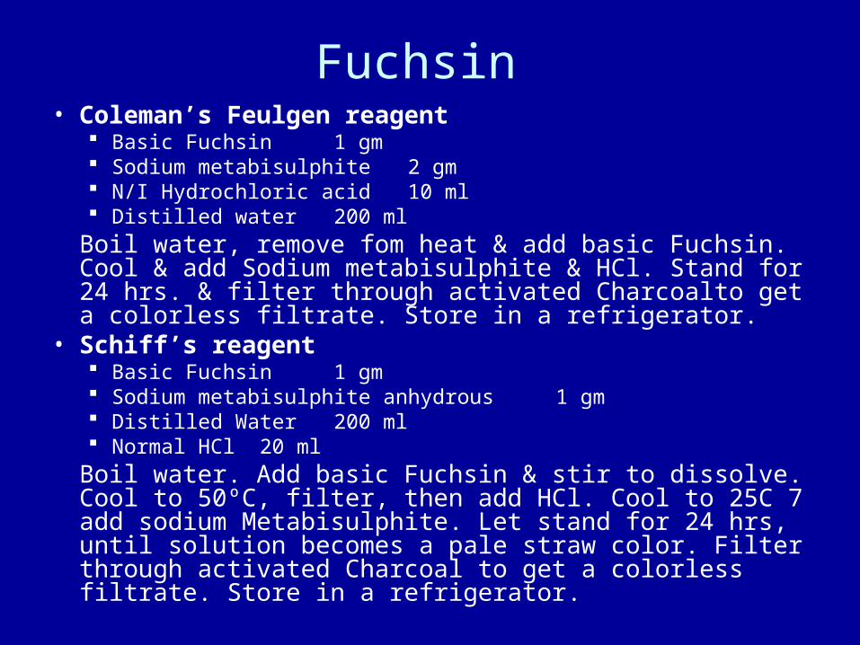
Fuchsin • Coleman’s Feulgen reagent
Basic Fuchsin 1 gm Sodium metabisulphite 2 gm N/I Hydrochloric acid 10 ml Distilled water 200 ml
Boil water, remove fom heat & add basic Fuchsin. Cool & add Sodium metabisulphite & HCl. Stand for 24 hrs. & filter through activated Charcoalto get a colorless filtrate. Store in a refrigerator.
• Schiff’s reagent Basic Fuchsin 1 gm Sodium metabisulphite anhydrous 1 gm Distilled Water 200 ml Normal HCl 20 ml
Boil water. Add basic Fuchsin & stir to dissolve. Cool to 50ºC, filter, then add HCl. Cool to 25C 7 add sodium Metabisulphite. Let stand for 24 hrs, until solution becomes a pale straw color. Filter through activated Charcoal to get a colorless filtrate. Store in a refrigerator.

Fuchsin • Mallory’s Fuchsin
Basic Fuchsin 0.5 gm 95% Ethanol 50 ml Distilled Water 50 ml
Gently heat Fuchsin in Alcohol to dissolve. Add water, Cool, Filter.
• Gomori’s (Aldehyde Fuchsin) Conc. HCl 1 ml Paraldehyde 1 ml Basic Fuchsin 0.5% in 70% Alcohol 100 ml
Mix all ingredients & stand at room temperature until mixture becomes deep purple, about 24 hrs. store in a refrigerator
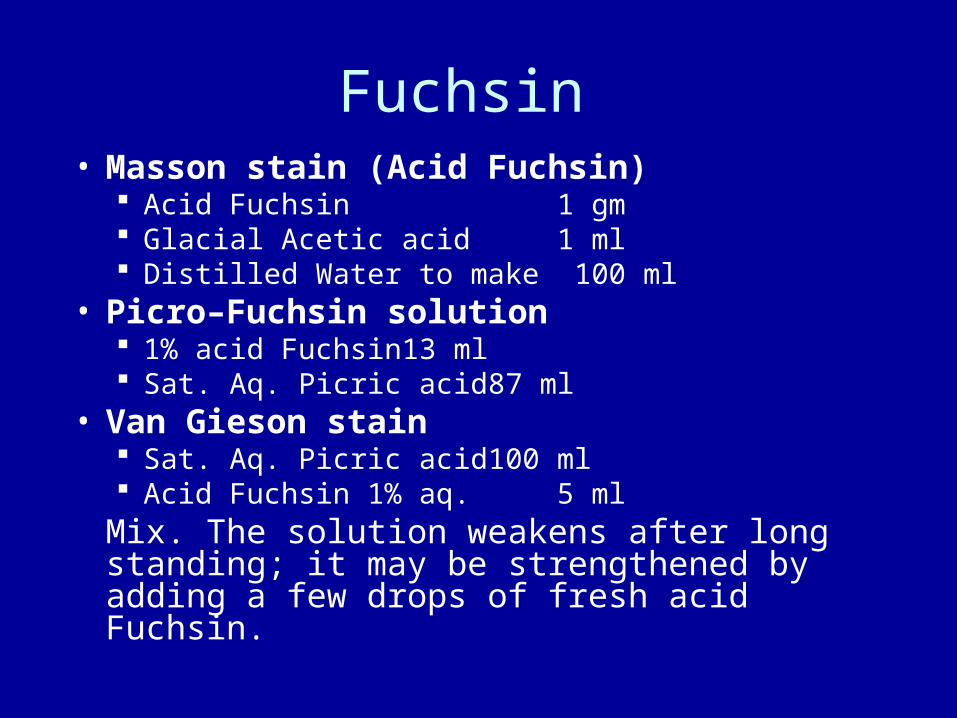
Fuchsin • Masson stain (Acid Fuchsin)
Acid Fuchsin 1 gm Glacial Acetic acid 1 ml Distilled Water to make 100 ml
• Picro–Fuchsin solution 1% acid Fuchsin 13 ml Sat. Aq. Picric acid 87 ml
• Van Gieson stain Sat. Aq. Picric acid 100 ml Acid Fuchsin 1% aq. 5 ml
Mix. The solution weakens after long standing; it may be strengthened by adding a few drops of fresh acid Fuchsin.
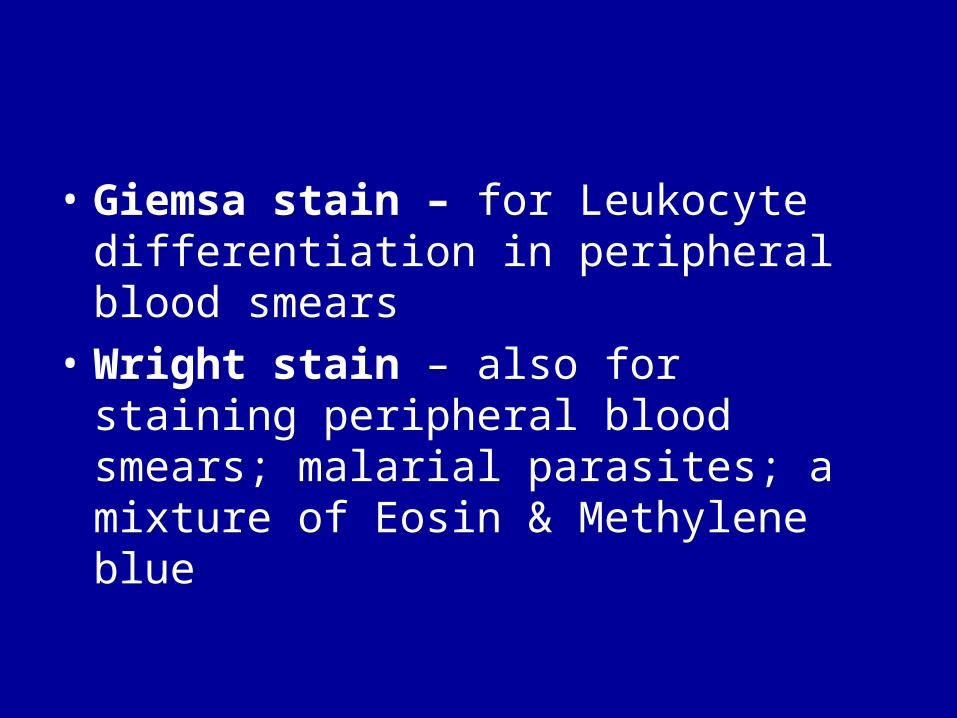
• Giemsa stain – for Leukocyte differentiation in peripheral blood smears
• Wright stain – also for staining peripheral blood smears; malarial parasites; a mixture of Eosin & Methylene blue
• Benzidine – for staining Hemoglobin• Rhodamine B – used by Griesbach
with Osmic acid to fix & stain blood & glandular tissues

Carmine • Carmine – used as a chromatin stain for
fresh materials in smears; slightly soluble in water at a neutral reaction; usually kept in ammoniacal solution which changes its properties due to oxidation; usually combined with Aluminum chloride to stain Glycogen (Best) & Mucin (Mucicarmine)
• Best Carmine stain (Stock solution) Carmine 2 gm ▪ conc. Ammonia 20 ml Potassium carbonate 1 gm ▪ distilled Water 60 ml Potassium Chloride 5 gm
• Best Carmine (Working solution): mix 2 parts each Best Carmine stock solution & conc. Ammonia, then add to 3 parts absolute Methanol

Iodine • Iodine – stains Amyloid, Cellulose, Starch, Carotenes
& Glycogen; widely used for removal of mercuric fixative artefact pigments & as a reagent to alter Crystal & Methyl violet so that they are retained by certain bacteria & fungi
• Gram’s Iodine – used in Gram–Weigert’s staining method for microorganisms & fibrin in tissue sections Iodine 1 gm. potassium Iodide 2 gm Distilled Water 300 mlDissolve Potassium Iodide in a little water. Add Iodine & dissolve in remaining water.
• Lugol’s Iodine – useful as a test for Glycogen, Amyloid, & Corpora amylacea (prostate) Iodine 1 gm Potassium iodide 2 gm Distilled Water 100 ml

Lysochromes (oil–soluble dyes )
• Lysochromes – stain lipids by virtue of their being more soluble in the tissue lipids than in their own medium (70% alcohol);
• Sudan black – slightly basic dye that colors neutral lipids (Triglycerides) by simple dissolution; very unstable, should be discarded if the usual blue-black color turns brownish black. prepared as a 0.5% solution boiled in 70% Ethanol for 10
mins under a reflux condenser & filtered before use;

Fat stains
• Sudan IV – stains Triglycerides a deep & intense red color prepared by saturating the dye Scharlach R in one part Benzoic
acid 2% in 70% Alcohol & one part Acetone, forming a very stable solution which may be used repeatedly as long as it is filtered
Addition of Benzoic acid intensifies fat & prevents rapid deterioration of the solutions
• Sudan III – useful for CNS tissues; less deep, lighter orange stain to lipids in these tissues
• Osmium tetroxide – osmic acid is a fixative also used to stain fat (reduces Osmium tetroxide to Osmium dioxide) black
Metallic Stains• Gold sublimate solution – for metallic
impregnation Mercuric chloride 0.4 gm Distilled Water 60 ml 1% Gold chloride (brown) 10 ml
Dissolve Mercuric chloride in water by gentle heating. Cool, then add Gold chloride, store in a dark place; or mix immediately before use
• Silver nitrate – 10% aqueous solution – used in staining Spirochetes, connective tissue fibers, especially Reticular; bluing agent is Ammonia Water (2 ml strong Ammonium hydroxide + 98 ml. tap water
H & E Staining Protocol

Hematoxylin & Eosin• routine staining method• differential, regressive staining – makes
use of a combination of dyes sequentially used to render contrasting colors to acidophilic & basophilic constituents
• results: Nuclei – blue to blue-black ▪ cartilage – pink-blue-dk blue Karyosome – dark blue ▪ Calcium, Bone – purple-blue Cytoplasm, Proteins – pale pink ▪ decal Bone, Collagen, RBCs, eosinophil granules – Osteoid – pink
bright red to orange-red ▪ muscle fibers – deep pink Keratin – dark pink ▪ plasma cells – purple pink
H & E staining method1. Clearing – xylene bath – 2nd xylene bath2. Absolute Ethanol – 95% Ethanol 3. Rinse – running water4. Hematoxylin (Harris – 5 min; Ehrlich’s – 15 to 30
min)5. Wash – running water to remove excess stain6. Differentiate – 1% acid Alcohol x 10-30 sec
until only nuclei are stained7. Rinse – tap water8. Blueing – ammonia water (ave. = 5 mins)9. Wash – running water x 5 min10. Counterstain – 5% aq. Eosin x 5 min11. Wash & Differentiate till nuclei are blue &
the rest of the tissue are in shades of pink12. Dehydrate13. Clear & Mount

Clearing (Differentiation)
Reagent for H&E Staining
• 1% Acid Alcohol Solution: Hydrochloric acid ---------------- 10 ml 70% ethanol ---------------------- 1000 mlMix well and store at room temperature.Differentiate 30 second to 2 minutes after hematoxylin
over-stain.50 um paraffin sections require 2 minutes differentiation.

Blueing reagents for H & E
• 0.1% Sodium Bicarbonate: Sodium bicarbonate 1 g + Distilled water 1000 ml Mix to dissolve. The pH will be around 8.0 Store this soln at room temp. Bluing for 30 sec to 1 min after hematoxylin staining and clearing/differentiation. Note: this sol’n worked better than ammonia water sol’n.
• 0.2% Ammonia Water Solution (Bluing): Ammonium hydroxide (conc) 2 ml + Distilled water 1000 ml Mix well. The pH will be around 10.0 Store this soln at room temp. Bluing for 30 sec to 1 min after hematoxylin staining and clearing/differentiation.
Note: this solution is not as good as sodium bicarbonate.
• Lithium Carbonate Solution (Saturated): Lithium carbonate 1.54 g + Distilled water 100 ml Mix to dissolve and store at room temp. Bluing for 30 sec to 1 min after hematoxylin staining and clearing/differentiation.

Routine H & E
• not suitable for tissues fixed in Osmic acid (inhibits Hematoxylin)
• for tissues fixed in Mercuric chloride, staining time in Htx should be increased slightly, in Eosin should be reduced
• staining time may be prolonged for Chromium-fixed tissues & those subjected to long acid decalcification or prolonged storage in acid Formalin / 70% Alcohol
• Neuroglia, axons, nerve endings, reticulum, Golgi bodies, mitochondria are not demonstrated.

Precautions • Avoid staining the Skin
Stains may be removed by prompt application of 0.5% acid alcohol & rinsing with tap water
• Ensure proper adhesion of the section to the slide Clean slide Adhesive should be fresh (stale Albumin loses its clear color & may
develop an odor) Sections must be completely dehydrated (Alcohol baths) Spread the paraffin section evenly & thoroughly
• Avoid failure of staining Sufficient “ripening” of Hematoxylin Purification & filtering of the dye to remove impurities Replace deteriorated dyes (precipitation & poor staining results are
indicators) Retained paraffin, fixative, adhesive, decolorizer, or decalcifying
agents Xylol, Absolute alcohol,

Thank You !Thank You