HIRA orchestrates a dynamic chromatin landscape in...
15
HIRA orchestrates a dynamic chromatin landscape in senescence and is required for suppression of neoplasia Taranjit Singh Rai, 1,2,3 John J. Cole, 1,2,5 David M. Nelson, 1,2,5 Dina Dikovskaya, 1,2 William J. Faller, 1 Maria Grazia Vizioli, 1,2 Rachael N. Hewitt, 1,2 Orchi Anannya, 1 Tony McBryan, 1,2 Indrani Manoharan, 1,2 John van Tuyn, 1,2 Nicholas Morrice, 1 Nikolay A. Pchelintsev, 1,2 Andre Ivanov, 1,2,4 Claire Brock, 1,2 Mark E. Drotar, 1,2 Colin Nixon, 1 William Clark, 1 Owen J. Sansom, 1 Kurt I. Anderson, 1 Ayala King, 1 Karen Blyth, 1 and Peter D. Adams 1,2 1 Beatson Institute for Cancer Research, Bearsden, Glasgow G61 1BD, United Kingdom; 2 Institute of Cancer Sciences, College of Medical, Veterinary, and Life Sciences, University of Glasgow, Glasgow G61 1BD, United Kingdom; 3 Institute of Biomedical and Environmental Health Research, University of West of Scotland, Paisley PA1 2BE, United Kingdom Cellular senescence is a stable proliferation arrest that suppresses tumorigenesis. Cellular senescence and associated tumor suppression depend on control of chromatin. Histone chaperone HIRA deposits variant histone H3.3 and histone H4 into chromatin in a DNA replication-independent manner. Appropriately for a DNA replication-independent chaperone, HIRA is involved in control of chromatin in nonproliferating senescent cells, although its role is poorly defined. Here, we show that nonproliferating senescent cells express and incorporate histone H3.3 and other canonical core histones into a dynamic chromatin landscape. Expression of canonical histones is linked to alternative mRNA splicing to eliminate signals that confer mRNA instability in non- proliferating cells. Deposition of newly synthesized histones H3.3 and H4 into chromatin of senescent cells depends on HIRA. HIRA and newly deposited H3.3 colocalize at promoters of expressed genes, partially redistributing between proliferating and senescent cells to parallel changes in expression. In senescent cells, but not proliferating cells, promoters of active genes are exceptionally enriched in H4K16ac, and HIRA is required for retention of H4K16ac. HIRA is also required for retention of H4K16ac in vivo and suppression of oncogene- induced neoplasia. These results show that HIRA controls a specialized, dynamic H4K16ac-decorated chromatin landscape in senescent cells and enforces tumor suppression. [Keywords: chromatin; senescence; HIRA; H4K16ac; dynamic; tumor suppression] Supplemental material is available for this article. Received June 15, 2014; revised version accepted November 4, 2014. Cellular senescence is a stable proliferation arrest asso- ciated with an altered proinflammatory secretory path- way (Salama et al. 2014). In response to acquisition of an activated oncogene, primary human cells enter this pro- liferation-arrested senescent state (oncogene-induced se- nescence [OIS]). By imposing proliferation arrest, OIS acts as a tumor suppressor mechanism (Braig et al. 2005; Chen et al. 2005; Collado et al. 2005; Michaloglou et al. 2005). Even in the absence of an activated oncogene, replicative senescence (RS) places an upper limit on the proliferative capacity of normal cells, also blocking tumor formation (Cosme-Blanco et al. 2007; Feldser and Greider 2007). The altered secretory program of senescent cells, comprised of proinflammatory cytokines, chemokines, and matrix pro- teases (the senescence-associated secretory phenotype [SASP]) (Krtolica et al. 2001; Acosta et al. 2008; Kuilman et al. 2008), can also contribute to tumor suppression by promoting clearance of some senescent cells by the immune system (Xue et al. 2007; Kang et al. 2011; Lujambio et al. 2013). Extensive chromatin changes are apparent in senescent cells, most obviously in the form of domains of compacted Ó 2014 Rai et al. This article is distributed exclusively by Cold Spring Harbor Laboratory Press for the first six months after the full-issue publication date (see http://genesdev.cshlp.org/site/misc/terms.xhtml). After six months, it is available under a Creative Commons License (Attribution-NonCommercial 4.0 International), as described at http:// creativecommons.org/licenses/by-nc/4.0/. 4 Present address: Barts, The London School of Medicine and Dentistry, Barts Cancer Institute, Queen Mary University of London, London EC1M 6BQ, United Kingdom 5 These authors contributed equally to this work. Corresponding authors: [email protected], taranjitsingh.rai@uws. ac.uk Article is online at http://www.genesdev.org/cgi/doi/10.1101/gad.247528.114. 2712 GENES & DEVELOPMENT 28:2712–2725 Published by Cold Spring Harbor Laboratory Press; ISSN 0890-9369/14; www.genesdev.org Cold Spring Harbor Laboratory Press on June 17, 2018 - Published by genesdev.cshlp.org Downloaded from
Transcript of HIRA orchestrates a dynamic chromatin landscape in...

HIRA orchestrates a dynamic chromatinlandscape in senescence and is requiredfor suppression of neoplasia
Taranjit Singh Rai,1,2,3 John J. Cole,1,2,5 David M. Nelson,1,2,5 Dina Dikovskaya,1,2 William J. Faller,1
Maria Grazia Vizioli,1,2 Rachael N. Hewitt,1,2 Orchi Anannya,1 Tony McBryan,1,2
Indrani Manoharan,1,2 John van Tuyn,1,2 Nicholas Morrice,1 Nikolay A. Pchelintsev,1,2
Andre Ivanov,1,2,4 Claire Brock,1,2 Mark E. Drotar,1,2 Colin Nixon,1 William Clark,1 Owen J. Sansom,1
Kurt I. Anderson,1 Ayala King,1 Karen Blyth,1 and Peter D. Adams1,2
1Beatson Institute for Cancer Research, Bearsden, Glasgow G61 1BD, United Kingdom; 2Institute of Cancer Sciences, Collegeof Medical, Veterinary, and Life Sciences, University of Glasgow, Glasgow G61 1BD, United Kingdom; 3Institute of Biomedicaland Environmental Health Research, University of West of Scotland, Paisley PA1 2BE, United Kingdom
Cellular senescence is a stable proliferation arrest that suppresses tumorigenesis. Cellular senescence andassociated tumor suppression depend on control of chromatin. Histone chaperone HIRA deposits variant histoneH3.3 and histone H4 into chromatin in a DNA replication-independent manner. Appropriately for a DNAreplication-independent chaperone, HIRA is involved in control of chromatin in nonproliferating senescent cells,although its role is poorly defined. Here, we show that nonproliferating senescent cells express and incorporatehistone H3.3 and other canonical core histones into a dynamic chromatin landscape. Expression of canonicalhistones is linked to alternative mRNA splicing to eliminate signals that confer mRNA instability in non-proliferating cells. Deposition of newly synthesized histones H3.3 and H4 into chromatin of senescent cellsdepends on HIRA. HIRA and newly deposited H3.3 colocalize at promoters of expressed genes, partiallyredistributing between proliferating and senescent cells to parallel changes in expression. In senescent cells, butnot proliferating cells, promoters of active genes are exceptionally enriched in H4K16ac, and HIRA is required forretention of H4K16ac. HIRA is also required for retention of H4K16ac in vivo and suppression of oncogene-induced neoplasia. These results show that HIRA controls a specialized, dynamic H4K16ac-decorated chromatinlandscape in senescent cells and enforces tumor suppression.
[Keywords: chromatin; senescence; HIRA; H4K16ac; dynamic; tumor suppression]
Supplemental material is available for this article.
Received June 15, 2014; revised version accepted November 4, 2014.
Cellular senescence is a stable proliferation arrest asso-ciated with an altered proinflammatory secretory path-way (Salama et al. 2014). In response to acquisition of anactivated oncogene, primary human cells enter this pro-liferation-arrested senescent state (oncogene-induced se-nescence [OIS]). By imposing proliferation arrest, OIS actsas a tumor suppressor mechanism (Braig et al. 2005; Chenet al. 2005; Collado et al. 2005; Michaloglou et al. 2005).Even in the absence of an activated oncogene, replicativesenescence (RS) places an upper limit on the proliferativecapacity of normal cells, also blocking tumor formation
(Cosme-Blanco et al. 2007; Feldser and Greider 2007). Thealtered secretory program of senescent cells, comprised ofproinflammatory cytokines, chemokines, andmatrix pro-teases (the senescence-associated secretory phenotype[SASP]) (Krtolica et al. 2001; Acosta et al. 2008; Kuilmanet al. 2008), can also contribute to tumor suppressionby promoting clearance of some senescent cells bythe immune system (Xue et al. 2007; Kang et al. 2011;Lujambio et al. 2013).Extensive chromatin changes are apparent in senescent
cells, most obviously in the form of domains of compacted
� 2014 Rai et al. This article is distributed exclusively by Cold SpringHarbor Laboratory Press for the first six months after the full-issuepublication date (see http://genesdev.cshlp.org/site/misc/terms.xhtml).After six months, it is available under a Creative Commons License(Attribution-NonCommercial 4.0 International), as described at http://creativecommons.org/licenses/by-nc/4.0/.
4Present address: Barts, The London School of Medicine and Dentistry,Barts Cancer Institute, Queen Mary University of London, London EC1M6BQ, United Kingdom5These authors contributed equally to this work.Corresponding authors: [email protected], [email protected] is online at http://www.genesdev.org/cgi/doi/10.1101/gad.247528.114.
2712 GENES & DEVELOPMENT 28:2712–2725 Published by Cold Spring Harbor Laboratory Press; ISSN 0890-9369/14; www.genesdev.org
Cold Spring Harbor Laboratory Press on June 17, 2018 - Published by genesdev.cshlp.orgDownloaded from
heterochromatin called senescence-associated hetero-chromatin foci (SAHF) (Narita et al. 2003, 2006; Braiget al. 2005; Zhang et al. 2005; O’Sullivan et al. 2010;Chandra et al. 2012; Cruickshanks et al. 2013; De Ceccoet al. 2013; Sadaie et al. 2013; Shah et al. 2013). These andother chromatin changes are thought to contribute to theonset and maintenance of viable senescence-associatedproliferation arrest (Braig et al. 2005; Narita et al. 2006;DiMicco et al. 2011; Benhamed et al. 2012; Cruickshankset al. 2013; Martin et al. 2013). Chromatin changes likelyalso contribute to activation of the SASP in senescentcells (Shah et al. 2013). Subunits of the SWI/SNF ATP-dependent chromatin remodeling complex are targets ofmutation and inactivation in cancer (Wilson and Roberts2011), and SWI/SNF is thought to contribute to tumorsuppression in part through induction of senescence(Chai et al. 2005). In sum, several lines of evidenceindicate that chromatin of senescent cells contributesto tumor suppression (Braig et al. 2005; Narita et al.2006). However, the molecular basis of this is poorlyunderstood.Specifically, SAHF have been suggested to contribute to
stable repression of proliferation genes and/or suppres-sion of DNA damage signaling in senescent cells (Naritaet al. 2003; Zhang et al. 2007; DiMicco et al. 2011). Eitherway, the apparent heterochromatinization of senescentcells intuitively suggests that their compacted, stablechromatin contributes to a senescence-mediated barrierto tumor progression. In line with this view of relativelystatic chromatin in senescent cells, compared with pro-liferating cells, senescent cells synthesize less totalhistone H3 and H4 (O’Sullivan et al. 2010). In fact, inmammalian cells, the canonical DNA replication-depen-dent histones—H2A, H2B, H3 (H3.1 and H3.2), and H4—are each coded for by 10–15 genes located mostly inclusters on chromosomes 1 and 6, and the correspondingmRNAs are typically unstable in nonproliferating cells(Marzluff et al. 2008). This is again consistent with theview that chromatin in nonproliferating senescent cellsis a relatively fixed entity, intuitively suggestive of animmobile barrier to tumor progression. However, theextent of chromatin dynamics in senescent cells has notbeen properly investigated.Histone variant H3.3 contributes to nucleosome de-
stabilization (Jin and Felsenfeld 2007) and so is thought tofacilitate nucleosome dynamics associated with tran-scription activation and ongoing transcription. HistoneH3.3 is enriched at nucleosomes at transcription startsites (TSSs) of genes and at enhancers and gene bodies ofactively transcribed genes (Ahmad and Henikoff 2002; Jinet al. 2009; Goldberg et al. 2010). H3.3 differs fromcanonical histones H3.1 and H3.2 by four or five aminoacids and is expressed constitutively throughout the cellcycle and in proliferating and nonproliferating cells fromtwo genes:H3F3A andH3F3B (Skene and Henikoff 2013).The HIRA chaperone complex, comprised of HIRA/UBN1/CABIN1, collaborates with histone-binding pro-tein ASF1a to incorporate H3.3 into chromatin in a DNAreplication-independent manner (Ray-Gallet et al. 2002;Tagami et al. 2004; Loppin et al. 2005). The HIRA protein
is required for deposition of histone H3.3 at active andpoised genes and enhancers (Goldberg et al. 2010; Ray-Gallet et al. 2011; Pchelintsev et al. 2013). Accordingly,HIRA is required for gene activation in some contexts(Placek et al. 2009; Dutta et al. 2010; Yang et al. 2011) aswell as recruitment and function of polycomb complexesat gene promoters and dynamic restoration of chromatinafter DNA damage repair (Adam et al. 2013; Banaszynskiet al. 2013). On the other hand, the HIRA complex and itsorthologs, together with histone H3.3, are also involvedin chromatin silencing (Sherwood et al. 1993; van derHeijden et al. 2007). Importantly, several recent studieshave shown histone H3.3 to be a target of recurrentmissense mutations in human cancer, and transcription-coupled methylation of histone H3.3 is also thought to betumor-suppressive (Schwartzentruber et al. 2012; Wuet al. 2012; Wen et al. 2014; Zhu et al. 2014). Appropri-ately for a DNA replication-independent chaperone,HIRA is also involved in control of chromatin in non-proliferating senescent cells (Zhang et al. 2005; Ye et al.2007; Banumathy et al. 2009; Rai et al. 2011; Duarte et al.2014). However, HIRA’s function in senescent cells ispoorly defined, and there is currently no direct evidencefor a role in tumor suppression.Here we set out to better understand dynamic chroma-
tin regulation in nonproliferating senescent cells and therole of HIRA in this process. We reveal chromatin insenescent cells to be a dynamic landscape linked tononcanonical regulation of histone mRNAs and excep-tional marking by H4K16ac. The histone chaperoneHIRA is required for regulation of this landscape and alsofor suppression of oncogene-induced neoplasia in amousemodel. This study points to shared roles of HIRA, histoneH3.3, and H4K16ac in both cellular senescence and tumorsuppression.
Results
The mRNAs encoding the canonical DNA replication-dependent histones are generally regarded as unstable innonproliferating cells (Marzluff et al. 2008). Moreover,compared with proliferating cells, senescent cells synthe-size less total histone H3 and H4 (O’Sullivan et al. 2010).We were surprised, therefore, when gene expressionmicroarray revealed that a subset of histone mRNAswas apparently up-regulated in RS cells (senescenceconfirmed by standard markers of senescence) (Fig. 1A;Supplemental Data Set 1; Supplemental Fig. 1A,B). Closeexamination showed that these are typically canonicalDNA replication-dependent histones from the HIST1cluster on chromosome 6 but are also annotated inEnsembl (hg19) to contain a noncoding second exon,and the Affymetrix probes target this second exon (Sup-plemental Data Set 2; Supplemental Fig. 1C). This secondexon lacks the specialized stem–loop that terminates thecanonical replication-dependent histone mRNAs to con-fer instability in nonproliferating cells (Marzluff et al.2008) but instead directs mRNA polyadenylation. In bothRS and OIS cells, we confirmed up-regulation of thesecond exon of several of these noncanonical histone
HIRA mediates tumor suppression
GENES & DEVELOPMENT 2713
Cold Spring Harbor Laboratory Press on June 17, 2018 - Published by genesdev.cshlp.orgDownloaded from

mRNAs (and two others that lacked second exon probeson the array, HIST4H4 and HIST1H2BN) by quantitativeRT–PCR (qRT–PCR) and RNA sequencing (RNA-seq)(Fig. 1B,C; Supplemental Data Sets 2, 3). Accumulationof these alternative spliced polyadenylated histone tran-scripts has been previously reported in some cancer celllines after treatment with ionizing radiation and on differ-entiation of mesenchymal progenitor cells (Kari et al. 2013).Moreover, we observed up-regulation of the second exonin quiescent cells (Supplemental Fig. 1D), suggesting thataccumulation of alternative spliced polyadenylated his-tone mRNAs occurs in other nonproliferating cells aswell as senescent cells.RNA-seq also confirmed splicing of the first coding
exon to the second exon in senescent cells, based on somereads spanning the two exons (Supplemental Fig. 1E).Significantly, qRT–PCR and RNA-seq data showed thatthe first exon of histone mRNAs spliced to a polyade-nylated noncoding second exon was down-regulated lessin senescence than canonical single coding exon stem–loophistone mRNAs, such as HIST1H2BM and HIST1H3B(Fig. 1B,D,E; Supplemental Data Set 3). Interestingly, theeight alternative-spliced histones include genes encodingtheH2A, H2B, andH4 subtypes but noH3 gene. However,
histone variant H3.3 is already known to be expressed innonproliferating cells (Urban and Zweidler 1983; Groveand Zweidler 1984; Pantazis and Bonner 1984; Brown et al.1985; Skene and Henikoff 2013). Indeed, substantial ex-pression of genes encoding DNA replication-independenthistone variant H3.3—H3F3A and H3F3B—was retainedin RS and OIS cells (Fig. 1F; Supplemental Fig. 1F). Thissuggests that the alternative-spliced mRNAs coding forthe canonical H2A,H2B, andH4 subtypes are counterpartsof histone variant H3.3 (Supplemental Data Sets 2, 3). Weconclude that senescent cells retain expression of a smallsubset of the >40 canonical histone genes whose expres-sion is typically classed as replication-dependent, linked toalternative mRNA splicing to bypass a signal known todirect mRNA degradation in nonproliferating cells.Next, we investigated whether newly synthesized
histones are deposited into chromatin in senescent cells.Previous studies have indicated that recruitment of his-tone chaperones and histones to PML bodies is indicativeof trafficking of newly synthesized histones into chroma-tin (Chang et al. 2013; Delbarre et al. 2013; Corpet et al.2014). Consistent with ongoing histone deposition andnucleosome assembly in nonproliferating senescent cells,histone H3 was enriched in PML bodies of RS and OIS
Figure 1. Expression of histone mRNAs in senescentcells linked to alternative mRNA splicing. (A) Expressionof multiple histone mRNAs assessed by Affymetrixmicroarray in proliferating (Prolif) and RS cells (fiveindependent replicates of each). Gene expression isscaled for each gene individually, and resulting intensi-ties are plotted between the green (low) and red (high)scale. (B) Fold change in abundance of first (coding) andsecond (noncoding, poly A) exons of selected HIST1
cluster mRNAs between proliferating and RS cells,determined by qRT–PCR. HIST1H2BM and HIST1H3B
(coding for canonical histone H2B and H3.1, respec-tively) are expressed only as single-exon stem–loopmRNAs. Values are means 6 standard deviation ofthree independent experiments normalized to GAPDH
as a housekeeping control. (C) Exon read counts (readsper kilobase of exon model per million mapped reads[RPKM]) of single-exon (HIST1H4I and HIST1H2AH;green) and two-exon (HIST1H2BK; first coding exon inred and second noncoding exon in yellow) histonemRNAs in proliferating, RS, control (Con), and OIScells. (D) Fold change in abundance of first coding exonbetween proliferating and RS cells, comparing single-exon, stem–loop, and no poly A mRNAs and thosemRNAs with a poly A second exon whose expressionincreases in RS, as determined by RNA-seq. See Sup-plemental Data Set 3 for gene lists. P-value = 0.0002016.(E) As in D, but analysis performed in OIS. See Supple-mental Data Set 3 for gene lists. P-value = 0.001837.(F) Expression of H3F3A and H3F3B genes assessed byRNA-seq in proliferating and RS cells.
Rai et al.
2714 GENES & DEVELOPMENT
Cold Spring Harbor Laboratory Press on June 17, 2018 - Published by genesdev.cshlp.orgDownloaded from

cells, where it colocalized with DNA replication-indepen-dent histone chaperone HIRA (Fig. 2A,B). While ectopi-cally expressed epitope-tagged histone H3.3 was notdetectably cell cycle inhibitory or toxic to proliferatingor RS cells (Supplemental Fig. 2A), ectopically expressedH3.3 and H4 were incorporated into detergent-insolublenuclear chromatin of senescent cells, directly demonstrat-ing that newly synthesized histones can be incorporatedinto chromatin in senescent cells (Fig. 2C; SupplementalFig. 2B). In contrast, DNA replication-dependent histonevariant H3.1 was incorporated into chromatin ofproliferating cells, but not senescent cells, despite com-parable expression of ectopically expressed H3.1 mRNAbetween proliferating and senescent cells (Fig. 2C,D).Chromatin immunoprecipitation (ChIP) combined withsequencing (ChIP-seq) of ectopically expressed HA-taggedhistone H3.3 in proliferating and RS cells confirmed thatits distribution between different genome features wascomparable with endogenous H3.3, as previously deter-mined by Allis and coworkers (Goldberg et al. 2010)(Fig. 2E; Supplemental Table 1). Incorporation of ectopi-cally expressed histones into chromatin implies a level ofhistone exchange in the chromatin of these senescent
cells. Consistent with this idea, fluorescence recoveryafter photobleaching (FRAP) showed that a proportion ofectopically expressed nuclear GFP-H3.3 recovered fromphotobleaching within seconds (Fig. 2F). Together, theseanalyses of endogenous and ectopic histones suggest thathistones H3.3 and H4 dynamically exchange at theirphysiological sites of incorporation in senescent cells.Given HIRA’s role in DNA replication-independent
histone H3.3 deposition, we set out to compare thegenomic distribution of HIRA and newly synthesizedhistone H3.3 in proliferating and senescent cells. HIRAChIP-seq was performed on chromatin from proliferating,RS, and OIS (and control) cells. Comparison of the HIRAand histone H3.3 genome distribution from proliferatingand RS cells confirmed a substantial and highly signifi-cant overlap of HIRA and H3.3 in both proliferating andsenescent cells (Supplemental Tables 1, 2). Consequently,initial analyses were performed on the intersect of theHIRA and H3.3 data sets (Supplemental Table 3). Consis-tent with previous analyses, a large proportion of HIRA/H3.3 peaks localized at and near coding regions (Supple-mental Fig. 3A; Ahmad and Henikoff 2002; Jin et al. 2009;Goldberg et al. 2010; Pchelintsev et al. 2013). In both
Figure 2. Dynamic incorporation of histonesinto chromatin of senescent cells. (A) Prolifer-ating (Prolif), RS, and OIS cells fluorescentlystained with antibodies to PML and histone H3.(B) Proliferating, RS, and OIS cells fluorescentlystained with antibodies to HIRA and histone H3.(C) RS cells were infected with lentivirus en-coding HA-histone H4, HA-histone H3.3, andHA-histone H3.1 and fluorescently stained withanti-HA. (D) Cells from C were analyzed formRNA abundance of HA-histone H3.1 by qRT–PCR. The bar chart displays the mean HA-H3.1
mRNA abundance in senescent cells comparedwith proliferating cells, normalized to GAPDH asa housekeeping control. Standard error fromthree independent experiments. (E) Percentageof base pairs covered by H3.3 peaks that overlapwith base pairs of specified genomic featuresfor human proliferating and RS cells expressingectopic HA-tagged H3.3 and mouse embryonicstem cells expressing endogenous HA-tagged H3.3.(F) The percentage of mobile fraction (Fm) andhalf-time of recovery in seconds (T1/2) for histoneslocated in GFP-H3.3 foci (likely PML bodies, basedonA,B) and nucleoplasm. Error bars represent6SEMfrom three independent experiments.
HIRA mediates tumor suppression
GENES & DEVELOPMENT 2715
Cold Spring Harbor Laboratory Press on June 17, 2018 - Published by genesdev.cshlp.orgDownloaded from

proliferating and RS cells, HIRA/H3.3 peaks localized to;10,000 coding regions (Supplemental Fig. 3B). Specificenrichment was observed at CpG islands, promoters,exons, and 59 untranslated regions (UTRs) (SupplementalFig. 3C). At promoters, HIRA and H3.3 were both mostenriched at the nucleosomes flanking the TSS, and forHIRA in proliferating, RS, control, and OIS cells and H3.3in proliferating and RS cells, binding correlated stronglywith gene expression, as determined by RNA-seq (Fig. 3A;Supplemental Fig. 3D; Supplemental Table 4).Since proliferating and senescent cells exhibit distinct
programs of gene expression, we asked whether changesin gene expression between proliferating and senescentcells correlated with change in HIRA and H3.3 binding.For HIRA, a change in gene expression, either up or down,directly correlated with a change in HIRA binding mea-sured by ChIP (Fig. 3B) in both OIS and RS. Similarly, forhistone H3.3, an increase in expression directly corre-lated with an increase in binding in RS (Fig. 3B; Supple-mental Fig. 3E). Notably, 1919 genes acquired both HIRAand H3.3 in senescent cells, and these are enriched ingenes related to TNF signaling, a component of the SASP(Supplemental Fig. 3B; Supplemental Data Set 4; Coppeet al. 2008). These correlations are also illustrated by a
comparison of the 250most up-regulated and down-regulatedgenes (Supplemental Fig. 3E). However, across all genes,a decrease in expression in senescence did not correlatewith decreased H3.3 binding, suggesting that genes thatare repressed on the transition from proliferating to RScells continue to incorporate histone H3.3 (Fig. 3B;Supplemental Fig. 3E). These phenomena were mostmarked at histone genes. Decreased expression of theDNA replication-dependent histones in senescent cellswas accompanied by a marked decrease in HIRA bindingin both RS and OIS (Fig. 3C,D). However, H3.3 did notsubstantially decrease at these genes in senescent cells(Fig. 3D). In sum, HIRA and newly synthesized H3.3 arelocalized to promoters of expressed genes in proliferatingand senescent cells and are partially relocalized betweenproliferating and senescent cells, in line with changes ingene expression—most notably in the case of HIRA atDNA replication-dependent histone genes.A recent study established a link between histone H3.3
and H4K16ac (Lin et al. 2013). Interestingly, H4K16ac alsohas candidate tumor suppressor activity (Fraga et al. 2005;Elsheikh et al. 2009). Therefore, to investigate this linkbetween HIRA/H3.3 and H4K16ac in tumor-suppressivesenescent cells, we performed ChIP-seq of H4K16ac in
Figure 3. Genome distribution of HIRA and histoneH3.3 between proliferating and senescent cells is regu-lated in parallel with gene expression. (A) HIRA andH3.3 enrichment profiles around (63 kb) the TSSs of thetop (red), middle (green), and bottom (blue) 1000 codinggenes ordered by expression for proliferating (Prolif), RS,control (Con), and OIS cells. ChIP-seq signal wasnormalized to input. (B) At significantly changingcoding genes (Benjamini and Hochberg false discoverrate 5%), the Pearson correlation coefficients (PCCs)between the difference in ChIP-seq signal and thedifference in expression. ChIP-seq signals were calcu-lated for promoter regions (TSS 62 kb) and normalizedto input. Differences in expression were calculated asfold change (FC; log2). Differences were calculated fromproliferating to senescent cells (RS or OIS, as indicated).The X-axis denotes the ChIP-seq data set used togenerate the PCCs. Bar colors indicate the gene setsused for the comparison: all genes (All) and genes thatgain expression (Exp+), lose expression (Exp�), gainChIP-seq signal (ChIP+), and lose ChIP-seq signal(ChIP�). An asterisk indicates significant correlationat P < 0.05. (C) Scatter plot of single-exon, stem–loop, nopoly A (red diamonds), and two-exon poly A (blackdiamonds) histone genes (Supplemental Data Set 3)showing the difference in input normalized ChIP-seqsignal (TSS 6 2 kb) versus the difference in expression(fold change log2) for proliferating to RS cells andcontrol to OIS cells. (D) Genome browser representa-tion of HIRA, ribodepleted RNA, and H3.3 in prolifer-ating and RS IMR90 cells at histone gene cluster one(HIST1).
Rai et al.
2716 GENES & DEVELOPMENT
Cold Spring Harbor Laboratory Press on June 17, 2018 - Published by genesdev.cshlp.orgDownloaded from

proliferating and RS cells using two different validatedantibodies to H4K16ac. There was a substantial and highlysignificant overlap between the distribution of H4K16acdetected by the two antibodies, particularly in RS cells (Fig.4A; Supplemental Tables 1, 5, 6). For downstream analyses,we evaluated the intersect of the two H4K16ac data sets.Qualitatively, the overall distribution of H4K16ac acrossthe genome was similar for proliferating and RS cells and,interestingly, was reminiscent of HIRA and histone H3.3.H4K16ac was most abundant at the coding regions andwas enriched at CpG islands, promoters, and 59 UTRs(cf. Supplemental Figs. 4A,B and 3A,C). However, bothantibodies reported a twofold to threefold increase in thenumber of H4K16ac peaks and the number of base pairscovered in RS cells (Fig. 4A; Supplemental Tables 5, 6).Indeed,;45,000 regions showed an increase in H4K16ac inRS, while <10,000 pre-existing peaks showed a decrease inRS (Fig. 4B). On a larger scale, in RS cells, H4K16ac wasdepleted from late replicating regions of the genome andSAHF (Supplemental Fig. 4C,D), in line with incorporationof late replicating regions into compacted SAHF, H4K16ac’sability to inhibit chromatin compaction, and a previousreport correlating global decreased H4K16ac to the forma-tion of SAHF (Shogren-Knaak et al. 2006; Chandra et al.2012; Contrepois et al. 2012).
There was a marked increase in overlap of HIRA/H3.3and H4K16ac in RS cells (Fig. 4C), and H4K16ac peaksacquired in RS mirrored HIRA/H3.3 peaks (Fig. 4D),indicating a substantial increase in H4K16ac in RS atregions bound by HIRA/H3.3. At H4K16ac peaks uniqueto RS cells, the HIRA and H4K16ac enrichments byChIP-seq correlated strongly (Pearson correlation co-efficient [PCC] = 0.64). Indeed, in RS cells but muchless so in proliferating cells, H4K16ac was enriched atearly replicating gene-rich regions (Fig. 4E; Supplemen-tal Fig. 4C), particularly on either side of the TSS (Fig.4F; Supplemental Fig. 4E). In senescent cells, ;12,500genes were bound by H4K16ac, compared with <5000genes in proliferating cells (Supplemental Fig. 4F). Re-markably, in RS cells, H4K16ac was enriched at allbut the lowest expressed genes (Fig. 4F; SupplementalFig. 4E,G), and expressed genes gained H4K16ac in RScells compared with proliferating cells regardless ofwhether their expression increased, decreased, or didnot change (Fig. 4G). These results show that in RS cells,H4K16ac is an especially prominent feature at pro-moters of expressed genes, where it colocalizes withHIRA and newly synthesized H3.3. However, ratherthan closely correlating with level of expression, anabundance of H4K16ac registers a binary pattern, either
Figure 4. HIRA controls H4K16ac, a marker ofexpressed genes in senescent cells. (A) The amount ofgenome (in megabases) covered by H4K16ac peaks inproliferating and RS cells, as determined by antibody1 (Ab1), Ab2, and the intersection between the two Abs.(B) The number of peaks that gain (up) or lose (down)H4K16ac in RS cells compared with proliferating cells.(C) The percentage of base pairs covered by both HIRAand H3.3 peaks that overlap with base pairs covered byH4K16ac peaks for proliferating and RS cells. (D) HIRA,H3.3, and H4K16ac enrichment profiles centered on themidpoint of intersected HIRA and H3.3 peaks (63 kb).The ChIP-seq signal was normalized to input (HIRA,H3.3) or H4 (H4K16ac). (E) Genome browser represen-tative region (Chr11: 65,602,592–65,707,036, hg19)showing HIRA, H3.3, and H4K16ac profiles in prolifer-ating and RS cells. (F) H4K16ac enrichment profilesaround (63 kb) the TSSs of genes separated intoquartiles according to expression in proliferating (left)and RS (right) (from high to low expression [Q1–Q4]).ChIP-seq signal was normalized to H4. (G) Box plotsshowing H4K16ac normalized to H4 ChIP-seq at pro-moters of up-regulated (up), down-regulated (down),unchanged, and unexpressed coding genes in RS cellscompared with proliferating cells. The bottom and top
of the boxes correspond to the 25th and 75th percen-tiles, respectively, and the internal band is the median.The plot whiskers correspond to the most extremevalue within 1.53 interquartile range.
HIRA mediates tumor suppression
GENES & DEVELOPMENT 2717
Cold Spring Harbor Laboratory Press on June 17, 2018 - Published by genesdev.cshlp.orgDownloaded from
‘‘high’’ or ‘‘low,’’ linked to expressed or very lowexpressed/off genes, respectively.To directly test a requirement for HIRA in the deposi-
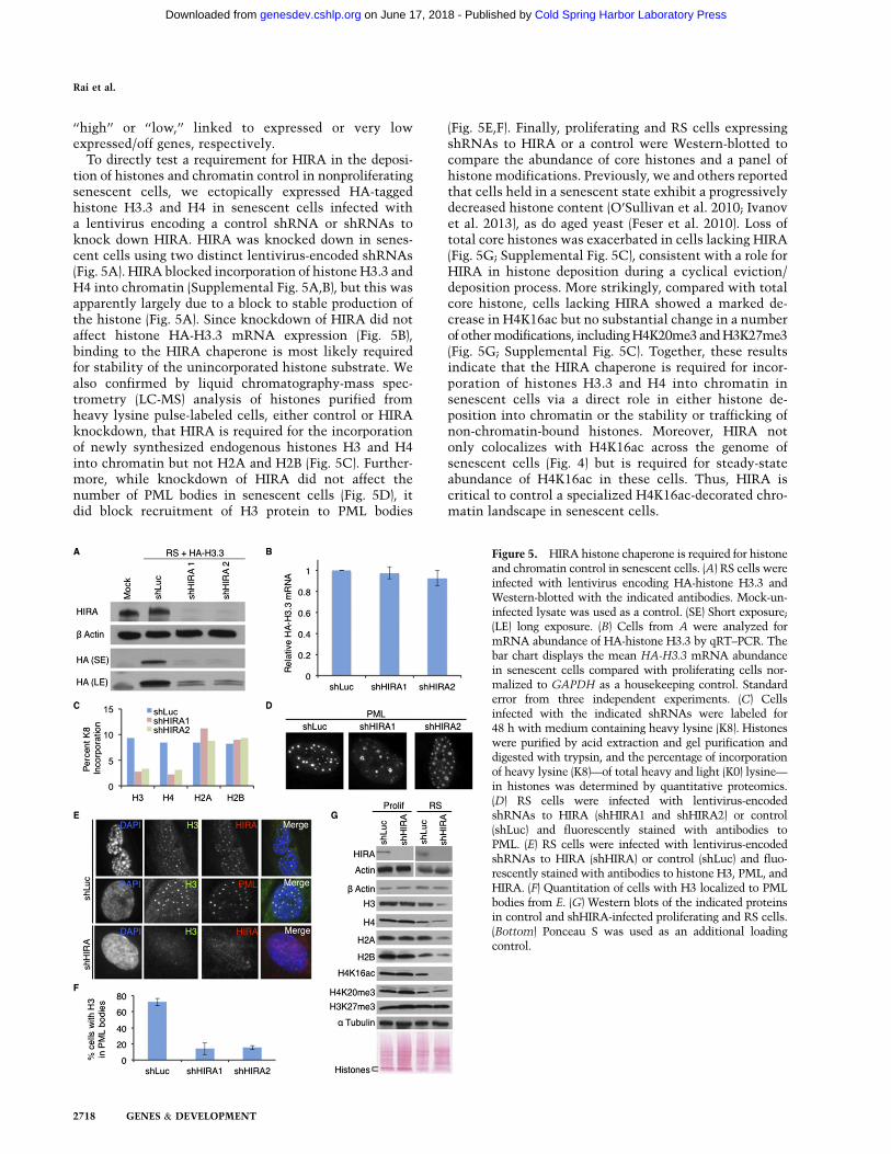
tion of histones and chromatin control in nonproliferatingsenescent cells, we ectopically expressed HA-taggedhistone H3.3 and H4 in senescent cells infected witha lentivirus encoding a control shRNA or shRNAs toknock down HIRA. HIRA was knocked down in senes-cent cells using two distinct lentivirus-encoded shRNAs(Fig. 5A). HIRA blocked incorporation of histone H3.3 andH4 into chromatin (Supplemental Fig. 5A,B), but this wasapparently largely due to a block to stable production ofthe histone (Fig. 5A). Since knockdown of HIRA did notaffect histone HA-H3.3 mRNA expression (Fig. 5B),binding to the HIRA chaperone is most likely requiredfor stability of the unincorporated histone substrate. Wealso confirmed by liquid chromatography-mass spec-trometry (LC-MS) analysis of histones purified fromheavy lysine pulse-labeled cells, either control or HIRAknockdown, that HIRA is required for the incorporationof newly synthesized endogenous histones H3 and H4into chromatin but not H2A and H2B (Fig. 5C). Further-more, while knockdown of HIRA did not affect thenumber of PML bodies in senescent cells (Fig. 5D), itdid block recruitment of H3 protein to PML bodies
(Fig. 5E,F). Finally, proliferating and RS cells expressingshRNAs to HIRA or a control were Western-blotted tocompare the abundance of core histones and a panel ofhistone modifications. Previously, we and others reportedthat cells held in a senescent state exhibit a progressivelydecreased histone content (O’Sullivan et al. 2010; Ivanovet al. 2013), as do aged yeast (Feser et al. 2010). Loss oftotal core histones was exacerbated in cells lacking HIRA(Fig. 5G; Supplemental Fig. 5C), consistent with a role forHIRA in histone deposition during a cyclical eviction/deposition process. More strikingly, compared with totalcore histone, cells lacking HIRA showed a marked de-crease in H4K16ac but no substantial change in a numberof othermodifications, includingH4K20me3 andH3K27me3(Fig. 5G; Supplemental Fig. 5C). Together, these resultsindicate that the HIRA chaperone is required for incor-poration of histones H3.3 and H4 into chromatin insenescent cells via a direct role in either histone de-position into chromatin or the stability or trafficking ofnon-chromatin-bound histones. Moreover, HIRA notonly colocalizes with H4K16ac across the genome ofsenescent cells (Fig. 4) but is required for steady-stateabundance of H4K16ac in these cells. Thus, HIRA iscritical to control a specialized H4K16ac-decorated chro-matin landscape in senescent cells.
Figure 5. HIRA histone chaperone is required for histoneand chromatin control in senescent cells. (A) RS cells wereinfected with lentivirus encoding HA-histone H3.3 andWestern-blotted with the indicated antibodies. Mock-un-infected lysate was used as a control. (SE) Short exposure;(LE) long exposure. (B) Cells from A were analyzed formRNA abundance of HA-histone H3.3 by qRT–PCR. Thebar chart displays the mean HA-H3.3 mRNA abundancein senescent cells compared with proliferating cells nor-malized to GAPDH as a housekeeping control. Standarderror from three independent experiments. (C) Cellsinfected with the indicated shRNAs were labeled for48 h with medium containing heavy lysine (K8). Histoneswere purified by acid extraction and gel purification anddigested with trypsin, and the percentage of incorporationof heavy lysine (K8)—of total heavy and light (K0) lysine—in histones was determined by quantitative proteomics.(D) RS cells were infected with lentivirus-encodedshRNAs to HIRA (shHIRA1 and shHIRA2) or control(shLuc) and fluorescently stained with antibodies toPML. (E) RS cells were infected with lentivirus-encodedshRNAs to HIRA (shHIRA) or control (shLuc) and fluo-rescently stained with antibodies to histone H3, PML, andHIRA. (F) Quantitation of cells with H3 localized to PMLbodies from E. (G) Western blots of the indicated proteinsin control and shHIRA-infected proliferating and RS cells.(Bottom) Ponceau S was used as an additional loadingcontrol.
Rai et al.
2718 GENES & DEVELOPMENT
Cold Spring Harbor Laboratory Press on June 17, 2018 - Published by genesdev.cshlp.orgDownloaded from
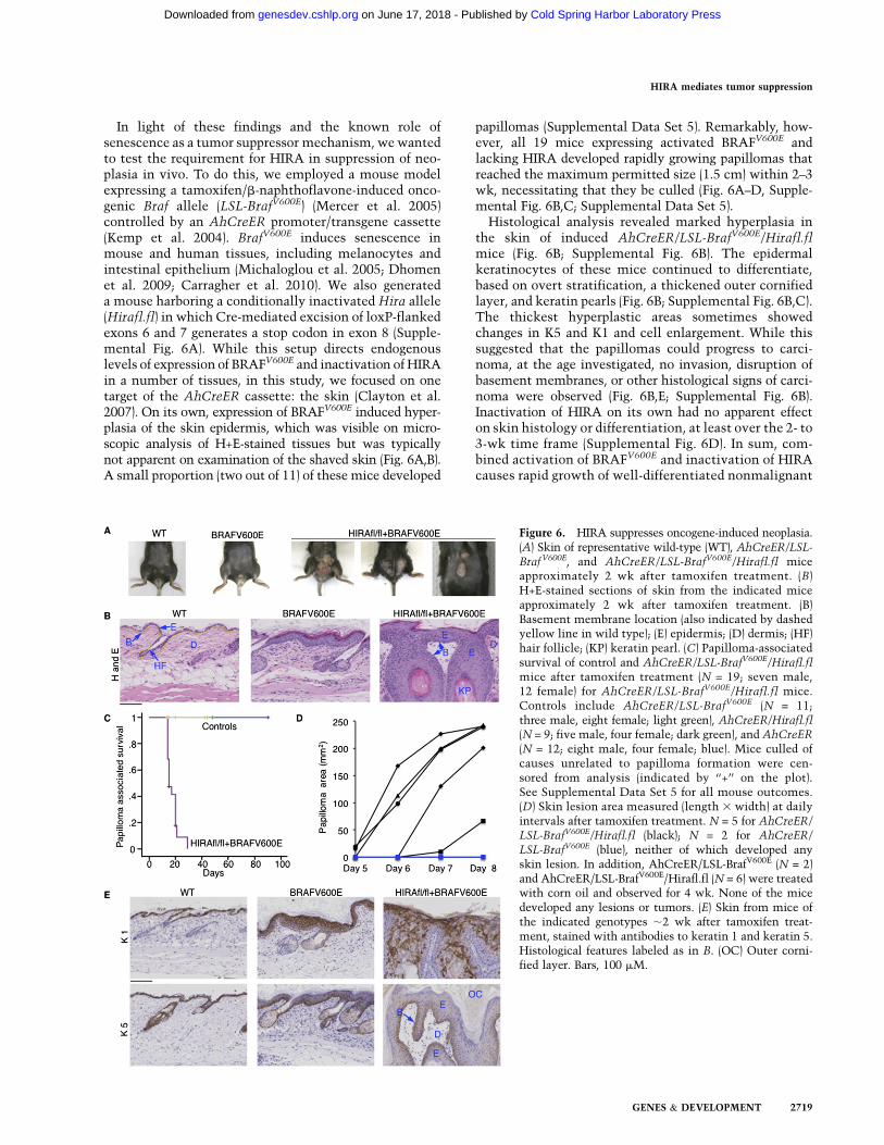
In light of these findings and the known role ofsenescence as a tumor suppressor mechanism, wewantedto test the requirement for HIRA in suppression of neo-plasia in vivo. To do this, we employed a mouse modelexpressing a tamoxifen/b-naphthoflavone-induced onco-genic Braf allele (LSL-BrafV600E) (Mercer et al. 2005)controlled by an AhCreER promoter/transgene cassette(Kemp et al. 2004). BrafV600E induces senescence inmouse and human tissues, including melanocytes andintestinal epithelium (Michaloglou et al. 2005; Dhomenet al. 2009; Carragher et al. 2010). We also generateda mouse harboring a conditionally inactivated Hira allele(Hirafl.fl) in which Cre-mediated excision of loxP-flankedexons 6 and 7 generates a stop codon in exon 8 (Supple-mental Fig. 6A). While this setup directs endogenouslevels of expression of BRAFV600E and inactivation of HIRAin a number of tissues, in this study, we focused on onetarget of the AhCreER cassette: the skin (Clayton et al.2007). On its own, expression of BRAFV600E induced hyper-plasia of the skin epidermis, which was visible on micro-scopic analysis of H+E-stained tissues but was typicallynot apparent on examination of the shaved skin (Fig. 6A,B).A small proportion (two out of 11) of these mice developed
papillomas (Supplemental Data Set 5). Remarkably, how-ever, all 19 mice expressing activated BRAFV600E andlacking HIRA developed rapidly growing papillomas thatreached the maximum permitted size (1.5 cm) within 2–3wk, necessitating that they be culled (Fig. 6A–D, Supple-mental Fig. 6B,C; Supplemental Data Set 5).Histological analysis revealed marked hyperplasia in
the skin of induced AhCreER/LSL-BrafV600E/Hirafl.flmice (Fig. 6B; Supplemental Fig. 6B). The epidermalkeratinocytes of these mice continued to differentiate,based on overt stratification, a thickened outer cornifiedlayer, and keratin pearls (Fig. 6B; Supplemental Fig. 6B,C).The thickest hyperplastic areas sometimes showedchanges in K5 and K1 and cell enlargement. While thissuggested that the papillomas could progress to carci-noma, at the age investigated, no invasion, disruption ofbasement membranes, or other histological signs of carci-noma were observed (Fig. 6B,E; Supplemental Fig. 6B).Inactivation of HIRA on its own had no apparent effecton skin histology or differentiation, at least over the 2- to3-wk time frame (Supplemental Fig. 6D). In sum, com-bined activation of BRAFV600E and inactivation of HIRAcauses rapid growth of well-differentiated nonmalignant
Figure 6. HIRA suppresses oncogene-induced neoplasia.(A) Skin of representative wild-type (WT), AhCreER/LSL-BrafV600E, and AhCreER/LSL-BrafV600E/Hirafl.fl miceapproximately 2 wk after tamoxifen treatment. (B)H+E-stained sections of skin from the indicated miceapproximately 2 wk after tamoxifen treatment. (B)Basement membrane location (also indicated by dashedyellow line in wild type); (E) epidermis; (D) dermis; (HF)hair follicle; (KP) keratin pearl. (C) Papilloma-associatedsurvival of control and AhCreER/LSL-BrafV600E/Hirafl.flmice after tamoxifen treatment (N = 19; seven male,12 female) for AhCreER/LSL-BrafV600E/Hirafl.fl mice.Controls include AhCreER/LSL-BrafV600E (N = 11;three male, eight female; light green), AhCreER/Hirafl.fl(N = 9; five male, four female; dark green), and AhCreER
(N = 12; eight male, four female; blue). Mice culled ofcauses unrelated to papilloma formation were cen-sored from analysis (indicated by ‘‘+’’ on the plot).See Supplemental Data Set 5 for all mouse outcomes.(D) Skin lesion area measured (length 3 width) at dailyintervals after tamoxifen treatment. N = 5 for AhCreER/LSL-BrafV600E/Hirafl.fl (black); N = 2 for AhCreER/
LSL-BrafV600E (blue), neither of which developed anyskin lesion. In addition, AhCreER/LSL-BrafV600E (N = 2)and AhCreER/LSL-BrafV600E/Hirafl.fl (N = 6) were treatedwith corn oil and observed for 4 wk. None of the micedeveloped any lesions or tumors. (E) Skin from mice ofthe indicated genotypes ;2 wk after tamoxifen treat-ment, stained with antibodies to keratin 1 and keratin 5.Histological features labeled as in B. (OC) Outer corni-fied layer. Bars, 100 mM.
HIRA mediates tumor suppression
GENES & DEVELOPMENT 2719
Cold Spring Harbor Laboratory Press on June 17, 2018 - Published by genesdev.cshlp.orgDownloaded from

epidermal hyperplasias. We conclude that HIRA is re-quired for efficient suppression of oncogene-induced neo-plasia in vivo.We asked whether HIRA’s role in suppression of neo-
plasia is linked to its regulation of chromatin structureand senescence. Inactivation of HIRA alone did notobviously affect proliferation or senescence markers(Supplemental Fig. 7A). In control and induced AhCreER/LSL-BrafV600E and AhCreER/LSL-BrafV600E/Hirafl.fl mice,the level of cell proliferation, measured by BrdUincorporation, correlated well with the degree of hyper-plasia (Fig. 7). Consistent with the histologically differ-entiated nature of the tissues, proliferation was largelyconfined to the basal layer, although this layer wasexpanded in induced AhCreER/LSL-BrafV600E/Hirafl.flmice (Fig. 7). Surprisingly, while we observed modestinduction of senescence markers p21 and p53 in inducedAhCreER/LSL-BrafV600E mice, expression of these pro-teins was, in regions, more marked in induced AhCreER/LSL-BrafV600E/Hirafl.fl mice (Fig. 7; Supplemental Fig. 7B).Most surprisingly, in mice harboring activated BRAFV600E
and inactivation of HIRA, there was a substantial overlap
of zones of proliferating cells marked by BrdU and zonesof cells expressing the highest levels of p21 and p53, withall three enriched in the basal layer of the epidermis(Fig. 7).Inactivation of HIRA alone had no detectable effect on
total histone H3 or histone modifications H3K9me3,H3K27me3, and H4K16ac (Supplemental Fig. 7C, cf.with wild type in Fig. 7 and Supplemental Fig. 7D).However, while inactivation of HIRA in induced AhCreER/LSL-BrafV600E/Hirafl.fl mice did not overtly affect totalhistone content and H3K9me3 and H3K27me3, papillo-mas of induced AhCreER/LSL-BrafV600E/Hirafl.fl micewere depleted of H4K16ac, particularly in the nonprolif-erative regions furthest from the basal layer (Fig. 7;Supplemental Fig. 7D). We conclude that, in the presenceof activated BRAFV600E, inactivation of HIRA expandsa population of cells that has engaged the p53–p21senescence effector pathway yet remains in a proliferativestate. Moreover, inactivation of HIRA in vivo recapitu-lates chromatin changes observed on HIRA inactivationin senescent cells in vitro; most notably, decreasedH4K16ac.
Figure 7. Suppression of oncogene-induced neoplasiaby HIRA is linked to control of H4K16ac. Sections ofskin from the indicated mice ;2 wk after tamoxifentreatment, stained with the indicated antibodies. Boxedareas are expanded in Supplemental Figure 6B. Bars,100 mM.
Rai et al.
2720 GENES & DEVELOPMENT
Cold Spring Harbor Laboratory Press on June 17, 2018 - Published by genesdev.cshlp.orgDownloaded from

Discussion
Cellular senescence acts as a barrier to malignant pro-gression of potentially neoplastic cells (Salama et al.2014), particularly those harboring activated oncogenes.This barrier function is facilitated by the exceptionalstability of the senescence-associated proliferation arrest,for example, in benign human nevi (Michaloglou et al.2005). The mechanisms by which chromatin contributesto barrier function are not fully understood. However,most efforts to date have focused on features of chromatinstructure and regulation that promote compacted hetero-chromatin, whose repressed ‘‘closed’’ state is intuitivelyinterpreted as a static barrier to proliferation and tumorprogression; for example, H3K9me3, HP1 proteins, andSAHF (Braig et al. 2005; Narita et al. 2006). In contrast,here we define chromatin of senescent cells as a dynam-ically maintained landscape, exhibiting previously un-anticipated dynamic features. We show that one criticalregulator of this dynamic landscape, histone chaperoneHIRA, is required for efficient suppression of oncogene-induced neoplasia.Several lines of evidence demonstrate the dynamic
nature of chromatin in senescent cells. First, histonemRNAs are expressed in senescent cells. Second, insenescent cells, histones are enriched in PML bodies.This has been previously shown to reflect trafficking ofnewly synthesized histones into chromatin (Chang et al.2013; Delbarre et al. 2013; Corpet et al. 2014). Third,newly synthesized ectopically expressed and endoge-nous histones are actively incorporated into chromatinin senescent cells. Fourth, FRAP analysis of a fluorescent-tagged histone H3.3 demonstrates a highly mobile sub-population of H3.3. Fifth, gene promoters in senescentcells show marked relative enrichment of H4K16ac insenescent cells. H4K16ac impedes higher-order chroma-tin packaging (Shogren-Knaak et al. 2006) and has alsobeen suggested to directly promote nucleosome exchange(Elliott et al. 2013). Together, these lines of evidenceindicate that chromatin of senescent cells is maintainedin a state of dynamic equilibrium.Chromatin of senescent cells exhibits at least two
unanticipated specialized features, indicating its funda-mentally distinct regulation from chromatin in prolifer-ating cells. First, nonproliferating senescent cells do,surprisingly, express a subset of so-called ‘‘replication-dependent’’ histone genes, linked to alternative mRNAsplicing to a second exon harboring a polyadenylationsignal instead of a stem/loop structure. Presumably, thissecond exon/poly A tail bypasses the normal stem–loop-mediated degradation of these mRNAs in nonprolifer-ating cells (Marzluff et al. 2008), facilitating theirpreferential expression in senescent cells. Second,H4K16ac is extraordinarily enriched at promoters ofexpressed genes in senescent cells, even at genes that areunchanged or decreased in expression compared withproliferating cells. As a result, in senescent cells, butnot proliferating cells, H4K16ac appears to flag allexpressed genes, regardless of whether those genes areexpressed at a high or low level. Curiously, H4K16ac is
similarly enriched at promoters and enhancers of embry-onic stem cells (Taylor et al. 2013), which have beenpreviously suggested to contain especially dynamic chro-matin (Meshorer et al. 2006). H4K16ac has been reportedto counter heterochromatin spreading (Wang et al. 2013).Conceivably, H4K16ac constitutes a local chromatin‘‘barrier’’ to prevent promoter silencing in unusual chro-matin environments in embryonic stem cells and senes-cent cells.We previously implicated the DNA replication-inde-
pendent histone chaperone HIRA and its binding partners(UBN1, CABIN1, and ASF1a) in chromatin regulation insenescent cells (Zhang et al. 2005; Banumathy et al. 2009;Rai et al. 2011). Here, we directly demonstrate thatmultiple features of the dynamic landscape in senescentcells are controlled by HIRA. HIRA colocalizes withhistones in PML bodies of senescent cells, a presumptivemarker of HIRA-mediated trafficking of histones intochromatin (Chang et al. 2013; Delbarre et al. 2013). HIRAis required for the accumulation of histones in PMLbodies and also for the deposition and/or accumulationof newly synthesized H3 (presumably H3.3) and H4 intochromatin of senescent cells. HIRA also colocalizes withnewly deposited histone H3.3 at gene promoters insenescent cells, particularly at highly expressed genes.HIRA and, to a lesser extent, H3.3 alter their target geneselection between proliferating and senescent cells, inline with changes in expression. HIRA is required for theretention of total histone content and, more specifically,H4K16ac in senescent cells. HIRA also colocalizes withH4K16ac at promoters of highly expressed genes. How-ever, HIRA is unlikely to act alone in nucleosome dynam-ics of senescent cells. An alternative chaperone, DAXX,has previously been suggested to target histone H3.3 topericentromeric heterochromatin in senescent cells (Corpetet al. 2014). A partially overlapping role for DAXX canexplain why repressed genes depleted of HIRA, such ashistone genes, continued to incorporate histone H3.3 insenescent cells. Nonetheless, numerous lines of evidenceindicate that the HIRA chaperone complex is a criticalhomeostatic regulator of the dynamic, H4K16ac-flaggedchromatin landscape of senescent cells.While previous constitutive knockout animals showed
that HIRA is required for early embryo development(Roberts et al. 2002; Szenker et al. 2012), these modelsdid not allow investigation of the functions in adulttissues. Here, in a novel conditional knockout HIRAmousemodel expressing an activated BRAFV600E oncogenein the skin, inactivation of HIRA dramatically enhancedoncogene-induced hyperplastic cell proliferation accompa-nied by p53- and p21-expressing proliferating (BrdU+) cells,consistent with impaired execution of the cellular senes-cence program despite p53–p21 activation. Just as in vitro,inactivation of HIRA also resulted in specific depletion ofH4K16ac. Inactivation of HIRA also appeared to modestlyimpair differentiation in BRAFV600E-expressing epidermis,based on altered K1 and K5 expression. Thus, both im-paired differentiation and senescence likely contribute toepidermal hyperplasia in AhCreER/LSL-BrafV600E/Hirafl.flmice. Regardless, these results indicate the importance
HIRA mediates tumor suppression
GENES & DEVELOPMENT 2721
Cold Spring Harbor Laboratory Press on June 17, 2018 - Published by genesdev.cshlp.orgDownloaded from

of proper chromatin regulation for tumor suppression,even in cells in which prosenescence effectors p53 andp21 are activated. Remarkably, they also unambiguouslydemonstrate a role for HIRA in efficient proliferationarrest to suppress neoplasia in the face of an activatedoncogene.Both histone H3.3 and H4K16ac are dysregulated in
cancer. The level of H4K16ac is reduced in many cancersand cancer cell lines (Fraga et al. 2005; Elsheikh et al.2009), suggesting a potential tumor suppressor function.Recent studies have revealed recurrent missense muta-tions in genes encoding histone H3.3 (H3F3A) and H3.1(HIST3H1B) in some cancers, in particular in pediatricgliomas (Schwartzentruber et al. 2012; Sturm et al. 2012;Wu et al. 2012; Yuen and Knoepfler 2013). These muta-tions often target specific residues and most often inhistone H3.3, specifically H3.3K27M and H3.3G34R/V. The H3.3K27M protein acts as a dominant inhibitorof the EZH2 histone methyltransferase responsible formethylation of histone H3 to generate H3K27me3 (Lewiset al. 2013). While H3.3K27M therefore acts to globallyreduce H3K27me3 levels, some regions do gain H3K27me3(Chan et al. 2013; Lewis et al. 2013; Venneti et al. 2013). Onesuch region is reported to be the CDKN2a locus, encodingthe tumor suppressor p16INK4a, and expression of p16INK4ais reduced in two glioma cell lines (Chan et al. 2013). Thep16INK4a/pRB pathway is a key effector of cellularsenescence, and its suppression by H3.3K27M would beexpected to impair senescence as a tumor suppressormechanism (Salama et al. 2014). Therefore, H3.3K27Mand inactivation of HIRA might in fact target similarsenescence-related pathways to promote tumorigenesis.Perhaps more relevant to HIRA, very recent studies haveindicated that transcription-coupled methylation of H3to yield H3K36me3 and, in particular, H3.3K36me3exerts a tumor-suppressive function (Wen et al. 2014;Zhu et al. 2014). HIRA is responsible for deposition ofhistone H3.3 at many transcribed genes (Goldberg et al.2010). Results reported here also invoke HIRA in themaintenance of H4K16ac levels in senescent cells. Thus,the tumor-suppressive functions of both histone H3.3 andH4K16ac might be controlled by the HIRA chaperonecomplex.In sum, our description of specialized and dynamic
chromatin in senescent cells presents a new model forchromatin control in senescent cells that is substan-tially different from the intuitive model of more staticcanonical heterochromatin enforcing a barrier to pro-liferation and tumor progression. Histone chaperoneHIRA is a key regulator of this landscape and exhibitspotent neoplastic suppressor activity in a mouse model.Extending this idea, we suggest that DNA replication-independent chromatin regulators, such as HIRA, alsomaintain the dynamic chromatin of senescent cells toretain the tumor-suppressive nonproliferating phenotypeover the long term, a process that we refer to as chromatinhomeostasis or ‘‘chromostasis.’’ In future studies, it willbe important to investigate the extent to which HIRA,histone H3.3, and H4K16ac act in concert to mediatetumor suppression.
Materials and methods
See the Supplemental Material for additional methods, details,and references.
Proliferating and senescent cells
IMR90 fibroblast cells were cultured according to American TypeCulture Collection guidelines in low oxygen (3%). Proliferatingfibroblasts (<30 population doublings) were cultured inDulbecco’smodified Eagle’s medium supplemented with 20% (v/v) fetalbovine serum until they reached RS (defined in SupplementalFig. 1A,B). To generate OIS and control cells, IMR90 cells stablyexpressing a fusion protein of the estrogen receptor (ER) ligand-binding domain and H-RASG12V (ER-RA-IMR90 cells [grownphenol red-free]) (Barradas et al. 2009) were supplemented with100 nM 4-hydroxytamoxifen (4-OHT) or ethanol vehicle,respectively.
ChIP-seq
For HIRA ChIP, native ChIP was performed using benzonaseendonuclease for chromatin solubilization and fragmentation.For HA-histone H3.3, H4K16ac, and H4 ChIP, cells were cross-linked with 1% formaldehyde for 5 min at room temperature.After quenching with glycine, the cells were harvested andsonicated to produce soluble chromatin with DNA fragmentsin the range of 300–500 base pairs. Fragmented chromatin wasincubated with antibodies to HIRA, HA, H4K16ac, or H4preimmobilized on Dynabeads. Libraries were prepared from10–20 ng of ChIP or input DNA using New England BiolabsChIP-seq kit according to the manufacturer’s instructions, andthe resulting libraries were sequenced on an Illumina GAIIx toyield ;30 million to 40 million raw reads.
RNA-seq
RNAwas isolated using a Qiagen RNeasy kit. Ribodepletion wasperformed using an Epicentre Ribo Zero kit (MRZH11124C).Libraries were prepared according to the Illumina TruSeq pro-tocol and sequenced on an Illumina GAIIx.
Massively parallel sequencing and data analysis
ChIP-seq or input reads were mapped to the human genome(hg19) using the Bowtie 2 alignment software. Only unique readsmapping to a single location were retained. The determination ofenriched regions was performed using the USeq and SICERpackages, and reads were visualized using the University ofCalifornia at Santa Cruz browser. The results presented wereanalyzed from a single ChIP-seq reaction for HIRA in each of RSand OIS and H3.3 in RS, but the results are representative of twodifferent experiments performed with two different antibodiesfor H4K16ac.
LC-MS analysis of extracted histones
The extracted histones were separated by SDS-PAGE, digestedwith trypsin, and analyzed by LC-MS on an AB-Sciex 5600 MSsystem as described previously (Gabrielsen et al. 2013). Proteinswere identified by searching the UniProt database using Mascot2.3 (MatrixScience), and SILAC ratios of lysine containingpeptides (K0/K8) were calculated using Mascot Distiller 2.5 runon a local server.
Rai et al.
2722 GENES & DEVELOPMENT
Cold Spring Harbor Laboratory Press on June 17, 2018 - Published by genesdev.cshlp.orgDownloaded from
Acknowledgments
We thank all members of the Adams laboratory for criticaldiscussions. We thank Jesus Gil for pLNC-RAS:ER, KatrinPritchard for LSL-BrafV600E mice, Philippe Collas for pH3.3-EGFP, and Doug Winton for AhCreER mice. The laboratory ofP.D.A. is funded by National Institute on Aging program projectAG031862 and Cancer Research UK program A16566. T.S.R. issupported by a start-up grant from Institute of Biomedical andEnvironmental Health Research, University of West Scotland,and the Carnegie Trust.
References
Acosta JC, O’Loghlen A, Banito A, Guijarro MV, Augert A,Raguz S, Fumagalli M, Da Costa M, Brown C, Popov N, et al.2008. Chemokine signaling via the CXCR2 receptor rein-forces senescence. Cell 133: 1006–1018.
Adam S, Polo SE, Almouzni G. 2013. Transcription recoveryafter DNA damage requires chromatin priming by the H3.3histone chaperone HIRA. Cell 155: 94–106.
Ahmad K, Henikoff S. 2002. The histone variant h3.3 marksactive chromatin by replication-independent nucleosomeassembly. Mol Cell 9: 1191–1200.
Banaszynski LA, Wen D, Dewell S, Whitcomb SJ, Lin M, Diaz N,Elsasser SJ, Chapgier A, Goldberg AD, Canaani E, et al. 2013.Hira-dependent histone H3.3 deposition facilitates PRC2 re-cruitment at developmental loci in ES cells. Cell 155: 107–120.
Banumathy G, Somaiah N, Zhang R, Tang Y, Hoffmann J,Andrake M, Ceulemans H, Schultz D, Marmorstein R, AdamsPD. 2009. Human UBN1 is an ortholog of yeast Hpc2p andhas an essential role in the HIRA/ASF1a chromatin-remodelingpathway in senescent cells. Mol Cell Biol 29: 758–770.
Barradas M, Anderton E, Acosta JC, Li S, Banito A, Rodriguez-Niedenfuhr M, Maertens G, Banck M, Zhou MM, Walsh MJ,et al. 2009. Histone demethylase JMJD3 contributes toepigenetic control of INK4a/ARF by oncogenic RAS. Genes
Dev 23: 1177–1182.Benhamed M, Herbig U, Ye T, Dejean A, Bischof O. 2012.
Senescence is an endogenous trigger for microRNA-directedtranscriptional gene silencing in human cells. Nat Cell Biol
14: 266–275.Braig M, Lee S, Loddenkemper C, Rudolph C, Peters AH,
Schlegelberger B, Stein H, Dorken B, Jenuwein T, SchmittCA. 2005. Oncogene-induced senescence as an initial barrierin lymphoma development. Nature 436: 660–665.
Brown DT, Wellman SE, Sittman DB. 1985. Changes in thelevels of three different classes of histone mRNA duringmurine erythroleukemia cell differentiation. Mol Cell Biol 5:2879–2886.
Carragher LA, Snell KR, Giblett SM, Aldridge VS, Patel B, CookSJ, Winton DJ, Marais R, Pritchard CA. 2010. V600EBrafinduces gastrointestinal crypt senescence and promotestumour progression through enhanced CpG methylation ofp16INK4a. EMBO Mol Med 2: 458–471.
Chai J, Charboneau AL, Betz BL, Weissman BE. 2005. Loss of thehSNF5 gene concomitantly inactivates p21CIP/WAF1 andp16INK4a activity associated with replicative senescence inA204 rhabdoid tumor cells. Cancer Res 65: 10192–10198.
Chan KM, Fang D, Gan H, Hashizume R, Yu C, Schroeder M,Gupta N, Mueller S, James CD, Jenkins R, et al. 2013. Thehistone H3.3K27M mutation in pediatric glioma reprogramsH3K27 methylation and gene expression. Genes Dev 27:985–990.
Chandra T, Kirschner K, Thuret JY, Pope BD, Ryba T, NewmanS, Ahmed K, Samarajiwa SA, Salama R, Carroll T, et al. 2012.
Independence of repressive histone marks and chromatincompaction during senescent heterochromatic layer forma-tion. Mol Cell 47: 203–214.
Chang FT, McGhie JD, Chan FL, Tang MC, Anderson MA,Mann JR, Andy Choo KH, Wong LH. 2013. PML bodiesprovide an important platform for the maintenance oftelomeric chromatin integrity in embryonic stem cells.Nucleic Acids Res 41: 4447–4458.
Chen Z, Trotman LC, Shaffer D, Lin HK, Dotan ZA, Niki M,Koutcher JA, Scher HI, Ludwig T, Gerald W, et al. 2005.Crucial role of p53-dependent cellular senescence in suppres-sion of Pten-deficient tumorigenesis. Nature 436: 725–730.
Clayton E, Doupe DP, Klein AM, Winton DJ, Simons BD, JonesPH. 2007. A single type of progenitor cell maintains normalepidermis. Nature 446: 185–189.
Collado M, Gil J, Efeyan A, Guerra C, Schuhmacher AJ, BarradasM, Benguria A, Zaballos A, Flores JM, Barbacid M, et al.2005. Tumour biology: senescence in premalignant tumours.Nature 436: 642.
Contrepois K, Thuret JY, Courbeyrette R, Fenaille F, Mann C.2012. Deacetylation of H4-K16Ac and heterochromatinassembly in senescence. Epigenetics Chromatin 5: 15.
Coppe JP, Patil CK, Rodier F, Sun Y, Munoz DP, Goldstein J,Nelson PS, Desprez PY, Campisi J. 2008. Senescence-associ-ated secretory phenotypes reveal cell-nonautonomous func-tions of oncogenic RAS and the p53 tumor suppressor. PLoSBiol 6: 2853–2868.
Corpet A, Olbrich T, Gwerder M, Fink D, Stucki M. 2014.Dynamics of histone H3.3 deposition in proliferating andsenescent cells reveals a DAXX-dependent targeting to PML-NBs important for pericentromeric heterochromatin organi-zation. Cell Cycle 13: 249–267.
Cosme-Blanco W, Shen MF, Lazar AJ, Pathak S, Lozano G,Multani AS, Chang S. 2007. Telomere dysfunction sup-presses spontaneous tumorigenesis in vivo by initiatingp53-dependent cellular senescence. EMBO Rep 8: 497–503.
Cruickshanks HA, McBryan T, Nelson DM, Vanderkraats ND,Shah PP, van Tuyn J, Singh Rai T, Brock C, Donahue G,Dunican DS, et al. 2013. Senescent cells harbour features ofthe cancer epigenome. Nat Cell Biol 15: 1495–1506.
De Cecco M, Criscione SW, Peckham EJ, Hillenmeyer S, HammEA, Manivannan J, Peterson AL, Kreiling JA, Neretti N,Sedivy JM. 2013. Genomes of replicatively senescent cellsundergo global epigenetic changes leading to gene silencingand activation of transposable elements. Aging Cell 12: 247–256.
Delbarre E, Ivanauskiene K, Kuntziger T, Collas P. 2013. DAXX-dependent supply of soluble (H3.3–H4) dimers to PML bodiespending deposition into chromatin. Genome Res 23: 440–451.
Dhomen N, Reis-Filho JS, da Rocha Dias S, Hayward R, SavageK, Delmas V, Larue L, Pritchard C, Marais R. 2009. Onco-genic Braf induces melanocyte senescence and melanoma inmice. Cancer Cell 15: 294–303.
Di Micco R, Sulli G, Dobreva M, Liontos M, Botrugno OA,Gargiulo G, dal Zuffo R, Matti V, d’Ario G, Montani E, et al.2011. Interplay between oncogene-induced DNA damageresponse and heterochromatin in senescence and cancer.Nat Cell Biol 13: 292–302.
Duarte LF, Young AR, Wang Z, Wu HA, Panda T, Kou Y,Kapoor A, Hasson D, Mills NR, Ma’ayan A, et al. 2014.Histone H3.3 and its proteolytically processed form drivea cellular senescence programme. Nat Commun 5: 5210.
Dutta D, Ray S, Home P, Saha B, Wang S, Sheibani N, Tawfik O,Cheng N, Paul S. 2010. Regulation of angiogenesis by histonechaperone HIRA-mediated Incorporation of lysine 56-acety-
HIRA mediates tumor suppression
GENES & DEVELOPMENT 2723
Cold Spring Harbor Laboratory Press on June 17, 2018 - Published by genesdev.cshlp.orgDownloaded from
lated histone H3.3 at chromatin domains of endothelialgenes. J Biol Chem 285: 41567–41577.
Elliott GO, Murphy KJ, Hayes JJ, Thiriet C. 2013. Replication-independent nucleosome exchange is enhanced by local andspecific acetylation of histone H4. Nucleic Acids Res 41:2228–2238.
Elsheikh SE, Green AR, Rakha EA, Powe DG, Ahmed RA,Collins HM, Soria D, Garibaldi JM, Paish CE, Ammar AA,et al. 2009. Global histone modifications in breast cancercorrelate with tumor phenotypes, prognostic factors, andpatient outcome. Cancer Res 69: 3802–3809.
Feldser DM, Greider CW. 2007. Short telomeres limit tumorprogression in vivo by inducing senescence. Cancer Cell 11:461–469.
Feser J, Truong D, Das C, Carson JJ, Kieft J, Harkness T, Tyler JK.2010. Elevated histone expression promotes life span exten-sion. Mol Cell 39: 724–735.
Fraga MF, Ballestar E, Villar-Garea A, Boix-Chornet M, Espada J,Schotta G, Bonaldi T, Haydon C, Ropero S, Petrie K, et al.2005. Loss of acetylation at Lys16 and trimethylation atLys20 of histone H4 is a common hallmark of human cancer.Nat Genet 37: 391–400.
Gabrielsen M, Schuldt M, Munro J, Borucka D, Cameron J,Baugh M, Mleczak A, Lilla S, Morrice N, Olson MF. 2013.Cucurbitacin covalent bonding to cysteine thiols: the fila-mentous-actin severing protein Cofilin1 as an exemplarytarget. Cell Commun Signal 11: 58.
Goldberg AD, Banaszynski LA, Noh KM, Lewis PW, Elsaesser SJ,Stadler S, Dewell S, Law M, Guo X, Li X, et al. 2010. Distinctfactors control histone variant H3.3 localization at specificgenomic regions. Cell 140: 678–691.
Grove GW, Zweidler A. 1984. Regulation of nucleosomal corehistone variant levels in differentiating murine erythroleu-kemia cells. Biochemistry 23: 4436–4443.
Ivanov A, Pawlikowski J, Manoharan I, van Tuyn J, Nelson DM,Rai TS, Shah PP, Hewitt G, Korolchuk VI, Passos JF, et al.2013. Lysosome-mediated processing of chromatin in senes-cence. J Cell Biol 202: 129–143.
Jin C, Felsenfeld G. 2007. Nucleosome stability mediated byhistone variants H3.3 and H2A. Z. Genes Dev 21: 1519–1529.
Jin C, Zang C, Wei G, Cui K, Peng W, Zhao K, Felsenfeld G.2009. H3.3/H2A.Z double variant-containing nucleosomesmark ‘nucleosome-free regions’ of active promoters andother regulatory regions. Nat Genet 41: 941–945.
Kang TW, Yevsa T, Woller N, Hoenicke L, Wuestefeld T, DauchD, Hohmeyer A, Gereke M, Rudalska R, Potapova A, et al.2011. Senescence surveillance of pre-malignant hepatocyteslimits liver cancer development. Nature 479: 547–551.
Kari V, Karpiuk O, Tieg B, Kriegs M, Dikomey E, Krebber H,Begus-Nahrmann Y, Johnsen SA. 2013. A subset of histoneH2B genes produces polyadenylated mRNAs under a varietyof cellular conditions. PLoS ONE 8: e63745.
Kemp R, Ireland H, Clayton E, Houghton C, Howard L, WintonDJ. 2004. Elimination of background recombination: somaticinduction of Cre by combined transcriptional regulation andhormone binding affinity. Nucleic Acids Res 32: e92.
Krtolica A, Parrinello S, Lockett S, Desprez PY, Campisi J. 2001.Senescent fibroblasts promote epithelial cell growth andtumorigenesis: a link between cancer and aging. Proc Natl
Acad Sci 98: 12072–12077.Kuilman T, Michaloglou C, Vredeveld LC, Douma S, van
Doorn R, Desmet CJ, Aarden LA, Mooi WJ, Peeper DS.2008. Oncogene-induced senescence relayed by an interleu-kin-dependent inflammatory network. Cell 133: 1019–1031.
Lewis PW,MullerMM, KoletskyMS, Cordero F, Lin S, BanaszynskiLA, Garcia BA, Muir TW, Becher OJ, Allis CD. 2013. Inhibitionof PRC2 activity by a gain-of-function H3 mutation found inpediatric glioblastoma. Science 340: 857–861.
Lin CJ, Conti M, Ramalho-Santos M. 2013. Histone variant H3.3maintains a decondensed chromatin state essential formouse preimplantation development. Development 140:3624–3634.
Loppin B, Bonnefoy E, Anselme C, Laurencon A, Karr TL,Couble P. 2005. The histone H3.3 chaperone HIRA isessential for chromatin assembly in the male pronucleus.Nature 437: 1386–1390.
Lujambio A, Akkari L, Simon J, Grace D, Tschaharganeh DF,Bolden JE, Zhao Z, Thapar V, Joyce JA, Krizhanovsky V, et al.2013. Non-cell-autonomous tumor suppression by p53. Cell
153: 449–460.Martin N, Popov N, Aguilo F, O’Loghlen A, Raguz S, Snijders
AP, Dharmalingam G, Li S, Thymiakou E, Carroll T, et al.2013. Interplay between Homeobox proteins and Polycombrepressive complexes in p16INK(4)a regulation. EMBO J 32:982–995.
Marzluff WF, Wagner EJ, Duronio RJ. 2008. Metabolism andregulation of canonical histone mRNAs: life without a poly(A)tail. Nat Rev Genet 9: 843–854.
Mercer K, Giblett S, Green S, Lloyd D, DaRocha Dias S, PlumbM, Marais R, Pritchard C. 2005. Expression of endogenousoncogenic V600EB-raf induces proliferation and developmen-tal defects in mice and transformation of primary fibroblasts.Cancer Res 65: 11493–11500.
Meshorer E, Yellajoshula D, George E, Scambler PJ, Brown DT,Misteli T. 2006. Hyperdynamic plasticity of chromatin proteinsin pluripotent embryonic stem cells. Dev Cell 10: 105–116.
Michaloglou C, Vredeveld LC, Soengas MS, Denoyelle C, KuilmanT, van der Horst CM, Majoor DM, Shay JW, Mooi WJ, PeeperDS. 2005. BRAFE600-associated senescence-like cell cycle ar-rest of human naevi. Nature 436: 720–724.
Narita M, Nunez S, Heard E, Lin AW, Hearn SA, Spector DL,Hannon GJ, Lowe SW. 2003. Rb-mediated heterochromatinformation and silencing of E2F target genes during cellularsenescence. Cell 113: 703–716.
Narita M, Narita M, Krizhanovsky V, Nunez S, Chicas A, HearnSA, Myers MP, Lowe SW. 2006. A novel role for high-mobility group a proteins in cellular senescence and hetero-chromatin formation. Cell 126: 503–514.
O’Sullivan RJ, Kubicek S, Schreiber SL, Karlseder J. 2010. Reducedhistone biosynthesis and chromatin changes arising from a dam-age signal at telomeres. Nat Struct Mol Biol 17: 1218–1225.
Pantazis P, Bonner WM. 1984. Specific alterations in the patternof histone-3 synthesis during conversion of human leukemiccells to terminally differentiated cells in culture. Differenti-ation 28: 186–190.
Pchelintsev NA, McBryan T, Rai TS, van Tuyn J, Ray-Gallet D,Almouzni G, Adams PD. 2013. Placing the HIRA histonechaperone complex in the chromatin landscape. Cell Reports
3: 1012–1019.Placek BJ, Huang J, Kent JR, Dorsey J, Rice L, Fraser NW, Berger
SL. 2009. The histone variant H3.3 regulates gene expressionduring lytic infection with herpes simplex virus type 1. J
Virol 83: 1416–1421.Rai TS, Puri A, McBryan T, Hoffman J, Tang Y, Pchelintsev NA,
van Tuyn J, Marmorstein R, Schultz DC, Adams PD. 2011.Human CABIN1 is a functional member of the humanHIRA/UBN1/ASF1a histone H3.3 chaperone complex. Mol
Cell Biol 31: 4107–4118.Ray-Gallet D, Quivy J-P, Scamps C, Martini EM-D, Lipinski M,
Almouzni G. 2002. HIRA is critical for a nucleosome assem-
Rai et al.
2724 GENES & DEVELOPMENT
Cold Spring Harbor Laboratory Press on June 17, 2018 - Published by genesdev.cshlp.orgDownloaded from

bly pathway independent of DNA synthesis. Mol Cell 9:1091–1100.
Ray-Gallet D, Woolfe A, Vassias I, Pellentz C, Lacoste N, Puri A,Schultz DC, Pchelintsev NA, Adams PD, Jansen LE, et al.2011. Dynamics of histone H3 deposition in vivo reveala nucleosome gap-filling mechanism for H3.3 to maintainchromatin integrity. Mol Cell 44: 928–941.
Roberts C, Sutherland HF, Farmer H, Kimber W, Halford S,Carey A, Brickman JM, Wynshaw-Boris A, Scambler PJ.2002. Targeted mutagenesis of the Hira gene results ingastrulation defects and patterning abnormalities of meso-endodermal derivatives prior to early embryonic lethality.Mol Cell Biol 22: 2318–2328.
Sadaie S, Salama R, Carroll T, Tomimatsu K, Chandra T, YoungARJ, Narita M, Perez-Mancera PA, Bennett DC, Chong H,et al. 2013. Redistribution of the Lamin B1 genomic bindingprofile affects rearrangement of heterochromatic domains andSAHF formation during senescence. Genes Dev 27: 1800–1808.
Salama R, Sadaie M, Hoare M, Narita M. 2014. Cellularsenescence and its effector programs. Genes Dev 28: 99–114.
Schwartzentruber J, Korshunov A, Liu XY, Jones DT, Pfaff E, JacobK, Sturm D, Fontebasso AM, Quang DA, Tonjes M, et al. 2012.Driver mutations in histone H3.3 and chromatin remodellinggenes in paediatric glioblastoma. Nature 482: 226–231.
Shah PP, Donahue G, Otte GL, Capell BC, Nelson DM, Cao K,Aggarwala V, Cruickshanks HA, Rai TS, McBryan T, et al.2013. Lamin B1 depletion in senescent cells triggers large-scale changes in gene expression and the chromatin land-scape. Genes Dev 27: 1787–1799.
Sherwood PW, Tsang SV, Osley MA. 1993. Characterization ofHIR1 and HIR2, two genes required for regulation of histonegene transcription in Saccharomyces cerevisiae. Mol Cell
Biol 13: 28–38.Shogren-Knaak M, Ishii H, Sun JM, Pazin MJ, Davie JR, Peterson
CL. 2006. Histone H4-K16 acetylation controls chromatinstructure and protein interactions. Science 311: 844–847.
Skene PJ, Henikoff S. 2013. Histone variants in pluripotency anddisease. Development 140: 2513–2524.
Sturm D, Witt H, Hovestadt V, Khuong-Quang DA, Jones DT,Konermann C, Pfaff E, Tonjes M, Sill M, Bender S, et al. 2012.Hotspot mutations in H3F3A and IDH1 define distinctepigenetic and biological subgroups of glioblastoma. Cancer
Cell 22: 425–437.Szenker E, Lacoste N, Almouzni G. 2012. A developmental
requirement for HIRA-dependent H3.3 deposition revealed atgastrulation in Xenopus. Cell Reports 1: 730–740.
Tagami H, Ray-Gallet D, Almouzni G, Nakatani Y. 2004.Histone H3.1 and H3.3 complexes mediate nucleosomeassembly pathways dependent or independent of DNAsynthesis. Cell 116: 51–61.
Taylor GC, Eskeland R, Hekimoglu-Balkan B, Pradeepa MM,Bickmore WA. 2013. H4K16 acetylation marks active genesand enhancers of embryonic stem cells, but does not alterchromatin compaction. Genome Res 23: 2053–2065.
Urban MK, Zweidler A. 1983. Changes in nucleosomal corehistone variants during chicken development and matura-tion. Dev Biol 95: 421–428.
van der Heijden GW, Derijck AA, Posfai E, Giele M, Pelczar P,Ramos L, Wansink DG, van der Vlag J, Peters AH, de Boer P.2007. Chromosome-wide nucleosome replacement and H3.3incorporation during mammalian meiotic sex chromosomeinactivation. Nat Genet 39: 251–258.
Venneti S, Garimella MT, Sullivan LM, Martinez D, Huse JT,Heguy A, Santi M, Thompson CB, Judkins AR. 2013. Evalu-ation of histone 3 lysine 27 trimethylation (H3K27me3) and
enhancer of Zest 2 (EZH2) in pediatric glial and glioneuronaltumors shows decreased H3K27me3 in H3F3A K27M mutantglioblastomas. Brain Pathol 23: 558–564.
Wang J, Tadeo X, Hou H, Tu PG, Thompson J, Yates JR 3rd, Jia S.2013. Epe1 recruits BET family bromodomain protein Bdf2 toestablish heterochromatin boundaries. Genes Dev 27: 1886–1902.
Wen H, Li Y, Xi Y, Jiang S, Stratton S, Peng D, Tanaka K, Ren Y,Xia Z, Wu J, et al. 2014. ZMYND11 links histoneH3.3K36me3 to transcription elongation and tumour sup-pression. Nature 508: 263–268.
Wilson BG, Roberts CW. 2011. SWI/SNF nucleosome remod-ellers and cancer. Nat Rev Cancer 11: 481–492.
Wu G, Broniscer A, McEachron TA, Lu C, Paugh BS, Becksfort J,Qu C, Ding L, Huether R, Parker M, et al. 2012. Somatichistone H3 alterations in pediatric diffuse intrinsic pontinegliomas and non-brainstem glioblastomas. Nat Genet 44:251–253.
Xue W, Zender L, Miething C, Dickins RA, Hernando E,Krizhanovsky V, Cordon-Cardo C, Lowe SW. 2007. Senes-cence and tumour clearance is triggered by p53 restoration inmurine liver carcinomas. Nature 445: 656–660.
Yang JH, Song Y, Seol JH, Park JY, Yang YJ, Han JW, Youn HD,Cho EJ. 2011. Myogenic transcriptional activation of MyoDmediated by replication-independent histone deposition.Proc Natl Acad Sci 108: 85–90.
Ye X, Zerlanko B, Kennedy A, Banumathy G, Zhang R, AdamsPD. 2007. Downregulation of Wnt signaling is a trigger forformation of facultative heterochromatin and onset of cellsenescence in primary human cells. Mol Cell 27: 183–196.
Yuen BT, Knoepfler PS. 2013. Histone H3.3 mutations: a variantpath to cancer. Cancer Cell 24: 567–574.
Zhang R, Poustovoitov MV, Ye X, Santos HA, Chen W, DaganzoSM, Erzberger JP, Serebriiskii IG, Canutescu AA, DunbrackRL, et al. 2005. Formation of MacroH2A-containing senes-cence-associated heterochromatin foci and senescencedriven by ASF1a and HIRA. Dev Cell 8: 19–30.
Zhang R, Chen W, Adams PD. 2007. Molecular dissection offormation of senescent associated heterochromatin foci. MolCell Biol 27: 2343–2358.
Zhu X, He F, Zeng H, Ling S, Chen A, Wang Y, Yan X, Wei W,Pang Y, Cheng H, et al. 2014. Identification of functionalcooperative mutations of SETD2 in human acute leukemia.Nat Genet 46: 287–293.
HIRA mediates tumor suppression
GENES & DEVELOPMENT 2725
Cold Spring Harbor Laboratory Press on June 17, 2018 - Published by genesdev.cshlp.orgDownloaded from

10.1101/gad.247528.114Access the most recent version at doi: 28:2014, Genes Dev.
Taranjit Singh Rai, John J. Cole, David M. Nelson, et al. is required for suppression of neoplasiaHIRA orchestrates a dynamic chromatin landscape in senescence and
Material
Supplemental
http://genesdev.cshlp.org/content/suppl/2014/12/12/28.24.2712.DC1
References
http://genesdev.cshlp.org/content/28/24/2712.full.html#ref-list-1
This article cites 88 articles, 29 of which can be accessed free at:
License
Commons Creative
.http://creativecommons.org/licenses/by-nc/4.0/at Creative Commons License (Attribution-NonCommercial 4.0 International), as described
). After six months, it is available under ahttp://genesdev.cshlp.org/site/misc/terms.xhtmlsix months after the full-issue publication date (see This article is distributed exclusively by Cold Spring Harbor Laboratory Press for the first
ServiceEmail Alerting
click here.right corner of the article or
Receive free email alerts when new articles cite this article - sign up in the box at the top
© 2014 Rai et al.; Published by Cold Spring Harbor Laboratory Press
Cold Spring Harbor Laboratory Press on June 17, 2018 - Published by genesdev.cshlp.orgDownloaded from