HIGHLIGHTS ON BREAST CANCER Eyad Alsaeed MD, FRCPC. Consultant Radiation Oncologist Acting Chairman...
87
HIGHLIGHTS ON BREAST CANCER Eyad Alsaeed MD, FRCPC. Consultant Radiation Oncologist Acting Chairman of Radiation Oncology Prince Sultan Hematology @ Oncology center KFMC
-
Upload
brice-goodwin -
Category
Documents
-
view
214 -
download
0
Transcript of HIGHLIGHTS ON BREAST CANCER Eyad Alsaeed MD, FRCPC. Consultant Radiation Oncologist Acting Chairman...
HIGHLIGHTS ON BREAST CANCER
Eyad Alsaeed MD, FRCPC.
Consultant Radiation Oncologist Acting Chairman of Radiation OncologyPrince Sultan Hematology @ Oncology centerKFMC
Breast Cancer In KSASaudis
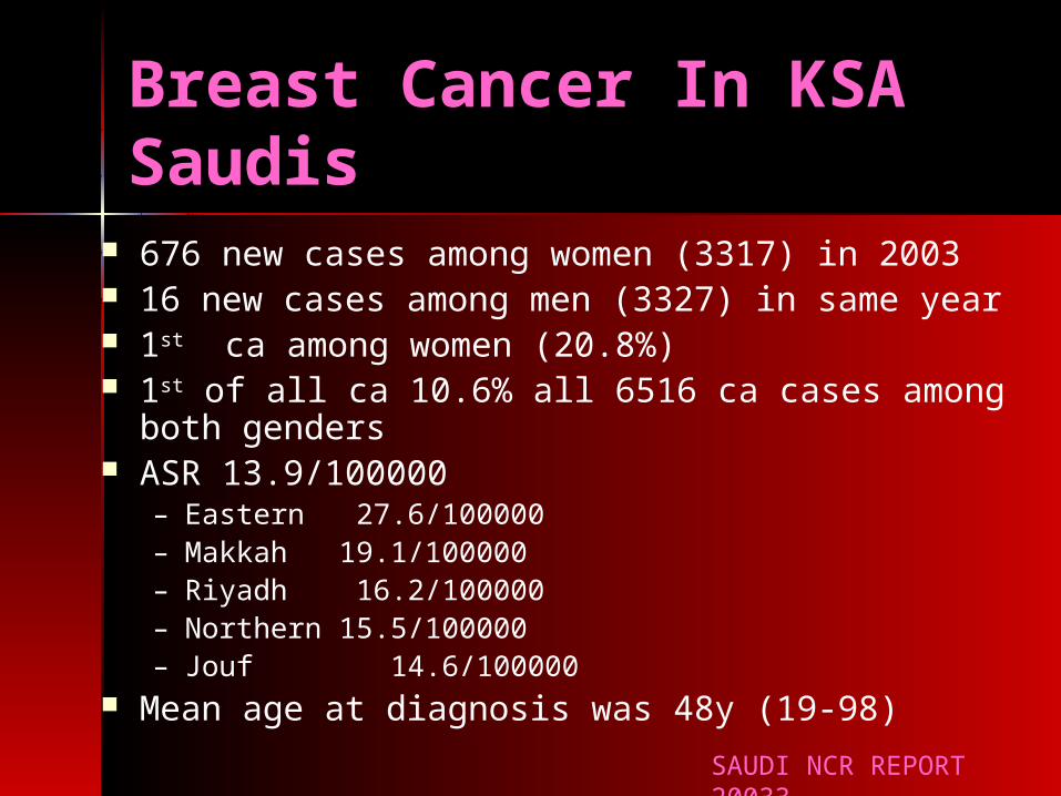
676 new cases among women (3317) in 2003 16 new cases among men (3327) in same year 1st ca among women (20.8%) 1st of all ca 10.6% all 6516 ca cases among both
genders ASR 13.9/100000
– Eastern 27.6/100000– Makkah 19.1/100000– Riyadh 16.2/100000– Northern 15.5/100000– Jouf 14.6/100000
Mean age at diagnosis was 48y (19-98)SAUDI NCR REPORT 20033

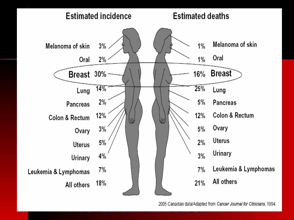
Cancer Incidence for Most Common Sites Cancer Incidence for Most Common Sites (2004)(2004)
CancerCancer MalMalee
FemalFemalee
All All %%
Breast Breast 1515 783783 798798 11.511.5
CRCCRC 366366 281281 647647 9.39.3
NHLNHL 332332 224224 556556 8.08.0
LeukemiaLeukemia 241241 194194 435435 6.26.2
ThyroidThyroid 8787 328328 415415 6.06.0
LiverLiver 231231 9393 324324 4.64.6
LungLung 233233 6363 296296 4.24.2
HDHD 166166 9898 264264 3.83.8
SkinSkin 136136 125125 261261 3.73.7
Brain, Brain, CNSCNS
147147 100100 247247 3.53.5
ProstateProstate 214214 -- 214214 3.13.1
StomachStomach 141141 7070 211211 3.03.0
BladderBladder 160160 4141 201201 2.92.9
UterusUterus -- 117117 117117 1.71.7
OvariesOvaries -- 108108 108108 1.51.5
All OthersAll Others 10091009 866866 18751875 26.926.9
TotalTotal 34783478 34913491 69606960 100100
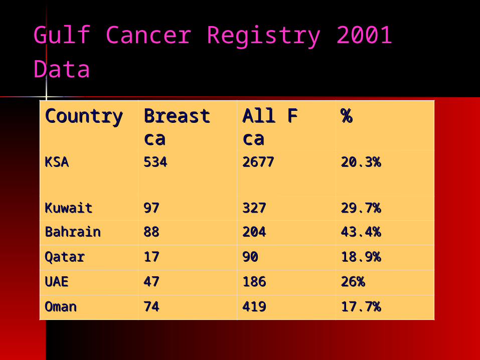
Country Country Breast caBreast ca All F ca All F ca %%
KSAKSA 534534 26772677 20.3%20.3%
KuwaitKuwait 9797 327327 29.7%29.7%
BahrainBahrain 8888 204204 43.4%43.4%
QatarQatar 1717 9090 18.9%18.9%
UAE UAE 4747 186186 26%26%
OmanOman 7474 419419 17.7%17.7%
Gulf Cancer Registry 2001 DataGulf Cancer Registry 2001 Data
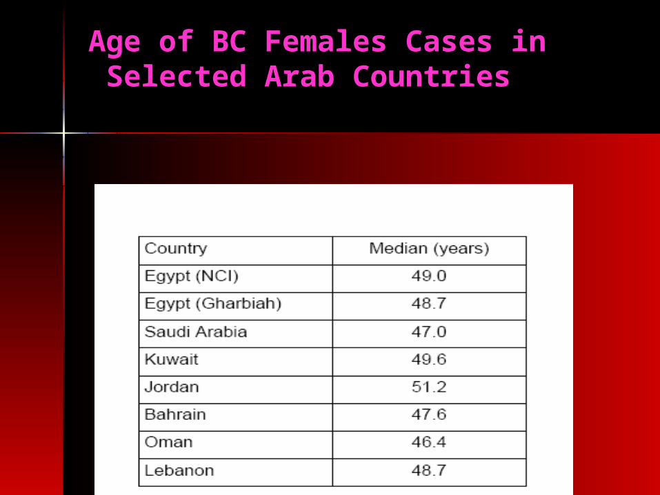
Age of BC Females Cases in Selected Arab Countries
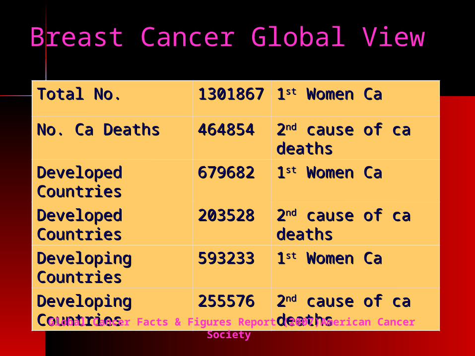
Breast Cancer Global View Breast Cancer Global View
Total No.Total No. 13018613018677
11stst Women Ca Women Ca
No. Ca DeathsNo. Ca Deaths 464854464854 22ndnd cause of ca cause of ca deaths deaths
Developed Developed Countries Countries
679682679682 11stst Women Ca Women Ca
Developed Developed CountriesCountries
203528203528 22ndnd cause of ca cause of ca deaths deaths
Developing Developing CountriesCountries
593233593233 11stst Women Ca Women Ca
Developing Developing CountriesCountries
255576255576 22ndnd cause of ca cause of ca deaths deaths Global Cancer Facts & Figures Report (2007)American Cancer
Society

Breast Cancer Global ViewBreast Cancer Global View
Incidence Rates varies By 25-30 Incidence Rates varies By 25-30 folds from as low as 3.9/100000 in folds from as low as 3.9/100000 in Mozambique to 10.1/100000 in USA Mozambique to 10.1/100000 in USA (2002)(2002)
This variation can be attributed to This variation can be attributed to under reporting as well as many under reporting as well as many factors are of importancefactors are of importance
Global Cancer Facts & Figures Report (2007)American Cancer Society

Breast cancer mortalityBreast cancer mortality(1980-2007)(1980-2007)

Histopathologic Subtypes
Morphology Total %
Infiltrating duct carcinoma (IDC) 518 76.6%
Lobular carcinoma 33 4.9%
Carcinoma, 20 3.0%
IDC mixed with other types of carcinoma 17 2.5%
Infiltrating duct & Lobular carcinoma 14 2.1%
Adenocarcinoma 13 1.9%
Medullary carcinoma 12 1.8%
Paget disease & Infiltrating duct carcinoma
10 1.5%
Infiltrating duct carcinoma (IDC) 7 1.0%
All Others 32 4.7%
SAUDI NCR REPORT 20033
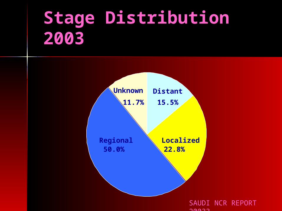
Stage Distribution 2003
11.7% 15.5%
50.0% 22.8%Regional Localized
Distant Unknown
SAUDI NCR REPORT 20033
Breast Cancer Facts
BC is most common lethal cancer in women.
2nd leading cause of death
BC is most common lethal cancer in women.
2nd leading cause of death
One out of eight women will be diagnosed with breast cancer
One out of eight women will be diagnosed with breast cancer
All women are at risk
Incidence increases with age
All women are at risk
Incidence increases with age
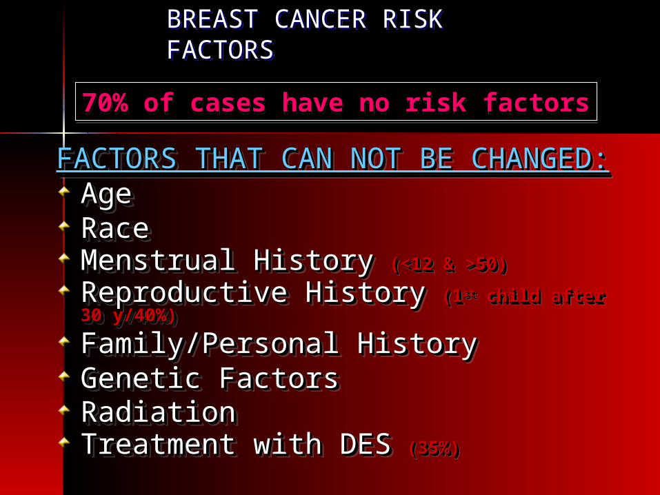
BREAST CANCER RISK FACTORS BREAST CANCER RISK FACTORS
FACTORS THAT CAN NOT BE CHANGED:FACTORS THAT CAN NOT BE CHANGED:AgeAgeRace Race Menstrual HistoryMenstrual History (<12 & >50)(<12 & >50)
Reproductive HistoryReproductive History (1(1stst child after 30 child after 30 y/40%)y/40%)
Family/Personal HistoryFamily/Personal History Genetic FactorsGenetic FactorsRadiationRadiation Treatment with DESTreatment with DES (35%)(35%)
70% of cases have no risk factors70% of cases have no risk factors
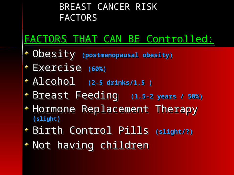
BREAST CANCER RISK FACTORS BREAST CANCER RISK FACTORS
FACTORS THAT CAN BE Controlled:FACTORS THAT CAN BE Controlled:
Obesity Obesity (postmenopausal obesity)(postmenopausal obesity) Exercise Exercise (60%)(60%)
Alcohol Alcohol (2-5 drinks/1.5 )(2-5 drinks/1.5 )
Breast Feeding Breast Feeding (1.5-2 years / 50%)(1.5-2 years / 50%)
Hormone Replacement Therapy Hormone Replacement Therapy ((slightslight))
Birth Control Pills Birth Control Pills (slight/?)(slight/?)
Not having children Not having children
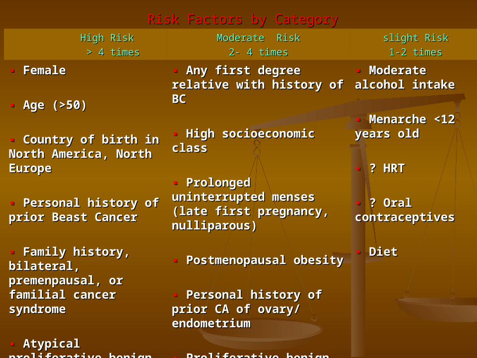
Risk Factors by CategoryRisk Factors by Category High RiskHigh Risk
> 4 times> 4 timesModerate RiskModerate Risk
2- 4 times2- 4 times slight Risk slight Risk
1-2 times1-2 times
▪▪ FemaleFemale
▪▪ Age (>50)Age (>50)
▪▪ Country of birth in North Country of birth in North America, North EuropeAmerica, North Europe
▪▪ Personal history of prior Personal history of prior Beast CancerBeast Cancer
▪▪ Family history, bilateral, Family history, bilateral, premenpausal, or familial premenpausal, or familial cancer syndromecancer syndrome
▪▪ Atypical proliferative Atypical proliferative benign breast disease/ benign breast disease/ family historyfamily history
▪▪ Any first degree relative with Any first degree relative with history of BChistory of BC
▪▪ High socioeconomic classHigh socioeconomic class
▪ ▪ Prolonged uninterrupted Prolonged uninterrupted menses (late first pregnancy, menses (late first pregnancy, nulliparous)nulliparous)
▪ ▪ Postmenopausal obesityPostmenopausal obesity
▪▪ Personal history of prior CA Personal history of prior CA of ovary/ endometriumof ovary/ endometrium
▪▪ Proliferative benign breast Proliferative benign breast disease, if no atypiadisease, if no atypia
▪ ▪ Moderate alcohol Moderate alcohol intakeintake
▪▪ Menarche <12 years Menarche <12 years oldold
▪ ▪ ? HRT? HRT
▪▪ ? Oral ? Oral contraceptivescontraceptives
▪ ▪ DietDiet

Histopathologic Subtypes
• Carcinoma Insitu (Cis)– Ductal carcinoma insitu (DCIS)– Lobular carcinoma insitu (LCIS)– Paegt`s Diease
• Invasive Cancer– Ductal– Lobular– Modularly– Papillary – Mucinous– Tubular
Inflammatory Carcinoma
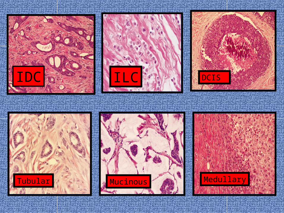
IDC ILC
Tubular Mucinous Medullary
DCIS
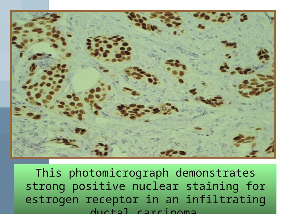
This photomicrograph demonstrates strong positive nuclear staining for estrogen
receptor in an infiltrating ductal carcinoma
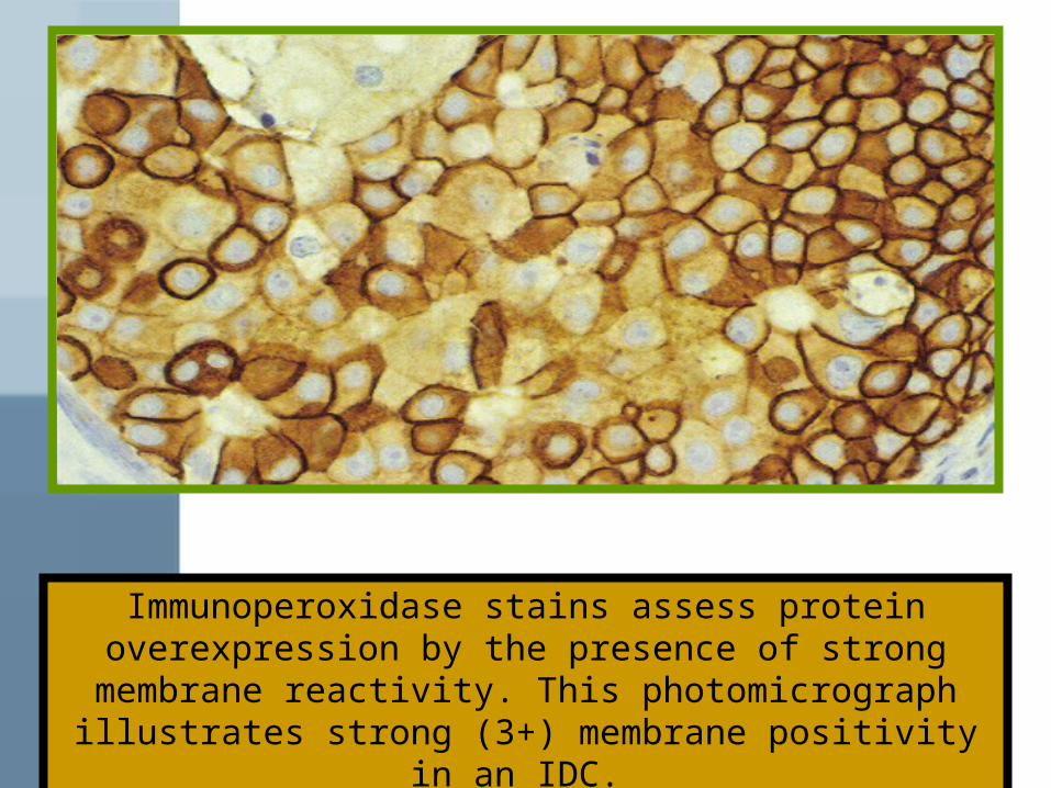
Immunoperoxidase stains assess protein overexpression by the presence of strong membrane
reactivity. This photomicrograph illustrates strong (3+) membrane positivity in an IDC.

Pathological and prognostic factors
Histological subtypes ( IDC = 85% = NOS),( ILC= 5-10%),.
Mucinous, Medullary, Tubular, Papillary
Nuclear and histological Grade
Estrogen and progesterone receptor status
HER-2 overexpression
Proliferation marker (s-phase fraction, Ki-67)
Aneuploidy / P53
LN +ve vs –ve numbers
LVI
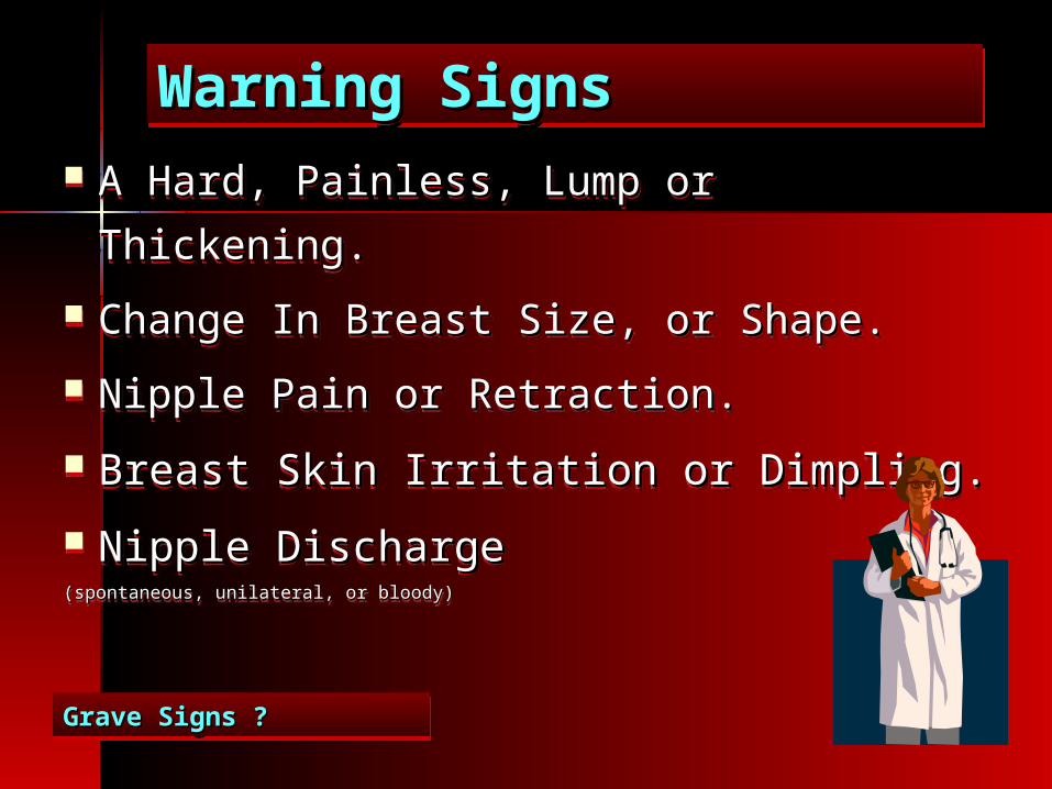
Warning SignsWarning Signs Warning SignsWarning Signs
A Hard, Painless, Lump or Thickening.A Hard, Painless, Lump or Thickening.
Change In Breast Size, or Shape.Change In Breast Size, or Shape.
Nipple Pain or Retraction.Nipple Pain or Retraction.
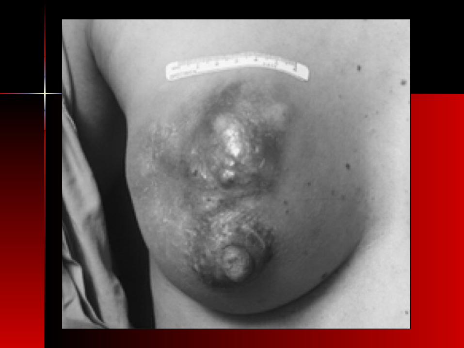
Breast Skin Irritation or Dimpling.Breast Skin Irritation or Dimpling.
Nipple DischargeNipple Discharge(spontaneous, unilateral, or bloody)(spontaneous, unilateral, or bloody)
A Hard, Painless, Lump or Thickening.A Hard, Painless, Lump or Thickening.
Change In Breast Size, or Shape.Change In Breast Size, or Shape.
Nipple Pain or Retraction.Nipple Pain or Retraction.
Breast Skin Irritation or Dimpling.Breast Skin Irritation or Dimpling.
Nipple DischargeNipple Discharge(spontaneous, unilateral, or bloody)(spontaneous, unilateral, or bloody)
Grave Signs ?Grave Signs ? Grave Signs ?Grave Signs ?

Core biopsy / fine-needle aspiration.
Experienced breast-imaging radiologist for tissue sampling ( ultrasound or mammogram guidance).
Manual Expression of spontaneous nipple discharge (Cytopathology examination).
Ductal lavage with saline solution to obtain nipple aspirate fluid.
FNAC cannot differentiate between invasive and noninvasive cancer , and even sometimes difficult to differentiate between Benign /Malignant lesion alone.
Request special studies for ER/PR (IHC) and, possibly, for HER2 over expression (FISH or IHC) if histopathology confirms invasive breast cancer.
BIOPSYBIOPSY

Management of palpable breast mass
Clinical breast examination by primary provider.
If pt is under 30 and mass feels benign ultrasound may be sufficient to verify cyst.
If mass is cystic, it can be aspirated with follow up in 3-4 months.
If pt is over 30, ultrasound and diagnostic mammogram are indicated.
If mass is solid, FNA or core needle biopsy.
If initial biopsy is negative, excisional biopsy mass be warranted.

WHAT TO DO NOW AFTER DIAGNOSIS WHAT TO DO NOW AFTER DIAGNOSIS WHAT TO DO NOW AFTER DIAGNOSIS WHAT TO DO NOW AFTER DIAGNOSIS
??

•Physical Examination
•Mammogram +/- US
•Core biopsy (Tue Cut)
Triple assessment
Diagnosis

•Bloods (CBC, LFTs, Bone profile, Markers, U & Es)
•Echo &/or Muga Scan
•CT CAP
•Bone Scan
Pretreatment workup
Staging & Fitness
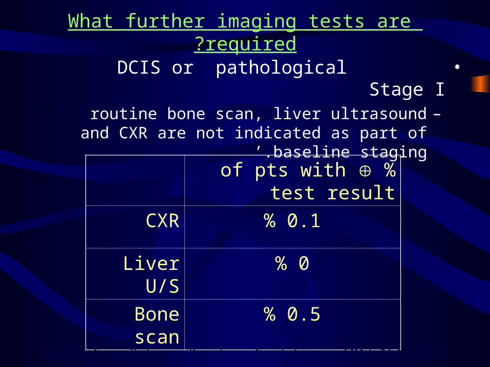
What further imaging tests are required?
• DCIS or pathological Stage I–routine bone scan, liver ultrasound and CXR
are not indicated as part of baseline staging.’
%of pts with test result
CXR 0.1%
Liver U/S 0%
Bone scan 0.5%
Cancer Care Ontario Practice Guidelines, CMAJ 164, 1439-1444, 2001.
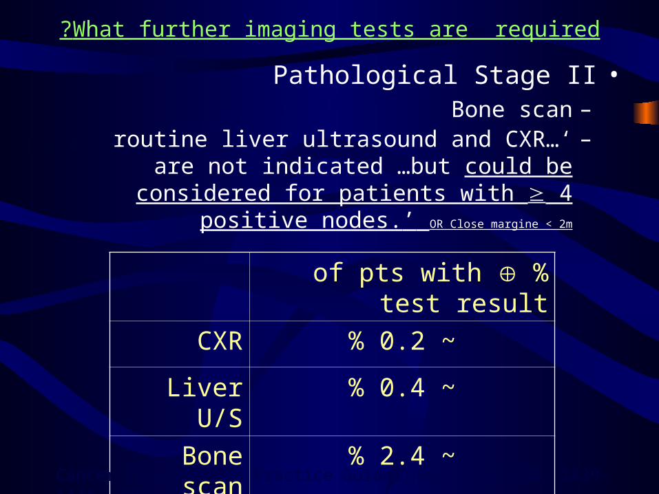
What further imaging tests are required?
•Pathological Stage II–Bone scan–…‘routine liver ultrasound and CXR are not
indicated …but could be considered for patients with 4 positive nodes.’ OR Close margine < 2m
%of pts with test result
CXR ~0.2%
Liver U/S ~0.4%
Bone scan ~2.4%
Cancer Care Ontario Practice Guidelines, CMAJ 164, 1439-1444, 2001.

STAGINGSTAGING

STAGINGSTAGING

STAGINGSTAGING

Stage Stage 0 0
cancer breastcancer breast
Stage Stage 0 0
cancer breastcancer breast
DCISDCIS
(± LCIS)(± LCIS)LCISLCIS Paget’sPaget’s
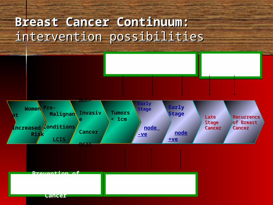
Breast Cancer Continuum: Breast Cancer Continuum: intervention possibilitiesintervention possibilitiesBreast Cancer Continuum: Breast Cancer Continuum: intervention possibilitiesintervention possibilities
Prevention of ClinicallyDetectable Breast Cancer
Pre- Malignant Conditions
LCIS
Women at Increased Risk
Women at Increased Risk
Non- Invasive Cancer
DCIS
Prevention of Contralateral Breast Cancer 3.2%
Tumors< 1cm
Early Stage
node -ve
Early Stage
node +ve
Prevention of RecurrencePrevention of Recurrence
Late StageCancer
Late StageCancer
Recurrenceof BreastCancer
Recurrenceof BreastCancer
Prevention of Progression
Prevention of Progression
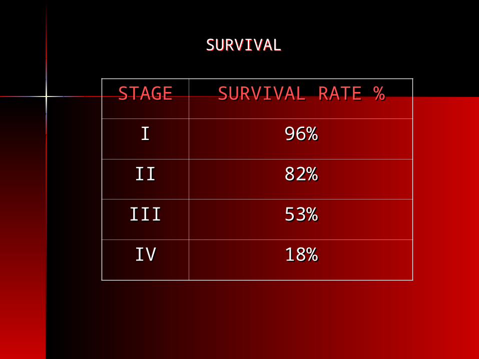
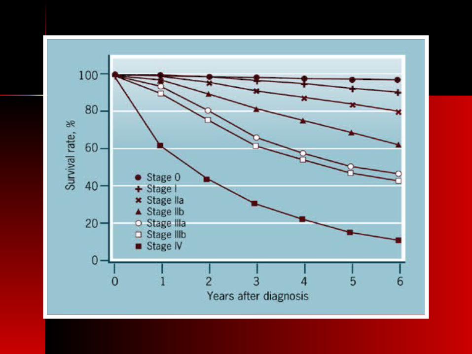
SURVIVALSURVIVAL
STAGESTAGE SURVIVAL RATE %SURVIVAL RATE %
II 96%96%
IIII 82%82%
IIIIII 53%53%
IVIV 18%18%

LOCO-REGIONAL CONTROL
LOCO-REGIONAL CONTROL
Surgery +RadiotherapySurgery +Radiotherapy
Treatment of early Breast CancerTreatment of early Breast Cancer
SYSTEMIC CONTROLSYSTEMIC CONTROL
Chemotherapy + HormonalNew Modalities
Chemotherapy + HormonalNew Modalities
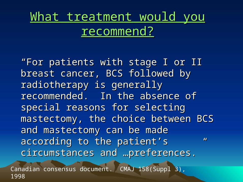
What treatment would you What treatment would you recommend?recommend?
““For patients with stage I or II breast cancer, For patients with stage I or II breast cancer, BCS followed by radiotherapy is generally BCS followed by radiotherapy is generally recommended. In the absence of special recommended. In the absence of special reasons for selecting mastectomy, the choice reasons for selecting mastectomy, the choice between BCS and mastectomy can be made between BCS and mastectomy can be made according to the patient’s circumstances and …according to the patient’s circumstances and …preferences.”preferences.”
Canadian consensus document. CMAJ 158(Suppl 3), 1998

What treatment would you recommend?What treatment would you recommend?
Advantages of BCT
• Cosmetic• Psychological
Disadvantages of BCT
• Time• Convenience• Late toxicity
*Major criteria for BCT,negative margin , mammography is also important

Stage I – II breast cancerStage I – II breast cancer
Outcome after BCS + RT Mastectomy• Survival
DFS
Distant metastases
Loco-Regional Control (except EORTC)
No Disadvantage in the use of BCS in Axillary N+ pt.
No different in the rate of ….breast or control ……
See next
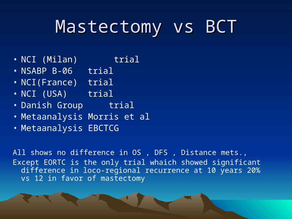
Mastectomy vs BCTMastectomy vs BCT
• NCI (Milan) trial • NSABP B-06 trial • NCI(France) trial • NCI (USA) trial • Danish Group trial • Metaanalysis Morris et al • Metaanalysis EBCTCG
All shows no difference in OS , DFS , Distance mets., Except EORTC is the only trial whaich showed significant difference in
loco-regional recurrence at 10 years 20% vs 12 in favor of mastectomy
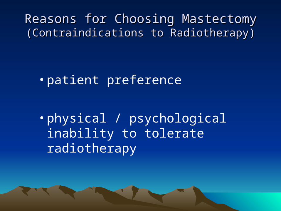
Reasons for Choosing MastectomyReasons for Choosing Mastectomy(Contraindications to Radiotherapy)(Contraindications to Radiotherapy)
• patient preference
• physical / psychological inability to tolerate radiotherapy

Reasons for Choosing MastectomyReasons for Choosing Mastectomy
(Contraindications to Radiotherapy(Contraindications to Radiotherapy) ) – 2– 2
High risk of recurrence
• Multicentric disease 2 primary tumors in separate quadrants
• Diffuse malignant calcifications
• Persistant positive margins
• Locally advanced disease (most T3, T4)

Reasons for Choosing MastectomyReasons for Choosing Mastectomy
(Contraindications to Radiotherapy(Contraindications to Radiotherapy) ) – 3– 3
Contraindications to radiotherapy
• Pregnancy• Scleroderma or active SLE• Previous breast radiotherapy (e.g. mantle RT)
Cosmetic
• Large tumor in small breast
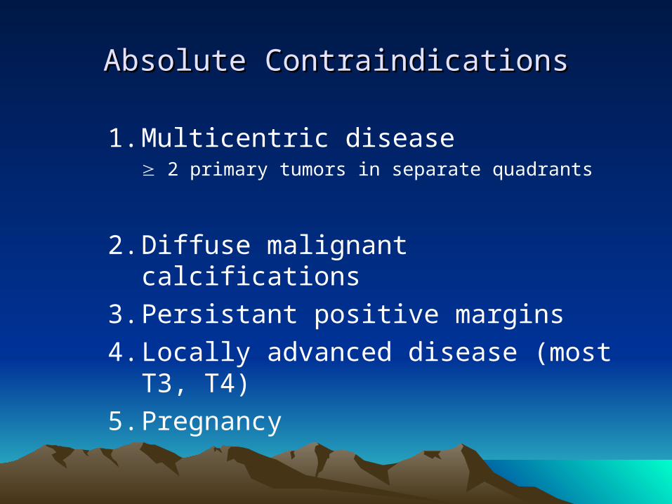
Absolute ContraindicationsAbsolute Contraindications
1. Multicentric disease 2 primary tumors in separate quadrants
2. Diffuse malignant calcifications
3. Persistant positive margins
4. Locally advanced disease (most T3, T4)
5. Pregnancy
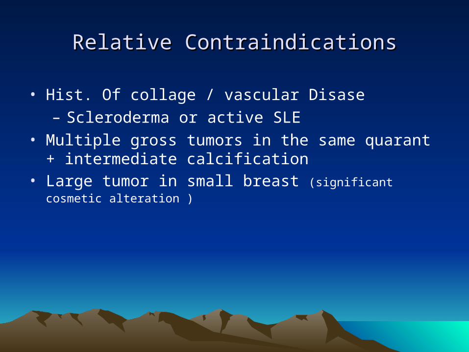
Relative ContraindicationsRelative Contraindications
• Hist. Of collage / vascular Disase – Scleroderma or active SLE
• Multiple gross tumors in the same quarant + intermediate calcification
• Large tumor in small breast (significant cosmetic alteration )
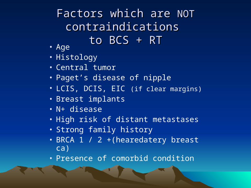
Factors which are Factors which are NOTNOT contraindications contraindications to BCS + RTto BCS + RT
• Age• Histology • Central tumor• Paget’s disease of nipple• LCIS, DCIS, EIC (if clear margins)
• Breast implants• N+ disease• High risk of distant metastases• Strong family history • BRCA 1 / 2 +(hearedatery breast ca)• Presence of comorbid condition
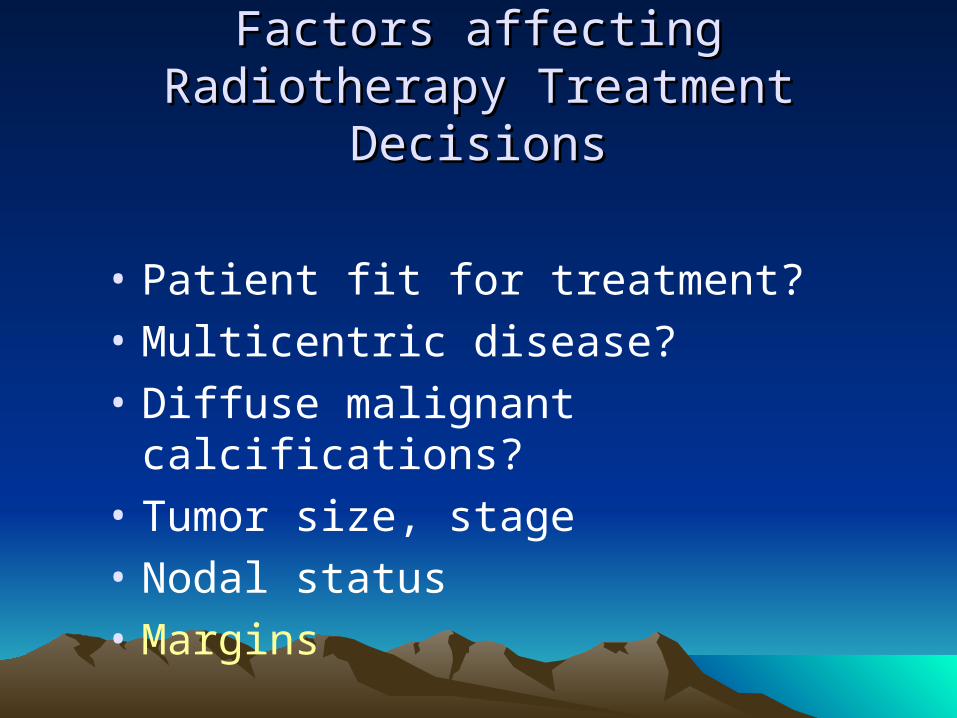
Factors affecting Radiotherapy Factors affecting Radiotherapy Treatment DecisionsTreatment Decisions
• Patient fit for treatment?
• Multicentric disease?
• Diffuse malignant calcifications?
• Tumor size, stage
• Nodal status
• Margins

MarginsMargins• Margins status remain one of the most important independent
factor involved with long term local recurrence .
Risk of local recurrence (5y) with xrt is approximately : 5% in negative
10% in focally+
20% in diffusely+.

Indications for Re-excisionIndications for Re-excision
1. Positive margina) Invasive tumor cells at marginb) DCIS at marginc) EIC with positive margin
2. Unknown margin
3. Inadequate excision by specimen mammography
4. Focally positive**But…focally positive margin ( 3 LPF) or close margin can be treated with many Rx. Option :1. Re-exceision2. Radiotherapy boost3. Chemotherapy
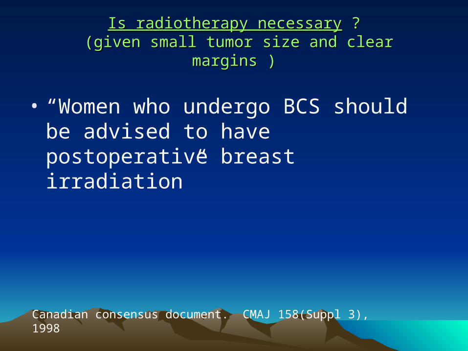
Is radiotherapy necessaryIs radiotherapy necessary ? ? (given small tumor size and clear margins ) (given small tumor size and clear margins )
• “Women who undergo BCS should be advised to have postoperative breast irradiation”
Canadian consensus document. CMAJ 158(Suppl 3), 1998

Randomized Trials of Randomized Trials of BCS vs. BCS + BCS vs. BCS + RTRT
n Local Recurrence Rate (%)
BCS BCS+RT
NSABP 1265 15 yr 36 12
OCOG 837 10 yr 40 18
See Table 2
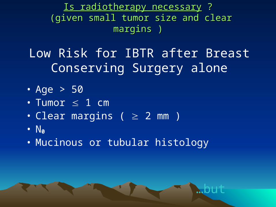
Is radiotherapy necessaryIs radiotherapy necessary ? ? (given small tumor size and clear margins ) (given small tumor size and clear margins )
• Age > 50• Tumor 1 cm• Clear margins ( 2 mm )• N0
• Mucinous or tubular histology
Low Risk for IBTR after Breast Conserving Surgery alone
…but
Is radiotherapy necessaryIs radiotherapy necessary ? ? (given small tumor size and clear margins ) (given small tumor size and clear margins )
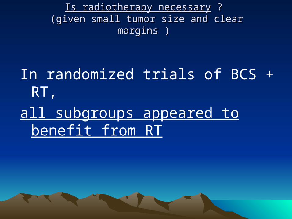
In randomized trials of BCS + RT,
all subgroups appeared to benefit from RT

INDICATIONS FOR ADJUVANT RADIOTHERAPY INDICATIONS FOR ADJUVANT RADIOTHERAPY TO THE BREASTTO THE BREAST
• Indications for Breast Adjuvant Radiotherapy post Lumpectomy?
All cases.
No subset has been found for which radiotherapy does not reduce local recurrence significantly.
• Indications for Chest Wall Adjuvant Radiotherapy?T3 especially if other risk factors present (2-3 lymph nodes, high grade, very large, etc.)T4
4 or more axillary lymph nodes +
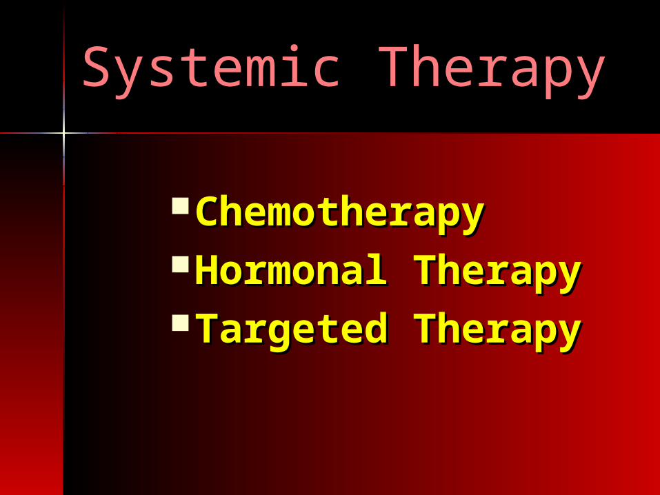
Systemic TherapySystemic Therapy
ChemotherapyChemotherapyHormonal TherapyHormonal TherapyTargeted TherapyTargeted Therapy

ChemotherapyChemotherapy
NeoadjuvantNeoadjuvantAdjuvantAdjuvantPalliativePalliative

Why Chemotherapy ? Why Chemotherapy ?
EBCTCG summarized results of 47 trials comparing chemotherapy
and no chemotherapy .
A significant reduction in mortality was seen in pts receiving
chemotherapy regardless of nodal status, and use of Tamoxifen.
Benefit of chemotherapy does vary with pt age and menopausal
status.
Women under age of 50 derive a greater survival benefit.
Tumor with higher “recurrence scores” may derive much greater
benefit whereas “low recurrence rate” tumors may derive little or no
benefit.
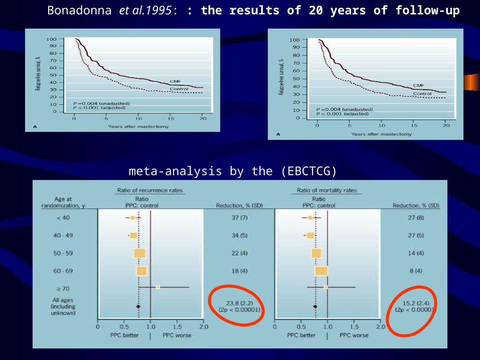
meta-analysis by the (EBCTCG)
Bonadonna et al.1995: : the results of 20 years of follow-up
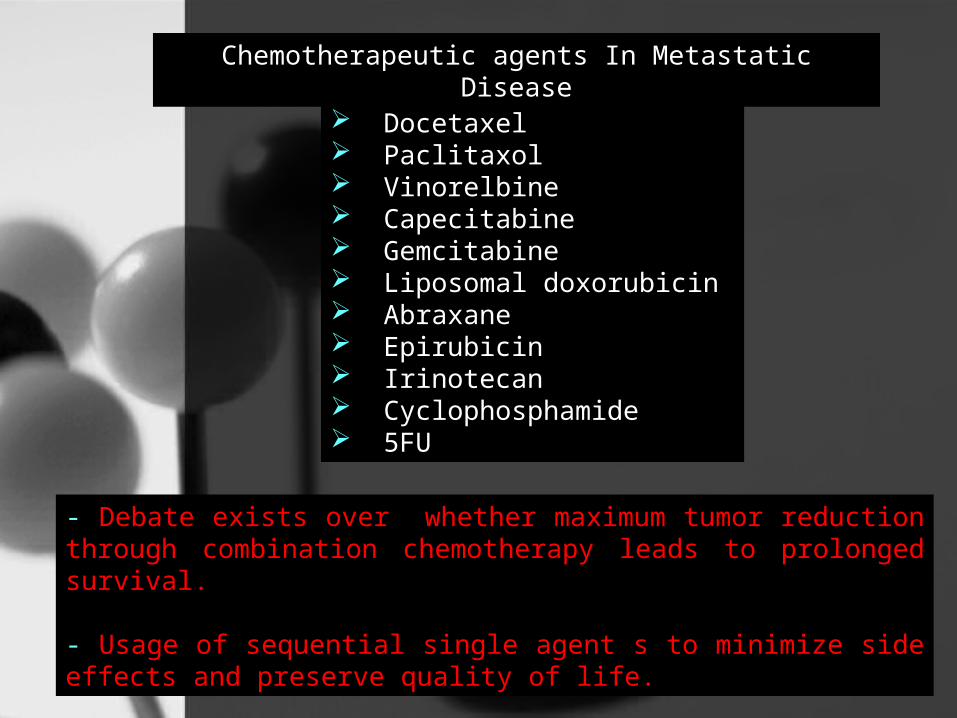
Chemotherapeutic agents In Metastatic Disease
Docetaxel Paclitaxol Vinorelbine Capecitabine Gemcitabine Liposomal doxorubicin Abraxane Epirubicin Irinotecan Cyclophosphamide 5FU
- Debate exists over whether maximum tumor reduction through combination chemotherapy leads to prolonged survival.
- Usage of sequential single agent s to minimize side effects and preserve quality of life.

Best regimen still (unknown) Node negative Node negative Node positiveNode positive
AC x 4 or 6 AC x 4 →Taxol x4
FAC or CAF x 6 TAC
FEC or CEF x 6 AC x 4 → Taxol/Herceptin
C MF FAC or FEC x 6
A/E →CMF
A → T → C
Retrospective evidence suggests that doxorubicin-based chemotherapy regimens may be superior to non-doxorubicin-based
regimens in patients with tumors over-expressing HER-2 by IHC. Am J Clin Oncol 1989; 12; 123-128
Retrospective evidence suggests that doxorubicin-based chemotherapy regimens may be superior to non-doxorubicin-based
regimens in patients with tumors over-expressing HER-2 by IHC. Am J Clin Oncol 1989; 12; 123-128
For node-positive patients, anthracycline-containing chemotherapy regimens are preferred.
J Clin Oncol 20:2812-2823, 2002
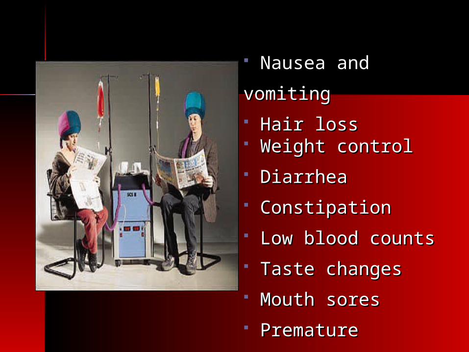
Nausea and vomitingNausea and vomiting Hair lossHair loss Weight controlWeight control DiarrheaDiarrhea ConstipationConstipation Low blood countsLow blood counts Taste changesTaste changes Mouth soresMouth sores Premature menopausePremature menopause

How should How should radiotherapy and radiotherapy and chemotherapy be chemotherapy be combined?combined?
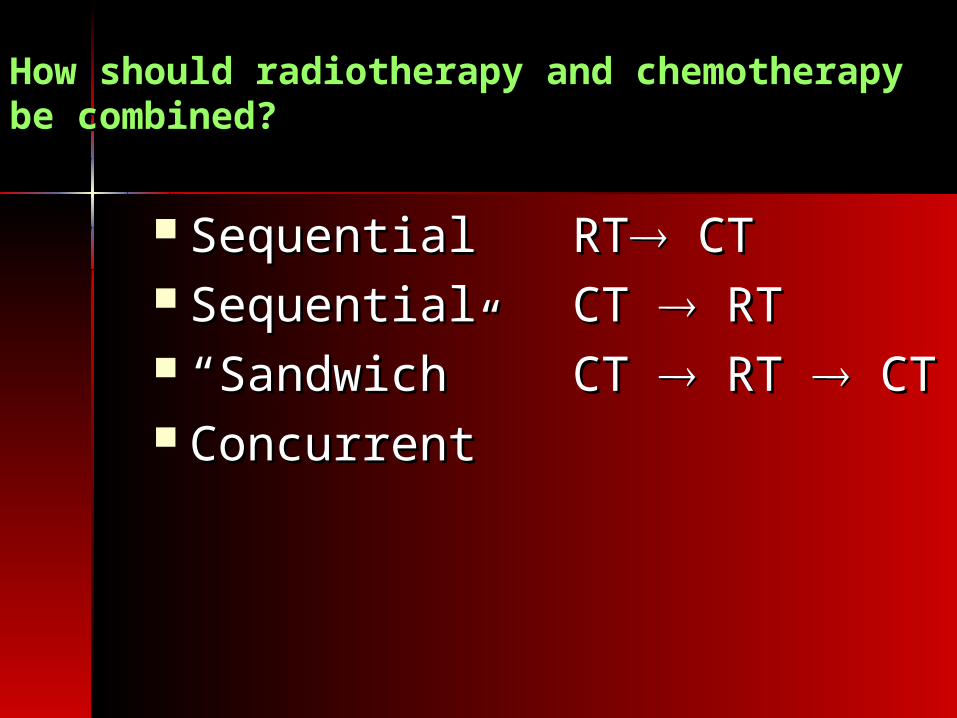
How should radiotherapy and chemotherapy How should radiotherapy and chemotherapy be combined?be combined?
Sequential Sequential RTRT CT CT SequentialSequential CT CT RT RT ““Sandwich”Sandwich” CT CT RT RT CT CT ConcurrentConcurrent
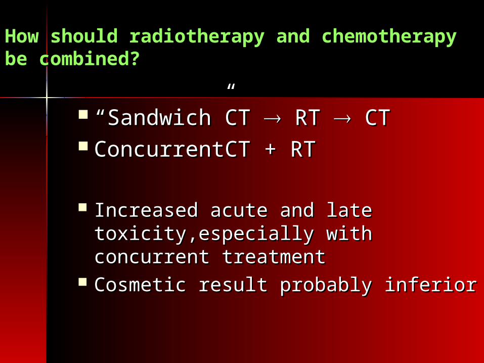
How should radiotherapy and chemotherapy How should radiotherapy and chemotherapy be combined?be combined?
““Sandwich”Sandwich” CT CT RT RT CT CT ConcurrentConcurrent CT + RTCT + RT
Increased acute and late Increased acute and late toxicity,especially with concurrent toxicity,especially with concurrent treatmenttreatment
Cosmetic result probably inferiorCosmetic result probably inferior

How should radiotherapy and chemotherapy How should radiotherapy and chemotherapy be combined?be combined?
Sequential Sequential RTRT CT CT SequentialSequential CT CT RT RT
? Delay in RT ? Delay in RT local local recurrencerecurrence
? Delay in CT ? Delay in CT distant metsdistant mets
How should radiotherapy and chemotherapy be How should radiotherapy and chemotherapy be combined?combined?
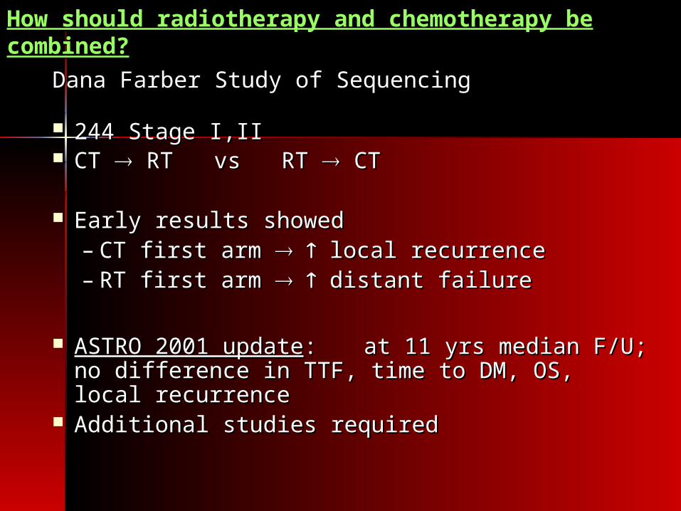
Dana Farber Study of SequencingDana Farber Study of Sequencing
244 Stage I,II244 Stage I,II CT CT RT vs RT RT vs RT CT CT
Early results showedEarly results showed– CT first arm CT first arm local recurrencelocal recurrence– RT first arm RT first arm distant failuredistant failure
ASTRO 2001 updateASTRO 2001 update: : at 11 yrs median F/U; at 11 yrs median F/U; no difference in TTF, time to DM, OS, local no difference in TTF, time to DM, OS, local recurrencerecurrence
Additional studies requiredAdditional studies required

Hormonal Therapy
NeoadjuvantAdjuvantPalliative

Endocrine Therapy: Approaches
• Estrogen receptor modulators– Tamoxifen (selective estrogen-receptor
modulator, or SERM)– Toremifene– Raloxifene
• Sex steroids• Aromatase inhibitors
– Nonselective AIs– Selective AIs
• pure anti-estrogens• ovarian ablation or suppression
– Medical– Surgical– Radiotherapy
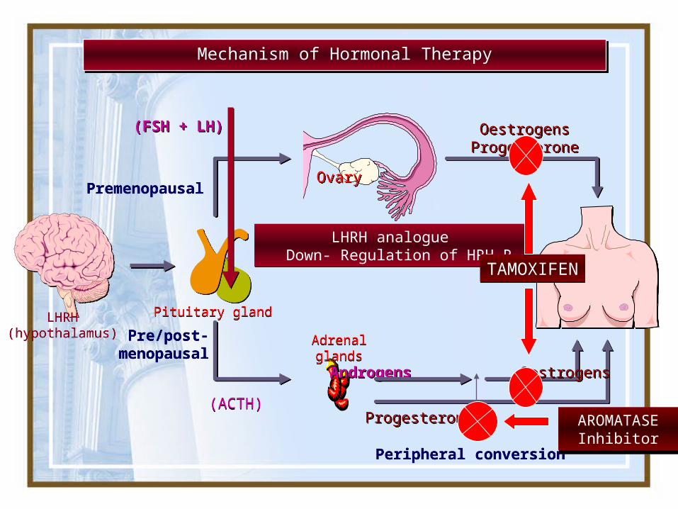
Mechanism of Hormonal TherapyMechanism of Hormonal Therapy
LHRH(hypothalamus)
LHRH(hypothalamus) Pre/post-
menopausalPre/post-
menopausal
Premenopausal Premenopausal
(ACTH)(ACTH)
AdrenalglandsAdrenalglands
Pituitary glandPituitary gland
OestrogensProgesteroneOestrogens
Progesterone
ProgesteroneProgesterone
Androgens OestrogensAndrogens Oestrogens
Peripheral conversionPeripheral conversion
OvaryOvary
LHRH analogue Down- Regulation of HRH R
LHRH analogue Down- Regulation of HRH R
(FSH + LH) (FSH + LH)
TAMOXIFENTAMOXIFEN
AROMATASEInhibitor
AROMATASEInhibitor
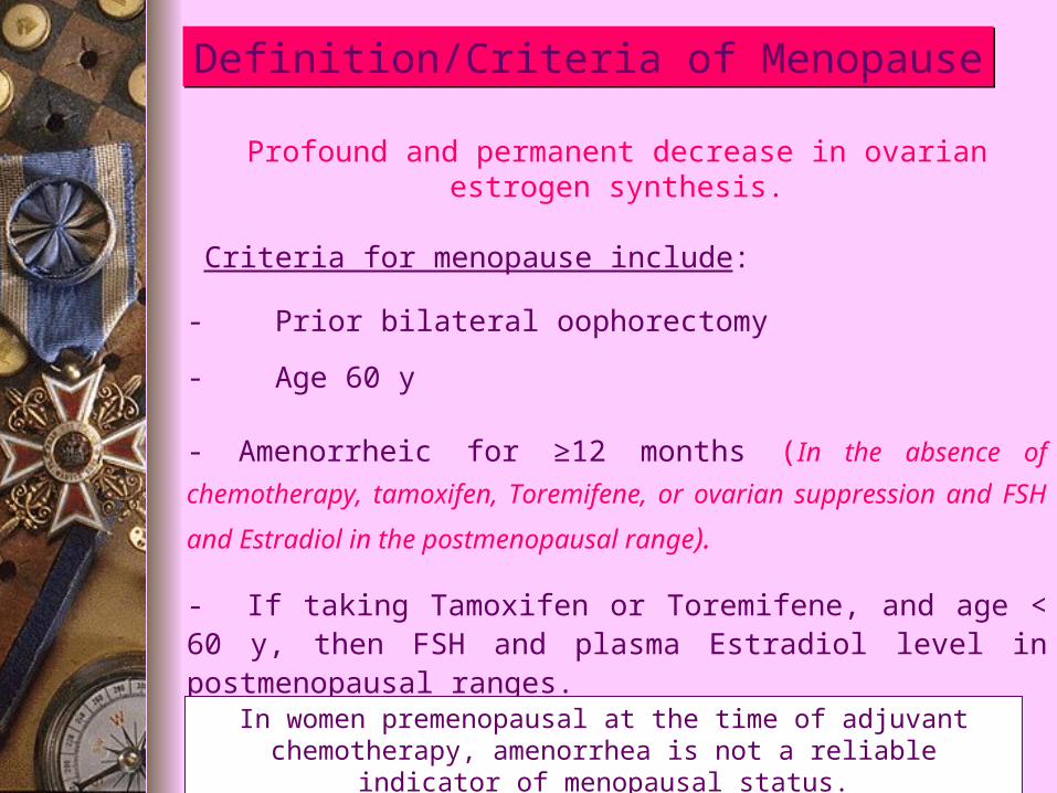
Definition/Criteria of MenopauseDefinition/Criteria of Menopause
Profound and permanent decrease in ovarian estrogen synthesis.
Criteria for menopause include:
- Prior bilateral oophorectomy
- Age 60 y
- Amenorrheic for ≥12 months (In the absence of
chemotherapy, tamoxifen, Toremifene, or ovarian suppression
and FSH and Estradiol in the postmenopausal range).
- If taking Tamoxifen or Toremifene, and age < 60 y, then FSH and plasma Estradiol level in postmenopausal ranges.
In women premenopausal at the time of adjuvant chemotherapy, amenorrhea is not a reliable indicator of menopausal status.

Mechanism of action of Tamoxifenas an antitumour agent
Antioestrogen effects - blockage of oestrogen
receptor
STROMALCELL
STROMALCELL
Local effects - independent of oestrogen receptor
Local effects - independent of oestrogen receptor
+
-
INCREASE TGF-βINCREASE TGF-β
DECREASE TGF- TGF-ααDECREASE TGF- TGF-αα
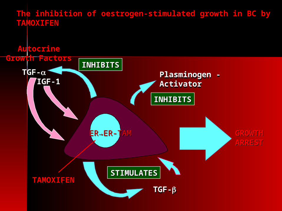
The inhibition of oestrogen-stimulated growth in BC by The inhibition of oestrogen-stimulated growth in BC by TAMOXIFENTAMOXIFEN
ER→ER-TAMER→ER-TAM
TAMOXIFENTAMOXIFEN
AutocrineGrowth Factors
AutocrineGrowth Factors
TGF-TGF-IGF-1IGF-1
Plasminogen -ActivatorPlasminogen -Activator
TGF-TGF-
STIMULATESSTIMULATES
INHIBITSINHIBITS
INHIBITSINHIBITS
GROWTHARRESTGROWTHARREST
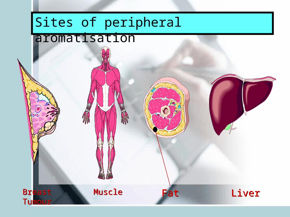
Sites of peripheral aromatisation
BreastTumourBreast
TumourMuscleMuscle FatFat LiverLiver

Figure AHypersecretion of LH following acute
administration of LHRH analogue
PituitaryPituitaryCellCell
PituitaryPituitaryCellCell LHLHLHLH
Figure BHyposecretion of LH following chronic
administration of LHRH analogue
LHLHLHLH
Mechanism of action of LHRH analogueMechanism of action of LHRH analogue
PituitaryPituitaryCellCell
PituitaryPituitaryCellCell

Targeted
Therapy
Targeted
Therapy
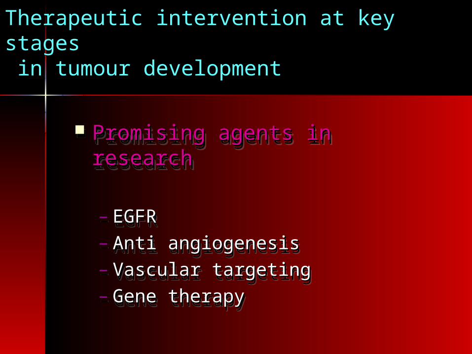
Promising agents in researchPromising agents in research
– EGFREGFR– Anti angiogenesisAnti angiogenesis– Vascular targetingVascular targeting– Gene therapyGene therapy
Promising agents in researchPromising agents in research
– EGFREGFR– Anti angiogenesisAnti angiogenesis– Vascular targetingVascular targeting– Gene therapyGene therapy
Therapeutic intervention at key stagesTherapeutic intervention at key stages in tumour development in tumour development

Epidermal Growth Factor Receptor (EGFR)
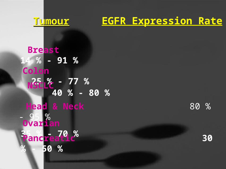
Breast 14 % - 91 %
Colon 25 % - 77 %
NSCLC 40 % - 80 %
Ovarian 35 % - 70 %
Pancreatic 30 % - 50 %
Head & Neck 80 % - 95 %
EGFR Expression Rate
TumourTumour
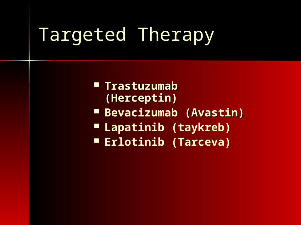
Targeted Therapy
Trastuzumab Trastuzumab (Herceptin)(Herceptin)
Bevacizumab (Avastin) (Avastin) Lapatinib (taykreb) Erlotinib (Tarceva)
MonthsMonths
0.2
0
0.4
0.6
0.8
1.0
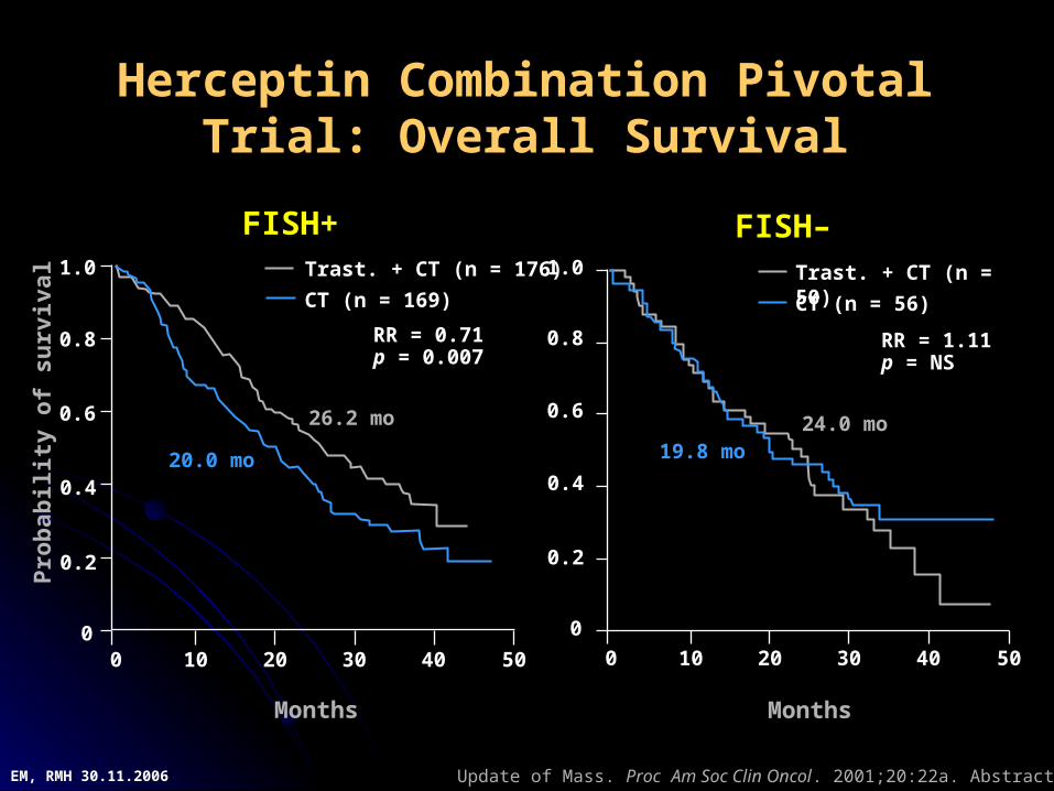
FISH+
Months
0.2
0
0.4
0.6
0.8
1.0 Trast. + CT (n = 176)
CT (n = 169)
Pro
bab
ility
of
surv
ival
RR = 0.71p = 0.007
0 10 20 30 40 50
20.0 mo
26.2 mo
FISH–
RR = 1.11p = NS
0 10 20 30 40 50
19.8 mo24.0 mo
Trast. + CT (n = 50)
CT (n = 56)
Herceptin Combination Pivotal Trial: Overall Survival
Update of Mass. Proc Am Soc Clin Oncol. 2001;20:22a. Abstract 85.EM, RMH 30.11.2006
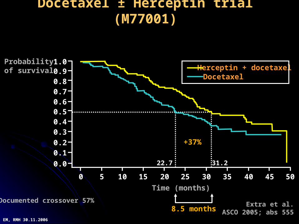
8.5 months
Time (months)
Extra et al.ASCO 2005; abs 555
Docetaxel ± Herceptin trial (M77001)
1.00.90.80.70.60.50.40.3
0.20.1
0.0
0 5 10 15 20 25 30 35 40 45 50
Herceptin + docetaxelDocetaxel
+37%
22.7 31.2
Documented crossover 57%
Probabilityof survival
EM, RMH 30.11.2006

100100
8080
6060
4040
2020
00
PatientsPatients
(%)(%)
Months from randomisationMonths from randomisation66 1122 1188 2244
16931693 11108108 767767 445445 22242416941694 11117272 885885 532532 268268
127127
220220
1-year Herceptin1-year Herceptin
ObservationObservation
00
No. No. at risk at risk
EventsEvents HRHR 95% CI95% CI p valuep value
0.540.54 0.43, 0.670.43, 0.67 <0.001<0.001
2-year2-yearDFSDFS
85.885.8
77.477.4
HERA DFS
Median follow-up: 1 yearMedian follow-up: 1 yearHR, hazard ratio; CI, confidence intervalHR, hazard ratio; CI, confidence interval
EM, RMH 30.11.2006
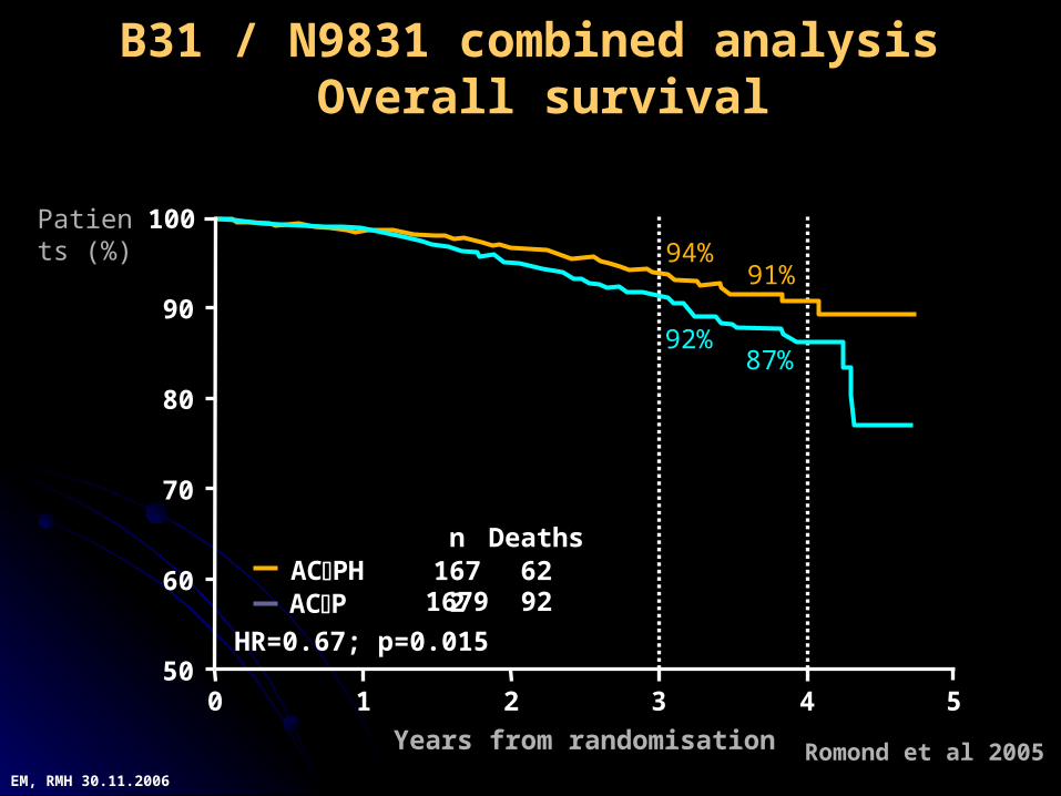
B31 / N9831 combined analysis Overall survival
94%94%91%91%
87%87%92%92%
0 1 2 3 4 550
60
70
80
90
100
Years from randomisation
HR=0.67; p=0.015
Deathsn6292
16721679
Patients Patients (%)(%)
ACPHACP
Romond et al 2005EM, RMH 30.11.2006

Nutritional Guidelines Nutritional Guidelines
Eat a variety of healthful foods, with an Eat a variety of healthful foods, with an emphasis on plant sources.emphasis on plant sources.
Adopt a physically active lifestyle.Adopt a physically active lifestyle.
Maintain a healthful weight throughout Maintain a healthful weight throughout life.life.
No alcoholic beverages.No alcoholic beverages.
Case 1Case 1
34 yr. 34 yr. oold, premenld, premenopausalopausal female female with upper outer quadrant mass with upper outer quadrant mass in left breast. Mammogram in left breast. Mammogram shows a 2 cm. spiculated nodule shows a 2 cm. spiculated nodule and core biopsy is positive for and core biopsy is positive for carcinoma. She is referred prior carcinoma. She is referred prior to surgery for a discussion of to surgery for a discussion of treatment options for the breast.treatment options for the breast.
Case 2Case 265 yr. 65 yr. oold, postmenld, postmenopausalopausal female with upper female with upper outer quadrant mass in left breastouter quadrant mass in left breast, undergoes , undergoes wide local excision and axillary node dissection. wide local excision and axillary node dissection. She is otherwise in good health. She is otherwise in good health.
Pathology: Pathology: – invasive lobular carcinoma, 2.5 x 2.1 x 1.8 invasive lobular carcinoma, 2.5 x 2.1 x 1.8
cm;cm;– Bloom- Richardson grade 2 (SBR 6/9)Bloom- Richardson grade 2 (SBR 6/9)– ERER+ + PRPR++– lymphovascular invasion presentlymphovascular invasion present– Margins clear by 1 mm. or moreMargins clear by 1 mm. or more– 2 lymph nodes contain metastases (of 10 2 lymph nodes contain metastases (of 10
resected nodes) with microscopic extranodal resected nodes) with microscopic extranodal extensionextension

Thank youThank you
THANK THANK YOUYOU