High-sensitivity troponin-T as a prognostic marker after...
22
High-sensitivity troponin-T as a prognostic marker after out-of-hospital cardiac arrest – A targeted temperature management (TTM) trial substudy Gilje, Patrik; Koul, Sasha; Thomsen, Jakob Hartvig; Devaux, Yvan; Friberg, Hans; Kuiper, Michael; Horn, Janneke; Nielsen, Niklas; Pellis, Tomasso; Stammet, Pascal; Wise, Matthew P.; Kjaergaard, Jesper; Hassager, Christian; Erlinge, David Published in: Resuscitation DOI: 10.1016/j.resuscitation.2016.06.024 2016 Document Version: Peer reviewed version (aka post-print) Link to publication Citation for published version (APA): Gilje, P., Koul, S., Thomsen, J. H., Devaux, Y., Friberg, H., Kuiper, M., ... Erlinge, D. (2016). High-sensitivity troponin-T as a prognostic marker after out-of-hospital cardiac arrest – A targeted temperature management (TTM) trial substudy. Resuscitation, 107, 156-161. DOI: 10.1016/j.resuscitation.2016.06.024 General rights Copyright and moral rights for the publications made accessible in the public portal are retained by the authors and/or other copyright owners and it is a condition of accessing publications that users recognise and abide by the legal requirements associated with these rights. • Users may download and print one copy of any publication from the public portal for the purpose of private study or research. • You may not further distribute the material or use it for any profit-making activity or commercial gain • You may freely distribute the URL identifying the publication in the public portal Take down policy If you believe that this document breaches copyright please contact us providing details, and we will remove access to the work immediately and investigate your claim.
Transcript of High-sensitivity troponin-T as a prognostic marker after...

LUND UNIVERSITY
PO Box 117221 00 Lund+46 46-222 00 00
High-sensitivity troponin-T as a prognostic marker after out-of-hospital cardiac arrest –A targeted temperature management (TTM) trial substudy
Gilje, Patrik; Koul, Sasha; Thomsen, Jakob Hartvig; Devaux, Yvan; Friberg, Hans; Kuiper,Michael; Horn, Janneke; Nielsen, Niklas; Pellis, Tomasso; Stammet, Pascal; Wise, MatthewP.; Kjaergaard, Jesper; Hassager, Christian; Erlinge, DavidPublished in:Resuscitation
DOI:10.1016/j.resuscitation.2016.06.024
2016
Document Version:Peer reviewed version (aka post-print)
Link to publication
Citation for published version (APA):Gilje, P., Koul, S., Thomsen, J. H., Devaux, Y., Friberg, H., Kuiper, M., ... Erlinge, D. (2016). High-sensitivitytroponin-T as a prognostic marker after out-of-hospital cardiac arrest – A targeted temperature management(TTM) trial substudy. Resuscitation, 107, 156-161. DOI: 10.1016/j.resuscitation.2016.06.024
General rightsCopyright and moral rights for the publications made accessible in the public portal are retained by the authorsand/or other copyright owners and it is a condition of accessing publications that users recognise and abide by thelegal requirements associated with these rights.
• Users may download and print one copy of any publication from the public portal for the purpose of private studyor research. • You may not further distribute the material or use it for any profit-making activity or commercial gain • You may freely distribute the URL identifying the publication in the public portalTake down policyIf you believe that this document breaches copyright please contact us providing details, and we will removeaccess to the work immediately and investigate your claim.
1
High-sensitivity Troponin-T as a prognostic marker after out-of-hospital cardiac
arrest – a Targeted Temperature Management (TTM) trial substudy.
Patrik Giljea, Sasha Koula, Jakob Hartvig Thomsenb, Yvan Devauxc, Hans Fribergd, Michael Kuipere,
Janneke Hornf, Niklas Nielseng, Tomasso Pellish, Pascal Stammeti, Matthew P Wisej, Jesper
Kjaergaardb, Christian Hassagerb and David Erlingea - on behalf of the TTM study group
a Department of Cardiology, Clinical Sciences, Lund University, Lund, Sweden.
b The Heart Centre, Copenhagen University Hospital, Copenhagen, Denmark.
c Laboratory of Cardiovascular Research, Public Research Centre – Health (CRP Santé), Luxembourg.
d Department of Anaesthesia and Intensive Care, Skåne University Hospital, Lund University, Lund,
Sweden.
e Department of Intensive Care, Leeuwarden Medical Centrum, Leeuwarden, The Netherlands.
f Department of Intensive Care, Academic Medical Center, Amsterdam, The Netherlands.
g Department of Anaesthesia and Intensive Care, Helsingborg Hospital, Helsingborg, Sweden.
h Intensive Care Unit, Santa Maria degli Angeli, Pordenone, Italy.
i Department of Anaesthesia and Intensive Care, Centre Hospitalier de Luxembourg, Luxembourg.
j Department of Intensive Care, University Hospital of Wales, Cardiff, United Kingdom.
Word count: 2918
Corresponding author:
Patrik Gilje, Department of Cardiology, Clinical Sciences, Lund University, Lund, Sweden.
Email: [email protected]

2
Abstract
Aim of the study:
Predicting outcome of unconscious patients after successful resuscitation is challenging and better prognostic
markers are highly needed. Ischemic heart disease is a common cause of out-of-hospital cardiac arrest (OHCA).
Whether or not high-sensitivity troponin T (hs-TnT) is a prognostic marker among survivors of OHCA with both
ischemic and non-ischemic aetiologies remains to be determined. We sought to evaluate the ability of hs-TnT to
prognosticate all-cause mortality, death due to cardiovascular causes or multi-organ failure and death due to
cerebral causes after OHCA. The influence of the level of target temperature management on hs-TnT as a marker
of infarct size was also assessed.
Methods:
A total of 699 patients from the Targeted Temperature Management (TTM) trial were included and hs-TnT was
analysed in blood samples from 24, 48 and 72 h after return of spontaneous circulation (ROSC). The endpoints
were 180 day all-cause mortality, death due to cardiovascular causes or multi-organ failure and death due to
cerebral causes. Subgroups based on the initial ECG after ROSC (STEMI vs all other ECG presentations) were
analysed.
Results:
Hs-TnT was independently associated with all-cause mortality which was driven by death due to cardiovascular
causes or multi-organ failure and not cerebral causes (at 48h: OR 1.10, CI 1.01-1.20, p<0.05). Hs-TnT was also an
independent predictor of death due to cardiovascular causes or multi-organ failure (at 48h: OR 1.13, CI 1.01-
1.26, p<0.05). In patients with STEMI, hs-TnT was independently associated with death due to cardiovascular
causes or multi-organ failure (at 48h: OR 1.47, CI 1.10-1.95, p<0.01). Targeted temperature management at 33
°C was not associated with hs-TnT compared to 36 °C.
Conclusions:
After OHCA due to both ischemic and non-ischemic causes, hs-TnT is an independent marker of both all-cause
mortality and death due to cardiovascular causes or multi-organ failure. Targeted temperature management at

3
33 °C did not reduce hs-TnT compared to 36 °C. Hs-TnT may be a marker of poor prognosis after OHCA and this
should be taken into consideration in patients that present with high troponin levels.
Trial Registration
The TTM-trial is registered and accessible at Clinicaltrials.gov (identifier: NCT01020916)
Keywords
Out-of-hospital cardiac arrest, hypothermia, prognosis, high-sensitivity Troponin T, TTM-trial
4
Introduction
Ischemic heart disease is a common cause of out-of-hospital cardiac arrest (OHCA), with an estimated 60-70% of
OHCA caused by coronary heart disease [1, 2]. Troponin T (TnT) is both a diagnostic and prognostic marker in ST-
elevation myocardial infarction (STEMI) as well as in non ST-elevation myocardial infarction (NSTEMI) [3, 4]. Since
shock and myocardial infarction are frequently encountered among survivors of OHCA, it is therefore plausible
that troponin might contribute to additional prognostic information. However, whether or not high-sensitivity
troponin T (hs-TnT) is a prognostic marker among survivors of OHCA with both ischemic and non-ischemic
aetiologies remains to be established. In a recent study including survivors of OHCA with ventricular fibrillation
as the primary rhythm, hs-TnT was not associated with neurological outcome and mortality after adjustment for
established risk factors [5]. Although high sensitivity troponins at admission are independently associated with
acute coronary occlusions, they do not seem to aid in the decision regarding coronary angiography when added
to other risk factors [6, 7].
Even though acute coronary syndromes are common in patients with OHCA, acute coronary angiography among
OHCA patients without STEMI is still a matter of debate. The issue is further complicated by difficulties
interpreting symptoms as well as the ECG in the post cardiac arrest setting since the post resuscitation ECG seem
to be a poor predictor of an acute coronary occlusion [8, 9]. No randomized trials have evaluated the potential
benefit of immediate coronary angiography in patients without STEMI and register studies have shown diverging
results [10-12]. A recent sub study of the Targeted Temperature Management (TTM) trial revealed no survival
benefit of coronary angiography within 6 hours of cardiac arrest among patients without STEMI at presentation
[13].
In the present study we hypothesized that hs-TnT would be an independent prognostic marker of all-cause
mortality as well as death due to cardiovascular causes or multi organ failure (MOF) at 6 months after OHCA.
Furthermore, the relationship between hs-TnT and cerebral causes of death as well as targeted temperature
management level, 33 °C vs 36 °C, was assessed.
Methods

5
Patient selection
The current study is a pre-defined observational substudy of the TTM-trial that investigated the effects of 33 °C
versus 36 °C treatment among 939 unconscious survivors of OHCA [14]. The TTM trial included all primary ECG
rhythms as well as both ischemic and non-ischemic cardiac arrest aetiologies. There was no difference in
outcome between the two temperature groups. The present study is a part of a pre-defined biomarker substudy
of the TTM trial. A total of 699 patients were included and serum blood samples were obtained at 24, 48 and 72
h after return of spontaneous circulation (ROSC). Out of the 36 participating sites, 29 took part in the biomarker
substudy. All samples were pre-analytically processed, aliquoted and frozen to - 80 °C at the different sites before
shipment to the integrated biobank in Luxembourg for analysis.
We pre-specified subgroups based on the initial ECG after ROSC (STEMI vs all other ECG presentations).
Information about ECG at admission was available for 691 patients (99%). Two hundred eighty two (282) patients
(40%) had STEMI at admission whereas 409 (59%) had other ECG presentations. For 8 patients, no information
about ECG at admission was available. Since a previous unrecognized left bundle branch block (LBBB) is rarely
associated with acute ischemia, it was not treated as a STEMI-equivalent [15].
Acute coronary angiography and PCI was defined as being performed within 6 hours from the cardiac arrest. In
total, acute coronary angiography was performed in 398 patients (57%) with ad-hoc PCI in 69% of cases (N=270).
In the group without STEMI, 164 patients (40%) underwent acute coronary angiography with ad-hoc PCI in 41%
of cases (N=67). Among STEMI patients, acute coronary angiography was performed in 222 patients (79%) with
ad-hoc PCI in 93% of cases (N=192). The decision to perform acute angiography or PCI was made by the treating
cardiologist.
Endpoints
Mortality and causes of death 180 days after OHCA were assessed by using public- and hospital health
registries as well as direct contact with patients and relatives at the end of the TTM-trial [14]. Autopsies were
not systematically performed, but may in some cases have been part of the adjudication of most likely cause of
death. As it can be difficult to differentiate between death due to multi-organ failure or cardiovascular causes,
these two entities were treated as one in the analysis.
TnT-assay

6
The Roche Cobas e601, high sensitivity Troponin T assay (Basel, Switzerland) was used for the troponin analyses,
with the detection limit 5 ng/l and with a 99th percentile conforming to the universal definition of myocardial
infarction of 14 ng/l [16]. These troponin data were not available to the treating physicians.
Statistical analysis
Data are presented as medians, interquartile range, counts and proportions (%). Since the distribution of hs-TnT
was skewed it was logarithmically transformed prior to analysis. Missing values were replaced with 5-fold
multiple imputations using the MCMC method with 10 iterations per imputation. Continuous variables with
normal distribution were compared using ANOVA. Non-parametric continuous data were compared using the
Mann-Whitney U test. Pearson´s chi Square test with the z-test as post-test were used for comparing differences
between categorical data. All confidence intervals were analysed at the 95% confidence level. The overall ability
of hs-TnT to discriminate between outcomes was described by receiving operating characteristic (ROC) curves.
Unadjusted mortality between quartiles of hs-TnT was compared using the Kaplan-Meier estimator with the log
rank test for significance testing. Troponin T at 24 h, 48 h and 72 h were added to a logistic regression model
containing temperature allocation, age, sex, bystander CPR, first prehospital rhythm, time from cardiac arrest
(CA) to return of spontaneous circulation (ROSC), pH at admission and shock at admission as covariates. There
was no multicollinearity as assessed with condition indices. As hs-TnT was expressed in the log2 scale, the odds
ratio corresponds to a 2-fold increase in hs-TnT. When the logistic regression model was used to analyse specific
causes of death, competing risk of dying was handled by censoring patients with other causes of death. All p-
values P<0.05 were considered statistically significant. IBM SPSS Statistics 22 (Foster City, USA) and GraphPad
Prism 6 (La Jolla, USA) were used for the analyses.
Results
Background demographics in the whole patient cohort
A total of 699 patients entered the study. Hs-TnT was available for 646 patients (92%) at 24h, 618 patients (88%)
at 48h and 573 patients (82%) at 72h. Hs-TnT peaked at 24 h after admission (848 ng/l (IQR 2685 ng/l). Median

7
values at 48h were 695 ng/l (IQR 2039 ng/l) and at 72h 756 ng/l (IQR 1786 ng/l)) (figure 1a). Non-survivors 180
days after OHCA were older, had more often PEA or asystole as the primary rhythm and longer duration between
cardiac arrest and ROSC. Moreover, comorbidities such as a history of heart disease, arterial hypertension and
diabetes were significantly more prevalent among non-survivors. Baseline characteristics for survivors compared
to non-survivors can be found in table 1.
All-cause mortality in the whole patient cohort
At follow up 6 months after OHCA, 322 patients (46%) had died. Hs-TnT was significantly higher among non-
survivors compared to survivors at the 48 h (p<0.05) and 72 h (p<0.01) after ROSC (Figure 1b). The Kaplan-Meier
analysis did not indicate a significant association between hs-TnT at any time point and all-cause mortality. ROC
curve analysis did not show significant AUC:s for predicting all-cause mortality (AUC 0.54-0.56; at 24h and 48h
p=ns, at 72h p<0.05). However, in the logistic regression model, hs-TnT at 48h and 72h were independently
associated with all-cause mortality (at 48h: OR 1.10, CI: 1.01-1.20, p<0.05; at 72h: OR 1.11, CI: 1.02-1.21, p<0.05)
(Table 2a). The relative hs-TnT change between 24h and 48h was not associated with all-cause mortality (OR:
1.05, CI: 0.97-1.15, p=ns).
Death from cardiovascular causes or multi-organ failure in the whole patient cohort
In total, 104 patients (14.9%) died due cardiovascular causes and multi-organ failure, which corresponded to 34%
of all deaths. Time to death was available for 309 patients (96%). The median duration from CA to death was 147
h (IQR:161 h) for the cohort as a whole. Among patients that died due to CV/MOF the duration from CA to death
was 138 h (IQR: 332 h) and for patients that died from cerebral causes 144 h (IQR:118 h) (p=ns).
Hs-TnT was significantly elevated at 48h and 72h in patients that died due to cardiovascular causes or multi-
organ failure compared to survivors (p<0.05) (figure 1c). The Kaplan-Meier analysis indicated a significant
unadjusted association between hs-TnT at 48h and 72h and cardiovascular death or multi-organ failure. ROC
curve analysis at 24, 48 and 72 h did not show significant AUC:s (0.54-0.59, p=ns) for predicting death due to

8
cardiovascular causes or multi-organ failure. However, in the logistic regression model, hs-TnT at 24, 48 and 72h
were independently associated with death from cardiovascular causes and multi-organ failure (at 24h: OR 1.11,
CI:1.00-1.23, p<0.05; at 48h: OR 1.13, CI: 1.01-1.26, p<0.05; at 72h: OR 1.16, CI:1.03-1.30, p<0.05) (table 2b). The
relative hs-TnT change between 24h and 48h was not associated with death from cardiovascular causes and
multi-organ failure (OR: 0.98, CI: 0.88-1.09, p=ns).
Death from cerebral causes in the whole patient cohort
In total, 194 patients died due to cerebral causes, which corresponded to 28% of all deaths. Hs-TnT was
significantly elevated at 72h in patients that died due to cerebral causes compared to survivors (p<0.05). The
Kaplan-Meier analysis could not detect an association between hs-TnT and death due to cerebral causes. In
concordance, the logistic regression could not detect any association between hs-TnT and death due to cerebral
causes at any time point. The relative hs-TnT change between 24h and 48h was borderline significantly associated
with death from cerebral causes (OR: 1.13, CI: 1.00-1.09, p=0.05).
Background demographics in patients with STEMI vs patients without STEMI
The STEMI group was slightly younger and had less prevalence of previous coronary artery bypass graft surgery
(CABG) and heart failure compared to the group without STEMI. STEMI was present in 282 patients (40%) at
admission and among those 216 (77%) underwent acute PCI compared to 86 patients (21%) without STEMI. The
STEMI group had significantly higher hs-TnT at 24h, 48h and 72h compared to the group without STEMI (p<0.001)
(figure 1d). Patients without STEMI that were selected for acute angiography had significantly higher hs-TnT than
the ones who did not (at 24 h: 718 vs 276 ng/l , p<0.001 ; at 48h: 449 vs 238 ng/l, p<0.05 : at 72h 492 vs 224 ng/l,
p<0.01) Furthermore, patients without STEMI that were treated with PCI had significantly higher hs-TnT at 48 h

9
and 72 h compared to the ones that only underwent acute angiography (at 48h: 899 ng/l vs 131 ng/l, p<0.05; at
72h: 918 ng/l vs 103 ng/l, p<0.05).
All-cause mortality in patients with STEMI vs patients without STEMI.
Patients with an ECG without STEMI at admission had higher all-cause mortality at follow up compared to the
STEMI group (49% vs 40%, p<0.05). The Kaplan-Meier curves revealed significant unadjusted correlations
between hs-TnT at 24h and 48h and all-cause mortality (p<0.05), but only among patients without STEMI.
However, adjusted analyses using a logistic regression model showed no association between hs-TnT and all-
cause mortality regardless of ECG presentation.
Death from cardiovascular causes or multi-organ failure in patients with STEMI vs patients without STEMI.
Death due to cardiovascular causes or multi-organ failure was significantly more prevalent among patients
without STEMI compared to the STEMI cohort (36% vs 25% of all deaths, p<0.05). The Kaplan-Meier analysis
indicated a significant unadjusted association between hs-TnT at 48h and 72h and cardiovascular death or multi-
organ failure in both groups. However, only in the STEMI group was hs-TnT predictive of death due to
cardiovascular causes or multi-organ failure when analysed in the logistic regression model (at 24h: OR 1.31, CI:
1.03-1.66, p<0.05; at 48h: OR 1.47, CI: 1.10-1.95, p<0.01; at 72h: OR 1.52, CI: 1.10-2.11, p<0.05).
Troponin T levels according to targeted temperature
Temperature assignment (33°C vs 36°C) was not associated with differences in hs-TnT levels and there was no
interaction between temperature allocation and hs-TnT in our logistic regression model (at 48h: the 33°C-group
782 ng/l (IQR 2192 ng/l); the 36°C-group 618 ng/l (IQR 1904 ng/l) (figure 2). This remained unchanged when
subgroups with and without STEMI were analysed separately.
Discussion
10
Treatment of patients who remain comatose after OHCA is challenging and novel markers to help guiding
treatment are highly desirable. In this study of patients with OHCA due to both ischemic and non-ischemic
aetiologies, hs-TnT was independently associated with all-cause mortality in the entire patient cohort. Hs-TnT
was also an independent predictor of death due to cardiovascular causes or multi-organ failure in the whole
patient cohort. In patients with STEMI, hs-TnT was independently associated with death due to cardiovascular
causes or multi-organ failure. In patients without STEMI, hs-TnT did not provide prognostic information. Targeted
temperature management at 33 °C did not reduce hs-TnT compared to 36 °C.
The association between hs-TnT and all-cause mortality is in line with the increased mortality observed with
elevated troponin in other critically ill populations [17]. It is tempting to speculate that hs-TnT reflect the severity
of the cardiac arrest syndrome and could be regarded as a marker of general hypo perfusion and systemic
inflammation. However, hs-TnT was not predictive of cerebral death at any time point and the prognostic
properties of-hs-TnT seem primarily to be related to cardiovascular death and multi organ failure.
The correlation between hs-TnT and death due to cardiovascular causes or multi-organ failure corresponds with
previous findings in patients with and without acute coronary syndromes [18, 19]. Furthermore, Troponin within
the first days after STEMI without OHCA is associated with infarct size, left ventricular function as well as
mortality in several studies [20-22]. However, in the present study hs-TnT did not predict death due to
cardiovascular causes or multi-organ failure in the group without STEMI. This is interesting since death due to
cardiovascular causes or multi-organ failure was significantly more common in these patients. One might
hypothesize that this increase in cardiovascular mortality amongst patients without STEMI might be due less
acute revascularization in this group. Thus, an ad-hoc analysis was performed focusing on the patients without
STEMI that did not undergo PCI within 6 hours from admission. Still, no correlation between hs-TnT and death
due to cardiovascular causes or multi-organ failure was found. This might indicate that acute PCI was not
underutilized in the TTM trial and complements a recent TTM substudy that could not find a benefit from acute
PCI in patients without STEMI [13]. The issue of whether or not patients without STEMI benefit from acute PCI
needs to be addressed in a randomized trial and a pilot trial is currently underway (DISCO, ClinicalTrials.gov
Identifier: NCT02309151).

11
There was no association between hs-TnT and the level of TTM. Since experimental studies show that
hypothermia decrease myocardial reperfusion damage [23], one would assume that the 33°C group should have
lower hs-TnT levels than the 36 °C group. Clinical and experimental results have previously shown the importance
of achieving a body temperature below 35 °C before reperfusion to reduce myocardial injury [24]. This
requirement was met by the 33 °C group in the TTM-trial as it reached a body temperature below 35 °C shortly
after randomization. However, the experimental models investigating the protective properties of hypothermia
have focused on coronary occlusion and reperfusion. In contrast, only 39% of the patients underwent acute PCI
making it difficult to extrapolate occlusion-reperfusion findings to our cohort.
Strengths of the present study include the use of a fourth generation high sensitivity troponin T assay that is
widely used in contemporary care. Furthermore, the relatively large sample size in combination with inclusion
of a wide range of cardiac arrest aetiologies and clinical characteristics increases its validity. Moreover, both
33°C and 36°C TTM was assessed. Limitations to the study include potential selection bias as not the whole
TTM-cohort was included in this biomarker substudy. For example, the median time to death of 147 hours is
longer than expected and could indicate that patients with shorter survival times are underrepresented.
Some values of hs-TnT were missing, although compensated for with multiple imputations. As with all post-hoc
and subgroups analysis, our results can only be regarded as hypothesis generating since they might suffer from
potential bias and confounding. Although hs-TnT is an independent predictor of all-cause mortality, it is
important to emphasize that there exists a considerable overlap of hs-TnT between the outcome groups.
Hence, hs-TnT should only be regarded as one of many variables to consider when assessing the prognosis of
an individual patient.
Conclusion
In this study of patients with OHCA due to both ischemic and non-ischemic aetiologies, hs-TnT was independently
associated with all-cause mortality which was driven by higher risk of death due to cardiovascular causes or multi
organ failure and not cerebral causes. In patients with STEMI, hs-TnT was independently associated with death
12
due to cardiovascular causes or multi organ failure, however this could not be seen for patients without STEMI.
Therapeutic hypothermia treatment at 33 °C did not reduce hs-TnT compared to 36 °C. Hs-TnT may be a marker
of poor prognosis after OHCA and special care should be given to patients that present with high troponin levels
regardless of initial ECG. The upcoming DISCO trial will assess whether or not patients without STEMI benefit
from acute revascularization (ClinicalTrials.gov Identifier: NCT02309151).
Acknowledgement
This work was funded by the Swedish Heart-Lung Foundation and the Swedish Research Council.

13
Figures and tables
24h
48h
72h
1
1 0
1 0 0
1 0 0 0
1 0 0 0 0
1 0 0 0 0 0
hs
-Tn
T (
ng
/l)
*** ***
A
E n tire c o h o rt
24h
48h
72h
1
1 0
1 0 0
1 0 0 0
1 0 0 0 0
1 0 0 0 0 0
hs
-Tn
T (
ng
/l)
S u rv iv o rs
N o n -su rv iv o rs
n s* **
S u rv iv o rs v s n o n -s u rv iv o rs
B
24h
48h
72h
1
1 0
1 0 0
1 0 0 0
1 0 0 0 0
1 0 0 0 0 0
hs
-Tn
T (
ng
/l)
S u rv iv o rs
D e a th fro m
C V /M O F
n s** **
S u rv iv o rs v s d e a th fro m c a rd io v a s c u la r c a u s e s
o r m u lti o rg a n fa ilu re
C
24h
48h
72h
1
1 0
1 0 0
1 0 0 0
1 0 0 0 0
1 0 0 0 0 0
hs
-Tn
T (
ng
/l)
N o S T E M I
S T E M I
*** *** ***
D
S T E M I v s w ith o u t S T E M I
Figure 1A. hs-TnT values at 24h, 48h and 72h after ROSC (p<0.001 for differences between timepoints).
Figure 1B. hs-TnT at 48h and 72h were significantly elevated among non-survivors compared to survivors (at
48h: p<0.05; at72h: p<0.01).
Figure 1C. hs-TnT was significantly elevated among patients that died due to cardiovascular causes and multi-
organ failure compared to survivors (at 48h and 72h: p<0.01). (CV: cardiovascular; MOF: multi-organ failure)
Figure 1D. hs-TnT was significantly elevated among patients with STEMI compared to patients without STEMI
(p<0.001 for differences between groups).

14
24h
48h
72h
1
1 0
1 0 0
1 0 0 0
1 0 0 0 0
1 0 0 0 0 0
hs
-Tn
T (
ng
/l)
3 6 C
3 3 C
n s n s n s
T e m p e ra tu re a llo c a tio n
Figure 2: Temperature allocation was not associated with differences in hs-TnT levels.
Survivors Non-survivors p-value
Age (years) 61 (16) 68 (14) <0.001
Sex (female) 62 (16%) 72 (22%) <0.05
Previous AMI (yes) 62 (16%) 78 (24%) <0.05
Arterial hypertension (yes) 130 (35%) 150 (47%) <0.01
Diabetes (yes) 42 (11%) 60 (19%) <0.05
Previous heart failure (yes) 14 (3.7%) 28 (9%) <0.01
Previous PCI (yes) 37 (10%) 48 (15%) ns
Previous CABG (yes) 20 (5%) 43 (13%) <0.01
Bystander CPR (yes) 301 (80%) 203 (63%) <0.001
First rhythm VT or VF 350 (94%) 204 (65%) <0.001
Time CA to ROSC (min) 20 (15) 31 (27) <0.001
pH at admission 7.27 (0.16) 7.18 (0.22) <0.001
Hs-troponin T day 1 (ng/l) 786 (2390) 996 (3014) ns
Hs-troponin T day 2 (ng/l) 602 (1895) 789 (2505) <0.01
Hs-troponin T day 3 (ng/l) 590 (1693) 859 (2124) <0.01
STEMI at admision (yes) 168 (45%) 114 (36%) <0.05
Acute coronary angiography (yes) 217 (58%) 156 (48%) <0.05
Acute PCI (yes) 161 (43%) 109 (34%) <0.05

15
Table 1. Baseline characteristics comparing survivors vs non-survivors at 180 days after OHCA. (AMI: acute
myocardial infarction; PCI: percutaneous coronary intervention; CABG: coronary artery bypass grafting; CPR:
cardiopulmonary resuscitation; VT: ventricular tachycardia; VF: ventricular fibrillation; CA: cardiac arrest; ROSC:
return of spontaneous circulation; STEMI: ST-elevation myocardial infarction)
Table 2a. Logistic regression analysis with hs-TnT and clinical variables to predict all-cause mortality. Hs-TnT at
48 h and 72 h were independent predictors. (CI: confidence interval; CPR: cardiopulmonary resuscitation; PEA:
pulseless electrical activity; CA: cardiac arrest; ROSC: return of spontaneous circulation)
Table 2b. Logistic regression model with hs-TnT and clinical variables to predict death due to cardiovascular
causes and multi organ failure. Hs-TnT was an independent predictor at all time points. (CI: confidence interval;
CPR: cardiopulmonary resuscitation; PEA: pulseless electrical activity; CA: cardiac arrest; ROSC: return of
spontaneous circulation)

16
References
[1] Zheng ZJ, Croft JB, Giles WH, Mensah GA. Sudden cardiac death in the United States, 1989 to
1998. Circulation. 2001;104:2158-63.
[2] Zipes DP, Wellens HJ. Sudden cardiac death. Circulation. 1998;98:2334-51.
[3] Mueller C, Neumann FJ, Perruchoud AP, Zeller T, Buettner HJ. Prognostic value of quantitative
troponin T measurements in unstable angina/non-ST-segment elevation acute myocardial infarction
treated early and predominantly with percutaneous coronary intervention. Am J Med. 2004;117:897-
902.
[4] Kurowski V, Hartmann F, Killermann DP, Giannitsis E, Wiegand UK, Frey N, et al. Prognostic
significance of admission cardiac troponin T in patients treated successfully with direct percutaneous
interventions for acute ST-segment elevation myocardial infarction. Crit Care Med. 2002;30:2229-35.
[5] Rosjo H, Vaahersalo J, Hagve TA, Pettila V, Kurola J, Omland T, et al. Prognostic value of high-
sensitivity troponin T levels in patients with ventricular arrhythmias and out-of-hospital cardiac
arrest: data from the prospective FINNRESUSCI study. Critical care. 2014;18:605.
[6] Geri G, Mongardon N, Dumas F, Chenevier-Gobeaux C, Varenne O, Jouven X, et al. Diagnosis
performance of high sensitivity troponin assay in out-of-hospital cardiac arrest patients. Int J Cardiol.
2013;169:449-54.
[7] Dumas F, Manzo-Silberman S, Fichet J, Mami Z, Zuber B, Vivien B, et al. Can early cardiac troponin
I measurement help to predict recent coronary occlusion in out-of-hospital cardiac arrest survivors?
Crit Care Med. 2012;40:1777-84.
[8] Spaulding CM, Joly LM, Rosenberg A, Monchi M, Weber SN, Dhainaut JF, et al. Immediate
coronary angiography in survivors of out-of-hospital cardiac arrest. N Engl J Med. 1997;336:1629-33.
[9] Salam I, Hassager C, Thomsen JH, Langkjaer S, Soholm H, Bro-Jeppesen J, et al. Is the pre-hospital
ECG after out-of-hospital cardiac arrest accurate for the diagnosis of ST-elevation myocardial
infarction? Eur Heart J Acute Cardiovasc Care. 2015.

17
[10] Bro-Jeppesen J, Kjaergaard J, Wanscher M, Pedersen F, Holmvang L, Lippert FK, et al. Emergency
coronary angiography in comatose cardiac arrest patients: do real-life experiences support the
guidelines? Eur Heart J Acute Cardiovasc Care. 2012;1:291-301.
[11] Dumas F, Cariou A, Manzo-Silberman S, Grimaldi D, Vivien B, Rosencher J, et al. Immediate
percutaneous coronary intervention is associated with better survival after out-of-hospital cardiac
arrest: insights from the PROCAT (Parisian Region Out of hospital Cardiac ArresT) registry. Circ
Cardiovasc Interv. 2010;3:200-7.
[12] Hollenbeck RD, McPherson JA, Mooney MR, Unger BT, Patel NC, McMullan PW, Jr., et al. Early
cardiac catheterization is associated with improved survival in comatose survivors of cardiac arrest
without STEMI. Resuscitation. 2014;85:88-95.
[13] Dankiewicz J, Nielsen N, Annborn M, Cronberg T, Erlinge D, Gasche Y, et al. Survival in patients
without acute ST elevation after cardiac arrest and association with early coronary angiography: a
post hoc analysis from the TTM trial. Intensive Care Med. 2015;41:856-64.
[14] Nielsen N, Wetterslev J, Cronberg T, Erlinge D, Gasche Y, Hassager C, et al. Targeted temperature
management at 33 degrees C versus 36 degrees C after cardiac arrest. N Engl J Med. 2013;369:2197-
206.
[15] Neeland IJ, Kontos MC, de Lemos JA. Evolving considerations in the management of patients
with left bundle branch block and suspected myocardial infarction. J Am Coll Cardiol. 2012;60:96-
105.
[16] Thygesen K, Alpert JS, Jaffe AS, Simoons ML, Chaitman BR, White HD, et al. Third universal
definition of myocardial infarction. Eur Heart J. 2012;33:2551-67.
[17] Lim W, Qushmaq I, Devereaux PJ, Heels-Ansdell D, Lauzier F, Ismaila AS, et al. Elevated cardiac
troponin measurements in critically ill patients. Arch Intern Med. 2006;166:2446-54.
[18] Omland T, de Lemos JA, Sabatine MS, Christophi CA, Rice MM, Jablonski KA, et al. A sensitive
cardiac troponin T assay in stable coronary artery disease. N Engl J Med. 2009;361:2538-47.

18
[19] Saunders JT, Nambi V, de Lemos JA, Chambless LE, Virani SS, Boerwinkle E, et al. Cardiac
troponin T measured by a highly sensitive assay predicts coronary heart disease, heart failure, and
mortality in the Atherosclerosis Risk in Communities Study. Circulation. 2011;123:1367-76.
[20] Hallen J, Buser P, Schwitter J, Petzelbauer P, Geudelin B, Fagerland MW, et al. Relation of cardiac
troponin I measurements at 24 and 48 hours to magnetic resonance-determined infarct size in
patients with ST-elevation myocardial infarction. Am J Cardiol. 2009;104:1472-7.
[21] Hall TS, Hallen J, Krucoff MW, Roe MT, Brennan DM, Agewall S, et al. Cardiac troponin I for
prediction of clinical outcomes and cardiac function through 3-month follow-up after primary
percutaneous coronary intervention for ST-segment elevation myocardial infarction. Am Heart J.
2015;169:257-65 e1.
[22] Giannitsis E, Steen H, Kurz K, Ivandic B, Simon AC, Futterer S, et al. Cardiac magnetic resonance
imaging study for quantification of infarct size comparing directly serial versus single time-point
measurements of cardiac troponin T. J Am Coll Cardiol. 2008;51:307-14.
[23] Erlinge D. A Review of Mild Hypothermia as an Adjunctive Treatment for ST-Elevation Myocardial
Infarction. Ther Hypothermia Temp Manag. 2011;1:129-41.
[24] Gotberg M, Olivecrona GK, Engblom H, Ugander M, van der Pals J, Heiberg E, et al. Rapid short-
duration hypothermia with cold saline and endovascular cooling before reperfusion reduces
microvascular obstruction and myocardial infarct size. BMC Cardiovasc Disord. 2008;8:7.
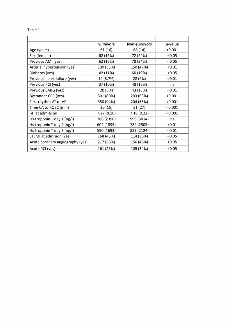
Table 1
Survivors Non-survivors p-value
Age (years) 61 (16) 68 (14) <0.001
Sex (female) 62 (16%) 72 (22%) <0.05
Previous AMI (yes) 62 (16%) 78 (24%) <0.05
Arterial hypertension (yes) 130 (35%) 150 (47%) <0.01
Diabetes (yes) 42 (11%) 60 (19%) <0.05
Previous heart failure (yes) 14 (3.7%) 28 (9%) <0.01
Previous PCI (yes) 37 (10%) 48 (15%) ns
Previous CABG (yes) 20 (5%) 43 (13%) <0.01
Bystander CPR (yes) 301 (80%) 203 (63%) <0.001
First rhythm VT or VF 350 (94%) 204 (65%) <0.001
Time CA to ROSC (min) 20 (15) 31 (27) <0.001
pH at admission 7.27 (0.16) 7.18 (0.22) <0.001
Hs-troponin T day 1 (ng/l) 786 (2390) 996 (3014) ns
Hs-troponin T day 2 (ng/l) 602 (1895) 789 (2505) <0.01
Hs-troponin T day 3 (ng/l) 590 (1693) 859 (2124) <0.01
STEMI at admision (yes) 168 (45%) 114 (36%) <0.05
Acute coronary angiography (yes) 217 (58%) 156 (48%) <0.05
Acute PCI (yes) 161 (43%) 109 (34%) <0.05

Table 2a
At 24h Odds ratio (95% CI) p-value
Temperature group (36°C) 1.23 (0.85-1.77) ns
Age 1.07 (1.05-1.09) <0.001
Sex (male) 0.75 (0.47-1.20) ns
Bystander CPR (yes) 0.64 (0.42-0.97) <0.05
Asystole/PEA (yes) 6.77 (3.91-11.73) <0.001
CA to ROSC (min) 1.03 (1.02-1.04) <0.001
Shock at admission (yes) 1.42 (0.81-2.50) ns
pH 0.12 (0.03-0.46) <0.01
hs-TnT at 24h 1.07 (0.99-1.17) ns
At 48h
Temperature group (36°C) 1.25 (0.87-1.80) ns
Age 1.07 (1.05-1.09) <0.001
Sex (male) 0.73 (0.45-1.17) ns
Bystander CPR (yes) 0.65 (0.43-0.98) <0.05
Asystole/PEA (yes) 6.80 (3.94-11.71) <0.001
CA to ROSC (min) 1.03 (1.02-1.04) <0.001
Shock at admission (yes) 1.39 (0.79-2.44) ns
pH 0.12 (0.03-0.49) <0.01
hs-TnT at 48h 1.10 (1.01-1.20) <0.05
At 72h
Temperature group (36°C) 1.26 (0.87-1.81) ns
Age 1.07 (1.05-1.09) <0.001
Sex (male) 0.72 (0.45-1.17) ns
Bystander CPR (yes) 0.65 (0.43-0.99) <0.05
Asystole/PEA (yes) 6.82 (3.95-11.58) <0.001
CA to ROSC (min) 1.03 (1.02-1.04) <0.001
Shock at admission (yes) 1.38 (0.78-2.44) ns
pH 0.13 (0.03-0.50) <0.01
hs-TnT at 72h 1.11 (1.02-1.21) <0.05
Table 2b
At 24h Odds ratio (95% CI) p-value
Temperature group (36°C) 0.87 (0.54-1.40) ns
Age 1.10 (1.07-1.13) <0.001
Sex (male) 0.58 (0.33-1.00) ns
Bystander CPR (yes) 0.76 (0.46-1.27) ns
Asystole/PEA (yes) 1.39 (0.79-2.47) ns
CA to ROSC (min) 1.00 (0.99-1.01) ns
Shock at admission (yes) 3.15 (1.76-5.63) <0.001
pH 0.14 (0.03-0.67) <0.05
hs-TnT at 24h 1.11 (1.00-1.23) <0.05
At 48h
Temperature group (36°C) 0.89 (0.55-1.42) ns
Age 1.10 (1.07-1.13) <0.001
Sex (male) 0.57 (0.33-0.99) <0.05
Bystander CPR (yes) 0.76 (0.46-1.27) ns

Asystole/PEA (yes) 1.38 (0.78-2.45) ns
CA to ROSC (min) 1.00 (0.99-1.00) ns
Shock at admission (yes) 3.10 (1.74-5.54) <0.001
pH 0.14 (0.03-0.71) <0.05
hs-TnT at 48h 1.13 (1.01-1.26) <0.05
At 72h
Temperature group (36°C) 1.26 (0.87-1.81) ns
Age 1.07 (1.05-1.09) <0.001
Sex (male) 0.72 (0.45-1.17) <0.05
Bystander CPR (yes) 0.65 (0.43-0.99) ns
Asystole/PEA (yes) 6.82 (3.95-11.58) ns
CA to ROSC (min) 1.03 (1.02-1.04) ns
Shock at admission (yes) 1.38 (0.78-2.44) <0.001
pH 0.13 (0.03-0.50) <0.05
hs-TnT at 72h 1.11 (1.02-1.21) <0.05