High Protective Efficacy of Probiotics and Rice Bran against … · fmicb-07-01699 October 31, 2016...
12
ORIGINAL RESEARCH published: 02 November 2016 doi: 10.3389/fmicb.2016.01699 Edited by: Luis Cláudio Nascimento Da Silva, University Centre of Maranhão, Brazil Reviewed by: Atte Von Wright, University of Eastern Finland, Finland Mario (Mago) Clerici, University of Milan, Italy Jesus Rodriguez-Diaz, University of Valencia, Spain *Correspondence: Lijuan Yuan [email protected] Specialty section: This article was submitted to Antimicrobials, Resistance and Chemotherapy, a section of the journal Frontiers in Microbiology Received: 13 September 2016 Accepted: 12 October 2016 Published: 02 November 2016 Citation: Lei S, Ramesh A, Twitchell E, Wen K, Bui T, Weiss M, Yang X, Kocher J, Li G, Giri -Rachman E, Trang NV, Jiang X, Ryan EP and Yuan L (2016) High Protective Efficacy of Probiotics and Rice Bran against Human Norovirus Infection and Diarrhea in Gnotobiotic Pigs. Front. Microbiol. 7:1699. doi: 10.3389/fmicb.2016.01699 High Protective Efficacy of Probiotics and Rice Bran against Human Norovirus Infection and Diarrhea in Gnotobiotic Pigs Shaohua Lei 1 , Ashwin Ramesh 1 , Erica Twitchell 1 , Ke Wen 1 , Tammy Bui 1 , Mariah Weiss 1 , Xingdong Yang 1 , Jacob Kocher 1 , Guohua Li 1 , Ernawati Giri-Rachman 1,2 , Nguyen Van Trang 3 , Xi Jiang 4 , Elizabeth P. Ryan 5 and Lijuan Yuan 1 * 1 Department of Biomedical Sciences and Pathobiology, Virginia-Maryland College of Veterinary Medicine, Virginia Tech, Blacksburg, VA, USA, 2 School of Life Science and Technology, Institut Teknologi, Bandung, West Java, Indonesia, 3 National Institute of Hygiene and Epidemiology, Hanoi, Vietnam, 4 Division of Infectious Diseases, Cincinnati Children’s Hospital Medical Center, Cincinnati, OH, USA, 5 Department of Environmental and Radiological Health Sciences, College of Veterinary Medicine and Biomedical Sciences, Colorado State University, Fort Collins, CO, USA Probiotics have been recognized as vaccine adjuvants and therapeutic agents to treat acute gastroenteritis in children. We previously showed that rice bran (RB) reduced human rotavirus diarrhea in gnotobiotic pigs. Human noroviruses (HuNoVs) are the major pathogens causing non-bacterial acute gastroenteritis worldwide. In this study, Lactobacillus rhamnosus GG (LGG) and Escherichia coli Nissle 1917 (EcN) were first screened for their ability to bind HuNoV P particles and virions derived from clinical samples containing HuNoV genotype GII.3 and GII.4, then the effects of LGG+EcN and RB on HuNoV infection and diarrhea were investigated using the gnotobiotic pig model. While LGG+EcN colonization inhibited HuNoV shedding, probiotic cocktail regimens in which RB feeding started 7 days prior to or 1 day after viral inoculation in the LGG+EcN colonized gnotobiotic pigs exhibited high protection against HuNoV diarrhea and shedding, characterized by significantly reduced incidence (89 versus 20%) and shorter mean duration of diarrhea (2.2 versus 0.2 days), as well as shorter mean duration of virus shedding (3.2 versus 1.0 days). In both probiotic cocktail groups, the diarrhea reduction rates were 78% compared with the control group, and diarrhea severity was reduced as demonstrated by the significantly lower cumulative fecal scores. The high protective efficacy of the probiotic cocktail regimens was attributed to stimulation of IFN- γ + T cell responses, increased production of intestinal IgA and IgG, and maintenance of healthy intestinal morphology (manifested as longer villi compared with the control group). Therefore, probiotic cocktail regimens containing LGG+EcN and RB may represent highly efficacious strategies to prevent and treat HuNoV gastroenteritis, and potentially other human enteric pathogens. Keywords: probiotics, rice bran, human norovirus, diarrhea, gnotobiotic pigs Abbreviations: EC, Enterobacter cloacae; EcN, Escherichia coli Nissle 1917; Gn, gnotobiotic; HBGA, histo-blood group antigen; HRV, human rotavirus; HuNoV, human norovirus; LA, Lactobacillus acidophilus; LB, Lactobacillus bulgaricus; LGG, Lactobacillus rhamnosus GG; LR, Lactobacillus reuteri; MuNoV, murine norovirus; PID, post-infection day; PPD, post-partum day; RB, rice bran; VLP, virus-like particle. Frontiers in Microbiology | www.frontiersin.org 1 November 2016 | Volume 7 | Article 1699
-
Upload
vuongduong -
Category
Documents
-
view
218 -
download
0
Transcript of High Protective Efficacy of Probiotics and Rice Bran against … · fmicb-07-01699 October 31, 2016...
fmicb-07-01699 October 31, 2016 Time: 15:3 # 1
ORIGINAL RESEARCHpublished: 02 November 2016
doi: 10.3389/fmicb.2016.01699
Edited by:Luis Cláudio Nascimento Da Silva,
University Centre of Maranhão, Brazil
Reviewed by:Atte Von Wright,
University of Eastern Finland, FinlandMario (Mago) Clerici,
University of Milan, ItalyJesus Rodriguez-Diaz,
University of Valencia, Spain
*Correspondence:Lijuan Yuan
Specialty section:This article was submitted to
Antimicrobials, Resistanceand Chemotherapy,
a section of the journalFrontiers in Microbiology
Received: 13 September 2016Accepted: 12 October 2016
Published: 02 November 2016
Citation:Lei S, Ramesh A, Twitchell E,
Wen K, Bui T, Weiss M, Yang X,Kocher J, Li G, Giri -Rachman E,Trang NV, Jiang X, Ryan EP and
Yuan L (2016) High ProtectiveEfficacy of Probiotics and Rice Branagainst Human Norovirus Infection
and Diarrhea in Gnotobiotic Pigs.Front. Microbiol. 7:1699.
doi: 10.3389/fmicb.2016.01699
High Protective Efficacy of Probioticsand Rice Bran against HumanNorovirus Infection and Diarrhea inGnotobiotic PigsShaohua Lei1, Ashwin Ramesh1, Erica Twitchell1, Ke Wen1, Tammy Bui1, Mariah Weiss1,Xingdong Yang1, Jacob Kocher1, Guohua Li1, Ernawati Giri-Rachman1,2,Nguyen Van Trang3, Xi Jiang4, Elizabeth P. Ryan5 and Lijuan Yuan1*
1 Department of Biomedical Sciences and Pathobiology, Virginia-Maryland College of Veterinary Medicine, Virginia Tech,Blacksburg, VA, USA, 2 School of Life Science and Technology, Institut Teknologi, Bandung, West Java, Indonesia, 3 NationalInstitute of Hygiene and Epidemiology, Hanoi, Vietnam, 4 Division of Infectious Diseases, Cincinnati Children’s HospitalMedical Center, Cincinnati, OH, USA, 5 Department of Environmental and Radiological Health Sciences, College of VeterinaryMedicine and Biomedical Sciences, Colorado State University, Fort Collins, CO, USA
Probiotics have been recognized as vaccine adjuvants and therapeutic agents to treatacute gastroenteritis in children. We previously showed that rice bran (RB) reducedhuman rotavirus diarrhea in gnotobiotic pigs. Human noroviruses (HuNoVs) are themajor pathogens causing non-bacterial acute gastroenteritis worldwide. In this study,Lactobacillus rhamnosus GG (LGG) and Escherichia coli Nissle 1917 (EcN) were firstscreened for their ability to bind HuNoV P particles and virions derived from clinicalsamples containing HuNoV genotype GII.3 and GII.4, then the effects of LGG+EcN andRB on HuNoV infection and diarrhea were investigated using the gnotobiotic pig model.While LGG+EcN colonization inhibited HuNoV shedding, probiotic cocktail regimensin which RB feeding started 7 days prior to or 1 day after viral inoculation in theLGG+EcN colonized gnotobiotic pigs exhibited high protection against HuNoV diarrheaand shedding, characterized by significantly reduced incidence (89 versus 20%) andshorter mean duration of diarrhea (2.2 versus 0.2 days), as well as shorter mean durationof virus shedding (3.2 versus 1.0 days). In both probiotic cocktail groups, the diarrheareduction rates were 78% compared with the control group, and diarrhea severity wasreduced as demonstrated by the significantly lower cumulative fecal scores. The highprotective efficacy of the probiotic cocktail regimens was attributed to stimulation of IFN-γ+ T cell responses, increased production of intestinal IgA and IgG, and maintenanceof healthy intestinal morphology (manifested as longer villi compared with the controlgroup). Therefore, probiotic cocktail regimens containing LGG+EcN and RB mayrepresent highly efficacious strategies to prevent and treat HuNoV gastroenteritis, andpotentially other human enteric pathogens.
Keywords: probiotics, rice bran, human norovirus, diarrhea, gnotobiotic pigs
Abbreviations: EC, Enterobacter cloacae; EcN, Escherichia coli Nissle 1917; Gn, gnotobiotic; HBGA, histo-blood groupantigen; HRV, human rotavirus; HuNoV, human norovirus; LA, Lactobacillus acidophilus; LB, Lactobacillus bulgaricus;LGG, Lactobacillus rhamnosus GG; LR, Lactobacillus reuteri; MuNoV, murine norovirus; PID, post-infection day; PPD,post-partum day; RB, rice bran; VLP, virus-like particle.
Frontiers in Microbiology | www.frontiersin.org 1 November 2016 | Volume 7 | Article 1699

fmicb-07-01699 October 31, 2016 Time: 15:3 # 2
Lei et al. Probiotics and Rice Bran against HuNoV
INTRODUCTION
Human noroviruses, non-enveloped viruses with a positive-strand RNA genome, are the major pathogens causing non-bacterial acute gastroenteritis worldwide (Pringle et al., 2015).In the United States, HuNoVs have replaced HRVs as the singlemost common cause of viral gastroenteritis in children and adults(Bresee et al., 2012; Payne et al., 2013). After approximately1.2 days of incubation (Lee et al., 2013), HuNoV gastroenteritisgenerally lasts for 2–3 days and consists of nausea, vomiting,and diarrhea (Lopman et al., 2004). More severe and prolongedillness can occur in specific risk groups, including infants, theelderly, and immunocompromised patients (Murata et al., 2007;Hall et al., 2012; Green, 2014). Given the tremendous diseaseburden and economic loss associated with HuNoVs infection(Patel et al., 2008; Ahmed et al., 2014), vaccines and therapeuticsare in great demand to prevent and treat these infections.However, due to the lack of a robust culturing system and asuitable small-animal model, HuNoVs vaccine development andantiviral research have long been hampered. Promising vaccineshave focused on recombinant capsid proteins, including VLPsand P particles (Kocher and Yuan, 2015). Appropriate animalmodels are essential tools to facilitate investigation of vaccinecandidates and therapeutic strategies. Neonatal gnotobiotic(Gn) pigs recapitulate the pathologic hallmarks of enteric viralinfection and associated immune responses in the gastrointestinaltract of young children (Yang and Yuan, 2014). Currently, asthe only animal model that supports the oral route of HuNoVinfection, develops diarrhea, and sheds virus in feces, Gn pigs areused to evaluate viral pathogenesis and vaccine efficacy with hightranslational validity to humans (Cheetham et al., 2006; Bui et al.,2013; Kocher et al., 2014).
Probiotic bacteria are increasingly recognized as vaccineadjuvants and therapeutic agents to treat acute gastroenteritisin children (Licciardi and Tang, 2011; Schnadower et al., 2015).The potential mechanisms include competing with pathogensfor nutrients and colonization sites, producing antimicrobialmetabolites, enhancing protective immune responses, andreducing intestinal permeability (Ng et al., 2009). Notably,Gram-positive probiotics Lactobacillus spp. have been extensivelyevaluated for their beneficial effects against viral infection anddiseases. These include reducing HRV and vesicular stomatitisvirus infection in cell cultures (Botic et al., 2007; Maragkoudakiset al., 2010) and promoting HRV-specific immune responses,which contribute to shortened HRV-induced diarrhea in animalmodels (Zhang et al., 2008; Wen et al., 2014, 2015) andhuman clinical trials (Guandalini et al., 2000; Sindhu et al.,2014; Szajewska et al., 2014). Gram-negative EcN is also awell-characterized probiotic used to treat diarrhea in infantsand young children (Henker et al., 2007, 2008), as well asin neonatal large animals (von Buenau et al., 2005; Schroederet al., 2006). The beneficial health effects are mediated viaimproving intestinal barrier function (Hering et al., 2014)or moderating inflammatory responses (Splichalova et al.,2011), which could protect Gn piglets from lethal infectionof Salmonella Typhimurium (Splichalova et al., 2011). Inaddition, EcN was recently shown to have HRV-binding and
immunomodulatory properties, resulting in significantly reducedHRV infection and diarrhea in Gn pigs (Kandasamy et al., 2016).Probiotics can act as adsorbents for HuNoV P particles, andthe presence of L. casei BL23 and EcN might inhibit P particleattachment to epithelial cells (Rubio-del-Campo et al., 2014).Enterobacter cloacae (EC) is a commensal bacterium that canbind to HuNoV by surface HBGA and inhibit HuNoV infectivityin Gn pigs (Miura et al., 2013; Lei et al., 2016b). Taken together,diarrhea-reducing probiotics may inhibit HuNoV infectivityin vivo, most likely by the binding between bacteria and virions.
Rice bran, an underutilized by-product of rice milling,contains a variety of prebiotic and bioactive componentsthat modulate gut microbiota and potentially prevent chronicdiseases, including diabetes, cancer, metabolic syndrome, andcardiovascular disease (Sheflin et al., 2015). In mouse studies,dietary RB feeding increased production of fecal and serumIgA (Henderson et al., 2012), and RB glycoproteins amelioratedcyclophosphamide-induced immunosuppression by restoringsplenic lymphocytes (Park et al., 2016), indicating that RBpromoted the development of mucosal and systemic adaptiveimmunity. Chemically engineered RB glucans possessed anti-cytomegalovirus activity by blocking viral entry of targetcells (Ray et al., 2013). Recently, therapeutic effects ofRB in inhibiting enteric infections and reducing diarrheahave been gaining attention. In a clinical trial, Biobran(modified arabinoxylan RB) improved irritable bowel syndromesymptoms, presumably resulting from its anti-inflammatoryand/or immunomodulatory effects (Kamiya et al., 2014). Inour previous Gn pig studies, dietary RB feeding significantlyenhanced HRV vaccine immunogenicity and reduced HRV-induced diarrhea (Yang et al., 2014). RB could also protectagainst HRV diarrhea in the presence of probiotics by preventingintestinal epithelial damage and promoting innate immuneresponses (Yang et al., 2015). Therefore, its beneficial effects ongastrointestinal health support RB as a promising agent againstHuNoV infection.
In this study, aiming to develop an effective and ready-to-use anti-HuNoV therapeutic strategy, we first screened agroup of probiotics to identify the virus-binding bacteria usingHuNoV P particles and native virions. Subsequently, probioticsand RB were evaluated individually or combined as cocktailregimens for their effects on HuNoV infection and diseases inthe well-established Gn pig model (Bui et al., 2013). Finally, themechanisms of antiviral and diarrhea-reducing activities fromthose treatments were explored.
MATERIALS AND METHODS
Viruses and BacteriaA human stool sample containing the HuNoV GII.4/2006bvariant 092895 (GenBank KC990829) was collected in 2008 atthe Cincinnati Children’s Hospital Medical Center from a childwith norovirus gastroenteritis. The sample pool was processedas an oral inoculum for HuNoV infection studies in Gn pigs(Bui et al., 2013). A human stool sample containing the HuNoVGII.3/20110200 (GenBank KX355506) was collected in 2011 at
Frontiers in Microbiology | www.frontiersin.org 2 November 2016 | Volume 7 | Article 1699

fmicb-07-01699 October 31, 2016 Time: 15:3 # 3
Lei et al. Probiotics and Rice Bran against HuNoV
the Thai Binh Pediatric Hospital (Thai Binh province, Vietnam)from a female child with norovirus gastroenteritis. L. reuteri(ATCC 23272), L. acidophilus (strain NCFM), L. rhamnosus GG(ATCC 53103), and L. bulgaricus (ATCC 11842) were cultured inlactobacilli MRS broth (Neogen Corporation) anaerobically usingBBLTM GasPakTM jar system with Anaerobe Sachets (BD) understatic condition at 37◦C. EcN (a gift from Dr. Jun Sun, RushUniversity, Chicago, IL, USA) and Enterobacter cloacae (ATCC13047) were cultured in Luria Bertani medium at 37◦C and innutrient broth at 30◦C, respectively, with shaking at 250 rpm.
Purification of HuNoVs and VP1SequencingThe pooled human stools containing HuNoVs were diluted10-fold with diluent #5 (Minimal Essential Medium with 1%penicillin-streptomycin and 1% HEPES) and mixed thoroughlywith an equal volume of Vertrel XF (Miller-Stephenson), andviruses were purified by CsCl gradient centrifugation as describedpreviously (Guix et al., 2007). VP1 of GII.4/2006b variant092895 was cloned and sequenced previously (Kocher et al.,2014). GII.3/20110200 viral RNA was extracted from the purifiedvirus by TRIzol LS and reverse transcribed by SuperScript IIIReverse Transcriptase (Thermo Fisher Scientific) using universalGII.3 reverse primer 5′-TAG CCC CTG CAT TAA CTA-3′ andfollowing the manufacturer’s instructions. The GII.3 VP1 wascloned by a nested PCR with primer set 1 (forward: 5′-TGAGCA CGT GGG AGG GCG-3′ and reverse: 5′-TAG CCC CTGCAT TAA CTA-3′) and primer set 2 (forward: 5′-CAC CAT GAAGAT GGC GTC GAA T-3′ and reverse: 5′-TTA TTG AAT CCTTCT ACG CC-3′) into pENTR directional TOPO vector (ThermoFisher Scientific). The GII.3 VP1 fragment in the recombinantplasmids were sequenced by Virginia Bioinformatics Institute atVirginia Tech, and the predominant sequence was used for thepreparation of P particles.
P Particles and Transmission ElectronMicroscopyThe region coding for the P domain was amplified fromthe recombinant plasmids containing VP1 capsid gene ofHuNoV GII.3 or GII.4 as described above. The P domainswere cloned into prokaryotic expression vector pET21a (EMDMillipore) as previously described (Rubio-del-Campo et al.,2014). A 6×His-Tag was incorporated to the N-terminus ofP proteins by forward primers, and a cysteine-rich peptideCDCRGDCFC was incorporated to the C-terminus of P proteinsby reverse primers to enhance the P particle stability (Tan andJiang, 2005). P proteins were expressed in E. coli strain BL21(New England Biolabs) and purified via HisPur Ni-NTA SpinColumns (Thermo Fisher Scientific) following the manufacturer’sinstructions. Protein production was monitored by SDS-PAGEand InVision His-tag In-gel Stain (Thermo Fisher Scientific).Protein concentrations were measured spectroscopically byQuick StartTM Bradford protein assay (Bio-Rad). Electronmicroscopy formvar carbon square grids (Electron MicroscopySciences) were pretreated with 1% aqueous Alcian blue for 5 min.After washes, P particles of genotype GII.3 or GII.4 were diluted
in PBS to 5 µg/ml and absorbed to the grids for 1 min. The gridswere stained with 3% phosphotungstic acid pH 7.0 for 1 min andviewed with a JEOL JEM 1400 transmission electron microscopy.
Binding of P Particles and Virions toBacteriaAfter the initial inoculation into fresh culture medium, bacteriawere grown overnight and sub-cultured at 1:50 for 2–3 h untilOD600 reached 0.4–1.0, which was the log phase of growth.Bacteria were washed three times and resuspended with PBS toan OD600 of 1.0. Then 10 µg P particles or 106 viral genomecopies of purified HuNoVs were incubated with 1 ml bacteriafor 1 h at 37◦C, then the mixture was centrifuged and washedthree times with PBS. To measure the remaining P particlesattached to bacteria, the bacterial pellets were resuspendedwith 100 µl Laemmli sample buffer (Bio-Rad) and boiled for10 min, and 20 µl of sample was loaded to 10% SDS-PAGE geland analyzed by Western Blot using HRP conjugated anti-His-Tag antibody (MA1-21315-HRP, Thermo Fisher Scientific). Tomeasure the remaining virions, the total RNA of the bacterialpellets was extracted by 750 µl TRIzol LS, and HuNoV genomeswere detected by a one-step TaqMan qRT-PCR with primerstargeting all GII viruses (Kageyama et al., 2003; Lei et al., 2016a).For controls, Enterobacter cloacae were heat-killed at 65◦C for40 min, then blocked with 5 µl of A antigen antibody (sc-69951,Santa Cruz) and H antigen antibody (sc-52369, Santa Cruz) at37◦C for 20 min before adding P particles or virions.
Gnotobiotic Pigs and Treatment GroupsNear-term Yorkshire cross-breed pigs were derived viahysterectomy by veterinarians and maintained in sterileisolator units as described previously (Meyer et al., 1964).Neonatal Gn pigs (male and female) were randomly assigned tothe five treatment groups upon derivation: cocktail-7d (n = 5),cocktail+1d (n = 5), RB-7d (n = 4), LGG+EcN (n = 5),control (n = 9). To initiate the colonization of LGG and EcN,104 CFU of each were mixed in five ml of Minimal EssentialMedium and administered orally to pigs on PPD 3, 5, and 7.The low dosage was chosen on purpose to be well below thetherapeutic practice (109 to 1012 CFU). LGG and EcN fecalshedding were determined by rectal swab sampling of pig fecesand enumeration of colonies grown on media agar plates asdescribed previously (Yang et al., 2015). For RB feeding of pigs,heat-stabilized and gamma-irradiated RB (Calrose variety) wasadded to pigs’ milk diet by replacing 10% daily calorie intake(Yang et al., 2015). Daily feeding started 7 days prior to or 1 dayafter HuNoV inoculation until euthanasia. All pigs were orallyinoculated on PPD33 with 6.43 × 10⁵ viral genome copies ofHuNoV GII.4/2006b variant 092895. To reduce gastric acidity,four ml 200 mM sodium bicarbonate were given to pigs 15 minprior to inoculation. Fecal consistency and virus shedding wereassessed daily until euthanasia on PPD40 where blood, tissues,and intestinal contents were collected. Fecal consistency scoreswere obtained based on previous scaling system (Bui et al., 2013),and fecal virus shedding was measured by a one-step TaqManqRT-PCR as described previously (Lei et al., 2016a).
Frontiers in Microbiology | www.frontiersin.org 3 November 2016 | Volume 7 | Article 1699

fmicb-07-01699 October 31, 2016 Time: 15:3 # 4
Lei et al. Probiotics and Rice Bran against HuNoV
Flow Cytometry AnalysisMononuclear cells (MNCs) were isolated from the duodenum,ileum, spleen, and blood as described previously (Yuan et al.,2008). 2× 10⁶ of MNCs were restimulated in vitro with P particle(12 µg/ml for the spleen and 6 µg/ml for others), positive controlPHA (10 µg/ml), or mock control in E-RPMI media for 17 hat 37◦C. Brefeldin A (B6542, Sigma-Aldrich, 5 µg/ml) and anti-CD49d monoclonal antibody (561892, BD Biosciences, 1 µl/ml)were added at 12 h post-incubation to block the secretion ofcytokines and enhance the stimulation, respectively. IFN-γ+CD4+ and CD8+ T cells were quantified by flow cytometryas described previously (Yuan et al., 2008). Isotype matchedirrelevant antibodies were included as negative gate controls.Mock-stimulated samples indicate the total IFN-γ+ T cells, whilethe increased cell populations of P particle-stimulated over mock-stimulated samples indicate HuNoV-specific IFN-γ+ T cells.
ELISA for Total Immunoglobulin andIFN-γThe total immunoglobulin (Ig) titers in intestinal contentswere determined by ELISA as described previously (Lei et al.,2016a). Intestinal IFN-γ titers were measured by Swine IFN-γVetSetTM ELISA development Kit (Kingfisher Biotech) followingthe manufacturer’s instructions.
Jejunum HistopathologyJejunum tissue was collected after pig euthanasia, fixed in 4%paraformaldehyde for 12–16 h, paraffin embedded, sectionedinto 5 µm slices and placed on positively charged slides, forroutine H&E staining. A pathologist who was blinded to thesample identifications evaluated the villus length using an ocularmicrometer under a light microscope.
Ethical StatementStool collection protocols were approved by the InstitutionalReview Boards of the Cincinnati Children’s Hospital MedicalCenter (IRB#: 2008-1131) and the National Institute of Hygieneand Epidemiology – Vietnam (IRB#: 15-IRB), written consentwas provided by parents or guardians of the children. Animalexperimental protocols were approved by the InstitutionalAnimal Care and Use Committee at Virginia Tech (IACUCprotocol: 13-187-CVM and 14-108-CVM). All sample collectionand experimental procedures were conducted in accordance withthe approved guidelines.
StatisticsStatistics were performed using GraphPad Prism 6.0 (GraphPadSoftware) with analyses indicated in table notes and figurelegends. Statistical significance was determined at the level ofP < 0.05.
RESULTS
Probiotic Bacteria Bind to HuNoVsAlthough HuNoV genotype GII.4 accounts for the mostglobal acute gastroenteritis outbreaks (Pringle et al., 2015),
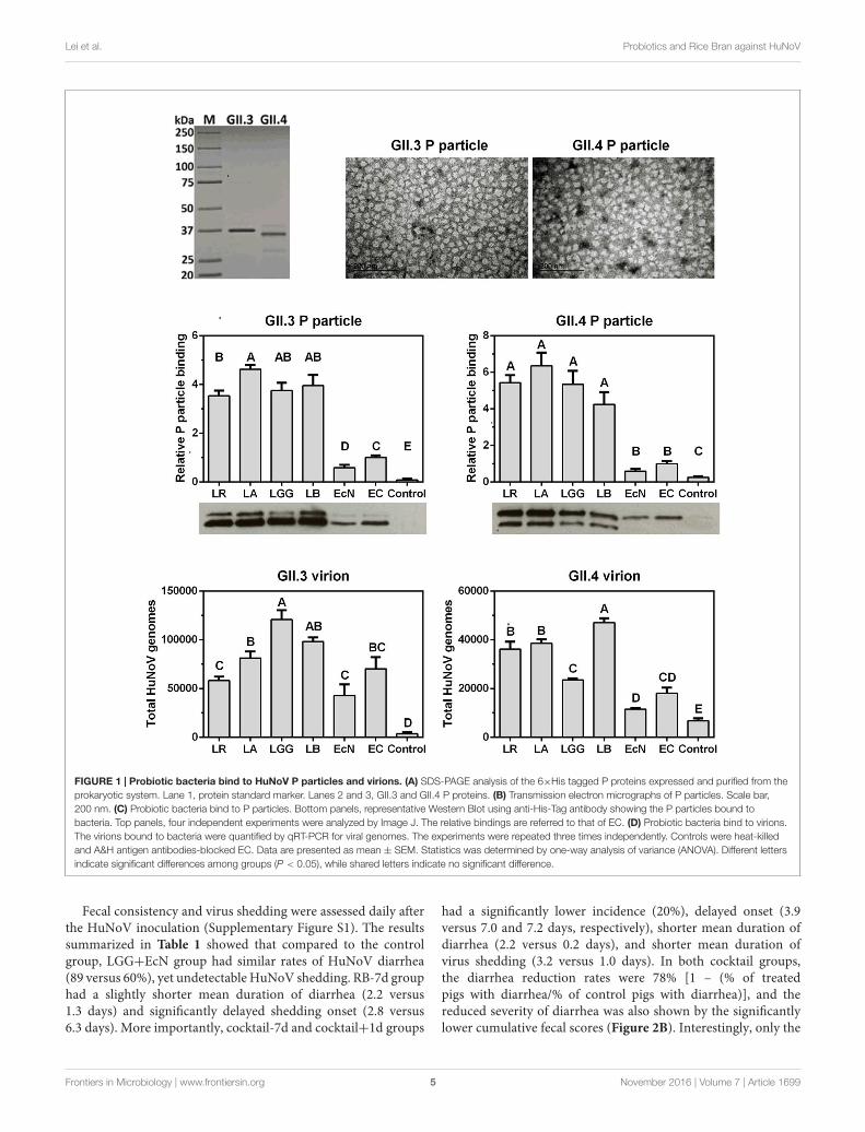
GII.3 is emerging and becoming predominant in someunderdeveloped areas (Liu et al., 2014; Van Trang et al.,2014; Ayouni et al., 2015). To explore the interactions betweenHuNoVs and probiotics, we first cloned the capsid VP1genes of GII.3 and GII.4 from clinical stool samples, thenthe P-domains were cloned and proteins were expressedwith N-terminal 6×His-Tag to facilitate their detection andpurification (Figure 1A). The formation of P particles wasnot compromised as indicated by a negative staining electronmicroscopy (Figure 1B).
P particles were first used as a model to determine HuNoVsinteractions with probiotics, including a Gram-negative strainEcN and four Gram-positive lactobacilli strains, i.e., L. reuteri(LR), L. acidophilus (LA), L. rhamnosus GG (LGG), andL. bulgaricus (LB). Since EC can bind to HuNoVs specificallyby surface HBGA (Miura et al., 2013), native EC was usedas a positive control and HBGA A&H antibodies-blocked ECwas a negative control in the binding assays. After incubationof P particles and bacteria, the P particles remaining on thebacterial surface were quantified by Western Blot using anti-His-Tag antibody. The results showed that all the tested bacteriawere able to bind to both GII.3 and GII.4 P particles, andlactobacilli strains had significantly higher binding capacitythan those of EcN and EC (Figure 1C). Additionally, LA wasstronger than LR in binding to GII.3 P particle, while EcNwas weaker than EC. The four lactobacilli strains did not differfrom each other in binding to GII.4 P particle, and neither didEcN and EC (Figure 1C). Similarly, the binding assays wereperformed using HuNoV virions purified from stool samples.Unlike the P particles, GII.3 virions had comparable bindingto all tested bacteria except for the higher binding to LGG,whereas GII.4 virions shared the binding pattern with P particleexcept for the lower binding to LGG and higher binding to LB(Figure 1D). These data suggest that probiotic bacteria can bindto HuNoVs.
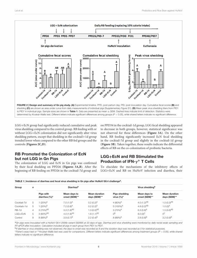
LGG+EcN Inhibited HuNoV Sheddingand RB Reduced Diarrhea in Gn PigsTo develop a ready-to-use anti-HuNoV therapeutic strategy,LGG and EcN were chosen for the evaluation of their potentialantiviral effects in the Gn pig model of HuNoV infectionand diarrhea, since they could bind HuNoVs in vitro and arecommercially available as diarrhea-reducing probiotics. Previousstudy showed that RB protected against HRV-induced diarrheain the presence of LGG and EcN (Yang et al., 2015), we testedRB feeding and/or LGG+EcN co-colonization in five treatmentgroups in this study: cocktail-7d (n = 5), pigs were pre-colonized with LGG and EcN, RB feeding started 7 days priorto HuNoV inoculation; cocktail+1d (n = 5), pigs were pre-colonized with LGG and EcN, RB feeding started 1 day aftervirus inoculation; RB-7d (n = 4), RB feeding started 7 days priorto inoculation; LGG+EcN (n = 5), pigs were colonized withLGG and EcN only; Control (n = 9), non-RB fed and non-LGG+EcN colonized. All pigs were inoculated with a HuNoVGII.4/2006b variant 092895 on PPD33/PID0 and euthanized onPID7 (Figure 2A).
Frontiers in Microbiology | www.frontiersin.org 4 November 2016 | Volume 7 | Article 1699
fmicb-07-01699 October 31, 2016 Time: 15:3 # 5
Lei et al. Probiotics and Rice Bran against HuNoV
FIGURE 1 | Probiotic bacteria bind to HuNoV P particles and virions. (A) SDS-PAGE analysis of the 6×His tagged P proteins expressed and purified from theprokaryotic system. Lane 1, protein standard marker. Lanes 2 and 3, GII.3 and GII.4 P proteins. (B) Transmission electron micrographs of P particles. Scale bar,200 nm. (C) Probiotic bacteria bind to P particles. Bottom panels, representative Western Blot using anti-His-Tag antibody showing the P particles bound tobacteria. Top panels, four independent experiments were analyzed by Image J. The relative bindings are referred to that of EC. (D) Probiotic bacteria bind to virions.The virions bound to bacteria were quantified by qRT-PCR for viral genomes. The experiments were repeated three times independently. Controls were heat-killedand A&H antigen antibodies-blocked EC. Data are presented as mean ± SEM. Statistics was determined by one-way analysis of variance (ANOVA). Different lettersindicate significant differences among groups (P < 0.05), while shared letters indicate no significant difference.
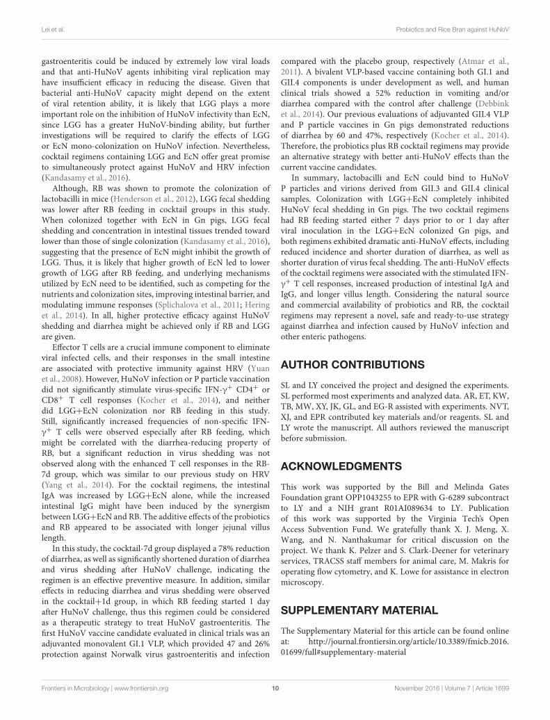
Fecal consistency and virus shedding were assessed daily afterthe HuNoV inoculation (Supplementary Figure S1). The resultssummarized in Table 1 showed that compared to the controlgroup, LGG+EcN group had similar rates of HuNoV diarrhea(89 versus 60%), yet undetectable HuNoV shedding. RB-7d grouphad a slightly shorter mean duration of diarrhea (2.2 versus1.3 days) and significantly delayed shedding onset (2.8 versus6.3 days). More importantly, cocktail-7d and cocktail+1d groups
had a significantly lower incidence (20%), delayed onset (3.9versus 7.0 and 7.2 days, respectively), shorter mean duration ofdiarrhea (2.2 versus 0.2 days), and shorter mean duration ofvirus shedding (3.2 versus 1.0 days). In both cocktail groups,the diarrhea reduction rates were 78% [1 – (% of treatedpigs with diarrhea/% of control pigs with diarrhea)], and thereduced severity of diarrhea was also shown by the significantlylower cumulative fecal scores (Figure 2B). Interestingly, only the
Frontiers in Microbiology | www.frontiersin.org 5 November 2016 | Volume 7 | Article 1699
fmicb-07-01699 October 31, 2016 Time: 15:3 # 6
Lei et al. Probiotics and Rice Bran against HuNoV
FIGURE 2 | Design and summary of Gn pig study. (A) Experimental timeline. PPD, post-partum day; PID, post-inoculation day. Cumulative fecal scores (B) andshedding (C) are shown as area under curve from daily measurements of individual pigs (Supplementary Figure S1). (D) Mean peak virus shedding titers from PID1to PID7 in individual pigs. Sample sizes are shown in Table 1. Data are presented as mean ± SEM. Dashed lines indicate limit of detection. Statistics weredetermined by Kruskal–Wallis test. Different letters indicate significant differences among groups (P < 0.05), while shared letters indicate no significant difference.
LGG+EcN group had significantly reduced cumulative and peakvirus shedding compared to the control group. RB feeding with orwithout LGG+EcN colonization did not significantly alter virusshedding pattern, except that shedding in the cocktail+1d grouptrended lower when compared to the other RB fed groups and thecontrols (Figures 2C,D).
RB Promoted the Colonization of EcNbut not LGG in Gn PigsThe colonization of LGG and EcN in Gn pigs was confirmedby their fecal shedding on PPD26 (Figures 3A,B). After thebeginning of RB feeding on PPD26 in the cocktail-7d group and
on PPD34 in the cocktail-1d group, LGG fecal shedding appearedto decrease in both groups, however, statistical significance wasnot observed for these differences (Figure 3A). On the otherhand, RB feeding significantly increased EcN fecal sheddingin the cocktail-7d group and slightly in the cocktail-1d group(Figure 3B). Taken together, these results indicate the differentialeffects of RB on the co-colonization of probiotic bacteria.
LGG+EcN and RB Stimulated theProduction of IFN-γ+ T CellsTo elucidate the mechanisms of the inhibitory effects ofLGG+EcN and RB on HuNoV infection and diarrhea, their
TABLE 1 | Incidence of diarrhea and fecal virus shedding in Gn pigs after HuNoV GII.4 challengea.
Group n Diarrheab Virus sheddingb
Pigs withdiarrhea (%)∗
Mean days toonset (SEM)∗∗
Mean durationdays (SEM)∗∗
Pigs sheddingvirus (%)∗
Mean days toonset (SEM)∗∗
Mean durationdays (SEM)∗∗
Cocktail-7d 5 1 (20%)A 7.0 (1.0)A 0.2 (0.2)A 4 (80%)A 4.0 (1.2)AB 1.0 (0.3)AB
Cocktail+1d 5 1 (20%)A 7.2 (0.8)A 0.2 (0.2)A 5 (100%)A 4.8 (0.2)AB 1.0 (0)A
RB-7d 4 3 (75%)AB 3.3 (1.6)AB 1.3 (0.5)AB 3 (75%)A 6.3 (0.8)A 1.5 (0.6)AB
LGG+EcN 5 3 (60%)AB 4.0 (1.6)AB 1.8 (1.1)AB 0B 8.0 (0)C 0C
Control 9 8 (89%)B 3.9 (0.7)B 2.2 (0.4)B 8 (89%)A 2.8 (0.8)B 3.2 (0.9)B
aGn pigs were inoculated with a HuNoV GII.4 2006b variant 092895 at 33 days of age. Diarrhea and virus shedding were monitored by daily rectal swab sampling andRT-qPCR after inoculation. Calculation included all pigs in each group from PID1 to PID7.b If diarrhea or virus shedding was not observed, the days to onset was recorded as 8 and the duration days was recorded as 0 for statistical purposes.∗Fisher’s exact test or ∗∗Kruskal–Wallis test was used for comparisons. Different letters indicate significant differences among treatment groups (P < 0.05), while sharedletters indicate no significant difference.
Frontiers in Microbiology | www.frontiersin.org 6 November 2016 | Volume 7 | Article 1699

fmicb-07-01699 October 31, 2016 Time: 15:3 # 7
Lei et al. Probiotics and Rice Bran against HuNoV
FIGURE 3 | LGG and EcN fecal shedding. Pig feces were collected by rectal swab and suspended in PBS. The concentrations of LGG (A) and EcN (B) weredetermined in serial dilution of fecal samples and enumeration of colony forming unit (CFU) grown on MRS or LB media agar plates. Sample sizes are shown inTable 1. Data are presented as mean ± SEM. Statistics were determined by Kruskal–Wallis test. NS, not significant, ∗P < 0.05.
FIGURE 4 | LGG+EcN and RB stimulated IFN-γ+ T cell responses. (A) Gating strategies for IFN-γ+ CD3+CD4+ (Th) cells and CD3+CD8+ (CTL) cells.Representative dot plots showing frequencies of HuNoV-specific (P particle stimulated) and non-specific (mock stimulated) IFN-γ+ T cells in ileum isolated fromLGG+EcN colonized Gn pigs. SSC-A, side scatter area; APC, allophycocyanin; FITC, fluorescein isothiocyanate; SPRD, spectral red; PE, phycoerythrin. (B)Non-specific IFN-γ+ T cells in intestinal (duodenum, ileum) and systemic (spleen, blood) tissues on PID7. Sample sizes are shown in Table 1. Data are presented asmeans ± SEM. Statistics were determined by Kruskal–Wallis test. NS, not significant, ∗P < 0.05, ∗∗P < 0.01.
Frontiers in Microbiology | www.frontiersin.org 7 November 2016 | Volume 7 | Article 1699

fmicb-07-01699 October 31, 2016 Time: 15:3 # 8
Lei et al. Probiotics and Rice Bran against HuNoV
FIGURE 5 | IgA, IgG, and IFN-γ levels in intestinal contents after HuNoV infection. Total IgA (A) and IgG (B) titers in small and large intestinal contents (SICand LIC) were measured by ELISA. (C) IFN-γ concentration in LIC was measured by ELISA. Sample sizes are shown in Table 1. Data are presented asmean ± SEM. Statistics were determined by Kruskal–Wallis test. Different letters indicate significant differences among groups (P < 0.05), while shared lettersindicate no significant difference.
immunomodulatory roles were first assessed regarding effector Tcells. After euthanasia on PID7, MNCs were isolated from bothintestinal and systemic lymphoid tissues, and the frequenciesof IFN-γ+ CD4+ and CD8+ T cells were determined byflow cytometry (Figure 4A). MNCs were stimulated with Pparticle to detect HuNoV-specific IFN-γ+ T cells, which wasthe increased frequency compared to the mock stimulatedsample. For pigs in the control, LGG+EcN, and RB-7dgroups, no significant increase of IFN-γ+ T cells was observedin P particle stimulated MNCs (Figure 4A and data notshown), suggesting low or short-term HuNoV-specific IFN-γ+ T cell responses. However, compared with control pigs,both LGG+EcN colonization and RB feeding significantlyincreased frequencies of non-specific total IFN-γ+ T cells(Figure 4B). In addition, compared with the LGG+EcN group,the RB-7 group had significantly higher frequencies of IFN-γ+CD8+ T cell population in ileum and IFN-γ+ CD4+ T cellpopulation in all assayed lymphoid tissues (duodenum, ileum,spleen, and blood; Figure 4B), indicating that RB has strongstimulatory effects on total IFN-γ+ T cell responses, whichmay contribute to the reduction of HuNoV diarrhea in Gnpigs.
Probiotics plus RB Cocktail RegimensEnhanced Gut ImmunityThe immunomodulatory roles of LGG+EcN and RB on gutimmunity were evaluated by testing total intestinal IgA, IgG, andIFN-γ levels, since PID 7 is too early to detect virus-specificIgA and IgG antibody responses. Compared with the controlgroup, the cocktail-7d, cocktail+1d, and LGG+EcN groups hadsignificantly higher IgA titers in both small and large intestinalcontents (SIC and LIC), but the increase was not observed inthe RB-7d group (Figure 5A), indicating that LGG+EcN butnot RB enhanced the production of IgA. The cocktail-7d andcocktail+1d groups had significantly higher IgG titers in bothSIC and LIC, whereas no differences were observed in eitherthe LGG+EcN or RB groups (Figure 5B). Consistent with thestrong stimulation of RB on total IFN-γ+ T cells (Figure 4B),significantly higher IFN-γ concentrations were detected inLIC from the cocktail-7d, cocktail+1d, and RB-7d groups(Figure 5C). In all, cocktail regimens remarkably enhanced gutimmunity in Gn pigs by secretion of intestinal immunoglobulinsand interferon, which might provide protection against HuNoVinfection.
Frontiers in Microbiology | www.frontiersin.org 8 November 2016 | Volume 7 | Article 1699

fmicb-07-01699 October 31, 2016 Time: 15:3 # 9
Lei et al. Probiotics and Rice Bran against HuNoV
FIGURE 6 | LGG+EcN and RB are associated with longer villi. (A–E) Representative images of H&E stained jejunum showing the villi length in the five groups.Scale bar, 0.25 mm. (F) 30 random villi including all pigs in each group were measured to quantify the villi length. Data are presented as means with individual points.Statistics were determined by Kruskal–Wallis test. Different letters indicate significant differences among groups (P < 0.05), while shared letters indicate no significantdifference.
Probiotics plus RB Cocktail RegimensIncreased Jejunal Villi LengthVillus blunting is a major manifestation of impaired intestinalhealth, such as in Crohn’s disease (Cadwell et al., 2010),celiac disease (Chand and Mihas, 2006), and virus-inducedgastroenteritis (Hodges and Gill, 2010). To examine the beneficialeffects of LGG+EcN and RB on the health of small intestinein Gn pigs, sections of jejunum were stained with H&E andevaluated for all the treatment groups after euthanasia. Comparedwith control, both LGG+EcN colonization and RB feedingwere associated with significantly longer jejunal villus length.Their stimulatory roles might be additive as the two cocktailgroups displayed greater villus length than either single treatment(Figure 6). These data indicate that the cocktail regimenspromote the growth and health of intestinal epithelium, whichmight contribute to the protection of HuNoV-induced disease.
DISCUSSION
Robust cell culture and animal models have long been lackingfor HuNoV propagation, as a result, clinical stool samplesfrom patients are the only resource for HuNoV infectionstudies. P particles are promising surrogates as they exhibitsurface conformation and receptor-binding profiles similar tothe corresponding VLPs (Tan et al., 2008), and they have beenvalidated as an in vitro model to evaluate viral binding withprobiotics (Rubio-del-Campo et al., 2014). In this study, we first
prepared HuNoV GII.3 and GII.4 P proteins, which displayeddouble bands as expected (Figures 1A,C; Rubio-del-Campo et al.,2014). The P particle structures were observed under electronmicroscopy. The binding assays with both P particles and nativevirions showed that their binding capacity with Gram-negativeEcN was lower than that with Gram-positive lactobacilli, EcNwas still included in this study due to its commercial availability,diarrhea-reducing properties on enteric pathogens such as HRV(Kandasamy et al., 2016), and potential inhibition of HuNoVattachment to epithelial cells (Rubio-del-Campo et al., 2014).It is likely that differential cell surface composition of Gram-negative and Gram-positive bacteria determines the observeddifferences, although surface components that are responsible forviral binding remain to be identified.
Bacterial microbiota was shown to facilitate persistent andacute MuNoV infection in mice (Jones et al., 2014; Baldridgeet al., 2015), but the effects of different bacteria on MuNoVinfectivity might vary as lactobacilli could inhibit MuNoVinfection in vitro using RAW264.7 cell culture model andvitamin A inhibited MuNoV replication in mice by upregulatinglactobacilli in gut microbiota (Lee and Ko, 2016). In this study,after HuNoV inoculation in Gn pigs colonized with LGG+EcN,virus fecal shedding was below the limit of detection, indicatingsignificant inhibition on HuNoV infection by their colonization.Similar to the reduced virus shedding but unaffected incidenceof diarrhea observed in EC colonized Gn pigs in the previousstudy (Lei et al., 2016b), LGG+EcN colonization did notalter the occurrence of diarrhea, suggesting that HuNoV
Frontiers in Microbiology | www.frontiersin.org 9 November 2016 | Volume 7 | Article 1699
fmicb-07-01699 October 31, 2016 Time: 15:3 # 10
Lei et al. Probiotics and Rice Bran against HuNoV
gastroenteritis could be induced by extremely low viral loadsand that anti-HuNoV agents inhibiting viral replication mayhave insufficient efficacy in reducing the disease. Given thatbacterial anti-HuNoV capacity might depend on the extentof viral retention ability, it is likely that LGG plays a moreimportant role on the inhibition of HuNoV infectivity than EcN,since LGG has a greater HuNoV-binding ability, but furtherinvestigations will be required to clarify the effects of LGGor EcN mono-colonization on HuNoV infection. Nevertheless,cocktail regimens containing LGG and EcN offer great promiseto simultaneously protect against HuNoV and HRV infection(Kandasamy et al., 2016).
Although, RB was shown to promote the colonization oflactobacilli in mice (Henderson et al., 2012), LGG fecal sheddingwas lower after RB feeding in cocktail groups in this study.When colonized together with EcN in Gn pigs, LGG fecalshedding and concentration in intestinal tissues trended towardlower than those of single colonization (Kandasamy et al., 2016),suggesting that the presence of EcN might inhibit the growth ofLGG. Thus, it is likely that higher growth of EcN led to lowergrowth of LGG after RB feeding, and underlying mechanismsutilized by EcN need to be identified, such as competing for thenutrients and colonization sites, improving intestinal barrier, andmodulating immune responses (Splichalova et al., 2011; Heringet al., 2014). In all, higher protective efficacy against HuNoVshedding and diarrhea might be achieved only if RB and LGGare given.
Effector T cells are a crucial immune component to eliminateviral infected cells, and their responses in the small intestineare associated with protective immunity against HRV (Yuanet al., 2008). However, HuNoV infection or P particle vaccinationdid not significantly stimulate virus-specific IFN-γ+ CD4+ orCD8+ T cell responses (Kocher et al., 2014), and neitherdid LGG+EcN colonization nor RB feeding in this study.Still, significantly increased frequencies of non-specific IFN-γ+ T cells were observed especially after RB feeding, whichmight be correlated with the diarrhea-reducing property ofRB, but a significant reduction in virus shedding was notobserved along with the enhanced T cell responses in the RB-7d group, which was similar to our previous study on HRV(Yang et al., 2014). For the cocktail regimens, the intestinalIgA was increased by LGG+EcN alone, while the increasedintestinal IgG might have been induced by the synergismbetween LGG+EcN and RB. The additive effects of the probioticsand RB appeared to be associated with longer jejunal villuslength.
In this study, the cocktail-7d group displayed a 78% reductionof diarrhea, as well as significantly shortened duration of diarrheaand virus shedding after HuNoV challenge, indicating theregimen is an effective preventive measure. In addition, similareffects in reducing diarrhea and virus shedding were observedin the cocktail+1d group, in which RB feeding started 1 dayafter HuNoV challenge, thus this regimen could be consideredas a therapeutic strategy to treat HuNoV gastroenteritis. Thefirst HuNoV vaccine candidate evaluated in clinical trials was anadjuvanted monovalent GI.1 VLP, which provided 47 and 26%protection against Norwalk virus gastroenteritis and infection
compared with the placebo group, respectively (Atmar et al.,2011). A bivalent VLP-based vaccine containing both GI.1 andGII.4 components is under development as well, and humanclinical trials showed a 52% reduction in vomiting and/ordiarrhea compared with the control after challenge (Debbinket al., 2014). Our previous evaluations of adjuvanted GII.4 VLPand P particle vaccines in Gn pigs demonstrated reductionsof diarrhea by 60 and 47%, respectively (Kocher et al., 2014).Therefore, the probiotics plus RB cocktail regimens may providean alternative strategy with better anti-HuNoV effects than thecurrent vaccine candidates.
In summary, lactobacilli and EcN could bind to HuNoVP particles and virions derived from GII.3 and GII.4 clinicalsamples. Colonization with LGG+EcN completely inhibitedHuNoV fecal shedding in Gn pigs. The two cocktail regimenshad RB feeding started either 7 days prior to or 1 day afterviral inoculation in the LGG+EcN colonized Gn pigs, andboth regimens exhibited dramatic anti-HuNoV effects, includingreduced incidence and shorter duration of diarrhea, as well asshorter duration of virus fecal shedding. The anti-HuNoV effectsof the cocktail regimens were associated with the stimulated IFN-γ+ T cell responses, increased production of intestinal IgA andIgG, and longer villus length. Considering the natural sourceand commercial availability of probiotics and RB, the cocktailregimens may represent a novel, safe and ready-to-use strategyagainst diarrhea and infection caused by HuNoV infection andother enteric pathogens.
AUTHOR CONTRIBUTIONS
SL and LY conceived the project and designed the experiments.SL performed most experiments and analyzed data. AR, ET, KW,TB, MW, XY, JK, GL, and EG-R assisted with experiments. NVT,XJ, and EPR contributed key materials and/or reagents. SL andLY wrote the manuscript. All authors reviewed the manuscriptbefore submission.
ACKNOWLEDGMENTS
This work was supported by the Bill and Melinda GatesFoundation grant OPP1043255 to EPR with G-6289 subcontractto LY and a NIH grant R01AI089634 to LY. Publicationof this work was supported by the Virginia Tech’s OpenAccess Subvention Fund. We gratefully thank X. J. Meng, X.Wang, and N. Nanthakumar for critical discussion on theproject. We thank K. Pelzer and S. Clark-Deener for veterinaryservices, TRACSS staff members for animal care, M. Makris foroperating flow cytometry, and K. Lowe for assistance in electronmicroscopy.
SUPPLEMENTARY MATERIAL
The Supplementary Material for this article can be found onlineat: http://journal.frontiersin.org/article/10.3389/fmicb.2016.01699/full#supplementary-material
Frontiers in Microbiology | www.frontiersin.org 10 November 2016 | Volume 7 | Article 1699

fmicb-07-01699 October 31, 2016 Time: 15:3 # 11
Lei et al. Probiotics and Rice Bran against HuNoV
REFERENCESAhmed, S. M., Hall, A. J., Robinson, A. E., Verhoef, L., Premkumar, P., Parashar,
U. D., et al. (2014). Global prevalence of norovirus in cases of gastroenteritis:a systematic review and meta-analysis. Lancet Infect. Dis. 14, 725–730. doi:10.1016/S1473-3099(14)70767-4
Atmar, R. L., Bernstein, D. I., Harro, C. D., Al-Ibrahim, M. S., Chen,W. H., Ferreira, J., et al. (2011). Norovirus vaccine against experimentalhuman Norwalk Virus illness. N. Engl. J. Med. 365, 2178–2187. doi:10.1056/NEJMoa1101245
Ayouni, S., Estienney, M., Sdiri-Loulizi, K., Ambert-Balay, K., de Rougemont, A.,Aho, S., et al. (2015). Relationship between GII.3 norovirus infections andblood group antigens in young children in Tunisia. Clin. Microbiol. Infect. 21,e871–e878. doi: 10.1016/j.cmi.2015.05.015
Baldridge, M. T., Nice, T. J., McCune, B. T., Yokoyama, C. C., Kambal, A.,Wheadon, M., et al. (2015). Commensal microbes and interferon-lambdadetermine persistence of enteric murine norovirus infection. Science 347,266–269. doi: 10.1126/science.1258025
Botic, T., Klingberg, T. D., Weingartl, H., and Cencic, A. (2007). A noveleukaryotic cell culture model to study antiviral activity of potential probioticbacteria. Int. J. Food Microbiol. 115, 227–234. doi: 10.1016/j.ijfoodmicro.2006.10.044
Bresee, J. S., Marcus, R., Venezia, R. A., Keene, W. E., Morse, D., Thanassi, M.,et al. (2012). The etiology of severe acute gastroenteritis among adults visitingemergency departments in the United States. J. Infect. Dis. 205, 1374–1381. doi:10.1093/infdis/jis206
Bui, T., Kocher, J., Li, Y., Wen, K., Li, G., Liu, F., et al. (2013). Medianinfectious dose of human norovirus GII.4 in gnotobiotic pigs is decreased bysimvastatin treatment and increased by age. J. Gen. Virol. 94, 2005–2016. doi:10.1099/vir.0.054080-0
Cadwell, K., Patel, K. K., Maloney, N. S., Liu, T. C., Ng, A. C., Storer, C. E.,et al. (2010). Virus-plus-susceptibility gene interaction determines Crohn’sdisease gene Atg16L1 phenotypes in intestine. Cell 141, 1135–1145. doi:10.1016/j.cell.2010.05.009
Chand, N., and Mihas, A. A. (2006). Celiac disease: current conceptsin diagnosis and treatment. J. Clin. Gastroenterol. 40, 3–14. doi:10.1097/01.mcg.0000190644.01661.2b
Cheetham, S., Souza, M., Meulia, T., Grimes, S., Han, M. G., and Saif, L. J. (2006).Pathogenesis of a genogroup II human norovirus in gnotobiotic pigs. J. Virol.80, 10372–10381. doi: 10.1128/JVI.00809-06
Debbink, K., Lindesmith, L. C., and Baric, R. S. (2014). The state of norovirusvaccines. Clin. Infect. Dis. 58, 1746–1752. doi: 10.1093/cid/ciu120
Green, K. Y. (2014). Norovirus infection in immunocompromised hosts. Clin.Microbiol. Infect. 20, 717–723. doi: 10.1111/1469-0691.12761
Guandalini, S., Pensabene, L., Zikri, M. A., Dias, J. A., Casali, L. G., Hoekstra, H.,et al. (2000). Lactobacillus GG administered in oral rehydration solutionto children with acute diarrhea: a multicenter European trial. J. Pediatr.Gastroenterol. Nutr. 30, 54–60. doi: 10.1097/00005176-200001000-00018
Guix, S., Asanaka, M., Katayama, K., Crawford, S. E., Neill, F. H., Atmar, R. L.,et al. (2007). Norwalk virus RNA is infectious in mammalian cells. J. Virol. 81,12238–12248. doi: 10.1128/JVI.01489-07
Hall, A. J., Curns, A. T., McDonald, L. C., Parashar, U. D., and Lopman, B. A.(2012). The roles of Clostridium difficile and norovirus among gastroenteritis-associated deaths in the United States, 1999-2007. Clin. Infect. Dis. 55, 216–223.doi: 10.1093/cid/cis386
Henderson, A. J., Kumar, A., Barnett, B., Dow, S. W., and Ryan, E. P.(2012). Consumption of rice bran increases mucosal immunoglobulin Aconcentrations and numbers of intestinal Lactobacillus spp. J. Med. Food 15,469–475. doi: 10.1089/jmf.2011.0213
Henker, J., Laass, M., Blokhin, B. M., Bolbot, Y. K., Maydannik, V. G., Elze, M.,et al. (2007). The probiotic Escherichia coli strain Nissle 1917 (EcN) stopsacute diarrhoea in infants and toddlers. Eur. J. Pediatr. 166, 311–318. doi:10.1007/s00431-007-0419-x
Henker, J., Laass, M. W., Blokhin, B. M., Maydannik, V. G., Bolbot, Y. K.,Elze, M., et al. (2008). Probiotic Escherichia coli Nissle 1917 versus placebofor treating diarrhea of greater than 4 days duration in infants andtoddlers. Pediatr. Infect. Dis. J. 27, 494–499. doi: 10.1097/INF.0b013e318169034c
Hering, N. A., Richter, J. F., Fromm, A., Wieser, A., Hartmann, S., Gunzel, D., et al.(2014). TcpC protein from E. coli Nissle improves epithelial barrier functioninvolving PKCzeta and ERK1/2 signaling in HT-29/B6 cells. Mucosal Immunol.7, 369–378. doi: 10.1038/mi.2013.55
Hodges, K., and Gill, R. (2010). Infectious diarrhea: cellular and molecularmechanisms. Gut Microbes 1, 4–21. doi: 10.4161/gmic.1.1.11036
Jones, M. K., Watanabe, M., Zhu, S., Graves, C. L., Keyes, L. R., Grau, K. R., et al.(2014). Enteric bacteria promote human and mouse norovirus infection of Bcells. Science 346, 755–759. doi: 10.1126/science.1257147
Kageyama, T., Kojima, S., Shinohara, M., Uchida, K., Fukushi, S., Hoshino, F. B.,et al. (2003). Broadly reactive and highly sensitive assay for Norwalk-like virusesbased on real-time quantitative reverse transcription-PCR. J. Clin. Microbiol. 41,1548–1557. doi: 10.1128/JCM.41.4.1548-1557.2003
Kamiya, T., Shikano, M., Tanaka, M., Ozeki, K., Ebi, M., Katano, T., et al. (2014).Therapeutic effects of biobran, modified arabinoxylan rice bran, in improvingsymptoms of diarrhea predominant or mixed type irritable bowel syndrome:a pilot, randomized controlled study. Evid. Based Complement. Alternat. Med.2014:828137. doi: 10.1155/2014/828137
Kandasamy, S., Vlasova, A. N., Fischer, D., Kumar, A., Chattha, K. S., Rauf, A.,et al. (2016). Differential Effects of Escherichia coli Nissle and Lactobacillusrhamnosus strain GG on human rotavirus binding, infection, and B cellimmunity. J. Immunol. 196, 1780–1789. doi: 10.4049/jimmunol.1501705
Kocher, J., Bui, T., Giri-Rachman, E., Wen, K., Li, G., Yang, X., et al. (2014).Intranasal P particle vaccine provided partial cross-variant protection againsthuman GII.4 norovirus diarrhea in gnotobiotic pigs. J. Virol. 88, 9728–9743.doi: 10.1128/JVI.01249-14
Kocher, J., and Yuan, L. (2015). Norovirus vaccines and potential antinorovirusdrugs: recent advances and future perspectives. Future Virol. 10, 899–913. doi:10.2217/fvl.15.57
Lee, H., and Ko, G. (2016). Antiviral effect of vitamin A on norovirus infection viamodulation of the gut microbiome. Sci. Rep. 6:25835. doi: 10.1038/srep25835
Lee, R. M., Lessler, J., Lee, R. A., Rudolph, K. E., Reich, N. G., Perl, T. M., et al.(2013). Incubation periods of viral gastroenteritis: a systematic review. BMCInfect. Dis. 13:446. doi: 10.1186/1471-2334-13-446
Lei, S., Ryu, J., Wen, K., Twitchell, E., Bui, T., Ramesh, A., et al. (2016a).Increased and prolonged human norovirus infection in RAG2/IL2RG deficientgnotobiotic pigs with severe combined immunodeficiency. Sci. Rep. 6:25222.doi: 10.1038/srep25222
Lei, S., Samuel, H., Twitchell, E., Bui, T., Ramesh, A., Wen, K., et al. (2016b).Enterobacter cloacae inhibits human norovirus infectivity in gnotobiotic pigs.Sci. Rep. 6:25017. doi: 10.1038/srep25017
Licciardi, P. V., and Tang, M. L. (2011). Vaccine adjuvant properties of probioticbacteria. Discov. Med. 12, 525–533.
Liu, P., Wang, X., Lee, J. C., Teunis, P., Hu, S., Paradise, H. T., et al.(2014). Genetic susceptibility to norovirus GII.3 and GII.4 infections inChinese pediatric diarrheal disease. Pediatr. Infect. Dis. J. 33, e305–e309. doi:10.1097/INF.0000000000000443
Lopman, B. A., Reacher, M. H., Vipond, I. B., Sarangi, J., and Brown, D. W. (2004).Clinical manifestation of norovirus gastroenteritis in health care settings. Clin.Infect. Dis. 39, 318–324. doi: 10.1086/421948
Maragkoudakis, P. A., Chingwaru, W., Gradisnik, L., Tsakalidou, E., and Cencic, A.(2010). Lactic acid bacteria efficiently protect human and animal intestinalepithelial and immune cells from enteric virus infection. Int. J. Food Microbiol.141(Suppl. 1), S91–S97. doi: 10.1016/j.ijfoodmicro.2009.12.024
Meyer, R. C., Bohl, E. H., and Kohler, E. M. (1964). Procurement and maintenanceof germ-free seine for microbiological investigations. Appl. Microbiol. 12,295–300.
Miura, T., Sano, D., Suenaga, A., Yoshimura, T., Fuzawa, M., Nakagomi, T., et al.(2013). Histo-blood group antigen-like substances of human enteric bacteriaas specific adsorbents for human noroviruses. J. Virol. 87, 9441–9451. doi:10.1128/JVI.01060-13
Murata, T., Katsushima, N., Mizuta, K., Muraki, Y., Hongo, S., andMatsuzaki, Y. (2007). Prolonged norovirus shedding in infants < or = 6months of age with gastroenteritis. Pediatr. Infect. Dis. J. 26, 46–49. doi:10.1097/01.inf.0000247102.04997.e0
Ng, S. C., Hart, A. L., Kamm, M. A., Stagg, A. J., and Knight, S. C. (2009).Mechanisms of action of probiotics: recent advances. Inflamm. Bowel Dis. 15,300–310. doi: 10.1002/ibd.20602
Frontiers in Microbiology | www.frontiersin.org 11 November 2016 | Volume 7 | Article 1699

fmicb-07-01699 October 31, 2016 Time: 15:3 # 12
Lei et al. Probiotics and Rice Bran against HuNoV
Park, H. Y., Yu, A. R., Hong, H. D., Kim, H. H., Lee, K. W., and Choi, H. D. (2016).Immunomodulatory effects of nontoxic glycoprotein fraction isolated from ricebran. Planta Med. 82, 606–611. doi: 10.1055/s-0042-101944
Patel, M. M., Widdowson, M. A., Glass, R. I., Akazawa, K., Vinje, J., and Parashar,U. D. (2008). Systematic literature review of role of noroviruses in sporadicgastroenteritis. Emerg. Infect. Dis. 14, 1224–1231. doi: 10.3201/eid1408.071114
Payne, D. C., Vinje, J., Szilagyi, P. G., Edwards, K. M., Staat, M. A., Weinberg,G. A., et al. (2013). Norovirus and medically attended gastroenteritis inU.S. children. N. Engl. J. Med. 368, 1121–1130. doi: 10.1056/NEJMsa1206589
Pringle, K., Lopman, B., Vega, E., Vinje, J., Parashar, U. D., and Hall, A. J. (2015).Noroviruses: epidemiology, immunity and prospects for prevention. FutureMicrobiol. 10, 53–67. doi: 10.2217/fmb.14.102
Ray, B., Hutterer, C., Bandyopadhyay, S. S., Ghosh, K., Chatterjee, U. R., Ray, S.,et al. (2013). Chemically engineered sulfated glucans from rice bran exert strongantiviral activity at the stage of viral entry. J. Nat. Prod. 76, 2180–2188. doi:10.1021/np4003977
Rubio-del-Campo, A., Coll-Marques, J. M., Yebra, M. J., Buesa, J., Perez-Martinez, G., Monedero, V., et al. (2014). Noroviral p-particles as an in vitromodel to assess the interactions of noroviruses with probiotics. PLoS ONE9:e89586. doi: 10.1371/journal.pone.0089586
Schnadower, D., Finkelstein, Y., and Freedman, S. B. (2015). Ondansetronand probiotics in the management of pediatric acute gastroenteritisin developed countries. Curr. Opin. Gastroenterol. 31, 1–6. doi:10.1097/MOG.0000000000000132
Schroeder, B., Duncker, S., Barth, S., Bauerfeind, R., Gruber, A. D., Deppenmeier, S.,et al. (2006). Preventive effects of the probiotic Escherichia coli strain Nissle 1917on acute secretory diarrhea in a pig model of intestinal infection. Dig. Dis. Sci.51, 724–731. doi: 10.1007/s10620-006-3198-8
Sheflin, A. M., Borresen, E. C., Wdowik, M. J., Rao, S., Brown, R. J., Heuberger,A. L., et al. (2015). Pilot dietary intervention with heat-stabilized rice branmodulates stool microbiota and metabolites in healthy adults. Nutrients 7,1282–1300. doi: 10.3390/nu7021282
Sindhu, K. N., Sowmyanarayanan, T. V., Paul, A., Babji, S., Ajjampur, S. S.,Priyadarshini, S., et al. (2014). Immune response and intestinal permeabilityin children with acute gastroenteritis treated with Lactobacillus rhamnosusGG: a randomized, double-blind, placebo-controlled trial. Clin. Infect. Dis. 58,1107–1115. doi: 10.1093/cid/ciu065
Splichalova, A., Trebichavsky, I., Rada, V., Vlkova, E., Sonnenborn, U., andSplichal, I. (2011). Interference of Bifidobacterium choerinum or Escherichiacoli Nissle 1917 with Salmonella Typhimurium in gnotobiotic piglets correlateswith cytokine patterns in blood and intestine. Clin. Exp. Immunol. 163,242–249. doi: 10.1111/j.1365-2249.2010.04283.x
Szajewska, H., Urbanska, M., Chmielewska, A., Weizman, Z., and Shamir, R.(2014). Meta-analysis: Lactobacillus reuteri strain DSM 17938 (and the originalstrain ATCC 55730) for treating acute gastroenteritis in children. Benef.Microbes 5, 285–293. doi: 10.3920/BM2013.0056
Tan, M., Fang, P., Chachiyo, T., Xia, M., Huang, P., Fang, Z., et al. (2008). NoroviralP particle: structure, function and applications in virus-host interaction.Virology 382, 115–123. doi: 10.1016/j.virol.2008.08.047
Tan, M., and Jiang, X. (2005). The p domain of norovirus capsid protein forms asubviral particle that binds to histo-blood group antigen receptors. J. Virol. 79,14017–14030. doi: 10.1128/JVI.79.22.14017-14030.2005
Van Trang, N., Vu, H. T., Le, N. T., Huang, P., Jiang, X., and Anh, D. D. (2014).Association between norovirus and rotavirus infection and histo-blood groupantigen types in Vietnamese children. J. Clin. Microbiol. 52, 1366–1374. doi:10.1128/JCM.02927-13
von Buenau, R., Jaekel, L., Schubotz, E., Schwarz, S., Stroff, T., and Krueger, M.(2005). Escherichia coli strain Nissle 1917: significant reduction of neonatal calfdiarrhea. J. Dairy Sci. 88, 317–323. doi: 10.3168/jds.S0022-0302(05)72690-4
Wen, K., Liu, F., Li, G., Bai, M., Kocher, J., Yang, X., et al. (2015). Lactobacillusrhamnosus GG dosage affects the adjuvanticity and protection against rotavirusDiarrhea in Gnotobiotic Pigs. J. Pediatr. Gastroenterol. Nutr. 60, 834–843. doi:10.1097/MPG.0000000000000694
Wen, K., Tin, C., Wang, H., Yang, X., Li, G., Giri-Rachman, E., et al. (2014).Probiotic Lactobacillus rhamnosus GG enhanced Th1 cellular immunity butdid not affect antibody responses in a human gut microbiota transplantedneonatal gnotobiotic pig model. PLoS ONE 9:e94504. doi: 10.1371/journal.pone.0094504
Yang, X., Twitchell, E., Li, G., Wen, K., Weiss, M., Kocher, J., et al. (2015). Highprotective efficacy of rice bran against human rotavirus diarrhea via enhancingprobiotic growth, gut barrier function, and innate immunity. Sci. Rep. 5:15004.doi: 10.1038/srep15004
Yang, X., Wen, K., Tin, C., Li, G., Wang, H., Kocher, J., et al. (2014). Dietaryrice bran protects against rotavirus diarrhea and promotes Th1-type immuneresponses to human rotavirus vaccine in gnotobiotic pigs. Clin. VaccineImmunol. 21, 1396–1403. doi: 10.1128/CVI.00210-14
Yang, X., and Yuan, L. (2014). Neonatal gnotobiotic pig models for studying viralpathogenesis, immune responses, and for vaccine evaluation. Br. J. Virol. 1,87–91. doi: 10.1371/journal.pone.0094504
Yuan, L., Wen, K., Azevedo, M. S., Gonzalez, A. M., Zhang, W., andSaif, L. J. (2008). Virus-specific intestinal IFN-gamma producing T cellresponses induced by human rotavirus infection and vaccines are correlatedwith protection against rotavirus diarrhea in gnotobiotic pigs. Vaccine 26,3322–3331. doi: 10.1016/j.vaccine.2008.03.085
Zhang, W., Azevedo, M. S., Wen, K., Gonzalez, A., Saif, L. J., Li, G., et al.(2008). Probiotic Lactobacillus acidophilus enhances the immunogenicity ofan oral rotavirus vaccine in gnotobiotic pigs. Vaccine 26, 3655–3661. doi:10.1016/j.vaccine.2008.04.070
Conflict of Interest Statement: The authors declare that the research wasconducted in the absence of any commercial or financial relationships that couldbe construed as a potential conflict of interest.
Copyright © 2016 Lei, Ramesh, Twitchell, Wen, Bui, Weiss, Yang, Kocher, Li, Giri-Rachman, Trang, Jiang, Ryan and Yuan. This is an open-access article distributedunder the terms of the Creative Commons Attribution License (CC BY). The use,distribution or reproduction in other forums is permitted, provided the originalauthor(s) or licensor are credited and that the original publication in this journalis cited, in accordance with accepted academic practice. No use, distribution orreproduction is permitted which does not comply with these terms.
Frontiers in Microbiology | www.frontiersin.org 12 November 2016 | Volume 7 | Article 1699