High Flow Nasal Cannula Therapy in Neonatologycdn-ecomm.dreamingcode.com/public/246/documents/...of...
10
High Flow Nasal Cannula Therapy in Neonatology Thomas L Miller, PhD TABLE OF CONTENTS Introduction ........................................................ 1 List of Terms ....................................................... 2 High Flow Therapy (HFT) Defined .................... 2 Multiple Mechanisms of Action ........................ 3 HFT in the Context of Current Practices in Neonatal Respiratory Care ............................ 4 HFT: A Unique Noninvasive Respiratory Support Modality ................................................ 5 CPAP versus HFT ............................................... 7 Application of HFT in the NICU: Flow Rate Titration and Rationale ...................................... 8 Cannula Research in Optimizing HFT: Fluid Dynamics and Flow Patterns................... 9 Summary ............................................................. 9 References ........................................................ 10 About Vapotherm Vapotherm, Inc. is the innovator of humidified high flow nasal cannula therapy (HFT) and manufactures devices for acute and chronic respiratory care. For more information visit www.vtherm.com Contact: [email protected] Introduction Over the past several years, there has been a marked increase in the use of nasal cannulae to deliver high flows of humidified respiratory gas to neonatal patients. During this period, research has been conducted and published examining safety and efficacy as well as exploring means of optimizing the therapeutic impact of high flow nasal cannula. This review provides definitions, an overview of the therapeutic approach and mechanisms of action, as well as a review of published research. Some key terms are listed on the following page which are important to distinguish specific concepts in discussing this therapy. About the Author Tom Miller, PhD, is Director of Clinical Research and Education at Vapotherm, Inc., Stevensville, Maryland and Assistant Professor of Pediatrics at Jefferson Medical College, Philadelphia, Pennsylvania. Contact: tom.miller@ vtherm.com
Transcript of High Flow Nasal Cannula Therapy in Neonatologycdn-ecomm.dreamingcode.com/public/246/documents/...of...

High Flow Nasal Cannula Therapy in Neonatology Thomas L Miller, PhD
TABLE OF CONTENTS
Introduction ........................................................ 1 List of Terms ....................................................... 2 High Flow Therapy (HFT) Defined .................... 2 Multiple Mechanisms of Action ........................ 3 HFT in the Context of Current Practices in Neonatal Respiratory Care ............................ 4 HFT: A Unique Noninvasive Respiratory Support Modality ................................................ 5 CPAP versus HFT ............................................... 7 Application of HFT in the NICU: Flow Rate Titration and Rationale ...................................... 8 Cannula Research in Optimizing HFT: Fluid Dynamics and Flow Patterns ................... 9 Summary ............................................................. 9 References ........................................................ 10
About Vapotherm Vapotherm, Inc. is the innovator of humidified high flow nasal cannula therapy (HFT) and manufactures devices for acute and chronic respiratory care. For more information visit www.vtherm.comContact: [email protected]
Introduction Over the past several years, there has been a marked increase in the use of nasal cannulae to deliver high flows of humidified respiratory gas to neonatal patients. During this period, research has been conducted and published examining safety and efficacy as well as exploring means of optimizing the therapeutic impact of high flow nasal cannula. This review provides definitions, an overview of the therapeutic approach and mechanisms of action, as well as a review of published research. Some key terms are listed on the following page which are important to distinguish specific concepts in discussing this therapy.
About the Author Tom Miller, PhD, is Director of Clinical Research and Education at Vapotherm, Inc., Stevensville, Maryland and Assistant Professor of Pediatrics at Jefferson Medical College, Philadelphia, Pennsylvania. Contact: [email protected]
HIGH FLOW NASAL CANNULA THERAPY IN NEONATALOGY
2 © 2012 Vapotherm 3100008 Rev New
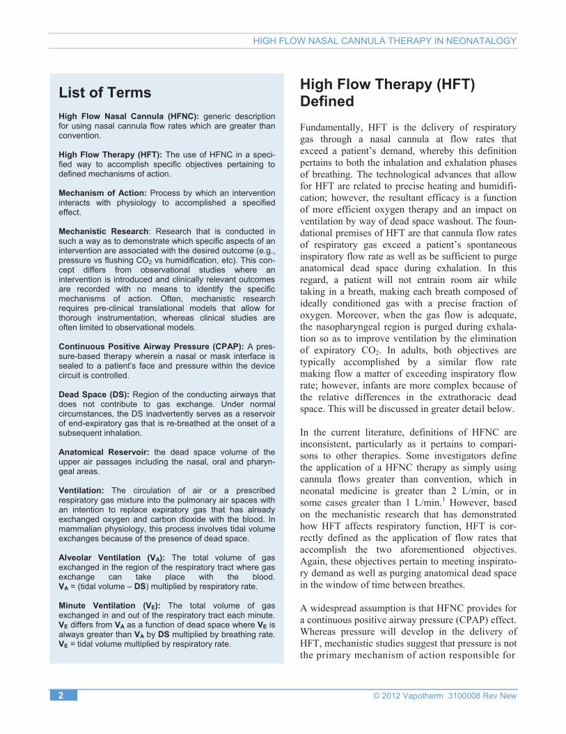
List of Terms High Flow Nasal Cannula (HFNC): generic description for using nasal cannula flow rates which are greater than convention.
High Flow Therapy (HFT): The use of HFNC in a speci-fied way to accomplish specific objectives pertaining to defined mechanisms of action.
Mechanism of Action: Process by which an intervention interacts with physiology to accomplished a specified effect.
Mechanistic Research: Research that is conducted in such a way as to demonstrate which specific aspects of an intervention are associated with the desired outcome (e.g., pressure vs flushing CO2 vs humidification, etc). This con-cept differs from observational studies where an intervention is introduced and clinically relevant outcomes are recorded with no means to identify the specific mechanisms of action. Often, mechanistic research requires pre-clinical translational models that allow for thorough instrumentation, whereas clinical studies are often limited to observational models.
Continuous Positive Airway Pressure (CPAP): A pres-sure-based therapy wherein a nasal or mask interface is sealed to a patient’s face and pressure within the device circuit is controlled.
Dead Space (DS): Region of the conducting airways that does not contribute to gas exchange. Under normal circumstances, the DS inadvertently serves as a reservoir of end-expiratory gas that is re-breathed at the onset of a subsequent inhalation.
Anatomical Reservoir: the dead space volume of the upper air passages including the nasal, oral and pharyn-geal areas.
Ventilation: The circulation of air or a prescribed respiratory gas mixture into the pulmonary air spaces with an intention to replace expiratory gas that has already exchanged oxygen and carbon dioxide with the blood. In mammalian physiology, this process involves tidal volume exchanges because of the presence of dead space.
Alveolar Ventilation (VA): The total volume of gas exchanged in the region of the respiratory tract where gas exchange can take place with the blood. VA = (tidal volume – DS) multiplied by respiratory rate.
Minute Ventilation (VE): The total volume of gas exchanged in and out of the respiratory tract each minute. VE differs from VA as a function of dead space where VE is always greater than VA by DS multiplied by breathing rate. VE = tidal volume multiplied by respiratory rate.
High Flow Therapy (HFT)Defined Fundamentally, HFT is the delivery of respiratory gas through a nasal cannula at flow rates that exceed a patient’s demand, whereby this definition pertains to both the inhalation and exhalation phases of breathing. The technological advances that allow for HFT are related to precise heating and humidifi-cation; however, the resultant efficacy is a function of more efficient oxygen therapy and an impact on ventilation by way of dead space washout. The foun-dational premises of HFT are that cannula flow rates of respiratory gas exceed a patient’s spontaneous inspiratory flow rate as well as be sufficient to purge anatomical dead space during exhalation. In this regard, a patient will not entrain room air while taking in a breath, making each breath composed of ideally conditioned gas with a precise fraction of oxygen. Moreover, when the gas flow is adequate, the nasopharyngeal region is purged during exhala-tion so as to improve ventilation by the elimination of expiratory CO2. In adults, both objectives are typically accomplished by a similar flow rate making flow a matter of exceeding inspiratory flow rate; however, infants are more complex because of the relative differences in the extrathoracic dead space. This will be discussed in greater detail below. In the current literature, definitions of HFNC are inconsistent, particularly as it pertains to compari-sons to other therapies. Some investigators define the application of a HFNC therapy as simply using cannula flows greater than convention, which in neonatal medicine is greater than 2 L/min, or in some cases greater than 1 L/min.1 However, based on the mechanistic research that has demonstrated how HFT affects respiratory function, HFT is cor-rectly defined as the application of flow rates that accomplish the two aforementioned objectives. Again, these objectives pertain to meeting inspirato-ry demand as well as purging anatomical dead space in the window of time between breathes. A widespread assumption is that HFNC provides for a continuous positive airway pressure (CPAP) effect. Whereas pressure will develop in the delivery of HFT, mechanistic studies suggest that pressure is not the primary mechanism of action responsible for

HIGH FLOW NASAL CANNULA THERAPY IN NEONATALOGY
© 2012 Vapotherm 3100008 Rev New 3
Figure 1. During inhalation (Top) the cannula flow needs to be adequate to meet inspiratory flow demand.
During exhalation (Bottom) the cannula flow needs to be adequate to purge the nasopharyngeal dead space volume between breaths.
observed physiologic outcomes. A more detailed comparison of HFT to CPAP is found in a later section of this paper. An example of why we need agreement on the definition of HFT is the 2011 Cochrane review on the use of “High flow nasal cannula for respiratory support in preterm infants.”1 These authors reviewed four studies and concluded that high flow nasal cannula may result in a higher rate of reintubation compared to CPAP. However, these reviews defined HFNC as flow rates greater than 1 L/Min, which may not exceed every infant’s inspiratory flow demand and certainly would not likely be sufficient to purge nasopharyngeal dead space during exhalation. The evidence cited to support that CPAP outperforms HFNC comes from the one study by Campbell and colleagues.2 These authors administered HFNC as if it were a CPAP therapy, and used an equation to assign flow rates. Specifically, this equation was proposed to predict flow needed to achieve a certain airway pressure, and as such, the mean cannula flow rate used in this study was only 1.8 L/min. It is fair to conclude from these data, as well as years of experience with nasal cannulae, that flow at rates less than 2 L/min may not be as effective as CPAP. Howev-er, this finding has little relevance to true high flow nasal cannula therapy (i.e. HFT) which is defined by the mechanistic literature to facilitate purging of the entire volume of nasal, oral and pharyngeal dead space. In this regard, the findings of Campbell and colleagues should not be unexpected and should not be used to represent the efficacy of HFT per its mechanistic definition.
Multiple Mechanisms of Action There are a number of mechanisms by which HFT can improve respiratory function in patients, including the neonatal population. In subsequent sections of this paper, the principle mechanisms of action are described in detail within the context of therapeutic application. Here, these mechanisms are briefly introduced to note the complex, multifactorial impact of HFT. Essentially, HFT makes the nasopharyngeal region a reservoir of fresh gas by way of purging the end-expiratory gas from this space during exhalation.

HIGH FLOW NASAL CANNULA THERAPY IN NEONATALOGY
4 © 2012 Vapotherm 3100008 Rev New
Therefore, the patient’s subsequent breath is more efficient in that it is composed of more fresh gas and less end-expiratory gas. With this improvement in efficiency, a patient can achieve adequate alveolar ventilation (VA) with less minute ventilation (VE), compared to pressure therapies that force greater lung expansion to achieve greater VE. Vapotherm recommends that HFT should not be used to produce a substantial distending airway pressure, although some pressure inevitably is generated. Rather, HFT should be used so as to minimize resistance to gas exhausting from the nasopharynx around the cannula and through the mouth. In other words, HFT should be used to maximize the purging of the nasopharynx with the least amount of flow and associated pres-sure. A recent publication validating the dead space washout concept as the principal mechanism of action showed that the least occlusive cannula geo-metry resulted in an optimal efficacy with less than 75% of the flow and pressure required when snug fitting prongs are used to generate distending pres-sure.3 Additional studies have shown how flow dynamics and heated humidification contribute to other mechanisms of action that reduce work of breathing and support airway function. These other mechanisms are summarized below and described in a review paper by Dysart et al.4
HFT in the Context of Current Practices in NeonatalRespiratory Care Since the 1980’s, there has been a focus on develop-ing strategies for noninvasive ventilation subsequent to the defining of bronchopulmonary dysplasia (BPD), the relationship to lung bio-inflammatory potential and the recognition of the need for lung protective ventilation strategies. Along these lines, there has been a major emphasis on CPAP and other noninvasive forms of ventilation, such as bilevel CPAP, that have reduced the need for mechanical ventilation.12 Other major developments have sur-faced in the last few decades, such as exogenous surfactant replacement therapy and inhaled nitric oxide, which have been widely adopted and used in conjunction with noninvasive respiratory support. For example, the INSURE technique (INtubate, SURfactant, Extubate) has allowed surfactant deli-very to be combined with noninvasive ventilation with notable success.13 Together these combinations of therapies have fostered tremendous improvements in infant mortality, but occurrence of BPD remains high.
High Flow Therapy Mechanisms of Action
MECHANISM DESCRIPTION
Dead space washout Reduce dead space making minute ventilation more efficient.3, 5
Reduce inspiratory work of breathing Exceed inspiratory flow thus eliminating nasal resistance.6
Improved lung mechanics Warmed, humidified gas has been shown to improve conductance, compliance and lung elasticity.7
Eliminate metabolic work associated with gas conditioning
Attenuates the energy and water loss associated with conditioning inspiratory gas.
Provision of mild distending pressure Flow can be restricted such as to provide positive distending pressure for lung recruitment.8-10
Improve secretion mobilization Ideal humidification of the inspired gas has been shown to restore mucocilliary function and reduce symptoms of airway exacerbations.11

HIGH FLOW NASAL CANNULA THERAPY IN NEONATALOGY
© 2012 Vapotherm 3100008 Rev New 5
In the context of this push for noninvasive ventila-tion strategies, dead space elimination and thus HFT is not a novel concept. Dead space elimination contributes to improved alveolar ventilation without forcing greater tidal volumes. In this regard, we need to reinforce that the term ventilation should not necessarily be synonymous with artificial breathing machines that deliver tidal breathes, but can encom-pass other, less invasive ways to facilitate exchange of respiratory gases within the lungs. Optimal gas conditioning capabilities has allowed for gas deli-very by nasal cannula to exceed the conventional limits without degradation of the nasal tissues.14 This advancement has opened the door for a noninvasive way to eliminate anatomical dead space, making ventilation more efficient. HFT, as we term the use of HFNC in a specified way so as to maximize the elimination anatomical dead space, has many peripheral advantages that are associated with the patient interface being easier to manage than a sealed CPAP system. These include patient tolerance, ease in nursing management, and accessibility for kangaroo care, as well as physiolog-ic concerns such as prone positioning to support spontaneous breathing.15, 16 As we better define and optimize HFT as primarily a therapy to eliminate dead space, and understand the coinciding ability to generate mild pressure and hydrate the air passages, HFT holds promise to emerge as a significant advancement in neonatal respiratory support.
HFT: A Unique Noninvasive Respiratory Support Modality The act of ventilation refers to the circulation of air so as to replace stale or noxious air with fresh air. In mammalian physiology this process involves tidal volumes and lung compliance because of our ana-tomical dead space. In other words, if we were to remove dead space entirely by putting our alveolar surface on the outside of our body (e.g. gills on a fish), we would not need to have tidal volume excur-sions to expose the alveolar surface to adequate VA in support of respiration. Obviously, this is not prac-tical for numerous reasons, including the need to condition gas before coming into contact with the blood, and our adaptation to use dead space for re-taining CO2 as our innate pH buffering mechanism.
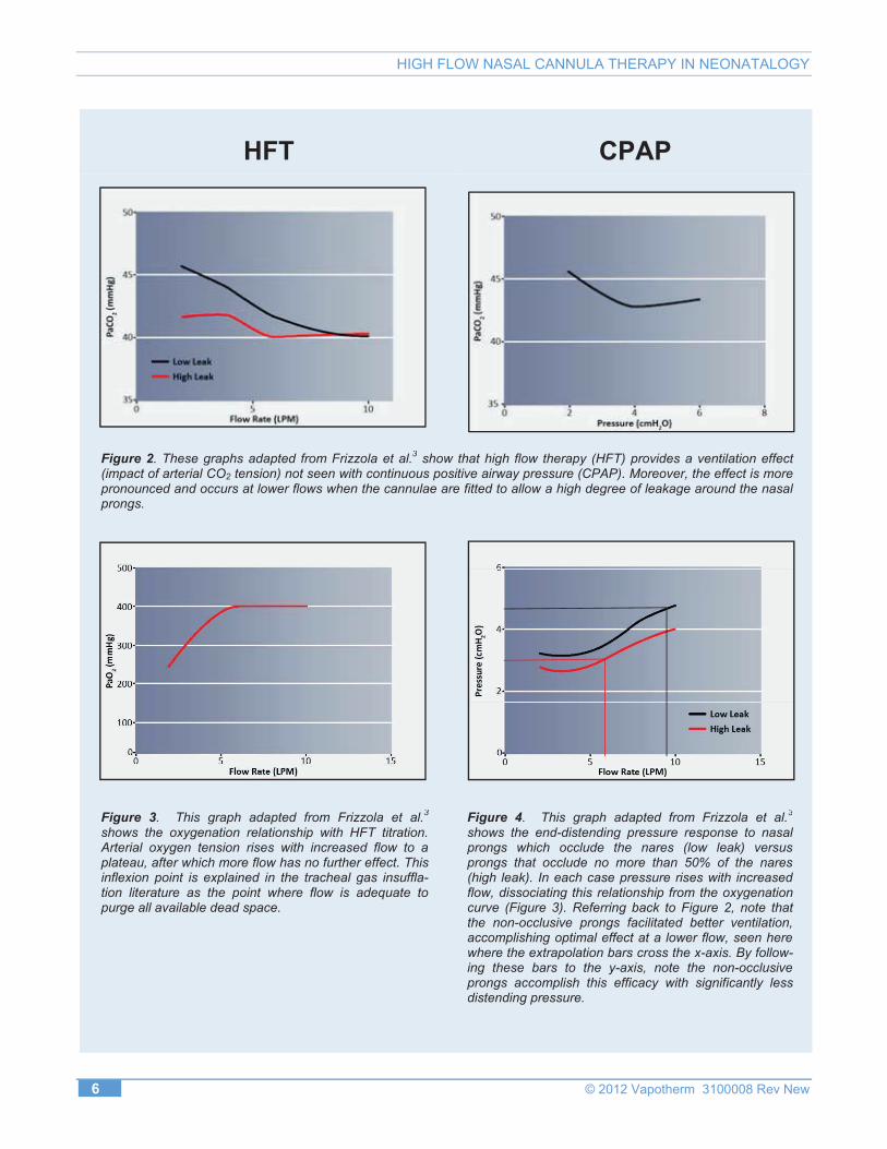
Nonetheless, by reducing dead space we can reduce the VE needed to accomplish adequate VA and there-fore reduce work of breathing. Dead space elimination tactics have been used for years in the form of tracheal gas insufflation17, 18 and transtra-cheal oxygen delivery.19 In the last 10 or more years, advancements in heated humidification devices have made it possible to accomplish ventilation by way of dead space elimination with a nasal cannula. Translational research has shown that the primary mechanism of action for HFT is purging anatomical dead space, thus achieving VA with lesser VE. A pivotal mechanistic study was done using neonatal piglets with a severe respiratory distress induced by central venous oleic acid delivery.3 In this model, three conditions were compared: HFT with a low leak around the prongs (i.e. snug fit in the nares), HFT where no more than 50% of the nares were occluded (i.e. non-occlusive prongs) and conven-tional mask CPAP. The low leak condition was created to mimic the situations where clinicians try to get a CPAP effect, whereas the ≤50% occlusion condition fits our recommendation for the applica-tion of HFT. Under these conditions, the model evaluated titration of flow/CPAP pressure on CO2 removal, oxygenation and pressure development. As shown in Figure 2, under both HFT conditions, arterial CO2 inversely correlated with flow rate wherein arterial CO2 tension (PaCO2) in these spon-taneous breathers could be reduced back to pre-injury levels. Moreover, the PaCO2 in the <50% oc-clusion condition was significantly reduced at lower flow rates compared to the low leak condition, indi-cating that a less occlusive prong design facilitates nasopharyngeal purge. CPAP alone was never able to achieve this ventilation effect. With CPAP, Pa-CO2 was slightly reduced with a mild pressure increase, but then PaCO2 rose as CPAP pressure went above 4 cmH2O, presumably due to over-distension. As shown in Figure 3, regarding oxygenation, under both HFT conditions a flow dependent increase in arterial oxygen tension (PaO2) was demonstrated until a plateau was reached. This saturation pattern is indicative of dead space washout and fits the hypo-thesis of the study based on the background modeling of tracheal gas insufflation.20 The concept
HIGH FLOW NASAL CANNULA THERAPY IN NEONATALOGY
6 © 2012 Vapotherm 3100008 Rev New
HFT CPAP
Figure 2. These graphs adapted from Frizzola et al.3 show that high flow therapy (HFT) provides a ventilation effect (impact of arterial CO2 tension) not seen with continuous positive airway pressure (CPAP). Moreover, the effect is more pronounced and occurs at lower flows when the cannulae are fitted to allow a high degree of leakage around the nasal prongs.
Figure 3. This graph adapted from Frizzola et al.3
shows the oxygenation relationship with HFT titration. Arterial oxygen tension rises with increased flow to a plateau, after which more flow has no further effect. This inflexion point is explained in the tracheal gas insuffla-tion literature as the point where flow is adequate to purge all available dead space.
Figure 4. This graph adapted from Frizzola et al.3
shows the end-distending pressure response to nasal prongs which occlude the nares (low leak) versus prongs that occlude no more than 50% of the nares (high leak). In each case pressure rises with increased flow, dissociating this relationship from the oxygenation curve (Figure 3). Referring back to Figure 2, note that the non-occlusive prongs facilitated better ventilation, accomplishing optimal effect at a lower flow, seen here where the extrapolation bars cross the x-axis. By follow-ing these bars to the y-axis, note the non-occlusive prongs accomplish this efficacy with significantly less distending pressure.

HIGH FLOW NASAL CANNULA THERAPY IN NEONATALOGY
© 2012 Vapotherm 3100008 Rev New 7
behind dead space purge techniques is that there is a finite amount of time (late stage exhalation and end-expiratory pause) to purge the space and a finite amount of dead space volume that can be purged. As flow is increased, more of the volume can be purged until flow is sufficient to purge all of the volume in the allotted time, after which additional flow pro-duces no additional effect. With respect to oxygenation, CPAP was as effective as HFT al-though not a function of pressure titration. Pressure in this study was measured by direct per-pendicular placement of a pressure catheter in the trachea through an anterior cervical cut-down. As shown in Figure 4, the pressure data from this study shows a direct relationship between flow and base-line pressure shift which is in agreement with the clinical studies. Here the pressure from the low leak condition is always greater than the ≤50% occlusion condition. Importantly, there was dissociation be-tween oxygenation and the pressure response where pressure continues to rise beyond the flow rate at which oxygenation response reaches a plateau. This dissociation between pressure and physiologic oxy-genation response supports dead space flush as the
Less occlusive prongs achievedmaximal efficacy with only 60% of the flow
needed for occlusive prongs, and abouthalf the inadvertent distending pressure.
Optimized prong fit produces betteroutcome with less pressure.
primary mechanism of action. Moreover, because the cannula fit impacted the flow rate needed to ac-complish optimal efficacy (i.e. flow rate where PaO2 plateaued and PaCO2 reached baseline levels), pres-sure was actually inversely related to physiologic improvement if we consider cannula design as a ca-tegorical variable. In other words, the less occlusive prong design accomplished maximal efficacy with approximately 60% of the flow needed to do so with the occlusive prong design, which translates to ap-proximately one-half of the inadvertent distending pressure. Optimized prong fit translates to better outcome with less pressure.
The clinical side to this translational modeling was done in COPD patients (data presented at the 2011 CHEST meeting and in review for publication). Adults were examined because they can be compliant in ways that an infant cannot, but the resulting evidence regarding ventilation is funda-mental to the concept of dead space and translates to the infant as well. This study shows that HFT with room air results in at least a 13% reduction in VE while maintaining the same PaCO2 compared to both no support and supplemental oxygen conditions. As discussed later, this ventilation effect is potentially greater in infants because of the greater relative extrathoracic dead space volume compared to adults.21
CPAP versus HFT CPAP systems are specifically designed to be a closed system in conjunction with the infant’s respi-ratory tract. The proposed mechanisms of action for CPAP are complex and multifactorial, but include the concept that pressure is able to recruit lung alveoli by increasing FRC, thus improving com-pliance so that a greater VE can be achieved to account for the necessary VA.
22 From a mechanical perspective, CPAP supports spontaneous breathing by making it less taxing to stretch the lung and by minimizing atelactrauma during lung stretch. HFT, on the other hand, is aimed at achieving VA with a lesser VE so as to reduce the necessary lung stretch. Nonetheless, the accompanying humidification and mild pressure effects with HFT would attenuate ate-lectrauma as well.7, 23 HFT is designed to be an open system, wherein the gas is not intended to be contained for the develop-ment of a pressurized patient airway. In an HFT system, pressure inside the device circuit is by necessity quite high, in the range of nearly 400 cmH2O.24 This is the result of pushing high flow though the substantial resistance of the relatively tiny nasal prong orifices. Because of this relatively enormous cannula resistance and the fact that the system circuit is not sealed with the patient’s airway, physics dictates that circuit pressure does not trans-mit to the patient. The development of patient airway pressure as a coinciding effect during HFT is

HIGH FLOW NASAL CANNULA THERAPY IN NEONATALOGY
8 © 2012 Vapotherm 3100008 Rev New
a function of the resistance to the flow exiting from the patient’s nasopharynx through the oral cavity and nose. To keep the coinciding nasal pressure from reaching levels that would need to be monitored, the literature dictates that a cannulae should not occlude more than 50% of the nares. This recommendation is based on the work of Dr. Locke and colleagues who showed that nasal prongs having an outside diameter that is no more than 50% of the internal diameter of the nares does not result in distending pressure during low flow O2 therapy. Conversely, cannula having an outside diameter that was three-quarters of the inside nare diameter resulted in significant pres-sure at low flows. The message here is that keeping nares open by 50% of the diameter represents ade-quate anatomic release. Note that this 50% diameter rule ensures that the surface area of the unconcluded region of the nares is greater than the surface area of the occluded area, based on the nonlinear, direct re-lationship between surface area and distance from the center of a circle. Vapotherm’s recommendations and cannulae offerings are consistent with this requirement. When applied correctly, mild airway pressure does develop during HFT and is considered a mechanism of action based on the rationale for CPAP.22 This pressure is a function of both the rate of flow through the patient’s upper air space and the anatom-ical resistance to this flow as it passes through the anatomy,25 however, the pressure is not at the level of closed CPAP system and varies regionally as a function of the gas flow patterns (preliminary data). From a review of the research related to airway pressures in neonates during HFT, data shows that airway pressure with HFT can be expected to be less than or approximately equivalent to airway pressure when a CPAP of 6 cmH2O is applied,8-10, 23, 24, 26 and equally as variable as airway pressure during CPAP.24 In interpreting these data it is important recognize that some investigators were trying to create CPAP by minimizing the leak through the nose and mouth. Nonetheless, the data showed only modest pressures.
Application of HFT in the NICU: Flow Rate Titration and Rationale Despite the inconsistency in the literature defining the flow rates needed for HFT, when used aggres-sively reports indicate improved extubation success and potentially a reduction in intubation rates.27, 28 In addition, the simplicity of the cannula interface with loose fitting nasal prongs reduces facial skin and nasal abrasions associated with more intense thera-pies. HFT is simple to administer and manage compared to positive airway pressure therapies that require intense monitoring to ensure that the patient interface remains properly placed. The range of flows to be used in infants is between 1-8 L/min. While infants have a very small tidal volume, in the range of 4-6 mL/kg, their respiratory rates are quite high. In sick children, respiratory rates can approach 100 breaths per minute, making peak inspiratory flows very high relative to minute volumes. Another consideration with infants, which pertains to the mechanisms of dead space purge, is the relative size of the anatomical reservoir which consists of the extra-thoracic dead space volume of the nasal, oral and pharyngeal cavities. Infants have a much larger anatomical reservoir compared to old-er children and adults.21 Small infants have an extrathoracic dead space volume around 2.3 mL/kg, whereas in children over six years of age and into adulthood this value drops to approximately 0.8 mL/kg. Therefore, as compared to an adult, an infant may need greater relative flow rates to realize the full benefits of purging the anatomical reservoir in the window of opportunity between breaths (flow rates that go beyond simply meeting inspiratory demand). This three-fold greater anatomical reser-voir volume in small infants translates to dead space making up a much greater fraction of their tidal volume as compared to larger children and adults. As a result of these factors, small infants have a greater propensity to benefit from HFT in that these patients are much more sensitive to changes in dead space. However, cannula flow rates needed to max-imize efficacy typically begin at greater than 3 L/min.

HIGH FLOW NASAL CANNULA THERAPY IN NEONATALOGY
© 2012 Vapotherm 3100008 Rev New 9
Cannula Research in Optimiz-ing HFT: Fluid Dynamics and Flow Patterns With an understanding that the mechanisms of action are based on creating an internal reservoir of conditioned gas, work has been done to refine the patient interface to optimize this effect. Some of the work that is currently underway involves using computation fluid dynamics modeling to learn more about gas flow characteristics in the nasopharynx with HFT. Using this model, we have already con-firmed what is suggested by animal data, that a less occlusive prong design allows for more rapid purge of the nasal cavity at any flow rate. Therefore, as we saw in the animal data, the nasopharynx can be purged in the time between breaths with a lesser flow rate when cannula design is optimized; in this case smaller prong diameter (data being prepared for publication).
Another topic addressed using the computational fluid dynamics modeling pertains to sheer force (or strain rate) on the walls of the nasopharynx as a result of the gas flow velocity from the cannula nozzle (commonly referred to as “jetting effect”). With his model we learned that the strain rate is absorbed between laminae of the gas, and with a smaller cannula dissipates before impacting the wall.
However, with the larger cannula, the strain rate impacts the wall just by nature of its closer proximi-ty (data being prepared for publication). Thus, the larger cannula is more likely to result in a jetting effect. To put this concept in another way, this “jet-ting effect” is often described as similar to turning a fire hose on a wall; however, this analogy is very
With the larger cannula, the strain rateimpacts the wall just by nature of its closer proximity. Thus, the larger cannula is more
likely to result in a jetting effect.
much incorrect because it involves jetting one medium (water) though another less dense medium (air). In the case of cannula gas flow jetting, air is jetting through air, and thus a more appropriate analogy would be similar to water jets that are under water such as in a hot tub. In this analogy, you can probably imagine that you would only experience significant strain if you were to hold your hand di-rectly on or around the water jet.
Summary HFT is a unique noninvasive respiratory support modality in the NICU. It is based on the concepts of dead space elimination for breathing efficiency and the delivery of ideally conditioned respiratory gases to an already fragile lung. A misconception that stifles the adaptation of HFT is that it is an uncon-trolled form of CPAP. The mechanistic literature, however, does not support this presumption and a significant amount of clinical data suggests that pressure is not a concern when HFT is applied correctly. Importantly, the neonatal community would benefit from the uniform adaptation of a definition that is based on research and guides the cannula design aspects and flow requirement. These studies suggest that cannula fit should not occlude more than 50% of the nares and that flows should be between 3 and 8 L/min.
Figure 5. This image from computational fluid dynamics modeling shows the patterns of gas flow through the nasopharynx from the cannula. Note the vortices and varied directionality of flow. These patterns define pressure and other forces through-out the cavity.

HIGH FLOW NASAL CANNULA THERAPY IN NEONATALOGY
10 © 2012 Vapotherm 3100008 Rev New
References
1. Wilkinson D, Andersen C, O'Donnell CP, De Paoli AG. High flow nasal cannula for respiratory support in preterm infants. Cochrane Database Syst Rev 2011:CD006405.
2. Campbell DM, Shah PS, Shah V, Kelly EN. Nasal continuous positive airway pressure from high flow cannula versus Infant Flow for Preterm infants. J Perinatol 2006;26:546-9.
3. Frizzola M, Miller TL, Rodriguez ME, et al. High-flow nasal cannula: Impact on oxygenation and ventilation in an acute lung injury model. Pediatr Pulmonol 2011;46:67-74.
4. Dysart K, Miller TL, Wolfson MR, Shaffer TH. Research in high flow therapy: mechanisms of action. Respir Med 2009;103:1400-5.
5. Dewan NA, Bell CW. Effect of low flow and high flow oxygen delivery on exercise tolerance and sensation of dyspnea. A study comparing the transtracheal catheter and nasal prongs. Chest 1994;105:1061-5.
6. Shepard JW, Jr., Burger CD. Nasal and oral flow-volume loops in normal subjects and patients with obstructive sleep apnea. Am Rev Respir Dis 1990;142:1288-93.
7. Greenspan JS, Wolfson MR, Shaffer TH. Airway responsiveness to low inspired gas temperature in preterm neonates. J Pediatr 1991;118:443-5.
8. Saslow JG, Aghai ZH, Nakhla TA, et al. Work of breathing using high-flow nasal cannula in preterm in-fants. J Perinatol 2006;26:476-80.
9. Spence KL, Murphy D, Kilian C, McGonigle R, Kilani RA. High-flow nasal cannula as a device to provide continuous positive airway pressure in infants. J Perinatol 2007;27:772-5.
10. Wilkinson DJ, Andersen CC, Smith K, Holberton J. Pharyngeal pressure with high-flow nasal cannulae in premature infants. J Perinatol 2008;28:42-7.
11. Hasani A, Chapman TH, McCool D, Smith RE, Dilworth JP, Agnew JE. Domiciliary humidification improves lung mucociliary clearance in patients with bronchiectasis. Chron Respir Dis 2008;5:81-6.
12. Mahmoud RA, Roehr CC, Schmalisch G. Current methods of non-invasive ventilatory support for neo-nates. Paediatr Respir Rev 2011;12:196-205.
13. Verder H, Robertson B, Greisen G, et al. Surfactant therapy and nasal continuous positive airway pres-sure for newborns with respiratory distress syndrome. Danish-Swedish Multicenter Study Group. N Engl J Med 1994;331:1051-5.
14. Woodhead DD, Lambert DK, Clark JM, Christensen RD. Comparing two methods of delivering high-flow gas therapy by nasal cannula following endotracheal extubation: a prospective, randomized, masked, crossover trial. J Perinatol 2006;26:481-5.
15. Martin RJ, Herrell N, Rubin D, Fanaroff A. Effect of supine and prone positions on arterial oxygen tension in the preterm infant. Pediatrics 1979;63:528-31.
16. Wolfson MR, Greenspan JS, Deoras KS, Allen JL, Shaffer TH. Effect of position on the mechanical interaction between the rib cage and abdomen in pre-term infants. J Appl Physiol 1992;72:1032-8.
17. Danan C, Dassieu G, Janaud JC, Brochard L. Efficacy of dead-space washout in mechanically venti-lated premature newborns. Am J Respir Crit Care Med 1996;153:1571-6.
18. Dassieu G, Brochard L, Agudze E, Patkai J, Janaud JC, Danan C. Continuous tracheal gas insufflation enables a volume reduction strategy in hyaline mem-brane disease: technical aspects and clinical results. Intensive Care Med 1998;24:1076-82.
19. Benditt J, Pollock M, Roa J, Celli B. Transtracheal delivery of gas decreases the oxygen cost of breath-ing. Am Rev Respir Dis 1993;147:1207-10.
20. Miller TL, Blackson TJ, Shaffer TH, Touch SM. Tra-cheal gas insufflation-augmented continuous positive airway pressure in a spontaneously breathing model of neonatal respiratory distress. Pediatr Pulmonol 2004;38:386-95.
21. Numa AH, Newth CJ. Anatomic dead space in infants and children. J Appl Physiol 1996;80:1485-9.
22. Morley C. Continuous distending pressure. Arch Dis Child Fetal Neonatal Ed 1999;81:F152-6.
23. Spentzas T, Minarik M, Patters AB, Vinson B, Stidham G. Children with respiratory distress treated with high-flow nasal cannula. J Intensive Care Med 2009;24:323-8.
24. Lampland AL, Plumm B, Meyers PA, Worwa CT, Mammel MC. Observational study of humidified high-flow nasal cannula compared with nasal continuous positive airway pressure. J Pediatr 2009;154:177-82.
25. Kahn DJ, Courtney SE, Steele AM, Habib RH. Unpredictability of Delivered Bubble Nasal Conti-nuous Positive Airway Pressure Role of Bias Flow Magnitude and Nares-Prong Air Leaks. Pediatr Res 2007;62:343-7.
26. Kubicka ZJ, Limauro J, Darnall RA. Heated, humidi-fied high-flow nasal cannula therapy: yet another way to deliver continuous positive airway pressure? Pediatrics 2008;121:82-8.
27. Holleman-Duray D, Kaupie D, Weiss MG. Heated humidified high-flow nasal cannula: use and a neo-natal early extubation protocol. J Perinatol 2007;27:776-81.
28. Shoemaker MT, Pierce MR, Yoder BA, DiGeronimo RJ. High flow nasal cannula versus nasal CPAP for neonatal respiratory disease: a retrospective study. J Perinatol 2007;27:85-91.