
High Efficiency, Homology-Directed Genome Editing in ... | INVESTIGATION High Efficiency,...
26
GENETICS | INVESTIGATION High Efficiency, Homology-Directed Genome Editing in Caenorhabditis elegans Using CRISPR-Cas9 Ribonucleoprotein Complexes Alexandre Paix, 1 Andrew Folkmann, Dominique Rasoloson, and Geraldine Seydoux Howard Hughes Medical Institute (HHMI) and Department of Molecular Biology and Genetics, Johns Hopkins University School of Medicine, Baltimore, Maryland 21205 ORCID IDs: 0000-0002-8080-7546 (A.P.); 0000-0003-2210-1569 (D.R.). ABSTRACT Homology-directed repair (HDR) of breaks induced by the RNA-programmed nuclease Cas9 has become a popular method for genome editing in several organisms. Most HDR protocols rely on plasmid-based expression of Cas9 and the gene-specific guide RNAs. Here we report that direct injection of in vitro–assembled Cas9-CRISPR RNA (crRNA) trans-activating crRNA (tracrRNA) ribonu- cleoprotein complexes into the gonad of Caenorhabditis elegans yields HDR edits at a high frequency. Building on our earlier finding that PCR fragments with 35-base homology are efficient repair templates, we developed an entirely cloning-free protocol for the generation of seamless HDR edits without selection. Combined with the co-CRISPR method, this protocol is sufficiently robust for use with low-efficiency guide RNAs and to generate complex edits, including ORF replacement and simultaneous tagging of two genes with fluorescent proteins. KEYWORDS CRISPR-Cas9; genome editing; homology-directed repair; ribonucleoprotein complexes; C. elegans T HE CRISPR-Cas9 system is a bacterial adaptive immune system that has been harnessed as a powerful genome editing tool (Doudna and Charpentier 2014). Cas9 is a nucle- ase that functions with two small RNAs: CRISPR RNA (crRNA), which guides Cas9 to complementary target se- quences, and trans-activating crRNA (tracrRNA), which binds to the crRNA and to Cas9 to form the ribonucleoprotein (RNP) complex (Deltcheva et al. 2011). For use in genome editing, the crRNA and tracrRNA are often combined into a single chimeric guide RNA (sgRNA) (Jinek et al. 2012). Expression of Cas9 and sgRNA in cells leads to cleavage of complementary genomic sequences. The double-strand breaks are repaired by endogenous cellular pathways, includ- ing end-joining mechanisms [e.g., nonhomologous end join- ing (NHEJ) and theta-mediated end joining (TMEJ)] and homology-dependent repair (HDR) mechanisms (van Schendel et al. 2015). End joining typically introduces random insertions/ deletions at the DNA break site, which can disrupt gene ac- tivity. HDR, in contrast, is a more precise repair process that uses a repair template. If the repair template contains edits flanked by sequences that are homologous to the cleavage site (homology arms), the edits will be incorporated by gene conversion. The high efficiency of Cas9 and the simplicity of guide RNA design have made it possible to develop end- joining protocols to systematically knock out genes (Hsu et al. 2014; Shah et al. 2015). In principle, scalable HDR protocols also could be used for systematic knock-ins of custom edits [such as green fluorescent protein (GFP)]. Unfortunately, current HDR protocols are inefficient (Hsu et al. 2014). First, most HDR protocols require cloning to create a repair tem- plate and a guide RNA expression vector for each gene to be targeted. Second, the efficiency of HDR typically is low, re- quiring the screening of large number of animals or the use of selection markers that are integrated alongside the desired edits (Dickinson et al. 2013, 2015). Recently, we found in Caenorhabditis elegans that linear DNAs with homology arms as short as 35 bases can support the efficient incorporation of HDR edits (Paix et al. 2014). This finding simplifies the construction of donor templates. Copyright © 2015 by the Genetics Society of America doi: 10.1534/genetics.115.179382 Manuscript received June 12, 2015; accepted for publication July 9, 2015; published Early Online July 17, 2015. Available freely online through the author-supported open access option. Supporting information is available online at www.genetics.org/lookup/suppl/ doi:10.1534/genetics.115.179382/-/DC1 1 Corresponding author: Department of Molecular Biology and Genetics, Johns Hopkins University School of Medicine, PCTB706, 725 N. Wolfe Street, Baltimore MD 21205. E-mail: [email protected] Genetics, Vol. 201, 47–54 September 2015 47
-
Upload
hoangkhanh -
Category
Documents
-
view
216 -
download
0
Transcript of High Efficiency, Homology-Directed Genome Editing in ... | INVESTIGATION High Efficiency,...
GENETICS | INVESTIGATION
High Efficiency, Homology-Directed Genome Editingin Caenorhabditis elegans Using CRISPR-Cas9
Ribonucleoprotein ComplexesAlexandre Paix,1 Andrew Folkmann, Dominique Rasoloson, and Geraldine Seydoux
Howard Hughes Medical Institute (HHMI) and Department of Molecular Biology and Genetics, Johns Hopkins University School ofMedicine, Baltimore, Maryland 21205
ORCID IDs: 0000-0002-8080-7546 (A.P.); 0000-0003-2210-1569 (D.R.).
ABSTRACT Homology-directed repair (HDR) of breaks induced by the RNA-programmed nuclease Cas9 has become a popular methodfor genome editing in several organisms. Most HDR protocols rely on plasmid-based expression of Cas9 and the gene-specific guideRNAs. Here we report that direct injection of in vitro–assembled Cas9-CRISPR RNA (crRNA) trans-activating crRNA (tracrRNA) ribonu-cleoprotein complexes into the gonad of Caenorhabditis elegans yields HDR edits at a high frequency. Building on our earlier findingthat PCR fragments with 35-base homology are efficient repair templates, we developed an entirely cloning-free protocol for thegeneration of seamless HDR edits without selection. Combined with the co-CRISPR method, this protocol is sufficiently robust for usewith low-efficiency guide RNAs and to generate complex edits, including ORF replacement and simultaneous tagging of two geneswith fluorescent proteins.
KEYWORDS CRISPR-Cas9; genome editing; homology-directed repair; ribonucleoprotein complexes; C. elegans
THE CRISPR-Cas9 system is a bacterial adaptive immunesystem that has been harnessed as a powerful genome
editing tool (Doudna and Charpentier 2014). Cas9 is a nucle-ase that functions with two small RNAs: CRISPR RNA(crRNA), which guides Cas9 to complementary target se-quences, and trans-activating crRNA (tracrRNA), whichbinds to the crRNA and to Cas9 to form the ribonucleoprotein(RNP) complex (Deltcheva et al. 2011). For use in genomeediting, the crRNA and tracrRNA are often combined intoa single chimeric guide RNA (sgRNA) (Jinek et al. 2012).Expression of Cas9 and sgRNA in cells leads to cleavageof complementary genomic sequences. The double-strandbreaks are repaired by endogenous cellular pathways, includ-ing end-joining mechanisms [e.g., nonhomologous end join-ing (NHEJ) and theta-mediated end joining (TMEJ)] and
homology-dependent repair (HDR) mechanisms (van Schendelet al. 2015). End joining typically introduces random insertions/deletions at the DNA break site, which can disrupt gene ac-tivity. HDR, in contrast, is a more precise repair process thatuses a repair template. If the repair template contains editsflanked by sequences that are homologous to the cleavagesite (homology arms), the edits will be incorporated by geneconversion. The high efficiency of Cas9 and the simplicityof guide RNA design have made it possible to develop end-joining protocols to systematically knock out genes (Hsu et al.2014; Shah et al. 2015). In principle, scalable HDR protocolsalso could be used for systematic knock-ins of custom edits[such as green fluorescent protein (GFP)]. Unfortunately,current HDR protocols are inefficient (Hsu et al. 2014). First,most HDR protocols require cloning to create a repair tem-plate and a guide RNA expression vector for each gene to betargeted. Second, the efficiency of HDR typically is low, re-quiring the screening of large number of animals or the use ofselection markers that are integrated alongside the desirededits (Dickinson et al. 2013, 2015).
Recently, we found in Caenorhabditis elegans that linearDNAs with homology arms as short as 35 bases can supportthe efficient incorporation of HDR edits (Paix et al. 2014).This finding simplifies the construction of donor templates.
Copyright © 2015 by the Genetics Society of Americadoi: 10.1534/genetics.115.179382Manuscript received June 12, 2015; accepted for publication July 9, 2015; publishedEarly Online July 17, 2015.Available freely online through the author-supported open access option.Supporting information is available online at www.genetics.org/lookup/suppl/doi:10.1534/genetics.115.179382/-/DC11Corresponding author: Department of Molecular Biology and Genetics, Johns HopkinsUniversity School of Medicine, PCTB706, 725 N. Wolfe Street, Baltimore MD 21205.E-mail: [email protected]
Genetics, Vol. 201, 47–54 September 2015 47
For example, a donor template to introduce GFP can be syn-thesized using two 55-base oligos, each containing 35 basesthat are homologous to the targeted locus and 20 bases thatare homologous to GFP. The PCR amplicon is injected along-side plasmids coding for Cas9 and the sgRNA into the gonadof adult hermaphrodites, and their progeny are screened forGFP expression by visual inspection or by PCR screening.Using this method, we were able to recover GFP fusions atsix of eight loci attempted. HDR frequencies were highenough to avoid the use of coselection makers but were stillrelatively low and variable (0.4–12%) (Paix et al. 2014).Other researchers also have reported difficulties in obtainingHDR edits, a problem that can be partially overcome byimprovements in the design of guide RNAs and repair tem-plates (Farboud and Meyer 2015; Katic et al. 2015).
Two additional factors are likely to also contribute to lowedit frequency. First, the efficiency of expressing and assem-bling Cas9–sgRNA complexes from plasmids is not knownand may be low given the tendency of C. elegans germ cellsto silence foreign DNA (Kelly et al. 1997). In mouse embryosand mammalian cells, direct delivery of Cas9 complexes as-sembled in vitro has been reported to yield a higher frequencyof edits (Lin et al. 2014; Aida et al. 2015; Liang et al. 2015).Injection of Cas9 complexes has been used in C. elegans tocreate edits by end-joining mechanisms (Cho et al. 2013) buthas not yet been compared directly to plasmid delivery orused for HDR.
A second factor that contributes to the lowedit frequency isthat many injected hermaphrodites generate no edits. Forreasons that remain unclear, we have found that edits tend tocluster among the progeny of a minority of injected mothers(jackpot broods) (Paix et al. 2014). Identification of jackpotbroods before screening would eliminate the need to screenthe broods of nonproductive hermaphrodites. Recently, threereports have described methods to enrich for desired edits byselecting or screening for editing at a second marker locus(Arribere et al. 2014; Kim et al. 2014; Ward 2015). In the
method described by Arribere et al. (2014), a dominant mu-tation is introduced by HDR in the marker locus dpy-10, lead-ing to a roller phenotype that is easily identified among theprogeny of injected hermaphrodites.
Here we report the development of a new direct-deliveryprotocol that combines injection of in vitro–synthesized and–assembled Cas9-crRNA-tracrRNA complexes with the dpy-10 co-CRISPR approach of Arribere et al. (2014) to identifyjackpot broods. The direct-delivery protocol is entirely cloning-free and generates edits at frequencies ranging from 2 to 70%of F1 progeny, a 10-fold improvement over our plasmid-basedearlier method (Paix et al. 2014). This new protocol permitsthe use of inefficient sgRNAs that failed in our previous studyand expands the repertoire of possible genome edits to in-clude ORF swaps and fluorescent protein (FP) tagging of twogenes at once.
Materials and Methods
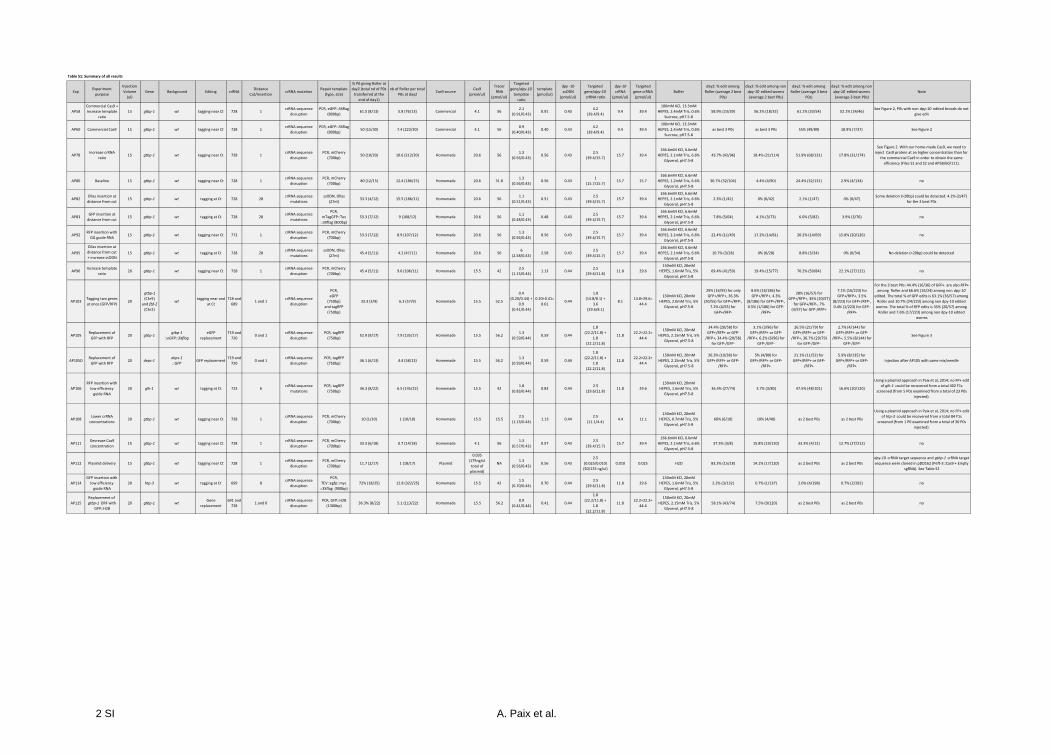
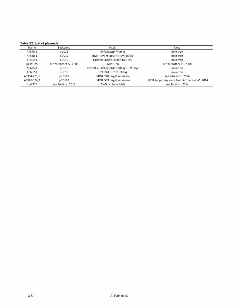
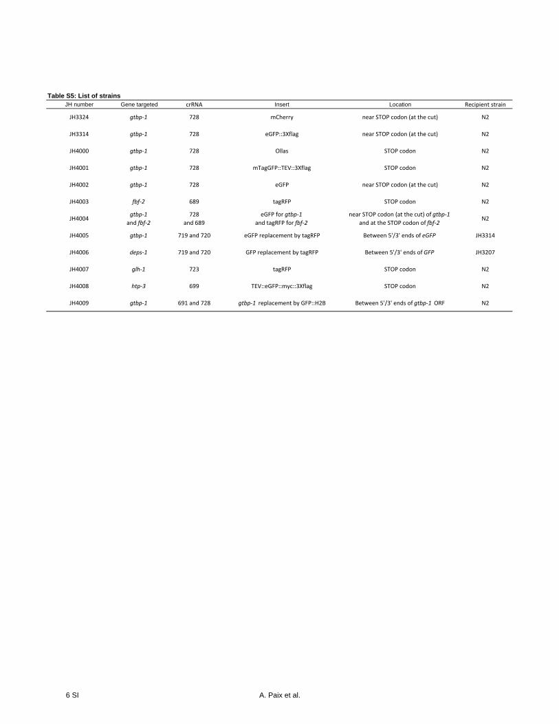
Supplementarymaterials include protocols for direct-deliveryediting (Supporting Information, File S1) and for Cas9 pro-tein purification (File S2), as well as lists of plasmids (TableS2), crRNAs (Table S3), rescue templates (Table S4), andstrains (Table S5).
Data availability
Strains and plasmids are available from the CaenorhabditisGenetics Center (CGC) and Addgene, or upon request.
Results and Discussion
Injection of Cas9 RNP complexes supports robust HDR
We developed a simple method to purify from Escherichia colirecombinant Cas9 [fused at its C-terminus with a nuclearlocalization sequence (Fu et al. 2014)] and assemble Cas9-crRNA-tracrRNA RNP complexes in the presence of repairtemplates and in a buffer suited for injection into C. elegans
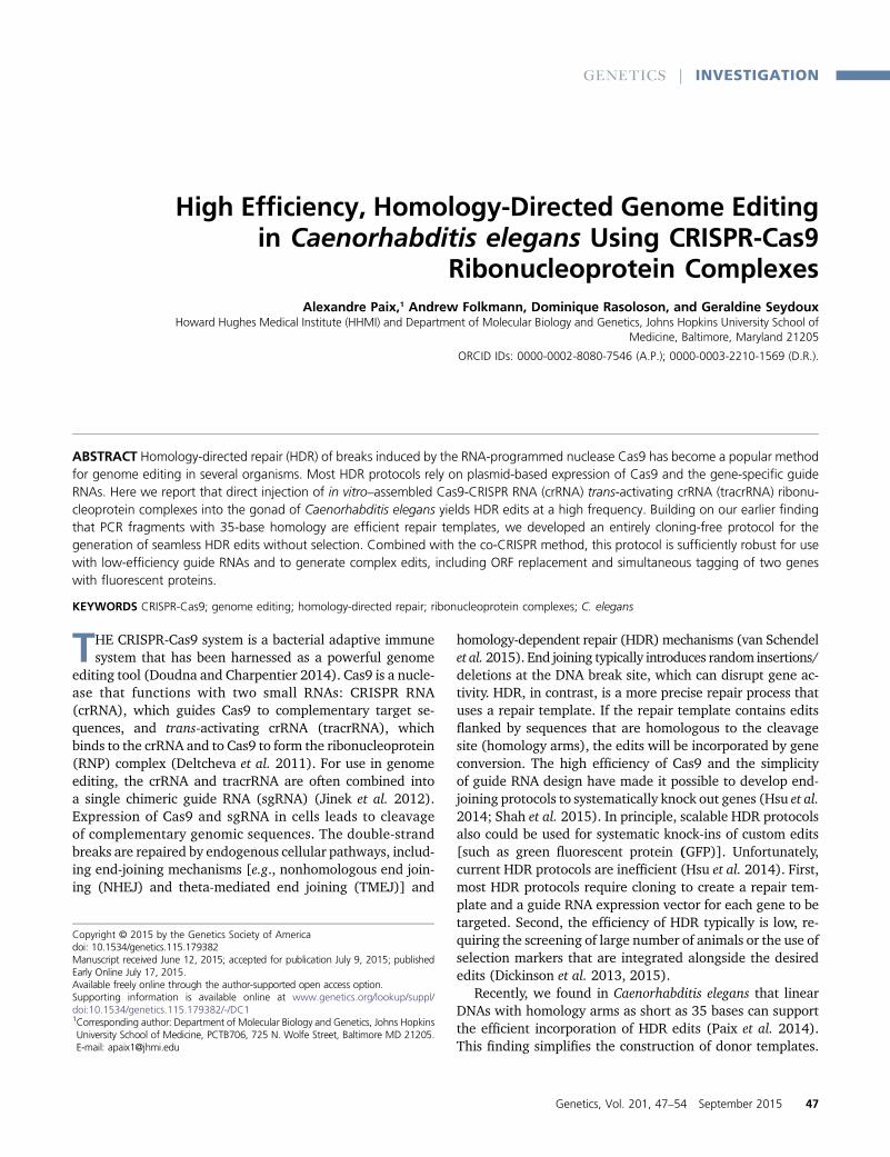
Figure 1 Co-CRISPR strategy. (A) We cotar-geted (1) the dpy-10 locus with a ssODN repairtemplate to introduce a missense mutationleading to the dominant roller phenotype(Arribere et al. 2014) and (2) a second locus(gene of interest) with a PCR repair templateto insert a fluorescent protein (FP) near theC-terminus. (B) Experimental outline. The gonadsof 10–20 hermaphrodites are injected, and theirbroods are examined for the presence of rollers(dpy-10 edits) and FP+ animals. In typical experi-ments, .50% of hermaphrodites segregate roll-ers. Jackpot broods are the broods with thehighest numbers of rollers. Edits at the gene ofinterest (pink) are found in both roller and non-roller worms, but only among broods that con-tain rollers.
48 A. Paix et al.
http://www.genetics.org/lookup/suppl/doi:10.1534/genetics.115.179382/-/DC1/genetics.115.179382-1.pdf
http://www.genetics.org/lookup/suppl/doi:10.1534/genetics.115.179382/-/DC1/genetics.115.179382-6.pdf
http://www.genetics.org/lookup/suppl/doi:10.1534/genetics.115.179382/-/DC1/genetics.115.179382-3.pdf
http://www.genetics.org/lookup/suppl/doi:10.1534/genetics.115.179382/-/DC1/genetics.115.179382-5.pdf
http://www.genetics.org/lookup/suppl/doi:10.1534/genetics.115.179382/-/DC1/genetics.115.179382-5.pdf
http://www.genetics.org/lookup/suppl/doi:10.1534/genetics.115.179382/-/DC1/genetics.115.179382-4.pdf
(File S1 and File S2). Cas9 also can be obtained from a com-mercial source, as in Cho et al. (2013). As in the co-CRISPRmethod of Arribere et al. (2014), we co-injected Cas9 com-plexes and repair templates targeting themarker gene dpy-10and a second target locus. dpy-10was repaired with a single-strand oligodeoxyribonucleotide (ssODN) that introducesa missense mutation in the dpy-10 ORF (Arribere et al.2014). The missense mutation causes a dominant roller phe-notype that is easily spotted under a dissecting microscope.For the second locus, we first used gtbp-1 (aka K08F4.2),a nonessential gene expressed in most tissues, which we tar-geted previously using plasmid delivery (Paix et al. 2014).We introduced a single cut near the C-terminus of gtbp-1 andrepaired the lesion with an �700-base double-stranded PCRfragment containing the ORF for a fluorescent protein [redfluorescent protein (RFP) or GFP] flanked by 35-base armsthat were homologous to sequences immediately surround-ing the cut site (Figure 1).
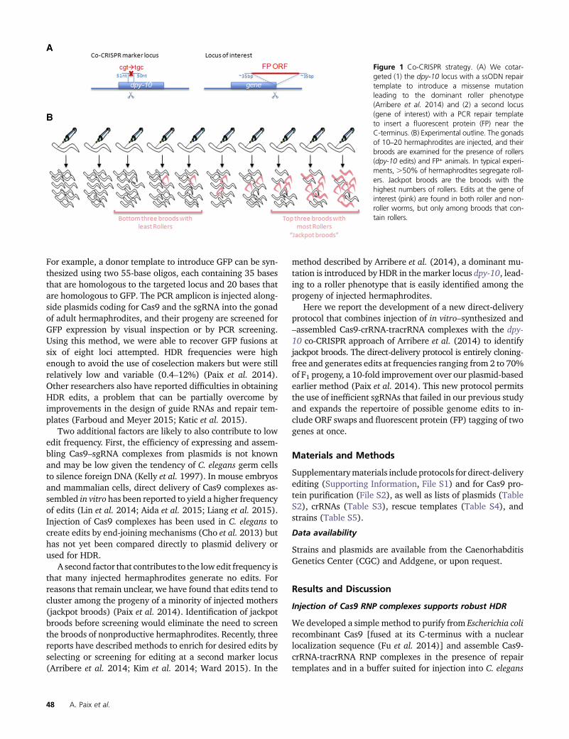
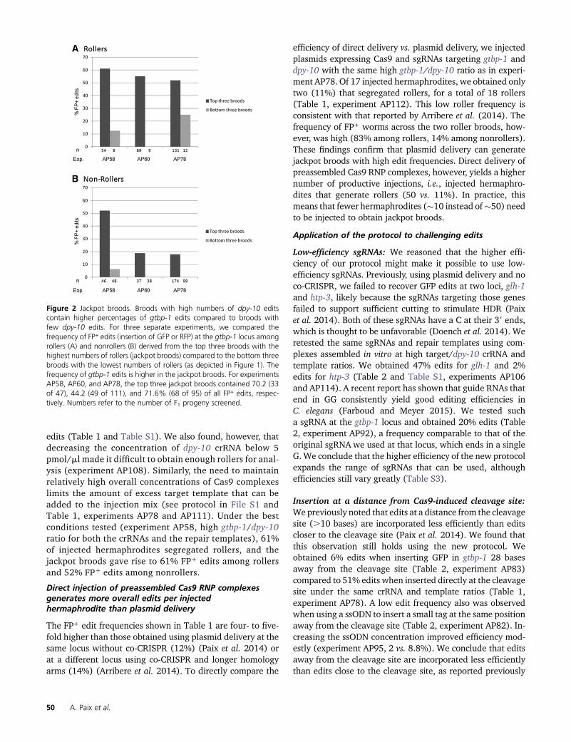
For three experiments (Table 1 and Table S1, experimentsAP58, AP60, and AP78), we examined the broods of all in-jected hermaphrodites by visual inspection for the presenceof roller and fluorescent (FP+) worms (Figure 2). We foundthat 50–61% of injected hermaphrodites segregated rollers(Table 1). Broods without rollers contained no FP+ edits(experiment AP58: 0 of 62 F1 progeny examined from fivebroods). Among broods with rollers, edits could be found inboth rollers and their nonroller siblings (Table 1). For eachexperiment, we found that the frequency of FP+ edits amongthe three broods that segregated the highest number of roll-ers was higher than that observed among the three broodswith the lowest numbers of rollers (Figure 2), as also report-ed by Arribere et al. (2014). For simplicity, in subsequentexperiments, we analyzed only the three broods with thehighest number of rollers (jackpot broods) and ignored allother broods. We conclude that direct delivery of Cas9 RNPcomplexes supports robust HDR and, like plasmid delivery,generates jackpot broods that are easily identified using thedpy-10 co-CRISPR marker (Arribere et al. 2014).
Increasing the target/marker ratio of crRNAs and repairtemplates increases HDR frequency at the target locus
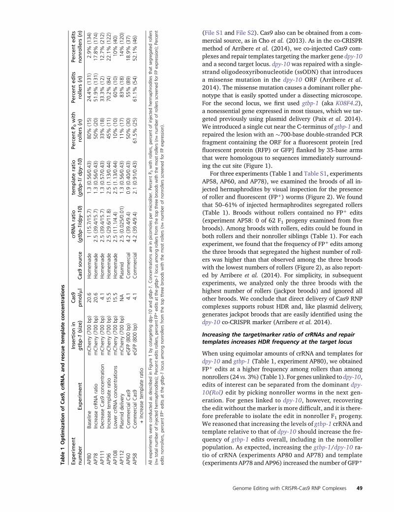
When using equimolar amounts of crRNA and templates fordpy-10 and gtbp-1 (Table 1, experiment AP80), we obtainedFP+ edits at a higher frequency among rollers than amongnonrollers (24 vs. 3%) (Table 1). For genes unlinked to dpy-10,edits of interest can be separated from the dominant dpy-10(Rol) edit by picking nonroller worms in the next gen-eration. For genes linked to dpy-10, however, recoveringthe edit without the marker is more difficult, and it is there-fore preferable to isolate the edit in nonroller F1 progeny.We reasoned that increasing the levels of gtbp-1 crRNA andtemplate relative to that of dpy-10 should increase the fre-quency of gtbp-1 edits overall, including in the nonrollerpopulation. As expected, increasing the gtbp-1/dpy-10 ra-tio of crRNA (experiments AP80 and AP78) and template(experiments AP78 and AP96) increased the number of GFP+Ta
ble
1OptimizationofCas9,
crRNA,an
drescuetemplate
concentrations
Experim
ent
number
Experim
ent
Insertionin
gtbp-1
(size)
Cas9
pmol/m
lCas9source
crRNA
ratio
(gtbp-1/dpy-10
)template
ratio
(gtbp-1/dpy-10
)Pe
rcen
tP 0
with
rolle
rs(n)
Percen
ted
its
rolle
rs(n)
Percen
ted
its
nonrolle
rs(n)
AP8
0Ba
seline
mChe
rry(700
bp)
20.6
Hom
emad
e1(15.7/15
.7)
1.3(0.56/0.43
)80
%(15)
24.4%
(131
)2.9%
(134
)AP7
8Increase
crRN
Aratio
mChe
rry(700
bp)
20.6
Hom
emad
e2.5(39.4/15
.7)
1.3(0.56/0.43
)50
%(20)
51.9%
(131
)17
.8%
(174
)AP1
11DecreaseCas9concen
tration
mChe
rry(700
bp)
4.1
Hom
emad
e2.5(39.4/15
.7)
1.3(0.57/0.43
)33
%(18)
33.3%
(12)
12.7%
(212
)AP9
6Increase
templateratio
mChe
rry(700
bp)
15.5
Hom
emad
e2.5(29.6/11
.8)
2.5(1.13/0.44
)45
%(11)
70.2%
(84)
22.1%
(122
)AP1
08Lower
crRN
Aconcen
trations
mChe
rry(700
bp)
15.5
Hom
emad
e2.5(11.1/4.4)
2.5(1.13/0.44
)10
%(10)
60%
(10)
10%
(40)
AP1
12Plasmid
delivery
mChe
rry(700
bp)
NA
Plasmid
2.5(0.025
/0.01)
1.3(0.56/0.43
)11
%(17)
83%
(18)
14%
(120
)AP6
0Com
mercial
Cas9
eGFP
(800
bp)
4.1
Com
mercial
4.2(39.4/9.4)
0.9(0.40/0.43
)50
%(30)
55%
(89)
18.9%
(37)
AP5
8Com
mercial
Cas9
+Increase
templateratio
eGFP
(800
bp)
4.1
Com
mercial
4.2(39.4/9.4)
2.1(0.91/0.43
)61
.5%
(25)
61.1%
(54)
52.1%
(46)
Allexpe
rimen
tswerecond
uctedas
describ
edin
Figu
re1by
cotargetingdp
y-10
andgtbp
-1.Con
centratio
nsarein
picomoles
permicroliter.Percen
tP 0
with
rollers,pe
rcen
tof
injected
herm
aphrod
itesthat
segreg
ated
rollers
(n=totaln
umbe
rof
injected
herm
aphrod
ites);Percen
ted
itsrollers,pe
rcen
tFP
+ed
itsat
thegtbp
-1locusam
ongrollers
from
thetopthreebroo
dswith
themostrollers
(n=nu
mbe
rof
rollers
screen
edforFP
expression
);Percen
ted
itsno
nrollers,pe
rcen
tFP
+ed
itsat
thegtbp
-1locusam
ongno
nrollers
from
thetopthreebroo
dswith
themostrollers
(n=nu
mbe
rof
nonrollers
screen
edforFP
expression
).
Genome Editing with CRISPR-Cas9 RNP Complexes 49
http://www.genetics.org/lookup/suppl/doi:10.1534/genetics.115.179382/-/DC1/genetics.115.179382-6.pdf
http://www.genetics.org/lookup/suppl/doi:10.1534/genetics.115.179382/-/DC1/genetics.115.179382-3.pdf
edits (Table 1 and Table S1). We also found, however, thatdecreasing the concentration of dpy-10 crRNA below 5pmol/ml made it difficult to obtain enough rollers for anal-ysis (experiment AP108). Similarly, the need to maintainrelatively high overall concentrations of Cas9 complexeslimits the amount of excess target template that can beadded to the injection mix (see protocol in File S1 andTable 1, experiments AP78 and AP111). Under the bestconditions tested (experiment AP58, high gtbp-1/dpy-10ratio for both the crRNAs and the repair templates), 61%of injected hermaphrodites segregated rollers, and thejackpot broods gave rise to 61% FP+ edits among rollersand 52% FP+ edits among nonrollers.
Direct injection of preassembled Cas9 RNP complexesgenerates more overall edits per injectedhermaphrodite than plasmid delivery
The FP+ edit frequencies shown in Table 1 are four- to five-fold higher than those obtained using plasmid delivery at thesame locus without co-CRISPR (12%) (Paix et al. 2014) orat a different locus using co-CRISPR and longer homologyarms (14%) (Arribere et al. 2014). To directly compare the
efficiency of direct delivery vs. plasmid delivery, we injectedplasmids expressing Cas9 and sgRNAs targeting gtbp-1 anddpy-10 with the same high gtbp-1/dpy-10 ratio as in experi-ment AP78. Of 17 injected hermaphrodites, we obtained onlytwo (11%) that segregated rollers, for a total of 18 rollers(Table 1, experiment AP112). This low roller frequency isconsistent with that reported by Arribere et al. (2014). Thefrequency of FP+ worms across the two roller broods, how-ever, was high (83% among rollers, 14% among nonrollers).These findings confirm that plasmid delivery can generatejackpot broods with high edit frequencies. Direct delivery ofpreassembled Cas9 RNP complexes, however, yields a highernumber of productive injections, i.e., injected hermaphro-dites that generate rollers (50 vs. 11%). In practice, thismeans that fewer hermaphrodites (�10 instead of�50) needto be injected to obtain jackpot broods.
Application of the protocol to challenging edits
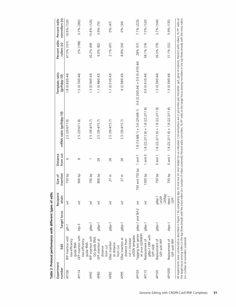
Low-efficiency sgRNAs: We reasoned that the higher effi-ciency of our protocol might make it possible to use low-efficiency sgRNAs. Previously, using plasmid delivery and noco-CRISPR, we failed to recover GFP edits at two loci, glh-1and htp-3, likely because the sgRNAs targeting those genesfailed to support sufficient cutting to stimulate HDR (Paixet al. 2014). Both of these sgRNAs have a C at their 39 ends,which is thought to be unfavorable (Doench et al. 2014). Weretested the same sgRNAs and repair templates using com-plexes assembled in vitro at high target/dpy-10 crRNA andtemplate ratios. We obtained 47% edits for glh-1 and 2%edits for htp-3 (Table 2 and Table S1, experiments AP106and AP114). A recent report has shown that guide RNAs thatend in GG consistently yield good editing efficiencies inC. elegans (Farboud and Meyer 2015). We tested sucha sgRNA at the gtbp-1 locus and obtained 20% edits (Table2, experiment AP92), a frequency comparable to that of theoriginal sgRNA we used at that locus, which ends in a singleG. We conclude that the higher efficiency of the new protocolexpands the range of sgRNAs that can be used, althoughefficiencies still vary greatly (Table S3).
Insertion at a distance from Cas9-induced cleavage site:Wepreviously noted that edits at a distance from the cleavagesite (.10 bases) are incorporated less efficiently than editscloser to the cleavage site (Paix et al. 2014). We found thatthis observation still holds using the new protocol. Weobtained 6% edits when inserting GFP in gtbp-1 28 basesaway from the cleavage site (Table 2, experiment AP83)compared to 51% edits when inserted directly at the cleavagesite under the same crRNA and template ratios (Table 1,experiment AP78). A low edit frequency also was observedwhen using a ssODN to insert a small tag at the same positionaway from the cleavage site (Table 2, experiment AP82). In-creasing the ssODN concentration improved efficiency mod-estly (experiment AP95, 2 vs. 8.8%). We conclude that editsaway from the cleavage site are incorporated less efficientlythan edits close to the cleavage site, as reported previously
Figure 2 Jackpot broods. Broods with high numbers of dpy-10 editscontain higher percentages of gtbp-1 edits compared to broods withfew dpy-10 edits. For three separate experiments, we compared thefrequency of FP+ edits (insertion of GFP or RFP) at the gtbp-1 locus amongrollers (A) and nonrollers (B) derived from the top three broods with thehighest numbers of rollers (jackpot broods) compared to the bottom threebroods with the lowest numbers of rollers (as depicted in Figure 1). Thefrequency of gtbp-1 edits is higher in the jackpot broods. For experimentsAP58, AP60, and AP78, the top three jackpot broods contained 70.2 (33of 47), 44.2 (49 of 111), and 71.6% (68 of 95) of all FP+ edits, respec-tively. Numbers refer to the number of F1 progeny screened.
50 A. Paix et al.
http://www.genetics.org/lookup/suppl/doi:10.1534/genetics.115.179382/-/DC1/genetics.115.179382-8.xls
http://www.genetics.org/lookup/suppl/doi:10.1534/genetics.115.179382/-/DC1/genetics.115.179382-6.pdf
http://www.genetics.org/lookup/suppl/doi:10.1534/genetics.115.179382/-/DC1/genetics.115.179382-8.xls
Table
2Protoco
lperform
ance
withdifferenttypes
ofed
its
Experim
ent
number
Edit
Target
locu
sRecipient
strain
Size
of
insertion
Distance
from
cut
crRNA
ratio(gof/dpy-10
)Te
mplate
ratio
(gof/dpy-10
)Pe
rcen
ted
its
rolle
rs(n)
Percen
ted
its
nonrolle
rs(n)
AP1
06RFPinsertionwith
low-efficien
cygu
ideRN
A
glh-1
wt
750bp
62.5(29.6/11
.8)
1.8(0.83/0.44
)47
.5%
(101
)16
.6%
(120
)
AP1
14GFP
insertionwith
low-efficien
cygu
ideRN
A
htp-3
wt
900bp
82.5(29.6/11
.8)
1.5(0.70/0.44
)2%
(198
)0.7%
(282
)
AP9
2RFPinsertionwith
GG
guideRN
Agtbp
-1wt
700bp
12.5(39.4/15
.7)
1.3(0.56/0.43
)20
.2%
(69)
15.8%
(126
)
AP8
3GFP
insertionat
distan
cefrom
cut
gtbp
-1wt
800bp
282.5(39.4/15
.7)
1.1(0.48/0.43
)6.0%
(82)
3.9%
(76)
AP8
2Ollasinsertion
atdistan
cefrom
cut
gtbp
-1wt
27nt
282.5(39.4/15
.7)
1.1(0.51/0.43
)2.1%
(47)
0%(47)
AP9
5Ollasinsertionat
distan
cefrom
cut+increa
sessODNtemplate
gtbp
-1wt
27nt
282.5(39.4/15
.7)
6(2.58/0.43
)8.8%
(34)
0%(34)
AP1
03Ta
ggingtw
oge
nes
aton
ce(GFP/RFP)
gtbp
-1an
dfbf-2
wt
750an
d75
0bp
1an
d1
1.8(14.8/8.1)
+3.6(29.6/8.1)
0.4(0.20/0.44
)+0.9(0.41/0.44
)28
%(57)
7.1%
(223
)
AP1
15Re
placem
entof
gtbp
-1ORF
with
GFP::H
2B
gtbp
-1wt
1300
bp1an
d0
1.8(22.2/11
.8)+1.8(22.2/11
.8)
0.9(0.41/0.44
)58
.1%
(74)
7.5%
(120
)
AP1
05Re
placem
entof
GFP
with
RFP
gtbp
-1gtbp
-175
0bp
0an
d1
1.8(22.2/11
.8)+1.8(22.2/11
.8)
1.3(0.59/0.44
)26
.5%
(79)
2.7%
(144
)::e
GFP
::3Xflag
AP1
05D
Replacem
entof
GFP
with
RFP
deps-1
deps-1
750bp
0an
d1
1.8(22.2/11
.8)+1.8(22.2/11
.8)
1.3(0.59/0.44
)21
.1%
(52)
5.9%
(135
)::G
FP
Allexpe
rimen
tswerecond
uctedas
describ
edin
Figu
re1by
cotargetingdp
y-10
andon
e(ortw
o)target
locias
indicated.
Con
centratio
nsarein
picomoles
permicroliter.go
f,ge
neof
interest;Percen
ted
itsrollers,%
FP+ed
itsat
thetarget
locusam
ongrollersin
thetopthreebroo
dswith
themostrollers(n=nu
mbe
rof
rollersscreen
ed);Percen
ted
itsno
nrollers:%
FP+ed
itsat
thetarget
locusam
ongno
nrollersin
thetopthreebroo
dswith
themostrollers
(n=nu
mbe
rof
nonrollers
screen
ed).
Genome Editing with CRISPR-Cas9 RNP Complexes 51
(Arribere et al. 2014; Paix et al. 2014). This inefficiency canbe compensated partially by increasing the template concen-tration in the injection mix.
Tagging two genes at once: We tested whether the newprotocol could be used to recover edits at two target locisimultaneously (in addition to the marker locus dpy-10). Wetargeted gtbp-1 and fbf-2 for GFP and RFP insertion, respec-tively. We obtained 28%GFP/RFP double edits among rollersand 7% GFP/RFP double edits among nonrollers (Table 2,experiment AP103). The frequency of GFP edits overall(63%) exceeded that of RFP edits (35%) almost by a factorof 2 (Table S1), possibly as a result of higher efficiency of thesgRNA targeting gtbp-1 compared to fbf-2, as also seen whenthese sgRNAs were used separately (Paix et al. 2014). Weconclude that protein delivery can support robust HDR atthree loci simultaneously (including dpy-10) even when us-ing sgRNAs with different editing efficiencies.
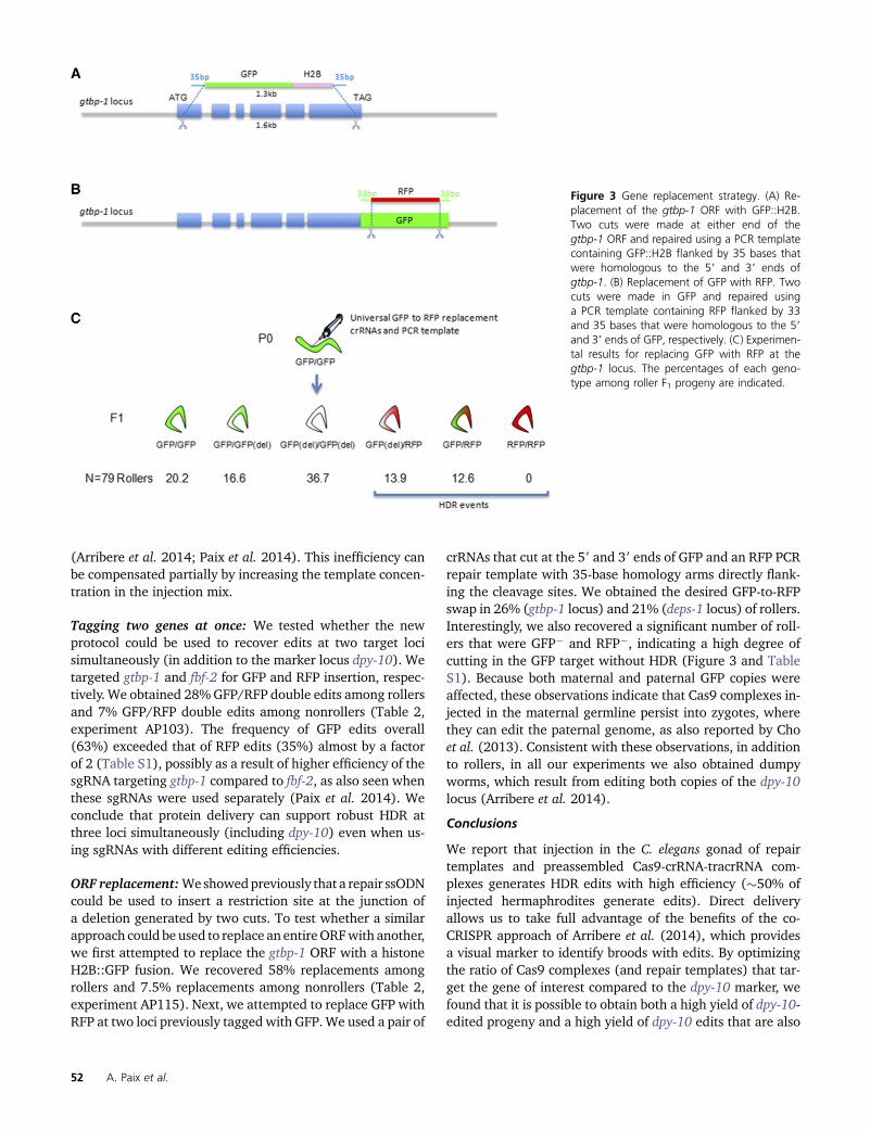
ORF replacement:Weshowedpreviously that a repair ssODNcould be used to insert a restriction site at the junction ofa deletion generated by two cuts. To test whether a similarapproach could be used to replace an entireORFwith another,we first attempted to replace the gtbp-1 ORF with a histoneH2B::GFP fusion. We recovered 58% replacements amongrollers and 7.5% replacements among nonrollers (Table 2,experiment AP115). Next, we attempted to replace GFP withRFP at two loci previously tagged with GFP.We used a pair of
crRNAs that cut at the 59 and 39 ends of GFP and an RFP PCRrepair template with 35-base homology arms directly flank-ing the cleavage sites. We obtained the desired GFP-to-RFPswap in 26% (gtbp-1 locus) and 21% (deps-1 locus) of rollers.Interestingly, we also recovered a significant number of roll-ers that were GFP2 and RFP2, indicating a high degree ofcutting in the GFP target without HDR (Figure 3 and TableS1). Because both maternal and paternal GFP copies wereaffected, these observations indicate that Cas9 complexes in-jected in the maternal germline persist into zygotes, wherethey can edit the paternal genome, as also reported by Choet al. (2013). Consistent with these observations, in additionto rollers, in all our experiments we also obtained dumpyworms, which result from editing both copies of the dpy-10locus (Arribere et al. 2014).
Conclusions
We report that injection in the C. elegans gonad of repairtemplates and preassembled Cas9-crRNA-tracrRNA com-plexes generates HDR edits with high efficiency (�50% ofinjected hermaphrodites generate edits). Direct deliveryallows us to take full advantage of the benefits of the co-CRISPR approach of Arribere et al. (2014), which providesa visual marker to identify broods with edits. By optimizingthe ratio of Cas9 complexes (and repair templates) that tar-get the gene of interest compared to the dpy-10 marker, wefound that it is possible to obtain both a high yield of dpy-10-edited progeny and a high yield of dpy-10 edits that are also
Figure 3 Gene replacement strategy. (A) Re-placement of the gtbp-1 ORF with GFP::H2B.Two cuts were made at either end of thegtbp-1 ORF and repaired using a PCR templatecontaining GFP::H2B flanked by 35 bases thatwere homologous to the 59 and 39 ends ofgtbp-1. (B) Replacement of GFP with RFP. Twocuts were made in GFP and repaired usinga PCR template containing RFP flanked by 33and 35 bases that were homologous to the 59and 39 ends of GFP, respectively. (C) Experimen-tal results for replacing GFP with RFP at thegtbp-1 locus. The percentages of each geno-type among roller F1 progeny are indicated.
52 A. Paix et al.
http://www.genetics.org/lookup/suppl/doi:10.1534/genetics.115.179382/-/DC1/genetics.115.179382-8.xls
http://www.genetics.org/lookup/suppl/doi:10.1534/genetics.115.179382/-/DC1/genetics.115.179382-8.xls
edited at the gene of interest (coedits, as high as 70%). Amongbroods that segregate the highest numbers of dpy-10 edits(jackpot broods), edits at the gene of interest also can befound in non-dpy-10-edited siblings (as high as 52%). Theability to recover edits in unmarked animals is particularlyuseful for linked loci that cannot be separated easily fromthe dominant dpy-10 marker in the next generation. Ourfindings also demonstrate that the co-CRISPR strategyworks efficiently even when using different types of tem-plates to repair the marker locus (ssODN to generate a pointmutation) and the gene of interest (PCR amplicon to inserta fluorescent protein). The efficiency of editing, however,still remains variable from locus to locus, likely owing todifferences in sgRNA efficiency and possibly differences inlocus competency for HDR. We also continue to find thatedits at a distance from the Cas9 cleavage site (.10 bases)are incorporated less efficiently than edits closer to thecleavage site, as reported previously (Arribere et al. 2014;Paix et al. 2014). Although protospacer adjacent motifs(PAMs) can be found within 30 bases of most edit sites,in cases where no PAMs are available, it may be preferableto use templates with longer homology arms and selectionmarkers, as in Dickinson et al. (2015).
Theefficiencyofourdirect-deliveryprotocol translates intoseveral practical advantages: workload is reduced at both theinjection (10–20 injected hermaphrodites vs. 50) and screen-ing (,50 F1 progeny compared to hundreds) steps, low-efficiency sgRNAs can be used, and complex edits, includinggene replacements and multigene edits, are possible. Re-placement of an ORF with GFP, as we report here for thegtbp-1 locus, is an effective method to generate marked nullalleles that can be maintained using the linked fluorescentmarker. We have also developed universal crRNAs and rescuetemplates to convert GFP fusions to RFP fusions (File S1).Another advantage of direct delivery is that the entire pro-tocol requires no cloning: tracrRNA, crRNAs, and ssODN re-pair templates for short edits (,50 bases) are all synthesizedchemically, and longer repair templates are made by PCRusing oligonucleotides to code for the short (35-base) homol-ogy arms. In principle, direct delivery of Cas9 RNP complexescould be combined with other CRISPR protocols (Dickinsonet al. 2015; Ward 2015) and also should be advantageousfor nematodes where promoters to drive expression of Cas9and the sgRNAs are not readily available (Chiu et al. 2013;Witte et al. 2015).
Acknowledgments
We thank Mike Nonet and Andy Fire for the Cas9::NLS::Hisbacterial expression plasmid and members of the SeydouxLaboratory for comments on the manuscript. Some strainswere provided by or deposited at the Caenorhabditis GeneticsCenter (CGC), which is funded by the National Institutesof Health Office of Research Infrastructure Programs (P40OD010440). We also thank WormBase for gene annota-tions and sequences. This work was supported by National
Institutes of Health grant R01HD37047. G.S. is an in-vestigator of the Howard Hughes Medical Institute.
Literature Cited
Aida, T., K. Chiyo, T. Usami, H. Ishikubo, R. Imahashi et al.,2015 Cloning-free CRISPR/Cas system facilitates functionalcassette knock-in in mice. Genome Biol. 16: 87.
Arribere, J. A., R. T. Bell, B. X. Fu, K. L. Artiles, P. S. Hartman et al.,2014 Efficient marker-free recovery of custom genetic modifi-cations with CRISPR/Cas9 in Caenorhabditis elegans. Genetics198: 837–846.
Chiu, H., H. T. Schwartz, I. Antoshechkin, and P. W. Sternberg,2013 Transgene-free genome editing in Caenorhabditis ele-gans using CRISPR-Cas. Genetics 195: 1167–1171.
Cho, S. W., J. Lee, D. Carroll, J. S. Kim, and J. Lee, 2013 Heritablegene knockout in Caenorhabditis elegans by direct injection ofCas9-sgRNA ribonucleoproteins. Genetics 195: 1177–1180.
Deltcheva, E., K. Chylinski, C. M. Sharma, K. Gonzales, Y. Chaoet al., 2011 CRISPR RNA maturation by trans-encoded smallRNA and host factor RNase III. Nature 471: 602–607.
Dickinson, D. J., J. D. Ward, D. J. Reiner, and B. Goldstein,2013 Engineering the Caenorhabditis elegans genome usingCas9-triggered homologous recombination. Nat. Methods 10:1028–1034.
Dickinson, D. J., A. M. Pani, J. K. Heppert, C. D. Higgins, and B.Goldstein, 2015 Streamlined genome engineering with a self-excising drug selection cassette. Genetics 115.178335.
Doench, J. G., E. Hartenian, D. B. Graham, Z. Tothova, M. Hegde et al.,2014 Rational design of highly active sgRNAs for CRISPR-Cas9-mediated gene inactivation. Nat. Biotechnol. 32: 1262–1267.
Doudna, J. A., and E. Charpentier, 2014 Genome editing. Thenew frontier of genome engineering with CRISPR-Cas9. Science346: 1258096.
Farboud, B., and B. J. Meyer, 2015 Dramatic enhancement ofgenome editing by CRISPR/Cas9 through improved guideRNA design. Genetics 199: 959–971.
Fu, B. X., L. L. Hansen, K. L. Artiles, M. L. Nonet, and A. Z. Fire,2014 Landscape of target:guide homology effects on Cas9-mediated cleavage. Nucleic Acids Res. 42: 13778–13787.
Hsu, P. D., E. S. Lander, and F. Zhang, 2014 Development andapplications of CRISPR-Cas9 for genome engineering. Cell 157:1262–1278.
Jinek, M., K. Chylinski, I. Fonfara, M. Hauer, J. A. Doudna et al.,2012 A programmable dual-RNA-guided DNA endonuclease inadaptive bacterial immunity. Science 337: 816–821.
Katic, I., L. Xu, and R. Ciosk, 2015 CRISPR/Cas9 genome editingin Caenorhabditis elegans: evaluation of templates for homology-mediated repair and knock-ins by homology-independent DNArepair. G3 115.019273.
Kelly, W. G., S. Xu, M. K. Montgomery, and A. Fire, 1997 Distinctrequirements for somatic and germline expression of a generallyexpressed Caernorhabditis elegans gene. Genetics 146: 227–238.
Kim, H., T. Ishidate, K. S. Ghanta, M. Seth, D. Conte, Jr. et al.,2014 A co-CRISPR strategy for efficient genome editing inCaenorhabditis elegans. Genetics 197: 1069–1080.
Liang, X., J. Potter, S. Kumar, Y. Zou, R. Quintanilla et al.,2015 Rapid and highly efficient mammalian cell engineeringvia Cas9 protein transfection. J. Biotechnol. 208: 44–53.
Lin, S., B. T. Staahl, R. K. Alla, and J. A. Doudna, 2014 Enhancedhomology-directed human genome engineering by controlledtiming of CRISPR/Cas9 delivery. eLife 3: e04766.
Paix, A., Y. Wang, H. E. Smith, C. Y. Lee, D. Calidas et al.,2014 Scalable and versatile genome editing using linear DNAswith microhomology to Cas9 Sites in Caenorhabditis elegans.Genetics 198: 1347–1356.
Genome Editing with CRISPR-Cas9 RNP Complexes 53

Shah, A. N., C. F. Davey, A. C. Whitebirch, A. C. Miller, and C. B.Moens, 2015 Rapid reverse genetic screening using CRISPR inzebrafish. Nat. Methods 12: 535–540.
van Schendel, R., S. F. Roerink, V. Portegijs, S. van den Heuvel, andM. Tijsterman, 2015 Polymerase theta is a key driver of ge-nome evolution and of CRISPR/Cas9-mediated mutagenesis.Nat. Commun. 6: 7394.
Ward, J. D., 2015 Rapid and precise engineering of the Caenor-habditis elegans genome with lethal mutation co-conversionand inactivation of NHEJ repair. Genetics 199: 363–377.
Witte, H., E. Moreno, C. Rodelsperger, J. Kim, J. S. Kim et al.,2015 Gene inactivation using the CRISPR/Cas9 system inthe nematode Pristionchus pacificus. Dev. Genes Evol. 225:55–62.
Communicating editor: O. Hobert
54 A. Paix et al.
GENETICSSupporting Information
www.genetics.org/lookup/suppl/doi:10.1534/genetics.115.179382/-/DC1
High Efficiency, Homology-Directed Genome Editingin Caenorhabditis elegans Using CRISPR-Cas9
Ribonucleoprotein ComplexesAlexandre Paix, Andrew Folkmann, Dominique Rasoloson, and Geraldine Seydoux
Copyright © 2015 by the Genetics Society of AmericaDOI: 10.1534/genetics.115.179382
ExpExperiment
purpose
Injection
Volume
(ul)
Gene Background Editing crRNADistance
Cut/InsertioncrRNA mutation
Repair template
(type, size)
% P0 giving Roller at
day2 (total nd of P0s
transferred at the
end of day1)
nb of Roller per total
P0s at day2Cas9 source
Cas9
(pmol/ul)
Tracer
RNA
(pmol/ul)
Targeted
gene/dpy‐10
template
ratio
template
(pmol/ul)
dpy ‐10
ssODN
(pmol/ul)
Targeted
gene/dpy‐10
crRNA ratio
dpy‐10
crRNA
(pmol/ul)
Targeted
gene crRNA
(pmol/ul)
Buffer
day2: % edit among
Roller (average 2 best
P0s)
day2: % edit among non
dpy‐10 edited worms
(average 2 best P0s)
day2: % edit among
Roller (average 3 best
P0s)
day2: % edit among non
dpy‐10 edited worms
(average 3 best P0s)
Note
AP58
Commercial Cas9 +
Increase template
ratio
15 gtbp‐1 wt tagging near Ct 728 1crRNA sequence
disruption
PCR, eGFP::3Xflag
(800bp)61.5 (8/13) 5.8 (76/13) Commercial 4.1 56
2.1
(0.91/0.43)0.91 0.43
4.2
(39.4/9.4)9.4 39.4
100mM KCl, 13.3mM
HEPES, 2.4mM Tris, 0.6%
Sucrose, pH7.5‐8
58.9% (23/39) 56.2% (18/32) 61.1% (33/54) 52.1% (24/46)See Figure 2, P0s with non dpy‐10 edited broods do not
give edit
AP60 Commercial Cas9 15 gtbp‐1 wt tagging near Ct 728 1crRNA sequence
disruption
PCR, eGFP::3Xflag
(800bp)50 (15/30) 7.4 (223/30) Commercial 4.1 56
0.9
(0.40/0.43)0.40 0.43
4.2
(39.4/9.4)9.4 39.4
100mM KCl, 13.3mM
HEPES, 2.4mM Tris, 0.6%
Sucrose, pH7.5‐8
as best 3 P0s as best 3 P0s 55% (49/89) 18.9% (7/37) See Figure 2
AP78Increase crRNA
ratio15 gtbp‐1 wt tagging near Ct 728 1
crRNA sequence
disruption
PCR, mCherry
(700bp)50 (10/20) 10.6 (212/20) Homemade 20.6 56
1.3
(0.56/0.43)0.56 0.43
2.5
(39.4/15.7)15.7 39.4
166.6mM KCl, 6.6mM
HEPES, 2.1mM Tris, 6.6%
Glycerol, pH7.5‐8
43.7% (42/96) 18.4% (21/114) 51.9% (68/131) 17.8% (31/174)
See Figure 2. With our home‐made Cas9, we need to
inject Cas9 protein at an higher concentration than for
the commercial Cas9 in order to obtain the same
efficiency (Files S1 and S2 and AP58/60/111).
AP80 Baseline 15 gtbp‐1 wt tagging near Ct 728 1crRNA sequence
disruption
PCR, mCherry
(700bp)80 (12/15) 12.4 (186/15) Homemade 20.6 31.8
1.3
(0.56/0.43)0.56 0.43
1
(15.7/15.7)15.7 15.7
166.6mM KCl, 6.6mM
HEPES, 1.2mM Tris, 6.6%
Glycerol, pH7.5‐8
30.7% (32/104) 4.4% (4/90) 24.4% (32/131) 2.9% (4/134) no
AP82Ollas insertion at
distance from cut15 gtbp‐1 wt tagging at Ct 728 28
crRNA sequence
mutations
ssODN, Ollas
(27nt)33.3 (4/12) 15.5 (186/12) Homemade 20.6 56
1.1
(0.51/0.43)0.51 0.43
2.5
(39.4/15.7)15.7 39.4
166.6mM KCl, 6.6mM
HEPES, 2.1mM Tris, 6.6%
Glycerol, pH7.5‐8
2.3% (1/42) 0% (0/42) 2.1% (1/47) 0% (0/47)Some deletion (>20bp) could be detected: 4.2% (2/47)
for the 3 best P0s
AP83GFP insertion at
distance from cut15 gtbp‐1 wt tagging at Ct 728 28
crRNA sequence
mutations
PCR,
mTagGFP::Tev
::3Xflag (800bp)
53.3 (7/12) 9 (108/12) Homemade 20.6 561.1
(0.48/0.43)0.48 0.43
2.5
(39.4/15.7)15.7 39.4
166.6mM KCl, 6.6mM
HEPES, 2.1mM Tris, 6.6%
Glycerol, pH7.5‐8
7.8% (5/64) 4.1% (3/73) 6.0% (5/82) 3.9% (3/76) no
AP92RFP insertion with
GG guide RNA15 gtbp‐1 wt tagging near Ct 772 1
crRNA sequence
disruption
PCR, mCherry
(700bp)53.3 (7/12) 8.9 (107/12) Homemade 20.6 56
1.3
(0.56/0.43)0.56 0.43
2.5
(39.4/15.7)15.7 39.4
166.6mM KCl, 6.6mM
HEPES, 2.1mM Tris, 6.6%
Glycerol, pH7.5‐8
22.4% (11/49) 17.2% (14/81) 20.2% (14/69) 15.8% (20/126) no
AP95
Ollas insertion at
distance from cut
+ increase ssODN
l
15 gtbp‐1 wt tagging at Ct 728 28crRNA sequence
mutations
ssODN, Ollas
(27nt)45.4 (5/11) 4.2 (47/11) Homemade 20.6 56
6
(2.58/0.43)2.58 0.43
2.5
(39.4/15.7)15.7 39.4
166.6mM KCl, 6.6mM
HEPES, 2.1mM Tris, 6.6%
Glycerol, pH7.5‐8
10.7% (3/28) 0% (0/28) 8.8% (3/34) 0% (0/34) No deletion (>20bp) could be detected
AP96Increase template
ratio20 gtbp‐1 wt tagging near Ct 728 1
crRNA sequence
disruption
PCR, mCherry
(700bp)45.4 (5/11) 9.6 (106/11) Homemade 15.5 42
2.5
(1.13/0.44)1.13 0.44
2.5
(29.6/11.8)11.8 29.6
150mM KCl, 20mM
HEPES, 1.6mM Tris, 5%
Glycerol, pH7.5‐8
69.4% (41/59) 19.4% (15/77) 70.2% (59/84) 22.1% (27/122) no
AP103Tagging two genes
at once (GFP/RFP)20
gtbp‐1
(Chr5)
and fbf‐2
(Chr2)
wttagging near and
at Ct
728 and
6891 and 1
crRNA sequence
disruption
PCR,
eGFP
(750bp)
and tagRFP
(750bp)
33.3 (3/9) 6.3 (57/9) Homemade 15.5 52.5
0.4
(0.20/0.44) +
0.9
(0.41/0.44)
0.20+0.41=
0.610.44
1.8
(14.8/8.1) +
3.6
(29.6/8.1)
8.114.8+29.6=
44.4
150mM KCl, 20mM
HEPES, 2.0mM Tris, 5%
Glycerol, pH7.5‐8
29% (16/55) for only
GFP+/RFP+, 36.3%
(20/55) for GFP+/RFP‐,
7.2% (4/55) for
GFP+/RFP‐
8.6% (16/186) for
GFP+/RFP+, 4.3%
(8/186) for GFP+/RFP‐,
0.5% (1/186) for GFP‐
/RFP+
28% (16/57) for
GFP+/RFP+, 35% (20/57)
for GFP+/RFP‐, 7%
(4/57) for GFP‐/RFP+
7.1% (16/223) for
GFP+/RFP+, 3.5%
(8/223) for GFP+/RFP‐,
0.4% (1/223) for GFP‐
/RFP+
For the 3 best P0s: 44.4% (16/36) of GFP+ are also RFP+
among Roller and 66.6% (16/24) among non dpy‐10
edited. The total % of GFP edits is 63.1% (36/57) among
Roller and 10.7% (24/223) among non dpy‐10 edited
worms. The total % of RFP edits is 35% (20/57) among
Roller and 7.6% (17/223) among non dpy‐10 edited
worms
AP105Replacement of
GFP with RFP20 gtbp‐1
gtbp‐1
::eGFP::3Xflag
eGFP
replacement
719 and
7200 and 1
crRNA sequence
disruption
PCR, tagRFP
(750bp)52.9 (9/17) 7.9 (135/17) Homemade 15.5 56.2
1.3
(0.59/0.44)0.59 0.44
1.8
(22.2/11.8) +
1.8
(22.2/11.8)
11.822.2+22.2=
44.4
150mM KCl, 20mM
HEPES, 2.15mM Tris, 5%
Glycerol, pH7.5‐8
34.4% (20/58) for
GFP+/RFP+ or GFP‐
/RFP+, 34.4% (20/58)
for GFP‐/GFP‐
3.1% (3/96) for
GFP+/RFP+ or GFP‐
/RFP+, 6.2% (6/96) for
GFP‐/GFP‐
26.5% (21/79) for
GFP+/RFP+ or GFP‐
/RFP+, 36.7% (29/79)
for GFP‐/GFP‐
2.7% (4/144) for
GFP+/RFP+ or GFP‐
/RFP+, 5.5% (8/144) for
GFP‐/GFP‐
See Figure 3
AP105DReplacement of
GFP with RFP20 deps‐1
deps‐1
::GFPGFP replacement
719 and
7200 and 1
crRNA sequence
disruption
PCR, tagRFP
(750bp)46.1 (6/13) 4.4 (58/13) Homemade 15.5 56.2
1.3
(0.59/0.44)0.59 0.44
1.8
(22.2/11.8) +
1.8
(22.2/11.8)
11.822.2+22.2=
44.4
150mM KCl, 20mM
HEPES, 2.15mM Tris, 5%
Glycerol, pH7.5‐8
26.3% (10/38) for
GFP+/RFP+ or GFP‐
/RFP+
5% (4/80) for
GFP+/RFP+ or GFP‐
/RFP+
21.1% (11/52) for
GFP+/RFP+ or GFP‐
/RFP+
5.9% (8/135) for
GFP+/RFP+ or GFP‐
/RFP+
Injection after AP105 with same mix/needle
AP106
RFP insertion with
low efficiency
guide RNA
20 glh‐1 wt tagging at Ct 723 6crRNA sequence
mutations
PCR, tagRFP
(750bp)36.3 (8/22) 6.5 (145/22) Homemade 15.5 42
1.8
(0.83/0.44)0.83 0.44
2.5
(29.6/11.8)11.8 29.6
150mM KCl, 20mM
HEPES, 1.6mM Tris, 5%
Glycerol, pH7.5‐8
36.4% (27/74) 3.7% (3/80) 47.5% (48/101) 16.6% (20/120)
Using a plasmid approach in Paix et al, 2014; no FP+ edit
of glh‐1 could be recovered from a total 402 F1s
screened (from 5 P0s examined from a total of 23 P0s
injected).
AP108Lower crRNA
concentrations20 gtbp‐1 wt tagging near Ct 728 1
crRNA sequence
disruption
PCR, mCherry
(700bp)10 (1/10) 1 (10/10) Homemade 15.5 15.5
2.5
(1.13/0.44)1.13 0.44
2.5
(11.1/4.4)4.4 11.1
150mM KCl, 20mM
HEPES, 0.7mM Tris, 5%
Glycerol, pH7.5‐8
60% (6/10) 10% (4/40) as 2 best P0s as 2 best P0s
Using a plasmid approach in Paix et al, 2014; no FP+ edit
of htp‐3 could be recovered from a total 84 F1s
screened (from 1 P0 examined from a total of 30 P0s
injected).
AP111Decrease Cas9
concentration15 gtbp‐1 wt tagging near Ct 728 1
crRNA sequence
disruption
PCR, mCherry
(700bp)33.3 (6/18) 0.7 (14/18) Homemade 4.1 56
1.3
(0.57/0.43)0.57 0.43
2.5
(39.4/15.7)15.7 39.4
166.6mM KCl, 6.6mM
HEPES, 2.1mM Tris, 6.6%
Glycerol, pH7.5‐8
37.5% (3/8) 15.8% (19/120) 33.3% (4/12) 12.7% (27/212) no
AP112 Plasmid delivery 15 gtbp‐1 wt tagging near Ct 728 1crRNA sequence
disruption
PCR, mCherry
(700bp)11.7 (2/17) 1 (18/17) Plasmid
0.035
(175ng/ul
total of
plasmid)
NA1.3
(0.56/0.43)0.56 0.43
2.5
(0.025/0.010)
(50/125 ng/ul)
0.010 0.025 H2O 83.3% (15/18) 14.1% (17/120) as 2 best P0s as 2 best P0s
dpy‐10 crRNA target sequence and gtbp‐1 crRNA target
sequence were cloned in pDD162 (Peft‐3::Cas9 + Empty
sgRNA). See Table S2
AP114
GFP insertion with
low efficiency
guide RNA
20 htp‐3 wt tagging at Ct 699 8crRNA sequence
disruption
PCR,
TEV::egfp::myc
::3Xflag (900bp)
72% (18/25) 12.8 (322/25) Homemade 15.5 421.5
(0.70/0.44)0.70 0.44
2.5
(29.6/11.8)11.8 29.6
150mM KCl, 20mM
HEPES, 1.6mM Tris, 5%
Glycerol, pH7.5‐8
2.2% (3/132) 0.7% (1/137) 2.0% (4/198) 0.7% (2/282) no
AP115
Replacement of
gtbp‐1 ORF with
GFP::H2B
20 gtbp‐1 wtGene
replacement
691 and
7281 and 0
crRNA sequence
disruption
PCR, GFP::H2B
(1300bp) 36.3% (8/22) 5.1 (113/22) Homemade 15.5 56.2
0.9
(0.41/0.44)0.41 0.44
1.8
(22.2/11.8) +
1.8
(22.2/11.8)
11.822.2+22.2=
44.4
150mM KCl, 20mM
HEPES, 2.15mM Tris, 5%
Glycerol, pH7.5‐8
58.1% (43/74) 7.5% (9/120) as 2 best P0s as 2 best P0s no
Table S1: Summary of all results
2 SI A. Paix et al.
Wendy
Typewritten Text
Also available for download as an Excel file at www.genetics.org/lookup/suppl/doi:10.1534/genetics.115.179382/-/DC1
Name Backbone Insert Note
AP575‐1 pUC19 3Xflag::tagRFP::myc no intron
AP588‐1 pUC19 myc::TEV::mTagGFP::TEV::3Xflag no intron
AP582‐1 pUC19 Ollas::mCherry::linker::H2B::V5 no intron
pCM1.35 see Merritt et al. 2008 GFP::H2B see Merritt et al. 2008
AP625‐1 pUC19 myc::TEV::3Xflag::eGFP::3Xflag::TEV::myc no intron
AP682‐1 pUC19 TEV::eGFP::myc::3Xflag no intron
AP334‐2/5/6 pDD162 crRNA 728 target sequence see Paix et al. 2014
AP568‐1/2/3 pDD162 crRNA 589 target sequence crRNA target sequence from Arribere et al. 2014
nm2973 see Fu et al. 2014 Cas9::NLSSV40::His6 see Fu et al. 2014
Table S2: List of plasmids
3 SI A. Paix et al.

crRNA
nameTarget gene Gene‐specific sequence Location % G/C
G at 3'end of the
crRNA
C at 3'end of the
crRNASens/Antisens Exp.
Efficiency of FP insertion (% Roller, 3
best P0s)
589 dpy‐10 gctaccataggcaccacgag in ORF 60 yes no AS All NA
728 gtbp‐1 ccacgaggtggtatgcgcag near STOP 65 yes no SAP80/78/111/96/108/60/5
8/83/82/95/103/11551.9% (AP78) and 70.2% (AP96)
723 glh‐1 tccctcaagatgaagaaggc near STOP 50 no yes S AP106 47.5%
699 htp‐3 agaggaaactgaacgatttc at STOP 40 no yes S AP114 2.0%
691 gtbp‐1 ggccttaacccagaataaga near ATG 45 no no S AP11558.1% (crRNAs 691 and 728 were used
for gene replacement)
719 gfp/egfp caaactcaagaaggaccatg near 3' end of gfp/egfp 45 yes no AS AP105/105D
720 gfp/egfp ccatctaattcaacaagaat near 5' end of gfp/egfp 30 no no AS AP105/105D
689 fbf‐2 ggtagtcacggcgatgatta at STOP 50 no no S AP10335.0% (was performed with gtbp‐1 co‐
edition)
772 gtbp‐1 tcgggtggtgctccacgagg near STOP 70 yes (GG) no S AP92 20.2%
26.5% (AP105) (crRNAs 719 and 720
were used for gene replacement)
Table S3: List of crRNAs
4 SI A. Paix et al.
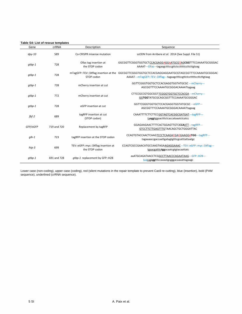
Table S4: List of rescue templates
Gene crRNA Description Sequence
dpy‐10 589 Co‐CRISPR misense mutation ssODN from Arribere et al. 2014 (See Suppl. File S1)
gtbp‐1 728Ollas tag insertion at
the STOP codon
GGCGGTTCGGGTGGTGCTCCACGAGGAGGAATGCGTAGCGGTTTCCAAAATGCGGGAC
AAAAT‐‐‐Ollas‐‐‐tagaagctttccgttctcctttttccttcttgtaag
gtbp‐1 728mTagGFP::TEV::3Xflag insertion at the
STOP codon
GGCGGTTCGGGTGGTGCTCCACGAGGAGGAATGCGTAGCGGTTTCCAAAATGCGGGAC
AAAAT‐‐‐mTagGFP::TEV::3Xflag‐‐‐tagaagctttccgttctcctttttccttcttgtaag
gtbp‐1 728 mCherry insertion at cutGGTTCGGGTGGTGCTCCACGAGGTGGTATGCGC ‐‐‐mCherry‐‐‐
AGCGGTTTCCAAAATGCGGGACAAAATtagaag
gtbp‐1 772 mCherry insertion at cutCTTCCGCCGTGGCGGTTCGGGTGGTGCTCCACGA‐‐‐mCherry‐‐‐
GGTGGTATGCGCAGCGGTTTCCAAAATGCGGGAC
gtbp‐1 728 eGFP insertion at cutGGTTCGGGTGGTGCTCCACGAGGTGGTATGCGC ‐‐‐eGFP‐‐‐
AGCGGTTTCCAAAATGCGGGACAAAATtagaag
fbf‐2 689tagRFP insertion at cut
(STOP codon)
CAAATTTTCTTCTTCCGGTAGTCACGGCGATGAT‐‐‐tagRFP‐‐‐
taaggtggaactttctcaccataaatctcatcc
GFP/eGFP 719 and 720 Replacement by tagRFPGGAGAAGAACTTTTCACTGGAGTTGTCCCAATT‐‐‐tagRFP‐‐‐
GTCCTTCTTGAGTTTGTAACAGCTGCTGGGATTAC
glh‐1 723 tagRFP insertion at the STOP codonCCAGTGTACCAACTCAAGTCCCTCAAGACGAGGAAGGATGG‐‐‐tagRFP‐‐‐
tagaaaaccgaccaattgatagtgtttcgcatttattaatgc
htp‐3 699TEV::eGFP::myc::3Xflag insertion at
the STOP codon
CCAGTCGCCGAACATGCCAAGTAGAAGAGGAAAC‐‐‐TEV::eGFP::myc::3Xflag‐‐‐
tgaacgatttctggacaatcgtgtacaattatc
gtbp‐1 691 and 728 gtbp‐1 replacement by GFP::H2BaaATGCAGATAACCTCGGCCTTAACCCAGAATAAG‐‐‐GFP::H2B‐‐‐
taacagcggtttccaaaatgcgggacaaaattagaagc
Lower case (non-coding), upper case (coding), red (silent mutations in the repair template to prevent Cas9 re-cutting), blue (insertion), bold (PAM sequence), underlined (crRNA sequence).
5 SI A. Paix et al.
JH number Gene targeted crRNA Insert Location Recipient strain
JH3324 gtbp‐1 728 mCherry near STOP codon (at the cut) N2
JH3314 gtbp‐1 728 eGFP::3Xflag near STOP codon (at the cut) N2
JH4000 gtbp‐1 728 Ollas STOP codon N2
JH4001 gtbp‐1 728 mTagGFP::TEV::3Xflag STOP codon N2
JH4002 gtbp‐1 728 eGFP near STOP codon (at the cut) N2
JH4003 fbf‐2 689 tagRFP STOP codon N2
JH4004gtbp‐1
and fbf‐2
728
and 689
eGFP for gtbp‐1
and tagRFP for fbf‐2
near STOP codon (at the cut) of gtbp‐1
and at the STOP codon of fbf‐2N2
JH4005 gtbp‐1 719 and 720 eGFP replacement by tagRFP Between 5'/3' ends of eGFP JH3314
JH4006 deps‐1 719 and 720 GFP replacement by tagRFP Between 5'/3' ends of GFP JH3207
JH4007 glh‐1 723 tagRFP STOP codon N2
JH4008 htp‐3 699 TEV::eGFP::myc::3Xflag STOP codon N2
JH4009 gtbp‐1 691 and 728 gtbp‐1 replacement by GFP::H2B Between 5'/3' ends of gtbp‐1 ORF N2
Table S5: List of strains
6 SI A. Paix et al.

7 SI A. Paix et al.
File S1: Direct delivery CRISPR-HDR editing protocol for C. elegans
(Paix et al. 2015)
Protocol updates will be posted on the Seydoux lab website:
http://www.bs.jhmi.edu/MBG/SeydouxLab/
Protocol Overview
‐ Design crRNA(s) and a repair template for your gene of interest.
‐ Inject Cas9/crRNA/tracrRNA complexes and repair templates targeting your gene of interest and
dpy‐10.
‐ Identify broods with Rollers (first generation after injection).
‐ Screen Rollers (and their non‐Roller siblings, if desired) for edits at your gene of interest
‐ See Section F for a positive control experiment – tagging gtbp‐1 with GFP using dpy‐10 co‐
CRISPR.
A. Preparation of reagents
Cas9
Recombinant Cas9::NLS can be purified from E. coli (see attached protocol File S2) or purchased from
commercial sources.
tracrRNA
The universal tracrRNA is a structural RNA that links the crRNA to Cas9. The same tracrRNA is used for all experiments.
We order it from Dharmacon #U‐002000‐05/20/50 (http://dharmacon.gelifesciences.com/gene‐editing/crispr‐cas9/edit‐r‐tracrrna/). The tracrRNA is 74nt long (Jinek et al. 2012): AACAGCAUAGCAAGUUAAAAUAAGGCUAGUCCGUUAUCAACUUGAAAAAGUGGCACCGAGUCGGUGCUUUUUUU
Upon receipt, briefly spin the tubes and reconstitute at 4µg/µl (0.17nmol/µl): add 29.8µl of Tris pH 7.5 to the 5nmol provided (U‐002000‐05). Other amounts of tracrRNA are available (U‐002000‐20/50). Store at ‐80°C.
8 SI A. Paix et al.
crRNA
The crRNA consist of a 20nt gene‐specific sequence followed by a universal sequence (GUUUUAGAGCUAUGCUGUUUUG) required to interact with the tracrRNA. The 20nt gene‐specific sequence must lie upstream of a PAM sequence (NGG) in genomic DNA. DO NOT INCLUDE THE PAM IN THE crRNA!
Remember to look for PAM sites on both DNA strands – crRNAs can target either strand.
Not all crRNAs work well (Table S3). Desired features include (in order of importance) 1) cleavage site as close as possible to the edit site, 2) good sequence (see below) and 3) few off‐target sites (we use the website http://crispr.mit.edu/ for off‐target prediction; Hsu et al. 2013). If there are off‐target sites, those sites should have 3 or more mismatches, preferentially close to the PAM.
In our hands, the most predictable determinant of guide RNA efficiency for HDR is distance between the cleavage site and the edit that you are trying to introduce. Optimal distance is <10 bases. We have obtained edits up to 30 bases away from the cleavage site, but the efficiency of edit incorporation drops by a factor of ~5‐10.
Several recommendations for crRNA sequence have been reported (Farboud et al. 2015; Gagnon et al. 2014; Doench et al. 2014), and we try to follow them when possible. These recommendations are:
‐ 50 to 75% overall GC content ‐ GG or G, but no C, for the 3’ most residue(s) immediately upstream of the PAM
The Broad Institute website implements these recommendations for guide RNA scoring (http://www.broadinstitute.org/rnai/public/analysis‐tools/sgrna‐design) (Doench et al. 2014).
Order your gene specific crRNA (20nt specific sequence + GUUUUAGAGCUAUGCUGUUUUG) from Dharmacon (http://dharmacon.gelifesciences.com/gene‐editing/crispr‐rna‐configurator/).
Also order the crRNA for dpy‐10: GCUACCAUAGGCACCACGAG + GUUUUAGAGCUAUGCUGUUUUG
Upon receipt, briefly spin the tubes and reconstitute at 8µg/µl (0.6nmol/µl): add 33.8µl of Tris pH 7.5 to the 20nmol provided. Store at ‐80°C.
Repair template design
Repair templates should contain ~35nt homology arms (Paix et al. 2014) (Table S4): sequences at the 5’ and 3’ end of the repair template that are homologous to sequences flanking the cut and edit in the genomic DNA. Ideally, flanking sequences should terminate with a C or G and contain good sequence diversity at their extremities (no hairpins).
The repair template should also contain mutations that make it resistant to re‐cutting by Cas9/crRNA‐tracrRNA complex after integration in the genome. These mutations can be 1) insertions that disrupt the crRNA sequence or separate the crRNA sequence from the PAM or 2) mismatches that disrupt the PAM or crRNA sequence (we typically create between 2 and 4 mismatches when disrupting the crRNA sequence, mutations closest to the PAM are the most effective) (Jinek et al. 2012).
9 SI A. Paix et al.
Be careful to introduce only silent changes using codons that are used at a frequency similar to the original codon (http://www.genscript.com/cgi‐bin/tools/codon_freq_table). If possible, avoid crRNAs that target non‐coding sequences since mutations in these sequences could possibly affect regulatory (splicing, promoter) motifs.
For small edits, engineer a restriction site in your repair template to facilitate screening (see Paix et al. 2014). For insertions >20bp, we typically identify the edits by size shift in the PCR product. When inserting a fluorescent protein, it is possible to screen directly by visual examination of F1s or F2s if the pattern is known.
Recommendation for antigenic peptide tag sequences can be found in Paix et al. 2014.
Repair template synthesis
A. Small edits (<100nt):
Use single‐stranded oligonucleotides (ssODNs, 200nt maximum size, 4nM ultramer, salt free) ordered
from IDT. Reconstitute ssODN at 1µg/µl according to the amount provided by the manufacturer. Sense‐
strand ssODNs have been reported to work better (Katic et al. 2015).
The ssODN repair template for dpy‐10 is:
CACTTGAACTTCAATACGGCAAGATGAGAATGACTGGAAACCGTACCGCATGCGGTGCCTATGGTAGCGGAGCTT
CACATGGCTTCAGACCAACAGCCTAT ‐ Use a working aliquot at 500ng/µl in H2O.
B. Large edits (100bp‐2kb): Use PCR amplicons.
Note that this type of template may not work efficiently for inserts > 3kb (A. Paix, unpublished).
‐Primer design: Design the primers so that they contain the desired homology arms (~35nt),
mutations in the crRNA site(s) and sequences complementary to insert. Be sure to have a C or G at the 3’
end of the primers. Where possible, limit the size of the primers to less than 65nt in order to avoid
primer contamination after PCR purification (see below). See Table S2 for available plasmids that can be
used as templates to amplify fluorescent proteins and tags.
‐PCR: Amplify the PCR template using this reaction mix: Mix 2µl of template plasmid (from a
standard miniprep of 1.5ml bacterial culture), 2µl of forward and reverse primers (100µM stock), 194µl
of H2O, 200µl of Phusion Master Mix 2X (NEB, #M0531L). Split the mix in 8 PCR tubes (50µl per tubes)
and do a gradient PCR as follow:
10 SI A. Paix et al.
‐‐‐‐‐‐‐‐‐‐‐‐‐
98°C, for 2min
‐‐‐‐‐‐‐‐‐‐‐‐‐
98°C, for 30s
60 to 72°C gradient, for 30s
72°C, for 45s
‐‐‐‐‐‐‐‐‐‐‐‐‐
Repeat 29 times
‐‐‐‐‐‐‐‐‐‐‐‐‐
72°C, for 10min
Hold at 10°C
‐‐‐‐‐‐‐‐‐‐‐‐‐
Add 10µl of 6X Orange loading dye (Bioworld, #10570024‐1) to each tube and run 8µl of it on an agarose
gel.
Pool the positive PCRs in one tube (up to 8) and purify them in one Qiagen minelute column (#28006),
elution with 10µl of H2O. For most templates, the 8 annealing temperatures will give good yield and
therefore we routinely pool the 8 reactions together. The expected yield ranges from 1 to 1.5µg/µl.
If the PCR primers are >65nt (typically when the edit is away from the cut), a second (nested) PCR step is
necessary because the long primers with be present in the purified PCR pool at a concentration high
enough to be toxic for injection. For the nested PCR, use forward and reverse primers of 18‐22nt
corresponding at the 5’ en 3’ ends of the template generated in the first PCR. Run a second PCR as
before but with the following master mix (for 8 annealing temperatures of PCR): 0.8µl of 1st round of
purified PCR, 2µl of forward and reverse primers, 195.2µl of H2O, 200µl of Phusion Master mix 2X.
B. Preparing injection mixes
I. One locus editing using home‐made Cas9
Cas9 prep (10µg/µl): 5µl
tracrRNA (4µg/µl): 5µl
dpy‐10 crRNA (8µg/µl): 0.4µl
11 SI A. Paix et al.
dpy‐10 ssODN (500ng/µl): 0.55µl
Targeted gene crRNA (8µg/µl): 1µl
PCR template (s) (several templates can be mixed): Up to 500ng/µl final in the mix
OR ssODN (s) (several templates can be mixed) (1µg/µl): 2.2µl total
KCl (1M): 0.5µl
Hepes pH7.4 (200mM): 0.75µl
H2O: add if necessary to reach a final volume of 20µl
II. Multi‐loci editing using home‐made Cas9
Cas9 prep (10µg/µl): 5µl
tracrRNA (4µg/µl): 6.7µl
dpy‐10 crRNA (8µg/µl): 0.4µl
dpy‐10 ssODN (500ng/µl): 0.55µl
Targeted gene crRNA1 (8µg/µl): 0.75µl
Targeted gene crRNA2 (8µg/µl): 0.75µl
PCR templates (to repair the cuts corresponding to crRNAs 1/2): Up to 500ng/µl final in the mix
OR ssODNs (to repair the cuts corresponding to crRNAs 1/2) (1µg/µl): 2.2µl total
KCl (1M): 0.5µl
Hepes pH7.4 (200mM): 0.75µl
H2O: add if necessary to reach a volume of 20µl
III. Note for edits on chromosome 2
For loci on LGII, the edits will be linked to the dpy‐10(Rol) edit. If you prefer to recover edits that are
unlinked to the dpy‐10 edit, use 0.28µl of dpy‐10 crRNA to maximize edits in non‐Roller animals.
Alternatively, you can use rescue of pha‐1(ts) mutation (LGIII) as an alternative co‐CRISPR strategy as
described in Ward, 2015.
12 SI A. Paix et al.
IV. Injection mixes processing
Add each components of the injection mix in a 0.5ml tube (add Cas9 last). Place the 0.5ml tube in a
1.5ml eppendorf tube and spin for 2min at 13000rpm. Incubate at 37°C for 10‐15min. Immediately load
the injection needles and process to injection.
Note that the volume of the injection mixes can be decreased if necessary as long as the molarity of
each component is maintained.
C. Injections, worm recovery and handling
Inject both arms of young adult hermaphrodites (with a few embryos). Be sure hermaphrodites are
young enough to lay eggs for next two days. See WormBook for injection protocol:
http://www.wormbook.org/chapters/www_transformationmicroinjection/transformationmicroinjection
.html
30min to 1h after injection, recover the injected hermaphrodites (P0s) as follows: Every 5‐10min, add:
5µl / 5µl / 10µl /10µl /20µl /20µl/ 40µl of 1X M9
Clone out the P0s onto NNGM plates (1 P0 per plate, at 20°C). Use fresh NNGM plates with a thin layer
of OP50 bacteria at the center to facilitate screening for Rollers. It is important to avoid that the P0s
touch the mineral oil on the injection pad because the oil will kill them. After 20‐23 hours, transfer the
P0s to second plate (again 1 P0 per plate). For experiments using large PCR repair templates, you may
find that edits arise more frequently on the second‐day plates (Paix et al. 2014).
Examine the F1s for Rollers 4‐5 days after cloning the P0s. You should recover Rollers from ~30‐70% of
injected hermaphrodites. Determine the number of Roller F1s per P0 and select the 3 P0s giving the
most Rollers. These are your “jackpot broods”.
Clone all the Roller F1s (from the jackpot broods or from all broods if you do not have that many). You
can also clone non‐Rollers (only from jackpot broods) if you prefer to isolate your edit without the dpy‐
10 edit (this is useful if your gene in on the same linkage group as dpy‐10: LGII).
D. Screening for edit of interest
For the insertion of a fluorescent protein, if you know the expected pattern, you can screen the F1s
(after you have allowed them to lay eggs) by placing them in a drop of M9 containing levamisole (1mM)
under the 10X objective of a compound microscope. You can also use no coverslip if you want to recover
the worms (in that case no need to clone them out first). Use 3 or 12‐well microscope slides.
13 SI A. Paix et al.
For smaller edits (antigenic tag or mutation), for each F1, pool 10 F2s in 15µl of lysis buffer and PCR the
edited locus. Avoid picking bacteria with the F2s. The edits can be detected by a size‐change or by
restriction‐enzyme digest if a restriction site was included in the rescue template. You can also PCR
directly each F1, but we have found in practice that it is easier to PCR cohorts (10 or so) of F2s.
E. Strain establishment
We recommend recovering at least two independent edits (derived from different P0s) for each
experiment.
If the edit was identified in a Roller worm, pick ~8 or more non‐Roller F2/3s to separate the edit from
dpy‐10 and recover homozygous edits.
For fluorescent protein integration, check the segregation of the fluorescent signal in F3 worms derived
from singled‐out F2s to identify homozygous lines.
Sequence‐verify the edits once the homozygous strains are established. Sequence at least the entire
sequence that was present in your rescue template. You may also want to sequence possible off‐target
loci.
Remember that mutants may not be viable when homozygous. We have also isolated edits that cause
dominant phenotypes in the F1 generation (dominant sterile or dominant maternal effect lethal/sterile).
Note that some tagged proteins are not fully functional ‐ check the homozygous edited lines for brood
size and viability at 20oC and 25oC.
F. Special applications of protocol
ORF replacement to obtain null allele and transcriptional reporter
Design crRNAs near the Start and Stop codons. Design repair template with homology arms that reach
up to the cleavage sites, and are in frame with the ORF of the gene of interest, if any coding sequence
remains after replacement. We recommend recoding any coding sequence remaining between the two
cuts to force gene conversion of the entire template.
Make the GFP::H2B repair template as described in Repair template synthesis. Use pCM1.35 (Available
at Addgene; Merritt et al. 2008) (Table S2) as a PCR plasmid template.
Process as described in Reagents for replacement of GFP (and eGFP variant) with tagRFP (or other FPs) for ORF replacement.
14 SI A. Paix et al.
Reagents for replacement of GFP (and eGFP variant) with tagRFP (or other FPs)
crRNA GFP Nt (#720 in Table S3): CCAUCUAAUUCAACAAGAAU + GUUUUAGAGCUAUGCUGUUUUG
crRNA GFP Ct (#719 in Table S3): CAAACUCAAGAAGGACCAUG + GUUUUAGAGCUAUGCUGUUUUG
Primers pairs to generate template (from RFP containing‐plasmid pAP575‐1):
Forward primer (5’ to 3’, lower case indicating the homology arm sequence):
ggagaagaacttttcactggagttgtcccaattGTGTCTAAGGGCGAAGAGCTG
Reverse primer (5’ to 3’, lower case indicating the homology arm sequence):
gtaatcccagcagctgttacaaactcaagaaggacATTAAGTTTGTGCCCCAGTTTG
PCR condition: use annealing temperature of 63oC, elongation step of 45s, pAP575‐1 as a plasmid
template.
Injection mix:
Cas9 home‐made prep (10µg/µl): 5µl
tracrRNA (4µg/µl): 6.7µl
dpy‐10 crRNA (8µg/µl): 0.4µl
dpy‐10 ssODN (500ng/µl): 0.55µl
crRNA GFP Nt (8µg/µl): 0.75µl
crRNA GFP Ct (8µg/µl): 0.75µl
PCR template for GFP (and eGFP variant) replacement: Up to 500ng/µl final in the mix
KCl (1M): 0.5µl
Hepes pH7.4 (200mM): 0.75µl
H2O: add if necessary to reach a volume of 20µl
Multi‐colors replacement (using the same homology arms than the one specified above ‐ lower
case in primer sequences) (Chudakov et al. 2010) can also be performed.
Note that the same injection mix / injection needle can be used on different GFP tagged strains.
15 SI A. Paix et al.
Positive control experiment (to test protocol in your hands and/or activity of your home‐made Cas9):
Tag gtbp‐1 with eGFP or mCherry using dpy‐10 co‐CRISPR.
Prepare the repair PCR template as indicated in Reagents for gtbp‐1 eGFP and mCherry tagging and Repair template synthesis.
Make the injection mix as indicated in injection mixes, part I. Use the crRNA gtbp‐1 Ct (#728 in Table S3).
Inject 15‐20 young adult N2 worms and recover as described in Worm recovery and handling. Pool the recovered worms (P0s) on one plate and incubate for 22‐23h at 20oC (day 1). Clone the P0s to individual
OP50 plates and incubate at 20oC for 4‐5 days (day 2).
When the F1s reach the adult stage, check for Rollers. At least 3 P0s should give a high number of Rollers
(>15) (Note that we do not count/examine Dumpy F1s since these are homozygous edits at the dpy‐10
locus). From those “jackpot broods”, screen the Rollers for fluorescent protein expression as described
in Screening for edit of interest. 50% or more of the Rollers (at day 2) should be positive for
fluorescence.
Reagents for gtbp‐1 eGFP and mCherry tagging
crRNA gtbp‐1 Ct (#728 in Table S3): CCACGAGGUGGUAUGCGCAG + GUUUUAGAGCUAUGCUGUUUUG
Primers pairs to generate template (from eGFP containing‐plasmid pAP682‐1):
Forward primer (5’ to 3’, lower case indicating the homology arm sequence) for eGFP insertion:
ggttcgggtggtgctccacgaggtggtatgcgcGTGAGTAAAGGAGAAGAAC
Reverse primer (5’ to 3’, lower case indicating the homology arm sequence) for eGFP insertion:
cttctaattttgtcccgcattttggaaaccgctCTTGTACAGCTCGTCCATGCC
Primers pairs to generate template (from mCherry containing‐plasmid pAP582‐1):
Forward primer (5’ to 3’, lower case indicating the homology arm sequence) for mCherry insertion:
ggttcgggtggtgctccacgaggtggtatgcgcGTCTCAAAGGGTGAAGAAGATAAC
Reverse primer (5’ to 3’ lower case indicating the homology arm sequence) for mCherry insertion:
cttctaattttgtcccgcattttggaaaccgctCTTATACAATTCATCCATGCC
PCR condition: use annealing temperature of 63oC, elongation step of 45s, pAP682‐1 (eGFP) or pAP582‐1
(mCherry) as a plasmid template. Do 8 PCR reactions and pool them (400µl total), purify on minelute
column (see Repair template synthesis).
16 SI A. Paix et al.
Additional references
Chudakov et al., Fluorescent Proteins and Their Applications in Imaging Living Cells and Tissues
Doench et al., Rational Design of Highly Active sgRNAs for CRISPR‐Cas9‐Mediated Gene Inactivation
Farboud and Meyer, Dramatic Enhancement of Genome Editing by CRISPR/Cas9 through Improved
Guide RNA Design
Gagnon et al., Efficient Mutagenesis by Cas9 Protein‐Mediated Oligonucleotide Insertion and Large‐
Scale Assessment of Single‐Guide RNAs
Hsu et al., DNA Targeting Specificity of RNA‐Guided Cas9 Nucleases
Jinek et al., A Programmable Dual‐RNA‐Guided DNA Endonuclease in Adaptive Bacterial Immunity
Katic, Xu, and Ciosk, CRISPR/Cas9 Genome Editing in Caenorhabditis Elegans
Merritt et al., 3’ UTRs Are the Primary Regulators of Gene Expression in the C. Elegans Germline
Paix et al., Scalable and Versatile Genome Editing Using Linear DNAs with Microhomology to Cas9 Sites
in Caenorhabditis Elegans
Ward, Rapid and precise engineering of the Caenorhabditis elegans genome with lethal mutation co‐
conversion and inactivation of NHEJ repair
17 SI A. Paix et al.
File S2: Cas9 preparation protocol (Paix et al. 2015)
Protocol updates will be posted on the Seydoux lab website:
http://www.bs.jhmi.edu/MBG/SeydouxLab/
Purification of Cas9::NLSSV40::His6:
1. Transform DE3 GOLD (Agilent, #230132) cells with nm2973 plasmid (Fu et al. 2014) and plate
on LB + 50μg/mL Carbenicillin.
2. Inoculate 25mL LB + 50 μg/mL Carbenicillin with bacteria from the fresh transformation and
incubate at 37C overnight.
3. Transfer 5mL of overnight culture to 1L LB + 0.1% glucose + 50 μg/mL Carbenicillin and grow
at 25C. Grow to OD600=~0.5.
3. Shift culture to 18C for 15‐25 minutes, then add IPTG to 0.2 mM. Incubate overnight.
4. Pellet culture and obtain wet weight. Resuspend at ~6 mL/g cells with Buffer A + protease
inhibitor (Roche, #11836170001) + 1mM PMSF.
5. Sonicate 6 x 45s (setting 3 at 30%, 1 second pulse‐2 second pause) with 1 minute cooling in
between.
6. Spin lysate 30 minutes at 16000xg and transfer supernatant to a fresh tube.
7. Equilibrate a 5mL Ni‐agarose (Qiagen, #30410) with column with Buffer A (at least 25mL).
8. Batch bind clarified lysate with Ni‐agarose 45 minutes at 4C.
9. Wash Ni‐agarose column with 100mL of Buffer B.
10. Elute protein with Buffer C. Determine fractions that have Cas9 protein using Bradford assay
or by running a small amount on SDS‐PAGE gel. Pool fractions.
11. To remove contaminating DNA in the prep. Equilibrate a 5mL Q Sepharose (Sigma, #Q1126)
column with 1M KCl (25mL, this charges the column). Then equilibrate Q Sepharose column with
Buffer C (25mL).
12. Flow eluent (from step 11) over Q Sepharose column. Collect flow‐through and dialyze into
1L Buffer D for 5 hours at 4C. Transfer into 1L Buffer D and dialyze overnight.
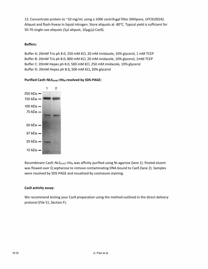
18 SI A. Paix et al.
13. Concentrate protein to ~10 mg/mL using a 100K centrifugal filter (Milipore, UFC910024).
Aliquot and flash‐freeze in liquid nitrogen. Store aliquots at ‐80°C. Typical yield is sufficient for
50‐70 single‐use aliquots (5μl aliquot, 10μg/μl Cas9).
Buffers:
Buffer A: 20mM Tris ph 8.0, 250 mM KCl, 20 mM imidazole, 10% glycerol, 1 mM TCEP
Buffer B: 20mM Tris ph 8.0, 800 mM KCl, 20 mM imidazole, 10% glycerol, 1mM TCEP
Buffer C: 20mM Hepes ph 8.0, 500 mM KCl, 250 mM imidazole, 10% glycerol
Buffer D: 20mM Hepes ph 8.0, 500 mM KCl, 20% glycerol Purified Cas9::NLSSV40::His6 resolved by SDS‐PAGE:
Recombinant Cas9::NLSSV40::His6 was affinity purified using Ni‐agarose (lane 1). Pooled eluent
was flowed over Q sepharose to remove contaminating DNA bound to Cas9 (lane 2). Samples
were resolved by SDS‐PAGE and visualized by coomassie staining.
Cas9 activity assay:
We recommend testing your Cas9 preparation using the method outlined in the direct delivery
protocol (File S1, Section F).