High Circulating Caspase-Cleaved Keratin 18 Fragments (M30...
9
Research Article High Circulating Caspase-Cleaved Keratin 18 Fragments (M30) Indicate Short-Term Mortality in Critically Ill Patients Alexander Koch, 1 Eray Yagmur, 2 Janine Linka, 1 Fabienne Schumacher, 1 Jan Bruensing, 1 Lukas Buendgens , 1 Ulf Herbers, 1 Ger H. Koek, 3 Ralf Weiskirchen , 4 Christian Trautwein, 1 and Frank Tacke 1 1 Department of Medicine III, RWTH-University Hospital Aachen, Pauwelsstrasse 30, 52074 Aachen, Germany 2 Medical Care Center, Dr. Stein and Colleagues, Mönchengladbach, Germany 3 Section of Gastroenterology and Hepatology, Department of Internal Medicine, Maastricht University Medical Centre (MUMC), Maastricht, Netherlands 4 Institute of Molecular Pathobiochemistry, Experimental Gene Therapy and Clinical Chemistry, RWTH-University Hospital Aachen, Pauwelsstrasse 30, 52074 Aachen, Germany Correspondence should be addressed to Frank Tacke; [email protected] Received 21 March 2018; Accepted 6 June 2018; Published 8 July 2018 Academic Editor: Emilia Hadziyannis Copyright © 2018 Alexander Koch et al. This is an open access article distributed under the Creative Commons Attribution License, which permits unrestricted use, distribution, and reproduction in any medium, provided the original work is properly cited. Caspase-cleaved fragments of the intermediate filament protein keratin 18 (cytokeratin-18 (CK18)) can be detected in serum as M30 levels and may serve as a circulating biomarker indicating apoptosis of epithelial and parenchymal cells. In order to evaluate M30 as a biomarker in critical illness, we analyzed circulating M30 levels in 243 critically ill patients (156 with sepsis, 87 without sepsis) at admission to the medical intensive care unit (ICU), in comparison to healthy controls (n = 32). M30 levels were significantly elevated in ICU patients compared with healthy controls. Circulating M30 was closely associated with disease severity but did not differ between patients with sepsis and ICU patients without sepsis. M30 serum levels were correlated with biomarkers of inflammation, cell injury, renal failure, and liver failure in critically ill patients. Patients that died at the ICU showed increased M30 levels at admission, compared with surviving patients. A similar trend was observed for the overall survival. Regression analyses confirmed that M30 levels are associated with mortality, and patients with M30 levels above 250.8 U/L displayed an excessive short-term mortality. Thus, our data support the utility of circulating levels of the apoptosis-related keratin fragment M30 as a prognostic biomarker at ICU admission. 1. Background Excessive systemic inflammation as a consequence of massive innate immune cell activation is a key characteristic in critically ill patients. This is triggered by infection-related molecules, termed pathogen-associated molecular patterns (PAMPs), and by the release of endogenous immuno- genic signals, termed danger-associated molecular patterns (DAMPs) [1]. Activated innate immune cells release abun- dant inflammatory cytokines (e.g., tumor necrosis factor and interleukins) that initiate inflammatory and also cell death signaling cascades in immune cells and parenchymal cells [2]. While excessive systemic inflammation is well recognized as a major driver of multiple organ failure and mortality in critical illness [3], the resulting mode of cell death, such as necrosis, apoptosis, necroptosis, pyroptosis, or ferroptosis, is incompletely understood [4]. Nonetheless, targeting apoptotic pathways has been suggested as a poten- tial novel therapeutic approach in sepsis therapy [5–7]. The systemic release of caspase-cleaved fragments of keratin 18 (K18), a major type I intermediate filament protein of the cytoskeleton, has been identified as a specific biomarker reflecting apoptotic cell death. K18 occurs in all single-layer epithelial cells such as the gut, urinary tract, or respiratory tract and in parenchymal cells like hepatocytes or cholangiocytes [8]. The caspase-cleaved K18 fragment Hindawi Disease Markers Volume 2018, Article ID 8583121, 8 pages https://doi.org/10.1155/2018/8583121
Transcript of High Circulating Caspase-Cleaved Keratin 18 Fragments (M30...
Research ArticleHigh Circulating Caspase-Cleaved Keratin 18 Fragments (M30)Indicate Short-Term Mortality in Critically Ill Patients
Alexander Koch,1 Eray Yagmur,2 Janine Linka,1 Fabienne Schumacher,1 Jan Bruensing,1
Lukas Buendgens ,1 Ulf Herbers,1 Ger H. Koek,3 Ralf Weiskirchen ,4
Christian Trautwein,1 and Frank Tacke 1
1Department of Medicine III, RWTH-University Hospital Aachen, Pauwelsstrasse 30, 52074 Aachen, Germany2Medical Care Center, Dr. Stein and Colleagues, Mönchengladbach, Germany3Section of Gastroenterology and Hepatology, Department of Internal Medicine, Maastricht University Medical Centre (MUMC),Maastricht, Netherlands4Institute of Molecular Pathobiochemistry, Experimental Gene Therapy and Clinical Chemistry, RWTH-University Hospital Aachen,Pauwelsstrasse 30, 52074 Aachen, Germany
Correspondence should be addressed to Frank Tacke; [email protected]
Received 21 March 2018; Accepted 6 June 2018; Published 8 July 2018
Academic Editor: Emilia Hadziyannis
Copyright © 2018 Alexander Koch et al. This is an open access article distributed under the Creative Commons Attribution License,which permits unrestricted use, distribution, and reproduction in any medium, provided the original work is properly cited.
Caspase-cleaved fragments of the intermediate filament protein keratin 18 (cytokeratin-18 (CK18)) can be detected in serum asM30 levels and may serve as a circulating biomarker indicating apoptosis of epithelial and parenchymal cells. In order toevaluate M30 as a biomarker in critical illness, we analyzed circulating M30 levels in 243 critically ill patients (156 with sepsis,87 without sepsis) at admission to the medical intensive care unit (ICU), in comparison to healthy controls (n = 32). M30 levelswere significantly elevated in ICU patients compared with healthy controls. Circulating M30 was closely associated with diseaseseverity but did not differ between patients with sepsis and ICU patients without sepsis. M30 serum levels were correlated withbiomarkers of inflammation, cell injury, renal failure, and liver failure in critically ill patients. Patients that died at the ICUshowed increased M30 levels at admission, compared with surviving patients. A similar trend was observed for the overallsurvival. Regression analyses confirmed that M30 levels are associated with mortality, and patients with M30 levels above250.8U/L displayed an excessive short-term mortality. Thus, our data support the utility of circulating levels of theapoptosis-related keratin fragment M30 as a prognostic biomarker at ICU admission.
1. Background
Excessive systemic inflammation as a consequence ofmassive innate immune cell activation is a key characteristicin critically ill patients. This is triggered by infection-relatedmolecules, termed pathogen-associated molecular patterns(PAMPs), and by the release of endogenous immuno-genic signals, termed danger-associated molecular patterns(DAMPs) [1]. Activated innate immune cells release abun-dant inflammatory cytokines (e.g., tumor necrosis factorand interleukins) that initiate inflammatory and also celldeath signaling cascades in immune cells and parenchymalcells [2]. While excessive systemic inflammation is well
recognized as a major driver of multiple organ failure andmortality in critical illness [3], the resulting mode of celldeath, such as necrosis, apoptosis, necroptosis, pyroptosis,or ferroptosis, is incompletely understood [4]. Nonetheless,targeting apoptotic pathways has been suggested as a poten-tial novel therapeutic approach in sepsis therapy [5–7].
The systemic release of caspase-cleaved fragments ofkeratin 18 (K18), a major type I intermediate filamentprotein of the cytoskeleton, has been identified as a specificbiomarker reflecting apoptotic cell death. K18 occurs in allsingle-layer epithelial cells such as the gut, urinary tract, orrespiratory tract and in parenchymal cells like hepatocytesor cholangiocytes [8]. The caspase-cleaved K18 fragment
HindawiDisease MarkersVolume 2018, Article ID 8583121, 8 pageshttps://doi.org/10.1155/2018/8583121

exposes a circulating neoepitope that is recognized by thespecific monoclonal antibody M30, which has been widelydescribed as a valid biomarker in the context of acute andchronic liver diseases [8, 9]. Few data exist on M30 levels incritical illness. Four independent observational studies inpatients with sepsis, two of them conducted in small patientcohorts, reported consistently elevated M30 levels comparedwith controls as well as an association with adverse clinicaloutcome [10–13]. However, it is currently less well definedif the clinical utility and prognostic relevance of M30 as abiomarker can be extrapolated to heterogeneous cohorts ofcritically ill patients, in which not only infectious threats(PAMPs) but also sterile inflammation (DAMPs) trigger celldeath mechanisms [1]. We therefore studied circulating M30in a large cohort of 243 critically ill patients at a medicalintensive care unit (ICU), in comparison to healthy controls,and assessed its potential as a diagnostic and prognosticbiomarker in medical ICU patients.
2. Methods
2.1. Study Design and Patient Characteristics. In our single-center prospective observational trial, we serially gathereddata and blood samples of consecutive patients uponadmission to the medical ICU in the University HospitalAachen, Germany. We excluded patients who had an elec-tive procedure or were admitted for postinterventionalobservational stays [14]. The study protocol was approvedby the local ethics committee and undertaken in accordancewith the ethical standards laid down in the Declaration ofHelsinki (ethics committee of the University HospitalAachen, RWTH Aachen University, Aachen, Germany, ref-erence number EK 150/06). We secured written informedconsent from the patient, the spouse, or the legal guardianaccording to the German civil code BGB §1896. Thepatients were categorized as sepsis and nonsepsis accordingto the Third International Consensus Definitions for Sepsisand Septic Shock (Sepsis-3) [15] and were treated followingthe current guidelines for treatment of sepsis (SurvivingSepsis Campaign) [16]. As a control cohort, we includedhealthy blood donors with normal blood count, normalvalues of liver enzymes, and a negative serology for viralhepatitis and HIV.
To assess the patients’ outcome, we defined the ICUmortality as well as overall mortality, which was based oncontacting the patients, relatives, and/or their general practi-tioners in approximately 6-month intervals after dischargefrom hospital for two years [17]. Short-term mortality wasdefined as mortality within 30 days after admission to theICU, independent from whether the patients died at theICU, in the hospital, or outside the hospital.
2.2. M30 Measurements. We obtained blood samplesimmediately at the time of admission to avoid influence ofany therapeutic procedures. After centrifugation, serum wasstored at −80°C. M30 was analyzed with a commercialenzyme immunoassay (M30 Apoptosense® ELISA, cat.number 10011, TECOmedical Group, Sissach, Germany).
2.3. Statistical Analysis. Because most samples were notnormally distributed, the Mann–Whitney U test was appliedto test for statistically significant differences between twogroups. Correlations were assessed by Spearman’s rankcorrelation method. All values, including outside values aswell as far-out values, were included. p values less than 0.05were considered as statistically significant.
The Cox regression model was employed to evaluate theprognostic value of M30 on the outcome. Furthermore, thesurvival was assessed using Kaplan-Meier analysis with aM30 cut-off level that had been calculated via the YoudenIndex [18]. All analyses were conducted using IBM SPSSStatistics (SPSS; Chicago, Illinois).
3. Results
3.1. M30 Levels Are Significantly Elevated in ICU PatientsCompared with Healthy Controls but Are Not Related toSepsis. Circulating M30 levels were significantly increasedin critically ill patients (n = 243, median 178.3U/L, range16.7–1001U/L, Table 1) compared to the healthy controlgroup (n = 32, median 107.1U/L, range 48.3–217.1U/L,p < 0 001, Figure 1(a)). A total of n = 156 patients fulfilledthe sepsis criteria, mostly with a pulmonary focus (n = 84).Nonsepsis patients (n = 87) were admitted due to cardiopul-monary disorders (n = 29), decompensated liver cirrhosis(n = 13), acute pancreatitis (n = 11), and other conditions(Table 2). M30 levels did not differ between patients withsepsis (median 161.8U/L, range 21.6–1000U/L) and criti-cal illness without sepsis (median 193.6U/L, range 16.7–1001U/L, Table 1, Figure 1(b)).
3.2. M30 Serum Concentrations Are Associated with DiseaseSeverity. To identify critical factors that impact M30 levels,we conducted correlation analyses with scoring systems fordisease severity as well as with clinically established labora-tory parameters representing disease severity (Table 3). Weobserved significant correlations between circulating M30levels at admission and clinical scores for disease severitysuch as the Acute Physiology And Chronic Health II(APACHE II) (r = 0 311, p < 0 001, Figure 1(c)), SequentialOrgan Failure Assessment (SOFA), or Simplified AcutePhysiology Score 2 (SAPS2) (Table 3). A positive associationwas also observed between M30 levels and soluble urokinaseplasminogen activator receptor (suPAR, Figure 1(d)), whichhad been identified as a marker of systemic inflammationand poor prognosis in critically ill patients [19].
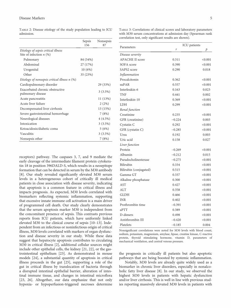
3.3. M30 Levels Are Correlated with Biomarkers of LiverFailure, Renal Failure, Inflammation, and Cell Injury inCritically Ill Patients. Furthermore, we found strong corre-lations between M30 levels and biomarkers that reflecthepatic and renal dysfunction (Table 3). More precisely,circulating M30 levels correlated with markers indicatingthe hepatic biosynthetic capacity (e.g., international normal-ized ratio (INR), antithrombin III, and pseudocholinester-ase), parenchymal damage (e.g., glutamate dehydrogenase(GLDH), aspartate transaminase, and alanine transami-nase) and parameters indicating cholestasis (e.g., gamma-
2 Disease Markers

glutamyltransferase, alkaline phosphatase, and bilirubin,Figure 1(e)) as well as markers tracing renal dysfunction(e.g., creatinine, cystatin C, and glomerular filtration rate).In addition, circulating M30 levels showed to correlate withparameters of systemic inflammation (e.g., tumor necrosisfactor, interleukins 6 and 10, and procalcitonin) and to thegeneral cell injury marker lactate dehydrogenase (LDH,r = 0 299, p < 0 001).
3.4. Patients with Underlying Liver Cirrhosis HaveSignificantly Elevated M30 Levels. M30 was indicated to beincreased in patients with hepatic dysfunction [11, 20]. Inaccordance, patients admitted to the ICU with liver cirrhosis(n = 25, median 391.4U/L, range 19.4–1000) had signifi-cantly increased M30 levels compared to critically ill patientswithout cirrhosis (n = 218, median 171U/L, range 16.7–1001,p = 0 006, Figure 1(f)). Although M30 levels had been specif-ically linked to nonalcoholic steatohepatitis [9], M30 serumconcentrations in our study were not associated with obesity(M30 in patients with BMI> 30 kg/m2 median 155.7U/Lversus BMI< 30 kg/m2 median 177.8U/L, not significant).
3.5. High M30 Serum Concentrations Are Associated withExcessive Short-Term Mortality. We found increased M30levels at admission in those patients who died at the ICU(n = 64, median 324.9U/L, range 16.7–1000U/L), comparedwith surviving patients (n = 179, median 166.5U/L, range21.6–1001U/L, p < 0 001; Figure 2(a)). Using Cox regres-sion analysis, high M30 levels significantly predicted ICUmortality (p = 0 005). Kaplan-Meyer curves were generatedby applying the optimal cut-off value (M30 of 250.8U/L)for the best ratio of sensitivity and specificity for mortalityusing the Youden index, displaying the prognostic valueof high M30 levels for short-term mortality (Figure 2(b)).
We detected a trend but no significant difference in M30levels regarding the overall mortality (nonsurvivors n =115, median 251U/L, range 16.7–1000U/L versus survivorsn = 115, median 166.5U/L, range 21.6–1001U/L, p = 0 059;Figure 2(c)). However, Cox regression analysis remainedsignificant for predicting overall survival as well (p = 0 004),and Kaplan-Meier curves displayed a separation betweenpatients with high versus low M30 levels in their overall sur-vival (Figure 2(d)). The validity and performance of M30 as abiomarker to predict ICU or overall survival in critically illpatients are summarized in Table 4.
As visible from the Kaplan-Meier curve analyses, themajority of deaths that separated patients with high fromlow M30 levels occurred within the first 30 days. In fact,patients that died within the first 30 days had significantlyhigher M30 levels (n = 74, median 294.6U/L, range 16.7–1000U/L) than patients that survived (n = 169, median166.5U/L, range 21.6–1001U/L, p = 0 001). This differenceremained significant at later time points (e.g., 60 days, 90days, 180 days, and 360 days mortality) but was mainlydriven by the difference within the first 30 days (detailed datanot shown). In addition, this difference regarding M30 levelsand 30 days mortality was significant also in the subgroup ofsepsis patients (p = 0 007), while the smaller subgroup ofnonsepsis patients showed a clear trend towards higherM30 levels in patients that died within 30 days after ICUadmission (p = 0 066).
4. Discussion
During the apoptotic mode of cell death, caspases, intracellu-lar proteases that cleave aspartate residues, become activatedeither via the intrinsic (mitochondrial release of cytochromeC) or the extrinsic (inflammatory cytokines and death
Table 1: Patient characteristics and M30 serum measurements at ICU admission.
Parameter All patients Nonsepsis Sepsis
Number 243 87 156
Sex (male/female) 154/89 55/32 99/57
Age median (range) (years) 64 (18–90) 61 (18–85) 65 (21–90)
APACHE II score median (range) 18 (2–43) 17 (2–34) 19 (3–43)
ICU days median (range) 7 (1–70) 5 (1–44) 10 (1–70)
Death during ICU n (%) 64 (26%) 15 (17%) 49 (31%)
Death during follow-up (total) n (%) 115 (47%) 29 (33%) 86 (55%)
Mechanical ventilation n (%) 168 (69%) 55 (63%) 113 (72%)
Preexisting diabetes n (%) 73 (30%) 30 (35%) 43 (28%)
Preexisting cirrhosis n (%) 25 (10%) 16 (18%) 9 (6%)
BMI median (range) (m2/kg) 25.9 (15.3–86.5) 25.4 (15.9–53.3) 26.0 (15.3–86.5)
WBC median (range) (×103/μL) 12.8 (0–208) 11.9 (2.5–27.7) 13.1 (0–208)
CRP median (range) (mg/dL) 93 (0–230) 18 (5–230) 153.3 (0–230)
Procalcitonin median (range) (μg/L) 1.2 (0–207.5) 0.35 (0.03–100) 3.4 (0–207.5)
Creatinine median (range) (mg/dL) 1.3 (0–21.6) 1.0 (0.2–15) 1.6 (0–21.6)
INR median (range) 1.17 (0–133) 1.15 (0.9–6.73) 1.18 (0–133)
M30 median (range) (U/L) 178.3 (16.7–1001) 161.8 (21.6–1000) 193.6 (16.7–1001)
For quantitative variables, median and range (in parenthesis) are given.
3Disease Markers

1200
1000p < 0.001
800
800
400
200
M30
(U/L
)
0
Controls(n = 32)
Patients(n = 243)
(a)
n.s
1200
1000
800
600
400
200
M30
(U/L
)
0
No(n = 87)
Sepsis
Yes(n = 156)
(b)
1000
800
600
400
200
M30
(U/L
)
0
0 10 20APACHE II score
30 40 50
r = 0.311p < 0.001
(c)
0 5 10suPAR (ng/mL)
15 20
1200
1000
800
600
400
200
M30
(U/L
)
0
r = 0.557p < 0.001
(d)
0 3 4Bilirubin (mg/dL)
6 8 10
1200
1000
800
600
400
200
M30
(U/L
)
0
r = 0.334p < 0.001
(e)
1200
1000
800
600
400
200
M30
(U/L
)
0
p = 0.006
No(n = 218)
Cirrhosis
Yes(n = 25)
(f)
Figure 1: Serum M30 levels in critically ill patients. (a) Serum levels of M30, at the time of admission to the ICU, were significantlyhigher in critically ill patients than in healthy controls (p < 0 001; U test). (b) M30 levels did not differ between ICU patients withor without sepsis. (c-d) M30 levels correlated with disease severity, as assessed by the APACHE II score (c) or serum concentrationsof soluble urokinase plasminogen activator receptor (suPAR, d). (e-f) M30 levels in critically ill patients correlated with serumbilirubin (e) and were particularly elevated in ICU patients with liver cirrhosis (f).
4 Disease Markers
receptors) pathway. The caspases 3, 7, and 9 mediate theearly cleavage of the intermediate filament protein cytokera-tin 18 in position 396DALD-S, which results in a neoepitopeformation that can be detected in serum by the M30 antibody[8]. Our study revealed significantly elevated M30 serumlevels in a heterogeneous cohort of critically ill medicalpatients in close association with disease severity, indicatingthat apoptosis is a common feature in critical illness andimpacts prognosis. As expected, M30 levels correlated withbiomarkers reflecting systemic inflammation, supportingthat excessive innate immune cell activation is a main driverof programmed cell death. Our study clearly demonstratesthat the serum apoptosis marker M30 is independent fromthe concomitant presence of sepsis. This contrasts previousreports from ICU patients, which have uniformly linkedelevated M30 to the clinical course of sepsis [10–13]. Inde-pendent from an infectious or noninfectious origin of criticalillness, M30 levels correlated with markers of organ dysfunc-tion and disease severity in our study. While these datasuggest that hepatocyte apoptosis contributes to circulatingM30 in critical illness [2], additional cellular sources mightinclude other epithelial cells, the kidney [21, 22], or the gas-trointestinal epithelium [23]. As demonstrated in mousemodels [24], a substantial quantity of apoptosis in criticalillness proceeds in the gut [25], supporting a role of thegut in critical illness by translocation of bacteria througha disrupted intestinal epithelial barrier, alteration of intes-tinal immune tissue, and changes in intestinal microflora[23, 26]. Altogether, our data emphasize that not onlyhypoxia- or hypoperfusion-triggered necroses determine
the prognosis in critically ill patients but also apoptoticpathways that are being boosted by systemic inflammation.
Notably, M30 levels are already quite widely used as abiomarker in chronic liver disorders, especially in nonalco-holic fatty liver disease [8]. In our study, we observed thehighest M30 levels in patients with hepatic dysfunctionand/or liver cirrhosis. This is well in line with previous stud-ies reporting massively elevated M30 levels in patients with
Table 2: Disease etiology of the study population leading to ICUadmission.
Sepsis Nonsepsis156 87
Etiology of sepsis critical illnessSite of infection n (%)
Pulmonary 84 (54%)
Abdominal 27 (17%)
Urogenital 10 (6%)
Other 35 (23%)
Etiology of nonsepsis critical illness n (%)
Cardiopulmonary disorder 29 (33%)
Exacerbated chronic obstructivepulmonary disease
3 (3.5%)
Acute pancreatitis 11 (13%)
Acute liver failure 2 (2%)
Decompensated liver cirrhosis 13 (15%)
Severe gastrointestinal hemorrhage 7 (8%)
Neurological diseases 4 (4.5%)
Intoxication 3 (3.5%)
Ketoacidosis/diabetic coma 5 (6%)
Vasculitis 3 (3.5%)
Nonsepsis other 7 (8%)
Table 3: Correlations of clinical scores and laboratory parameterswith M30 serum concentrations at admission day (Spearman rankcorrelation test, only significant results are shown).
ParametersICU patients
r p
Disease severity
APACHE II score 0.311 <0.001SOFA score 0.390 <0.001SAPS2 score 0.290 0.018
Inflammation
Procalcitonin 0.362 <0.001suPAR 0.557 <0.001Interleukin-6 0.163 0.023
TNF 0.441 0.002
Interleukin-10 0.369 <0.001LDH 0.299 <0.001Renal function
Creatinine 0.235 <0.001GFR (creatinine) −0.224 0.003
Cystatin C 0.292 <0.001GFR (cystatin C) −0.285 <0.001Urea 0.192 0.003
Uric acid 0.158 0.027
Liver function
Protein −0.269 <0.001Albumin −0.212 0.015
Pseudocholinesterase −0.275 <0.001Bilirubin 0.334 <0.001Bilirubin (conjugated) 0.515 <0.001Gamma GT 0.337 <0.001Alkaline phosphatase 0.300 <0.001AST 0.427 <0.001ALT 0.358 <0.001GLDH 0.466 <0.001INR 0.402 <0.001Prothrombin time −0.391 <0.001aPTT 0.389 <0.001D-dimers 0.498 <0.001Antithrombin III −0.420 <0.001Platelets −0.185 0.004
Nonsignificant correlations were noted for M30 levels with blood count,sodium, potassium, magnesium, amylase, lipase, creatine kinase, C-reactiveprotein, thyroid stimulating hormone, vitamin D, parameters ofmechanical ventilation, and central venous pressure.
5Disease Markers

acute liver failure [27], acute-on-chronic liver failure [20],and decompensated liver cirrhosis [9]. These associationsemphasize the crucial role of the hepatic function for theprognosis in critical illness, as the assessment of liver failureis already included in several prognostic ICU scores such asthe SOFA score [2].
In our study, circulating M30 was an early predictor ofadverse outcome upon admission of medical patients to theICU. M30 levels are correlated to disease severity, organfailure, and short-term mortality at the ICU, independentof the presence of sepsis. Our findings indicate a broadclinical relevance of apoptosis in critically ill patients and giveimpulses for further research. With its strong prognosticvalue already at ICU admission, M30 is likely to improverisk assessment, if included in novel multimarker panelsor clinical scoring systems.
1200
1000p < 0.001
800
600
400
200
M30
(U/L
)
0
Survivor ICU(n = 179)
Death ICU(n = 64)
(a)
1.0
1.8
1.6
0.4
0.2
0
0 20 40Time (days)
60
M30 > 250.8 U/L
Log rank 10.037p = 0.002
M30 ≤ 250.8 U/L
ICU
surv
ival
(%)
(b)
p = 0.059
Survivor overall(n = 115)
Death overall(n = 115)
1200
1000
800
600
400
200
M30
(U/L
)
0
⁎⁎
(c)
0 200 400Time (days)
600 800 1000
Log rank 15.301p < 0.001
M30 > 250.8 U/LM30 ≤ 250.8 U/L
1.0
1.8
1.6
0.4
0.2
0
Ove
rall
surv
ival
(%)
(d)
Figure 2: Prediction of mortality by M30 serum levels. (a) Patients that died during the course of ICU treatment had significantly higherserum M30 levels on ICU admission than survivors (p < 0 001). (b) On Kaplan-Meier survival curve analysis, ICU patients with M30levels above 250.8U/L had increased ICU mortality. (c) Patients that died during the total observation period displayed a trend towardshigher serum M30 levels at admission to the ICU than survivors (p = 0 059). (d) On Kaplan-Meier survival curve analysis, ICU patientswith M30 levels above 250.8U/L had increased overall mortality, which was apparent especially during the first 30 days after admission.The symbols “o” and “∗” indicate outliers.
Table 4: SerumM30 performance as a biomarker to predict ICU oroverall mortality.
ICU mortality Overall mortality
M30 (U/L) optimal cut-off 250.8 250.8
Sensitivity 0.61 0.54
Specificity 0.68 0.70
Positive predictive value 0.41 0.63
Negative predictive value 0.83 0.59
Youden index 0.29 0.21
LHR+ 1.92 1.82
LHR− 0.57 0.65
Diagnostic odds ratio 3.34 2.79
LHR: likelihood ratio.
6 Disease Markers

Furthermore, the association between high circulatingM30 and increased mortality at the ICU in our study impliesthat a transient inhibition of apoptotic pathways couldpotentially reduce the excessive short-term mortality in thesepatients. While no such clinical trials, stratified by M30levels, are available in critically ill patients, experimentalevidence from animal models suggests that prevention ofapoptosis might improve survival in endotoxemia and sepsis[7]. A selective caspase 3 as well as a pan-caspase inhibitorreduced mortality in a mouse model of polymicrobial sepsis,but this effect has been primarily linked to apoptosis oflymphocytes [28]. Similarly, hydrodynamic injection of smallinterfering RNA (siRNA) against either the Fas deathreceptor or caspase-8, which effectively targets hepatocytes,reduced mortality in septic mice [29]. On the other hand,apoptosis is a physiological process essential for tissue regen-eration as well as a crucial regulator preventing persistent(over-) activation of immune cells, making it challenging totherapeutically interfere with this complex and incompletelyunderstood network [5, 30].
5. Conclusions
Our study demonstrated that circulating levels of theapoptosis-related keratin fragment M30 are significantlyelevated in critically ill patients as compared with healthycontrols, independent of the presence of sepsis. M30 levelsare correlated with clinical scoring systems for diseaseseverity as well as biomarkers indicating organ dysfunctionand inflammation. The remarkably high levels in patientswith cirrhosis and the association with liver function testsindicate that hepatocyte apoptosis might contribute substan-tially to high circulating M30 in critically ill patients. M30levels above 250.8U/L at admission to the ICU indicate anunfavourable short-term prognosis. Further research mayexplore how this biomarker could be implemented in a mul-timarker risk assessment panel at the ICU or help in guidinginterventional strategies targeting apoptotic pathways incritical illness.
Data Availability
All data are available upon request to the correspondingauthor, except for data that could possibly link individualpatients to the experimental measurements.
Conflicts of Interest
The M30 ELISA kits were a kind gift from the TECOmedicalGroup. The authors disclose no further competing interests.
Acknowledgments
This work was supported by the German Research Founda-tion (DFG; Ta434/5-1 and SFB/TRR57) and the Interdisci-plinary Center for Clinical Research (IZKF) Aachen.
References
[1] K. C. Ma, E. J. Schenck, M. A. Pabon, and A. M. K. Choi,“The role of danger signals in the pathogenesis and perpetu-ation of critical illness,” American Journal of Respiratory andCritical Care Medicine, vol. 197, no. 3, pp. 300–309, 2018.
[2] P. Strnad, F. Tacke, A. Koch, and C. Trautwein, “Liver—guardian, modifier and target of sepsis,” Nature ReviewsGastroenterology & Hepatology, vol. 14, no. 1, pp. 55–66, 2017.
[3] B. G. Chousterman, F. K. Swirski, and G. F. Weber, “Cytokinestorm and sepsis disease pathogenesis,” Seminars in Immuno-pathology, vol. 39, no. 5, pp. 517–528, 2017.
[4] M. Aziz, A. Jacob, and P.Wang, “Revisiting caspases in sepsis,”Cell Death & Disease, vol. 5, no. 11, article e1526, 2014.
[5] D. E. Wesche-Soldato, R. Z. Swan, C. S. Chung, and A. Ayala,“The apoptotic pathway as a therapeutic target in sepsis,”Current Drug Targets, vol. 8, no. 4, pp. 493–500, 2007.
[6] R. Huttunen and J. Aittoniemi, “New concepts in the patho-genesis, diagnosis and treatment of bacteremia and sepsis,”The Journal of Infection, vol. 63, no. 6, pp. 407–419, 2011.
[7] M. Harjai, J. Bogra, M. Kohli, and A. B. Pant, “Is suppression ofapoptosis a new therapeutic target in sepsis?,” Anaesthesia andIntensive Care, vol. 41, no. 2, pp. 175–183, 2013.
[8] N. O. Ku, P. Strnad, H. Bantel, and M. B. Omary, “Keratins:biomarkers and modulators of apoptotic and necrotic celldeath in the liver,” Hepatology, vol. 64, no. 3, pp. 966–976, 2016.
[9] G. Mazzolini, J. P. Sowa, and A. Canbay, “Cell death mecha-nisms in human chronic liver diseases: a far cry from clinicalapplicability,” Clinical Science, vol. 130, no. 23, pp. 2121–2138, 2016.
[10] G. A. Roth, C. Krenn, M. Brunner et al., “Elevated serum levelsof epithelial cell apoptosis-specific cytokeratin 18 neoepitopem30 in critically ill patients,” Shock, vol. 22, no. 3, pp. 218–220, 2004.
[11] S. Hofer, T. Brenner, C. Bopp et al., “Cell death serum bio-markers are early predictors for survival in severe septicpatients with hepatic dysfunction,” Critical Care, vol. 13,no. 3, article R93, 2009.
[12] L. Lorente, M. M. Martín, A. F. González-Rivero et al., “Serumlevels of caspase-cleaved cytokeratin-18 and mortality areassociated in severe septic patients: pilot study,” PLoS One,vol. 9, no. 10, article e109618, 2014.
[13] D. J. Moore, A. Greystoke, F. Butt et al., “A pilot study asses-sing the prognostic value of CK18 and nDNA biomarkers insevere sepsis patients,” Clinical Drug Investigation, vol. 32,no. 3, pp. 179–187, 2012.
[14] A. Koch, R. Weiskirchen, J. Kunze et al., “Elevated asymmetricdimethylarginine levels predict short- and long-term mortalityrisk in critically ill patients,” Journal of Critical Care, vol. 28,no. 6, pp. 947–953, 2013.
[15] M. Singer, C. S. Deutschman, C. W. Seymour et al., “The thirdinternational consensus definitions for sepsis and septic shock(Sepsis-3),” JAMA, vol. 315, no. 8, pp. 801–810, 2016.
[16] A. Rhodes, L. E. Evans, W. Alhazzani et al., “Surviving sepsiscampaign: international guidelines for management of sepsisand septic shock: 2016,” Intensive Care Medicine, vol. 43,no. 3, pp. 304–377, 2017.
[17] A. Koch, S. Voigt, C. Kruschinski et al., “Circulating solubleurokinase plasminogen activator receptor is stably elevatedduring the first week of treatment in the intensive care unit
7Disease Markers

and predicts mortality in critically ill patients,” Critical Care,vol. 15, no. 1, article R63, 2011.
[18] L. Buendgens, E. Yagmur, J. Bruensing et al., “C-terminalproendothelin-1 (CT-proET-1) is associated with organ failureand predicts mortality in critically ill patients,” Journal ofIntensive Care, vol. 5, no. 1, p. 25, 2017.
[19] Y. Backes, K. F. van der Sluijs, D. P. Mackie et al., “Usefulnessof suPAR as a biological marker in patients with systemicinflammation or infection: a systematic review,” Intensive CareMedicine, vol. 38, no. 9, pp. 1418–1428, 2012.
[20] D. Adebayo, V. Morabito, F. Andreola et al., “Mechanismof cell death in acute-on-chronic liver failure: a clinico-pathologic-biomarker study,” Liver International, vol. 35,no. 12, pp. 2564–2574, 2015.
[21] P. G. Chu and L. M. Weiss, “Keratin expression in humantissues and neoplasms,” Histopathology, vol. 40, no. 5,pp. 403–439, 2002.
[22] G. A. Roth, D. Lebherz-Eichinger, H. J. Ankersmit et al.,“Increased total cytokeratin-18 serum and urine levels inchronic kidney disease,” Clinica Chimica Acta, vol. 412,no. 9-10, pp. 713–717, 2011.
[23] R. S. Hotchkiss, R. E. Schmieg Jr., P. E. Swanson et al., “Rapidonset of intestinal epithelial and lymphocyte apoptotic celldeath in patients with trauma and shock,” Critical CareMedicine, vol. 28, no. 9, pp. 3207–3217, 2000.
[24] M. Hiramatsu, R. S. Hotchkiss, I. E. Karl, and T. G. Buchman,“Cecal ligation and puncture (CLP) induces apoptosis inthymus, spleen, lung, and gut by an endotoxin and TNF-independent pathway,” Shock, vol. 7, no. 4, pp. 247–253, 1997.
[25] J. A. Clark and C. M. Coopersmith, “Intestinal crosstalk: a newparadigm for understanding the gut as the "motor" of criticalillness,” Shock, vol. 28, no. 4, pp. 384–393, 2007.
[26] B. W. Haak and W. J. Wiersinga, “The role of the gut microbi-ota in sepsis,” The Lancet Gastroenterology & Hepatology,vol. 2, no. 2, pp. 135–143, 2017.
[27] L. P. Bechmann, C. Jochum, P. Kocabayoglu et al., “Cytokera-tin 18-based modification of theMELD score improves predic-tion of spontaneous survival after acute liver injury,” Journal ofHepatology, vol. 53, no. 4, pp. 639–647, 2010.
[28] R. S. Hotchkiss, K. C. Chang, P. E. Swanson et al., “Caspaseinhibitors improve survival in sepsis: a critical role of the lym-phocyte,”Nature Immunology, vol. 1, no. 6, pp. 496–501, 2000.
[29] D. E. Wesche-Soldato, C. S. Chung, J. Lomas-Neira, L. A.Doughty, S. H. Gregory, and A. Ayala, “In vivo delivery ofcaspase-8 or Fas siRNA improves the survival of septic mice,”Blood, vol. 106, no. 7, pp. 2295–2301, 2005.
[30] R. S. Hotchkiss and D. W. Nicholson, “Apoptosis and caspasesregulate death and inflammation in sepsis,” Nature ReviewsImmunology, vol. 6, no. 11, pp. 813–822, 2006.
8 Disease Markers

Stem Cells International
Hindawiwww.hindawi.com Volume 2018
Hindawiwww.hindawi.com Volume 2018
MEDIATORSINFLAMMATION
of
EndocrinologyInternational Journal of
Hindawiwww.hindawi.com Volume 2018
Hindawiwww.hindawi.com Volume 2018
Disease Markers
Hindawiwww.hindawi.com Volume 2018
BioMed Research International
OncologyJournal of
Hindawiwww.hindawi.com Volume 2013
Hindawiwww.hindawi.com Volume 2018
Oxidative Medicine and Cellular Longevity
Hindawiwww.hindawi.com Volume 2018
PPAR Research
Hindawi Publishing Corporation http://www.hindawi.com Volume 2013Hindawiwww.hindawi.com
The Scientific World Journal
Volume 2018
Immunology ResearchHindawiwww.hindawi.com Volume 2018
Journal of
ObesityJournal of
Hindawiwww.hindawi.com Volume 2018
Hindawiwww.hindawi.com Volume 2018
Computational and Mathematical Methods in Medicine
Hindawiwww.hindawi.com Volume 2018
Behavioural Neurology
OphthalmologyJournal of
Hindawiwww.hindawi.com Volume 2018
Diabetes ResearchJournal of
Hindawiwww.hindawi.com Volume 2018
Hindawiwww.hindawi.com Volume 2018
Research and TreatmentAIDS
Hindawiwww.hindawi.com Volume 2018
Gastroenterology Research and Practice
Hindawiwww.hindawi.com Volume 2018
Parkinson’s Disease
Evidence-Based Complementary andAlternative Medicine
Volume 2018Hindawiwww.hindawi.com
Submit your manuscripts atwww.hindawi.com