High carbohydrate diets and Alzheimer’s disease · · 2018-03-24High carbohydrate diets and...
12
High carbohydrate diets and Alzheimer’s disease Samuel T. Henderson * Accera Inc. and Institute for Behavioral Genetics, University of Colorado, 1480 30th Street, Boulder, CO 80303, USA Received 30 June 2003; accepted 26 November 2003 Summary Alzheimer’s disease (AD) is a common, progressive, neurodegenerative disease that primarily afflicts the elderly. A well-defined risk factor for late onset AD is possession of one or more alleles of the epsilon-4 variant (E4) of the apolipoprotein E gene. Meta-analysis of allele frequencies has found that E4 is rare in populations with long historical exposure to agriculture, suggesting that consumption of a high carbohydrate (HC) diet may have selected against E4 carriers. The apoE4 protein alters lipid metabolism in a manner similar to a HC diet, suggesting a common mechanism for the etiology of AD. Evolutionarily discordant HC diets are proposed to be the primary cause of AD by two general mechanisms. (1) Disturbances in lipid metabolism within the central nervous system inhibits the function of membrane proteins such as glucose transporters and the amyloid precursor protein. (2) Prolonged excessive insulin/IGF signaling accelerates cellular damage in cerebral neurons. These two factors ultimately lead to the clinical and pathological course of AD. This hypothesis also suggests several preventative and treatment strategies. A change in diet emphasizing decreasing dietary carbohydrates and increasing essential fatty acids (EFA) may effectively prevent AD. Interventions that restore lipid homeostasis may treat the disease, including drugs that increase fatty acid metabolism, EFA repletion therapy, and ketone body treatment. c 2004 Elsevier Ltd. All rights reserved. Introduction The clinical course of Alzheimer’s disease (AD) typically begins in the seventh or eighth decade and is characterized by disturbances in memory, language, and spatial skills, all of which worsen as the disease progresses. Upon autopsy, extensive neuritic plaques and neurofibrillar tangles are found in the brain, as well as gross structural changes, such as loss of neurons in the hippocam- pus, nucleus basalis and other areas (for overview see [1]). There are no effective treatments and the disease invariably progresses until death. The cause of AD has been the subject of intense debate. The current favored model is the amyloid cascade hypothesis, which proposes that peptides generated from the amyloid precursor protein (APP) are the causative factor and reducing the generation or accumulation of these peptides will treat the disease (for overview see [2]). However, others have proposed that diet may be the primary cause. In 1997, William Grant correlated the amounts and types of foods consumed in different countries with the prevalence of AD and found a positive association between both total calories and total fat and the incidence of the disease [3]. Kalmijn et al. [4] also noted a correlation between fat intake and dementia in a study of 5386 partic- ipants in Rotterdam. These important studies pointed toward a strong environmental component * Tel.: +1-303-492-5159; fax: +1-303-492-8063. E-mail address: [email protected] (S.T. Hen- derson). 0306-9877/$ - see front matter c 2004 Elsevier Ltd. All rights reserved. doi:10.1016/j.mehy.2003.11.028 Medical Hypotheses (2004) 62, 689–700 http://intl.elsevierhealth.com/journals/mehy
-
Upload
hoangduong -
Category
Documents
-
view
218 -
download
0
Transcript of High carbohydrate diets and Alzheimer’s disease · · 2018-03-24High carbohydrate diets and...

Medical Hypotheses (2004) 62, 689–700
http://intl.elsevierhealth.com/journals/mehy
High carbohydrate diets and Alzheimer’s disease
Samuel T. Henderson*
Accera Inc. and Institute for Behavioral Genetics, University of Colorado, 1480 30th Street, Boulder,CO 80303, USA
Received 30 June 2003; accepted 26 November 2003
Summary Alzheimer’s disease (AD) is a common, progressive, neurodegenerative disease that primarily afflicts theelderly. A well-defined risk factor for late onset AD is possession of one or more alleles of the epsilon-4 variant (E4) ofthe apolipoprotein E gene. Meta-analysis of allele frequencies has found that E4 is rare in populations with longhistorical exposure to agriculture, suggesting that consumption of a high carbohydrate (HC) diet may have selectedagainst E4 carriers. The apoE4 protein alters lipid metabolism in a manner similar to a HC diet, suggesting a commonmechanism for the etiology of AD. Evolutionarily discordant HC diets are proposed to be the primary cause of AD by twogeneral mechanisms. (1) Disturbances in lipid metabolism within the central nervous system inhibits the function ofmembrane proteins such as glucose transporters and the amyloid precursor protein. (2) Prolonged excessive insulin/IGFsignaling accelerates cellular damage in cerebral neurons. These two factors ultimately lead to the clinical andpathological course of AD. This hypothesis also suggests several preventative and treatment strategies. A change indiet emphasizing decreasing dietary carbohydrates and increasing essential fatty acids (EFA) may effectively preventAD. Interventions that restore lipid homeostasis may treat the disease, including drugs that increase fatty acidmetabolism, EFA repletion therapy, and ketone body treatment.
�c 2004 Elsevier Ltd. All rights reserved.
Introduction
The clinical course of Alzheimer’s disease (AD)typically begins in the seventh or eighth decadeand is characterized by disturbances in memory,language, and spatial skills, all of which worsen asthe disease progresses. Upon autopsy, extensiveneuritic plaques and neurofibrillar tangles arefound in the brain, as well as gross structuralchanges, such as loss of neurons in the hippocam-pus, nucleus basalis and other areas (for overviewsee [1]). There are no effective treatments and thedisease invariably progresses until death.
* Tel.: +1-303-492-5159; fax: +1-303-492-8063.E-mail address: [email protected] (S.T. Hen-
derson).
0306-9877/$ - see front matter �c 2004 Elsevier Ltd. All rights reserdoi:10.1016/j.mehy.2003.11.028
The cause of AD has been the subject of intensedebate. The current favored model is the amyloidcascade hypothesis, which proposes that peptidesgenerated from the amyloid precursor protein(APP) are the causative factor and reducing thegeneration or accumulation of these peptides willtreat the disease (for overview see [2]). However,others have proposed that diet may be the primarycause. In 1997, William Grant correlated theamounts and types of foods consumed in differentcountries with the prevalence of AD and found apositive association between both total caloriesand total fat and the incidence of the disease [3].Kalmijn et al. [4] also noted a correlation betweenfat intake and dementia in a study of 5386 partic-ipants in Rotterdam. These important studiespointed toward a strong environmental component
ved.

690 Henderson
to AD and suggested that dietary modificationmight prevent the disease. However, follow-upstudies have failed to confirm this link [5]. Thishighlights the difficulties in identifying environ-mental risk factors in large diverse populationswith many variables, some of which may be omit-ted or hidden by cultural bias. For example, as-sumptions on what is considered normal intake offat, protein and carbohydrate depends greatly onwhere and when you look.
The analysis presented here suggests that ADresults not from high-fat diets, but rather fromhigh-carbohydrate diets (HC). This view is sup-ported by the genetic association of AD with theepsilon 4 allele of the apolipoprotein E gene (E4),the role of lipids in APP processing, and the role ofinsulin/IGF signaling in aging. A molecular model ispresented as well as preventative and treatmentstrategies. Furthermore, this analysis supports theview that AD is similar to type II diabetes, obesity,and coronary heart disease, in that it results fromthe conflict between our Paleolithic genetic ma-keup and our current Neolithic diet.
Agriculture was abomination
The conflict between our genetic makeup and ourdiet is similar to the concept of the “thrifty geno-type” proposed by James Neel in a landmark workto explain prevalence of type II diabetes in modernsociety. “thrifty” was used to mean “. . . being ex-ceptionally efficient in the intake and/or utilizationof food.” [6]. He proposed that pre-agriculturalhunter-gatherers went through cycles of feast orfamine which led to the selection of a metabolismthat would readily store fat, and obesity and type IIdiabetes result when this genetic makeup is con-fronted with the modern abundance of food. Analternative to this model is that the abundance offood did not change, rather the type of food did.
In a translation of the classic Indian text TheRamayana, the adoption of agriculture is depictednot as a revolution, but as an abomination. “In theGolden Age, agriculture was abomination. . . For theexistence of sin in the form of cultivation, thelifespan of people became shortened.” [7]. Such aview is consistent with the hypothesis that agri-culture (The Neolithic Revolution) arose out of ne-cessity rather than cleverness. Several authors haveargued that present day hunter-gatherers are wellaware of the concepts of agriculture but do notpractice it because it requires too much labor [8].Instead they propose that humans adopted agri-culture only when wild game became scarce and
they had no other choice (for overview see [9]). Infact, Paleolithic hunter-gatherers are likely to havecaused the scarcity of wild game. Recent evidencesuggests that they were extremely productivehunters, especially of big game, and over huntingwas a major factor in the extinction of mega-faunain North America [10] and Australia [11].
To understand the dietary shift brought about bythe Neolithic Revolution it is necessary to recon-struct the Paleolithic diet. In an earlier work, Eatonand Konner estimated a plant:animal ratio of 65:35and a fat:protein:carbohydrate ratio of 21:34:45[12]. In an updated analysis, Cordain et al. [13]estimate a much higher fat intake. They concludethat most hunter-gatherers (73%) derived greaterthan 50% of their diet from animal sources, sug-gesting a reversal of the plant:animal ratio from65:35 to 35:65. They also propose a macronutrientrange of approximately 40:30:30. Both studiesconclude that protein was a significant part of thediet, while fat and carbohydrate content varied bylocation. Those living at higher latitudes tended toeat more fat, while those in more tropical latitudestended to eat more plant matter. Yet, most hunter-gatherers ate animal matter when available [13].Such a large protein intake is consistent with thetall stature of Upper Paleolithic humans [14], and ofpre-agricultural Native Americans [15]. Male LatePaleolithic hunter-gatherers are estimated to havebeen an average of 177 cm tall, similar to averagemale heights in the developed world today [16].
It should be noted that the carbohydrates con-sumed during the Paleolithic period were very dif-ferent from the high-glycemic carbohydrates foundin modern diets and from the breads and grainsconsumed by Neolithic farmers. Consumption ofplant matter does not necessarily result in a largeintake of carbohydrates. For some plant matter, asmuch as 30–100% of the energy is released in theform of short chain fatty acids produced by hind gutfermentation of fiber [17]. A good analogy might bemodern primate diets. In an analysis of gorilla diet,which consists almost exclusively of fruits andvegetables, a macronutrient profile of fat:pro-tein:carbohydrate was calculated at approximately3:24:16, with the remaining 57% of the energy in theform of short chain fatty acids derived from fiber[18]. Therefore, Paleolithic diets, rich in animalproducts and fruits and vegetables, may have beena low-carbohydrate diet (~20% of energy).
The diets of Neolithic farmers were of muchpoorer quality than Late Paleolithic hunter-gatherers and this has been implicated in theoverall decline in health during the Neolithic period(for overview see [19]). For example, averageheight of a male Late Neolithic farmer was 161 cm,

691High carbohydrate diets and Alzheimer’s disease
a full 16 cm shorter than a male Late Paleolithichunter-gatherer [16]. Stature is known to bestrongly influenced by diet, especially protein in-take, and is frequently used as a measure of nu-tritional status [16]. It is likely that the Neolithicdiet was very high in carbohydrates and low inprotein, consistent with depletion of wild game asa major motivator for the development of agri-culture. This dependence on grain-based agricul-ture resulted in a long period of reduced stature inhumans. Only in modern times has average heightreturned to Late Paleolithic standards [12].
ApoE4 and agriculture
While the shift to HC diets during the Neolithic Rev-olution resulted in a general decline in health, itproved particularly disastrous to carriers of the ep-silon 4 allele of apolipoprotein E. Currently, the onlywell defined genetic risk factor for late onset Alz-heimer’s disease is allelic variation in the apolipo-protein E gene (apoE). The main function of the apoEprotein is lipid transport, but as such, it has an im-pact on a variety of cellular processes. There arethree common allelic variants of apoE: epsilon 2 (E2),
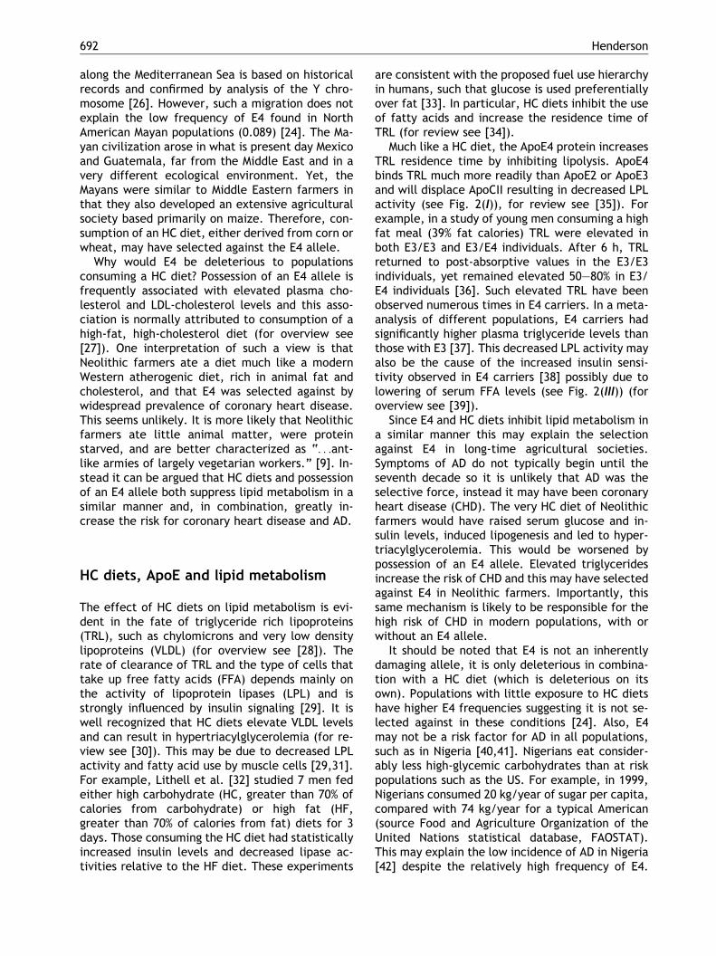
Figure 1 Distribution of apolipoprotein E epsilon 4 allele (regions) in historically agriculture-based societies in the Midmigration of Neolithic farmers from the Middle East along th
3 (E3) and 4 (E4) (for review see [20]). Possession ofthe E4 variant increases the risk of developing AD andbehaves in a dominant dose dependent manner [21].E4 is also a risk factor for coronary heart disease [22]and poor recovery from head trauma [23]. Why sucha “deleterious” allele would be selected against insome populations but not in others may provide animportant clue to the etiology of AD.
The alleles E2, E3 and E4 are not evenly dis-tributed in all populations. In a meta-analysis ofpublished apoE allele frequencies, Corbo andScacchi noted that E4 is under-represented inpopulations with long historical exposure to agri-culture, and they proposed that E4 may be a thriftyallele [24]. Populations with the lowest frequenciesof E4 include long time agriculturalists, such asGreeks (0.068) and Turks (0.079), while popula-tions with the highest frequencies include longtime hunter-gatherers, such as African Pygmies(0.407), Papuans (0.368), and Inuits (0.214) [24].This has been supported by a study of Arab popu-lations living in northern Israel who had the lowestE4 frequency ever recorded (0.04) [25]. One in-terpretation of this distribution is genetic drift dueto migration of populations out of the Middle East(see Fig. 1). The migration of Neolithic farmers
E4), adapted from [24,25]. Frequency of E4 is low (lightdle East and in Central America (inset). Arrows indicatee Mediterranean Sea.
692 Henderson
along the Mediterranean Sea is based on historicalrecords and confirmed by analysis of the Y chro-mosome [26]. However, such a migration does notexplain the low frequency of E4 found in NorthAmerican Mayan populations (0.089) [24]. The Ma-yan civilization arose in what is present day Mexicoand Guatemala, far from the Middle East and in avery different ecological environment. Yet, theMayans were similar to Middle Eastern farmers inthat they also developed an extensive agriculturalsociety based primarily on maize. Therefore, con-sumption of an HC diet, either derived from corn orwheat, may have selected against the E4 allele.
Why would E4 be deleterious to populationsconsuming a HC diet? Possession of an E4 allele isfrequently associated with elevated plasma cho-lesterol and LDL-cholesterol levels and this asso-ciation is normally attributed to consumption of ahigh-fat, high-cholesterol diet (for overview see[27]). One interpretation of such a view is thatNeolithic farmers ate a diet much like a modernWestern atherogenic diet, rich in animal fat andcholesterol, and that E4 was selected against bywidespread prevalence of coronary heart disease.This seems unlikely. It is more likely that Neolithicfarmers ate little animal matter, were proteinstarved, and are better characterized as “. . .ant-like armies of largely vegetarian workers.” [9]. In-stead it can be argued that HC diets and possessionof an E4 allele both suppress lipid metabolism in asimilar manner and, in combination, greatly in-crease the risk for coronary heart disease and AD.
HC diets, ApoE and lipid metabolism
The effect of HC diets on lipid metabolism is evi-dent in the fate of triglyceride rich lipoproteins(TRL), such as chylomicrons and very low densitylipoproteins (VLDL) (for overview see [28]). Therate of clearance of TRL and the type of cells thattake up free fatty acids (FFA) depends mainly onthe activity of lipoprotein lipases (LPL) and isstrongly influenced by insulin signaling [29]. It iswell recognized that HC diets elevate VLDL levelsand can result in hypertriacylglycerolemia (for re-view see [30]). This may be due to decreased LPLactivity and fatty acid use by muscle cells [29,31].For example, Lithell et al. [32] studied 7 men fedeither high carbohydrate (HC, greater than 70% ofcalories from carbohydrate) or high fat (HF,greater than 70% of calories from fat) diets for 3days. Those consuming the HC diet had statisticallyincreased insulin levels and decreased lipase ac-tivities relative to the HF diet. These experiments
are consistent with the proposed fuel use hierarchyin humans, such that glucose is used preferentiallyover fat [33]. In particular, HC diets inhibit the useof fatty acids and increase the residence time ofTRL (for review see [34]).
Much like a HC diet, the ApoE4 protein increasesTRL residence time by inhibiting lipolysis. ApoE4binds TRL much more readily than ApoE2 or ApoE3and will displace ApoCII resulting in decreased LPLactivity (see Fig. 2(I)), for review see [35]). Forexample, in a study of young men consuming a highfat meal (39% fat calories) TRL were elevated inboth E3/E3 and E3/E4 individuals. After 6 h, TRLreturned to post-absorptive values in the E3/E3individuals, yet remained elevated 50–80% in E3/E4 individuals [36]. Such elevated TRL have beenobserved numerous times in E4 carriers. In a meta-analysis of different populations, E4 carriers hadsignificantly higher plasma triglyceride levels thanthose with E3 [37]. This decreased LPL activity mayalso be the cause of the increased insulin sensi-tivity observed in E4 carriers [38] possibly due tolowering of serum FFA levels (see Fig. 2(III)) (foroverview see [39]).
Since E4 and HC diets inhibit lipid metabolism ina similar manner this may explain the selectionagainst E4 in long-time agricultural societies.Symptoms of AD do not typically begin until theseventh decade so it is unlikely that AD was theselective force, instead it may have been coronaryheart disease (CHD). The very HC diet of Neolithicfarmers would have raised serum glucose and in-sulin levels, induced lipogenesis and led to hyper-triacylglycerolemia. This would be worsened bypossession of an E4 allele. Elevated triglyceridesincrease the risk of CHD and this may have selectedagainst E4 in Neolithic farmers. Importantly, thissame mechanism is likely to be responsible for thehigh risk of CHD in modern populations, with orwithout an E4 allele.
It should be noted that E4 is not an inherentlydamaging allele, it is only deleterious in combina-tion with a HC diet (which is deleterious on itsown). Populations with little exposure to HC dietshave higher E4 frequencies suggesting it is not se-lected against in these conditions [24]. Also, E4may not be a risk factor for AD in all populations,such as in Nigeria [40,41]. Nigerians eat consider-ably less high-glycemic carbohydrates than at riskpopulations such as the US. For example, in 1999,Nigerians consumed 20 kg/year of sugar per capita,compared with 74 kg/year for a typical American(source Food and Agriculture Organization of theUnited Nations statistical database, FAOSTAT).This may explain the low incidence of AD in Nigeria[42] despite the relatively high frequency of E4.

Figure 2 ApoE4 and a high carbohydrate diet inhibit lipid metabolism. Bold Roman numerals indicate key points inthe model. (I) E4 preferentially binds triglyceride rich particles such as VLDL and chylomicrons reducing ApoCII binding.(II) Decreased LPL activity inhibits delivery of FFA to astrocytes. (III) Low FFA levels increase insulin sensitivity andfurther decrease LPL activity and ketone body transport. (IV) Inefficient delivery of EFA to cerebral neurons inhibitsfunction of glucose transporters (GLUT). (V) Decreased metabolism lowers acetyl-CoA pools and levels of ATP andacetylcholine. (VI) Increased insulin signaling inhibits Foxo proteins from entering the nucleus and prevents activationof stress response genes, such as antioxidant proteins. Abbreviations: LRP – LDL receptor related protein, VLDL – verylow density lipoprotein, HDL – high density lipoprotein, LPL – lipoprotein lipase, FFA – free fatty acid, MCTr –monocarboxylate transporter, E4 – ApoE4, FATP – fatty acid transport protein, KB – ketone bodies, ACh – acetyl-choline, ROS – reactive oxygen species.
693High carbohydrate diets and Alzheimer’s disease
Prior to the development of agriculture, E2, E3 andE4 may have been neutral alleles that arose whenour human ancestors began to eat more animalmatter, and hence more fat, and this relaxed se-lection on apoE. The development of agriculturethen imposed a new selection on apoE reducing E4in Middle Eastern and Mayan populations.
Overview of the etiology of AD
HC diets are proposed as the primary cause of ADby two basic mechanisms (see Fig. 3 for overview).The first is disturbed lipid homeostasis within theCNS, especially decreased delivery of essentialfatty acids (EFA) (see Fig. 3(I)). This compromisesthe integrity of cellular membranes, decreasing thefunction of membrane proteins such as glucosetransporters and APP. The second is mild chronicelevated insulin/IGF signaling, which acceleratescellular damage (Fig. 3(II)). These two mechanismscontribute to two stages of the disease. Stage Ibegins when altered lipid metabolism inhibits thefunction of membrane proteins such as glucosetransporters, resulting in decreased glucose utili-zation and lowered metabolism in susceptible re-gions of the brain. At this stage no clinical signs ofdementia are evident, yet the disease has begun.Stage II begins when the inhibition of cellularfunction can no longer be compensated for, either
due to excessive cellular damage, or age impairedloss of homeostatic mechanisms. In stage II, acetyl-CoA levels are lowered below critical levels, af-fecting the production of a variety of cellularcomponents such as cholesterol and acetylcholineand clinical signs of dementia become evident. Thedisturbances in cholesterol metabolism result inlarge scale aberrant processing of APP, decreasesin cellular trafficking, and generation of amyloidbeta peptides (Ab). As the disease progresses, thefailure to transport neurotrophin receptors and theproduction of increasing amounts of Ab ultimatelyresults in large scale cell death and the charac-teristic pathology of AD.
Stage I – essential fatty acids andmembrane function
Despite the importance of fatty acids in cerebralneurons little de novo fatty acid synthesis occurs inthe adult brain (for overview see [43]). Most fattyacids are imported as phospholipids or unesterifiedFFA from the plasma through the use of fatty acidtransport proteins (for review see [44]). One im-portant class of fatty acids required by the CNS areEFA. For example, docosahexanoic acid (DHA) isfound extensively in phospholipids of neuronalmembranes (for overview see [45]). Inhibition oflipid metabolism by HC diets may mimic dietary

Figure 3 HC diet and Alzheimer’s disease model overview. Light and shaded areas indicate stages of the disease.Bold Roman numerals highlight major mechanisms of disease progression. (I) HC diet and ApoE4 contribute to de-creased lipid metabolism in central nervous system, altering the function of glucose transporters and amyloid pre-cursor protein (APP). (II) Chronic excessive insulin/IGF signaling inhibits the functioning of Foxo proteins therebyincreasing cellular damage.
694 Henderson
deficiencies of EFA which are known to alter thecomposition of neuronal membranes, disturb theactivity of membrane proteins [46], and lead tobehavioral defects such as poor performance inlearning tasks (for overview see [47]). This is con-sistent with the growing evidence that EFA play arole in AD. Low serum DHA levels have been im-plicated as a significant risk factor [48,49], andconsumption of fish (a rich source of DHA and EPA)may prevent the disease [50]. Additionally, alteredlipid metabolism may be responsible for theextensive membrane deterioration seen in AD[51,52].
In addition to metabolic changes induced by HCdiets, the development of agriculture has directlychanged the normal dietary balance of EFA. It hasbeen estimated that Paleolithic hunter-gatherersate roughly equal amounts of n� 6 and n� 3 fattyacids [12]. However, the modern Western foodsupply is much richer in n� 6 fatty acids due to theuse of grains both in the diet and as animal feed.This has greatly altered the ratio of dietary n� 6 ton� 3 fatty acids from roughly 1:1 for Paleolithichunter-gatherers to �20:1 for a modern diet. Then� 6 fatty acids compete for desaturases used byn� 3 fatty acids to produce products such as DHA,essentially lowering their levels (for review see[53]).
One class of protein known to be effected by EFAlevels are glucose transporters. For example, rats
raised on a n� 3 deficient diet for three monthsexhibit a 30–35% decrease in glucose uptake in thecortex, hippocampus and SCN compared to ad libfed controls, due to inefficient function of glucosetransporters [54]. Such decreases in cerebral glu-cose utilization are one of the earliest signs of ADand are evident in at risk populations well beforeclinical signs of dementia occur, particularly in E4carriers [55]. Yet, at this early stage, poor cogni-tive performance may be masked by recruitinglarger regions of the brain to accomplish mentaltasks [56]. As the disease progresses, inhibition ofglucose use worsens (for overview see [57]), and atsome point declines to where recruitment can nolonger compensate for energy loss (Fig. 3(IV)). Thisis the beginning of Stage II.
Stage II – metabolism, cholesterol andAPP
Cerebral neurons are normally considered to deriveacetyl-CoA almost exclusively from glucose. Asglucose utilization worsens it will begin to depleteneuronal acetyl-CoA pools leading to decreasedsynthesis of acetylcholine (Fig. 3(IV)) and the wellrecognized cholinergic defects found in AD [58].Another less obvious, but perhaps more important,consequence of lower acetyl-CoA levels is altera-

695High carbohydrate diets and Alzheimer’s disease
tions in cholesterol homeostasis (Fig. 4(II)). Thehuman brain contains large amounts of unesterifiedcholesterol, roughly 25% of the total amount in thebody. Unlike FFA, cholesterol is synthesized denovo within neuronal cells from condensation ofacetyl-CoA and is part of a complex regulatoryprocess of cholesterol homeostasis (for review see[59]). Disturbance in this process has been impli-cated in several neurological disorders, includingAD. For example, allelic variation in the cyp46 gene(a cholesterol 24-hydroxylase) has been identifiedas a risk factor [60] and decreased cholesterollevels are found in affected regions of the brain[61].
One important protein that is sensitive to dis-turbances in cholesterol homeostasis is APP(Fig. 4(III)). Early onset AD is frequently associatedwith mutations in three genes; APP, presenilin 1(PS1) and presenilin 2 (PS2). These mutations leadto aberrant processing of the APP protein and ac-cumulation of the Ab peptide (for review see [2]).Recent evidence has suggested that excess cho-lesterol leads to increased APP cleavage. Diet in-duced hypercholesterolemia increases the levels ofAb and amyloid deposits in the CNS of transgenicmouse models of AD [62,63]. Addition of excesscholesterol to cells in culture increases Ab pro-duction, while depleting cells of cholesterol de-
Figure 4 Lipid homeostasis and APP. Bold Roman numeralsdiets inhibit efficient lipid delivery to the brain, inhibiting fpools. (II) Low EFA and acetyl-CoA levels inhibit cholesteroInability to maintain lipid homeostasis results in improper proAPP results in failure to deliver neurotrophin receptors to ceAbbreviations: EFA – essential fatty acid, MCTr – monocarb
creases Ab production (for overview see [64]).Also, treating animals with cholesterol loweringdrugs (statins) decreases the levels of Ab in theblood [65] and may decrease the risk of developingAD up to 70% [66]. Yet, most statin drugs do notcross the blood brain barrier and are predicted tohave a weak, if any, effect on cerebral cholesterolproduction (for overview see [67]).
Alternatively, statins may protect against AD byimproving cerebral lipid metabolism. In addition toinhibition of 3-hydroxy-3-methylglutaryl CoA re-ductase, statins have other physiologic effects,such as vasodilatory and anti-inflammatory. Im-portantly, statins also cause a reduction in circu-lating TRL by increasing the levels of lipoproteinlipase while also decreasing apolipoprotein C-III (aninhibitor of lipoprotein lipase) [68]. Thereforestatins may directly counteract the effects of HCdiets by increasing the activity of LPL.
Ab may not be the only toxic result of aberrantAPP processing. APP is proposed to function as amembrane cargo receptor for kinesin-I during ax-onal transport, delivering several cellular factors,including Bace (beta secretase), Ps1 (Presenilin 1),and the neurotrophin receptor TrkA [69–71]. Mu-tations in APP and the presenilins, as well as dis-turbed cholesterol homeostasis, may lead topremature cleavage of APP and inhibition of cel-
indicate key points in the model. (I) High carbohydrateunction of glucose transporters and lowering acetyl-CoAl metabolism and membrane function and integrity. (III)cessing of APP and Ab generation. Premature cleavage ofll surface and cell death. (IV) Accumulation of toxic Ab.oxylate transporter, KB – ketone bodies.
696 Henderson
lular trafficking [72]. Failure to deliver neurotro-phin receptors would lead to widespread neuronalcell death (Fig. 4, for review see [73]). In fact,inhibiting NGF in the brains of mice results in anage dependent pathology very similar to AD [74].
Stage I/II – insulin/IGF signaling andaging
HC diets are well known to increase glucose andinsulin levels in humans [31] and this elevated in-sulin signaling may lead to rapid aging of suscep-tible tissues. In mammals and lower organismsthere is growing evidence that insulin/IGF signalingmodulates lifespan (for overview see [75]). Forexample, reducing the caloric intake of mice andrats reduces insulin/IGF levels and increases lifespan (for review see [76]). More direct evidencecomes from the observation that mice heterozy-gous for the IGF-1 receptor live �33% longer thantheir wild-type littermates [77] and mice lackingthe insulin receptor in fat cells live �18% longer[78].
The insulin-like signaling pathway shows re-markable conservation across phyla. In both nem-atodes and mammals insulin/IGF signalingnegatively regulates the activity of the Foxo familyof transcription factors by sequestration in thecytoplasm (for review see [79,80]). Activation ofFoxo proteins increases stress resistance and lon-gevity in mice and nematodes [75]. The long-livedp66shc()/)) mouse may have increased Foxo ac-tivation and increased resistance to oxidativestress [81]. Activation of FKHR (a Foxo protein)increases expression of stress response genes, suchas Gadd45a, a gene involved in DNA repair [82]. Ithas been proposed that insulin/IGF signaling func-tions, via Foxo proteins, to adjust metabolism andultimately lifespan in response to nutritional andenvironmental cues [83,84]. Low food availabilitywill increase the proportion of Foxo in the nucleusand increase the expression of a variety of stressresistance genes, resulting in more stress resistantlonger lived individuals. High food availability willdecrease the expression of stress genes, resultingin less stress resistant shorter lived individuals.
The mammalian brain is well supplied with in-sulin receptors where insulin appears to signalabundant food and not trigger glucose uptake as itdoes in muscle and fat [85,86]. For example,chronic infusion of insulin into the brains of ba-boons reduces food intake [87], while inhibition ofthe insulin receptor in the brains of mice in-creases food intake [88,89]. Therefore, the strong
increases in postprandial glucose and insulin levelsinduced by HC diets may continuously signal thatnutrients are plentiful, exclude Foxo from the nu-cleus, and accelerate aging of susceptible neurons(Fig. 3(VI)). This condition will be exacerbated inE4 individuals who are more insulin sensitive.
Treatment and prevention
This hypothesis suggests several treatment andpreventative measures that may be beneficial forAD and other disorders resulting from what can becollectively called the “Neolithic Syndrome”.Such treatment may be especially effective incombination.
The Paleolithic prescription
A modified “Paleolithic prescription” [90] mayprevent AD. The Paleolithic prescription proposes achange in diet and activity to a level more similarto our Late Paleolithic ancestors, and emphasizesreducing fat and increasing dietary fiber as the keysto better health [90,91]. However, the inhibition oflipid metabolism by HC diets may be the mostdetrimental aspect of modern diets. Therefore,reducing dietary intake of high-glycemic carbohy-drates and increasing protein, fiber and fat wouldbe preferred. Similar diets appear to reduce therisk of AD [92]. Since HC diets are proposed to bethe primary cause of AD regardless of apoE geno-type, such a diet would generally reduce the risk ofAD. However, this diet is predicted to be particu-larly beneficial to carriers of apoE4, and suggeststhat individuals should “eat right for your apoEtype”. Dietary change would be the preventativetreatment of choice, since it would not only lowerthe incidence of AD, but many other harmful con-ditions. Yet such a change would require dramaticdecreases in carbohydrate intake (to < 30% of dailycaloric intake) and would be difficult to implementwithout drastic changes in dietary thinking.
EFA repletion diet
Increasing evidence has implicated consumption offish (a source of EFA) as protective against AD [50].For individuals in Stage I or II, an EFA repletionregime, consisting of high doses of EFA, may re-plenish EFA in neuronal membranes and preventand/or treat the disease [93]. In particular, ele-vation of n� 3 EFA may allow for more efficient

697High carbohydrate diets and Alzheimer’s disease
function of glucose transporters and the APPprotein.
Ketone body treatment
While increasing fatty acid metabolism may helpprevent the disease, by the time clinical dementiais diagnosed (Stage II) irreparable damage mayhave occurred and reversal will be difficult. Onestrategy that might be effective is direct elevationof acetyl-CoA levels using ketone bodies (KB). In-creasing acetyl-CoA levels will provide a substratefor acetylcholine and cholesterol synthesis and canbe used in the TCA cycle [94]. A simple way to el-evate plasma KB levels is through consumption ofmedium chain triglycerides, which are readily me-tabolized to KB. We have found that exogenousadministration of medium chain triglycerides in-creased cognitive performance in early stage non-E4 AD patients [95].
Increasing fatty acid metabolism
Drugs that increase the use of fatty acids, espe-cially in glia, may be beneficial for AD. This mayexplain the beneficial effects of statins (as dis-cussed) and non-steroid anti-inflammatory drugs(NSAIDS) [96]. NSAIDS function, in part, as PPAR-gamma agonists. Increasing PPAR-gamma activityincreases the expression of genes associated withfatty acid metabolism such as FATP (for review see[97]). Other drugs may have similar effects. Fibratedrugs, such as Bezafibrate, ciprofibrate, fenofi-brate and Gemfibrozil may also prove beneficial.Fibrates act as PPAR-alpha agonists and like statinsthey increase lipoprotein lipase, apoAI and apoAIItranscription and reduce levels of apoCIII, therebyincreasing lipid availability to the brain [98].
Conclusion
AD is a devastating neurodegenerative disorderthat will reach epidemic proportions in the next 50years. While tremendous progress has been madein our molecular understanding of the disease, noeffective treatments exist. Much of the currentresearch centers on modulating the processing ofthe APP protein and correcting the imbalance be-tween Ab production and clearance. This approach,while promising, has many drawbacks. Altering theprocessing of APP may affect other proteins such asNotch and is technically difficult [99]. Here it is
argued that the primary event leading to the de-velopment of AD is consumption of an evolution-arily discordant HC diet. This hypothesis predictsthat relatively simple preventative measures, suchas lowering the consumption of starchy carbohy-drates and increasing EFA in the diet will be ef-fective. Yet, in practice this may be difficultwithout sufficient public awareness. Other treat-ments may also be effective, such as ketone bodytherapy, EFA repletion diets, and statin drugs.Hopefully, in the future more research will focuson the role of diet in AD.
Acknowledgements
This work is dedicated to Florence Tomlins Hen-derson. I wish to thank members of the Johnson labfor critical reading of the manuscript and activediscussion. I am grateful to Dr. Thomas Johnson forallowing me the time and freedom to pursue myinterests.
References
[1] Sisodia SS, Martin LJ, Walker LC, Borchelt DR, Price DL.Cellular and molecular biology of Alzheimer’s disease andanimal models. Neuroimaging Clin N Am 1995;5:59–68.
[2] Selkoe DJ. Alzheimer’s disease: genes, proteins, andtherapy. Physiol Rev 2001;81:741–66.
[3] Grant WB. Dietary links to Alzheimer’s disease. Alzheimer’sDisease Rev 1997;2:42–55.
[4] Kalmijn S, Launer LJ, Ott A, Witteman JC, Hofman A,Breteler MM. Dietary fat intake and the risk of incidentdementia in the Rotterdam Study. Ann Neurol1997;42:776–82.
[5] Engelhart MJ, Geerlings MI, Ruitenberg A, et al. Diet andrisk of dementia: Does fat matter?: The Rotterdam study.Neurology 2002;59:1915–21.
[6] Neel JV. Diabetes mellitus: A thrifty genotype rendereddetrimental by progress? Am J Hum Genet 1962;14:353–62.
[7] Mehta NG. Did agriculture reduce human lifespan? Nature2001;409:131.
[8] Gowdy JM. Limited wants, unlimited means: a reader onhunter-gatherer economics and the environment. Washing-ton, DC: Island Press; 1998.
[9] Harris M. Cannibals and Kings: the origins of cultures. NewYork: Random House; 1977.
[10] Alroy J. A multispecies overkill simulation of the end-Pleistocene megafaunal mass extinction. Science2001;292:1893–6.
[11] Roberts RG, Flannery TF, Ayliffe LK, et al. New ages for thelast Australian megafauna: continent-wide extinction about46,000 years ago. Science 2001;292:1888–92.
[12] Eaton SB, Konner M. Paleolithic nutrition. A considerationof its nature and current implications. N Engl J Med1985;312:283–9.
[13] Cordain L, Miller JB, Eaton SB, Mann N. Macronutrientestimations in hunter-gatherer diets. Am J Clin Nutr2000;72:1589–92.

698 Henderson
[14] Formicola V, Giannecchini M. Evolutionary trends of staturein upper Paleolithic and Mesolithic Europe. J Hum Evol1999;36:319–33.
[15] Steckel R, Prince JM. Tallest in the World: Native Ameri-cans of the Great Plains in the Nineteenth Century. AER2001;91:287–94.
[16] Angel LJ. Health as a crucial factor in the changes fromhunting to developed farming in the eastern Mediterranean.In: Cohen MN, Armelagos GJ, editors. Paleopathology at theorigins of agriculture. Orlando: Academic Press; 1984. p.51–73.
[17] Cummings JH. Short chain fatty acids in the human colon.Gut 1981;22:763–79.
[18] Popovich DG, Jenkins DJ, Kendall CW, et al. The westernlowland gorilla diet has implications for the health ofhumans and other hominoids. J Nutr 1997;127:2000–5.
[19] Cohen MN, Armelagos GJ, editors. Paleopathology at theorigins of agriculture. Orlando, FL: Academic Press; 1984.
[20] Mahley RW, Rall Jr SC. Apolipoprotein E: far more than alipid transport protein. Ann Rev Genomics Hum Genet2000;1:507–37.
[21] Corder EH, Saunders AM, Strittmatter WJ, et al. Gene doseof apolipoprotein E type 4 allele and the risk of Alzheimer’sdisease in late onset families. Science 1993;261:921–3.
[22] Davignon J, Gregg RE, Sing CF. Apolipoprotein E polymor-phism and atherosclerosis. Arteriosclerosis 1988;8:1–21.
[23] Nicoll JA, Roberts GW, Graham DI. Amyloid beta-protein,APOE genotype and head injury. Ann N Y Acad Sci1996;777:271–5.
[24] Corbo RM, Scacchi R. Apolipoprotein E (APOE) alleledistribution in the world. Is APOE*4 a ‘thrifty’ allele? AnnHum Genet 1999;63(Pt 4):301–10.
[25] Bowirrat A, Friedland RP, Chapman J, Korczyn AD. The veryhigh prevalence of AD in an Arab population is not explainedby APOE epsilon4 allele frequency. Neurology 2000;55:731.
[26] Semino O, Passarino G, Oefner PJ, et al. The genetic legacyof Paleolithic Homo sapiens sapiens in extant Europeans: aY chromosome perspective. Science 2000;290:1155–9.
[27] Ordovas JM. The genetics of serum lipid responsiveness todietary interventions. Proc Nutr Soc 1999;58:171–87.
[28] Murray RK, Granner DK, Mayes PA, Rodwell VW. Harper’sbiochemistry. 25th ed. New York: McGraw-Hill; 1999. p.927.
[29] Campos H, Dreon DM, Krauss RM. Associations of hepaticand lipoprotein lipase activities with changes in dietarycomposition and low density lipoprotein subclasses. J LipidRes 1995;36:462–72.
[30] Parks EJ, Hellerstein MK. Carbohydrate-induced hypertria-cylglycerolemia: historical perspective and review of bio-logical mechanisms. Am J Clin Nutr 2000;71:412–33.
[31] Ginsberg H, Olefsky JM, Kimmerling G, Crapo P, Reaven GM.Induction of hypertriglyceridemia by a low-fat diet. J ClinEndocrinol Metab 1976;42:729–35.
[32] Lithell H, Jacobs I, Vessby B, Hellsing K, Karlsson J.Decrease of lipoprotein lipase activity in skeletal musclein man during a short-term carbohydrate-rich dietaryregime. With special reference to HDL-cholesterol, apoli-poprotein and insulin concentrations. Metabolism1982;31:994–8.
[33] Hellerstein MK. No common energy currency: de novolipogenesis as the road less traveled. Am J Clin Nutr2001;74:707–8.
[34] Hellerstein MK. Carbohydrate-induced hypertriglyceride-mia: modifying factors and implications for cardiovascularrisk. Curr Opin Lipidol 2002;13:33–40.
[35] Weisgraber KH. Apolipoprotein E: structure–function rela-tionships. Adv Protein Chem 1994;45:249–302.
[36] Bergeron N, Havel RJ. Prolonged postprandial responses oflipids and apolipoproteins in triglyceride-rich lipoproteinsof individuals expressing an apolipoprotein epsilon 4 allele.J Clin Invest 1996;97:65–72.
[37] Dallongeville J, Lussier-Cacan S, Davignon J. Modulation ofplasma triglyceride levels by apoE phenotype: a meta-analysis. J Lipid Res 1992;33:447–54.
[38] Craft S, Asthana S, Schellenberg G, et al. Insulin metabo-lism in Alzheimer’s disease differs according to apolipo-protein E genotype and gender. Neuroendocrinology1999;70:146–52.
[39] Randle PJ. Regulatory interactions between lipids andcarbohydrates: the glucose fatty acid cycle after 35 years.Diabetes Metab Rev 1998;14:263–83.
[40] Osuntokun BO, Sahota A, Ogunniyi AO, et al. Lack of anassociation between apolipoprotein E epsilon 4 and Alzhei-mer’s disease in elderly Nigerians. Ann Neurol1995;38:463–5.
[41] Kalaria RN, Ogeng’o JA, Patel NB, et al. Evaluation of riskfactors for Alzheimer’s disease in elderly east Africans.Brain Res Bull 1997;44:573–7.
[42] Hendrie HC, Ogunniyi A, Hall KS, et al. Incidence ofdementia and Alzheimer disease in 2 communities: Yorubaresiding in Ibadan, Nigeria, and African Americans residingin Indianapolis, Indiana. Jama 2001;285:739–47.
[43] Edmond J. Essential polyunsaturated fatty acids and thebarrier to the brain: the components of a model fortransport. J Mol Neurosci 2001;16:181–93, discussion 215-21.
[44] Zhou L, Nilsson A. Sources of eicosanoid precursor fattyacid pools in tissues. J Lipid Res 2001;42:1521–42.
[45] Agranoff BW, Hajra AK. Lipids. In: Siegel GJ, editor. Basicneurochemistry. New York: Raven; 1993.
[46] Bourre JM, Pascal G, Durand G, Masson M, Dumont O,Piciotti M. Alterations in the fatty acid composition of ratbrain cells (neurons, astrocytes, and oligodendrocytes) andof subcellular fractions (myelin and synaptosomes) inducedby a diet devoid of n� 3 fatty acids. J Neurochem1984;43:342–8.
[47] Youdim KA, Martin A, Joseph JA. Essential fatty acids andthe brain: possible health implications. Int J Dev Neurosci2000;18:383–99.
[48] Kyle DJ, Schaefer E, Patton G, Beiser A. Low serumdocosahexaenoic acid is a significant risk factor for Alzhei-mer’s dementia. Lipids 1999;34(Suppl):S245.
[49] Conquer JA, Tierney MC, Zecevic J, Bettger WJ, Fisher RH.Fatty acid analysis of blood plasma of patients withAlzheimer’s disease, other types of dementia, and cogni-tive impairment. Lipids 2000;35:1305–12.
[50] Morris MC, Evans DA, Bienias JL, et al. Consumption of fishand n� 3 fatty acids and risk of incident Alzheimer disease.Arch Neurol 2003;60:940–6.
[51] Nitsch RM, Blusztajn JK, Pittas AG, Slack BE, Growdon JH,Wurtman RJ. Evidence for a membrane defect in Alzheimerdisease brain. Proc Natl Acad Sci USA 1992;89:1671–5.
[52] Prasad MR, Lovell MA, Yatin M, Dhillon H, Markesbery WR.Regional membrane phospholipid alterations in Alzheimer’sdisease. Neurochem Res 1998;23:81–8.
[53] Simopoulos AP. Essential fatty acids in health and chronicdisease. Am J Clin Nutr 1999;70:560S–9S.
[54] Ximenes da Silva A, Lavialle F, Gendrot G, Guesnet P,Alessandri JM, Lavialle M. Glucose transport and utilizationare altered in the brain of rats deficient in n� 3 polyun-saturated fatty acids. J Neurochem 2002;81:1328–37.
[55] Reiman EM, Caselli RJ, Chen K, Alexander GE, Bandy D,Frost J. Declining brain activity in cognitively normalapolipoprotein E epsilon 4 heterozygotes: a foundation for

699High carbohydrate diets and Alzheimer’s disease
using positron emission tomography to efficiently testtreatments to prevent Alzheimer’s disease. Proc Natl AcadSci USA 2001;98:3334–9.
[56] Bookheimer SY, Strojwas MH, Cohen MS, et al. Patterns ofbrain activation in people at risk for Alzheimer’s disease. NEngl J Med 2000;343:450–6.
[57] Swaab DF, Lucassen PJ, Salehi A, Scherder EJ, van SomerenEJ, Verwer RW. Reduced neuronal activity and reactivationin Alzheimer’s disease. Prog Brain Res 1998;117:343–77.
[58] White P, Hiley CR, Goodhardt MJ, et al. Neocorticalcholinergic neurons in elderly people. Lancet1977;1:668–71.
[59] Dietschy JM, Turley SD. Cholesterol metabolism in thebrain. Curr Opin Lipidol 2001;12:105–12.
[60] Papassotiropoulos A, Streffer JR, Tsolaki M, et al. Increasedbrain beta-amyloid load, phosphorylated tau, and risk ofAlzheimer disease associated with an intronic CYP46 poly-morphism. Arch Neurol 2003;60:29–35.
[61] Mason RP, Shoemaker WJ, Shajenko L, Chambers TE,Herbette LG. Evidence for changes in the Alzheimer’sdisease brain cortical membrane structure mediated bycholesterol. Neurobiol Aging 1992;13:413–9.
[62] Howland DS, Trusko SP, Savage MJ, et al. Modulation ofsecreted beta-amyloid precursor protein and amyloid beta-peptide in brain by cholesterol. J Biol Chem1998;273:16576–82.
[63] Refolo LM, Malester B, LaFrancois J, et al. Hypercholester-olemia accelerates the Alzheimer’s amyloid pathology in atransgenic mouse model. Neurobiol Dis 2000;7:321–31.
[64] Pappolla MA, Smith MA, Bryant-Thomas T, et al. Choles-terol, oxidative stress, and Alzheimer’s disease: expandingthe horizons of pathogenesis (1). Free Radic Biol Med2002;33:173–81.
[65] Friedhoff LT, Cullen EI, Geoghagen NS, Buxbaum JD.Treatment with controlled-release lovastatin decreasesserum concentrations of human beta-amyloid (A beta)peptide. Int J Neuropsychopharmacol 2001;4:127–30.
[66] Wolozin B, Kellman W, Ruosseau P, Celesia GG, Siegel G.Decreased prevalence of Alzheimer disease associated with3-hydroxy-3-methyglutaryl coenzyme A reductase inhibi-tors. Arch Neurol 2000;57:1439–43.
[67] Wolozin B. Cholesterol and Alzheimer’s disease. BiochemSoc Trans 2002;30:525–9.
[68] Schoonjans K, Peinado-Onsurbe J, Fruchart JC, Tailleux A,Fievet C, Auwerx J. 3-Hydroxy-3-methylglutaryl CoA reduc-tase inhibitors reduce serum triglyceride levels throughmodulation of apolipoprotein C-III and lipoprotein lipase.FEBS Lett 1999;452:160–4.
[69] Kamal A, Stokin GB, Yang Z, Xia CH, Goldstein LS. Axonaltransport of amyloid precursor protein is mediated bydirect binding to the kinesin light chain subunit of kinesin-I.Neuron 2000;28:449–59.
[70] Gunawardena S, Goldstein LS. Disruption of axonal trans-port and neuronal viability by amyloid precursor proteinmutations in Drosophila. Neuron 2001;32:389–401.
[71] Kamal A, Almenar-Queralt A, LeBlanc JF, Roberts EA,Goldstein LS. Kinesin-mediated axonal transport of amembrane compartment containing beta-secretase andpresenilin-1 requires APP. Nature 2001;414:643–8.
[72] Lynch C, Mobley W. Comprehensive theory of Alzheimer’sdisease. The effects of cholesterol on membrane receptortrafficking. Ann N Y Acad Sci 2000;924:104–11.
[73] Huang EJ, Reichardt LF. Neurotrophins: roles in neuronaldevelopment and function. Annu Rev Neurosci2001;24:677–736.
[74] Capsoni S, Ugolini G, Comparini A, Ruberti F, Berardi N,Cattaneo A. Alzheimer-like neurodegeneration in aged
antinerve growth factor transgenic mice. Proc Natl AcadSci USA 2000;97:6826–31.
[75] Guarente L, Kenyon C. Genetic pathways that regulateageing in model organisms. Nature 2000;408:255–62.
[76] Masoro EJ. Caloric restriction. Aging (Milano) 1998;10:173–4.
[77] Holzenberger M, Dupont J, Ducos B, et al. IGF-1 receptorregulates lifespan and resistance to oxidative stress inmice. Nature 2003;421:182–7.
[78] Bluher M, Kahn BB, Kahn CR. Extended longevity in micelacking the insulin receptor in adipose tissue. Science2003;299:572–4.
[79] Kaestner KH, Knochel W, Martinez DE. Unified nomencla-ture for the winged helix/forkhead transcription factors.Genes Dev 2000;14:142–6.
[80] Birkenkamp KU, Coffer PJ. Regulation of cell survival andproliferation by the FOXO (Forkhead box, class O) subfamilyof Forkhead transcription factors. Biochem Soc Trans2003;31:292–7.
[81] Nemoto S, Finkel T. Redox regulation of forkhead proteinsthrough a p66shc-dependent signaling pathway. Science2002;295:2450–2.
[82] Tran H, Brunet A, Grenier JM, et al. DNA repair pathwaystimulated by the forkhead transcription factor FOXO3athrough the Gadd45 protein. Science 2002;296:530–4.
[83] Kimura KD, Tissenbaum HA, Liu Y, Ruvkun G. daf-2, aninsulin receptor-like gene that regulates longevity anddiapause in Caenorhabditis elegans. Science 1997;277:942–6.
[84] Henderson ST, Johnson TE. daf-16 integrates developmentaland environmental inputs to mediate aging in the nematodeCaenorhabditis elegans. Curr Biol 2001;11: 1975–80.
[85] Havrankova J, Roth J, Brownstein M. Insulin receptors arewidely distributed in the central nervous system of the rat.Nature 1978;272:827–9.
[86] Wickelgren I. Tracking insulin to the mind. Science1998;280:517–9.
[87] Woods SC, Lotter EC, McKay LD, Porte Jr D. Chronicintracerebroventricular infusion of insulin reduces foodintake and body weight of baboons. Nature 1979;282:503–5.
[88] Obici S, Feng Z, Karkanias G, Baskin DG, Rossetti L.Decreasing hypothalamic insulin receptors causes hyper-phagia and insulin resistance in rats. Nat Neurosci2002;5:566–72.
[89] Bruning JC, Gautam D, Burks DJ, et al. Role of brain insulinreceptor in control of body weight and reproduction.Science 2000;289:2122–5.
[90] Eaton SB, Shostak M, Konner M. The Paleolithic prescrip-tion: a program of diet & exercise and a design for living.New York: Harper & Row; 1988,.
[91] Cordain L. The Paleo diet: lose weight and get healthy byeating the food you were designed to eat. New York: Wiley;2002.
[92] Engelhart MJ, Geerlings MI, Ruitenberg A, et al. Dietaryintake of antioxidants and risk of Alzheimer disease. Jama2002;287:3223–9.
[93] Conner WE, Anderson GJ, Lin DS. Dietary n� 3 fatty aciddeficiency and its reversibility. In: Mostofsky DI, Yehuda S,Salem N, editors. Fatty Acids. Totowa: Humana Press;2001. p. 177–92.
[94] Hasselbalch SG, Madsen PL, Hageman LP, et al. Changes incerebral blood flow and carbohydrate metabolism duringacute hyperketonemia. Am J Physiol 1996;270:E746–51.
[95] Reger MA, Henderson ST, Hale C, et al. Effects of beta-hydroxybutyrate on cognition in memory-impaired adults.Neurobiol Aging 2004;25:311–4.

700 Henderson
[96] Zandi PP, Breitner JC. Do NSAIDs prevent Alzheimer’sdisease? And, if so, why? The epidemiological evidence.Neurobiol Aging 2001;22:811–7.
[97] Gelman L, Fruchart JC, Auwerx J. An update on themechanisms of action of the peroxisome proliferator-activated receptors (PPARs) and their roles in inflammationand cancer. Cell Mol Life Sci 1999;55:932–43.
[98] Staels B, Dallongeville J, Auwerx J, Schoonjans K, Leiters-dorf E, Fruchart JC. Mechanism of action of fibrates onlipid and lipoprotein metabolism. Circulation 1998;98:2088–93.
[99] Hardy J, Selkoe DJ. The amyloid hypothesis of Alzheimer’sdisease: progress and problems on the road to therapeutics.Science 2002;297:353–6.