High alkaline phosphatase activity and growth in...
8
Archives of Disease in Childhood, 1989, 64, 902-909 High alkaline phosphatase activity and growth in preterm neonates A LUCAS, 0 G BROOKE, B A BAKER, N BISHOP, AND R MORLEY MRC Dunn Nutrition Unit and University Department of Paediatrics, Cambridge SUMMARY In a study on 857 infants born preterm, high peak plasma alkaline phosphatase activity was independently related to slower growth rate in the neonatal period, and to a highly significant reduction in attained length at 9 months and 18 months post term. At 18 months the deficit in body length associated with peak neonatal plasma alkaline phosphase activity of 1200 IU/l or more was 1 6 cm (95% confidence interval 0-9 to 2-3 cm) after adjusting for confounding factors. The strength and magnitude of this association between high plasma alkaline phosphase activity and body length was greater than that for any other factor identified, including the infant's sex and the presence of fetal growth retardation. Data are presented that support the view that the high plasma alkaline phosphatase activity reflected early bone mineral substrate deficiency resulting in metabolic bone disease. We speculate that even silent early bone disease may interfere with the control of subsequent linear growth and emphasise the potential importance of providing preterm infants, especially those fed human milk, with adequate substrate for bone mineralisation. Metabolic bone disease in preterm infants is common.' Though this has often been described as 'rickets', current evidence suggests that in most cases it is caused by a deficiency of mineral substrate rather than of vitamin D. 4 It is particularly common in very preterm infants who are being fed on breast milk.5 In most instances metabolic bone disease is not associated with obvious clinical signs, and the diagnosis has been based on radiographs, photonabsorptiometry, and biochemical changes including high plasma alkaline phosphatase activity. There is little information about whether such asymptomatic bone disease is benign or associated with an adverse outcome. Although there is evidence that linear growth is impaired in classical rickets,6 the effects of metabolic bone disease on growth performance in premature infants have not been sufficiently investigated. Preterm infants fed their own mothers' milk have raised plasma alkaline phosphatase activity and reduced linear growth in comparison with formula fed babies,7 and there is a negative correlation between plasma alkaline phosphatase activity and linear growth at the age of 12 weeks in a small group of preterm infants8: there are, however, no longer term studies of growth in relation to early evidence of metabolic bone disease. In this paper we present such data in 857 infants from a large multicentre outcome study on the long term effects of early diet in low birthweight babies. This cohort has now been studied to 18 months corrected age. Patients and methods Infants from five centres (Cambridge, Ipswich, King's Lynn, Norwich, and Sheffield) who were participating in a larger multicentre trial of preterm infant feeding were studied.9 In all, 857 infants had sequential plasma alkaline phosphatase measure- ments made, and these are reported here. All infants weighed under 1850 g at birth, and the only exclusions were infants who died within 72 hours of birth and those with serious malformations known to impair growth or development. Parental consent was obtained in all cases, and approval was obtained from the local health authority ethics committees in each of the five centres. Infants were randomly assigned to diet as described previously.9 Briefly, infants in three of the centres were assigned to receive banked donor breast milk or a preterm formula (Osterprem) as sole diets (trial 1; n= 140) or as supplements to maternal milk (trial 2; n=308). In the other two centres a standard full 902 copyright. on 17 July 2018 by guest. Protected by http://adc.bmj.com/ Arch Dis Child: first published as 10.1136/adc.64.7_Spec_No.902 on 1 July 1989. Downloaded from
Transcript of High alkaline phosphatase activity and growth in...

Archives of Disease in Childhood, 1989, 64, 902-909
High alkaline phosphatase activity and growth inpreterm neonates
A LUCAS, 0 G BROOKE, B A BAKER, N BISHOP, AND R MORLEY
MRC Dunn Nutrition Unit and University Department of Paediatrics, Cambridge
SUMMARY In a study on 857 infants born preterm, high peak plasma alkaline phosphataseactivity was independently related to slower growth rate in the neonatal period, and to a highlysignificant reduction in attained length at 9 months and 18 months post term. At 18 monthsthe deficit in body length associated with peak neonatal plasma alkaline phosphase activity of1200 IU/l or more was 1 6 cm (95% confidence interval 0-9 to 2-3 cm) after adjusting forconfounding factors. The strength and magnitude of this association between high plasmaalkaline phosphase activity and body length was greater than that for any other factor identified,including the infant's sex and the presence of fetal growth retardation. Data are presented thatsupport the view that the high plasma alkaline phosphatase activity reflected early bone mineralsubstrate deficiency resulting in metabolic bone disease.We speculate that even silent early bone disease may interfere with the control of subsequent
linear growth and emphasise the potential importance of providing preterm infants, especiallythose fed human milk, with adequate substrate for bone mineralisation.
Metabolic bone disease in preterm infants iscommon.' Though this has often been described as'rickets', current evidence suggests that in mostcases it is caused by a deficiency of mineral substraterather than of vitamin D. 4 It is particularlycommon in very preterm infants who are being fedon breast milk.5 In most instances metabolic bonedisease is not associated with obvious clinical signs,and the diagnosis has been based on radiographs,photonabsorptiometry, and biochemical changesincluding high plasma alkaline phosphatase activity.There is little information about whether suchasymptomatic bone disease is benign or associatedwith an adverse outcome. Although there is evidencethat linear growth is impaired in classical rickets,6the effects of metabolic bone disease on growthperformance in premature infants have not beensufficiently investigated.
Preterm infants fed their own mothers' milk haveraised plasma alkaline phosphatase activity andreduced linear growth in comparison with formulafed babies,7 and there is a negative correlationbetween plasma alkaline phosphatase activity andlinear growth at the age of 12 weeks in a small groupof preterm infants8: there are, however, no longerterm studies of growth in relation to early evidenceof metabolic bone disease.
In this paper we present such data in 857 infantsfrom a large multicentre outcome study on the longterm effects of early diet in low birthweight babies.This cohort has now been studied to 18 monthscorrected age.
Patients and methods
Infants from five centres (Cambridge, Ipswich,King's Lynn, Norwich, and Sheffield) who wereparticipating in a larger multicentre trial of preterminfant feeding were studied.9 In all, 857 infants hadsequential plasma alkaline phosphatase measure-ments made, and these are reported here. Allinfants weighed under 1850 g at birth, and the onlyexclusions were infants who died within 72 hours ofbirth and those with serious malformations knownto impair growth or development. Parental consentwas obtained in all cases, and approval was obtainedfrom the local health authority ethics committees ineach of the five centres.
Infants were randomly assigned to diet as describedpreviously.9 Briefly, infants in three of the centreswere assigned to receive banked donor breast milkor a preterm formula (Osterprem) as sole diets (trial 1;n= 140) or as supplements to maternal milk (trial 2;n=308). In the other two centres a standard full
902
copyright. on 17 July 2018 by guest. P
rotected byhttp://adc.bm
j.com/
Arch D
is Child: first published as 10.1136/adc.64.7_S
pec_No.902 on 1 July 1989. D
ownloaded from

High alkaline phosphatase activity and growth in preterm neonates 903
term adapted formula (Osterfeed) or pretermformula were used as sole diets (trial 3; n= 156) or assupplements to maternal milk (trial 4; n=253). Thepreterm formula contained 35 mg phosphorus and75 mg calcium/100 ml. The full term formulacontained 29 mg phosphorus and 35 mg calcium/100 ml. All infants received a minimum dailyvitamin D intake of 800 IU. The median period(quartiles) to attainment of full enteral feeding was8 (6, 12) days; and to discontinuation of the as-signed diet 29 (19, 46) days. After this, infants wereeither fed by breast or on a standard 'term' infantformula and received 400 IU vitamin D daily untilweaning.The principal objective in this paper was to relate
plasma alkaline phosphase activity to neonatal andlater growth. Data on the effects of diet on plasmaalkaline phosphatase activity and growth are includedhere only to add weight to the argument that highneonatal alkaline phosphatase activity reflectsmetabolic bone disease.
Extensive data were collected on obstetric, peri-natal, and postnatal events, including the followinggrowth measurements in the neonatal period: bodyweight (daily to the nearest 1 g on an electronicbalance, Sartorius MP series); occipitofrontal headcircumference (twice weekly to the next succeeding1 mm using paper tape); and crown-heel length(once or twice weekly to the next succeeding 1 mmusing a horizontal stadiometer). Weight gain (g/kg/day) was calculated from the age when birth weighthad been regained. Head growth and length gainwere calculated by a similar analysis of measure-ments made over a period of at least two weeks afterregaining birth weight.Blood samples were drawn weekly at the time of
routine monitoring from all infants during their stayin hospital. Plasma calcium concentrations weremeasured by atomic absorption spectrophotometryand phosphorus was determined by an end pointmethod on 5 ,ul of plasma using a direct phospho-molybdate reaction without deproteinisation (Smith,Kline Instrument Co Ltd). Alkaline phosphataseactivity was measured kinetically on 2-5 I,l plasma bythe catalysed hydrolysis of p-nitro-phenylphosphateto p-nitrophenol and phosphoric acid (RocheProducts Ltd).
All surviving infants from three centres (Cam-bridge, Ipswich, and King's Lynn) were invited forfollow up examinations at 9 months post term, andchildren from all five centres were examined at18 months post term. Measurements of weight,crown-heel length, and occipitofrontal head circum-ference were included at these examinations'.
Statistical analysis was by the x2 test, Student'st test, and regression analysis.
Results
Details of the study population are shown in table 1.The group has been divided into two accordingto peak plasma alkaline phosphatase activities(¢1200 IU/l). The value of 1200 IU/1, was chosenbecause, as described below, this was shown to bethe cut off point that best discriminated between twogroups of infants in terms of their growth perform-ance.
ASSOCIATION BETWEEN INCREASED ALKALINEPHOSPHATASE ACTIVITY AND EVIDENCE OF METABOLICBONE DISEASEWe used sequential measurement of plasma alkalinephosphatase activity as an enzymatic marker ofmetabolic bone disease. Previously we conducted asurvey in 26 infants (unpublished observations).Thatshowed a highly significant correlation between ascore for metabolic bone disease based on radio-graphic appearances,10 and maximum plasmaalkaline phosphatase activity, confirming the findingsof Glass et al that alkaline phosphatase activity is a
Table 1 Clinical details ofstudy population; raw data havebeen divided according topeak alkaline phosphataseactivity (up to, or above 1200 lU/I) during the neonatalperiod
Alkaline phosphatase p Valueactivity
<1200 IUII 41200 IUI
Mean (SEM) birthweight (g) 1420 (11) 1082 (24) <0-001
Mean (SEM) gestation(weeks) 31-0 (0-1) 29-2 (0-2) <0-001
Duration of ventilation:No (%) <24 hours(n=474) 427 (90) 47 (10) <0001
No (%) 424 hours(n=381) 298 (78) 83 (22)
Sex:No (%) boys
(n=425) 357 (84) 68 (16) >05No (%) girls(n=432) 370 (86) 62 (14)
Weight for gestationalage:No (%) appropriate
(n=567) 485 (86) 82 (15)No (%) small for >0 4
gestational age(n=289) 241 (83) 48 (17)
Pre-eclampsia:No (%) without(n=607) 519 (86) 40 (17) >o04
No (%) with(n=240) 200 (83) 40 (17)
copyright. on 17 July 2018 by guest. P
rotected byhttp://adc.bm
j.com/
Arch D
is Child: first published as 10.1136/adc.64.7_S
pec_No.902 on 1 July 1989. D
ownloaded from
904 Lucas, Brooke, Baker, Bishop, and Morley
Table 2 Association between peak plasma alkalinephosphatase activity and incidence ofhypophosphataemia,low urinary phosphorus concentration, and hypercalcaemia
Peak plasma alkaline p Value*phosphatase activity
<1200 lUII >1200 lU/INo (%) No (%)
Low plasma phosphorusconcentration(<1.0 mmoVl) 109/725 (15) 78/130 (60) <0-0001
tLow urine phosphorusconcentration(<0-02 mmol/l) 62/246 (25) 16/36 (44) <0-02
High plasma calciumconcentration(>3.0 mmol/l) 9/549 (1-6) 12/116 (10) <0 0001
*X2 test, ldf; testimates made on only-a segmental subsample.
satisfactory measure of active metabolic bone diseasein such studies." We provide here further strongevidence that high plasma alkaline phosphataseactivity was related to bone mineral substratedeficiency.
Analysis of raw data showed that infants withhigh peak plasma alkaline phosphatase activity(>1200 IU/1) had a higher incidence of low plasmaand urinary phosphorus concentrations (minimumrecorded values less than 1 -0 mmol/l, and 0*02 mmol/Il,respectively), and a higher incidence of hyper-calcaemia (peak plasma calcium concentration morethan 3*0 mmol/l), each suggesting low phosphorusintake or phosphorus deficiency (table 2). These
Table 3 Effect ofdonor breast milk or preterm formula,assigned randomly (as sole diets or supplements to maternalexpressed milk), on the incidence ofhigh peak plasmaalkaline phosphatase values, hypophosphataemia, and lowor high urinary phosphorus concentrations
Donor breast Preterm p Value*milk formulaNo (%) No (%)
High plasma alkalinephosphatase activity(>1200 1U/1) 57/226 (25) 31/222 (14) <001
Low plasma phosphorusconcentration(<1-0 mmoVIl) 49/153 (32) 27/155 (17) <001
Low urine phosphorusconcentration(<0-02 mmoVl) 39/78 (50) 16/70 (23) <0-001
High urine phosphorusconcentration(>3 mmoVl) 1/78 (1-3) 16/70 (23) <00001
*x2, 1 df.
associations were still significant (p<005) afterusing logistic regression to adjust for birth weight,gestation, duration of ventilatory assistance, andfetal growth retardation.
In the randomised comparison of infants feddonor breast milk or preterm formula (as sole dietsor supplements to maternal milk; trials 1 and 2combined), those fed banked milk with its lowercontent of phosphorus and calcium had a higherincidence of high plasma alkaline phosphataseactivity (table 3). Fig 1 shows that in infantsweighing under 1500 g (the high risk group for bonedisease) plasma alkaline phosphatase valuesremained consistently higher in infants fed donormilk than in those fed a preterm formula throughoutthe first nine weeks postpartum. High alkalinephosphatase activity in infants fed human milk wasassociated with a higher incidence of hypophos-phataemia and low urinary phosphorus values in thisgroup (table 3).
1500
-
" 1000-
50
-C
c
Q
_u-CQ
0-
o
PITPTF
Ii
1 2 3 4 5
Week
6 7 8 9+
Fig 1 Mean (SEM) plasma alkaline phosphatase activityfrom week I to week 9 and beyond in infants weighingunder 1500 g at birth, according to diet. Infants wererandomly assigned to banked breast milk (BBM) orpretermformula (PTF) as sole diets or supplements to their mother'sown milk, Sample size at week I forBBM was 129 andforPTF126, falling respectively (as the infants were dischargedor died) to 101 and 95 at week 4 and to 36 and 19 at week 9and beyond; *p<0o05; **p<0.0JI
i i
copyright. on 17 July 2018 by guest. P
rotected byhttp://adc.bm
j.com/
Arch D
is Child: first published as 10.1136/adc.64.7_S
pec_No.902 on 1 July 1989. D
ownloaded from
High alkaline phosphatase activity and growth in preterm neonates 905
8-5E l
.I
.
71
70......
69 r
68-
67-
66-
65
AS /
32 8274
.I........
90
64
............................................
58
2317
4
42
42.1....................................1 7
30
on- veZZA roZZZ vZZA KZZZA v^ZZ rttt rtzzz V.,2A VZZA81 ( 118
77 17380- 8
.68
..
28 20
79-5
78-
77-
76-
400 400- 600- 800- 1000- 1200- 1400- 1600- 1800+599 799 999 1199 1399 1599 1799Peak alkaline phosphatase activity (IU I)
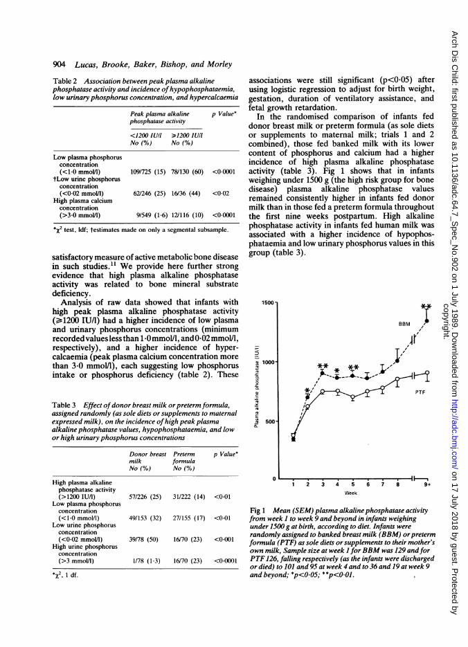
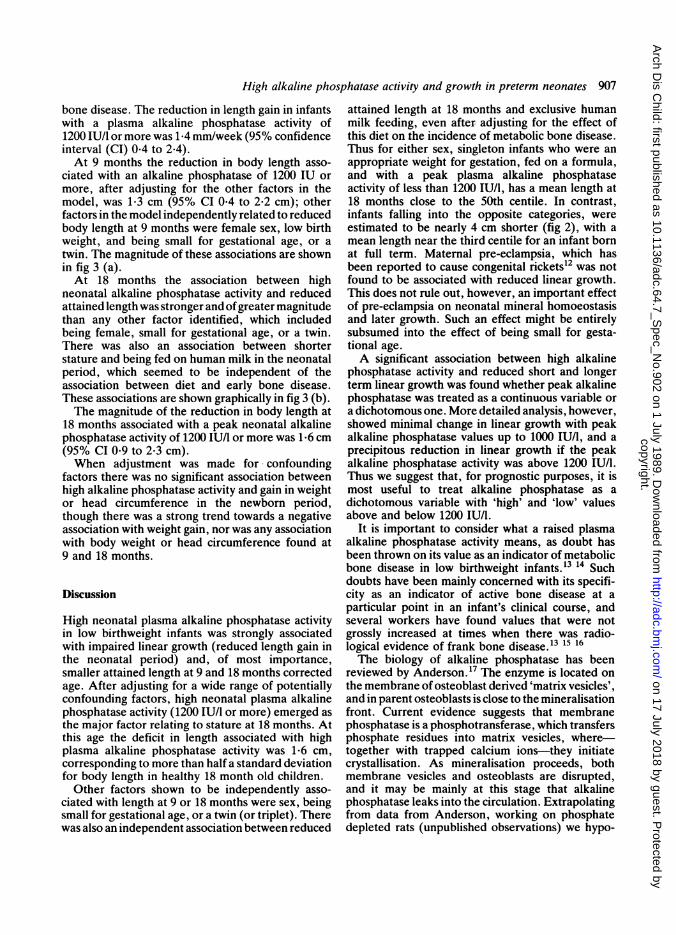
Fig 2 Association between peak plasma alkalinephosphatase activity and (a) rate ofneonatal length gain(b) length at 9 months corrected age and (c) length at18 months corrected age. The horizontal line represents themean value for length gain or length at 9 or 18 months in allinfants whose peak alkaline phosphatase value was<1000 IUII (firstfour data bars ofeach group).
ASSOCIATION BETWEEN INCREASED PLASMA ALKALINEPHOSPHATASE ACTIVITY AND GROWTHGrowth was assessed in the neonatal period asweight gain (g/kg/day), length gain (mm/day), andhead circumference gain (mm/day). Body weight(kg), length (mm), and head circumference (mm)were recorded at 9 months in three of the fivecentres, and at 18 months in all centres. The 719subjects seen at 18 months represented a 94% followup rate among survivors.The principle outcome of interest in this study was
linear growth. Analysis of raw data (fig 2) indicated
that there was no trend in neonatal length gain or inlength at 9 at 18 months with increasing peakalkaline phosphatase activities up to 1000 IU/l.When values were above this, linear growth andlater body length were lower with the most dramaticreductions seen in babies with peak alkaline phos-phatase activites of over 1200 IU/l.
In table 4 raw data are presented for these growthvariables according to whether or not peak alkalinephosphatase exceeded 1200 IU/1. In the group ofinfants with high peak plasma alkaline phosphataseactivity, gains in weight, length, and head circum-ference were significantly reduced in the neonatalperiod. At 9 and 18 months body length and bodyweight, though not head circumference, were signi-ficantly lower.These comparisons of raw data are confounded by
other differences (including those shown in table 1)between infants with high and low alkaline phospha-tase activities, which could contribute to differencesin growth performance. We therefore constructed aseries of regression models, each of which includedone of the above growth variables as the dependentvariable; independent variables were plasma alkaline
Table 4 Association between high plasma alkalinephosphatase activity (peak value >1200 IUIl), growth in theneonatalperiod, and anthropometric indices at 9 and18 months (corrected post term age). Data expressed asmean (SEM)
Peak plasma alkaline p Valuephosphatase activity
<1200 IU/I >s'1200 IU/I
Neonatal period*:Weight gain(g/kg per day)(n=703) 14-6 (0-2) 13-7 (0-2) <0-02Head circumferencegain (mm/day)(n=581) 1.36 (0.03) 1-16 (0.04) <0-01Length gain(mm/day) (n=481) 1-40 (0-02) 1-29 (0.03) <0.05
At 9 months (n=362):Weight (kg) 8-03 (0-07) 7-35 (0-13) <0-001Head circumference(mm) 45 5 (0-1) 45-3 (0-2) >0*4
Length (mm) 69-7 (0-2) 67-6 (0-4) <0.001At 18 months (n=719):Weight (kg) 10-21 (0-05) 9-68 (0-13) <0-001Head circumference(mm) 47-9 (0-1) 47-8 (0-2) >0-7
Length (mm) 79-8 (0-1) 78-2 (0-4) <0-001
*Number of subjects analysed for weight, head circumference, andlength gain were the number for whom more than three data pointshad been collected over at least two weeks beyond the age ofregaining birth weight.
IZ ,/LIZZX llzzz|oot4X{te svo
I
I
3
7
750ETa)
.Ecw-i
E
c-C0E
1;of.ca:-i
copyright. on 17 July 2018 by guest. P
rotected byhttp://adc.bm
j.com/
Arch D
is Child: first published as 10.1136/adc.64.7_S
pec_No.902 on 1 July 1989. D
ownloaded from
906 Lucas, Brooke, Baker, Bishop, and Morley
phosphatase activity (either as a continuous ordichotomous variable), together with birth weight,gestation, fetal growth retardation (above or below10th centile of birth weight for gestation), sex, daysof ventilation, and days in oxygen (expressed aslogarithm, plus 1 to avoid log 0), number of fetuses(singletons or twins/triplets) and maternal hyper-tension. As a further independent variable, diet(exclusively fed human milk or not) was added tothe models.Plasma alkaline phosphatase activity was initially
entered into these models as a continuous variableto assess its relationship, after adjusting for poten-tially confounding factors, with neonatal lengthgain, and body length, at 9 or 18 months. Thoughthere was a significant linear relationship betweenincreasing peak alkaline phosphatase activity andtwo of these growth variables (neonatal length gain,p<001, and length at 18 months, p<00005), therewas a better fit using a quadratic regression, with thesquare of peak alkaline phosphatase activity relatingsignificantly to length gain (p<0.01), length at9 months (p<0-01), and length at 18 months(p<0.OOOl). Nevertheless, from further modelling,
( ) 9 Months
qj 0.4rta'*
0.0,
_ _ _
E-acmcs
c
._1
aa: l
.-Z- A
7
'7,a'
a' f"~Fa Q-'
o'-OS,~~ Ci,"~ -"~
and inspection of the raw data in fig 3, we observeda dramatic reduction in neonatal length gain and inbody length at 9 or 18 months in infants whose peakalkaline phosphatase was above 1000 IU/l. Thisjustifies treating peak alkaline phosphatase activityas a dichotomous variable. Using a variety of cutoff values, significant relationships between peakalkaline phosphatase and all three of the lineargrowth outcome variables was found for cut offvalues between 900 IU/l and 1350 IU/l; the bestmodelled cut off for predicting outcome, however(taking both the size of the regression coefficientand significance into account) was for a cut off at1200 IU/l (as suggested also by the data in fig 3). Insubsequent analysis this value of 1200 IU/l was usedto divide infants into those with 'high' or 'low' peakalkaline phosphatase activities.
In the neonatal period (up to the time ofdischarge) the only factors among those included inthe regression model that were independently relatedto length gain were high alkaline phosphataseactivity (p<001), and diet (<0.05), both of whichseemed to have a small additional association withlinear growth independent of its relationship with
18 Months e
-Psi1'el - 1
9.1~~~~QlA~~~a
7
*-b
sl!~ I'0,{si
7
-2pO J
p Value < 0 0001 <0-001 <0-005 <0-001 <0-005 <0-0001 <0-0001 <0-0005 <0002 <002
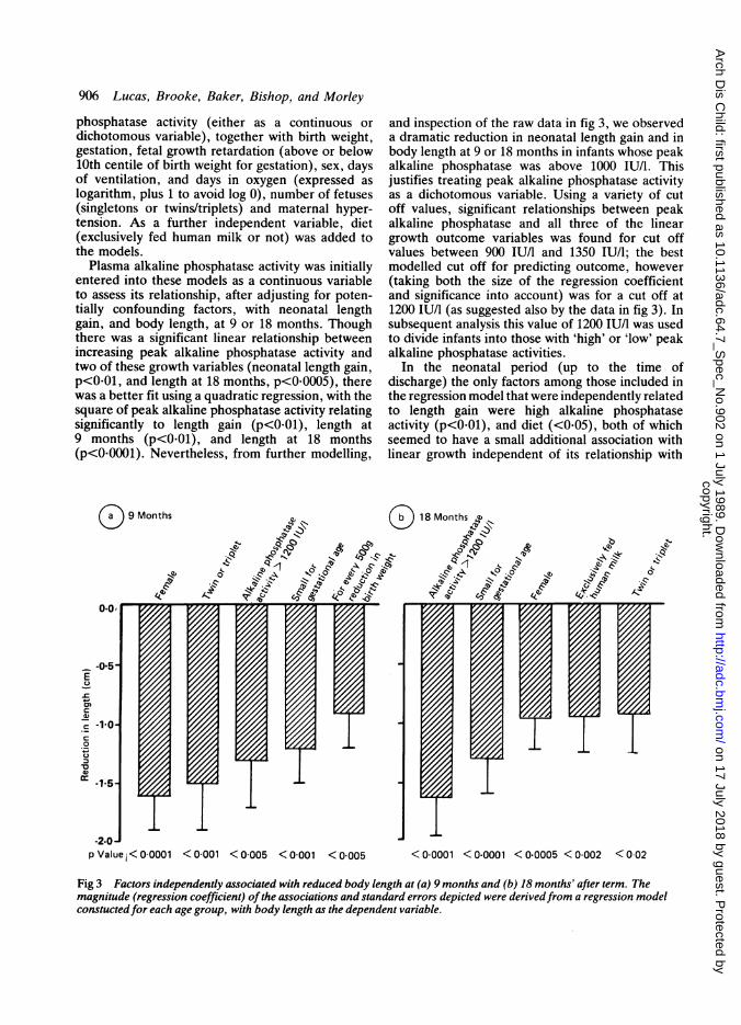
Fig 3 Factors independently associated with reduced body length at (a) 9 months and (b) 18 months' after term. Themagnitude (regression coefficient) ofthe associations and standard errors depicted were derivedfrom a regression modelconstucted for each age group, with body length as the dependent variable.
1~~~~---- - -.1vssss] ~ ~ ~ ~ ~ ~ ~ ~ ~ ~ ~ ~ ~ ~ 1IsseIsss I.Jss w s
copyright. on 17 July 2018 by guest. P
rotected byhttp://adc.bm
j.com/
Arch D
is Child: first published as 10.1136/adc.64.7_S
pec_No.902 on 1 July 1989. D
ownloaded from
High alkaline phosphatase activity and growth in preterm neonates 907
bone disease. The reduction in length gain in infantswith a plasma alkaline phosphatase activity of1200 IU/l or more was 1*4 mm/week (95% confidenceinterval (CI) 0.4 to 2.4).At 9 months the reduction in body length asso-
ciated with an alkaline phosphatase of 1200 IU ormore, after adjusting for the other factors in themodel, was 1*3 cm (95% CI 0*4 to 2*2 cm); otherfactors in the model independently related to reducedbody length at 9 months were female sex, low birthweight, and being small for gestational age, or atwin. The magnitude of these associations are shownin fig 3 (a).At 18 months the association between high
neonatal alkaline phosphatase activity and reducedattained length was stronger and ofgreater magnitudethan any other factor identified, which includedbeing female, small for gestational age, or a twin.There was also an association between shorterstature and being fed on human milk in the neonatalperiod, which seemed to be independent of theassociation between diet and early bone disease.These associations are shown graphically in fig 3 (b).The magnitude of the reduction in body length at
18 months associated with a peak neonatal alkalinephosphatase activity of 1200 IU/I or more was 1-6 cm(95% CI 0-9 to 2-3 cm).When adjustment was made for confounding
factors there was no significant association betweenhigh alkaline phosphatase activity and gain in weightor head circumference in the newborn period,though there was a strong trend towards a negativeassociation with weight gain, nor was any associationwith body weight or head circumference found at9 and 18 months.
Discussion
High neonatal plasma alkaline phosphatase activityin low birthweight infants was strongly associatedwith impaired linear growth (reduced length gain inthe neonatal period) and, of most importance,smaller attained length at 9 and 18 months correctedage. After adjusting for a wide range of potentiallyconfounding factors, high neonatal plasma alkalinephosphatase activity (1200 IU/I or more) emerged asthe major factor relating to stature at 18 months. Atthis age the deficit in length associated with highplasma alkaline phosphatase activity was 1-6 cm,corresponding to more than half a standard deviationfor body length in healthy 18 month old children.Other factors shown to be independently asso-
ciated with length at 9 or 18 months were sex, beingsmall for gestational age, or a twin (or triplet). Therewas also an independent association between reduced
attained length at 18 months and exclusive humanmilk feeding, even after adjusting for the effect ofthis diet on the incidence of metabolic bone disease.Thus for either sex, singleton infants who were anappropriate weight for gestation, fed on a formula,and with a peak plasma alkaline phosphataseactivity of less than 1200 IU/l, has a mean length at18 months close to the 50th centile. In contrast,infants falling into the opposite categories, wereestimated to be nearly 4 cm shorter (fig 2), with amean length near the third centile for an infant bornat full term. Maternal pre-eclampsia, which hasbeen reported to cause congenital rickets12 was notfound to be associated with reduced linear growth.This does not rule out, however, an important effectof pre-eclampsia on neonatal mineral homoeostasisand later growth. Such an effect might be entirelysubsumed into the effect of being small for gesta-tional age.A significant association between high alkaline
phosphatase activity and reduced short and longerterm linear growth was found whether peak alkalinephosphatase was treated as a continuous variable ora dichotomous one. More detailed analysis, however,showed minimal change in linear growth with peakalkaline phosphatase values up to 1000 IU/l, and aprecipitous reduction in linear growth if the peakalkaline phosphatase activity was above 1200 IU/I.Thus we suggest that, for prognostic purposes, it ismost useful to treat alkaline phosphatase as adichotomous variable with 'high' and 'low' valuesabove and below 1200 IU/l.
It is important to consider what a raised plasmaalkaline phosphatase activity means, as doubt hasbeen thrown on its value as an indicator of metabolicbone disease in low birthweight infants.'3 14 Suchdoubts have been mainly concerned with its specifi-city as an indicator of active bone disease at aparticular point in an infant's clinical course, andseveral workers have found values that were notgrossly increased at times when there was radio-logical evidence of frank bone disease.13 15 16The biology of alkaline phosphatase has been
reviewed by Anderson.17 The enzyme is located onthe membrane of osteoblast derived 'matrix vesicles',and in parent osteoblasts is close to the mineralisationfront. Current evidence suggests that membranephosphatase is a phosphotransferase, which transfersphosphate residues into matrix vesicles, where-together with trapped calcium ions-they initiatecrystallisation. As mineralisation proceeds, bothmembrane vesicles and osteoblasts are disrupted,and it may be mainly at this stage that alkalinephosphatase leaks into the circulation. Extrapolatingfrom data from Anderson, working on phosphatedepleted rats (unpublished observations) we hypo-
copyright. on 17 July 2018 by guest. P
rotected byhttp://adc.bm
j.com/
Arch D
is Child: first published as 10.1136/adc.64.7_S
pec_No.902 on 1 July 1989. D
ownloaded from

908 Lucas, Brooke, Baker, Bishop, and Morley
thesise that when bone mineral substrate is deficientan adaptive increase in synthesis or activity ofalkaline phosphatase occurs, resulting in greatlyincreased plasma activity despite reduced bonemineralisation. Certainly it would not be surprisingif the complex events leading to increased alkalinephosphatase activity in the plasma were not preciselyrelated in time to structural evidence of bonemineral deficiency. Nevertheless, when grosslyelevated, plasma alkaline phosphatase activity inneonates is predominantly of bone origin18 andthere are abundant data to show that its activity isusually increased during frank metabolic bonedisease, often exceeding the maximum adult valueseveral fold.1'13-1619-2 It is increased whenbone mineral intake is inadequate and growth isslowed.2 7 23-26 Many workers have regarded raisedplasma alkaline phosphatase activity as an importantindicator of metabolic bone disease,1' 16 21 2 and inparticular when sequential monitoring is under-taken.16 Conversely, a reduction in plasma alkalinephosphatase activity has been observed during catchup growth following periods of slow growth inpreterm infants.8Our data add further weight to the view that high
plasma alkaline phosphatase activity is associatedwith bone mineral substrate deficiency. In a ran-domised comparison infants fed human milk hadsignificantly higher plasma alkaline phosphataseactivity than those receiving a formula providingtwice the calcium and phosphorus contents of breastmilk (fig 1). The human milk fed group showedfurther evidence of phosphorus depletion, with asignificantly higher incidence of hypophosphataemiaand low urine phosphorus concentrations. Moreover,high alkaline phosphatase activity itself was asso-ciated with hypophosphataemia, low urinary phos-phorus concentrations, and an increased incidenceof hypercalcaemia, which has been described withphosphorus depletion.28 29
Regardless of the debate on whether high plasmaalkaline phosphatase activity is a valuable neonatalscreening test for metabolic bone disease in indivi-dual preterm infants, we have shown that it is animportant indicator of outcome in terms of laterlinear growth. Few previous studies have relatedearly bone disease to long term outcome; work inthis field has focused mainly on identification andquantification of active bone disease. Metabolicbone disease in preterm infants, however, is usuallyasymptomatic and is frequently missed. Indeed, inthis investigation high plasma alkaline phosphataseactivity often occurred when the baby was well, andmight have been overlooked had the infant not beentaking part in a scientific study. We suggest poorlinear growth performance both in the short and
longer term could be viewed as an important,adverse, clinical outcome response to even silentearly metabolic bone disease. We speculate also thatthe trend towards reduced neonatal weight gain seenwith increased plasma alkaline phosphatase activityis linked to the effects of phosphorus depletion onsoft tissue growth and metabolism.30 Though wehave shown that high alkaline phosphatase activityrelates well to these adverse responses, it is pertinentto note that other tests for metabolic bone disease,namely radiographs of long bones and photon-absorptiometry, as yet have unknown predictivevalue for later outcome. Furthermore, radiographyand densitometry both include irradiation of thepatient.Our hypothesis, strongly supported by the data
presented, that neonatal metabolic bone disease hasa long lasting and important effect on linear growth,is perhaps a surprising one as excellent catch up isusually seen during the treatment of classical rickets.3'Early bone disease in preterm infants seems increas-ingly unlikely to be a vitamin D related problem inmost cases1 so cannot be directly compared withclassical rickets. Even so, if we assume that thecause of metabolic bone disease in primarily adeficiency of bone mineral substrate, it still requiresexplanation that catch up growth and a return tonormal growth velocity do not apparently take placeonce calcium and phosphorus intake and absorptionhave reached normal infant values32 and the boneshave remineralised.33We have considered that our findings could be
explained by some completely different and as yetunidentified confounding factor or factors that wereassociated with early bone disease and had acontinued influence on growth. Arguably it isdifficult to adjust statistically for the variable degreeof illness seen in preterm infants. We used days ofventilation and days in oxygen as such indicators.Though these measures (especially days of ventila-tion) have proved to be sensitive markers of poorlonger term prognosis for other outcome measuresin this cohort (for example neurodevelopment34),interestingly we found here that requirement forrespiratory support was unrelated to linear growthup to 18 months after adjustment was made forother factors such as birth weight, fetal growthretardation, and twinning. Alternatively, we suggestthat early metabolic bone disease itself may interferewith the control mechanisms for subsequent lineargrowth. This might occur either as a direct conse-quence of the bone disease itself, or because of thesecondary effects such disease may have on thehormonal milieu during a critical growth periodnormally protected in the intrauterine environment.Indeed, it is possible that any process that results in
copyright. on 17 July 2018 by guest. P
rotected byhttp://adc.bm
j.com/
Arch D
is Child: first published as 10.1136/adc.64.7_S
pec_No.902 on 1 July 1989. D
ownloaded from

High alkaline phosphatase activity and growth in preterm neonates 909
lasting retardation of linear growth at an early-perhaps critical-stage could have an adverse 'pro-gramming' or 'imprinting' effect on later lineargrowth performance. In this regard it is worthconsidering the serious effect on long term lineargrowth that may occur in infants who have beenborn stunted at term after prolonged placentalinsufficiency.35 Our data presented here indicate anindependent detrimental effect of fetal growthretardation on linear growth to 18 months.We suggest that even entirely subclinical metabolic
bone disease may have prolonged consequences forgrowth performance. Though our future plannedfollow up of this cohort will be important inassessing the longer term significance of our obser-vations, at present it would seem prudent to makeevery effort to achieve an optimal intake of bonemineral substrate in the neonatal period. Thiscannot be achieved with unsupplemented humanmilk.
We wish to thank Drs MFM Bamford, JFB Dossetor, AW Boon,RG Pearse, and PM Crowle for their assistance in the neonatalunits at Ipswich, King's Lynn, Sheffield, and Norwich, and alsoFarley Health Products for financial assistance.
References
'Brooke OG, Lucas A. Metabolic bone disease in preterminfants. Arch Dis Child 1985;60:682-5.
2 Rowe J, Rowe D, Horak E, et al. Hypophosphatemia andhypercalciuria in small premature infants fed human milk:evidence for inadequate dietary phosphorus. J Pediatr 1984;104:112-7.
3Chan GM, Mileur L, Hansen JW. Effects of increased calciumand phosphorus formulas and human milk on bone mineralizationin preterm infants. J Pediatr Gastroenterol Nutr 1986;5:444-9.
4 Gross SJ. Bone mineralization in preterm infants fed humanmilk with and without mineral supplementation. J Pediatr1987;111:450-8.
5McIntosh N, Livesay A, Brooke OG. Plasma 25-hydroxyvitaminD and rickets in infants of extremely low birthweight. Arch DisChild 1982;57:848-50.
6 Steams G, Jeans PC, Vandecar V. The effect of vitamin D onlinear growth in infancy. J Pediatr 1936;9:1-10.Cooper PA, Rothberg AD, Davies VA, Argent AC. Compara-tive growth and biochemical response of very low birthweightinfants fed own mother's milk, a premature infant formula, andone of two standard formulas. J Pediatr Gastroenterol Nutr1985;4:786-94.
8 James JA, Mayne PD, Barnes IC, Kovar IZ. Growth velocityand plasma alkaline phosphatase activity in the preterm infant.Early Hum Dev 1985;11:27-32.
9Lucas A, Gore SM, Cole TJ, et al. Multicentre trial on feedinglow birthweight infants: effects of diet on early growth. Arch DisChild 1984;59:722-30.
'o Pohlandt F. A radiological sign of bone demineralization inpreterm infants. Klin Padiatr 1985;197:155-6.
" Glass EJ, Hume R, Hendry GM, Strange RC, Forfar JO.Plasma alkaline phosphatase activity in rickets of prematurity.Arch Dis Child 1982;57:373-6.
12 Zeidan S, Bamford M. Congenital rickets with maternal pre-eclampsia. J R Soc Med 1984;77:426-7.
13 McIntosh N, Williams JE, Lyon AJ, Wheeler KA. Diagnosis ofrickets of prematurity. Lancet 1984;ii:869.
14 James JR, Congdon PJ, Truscott J, Horsman A, Arthur R.Osteopenia of prematurity. Arch Dis Child 1986;61:871-6.
15 Koo WW, Gupta JM, Nayaner VV, Wilkinson M, Posen A.Skeletal changes in preterm infants. Arch Dis Child 1982;57:447-52.
16 Walters EG, Murphy JF, Henry P, Gray OP, Elder GH. Plasmaalkaline phosphatase activity and its relation to rickets inpreterm infants. Ann Clin Biochem 1986;23:652-6.
17 Anderson HC. Matrix vesicle calcification: review and update.In: Peck WA, ed. Bone and mineral research 3. Amsterdam:Elsevier, 1985:109-49.
18 Brooke OG, Brown IRF, Bone CDM, et al. Vitamin Dsupplements in pregnant Asian women: effects on calcium statusand fetal growth. Br Med J 1980;280:751-4.
19 Kovar IZ, Mayne P, Wallis J. Neonatal rickets in one ofidentical twins. Arch Dis Child 1982;57:792-4.
20 Chudler AE, Brown DR, Holzman IR, Oh KS. Nutritionalrickets in two very low birthweight infants with chronic lungdisease. Arch Dis Child 1980;55:687-90.
21 Kovar IZ, Mayne P, Barltrop D. Plasma alkaline phosphataseactivity: a screening test for rickets in preterm neonates. Lancet1982;i:373-6.
22 Callenback JC, Sheehan MB, Abramson SJ, Hall RT. Etiologicfactors in rickets of very low-birthweight infants. J Pediatr1981 ;98:800-5.
23 Eek S, Gabrielsen LH, Halvorsen S. Prematurity and rickets.Pediatrics 1957;20:63-77.
24 Schanler RJ, Garza C, O'Brien Smith E. Fortified mother's milkfor very low birthweight infants: results of macromineralbalance studies. J Pediatr 1985;107:767-74.
25 Hillman LS, Hoff NH, Salmons S, Martin L, McAlister W,Haddad J. Mineral homeostasis in very premature infants: serialevaluation of serum 25-hydrocyvitamin D, serum minerals, andbone mineralization. J Pediatr 1985;106:970-80.
2 Pettifor JM, Stein H, Herman A, Ross FP, Blumenfield T,Moodely GP. Mineral homeostasis in very low birthweightinfants fed either own mother's milk or pooled pasteurizedpreterm milk. J Pediatr Gastroenterol Nutr 1986;5:248-53.
27 Hillman LS, Salmons SJ, Slatopolsky E, McAlister WH. Serialserum 25-hydroxyvitamin D and mineral homeostasis in verypremature infants fed preterm human milk. J Pediatr Gastro-enterol Nutr 1985;4:762-70.
2' Miller RR, Menke JA, Mentser MI. Hypercalcemia associatedwith phosphate depletion in the neonate. J Pediatr 1984;105:814-7.
29 Lyon AJ, McIntosh N, Wheeler K, Brooke OG. Hypercal-caemia in extremely low birthweight infants. Arch Dis Child1984;59: 1141-4.
30 Wharton BA. Nutrition and feeding ofpreterm infants. Oxford:Blackwell, 1987:117-132.
31 Hess AF. Rickets including osteomalacia and tetany. London:Henry Kimpton, 1930: 401-29.
32 Salle B, Senterre J, Putet G, Rigo J. Effects of calcium andphosphorus supplementation on calcium retention and fatabsorption in preterm infants fed pooled human milk. J PediatrGastroenterol Nutr 1986;5:638-42.
33 Chan GM, Mileu LJ. Post hospitalization growth and bonemineral status of normal preterm infants. Am J Dis Child1985;139:896-8.
3 Lucas A, Morley R, Cole TJ. Adverse neurodevelopmentaloutcome of moderate neonatal hypoglycaemia. Br Med J1988;297:1304-8.
35 Neligan GA, Kolvin I, Scott DM, Garside RF. Born too soon orborn too small. Clinics in developmental medicine No 61.London: Heineman, 1976:54-60.
Correspondence to Dr A Lucas, Dunn Nutritional Laboratory,Downhams Lane, Milton Road, Cambridge CB4 1XJ.
Accepted 10 February 1989
copyright. on 17 July 2018 by guest. P
rotected byhttp://adc.bm
j.com/
Arch D
is Child: first published as 10.1136/adc.64.7_S
pec_No.902 on 1 July 1989. D
ownloaded from


